Embed Size (px)
Citation preview
DOI: 10.1126/science.1235357, 1281 (2013);339 Science
Ingo SethmannCreating Flexible Calcite Fibers with Proteins
This copy is for your personal, non-commercial use only.
clicking here.colleagues, clients, or customers by , you can order high-quality copies for yourIf you wish to distribute this article to others
here.following the guidelines
can be obtained byPermission to republish or repurpose articles or portions of articles
): November 11, 2014 www.sciencemag.org (this information is current as of
The following resources related to this article are available online at
http://www.sciencemag.org/content/339/6125/1281.full.htmlversion of this article at:
including high-resolution figures, can be found in the onlineUpdated information and services,
http://www.sciencemag.org/content/339/6125/1281.full.html#relatedfound at:
can berelated to this article A list of selected additional articles on the Science Web sites
http://www.sciencemag.org/content/339/6125/1281.full.html#ref-list-1, 2 of which can be accessed free:cites 7 articlesThis article
http://www.sciencemag.org/cgi/collection/mat_sciMaterials Science
subject collections:This article appears in the following
registered trademark of AAAS. is aScience2013 by the American Association for the Advancement of Science; all rights reserved. The title
CopyrightAmerican Association for the Advancement of Science, 1200 New York Avenue NW, Washington, DC 20005. (print ISSN 0036-8075; online ISSN 1095-9203) is published weekly, except the last week in December, by theScience
on
Nov
embe
r 11
, 201
4w
ww
.sci
ence
mag
.org
Dow
nloa
ded
from
o
n N
ovem
ber
11, 2
014
ww
w.s
cien
cem
ag.o
rgD
ownl
oade
d fr
om
on
Nov
embe
r 11
, 201
4w
ww
.sci
ence
mag
.org
Dow
nloa
ded
from
www.sciencemag.org SCIENCE VOL 339 15 MARCH 2013 1281
PERSPECTIVES
be equipped to monitor processes affecting
global mercury transport and cycling. The
United Nations Environment Programme
recently identif ied two pressing global
issues with regard to mercury pollution:
establishing the link among deposition,
methylation, and uptake by living organ-
isms, and characterizing methylation and
demethylation and how these reactions are
affected by climate change ( 14).
The study by Parks et al. is important
and timely for its promise to inform the
development of such monitoring and man-
agement strategies. Knowing the sequences
of mercury methylation genes will be use-
ful for the development of molecular bio-
markers for the detection and quantifi cation
of mercury methylation and the elucidation
of the environmental triggers of hgcA/hgcB
expression. Given that quantitative and tra-
ditional polymerase chain reactions can
now be performed in the fi eld, these bio-
markers would offer specific, fast analy-
ses of whether or not methylation is likely
to occur in a given environment, as well as
enable evaluation of the effi ciency of poten-
tial mitigation strategies. Further work may
reveal additional determinants of mercury
methylation under anoxic conditions and
might explain puzzling observations of
methylation under oxic conditions in sur-
face marine waters ( 15).
References
1. H. Hintelmann, K. Keppel-Jones, R. D. Evans, Environ.
Toxicol. Chem. 19, 2204 (2000).
2. B. A. Bergquist, J. D. Blum, Science 318, 417 (2007).
3. J. M. Parks et al., Science 339, 1332 (2013);
10.1126/science.1230667.
4. S. Jensen, A. Jernelöv, Nature 223, 753 (1969).
5. G. Compeau, R. Bartha, Appl. Environ. Microbiol. 50, 498
(1985).
6. E. J. Kerin et al., Appl. Environ. Microbiol. 72, 7919 (2006).
7. S.-C. Choi, T. Chase Jr., R. Bartha, Appl. Environ. Microbiol.
60, 4072 (1994).
8. J. M. Wood, F. S. Kennedy, C. G. Rosen, Nature 220, 173
(1968).
9. S. Hamelin, M. Amyot, T. Barkay, Y. Wang, D. Planas, Envi-
ron. Sci. Technol. 45, 7693 (2011).
10. C. C. Gilmour et al., Appl. Environ. Microbiol. 77, 3938
(2011).
11. L. Landner, Nature 230, 452 (1971).
12. J. K. Schaefer et al., Proc. Natl. Acad. Sci. U.S.A. 108, 8714
(2011).
13. L. Boto, I. Doadrio, R. Diogo, Biol. Philos. 24, 119 (2009).
14. U.N. Environment Programme DTI/1636/GE (2013).
15. I. Lehnherr, V. L. St. Louis, H. Hintelmann, J. L. Kirk, Nat.
Geosci. 4, 298 (2011).
Creating Flexible Calcite Fibers with Proteins
MATERIALS SCIENCE
Ingo Sethmann
Fracture-resistant calcium carbonate fi bers
were made by using a protein that normally
directs silica spicule formation in sponges.
The process of biomineralization that
forms structures such as bones, teeth,
and shells of organisms incorporates
biomacromolecules (proteins) into miner-
als as they precipitate. The composite nature
of these materials confers flexibility and
elasticity on otherwise brittle minerals, so
biominerals can exhibit high performance
rarely reproduced by synthetic or biomi-
metic materials. On page 1298 of this issue,
Natalio et al. ( 1) describe the synthesis of
intriguingly fl exible fi brous spicules of cal-
cite (CaCO3) using silicatein-α, a protein
involved in the formation of skeletal silica
(hydrated SiO2) spicules in sponges, to facil-
itate biomimetic precipitation. Transferring
a protein from a biological silicifi cation sys-
tem to in vitro calcite precipitation led to the
formation of spicules with extremely high
fl exibility. The high quality of these spicules
allows them to be used as waveguides for
visible light.
Organisms most commonly precipitate
calcium carbonates, calcium phosphates,
or silica to produce functional hard struc-
tures. A high degree of control is required
to reproducibly form a biomineral structure
consisting of a specifi c mineral phase with a
specifi c size and morphology in the correct
location. This control is exerted by com-
plex cellular machineries in which polyan-
ionic macromolecules play a crucial role
by binding dissolved mineral constituents
and transporting them to the intended min-
eralization site in a chemically controlled
microenvironment.
Controlled biomineralization is per-
formed even by relatively simple organ-
isms such as sponges, which lack differ-
entiated tissues or organs. Most sponges
form a multitude of mineral spicules with
specifi c morphologies as an internal skel-
eton that prevents the body from collaps-
ing. Sponges can be subdivided into two
major groups: siliceous sponges producing
spicules of amorphous silica (also known
as bio-opal), and calcareous sponges that
form calcite spicules. Silica spicules are ini-
tially formed inside specialized sponge cells
where silicatein proteins catalytically medi-
ate the polymerization of silica, leading to
the formation of hydrated nanoparticles.
These particles arrange in concentric clouds
around a preformed central fi lament consist-
ing of silicatein as well ( 2). Upon condensa-
tion, a silica-silicatein composite is formed
in concentric cylindrical layers that build
up the spicule ( 3). Highly laminated silica
spicules show exceptionally high fracture
resistance because the silicatein-reinforced
layered structure counteracts catastrophic
failure through enhanced toughness and
reduced hardness, as well as through its abil-
ity to arrest microcracks ( 4).
In contrast to the silica spicules consist-
ing of noncrystalline material, calcareous
sponges produce spicules that represent
single crystals of calcite but with specifi c
elaborate morphologies, basically elon-
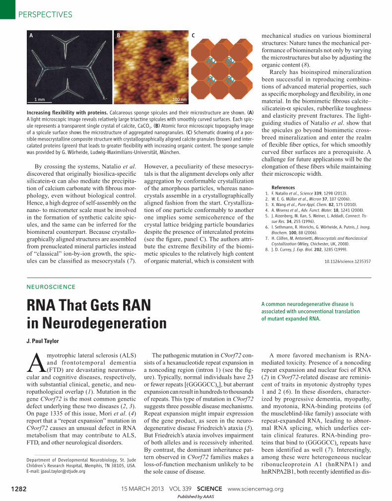
gated single rods or triradiate stars (see the
fi gure, panel A). Despite being well-defi ned
crystals, calcite sponge spicules contain
small amounts of polyanionic macromole-
cules incorporated within the mineral phase
( 5). These proteins presumably play a role
in the precipitation mechanism, equivalent
to that of silicatein in silica spicule for-
mation, because calcite sponge spicules
also appear to be constructed by aggrega-
tion of nanoparticles (see the fi gure, panel
B) to create smoothly curved morpholo-
gies ( 6). The proteins are assumed to tem-
porarily stabilize the particles as an amor-
phous phase of CaCO3, which then crystal-
lizes after particle aggregation into a calcite
single crystal. The granular structure with
intercalated proteins defl ects fracture prop-
agation and dissipates strain energy, reduc-
ing the brittleness of the material.
Institut für Angewandte Geowissenschaften, Technische Universität Darmstadt, 64287 Darmstadt, Germany. E-mail: [email protected]
10.1126/science.1235591
Published by AAAS
15 MARCH 2013 VOL 339 SCIENCE www.sciencemag.org 1282
PERSPECTIVES
By crossing the systems, Natalio et al.
discovered that originally biosilica-specifi c
silicatein-α can also mediate the precipita-
tion of calcium carbonate with fi brous mor-
phology, even without biological control.
Hence, a high degree of self-assembly on the
nano- to micrometer scale must be involved
in the formation of synthetic calcite spic-
ules, and the same can be inferred for the
biomineral counterpart. Because crystallo-
graphically aligned structures are assembled
from prenucleated mineral particles instead
of “classical” ion-by-ion growth, the spic-
ules can be classifi ed as mesocrystals ( 7).
However, a peculiarity of these mesocrys-
tals is that the alignment develops only after
aggregation by conformable crystallization
of the amorphous particles, whereas nano-
crystals assemble in a crystallographically
aligned fashion from the start. Crystalliza-
tion of one particle conformably to another
one implies some semicoherence of the
crystal lattice bridging particle boundaries
despite the presence of intercalated proteins
(see the fi gure, panel C). The authors attri-
bute the extreme fl exibility of the biomi-
metic spicules to the relatively high content
of organic material, which is consistent with
mechanical studies on various biomineral
structures: Nature tunes the mechanical per-
formance of biominerals not only by varying
the microstructures but also by adjusting the
organic content ( 8).
Rarely has bioinspired mineralization
been successful in reproducing combina-
tions of advanced material properties, such
as specifi c morphology and fl exibility, in one
material. In the biomimetic fi brous calcite–
silicatein-α spicules, rubberlike toughness
and elasticity prevent fractures. The light-
guiding studies of Natalio et al. show that
the spicules go beyond biomimetic cross-
breed mineralization and enter the realm
of fl exible fi ber optics, for which smoothly
curved fi ber surfaces are a prerequisite. A
challenge for future applications will be the
elongation of these fi bers while maintaining
their microscopic width.
References 1. F. Natalio et al., Science 339, 1298 (2013). 2. W. E. G. Müller et al., Micron 37, 107 (2006). 3. X. Wang et al., Pure Appl. Chem. 82, 175 (2010). 4. A. Miserez et al., Adv. Funct. Mater. 18, 1241 (2008). 5. J. Aizenberg, M. Ilan, S. Weiner, L. Addadi, Connect. Tis-
sue Res. 34, 255 (1996). 6. I. Sethmann, R. Hinrichs, G. Wörheide, A. Putnis, J. Inorg.
Biochem. 100, 88 (2006). 7. H. Cölfen, M. Antonietti, Mesocrystals and Nonclassical
Crystallization (Wiley, Chichester, UK, 2008). 8. J. D. Currey, J. Exp. Biol. 202, 3285 (1999).
1 mm 100 nm
A B C
Increasing fl exibility with proteins. Calcareous sponge spicules and their microstructure are shown. (A) A light microscopic image reveals relatively large triactine spicules with smoothly curved surfaces. Each spic-ule represents a transparent single crystal of calcite, CaCO3. (B) Atomic force microscopic topography image of a spicule surface shows the microstructure of aggregated nanogranules. (C) Schematic drawing of a pos-sible mesocrystalline composite structure with crystallographically aligned calcite granules (brown) and inter-calated proteins (green) that leads to greater fl exibility with increasing organic content. The sponge sample was provided by G. Wörheide, Ludwig-Maximilians-Universität, München.
RNA That Gets RAN in Neurodegeneration
NEUROSCIENCE
J. Paul Taylor
A common neurodegenerative disease is
associated with unconventional translation
of mutant expanded RNA.
Amyotrophic lateral sclerosis (ALS)
and frontotemporal dementia
(FTD) are devastating neuromus-
cular and cognitive diseases, respectively,
with substantial clinical, genetic, and neu-
ropathological overlap ( 1). Mutation in the
gene C9orf72 is the most common genetic
defect underlying these two diseases ( 2, 3).
On page 1335 of this issue, Mori et al. ( 4)
report that a “repeat expansion” mutation in
C9orf72 causes an unusual defect in RNA
metabolism that may contribute to ALS,
FTD, and other neurological disorders.
The pathogenic mutation in C9orf72 con-
sists of a hexanucleotide repeat expansion in
a noncoding region (intron 1) (see the fi g-
ure). Typically, normal individuals have 23
or fewer repeats [(GGGGCC)n], but aberrant
expansion can result in hundreds to thousands
of repeats. This type of mutation in C9orf72
suggests three possible disease mechanisms.
Repeat expansion might impair expression
of the gene product, as seen in the neuro-
degenerative disease Friedreich’s ataxia ( 5).
But Friedreich’s ataxia involves impairment
of both alleles and is recessively inherited.
By contrast, the dominant inheritance pat-
tern observed in C9orf72 families makes a
loss-of-function mechanism unlikely to be
the sole cause of disease.
A more favored mechanism is RNA-
mediated toxicity. Presence of a noncoding
repeat expansion and nuclear foci of RNA
( 2) in C9orf72-related disease are reminis-
cent of traits in myotonic dystrophy types
1 and 2 ( 6). In these disorders, character-
ized by progressive dementia, myopathy,
and myotonia, RNA-binding proteins (of
the muscleblind-like family) associate with
repeat-expanded RNA, leading to abnor-
mal RNA splicing, which underlies cer-
tain clinical features. RNA-binding pro-
teins that bind to (GGGGCC)n repeats have
been identified as well ( 7). Interestingly,
among these were heterogeneous nuclear
ribonucleoprotein A1 (hnRNPA1) and
hnRNPA2B1, both recently identifi ed as dis-
Department of Developmental Neurobiology, St. Jude Children’s Research Hospital, Memphis, TN 38105, USA. E-mail: [email protected]
10.1126/science.1235357
Published by AAAS