Embed Size (px)
Citation preview
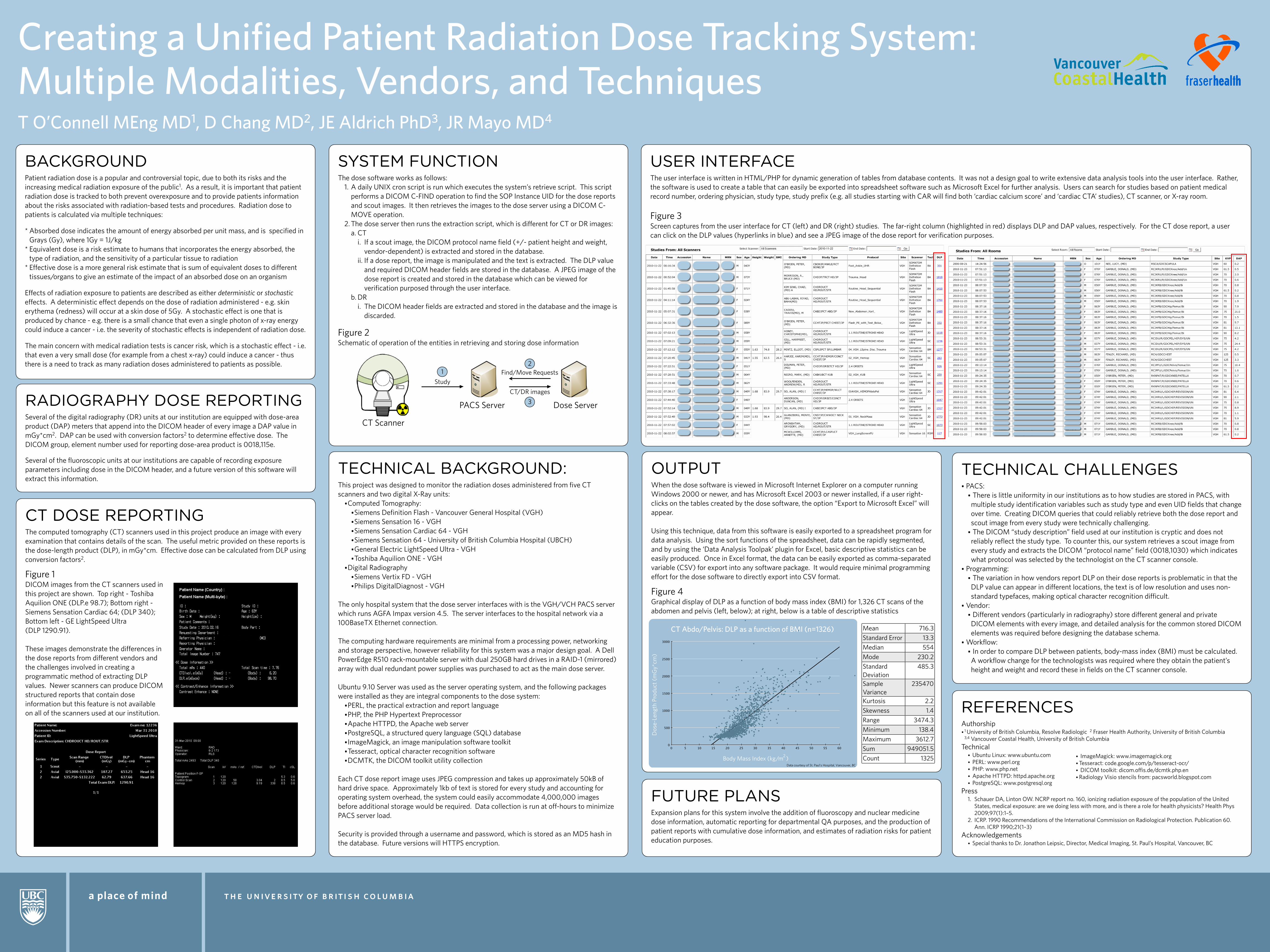
Creating a Unified Patient Radiation Dose Tracking System: Multiple Modalities, Vendors, and TechniquesT O’Connell MEng MD1, D Chang MD2, JE Aldrich PhD3, JR Mayo MD4
BACKGROUNDPatient radiation dose is a popular and controversial topic, due to both its risks and the increasing medical radiation exposure of the public1. As a result, it is important that patient radiation dose is tracked to both prevent overexposure and to provide patients information about the risks associated with radiation-based tests and procedures. Radiation dose to patients is calculated via multiple techniques:
* Absorbed dose indicates the amount of energy absorbed per unit mass, and is specified in Grays (Gy), where 1Gy = 1J/kg
* Equivalent dose is a risk estimate to humans that incorporates the energy absorbed, the type of radiation, and the sensitivity of a particular tissue to radiation
* Effective dose is a more general risk estimate that is sum of equivalent doses to different tissues/organs to give an estimate of the impact of an absorbed dose on an organism
Effects of radiation exposure to patients are described as either deterministic or stochastic effects. A deterministic effect depends on the dose of radiation administered - e.g. skin erythema (redness) will occur at a skin dose of 5Gy. A stochastic effect is one that is produced by chance - e.g. there is a small chance that even a single photon of x-ray energy could induce a cancer - i.e. the severity of stochastic effects is independent of radiation dose.
The main concern with medical radiation tests is cancer risk, which is a stochastic effect - i.e. that even a very small dose (for example from a chest x-ray) could induce a cancer - thus there is a need to track as many radiation doses administered to patients as possible.
TECHNICAL BACKGROUND:This project was designed to monitor the radiation doses administered from five CT scanners and two digital X-Ray units:
•Computed Tomography:•Siemens Definition Flash - Vancouver General Hospital (VGH)•Siemens Sensation 16 - VGH•Siemens Sensation Cardiac 64 - VGH•Siemens Sensation 64 - University of British Columbia Hospital (UBCH)•General Electric LightSpeed Ultra - VGH•Toshiba Aquilion ONE - VGH
•Digital Radiography•Siemens Vertix FD - VGH•Philips DigitalDiagnost - VGH
The only hospital system that the dose server interfaces with is the VGH/VCH PACS server which runs AGFA Impax version 4.5. The server interfaces to the hospital network via a 100BaseTX Ethernet connection.
The computing hardware requirements are minimal from a processing power, networking and storage perspective, however reliability for this system was a major design goal. A Dell PowerEdge R510 rack-mountable server with dual 250GB hard drives in a RAID-1 (mirrored) array with dual redundant power supplies was purchased to act as the main dose server.
Ubuntu 9.10 Server was used as the server operating system, and the following packages were installed as they are integral components to the dose system:
•PERL, the practical extraction and report language•PHP, the PHP Hypertext Preprocessor•Apache HTTPD, the Apache web server•PostgreSQL, a structured query language (SQL) database•ImageMagick, an image manipulation software toolkit•Tesseract, optical character recognition software•DCMTK, the DICOM toolkit utility collection
Each CT dose report image uses JPEG compression and takes up approximately 50kB of hard drive space. Approximately 1kb of text is stored for every study and accounting for operating system overhead, the system could easily accommodate 4,000,000 images before additional storage would be required. Data collection is run at off-hours to minimize PACS server load.
Security is provided through a username and password, which is stored as an MD5 hash in the database. Future versions will HTTPS encryption.
CT DOSE REPORTINGThe computed tomography (CT) scanners used in this project produce an image with every examination that contains details of the scan. The useful metric provided on these reports is the dose-length product (DLP), in mGy*cm. Effective dose can be calculated from DLP using conversion factors2.
Figure 1 DICOM images from the CT scanners used in this project are shown. Top right - Toshiba Aquilion ONE (DLP.e 98.7); Bottom right - Siemens Sensation Cardiac 64; (DLP 340); Bottom left - GE LightSpeed Ultra (DLP 1290.91).
These images demonstrate the differences in the dose reports from different vendors and the challenges involved in creating a programmatic method of extracting DLP values. Newer scanners can produce DICOM structured reports that contain dose information but this feature is not available on all of the scanners used at our institution.
RADIOGRAPHY DOSE REPORTINGSeveral of the digital radiography (DR) units at our institution are equipped with dose-area product (DAP) meters that append into the DICOM header of every image a DAP value in mGy*cm2. DAP can be used with conversion factors2 to determine effective dose. The DICOM group, element number used for reporting dose-area product is 0018,115e.
Several of the fluoroscopic units at our institutions are capable of recording exposure parameters including dose in the DICOM header, and a future version of this software will extract this information.
TECHNICAL CHALLENGES• PACS:
• There is little uniformity in our institutions as to how studies are stored in PACS, with multiple study identification variables such as study type and even UID fields that change over time. Creating DICOM queries that could reliably retrieve both the dose report and scout image from every study were technically challenging.
• The DICOM “study description” field used at our institution is cryptic and does not reliably reflect the study type. To counter this, our system retrieves a scout image from every study and extracts the DICOM “protocol name” field (0018,1030) which indicates what protocol was selected by the technologist on the CT scanner console.
• Programming:• The variation in how vendors report DLP on their dose reports is problematic in that the DLP value can appear in different locations, the text is of low resolution and uses non-standard typefaces, making optical character recognition difficult.
• Vendor:• Different vendors (particularly in radiography) store different general and private DICOM elements with every image, and detailed analysis for the common stored DICOM elements was required before designing the database schema.
• Workflow:• In order to compare DLP between patients, body-mass index (BMI) must be calculated. A workflow change for the technologists was required where they obtain the patient’s height and weight and record these in fields on the CT scanner console.
REFERENCESAuthorship• 1 University of British Columbia, Resolve Radiologic 2 Fraser Health Authority, University of British Columbia
3,4 Vancouver Coastal Health, University of British Columbia Technical
• Ubuntu Linux: www.ubuntu.com• PERL: www.perl.org• PHP: www.php.net• Apache HTTPD: httpd.apache.org• PostgreSQL: www.postgresql.org
Press1. Schauer DA, Linton OW. NCRP report no. 160, ionizing radiation exposure of the population of the United
States, medical exposure: are we doing less with more, and is there a role for health physicists? Health Phys 2009;97(1):1–5.
2. ICRP. 1990 Recommendations of the International Commission on Radiological Protection. Publication 60. Ann. ICRP 1990;21(1–3)
Acknowledgements• Special thanks to Dr. Jonathon Leipsic, Director, Medical Imaging, St. Paul’s Hospital, Vancouver, BC
• ImageMagick: www.imagemagick.org• Tesseract: code.google.com/p/tesseract-ocr/• DICOM toolkit: dicom.offis.de/dcmtk.php.en • Radiology Visio stencils from: pacsworld.blogspot.com
OUTPUTWhen the dose software is viewed in Microsoft Internet Explorer on a computer running Windows 2000 or newer, and has Microsoft Excel 2003 or newer installed, if a user right-clicks on the tables created by the dose software, the option “Export to Microsoft Excel” will appear.
Using this technique, data from this software is easily exported to a spreadsheet program for data analysis. Using the sort functions of the spreadsheet, data can be rapidly segmented, and by using the ‘Data Analysis Toolpak’ plugin for Excel, basic descriptive statistics can be easily produced. Once in Excel format, the data can be easily exported as comma-separated variable (CSV) for export into any software package. It would require minimal programming effort for the dose software to directly export into CSV format.
Figure 4 Graphical display of DLP as a function of body mass index (BMI) for 1,326 CT scans of the abdomen and pelvis (left, below); at right, below is a table of descriptive statistics
600 5 10 15 20 25 30 35 40 45 50 55
3000
0
500
1000
1500
2000
2500
Body Mass Index (kg/m )
Dos
e-Le
ngth
Pro
duct
(m
Gy*
cm)
CT Abdo/Pelvis: DLP as a function of BMI (n=1326)
2
Data courtesy of St. Paul's Hospital, Vancouver, BC
Mean 716.3Standard Error 13.3Median 554Mode 230.2Standard Deviation
485.3
Sample Variance
235470
Kurtosis 2.2Skewness 1.4Range 3474.3Minimum 138.4Maximum 3612.7Sum 949051.5Count 1325
FUTURE PLANSExpansion plans for this system involve the addition of fluoroscopy and nuclear medicine dose information, automatic reporting for departmental QA purposes, and the production of patient reports with cumulative dose information, and estimates of radiation risks for patient education purposes.
USER INTERFACEThe user interface is written in HTML/PHP for dynamic generation of tables from database contents. It was not a design goal to write extensive data analysis tools into the user interface. Rather, the software is used to create a table that can easily be exported into spreadsheet software such as Microsoft Excel for further analysis. Users can search for studies based on patient medical record number, ordering physician, study type, study prefix (e.g. all studies starting with CAR will find both ‘cardiac calcium score’ and ‘cardiac CTA’ studies), CT scanner, or X-ray room.
Figure 3 Screen captures from the user interface for CT (left) and DR (right) studies. The far-right column (highlighted in red) displays DLP and DAP values, respectively. For the CT dose report, a user can click on the DLP values (hyperlinks in blue) and see a JPEG image of the dose report for verification purposes.
SYSTEM FUNCTIONThe dose software works as follows:
1. A daily UNIX cron script is run which executes the system’s retrieve script. This script performs a DICOM C-FIND operation to find the SOP Instance UID for the dose reports and scout images. It then retrieves the images to the dose server using a DICOM C-MOVE operation.
2. The dose server then runs the extraction script, which is different for CT or DR images:a. CT
i. If a scout image, the DICOM protocol name field (+/- patient height and weight, vendor-dependent) is extracted and stored in the database.
ii. If a dose report, the image is manipulated and the text is extracted. The DLP value and required DICOM header fields are stored in the database. A JPEG image of the dose report is created and stored in the database which can be viewed for verification purposed through the user interface.
b. DRi. The DICOM header fields are extracted and stored in the database and the image is
discarded.
Figure 2Schematic of operation of the entities in retrieving and storing dose information
CT Scanner
PACS Server Dose Server
Study
CT/DR images
Find/Move Requests12
3