Embed Size (px)
Citation preview

Photographic Sciences Capstone
Creating a Low-Cost, Portable, Non-Mydriatic Fundus Camera Research Paper
Daniel Beim
Abstract
Fundus cameras are a key part of any ophthalmic practice’s equipment, as they can be
used to locate and document a variety of ocular defects with relative ease. As common as they
are though, fundus cameras are often times bulky and expensive pieces of equipment that may
not be feasible for use in smaller offices, preventing many populations who may be at risk from
getting the help they need. Additionally, most systems are mydriatic systems which means that
they require the patient’s eye to be dilated, placing further restrictions on the likelihood of
smaller practices investing in a system.
The creation of a low-cost, portable, non-mydriatic system would allow for the device to
become much more accessible, allowing for more practices and organizations to own a device
and perform tests to document and diagnose more patients. This is especially important with the
worldwide prevalence of diabetic retinopathy, which can occur as a result of uncontrolled
diabetes and may lead to blindness if left untreated. The implementation of a low-cost, portable
system would allow for at-risk populations to get examined for the defect and determine if they
need to seek treatment, helping to reduce the risk of blindness.
My aim is assist this year’s Multidisciplinary Senior Design (MSD) team in the creation
of their fundus camera system, using my knowledge of ophthalmic systems gathered from my
time in the ophthalmic photography class and my co-op this past summer at the Manhattan Eye,
Ear, & Throat hospital. I will provide design advice as well as assist in testing the system, acting
as an “expert buyer” and evaluating the system’s clinical feasibility.
The Problem
A typical fundus camera system costs in the tens of thousands of dollars, and most
systems come as part of a larger system consisting of the fundus camera itself attached to a
variable height table which is also attached in some way to a computer system to store data.
These systems can be out of the realm of possibility for practices and organizations with limited
budgets and space or who are mobile. On top of size and pricing constraints, many fundus
cameras require the patient to be dilated which can cause additional hardships with budget as
well as with patients who may not want to undergo the dilation process. The need for dilation
with many systems creates further complications due to the risk of patients having a
contraindication related to increased intraocular pressure, which could be deadly. There are a
variety of non-mydriatic systems currently on the market in order to try and circumvent this risk,
however the issue of price and sometimes weigh still remain. Due to these factors, fundus
systems are not always available and many people with conditions like diabetic retinopathy
continue living with the threat of severely decreased vision or blindness.
Current Commercial Technologies
Research into developing newer and more novel fundus imaging systems has been
ongoing for many years and has yielded a variety of systems, ranging from miniature systems to
smartphone-based devices. I will only cover a small sample of the available non-mydriatic and
mobile systems for this paper, in order to give a rough overview of what is currently available to
compare to the MSD team’s proposed system.
One currently available commercial device is the 3nethra Classic (image 1), which is a
lightweight non-mydriatic system capable of color and near-infrared imaging, requiring a

minimum pupil diameter of 3mm and a field of view of 45°. It uses reduced optics to reach its
moderate price point of $7,400, as well as utilizing an infrared light emitting diode (LED) for
observation, a white LED as the imaging flash, automatic and manual focus controls, a three
megapixel image sensor, and a computer interface for data storage and viewing.1 2 The device
weighs about 15kg2, which is significantly less than a mydriatic system such as the ZEISS PRO
NM which weighs 30kg3.
Image 1: 3nethra Classic fundus camera1
Another commercially available non-mydriatic system is the DRS (Digital Retinopathy
System)(image 2), which requires a minimum pupil diameter of 4mm and has a horizontal field
of view of 45° and a vertical field of view of 40°. Unlike most systems the DRS is fully
automatic, giving it the ability to “sense the patient, self-align to the target eye, focus the retina,
adjust the flash level and capture the image in less than 30 seconds.”4. The system utilizes white
light reflective imaging to capture an image, has a five megapixel image sensor, and utilizes a
touchscreen tablet for image capture and storage. The device weighs 19kg and costs about
$10,000, putting it on the higher end of available non-mydriatic systems. 2 4

Image 2: DRS fundus camera4
The Optomed SmartScope Pro (image 3) is a fully handheld non-mydriatic fundus
camera capable of color, red-free, and infrared imaging, requiring a minimum pupil diameter of
3.5mm with a field of view of 40°. In addition to being handheld, the device can also fit onto a
slit lamp camera mount for imaging. The device can autofocus, has a five megapixel sensor,
uses a rechargeable Ni-MH battery, and uses an LCD screen on the back for image viewing but
can also be connected to a PC via USB.2 5 The devices weight and price are currently
unavailable.
Image 3: Optomed Smartscope Pro5
The iExaminer and panoptic ophthalmoscope (image 4) are two devices which when
combined with an iPhone create a handheld non-mydriatic fundus camera capable of color
images with a field of view of 25°.2 6 The device’s specifications depend on the phone being
used, but the panoptic ophthalmoscope portion has a halogen lamp for illumination and a focus

wheel, claiming a “view of the fundus that's 5X greater than you see with a standard
ophthalmoscope in an undilated eye.”6. The system weighs about one kilogram and costs about
$1,000 plus the cost of an iPhone.2
Image 4: iExaminer and panoptic ophthalmoscope6
Previous Academic Work
This will be the third MSD team to attempt to create a low-cost fundus camera, with two
other teams having made their own prototypes which they displayed at ImagineRIT in their
respective years, passing along their findings to the next year’s team to help them develop their
system.
The MSD team for 2014-2015 consisted of mechanical engineers Kevin Labourdette and
Thomas Casero, electrical engineers Daniel Sui and Quang Huynh, biomedical engineer Ian
Morency, and computer engineer Kyle Burden. Together they set out to “develop a prototype of
a low-cost fundus camera using off-the-shelf optical components” that could be used without any
special training, was portable/compact enough for use in a general practitioner’s office, and
could take pictures of similarly high quality to those from a commercial system. The team ended
up deciding on a standing, vertically adjustable frame model on retractable wheels that the
camera mounts to the top of and can tilt so that virtually all patients can rest on the chinrest. The
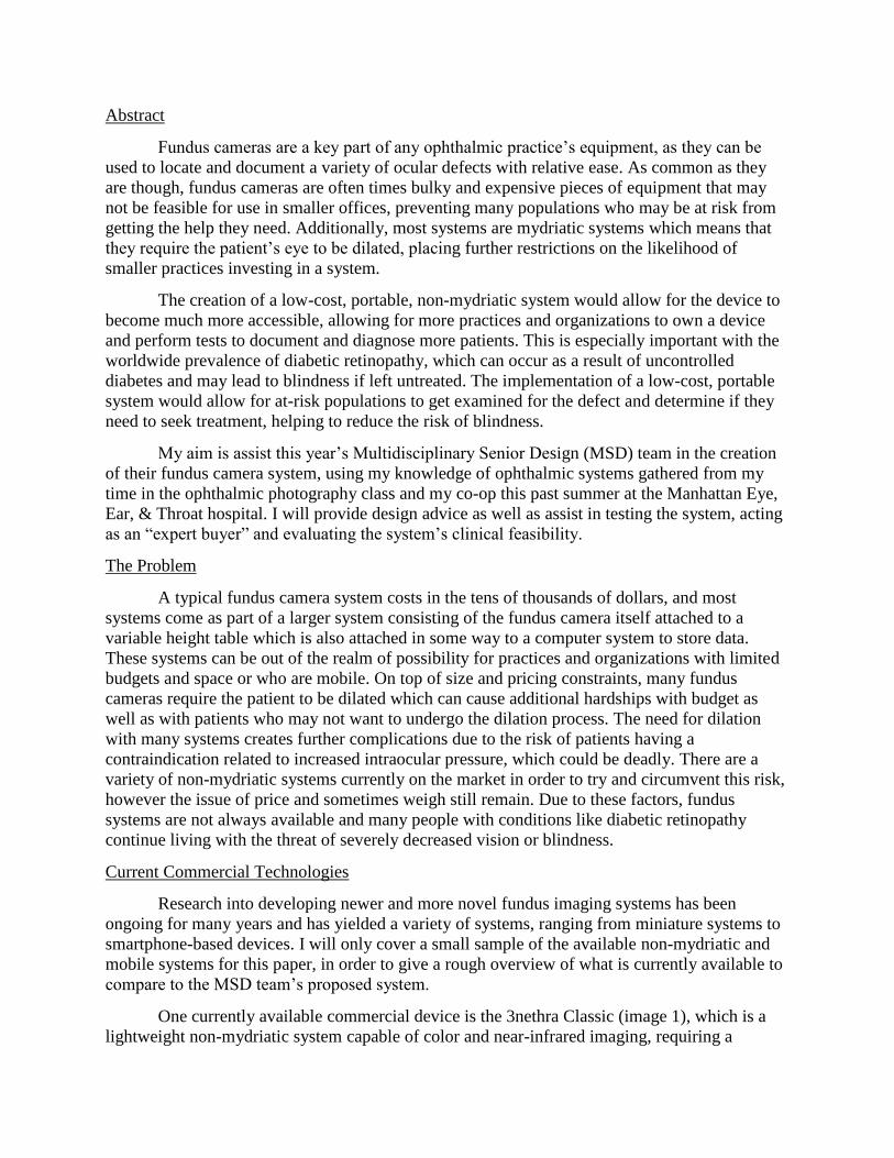
optical system – as seen in image 5 – they created used a 20 diopter lens to magnify the view, a
42 diopter convex lens to increase the angle of view on the retina, and a Raspberry Pi NoIR (no
infrared) camera module to capture the images, with a working distance of 40mm and a field of
view of 26.5° in a 6mm pupil. The system was able to be focused by moving the camera lens on
a stepper motor controlled by the Raspberry Pi, which in turn could be controlled via an attached
touchscreen. The system also used an IR LED for focusing and a white LED Bright Pi lamp for
illumination of the retina when imaging. The team created a simple application to input patient
data using a keyboard plugged into the Raspberry Pi, as well as provide a preview of the
camera’s current positioning so that it can be properly adjusted and then capture an image when
ready. The camera housing design – as seen in image 6 – was designed to be a standing design
that allows the patient to sit and lean into it, allowing for it to be a light and collapsible system
for easy storage. The team received some help with deciding on this design from Linda Deng, an
industrial design student, and Brittany Bateman, a biomedical photography student. In the end
the final product ended up being a rough prototype which was displayed at ImagineRIT, still
requiring an improved optics system that cut down on issues like reflections in the captured
images but otherwise functional; the project became a proof of concept, showing that it is
possible to build a low-cost camera.7
The MSD team for 2015-2016 consisted of lead software engineer Cecilia Demarest, lead
biomedical specialist Kelly Dunn, lead hardware engineer Matthew Little, lead systems engineer
Casey Petzel, industrial designer Zachary Olmstead, and lead purchasing and communication
specialist Ryan Morgan. Their goals were practically identical to those of the previous team: to
create a low-cost, non-mydriatic fundus camera that could be used by non-specialized personnel
in a general practitioner’s office and provides images of similar quality to those from a regular
system, in order to attempt to increase the number of screenings for disorders like diabetic
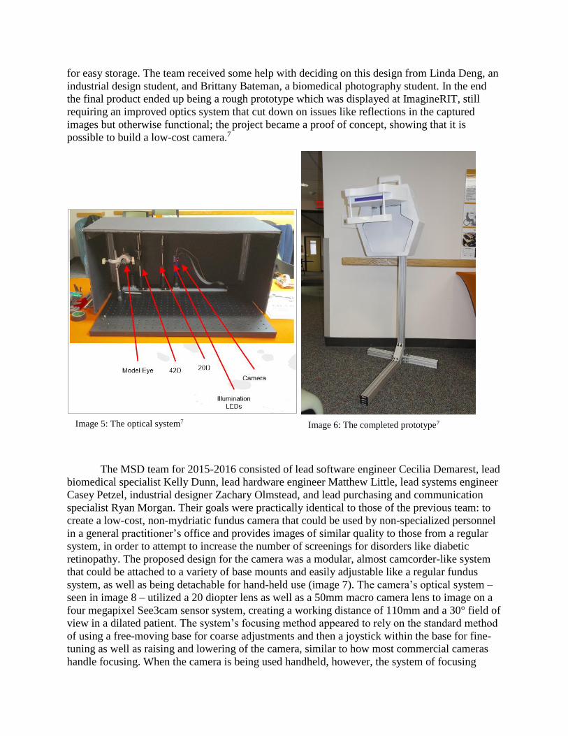
retinopathy. The proposed design for the camera was a modular, almost camcorder-like system
that could be attached to a variety of base mounts and easily adjustable like a regular fundus
system, as well as being detachable for hand-held use (image 7). The camera’s optical system –
seen in image 8 – utilized a 20 diopter lens as well as a 50mm macro camera lens to image on a
four megapixel See3cam sensor system, creating a working distance of 110mm and a 30° field of
view in a dilated patient. The system’s focusing method appeared to rely on the standard method
of using a free-moving base for coarse adjustments and then a joystick within the base for fine-
tuning as well as raising and lowering of the camera, similar to how most commercial cameras
handle focusing. When the camera is being used handheld, however, the system of focusing
Image 5: The optical system7 Image 6: The completed prototype7
would have been similar, but less precisely controlled as it depends on the stability of the user’s
hand and the patient. For illumination the system utilized a ring of IR and white LEDs directed
down an acrylic tube in order to reduce non-image forming light. In order to capture and process
images the system used an ODROID-XU4 single board computer attached to the sensor and
lights as well, a solid state drive to store the images, and to an ODROID-VU7 touch screen for
viewing and control. The team created an application called FUNdusCam in order to control the
illumination and camera, as well as add necessary data to and transfer/save files. The team
received some design and lens system as well as clinical setting feasibly advice from biomedical
photographic communications student Amber Kates during the design process. In the end the
project was largely conceptual, ending up with a functional optical system and a start on the base
mount parts but otherwise no real physical prototype.
The Plan
This year’s MSD team hopes to continue improving on the systems created by the past
two teams to end up with a functional prototype approved for human testing. The project is still
in the conceptual stage, but the current plan for components and layout is thus: a miniature
tabletop structure with a headrest, IR LEDs for focus illumination, visible/white LEDs for
imaging illumination via on-axis “donut ring” illumination, a physical shift device for switching
between IR and visible light, reduction of noise caused by reflected light by blocking the center
of the light, a pellicle beam splitter, manual adjustment knobs for focusing, a CCD micro-camera
for image capture (likely the same one used last year), and a touch screen for image capture and
metadata input. As with the previous years’ goals, the device is being designed with non-
mydriatic use in mind as well as an emphasis on portability and a low-cost without sacrificing
image quality or comfort. The team is currently working on benchmarking their optics,
Image 7: Conceptual housing on base8 Image 8: Optical system8

reviewing requirements for FDA and human subject research approval, and creating conceptual
models, of which the current ideas can be seen in image 9 below.2
Image 9: Current conceptual sketches2
The previous teams have cited some issues with understanding the optics and other
aspects of the system in their final papers, and my goal in joining this year’s team is to use my
biomedical photography background to help them develop a clinically viable system. I will keep
up with the team’s progress as they continue designing their system, offering advice on the
design and other aspects of the system in a similar fashion to the contributions of Brittany
Bateman and Amber Kates with their respective year’s team. I also plan to undergo the human
subject research approval process myself in the hopes that if a functioning system is developed I
can assist in testing it, using my experiences from RIT’s ophthalmic photography class as well as
my co-op in the ophthalmology department at Manhattan Eye, Ear, & Throat Hospital this past
summer. If such testing is possible I will use the opportunity to provide valuable feedback on
image quality, ease of use as an operator, and potentially even patient-experience feedback. It
will be an enriching experience to see the construction side of the fundus camera, perhaps even
leading to further research into the creating of low-cost fundus cameras beyond this project, and I
look forward to seeing where it goes.
Mentor Choices
1) Christye Sisson
2) Nitin Sampat
3) Bob Rose

Sources
1Forus Health - 3nethra classic. (n.d.) 3nethra Classic [Brochure]. Retrieved October 5, 2016,
from http://www.forushealth.com/new/3nethra-classic.html
2Neuberger, B., Lanza, D., Rosengrant, N., Axel, P., & Olmsted, Z. (2016, October 5). Design
Review II [PDF].
3VISUCAM PRO NM. (n.d.). Retrieved October 19, 2016, from
http://www.zeiss.com/meditec/us/products/ophthalmology-optometry/retina/diagnostics/fundus-
imaging/visucam-pro-nm.html#technical-data
4 CenterVue. (n.d) drs: Digital Retinopathy System [Brochure]. (n.d.). CenterVue.
5Optomed. (2016). Smartscope PRO Camera [Brochure]. Retrieved October 5, 2016, from
http://www.optomed.com/blog/module/smartscope-pro-camera/
6 PanOptic™ Ophthalmoscope - Welch Allyn. (n.d.). Retrieved October 5, 2016, from
http://tinyurl.com/handheld-fundus-phone2016
7 Labourdette, K., Morency, I., Huynh, Q., Sui, D., Burden, K., & Casero, T. (2014, December
11). Low-Cost Fundus Camera [Scholarly project]. In P15590: Low Cost Fundus Camera.
Retrieved October 18, 2016, from http://edge.rit.edu/edge/P15590/public/Home
8Low-Cost Fundus Camera 2 [Scholarly project]. (2016, May 13). In P16590: Low-Cost Fundus
Camera 2. Retrieved October 18, 2016, from edge.rit.edu/edge/P16590/public/Home
![Clinical Application of Multicolor Imaging Technology · phy and commercial availability of fundus cameras [1] . Numerous enhancements such as digital imaging, non-mydriatic functions,](https://img.pdfslide.us/doc/110x75/5f068f247e708231d41896c5/clinical-application-of-multicolor-imaging-technology-phy-and-commercial-availability.jpg)