Embed Size (px)
Citation preview
Psychiatry 2009 [ V O L U M E 6 , N U M B E R 1 1 , N O V E M B E R ]34
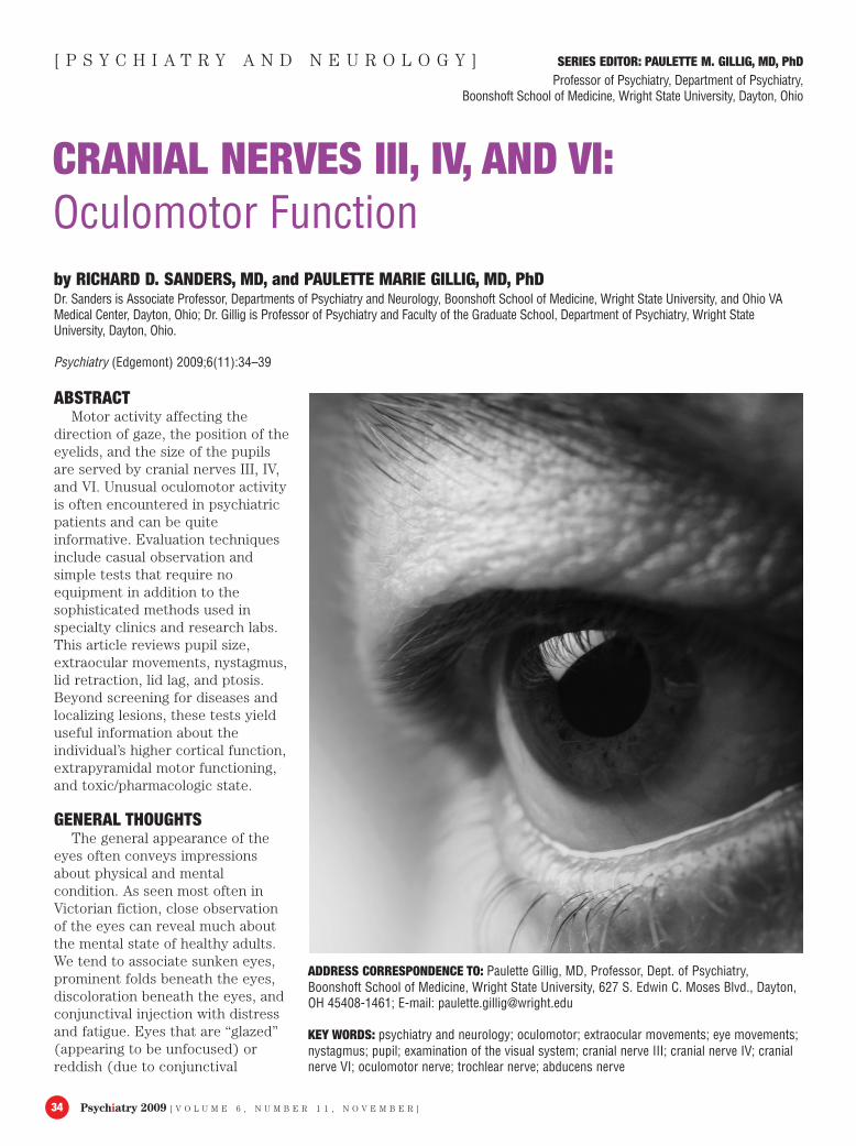
ABSTRACTMotor activity affecting the
direction of gaze, the position of theeyelids, and the size of the pupilsare served by cranial nerves III, IV,and VI. Unusual oculomotor activityis often encountered in psychiatricpatients and can be quiteinformative. Evaluation techniquesinclude casual observation andsimple tests that require noequipment in addition to thesophisticated methods used inspecialty clinics and research labs.This article reviews pupil size,extraocular movements, nystagmus,lid retraction, lid lag, and ptosis.Beyond screening for diseases andlocalizing lesions, these tests yielduseful information about theindividual’s higher cortical function,extrapyramidal motor functioning,and toxic/pharmacologic state.
GENERAL THOUGHTSThe general appearance of the
eyes often conveys impressionsabout physical and mentalcondition. As seen most often inVictorian fiction, close observationof the eyes can reveal much aboutthe mental state of healthy adults.We tend to associate sunken eyes,prominent folds beneath the eyes,discoloration beneath the eyes, andconjunctival injection with distressand fatigue. Eyes that are “glazed”(appearing to be unfocused) orreddish (due to conjunctival
CRANIAL NERVES III, IV, AND VI:Oculomotor Functionby RICHARD D. SANDERS, MD, and PAULETTE MARIE GILLIG, MD, PhD Dr. Sanders is Associate Professor, Departments of Psychiatry and Neurology, Boonshoft School of Medicine, Wright State University, and Ohio VAMedical Center, Dayton, Ohio; Dr. Gillig is Professor of Psychiatry and Faculty of the Graduate School, Department of Psychiatry, Wright StateUniversity, Dayton, Ohio.
Psychiatry (Edgemont) 2009;6(11):34–39
ADDRESS CORRESPONDENCE TO: Paulette Gillig, MD, Professor, Dept. of Psychiatry,Boonshoft School of Medicine, Wright State University, 627 S. Edwin C. Moses Blvd., Dayton,OH 45408-1461; E-mail: [email protected]
KEY WORDS: psychiatry and neurology; oculomotor; extraocular movements; eye movements;nystagmus; pupil; examination of the visual system; cranial nerve III; cranial nerve IV; cranialnerve VI; oculomotor nerve; trochlear nerve; abducens nerve
[ P S Y C H I A T R Y A N D N E U R O L O G Y ] SERIES EDITOR: PAULETTE M. GILLIG, MD, PhDProfessor of Psychiatry, Department of Psychiatry,
Boonshoft School of Medicine, Wright State University, Dayton, Ohio
[ V O L U M E 6 , N U M B E R 1 1 , N O V E M B E R ] Psychiatry 2009 35
injection) elicit suspicions of fatigueand/or the use of intoxicatingsubstances.
Spontaneous eye movements alsohave conventionally acceptedimplications. A lack of direct eyecontact is taken to indicate a lack ofconfidence, authenticity, or interest;excessive eye contact can be takenas intimidating. Frequent lateralgazes (“shifty eyes”) sometimesimplies anxiety or deception, butalso may be assumed to reflecthypervigilance, paranoia, orhallucination. Because thesefeatures of the eyes can have somany causes, it is more useful tostart with specific observations(e.g., frequent, rapid, spontaneous,lateral eye movements) rather thanan inference (e.g., appears to beresponding to internal stimuli) andthen consider explanations.
Cranial nerves III (CNIII)(oculomotor), IV (trochlear), and VI(abducens) control the position ofthe eyeballs; CNIII influences theposition of the eyelids and the sizeof the pupils. In addition to theirvalue in localizing lesions, thesethree oculomotor nerves (sensoryfunction is limited toproprioception) can reveal subtlechanges in general skeletal andsmooth muscle activity. Forexample, even minor weakness inone extraocular muscle can causediplopia, and the eyelids reveal evenmild variations in the activity ofskeletal or smooth muscle.1,2 Motoractivity controlling the direction ofgaze, the elevation of the eyelids,and the size of the pupils alsoreflect higher cortical activity, andare sensitive to drug effects. Assuch, they are informative incommon psychiatric conditions.
RELEVANT ANATOMY ANDPHYSIOLOGY
Pupils. As discussed in theprevious article of this series, lightenergy is transduced into neuralactivity in the retina, coursesthrough the optic nerve (CNII),through the optic chiasm (wherethe nasal retinal projections,containing information from the
lateral visual fields, cross over), andthen the optic tract (containing allinformation from the opposite visualhemifield).
The optic tract synapses in thepretectal nucleus, which projectsequally to the Edinger-Westphalnucleus (part of CNIII) on bothsides. The Edinger-Westphalnucleus sends efferent projectionsthrough CNIII to the ciliaryganglion, then to the pupil. Becauseprojections from the pretectal to theEdinger-Westphal nuclei arebilateral, the pupils should respondabout equally to light shined oneither eye. Even a blind eye shouldconstrict in response to light shinedon the other eye. Unequal pupils(anisocoria) are due to the efferent(motor) system, which includesCNIII, somatic and parasympatheticcomponents, sympathetic nervesoriginating in the cervical spine, andthe smooth muscle of the iris.1,2
Extraocular muscles. Thereare six extraocular muscles: fourrectus muscles (superior, inferior,lateral, medial) and two oblique(superior and inferior). Theoculomotor nerve (CNIII)innervates all but two of these,ipsilaterally. Partial lesions of CNIIIare rare, so a lesion of the nucleusor nerve will result in a unilateralfailure of almost all eye movements(as well as dilated pupil and ptosis).The nucleus is in the midbrain. Thetrochlear nerve (CNIV), alsooriginating in midbrain, innervatesthe contralateral superior oblique,enabling the eye to point downwhile it is pointed medially. Lesionsare rare, causing vertical diplopiaand a compensatory tilt of the headthat could be mistaken for dystonia.The abducens nerve (CNVI)originates in the pons, thustravelling farther to its destination(the lateral rectus muscle) than theothers. Perhaps because of this,isolated lesions of CNVI, manifestedby ipsilateral loss of lateral gaze(and inward deviation or esotropiaat rest), are more common than ofthe others. The median longitudinalfasciculus links these nuclei,enabling coordination of the three
nuclei. Influencing the nuclei arewidespread, higher (supranuclear)systems.1,2
There are several anatomicallydistinguishable types of eyemovement.3,4 Saccades are high-velocity movements used for visualsearch. They are elicited by havingthe patient rapidly shift gazebetween two targets. Burst neurons,which activate saccades, are in thepons and the midbrain. Omnipauseneurons, which inhibit the burstneurons, are in the pons. Thesebrainstem systems are in turncontrolled by the superior colliculusand frontal eye fields. Saccades arethe most vulnerable to damage tothe supranuclear systems.
In the vestibulo-ocular reflex,brief, rapid head movementsprovoke rapid compensatory eyemovements. It is elicited at thebedside with the head impulse test.This is a simple circuit involving thevestibular receptors, the vestibularnucleus, and the three oculomotornuclei (III, IV, and VI). It is free ofinfluence from outside thebrainstem, which is why a normalhead impulse test supports thediagnosis of a supranucleardisorder.
The optokinetic responseconsists of slow tracking pursuitmovements and quick resettingsaccades. As there are now doubtsabout the validity of bedsideassessments, it is probably best leftto specialists. Retinal responses tolarge moving fields activate cellsgroups in the diencephalon andmidbrain and generate theoptokinetic response by influencingthe vestibulo-ocular circuit.
Smooth pursuit is a low-velocitytracking activity. It is elicited at thebedside by asking the patient tofollow a slowly moving target. Thisinvolves a complex circuit: Dorsalstream signals proceed from thediencephalon’s lateral geniculatenucleus to occipital and posteriorparietal cortex. Posterior parietaloutput to the pons is relayed tocerebellum, which projects to theoculomotor nuclei. The frontal eyefields also contribute. Smooth
Psychiatry 2009 [ V O L U M E 6 , N U M B E R 1 1 , N O V E M B E R ]36
pursuit is famously impaired inschizophrenia, as well as in mooddisorders.5
In vergence movements, the eyesmove toward each other(convergence) or away from eachother (divergence). It can be testedby having the patient continuetracking a target to a near midlineposition. Vergence movementsrequire nuclei in midbrain anddiencephalon, which receive visualinput and relay to the oculomotornuclei.
Gaze fixation “freezes” the gazein a new position after a saccade. Itdepends on midbrain and medullarynuclei, which interact withvestibular and cerebellar centersand the frontal eye fields.
Eyelids. CNIII innervates thelevator palpebrae superioris(skeletal) muscle, and sympatheticnerves innervate tarsal (smooth)muscles in both upper and lowerlids. Thus the width of the eyeopening is under both voluntary andautonomic control.1,2 Blinking, eitherspontaneously or in response to astimulus, is affected by theorbicularis oculi, innervated byCNVII, and will be covered in thenext article of this series.
SIGNS AND SYMPTOMSPupils. The size of pupils
decreases from childhood tosenescence. From moment tomoment, normal pupils vary in sizeas mediated by cholinergicparasympathetic neurons (whichconstrict) and adrenergicsympathetic neurons (which dilate).Important factors in pupil size arethe level of illumination andphysiological arousal, and it alsovaries unaccountably (hippus).Bilaterally large pupils mayrepresent stimulant, hallucinogen,or anticholinergic intoxication.Unusually small pupils are due toopiate intoxication. Asymmetricpupils usually result from iristrauma or surgery.1,2
Pupillary responses to light(“response”) and near vision(“accommodation”) can beimportant. The swinging flashlight
test is actually a test of afferentlight reception. As the light goesfrom the healthy eye to theimpaired eye (or impaired afferentsystem), both pupils dilate; bothconstrict again when the lightpasses back to the intact side. Thisfinding, known as the Marcus Gunnpupil, is due to optic nerve disease(optic neuritis in particular) orsevere retinal disease. Pupillaryconstriction normally accompaniesconvergence on a near target. Light-near dissociation, an uncommonfinding of pupils constricting brisklywith near vision but not onexposure to light, might be due toan afferent CNII lesion (Argyll-Robertson pupil), disruptedparasympathetic efferents (Adie’stonic pupil), or injury to the dorsalmidbrain (dorsal midbrainsyndrome or Parinaud’s syndrome).Other features of dorsal midbrainsyndrome include lid retraction, lossof upgaze, and other extraoculardeficits.1,2,6
Earlier studies in schizophreniaincluded numerous observations onpupil size, including reactions topsychological stimuli. Anisocoriawas found in 5 to 11 percent ofpatients with schizophrenia, morethan in healthy persons.7,8 Morerecently, pupillary findings were nomore prevalent in schizophreniathan in health.9 Pupil studies arenow generally conducted usingtightly controlled conditions andinstrumentation that cannot bematched in usual clinical settings.
Extraocular movements.Significant extraocular movementproblems generally cause diplopia(although diplopia is due to cranialnerve problems in less than half ofcases). Heterotropia (strabismus) ofearly onset is not associated withdiplopia, as only one eyefunctions.1,2
Impaired upgaze, or raising theeyebrows or forehead duringupgaze, may be a Parkinsoniansign,10 but is also attributed tonormal aging. If marked andaccompanied by ptosis andcomplaints of diplopia, myastheniagravis is likely. Impaired down-gaze
suggests progressive supranuclearpalsy,10 but thyroid myopathy andorbital fractures may be responsible.Isolated lateral gaze palsy is usuallydue to abducens nerve (CNVI)palsy, commonly associated withdiabetes mellitus; sometimesmyasthenia gravis and thyroidmyopathy can be the cause.1,2
Gaze deviation is fairly common.The frontal eye fields project tocontralateral burst neurons in thebrainstem. Thus, with a destructivelesion (e.g., stroke) of the frontallobe, the patient “looks at thelesion.” With an irritative lesion(e.g., hemorrhage), the patient“looks away from the lesion.”4
Sustained lateral gaze nystagmusis a sensitive (if nonspecific) sign ofmetabolic or cerebellar insult.Sedative-tranquilizers, alcohol,diphenylhydantoin, andphencyclidine commonly causenystagmus. If lateral nystagmus isaccompanied by weak adduction ofthe other eye (difficulty lookingacross the midline), the problem isprobably internuclearophthalmoplegia; if bilateral, it isstrongly indicative of multiplesclerosis, and if unilateral manyexplanations are possible.1,2
Several oddities of extraocularmovement have long been noticedin schizophrenia, includingsustained deviations of gaze andabnormal ocular pursuit.11 Smoothpursuit abnormalities have beenstudied for the past century.12 Oneoccasionally notes jerky saccadesduring bedside testing,13,14 butsmooth pursuit eye movements arenow usually the domain ofspecialized labs.
Failures of convergence seem tobe common in schizophrenia: sixpercent in acute schizophrenia15 and18 to 24 percent in chronicschizophrenia.16–18
Nystagmus comes in manyvarieties, the most common beingconjugate horizontal jerk nystagmuswith sustained lateral gaze. Thistype of nystagmus, in which botheyes move together (conjugate)with a slower movement away fromthe target followed by a rapid
[ V O L U M E 6 , N U M B E R 1 1 , N O V E M B E R ] Psychiatry 2009 37
movement (jerk) toward the target,is attributed to cerebellar disease.Intoxication with sedative-tranquilizers, alcohol, andphencyclidine also cause this type ofnystagmus. It can also be seen inneurologically sound people,particularly with fatigue. At anglesgreater than 45 to 50 degrees fromdirectly forward nystagmus has nopathological significance. If reboundnystagmus is seen, cerebellardisease is virtually confirmed.Spontaneous nystagmus, notedwithout stimulus or demand, is mostoften seen in vestibular disease, butis also common in those with severevisual impairment.
Nystagmus is more common inschizophrenia than in health andmood disorders, with rates of 5 to20 percent.8,19,20 It is more commonin alcoholism (not duringintoxication) than commonpsychiatric disorders.20
Phencyclidine intoxication, whichoften resembles idiopathicpsychosis, presents with nystagmusin more than 50 percent of acutecases.21
Strabismus was more frequent inone large sample with schizophrenia(13%) than in healthy controls(4.4%); this was particularly true ofexotropia (8.2% vs. 0.6%).22
Strabismus in childhood alsopredicts adult schizophrenia-spectrum disorder.23
Eyelids. In the awake person atrest, eyelids usually slightly coverthe superior iris but leave a smallwhite gap beneath the inferior iris.Widely separated eyelids, giving theimpression of over-arousal orprotuberant eyes, is called lidretraction. Related to this is lid lag,in which white cornea is visiblebetween lid and iris during down-gaze. Whether unilateral or bilateral,either sign suggestshyperthyroidism. In fact, theseappear to have the highest positivelikelihood ratios and specificityamong all physical signs ofhyperthyroidism.24 Other causesinclude unilateral ptosis, weaknessof the orbicularis oculi or CNVII(nothing opposes lid opening),
previous eyelid surgery, irritatingcontact lenses, and lesions of thedorsal midbrain.
Ptosis suggests neuromusculardisease, such as myasthenia gravis(pupils are normal), and also alesion of the oculomotor nucleus ornerve (unilateral, pupil dilated,exotropia, diffuse paresis of eyemovements) and Horner’s syndrome(sympathetic denervation, pupilconstricted, lack of facial sweat).1,2
EXAMINATION METHODSGeneral inspection of the
eyes. Proptosis or exopthalmia canreflect thyroid disease. This ispresent when the eyes protrudefrom the contour of the face, whenviewed from behind and directlyover the crown. The lateral width ofthe palpebral fissure (apparent sizeof the eye) can be reduced in fetalalcohol syndrome and several otherdevelopmental syndromes. At themedial corner of each eye, theepicanthal fold (obscuring thecaruncle) is a common anomaly; itsmost common pathologicalsignificance is Down syndrome(trisomy 21).
Discolorations at the outermargin of the iris can be important.Most common among these is arcussenilis, a brownish-yellow ring. TheKaiser-Fleischer ring, morebrownish-green than arcus senilis,strongly suggests Wilson disease.
Pupils. Because of factors suchas ambient light, arousal, andattention, subtle variations in pupilsize are difficult to interpret inclinical practice. Unusually largeand small pupils and anisocoriamight be noted with simpleobservation. Testing the lightresponse should be done in a dimlylit room. The patient is asked tofocus on some distant object, even ifthe view of the object is obstructed.A bright light is passed over thepatient’s eyes, shining it into eacheye for about one second beforeshifting to the other eye. Thenormal response is for both eyes toconstrict promptly and equally andto dilate slightly during the shiftfrom one eye to the other. In normal
light, during extraocular motortesting, observe the pupils’ responseduring convergence. The normalresponse is bilateral constriction. Itshould be noted that in specializedlabs pupilometry can reveal theextent of a subject’s interest oreffort, and can reveal much aboutautonomic nervous function.25–28
Extraocular motor function.Directly elicited extraocularmovements are readily measuredand have numerous implications fordiagnoses.29,30
Misalignment of the eyes (knownas strabismus or heterotropia) isoften obvious. In patients who makeeye contact, one readily noticesthose making contact with only oneeye. Milder heterotropia is revealedby the corneal light reflex test:Shine a light onto the eyes from adistance and observe the reflectionon the cornea with respect to thepupil. The reflection from both eyesshould appear symmetric andgenerally slightly nasal to the centerof the pupil. The specific deviationof the poorly functioning eye shouldbe described: exotropia if external(temporal), esotropia if internal(nasal), and hypertropia if superior.
Smooth pursuit movements, themost commonly tested extraocularmovements, are tested by asking thepatient to visually track a target(“follow this with your eyes only”),and then the clinician moves thetarget (e.g., the tip of your finger orpen) smoothly to the vertical andhorizontal extremes of the patient’stracking ability. The target is heldabout 12 to 18 inches from the eyes.Also, the target is brought slowlytoward the eyes until it is aboutthree inches away or until one orboth eyes break convergence anddrift outward. During thesemaneuvers, observe for limitationsof movement, loss of fixation, headmovements, jerky (rather thansmooth) movement of the eyes asthey track the target, andnystagmus.
Nystagmus (in this case, gaze-evoked nystagmus) consists of moreor less rhythmic excursions fromand back to the target. Note
Psychiatry 2009 [ V O L U M E 6 , N U M B E R 1 1 , N O V E M B E R ]38
whether or not it is jerk nystagmus(movements from target are slowerthan movements to target), whetherit is conjugate (synchronizedbilaterally), and in which gazedirection it occurs. If conjugatelateral jerk nystagmus is prominent,have the patient hold the positionfor 20 to 30 seconds, by which timejerks should have ceased. When theeyes revert to their resting forwardposition, observe for reboundnystagmus (a few beats of conjugatejerk nystagmus in a directionopposite to the previous lateralgaze).
When the patient has been askedto track an object with eyes only,head movement (synkinesis) canreflect disinhibition. In this case, tryit again after asking the patientmore explicitly to hold his or herhead still. Asking the patient tomaintain fixation on an off-centertarget is a test for motorimpersistence.
Saccades, being more sensitive tosupranuclear dysfunction, areprobably more useful in psychiatricassessment. Ask the patient to lookat one target, then another, then tolook quickly back and forth betweenthe two targets. Targets aretypically 18 to 24 inches from theeyes. Two sets of targets, separatedhorizontally and vertically, can betested.
If saccades or smooth pursuit areimpaired, the head impulse test canbe useful for localizing further. Thepatient relaxes the neck as theexaminer holds the head on eitherside. The examiner quickly displacesthe head a short distance andobserves the eyes. Normally theeyes will displace in such a way asto maintain the direction of gaze (inspite of the changed position of thehead). Having been unable to movethe eyes in a given direction duringsaccade or pursuit testing, eyemovement in that direction duringhead impulse testing shows that theproblem is supranuclear. Anexample is supranuclear palsy (aParkinsonian syndrome), in whichdownward gaze is impaired exceptwith the head impulse test.1,2
Eyelids. Simple observation ofthe eyes identifies lid retraction, inwhich white is visible above the iris.The eyes have a startled, bulgingappearance, but without trueproptosis. To detect lid lag, observethe upper eyelid as the patienttracks downward during extraocularmotor testing. Lid lag is presentwhen the white becomes visibleabove the iris (or increases in visibleextent) during downgaze.
Ptosis is generally unmistakable,with half or more of the pupilobscured in the alert patient.1,2
CONCLUSIONAttention to the functional
integrity of cranial nerves III, IV, andVI can yield important informationto the psychiatrist. Unusualoculomotor activity is oftenencountered in psychiatric patients.Pupil size, extraocular movements,nystagmus, lid retraction, lid lag,and ptosis are significant factors toconsider when evaluatingpsychiatric patients. Beyondscreening for diseases and localizinglesions, certain tests yield usefulinformation about the individual’shigher cortical function,extrapyramidal motor functioning,and toxic/pharmacologic state.There are evaluation techniquesthat include casual observation andsimple tests that require noequipment and can be done in thepsychiatrist's office.
REFERENCES1. Kaufman DM. Clinical Neurology
for Psychiatrists. Philadelphia,PA: Saunders/Elsevier;2007.
2. McGee S. Evidence-basedPhysical Diagnosis. Philadelphia:Saunders;2001.
3. Büttner-Ennever JA, Horn AK.Anatomical substrates ofoculomotor control. Curr OpinNeurobiol. 1997;7:872–879.
4. Rucker JC. Oculomotor disorders.Semin Neurol. 2007;27:244–256.
5. Tregellas JR, Tanabe JL, MillerDE, et al. Neurobiology of smoothpursuit eye movement deficits inschizophrenia: an fMRI study. AmJ Psychiatry. 2004;161:315–321.
6. Dacso CC, Bortz DL. Significanceof the Argyll Robertson pupil inclinical medicine. Am J Med.1989;86:199–202.
7. Muehlig WA. Schizophrenia:neurological signs. J Mich StateMed Soc. 1940;39:116–142.
8. Lemke VR. Neurologische befundebei schizophrenen. PsychiatrieNeurologie und MedizinischePsychologie. 1955;7:226–229.
9. Rubin P, Vorstrup S, HemmingsenR, et al. Neurologicalabnormalities in patients withschizophrenia or schizophreniformdisorder at first admission tohospital: correlations withcomputerized tomography andregional cerebral blood flowfindings. Acta Psychiat Scand.1994;90:385–390.
10. Stowe RM. Assessment methods inbehavioral neurology andneuropsychiatry. In: Goldstein G,Nussbaum P, Beers S (eds).Neuropsychology. New York:Plenum Press;1998:439-485
11. Stevens JR. Disturbances of ocularmovements and blinking inschizophrenia. J NeurolNeurosurg Psychiatry.1978;41:1024–1030.
12. Levy DL, Holzman PS, Proctor LR.Vestibular responses inschizophrenia. Arch GenPsychiatry. 1978;35:972–981.
13. Griffiths TD, Sigmundsson T,Takei T, et al. Neurologicalabnormalities in familial andsporadic schizophrenia. Brain.1998;121:191–203.
14. Flyckt L, Sydow O, BjerkenstedtL, et al. Neurological signs andpsychomotor performance inpatients with schizophrenia, theirrelatives and healthy controls.Psychiatry Res. 1999;86:113–129.
15. Shibre T, Kebede D, Alem A, et al.Neurological soft signs (NSS) in200 treatment-naïve cases withschizophrenia: a community-basedstudy in a rural setting. Nordic JPsychiatry. 2002;56:425–431.
16. Buchanan RW, Heinrichs DW.: Theneurological evaluation scale(NES): a structured instrumentfor the assessment of neurologicalsigns in schizophrenia. Psychiatry
[ V O L U M E 6 , N U M B E R 1 1 , N O V E M B E R ] Psychiatry 2009 39
Res. 1989;27:335-35017. Mohr F, Hubmann W, Cohen R, et
al. Neurological soft signs inschizophrenia: assessment andcorrelates. Eur Arch ClinNeurosci. 1996;246:240–249.
18. Lawrie SM, Byrne M, Miller P, etal. Neurodevelopmental indicesand the development of psychoticsymptoms in subjects at high riskof schizophrenia. Br J Psychiatry.2001;178:524–530.
19. Kennard M. The value of equivocalsigns in neurological diagnosis.Neurology. 1960;10:753–764.
20. Kiyomizu K, Matsuda K, ToriharaK. Nystagmus using video-oculography in psychiatricpatients. Eur ArchOtorhinolaryngol.2009;266:1167–1174.
21. McCarron MM, Schulze BW,Thompson GA, et al. Acutephencyclidine intoxication:incidence of clinical findings in
1,000 cases. Ann Emerg Med.1981;10:237-42
22. Yoshitsugu K, Yamada K, Toyota T,et al. A novel scale includingstrabismus and ‘cuspidal ear’ fordistinguishing schizophreniapatients from controls using minorphysical anomalies. PsychiatryRes. 2006;145:249–258.
23. Schiffman J, Maeda JA, Hayashi K,et al. Premorbid childhood ocularalignment abnormalities and adultschizophrenia-spectrum disorder.Schizophr Res. 2006;81:253–260.
24. Nordyke RA, Gilbert FI, HaradaAS. Graves’ disease. Influence ofage on clinical findings. ArchIntern Med. 1988;148:626–631.
25. Granholm E, Fish SC, Verney SP.Pupillometric measures ofattentional allocation to target andmask processing on the backwardmasking task in schizophrenia.Psychophysiology.2009;46:510–520.
26. Steinhauer SR, Hakerem G. Thepupillary response in cognitivepsychophysiology andschizophrenia. Ann N Y Acad Sci.1992;658:182–204.
27. Siegle GJ, Steinhauer SR, ThaseME. Pupillary assessment andcomputational modeling of theStroop task in depression. Int JPsychophysiol. 2004;52:63–76.
28. Silk JS, Dahl RE, Ryan ND, et al.Pupillary reactivity to emotionalinformation in child andadolescent depression: links toclinical and ecological measures.Am J Psychiatry.2007;164:1873–1880.
29. Shaunak S, O’Sullivan E, KennardC. Eye movements. J Neurol,Neurosurg, and Psychiatry.1995;59:115–125.
30. Pierrot-Deseilligny C, Gaymard B,Muri R, et al. Cerebral ocularmotor signs. J Neurol.1997;244:65–70.