Embed Size (px)
Citation preview
CORRESPONDENCEJ. Craig Garrison, PT, PhD, SCS, ATC University of South Florida12901 Bruce B. Downs Blvd. MDC 77Tampa, FL 33612Email: [email protected]
CLINICAL COMMENTARY
REHABILITATION AFTER ARTHROSCOPY OFAN ACETABULAR LABRAL TEARJ. Craig Garrison, PT, PhD, SCS, ATC a
Michael T. Osler, PT, DPT, CSCSb
Steven B. Singleton, MD, FACSc
241
a School of Physical Therapy & Rehabilitation Sciences College of MedicineTampa, FL
b Proaxis TherapyGreenville, SC
c Steadman Hawkins Clinic of the CarolinasOrthopaedic SurgeonSpartanburg, SC
ABSTRACT
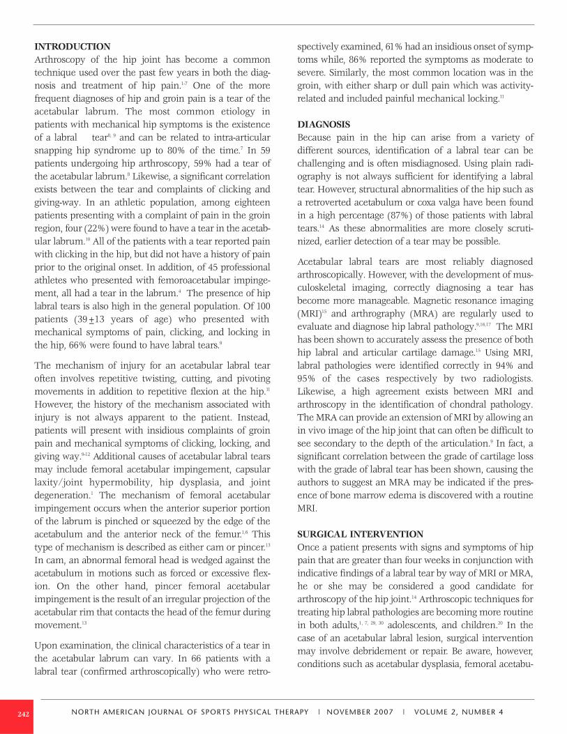
Over the past few years, arthroscopy of the hipjoint is becoming more common as a tech-nique in both the diagnosis and treatment ofhip pain. A frequent cause of hip and groinpain is a tear of the acetabular labrum. Patientswith labral tears complain of pain in the groinregion and pain with clicking in the hip with-out a history of pain prior to the original onset.Once a patient presents with signs and symp-toms of hip pain that are greater than fourweeks in conjunction with indicative findingsof a labral tear by way of MRI, he or she maybe considered a good candidate forarthroscopy of the hip joint. Little evidenceexists in the current literature on rehabilitativeprocedures performed after arthroscopy of theacetabular labrum. The purpose of this clinicalcommentary is to suggest a rehabilitation pro-tocol after acetebular labral debridement orrepair.
NORTH AMERICAN JOURNAL OF SPORTS PHYSICAL THERAPY | NOVEMBER 2007 | VOLUME 2, NUMBER 4
INTRODUCTIONArthroscopy of the hip joint has become a commontechnique used over the past few years in both the diag-nosis and treatment of hip pain.1-7 One of the morefrequent diagnoses of hip and groin pain is a tear of theacetabular labrum. The most common etiology inpatients with mechanical hip symptoms is the existenceof a labral tear8, 9 and can be related to intra-articularsnapping hip syndrome up to 80% of the time.7 In 59patients undergoing hip arthroscopy, 59% had a tear ofthe acetabular labrum.8 Likewise, a significant correlationexists between the tear and complaints of clicking andgiving-way. In an athletic population, among eighteenpatients presenting with a complaint of pain in the groinregion, four (22%) were found to have a tear in the acetab-ular labrum.10 All of the patients with a tear reported painwith clicking in the hip, but did not have a history of painprior to the original onset. In addition, of 45 professionalathletes who presented with femoroacetabular impinge-ment, all had a tear in the labrum.4 The presence of hiplabral tears is also high in the general population. Of 100patients (39±13 years of age) who presented withmechanical symptoms of pain, clicking, and locking inthe hip, 66% were found to have labral tears.9
The mechanism of injury for an acetabular labral tearoften involves repetitive twisting, cutting, and pivotingmovements in addition to repetitive flexion at the hip.11
However, the history of the mechanism associated withinjury is not always apparent to the patient. Instead,patients will present with insidious complaints of groinpain and mechanical symptoms of clicking, locking, andgiving way.9-12 Additional causes of acetabular labral tearsmay include femoral acetabular impingement, capsularlaxity/joint hypermobility, hip dysplasia, and jointdegeneration.1 The mechanism of femoral acetabularimpingement occurs when the anterior superior portionof the labrum is pinched or squeezed by the edge of theacetabulum and the anterior neck of the femur.1,6 Thistype of mechanism is described as either cam or pincer.13
In cam, an abnormal femoral head is wedged against theacetabulum in motions such as forced or excessive flex-ion. On the other hand, pincer femoral acetabularimpingement is the result of an irregular projection of theacetabular rim that contacts the head of the femur duringmovement.13
Upon examination, the clinical characteristics of a tear inthe acetabular labrum can vary. In 66 patients with alabral tear (confirmed arthroscopically) who were retro-
spectively examined, 61% had an insidious onset of symp-toms while, 86% reported the symptoms as moderate tosevere. Similarly, the most common location was in thegroin, with either sharp or dull pain which was activity-related and included painful mechanical locking.11
DIAGNOSISBecause pain in the hip can arise from a variety ofdifferent sources, identification of a labral tear can bechallenging and is often misdiagnosed. Using plain radi-ography is not always sufficient for identifying a labraltear. However, structural abnormalities of the hip such asa retroverted acetabulum or coxa valga have been foundin a high percentage (87%) of those patients with labraltears.14 As these abnormalities are more closely scruti-nized, earlier detection of a tear may be possible.
Acetabular labral tears are most reliably diagnosedarthroscopically. However, with the development of mus-culoskeletal imaging, correctly diagnosing a tear hasbecome more manageable. Magnetic resonance imaging(MRI)15 and arthrography (MRA) are regularly used toevaluate and diagnose hip labral pathology.9,16,17 The MRIhas been shown to accurately assess the presence of bothhip labral and articular cartilage damage.15 Using MRI,labral pathologies were identified correctly in 94% and95% of the cases respectively by two radiologists.Likewise, a high agreement exists between MRI andarthroscopy in the identification of chondral pathology.The MRA can provide an extension of MRI by allowing anin vivo image of the hip joint that can often be difficult tosee secondary to the depth of the articulation.9 In fact, asignificant correlation between the grade of cartilage losswith the grade of labral tear has been shown, causing theauthors to suggest an MRA may be indicated if the pres-ence of bone marrow edema is discovered with a routineMRI.
SURGICAL INTERVENTIONOnce a patient presents with signs and symptoms of hippain that are greater than four weeks in conjunction withindicative findings of a labral tear by way of MRI or MRA,he or she may be considered a good candidate forarthroscopy of the hip joint.14 Arthroscopic techniques fortreating hip labral pathologies are becoming more routinein both adults,1, 7, 29, 30 adolescents, and children.20 In thecase of an acetabular labral lesion, surgical interventionmay involve debridement or repair. Be aware, however,conditions such as acetabular dysplasia, femoral acetabu-
242 NORTH AMERICAN JOURNAL OF SPORTS PHYSICAL THERAPY | NOVEMBER 2007 | VOLUME 2, NUMBER 4
lar impingement, degenerative conditions of the joint,fractures, arthrofibrosis, or non-compliance with therehabilitation program may preclude the surgical inter-vention.
Both debridement and repair of an acetabular labrallesion may be performed in either the supine or lateralposition. In the supine position, a standard fracture tableis used with an oversized perineal post to apply traction.The affected hip is placed into slight extension and adduc-tion to allow approach to the joint. Care is taken tominimize pressure in the perineal area as well as to care-fully monitor the amount and duration of traction to avoidneurologic complications. The procedure is performedunder the guidance of fluoroscopy. After an adequateamount of distraction is obtained, a 14 or 16 gauge spinalneedle is inserted into the joint to break the vacuum sealand allow further distraction. Three portals are generallyused, the anterolateral (which is established first), theanterior, and the distal lateral accessory or para-trochanteric portal.
After complete evaluation of the joint, including thearticular cartilage surfaces of the acetabulum and femoralhead, ligament fraying or tearing, and assessment of theposterior recess, any labral abnormality may then beaddressed. Most labral tears occur in the anterior superi-or quadrant of the acetabulum. Acute, longitudinal, andperipheral tears are most amenable to repair. For repair ofa detached labrum, the edges of the tear are delineatedand suture anchors are placed on the top of the acetabu-lar rim in the area of detachment.1 Conversely, if the tearin the labrum has a secure outer rim and is still attachedto the acetabulum, a suture in the midsubstance of thetear can be used to secure the tissue. Radial splits, signif-icantly macerated or degenerative labral tissue, or labraltissue that appears unviable should be debrided. Notuncommonly, chondral lesions may be noted adjacent tolabral pathology that may require either debridement,common microfracture, or other cartilage resurfacingtechniques.4,21
The natural history of untreated labral tears is unknown.Recently, authors9,21,22 have attempted to correlate thepresence of labral tears with the development of degen-erative osteoarthritis of the hip joint. Although this corre-lation remains unclear, some observations suggest labrallesions may be a contributory factor to the evolution andprogression of osteoarthritis.21 Although debridementmay be successful from the standpoint of pain relief, the
surgery could potentially lead to joint load alteration andthe progression of articular cartilage changes.23 In addi-tion, alterations to the structure of the labrum, or disrup-tion in the acetabular labral junction, may lead to the lossof the pressurized fluid film layer within the joint anduneven force distribution across the articular cartilagesurfaces of the acetabulum and femoral head.24
REHABILITATIONLittle evidence exists in the current literature to supportrehabilitative procedures performed after arthroscopy ofthe acetabular labrum. Likewise, because new surgicalprocedures are constantly evolving, it is the responsibilityof the physical therapist to stay up to date with the mostcurrent techniques as well as to establish and maintaingood communication with the orthopaedic surgeon.
Surgical techniques and outcomes have been reported inthe literature with little or no attention to post-operativerehabilitation.25-27 Currently, the best evidence for post-operative rehabilitation is based upon surgeon and physi-cal therapist experience. With a labral repair, the locationand size of the tear should be noted.28 Most of these tearsare located in the anterior or anterosuperior portion ofthe labrum;9,29 movements that stress this area should beavoided. However, communication with the surgeonabout the location of the tear and surgical technique usedis vital to the treating physical therapist.
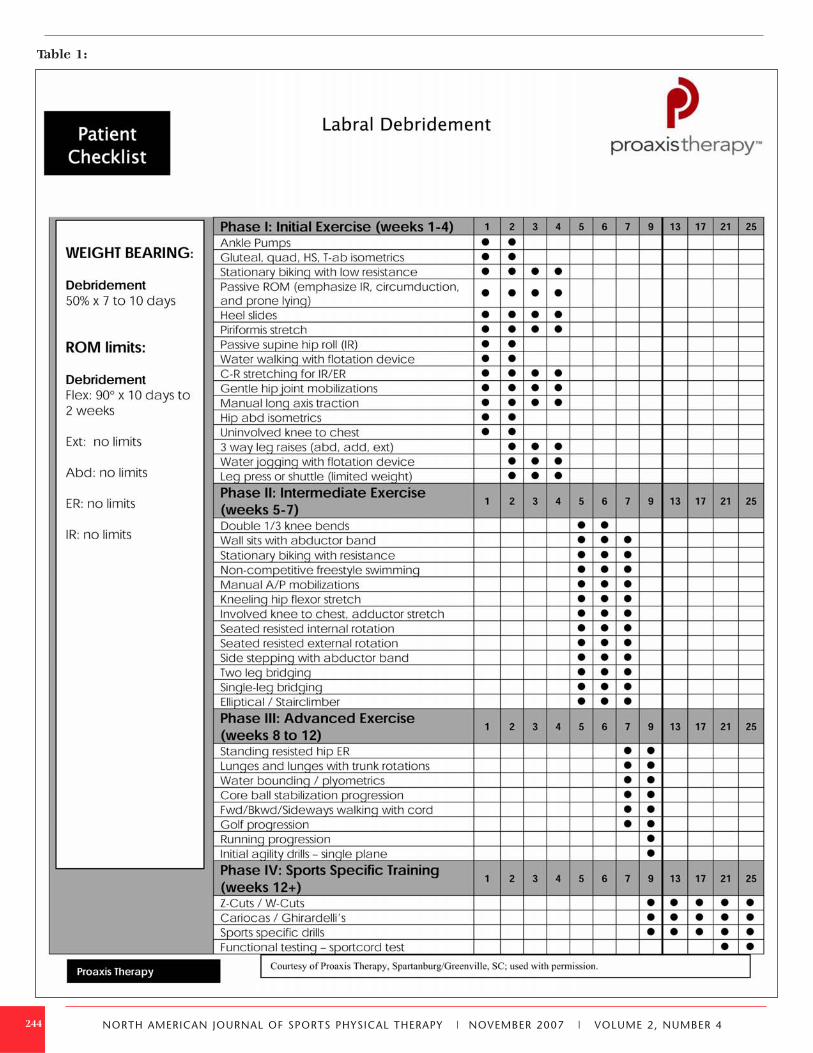
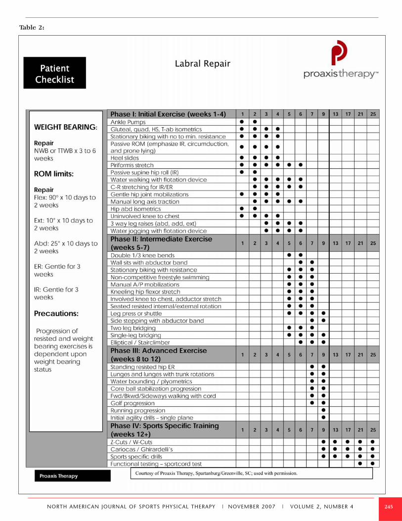
Rehabilitation protocols following acetabular labraldebridement (Table 1) or repair (Table 2) can be dividedinto four phases. The progression with both procedures issimilar with the exception of differences which are notedwithin the protocol. Exercise, and particularly strength-ening, progression in a repair of the labrum may bedelayed by a few weeks depending upon tissue healing.The timelines for each phase are based on clinical find-ings and presentations of active, healthy individuals. Ifclinical presentation meets objective criteria an athletemay move through the phases at a faster rate, alwayskeeping basic tissue healing physiology in mind.
Phase I – Initial Exercises. Weeks 1 to 4. The primary goals immediately following acetabularlabral debridement or repair are to minimize pain andinflammation, protect the surgically repaired tissue, andinitiate early motion exercises. Patients are typically 50%weight-bearing for 7 to 10 days progressing to weight-bear-ing as tolerated. For a labral repair, the weight-bearingrestrictions include toe-touch weight bearing for 3 weeks,
243NORTH AMERICAN JOURNAL OF SPORTS PHYSICAL THERAPY | NOVEMBER 2007 | VOLUME 2, NUMBER 4
244
Table 1:
NORTH AMERICAN JOURNAL OF SPORTS PHYSICAL THERAPY | NOVEMBER 2007 | VOLUME 2, NUMBER 4
245
Table 2:
NORTH AMERICAN JOURNAL OF SPORTS PHYSICAL THERAPY | NOVEMBER 2007 | VOLUME 2, NUMBER 4
but may last up to 6 weeks depending upon the progres-sion of healing and pain level of the patient. If the surgeryinvolved additional procedures to the hip such as amicrofracture, the weight-bearing restrictions canincrease to 6 more weeks. However, for purposes of thispaper, labral debridement and repair will be the mainfocus.
It is important to maintain a symmetrical gait pattern toprevent concomitant stress throughout the lower extrem-ity and spine. If this gait pattern is not established, amuscular imbalance of tight hip flexors and erectorspinae with inhibition of the gluteals and abdominals(lower crossed syndrome) could develop.30 The potentialramifications include increased weight-bearing throughthe acetabulum with labral tissue stresses secondary tohip flexor tightness.31 Consequently, continued crutch usemay be a necessary prophylactic. Similarly, the patientshould be instructed to control the hip in all three planesof motion. Decreased core and hip strength have beenimplicated in alterations of lower extremity alignmentduring functional activities.32-34 Also, hip abductor strengthhas been shown to be a predictor of frontal plane motionin the knee while weak hip extensors can lead to quadri-ceps overuse and increased compression and shear forceat the knee.32 A correlation between weakness in the hipand core and lower extremity injuries has been estab-lished.35 Thus, when these studies32,34-36 are consideredtogether, the suggestion is that rehabilitation of the hipshould include a component of hip and core strengthen-ing in each phase.
Aquatic therapy is an excellent resource, if available, oncesurgical incisions are well-healed. Ambulation in thewater allows for improvements in gait by allowing appro-priate loads to the joint while minimizing unnecessarystresses to the healing tissue.19,28 Light jogging in the waterusing a flotation device may begin as early as 2-3 weeksif pain is not an issue. Range of motion (ROM) precau-tions may vary, but typically include limiting flexionbeyond 90º for 10 days to avoid undue compression of theanterior labrum. If treating a labral repair, in addition tothe 90º of flexion, the patient is limited to 25º of abduc-tion and 10º of extension for 10 days to 2 weeks. Anemphasis should be placed on manual therapy for painreduction and improvements in joint mobility and pro-prioception.31 Considerations include gentle hip jointmobilizations, contract-relax stretching for internal andexternal rotation, long axis distraction, and assessment oflumbo-sacral mobility. Particular attention should be
paid to the posterlateral soft tissue structures and thelumbo-sacral spine (as each can contribute to pain) andunnecessary hypomobility which will limit progress infuture phases.
Modalities for pain control such as immediate post-operative transcutaneous electrical nerve stimulation(TENS) units {preferably applied in recovery room (basedon clinical experiences of the authors)}, cryotherapy, andappropriate pain management through medication areimportant, as well. Cautious and gentle stretching of hipmuscle groups including piriformis, psoas, quadriceps,and hamstring muscles should begin with hip passiverange of motion exercises, respecting the patient’s painthreshold. The risk of tissue damage must be consid-ered,37 and the patient should provide verbal feedbackfollowing mobilization and ROM exercises. Prone lying for1 to 2 hours per day and passive ROM exercises empha-sizing internal rotation are beneficial to preventadhesions.19 Stationary bike begins with no resistance andgradually progresses in resistance over the first 4 weekswith a seat height that limits hip flexion to less than 90º.
Strengthening in Phase I initially consists of isometriccontractions for the hip adductors, abductors, extensors,and transverse abdominals, but progresses to straight legraises for abduction, adduction, and extension. To preventirritation of the psoas muscle,19 hip flexion straight legraise is not performed early on. Seated hip flexion usinga short lever arm might be an alternative. For labraldebridement, closed-chain activities of low-level leg pressor shuttle can also begin with limited resistance. This typeof exercise allows weight-bearing through the lowerextremity joints with the application of appropriate stress-es to the tissue.38
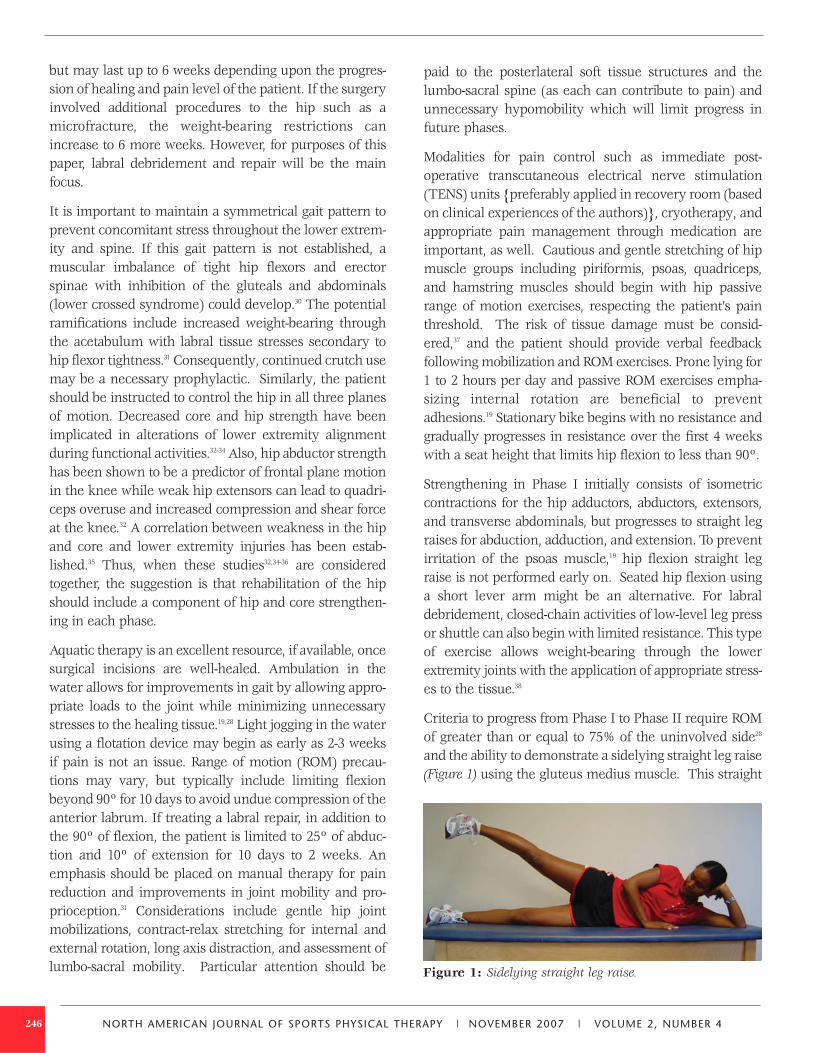
Criteria to progress from Phase I to Phase II require ROMof greater than or equal to 75% of the uninvolved side28
and the ability to demonstrate a sidelying straight leg raise(Figure 1) using the gluteus medius muscle. This straight
246
Figure 1: Sidelying straight leg raise.
NORTH AMERICAN JOURNAL OF SPORTS PHYSICAL THERAPY | NOVEMBER 2007 | VOLUME 2, NUMBER 4
let raise should be performed without compensationfrom the tensor fascia lata and quadratus lumborum.
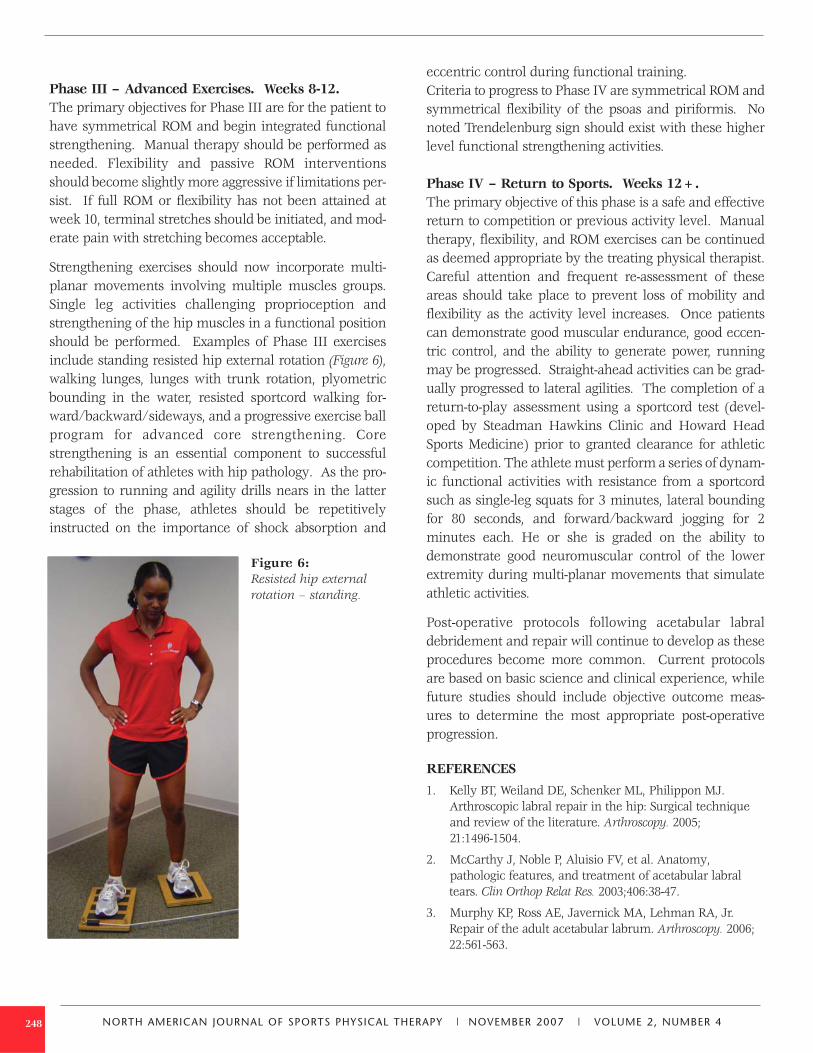
Phase II – Intermediate Exercises. Weeks 5-7. The primary focus of the second phase is to continueprogressing ROM and soft tissue flexibility while begin-ning to transition the emphasis to strengthening. Manualtherapy should continue with mobilization becomingmore aggressive, as appropriate. If capsular laxity wasthought to be a contributing factor to developing labralpathology, normal mobility (but not hypermobility)should be achieved. Flexibility exercises involving thepiriformis, adductor group, and psoas/rectus femorisshould continue. The kneeling hip flexor stretch (Figure2) can be particularly beneficial for psoas and rectusfemoris once tolerated in this phase of rehabilitation.Passive ROM exercises should become more aggressive,as needed, for internal and external rotation.
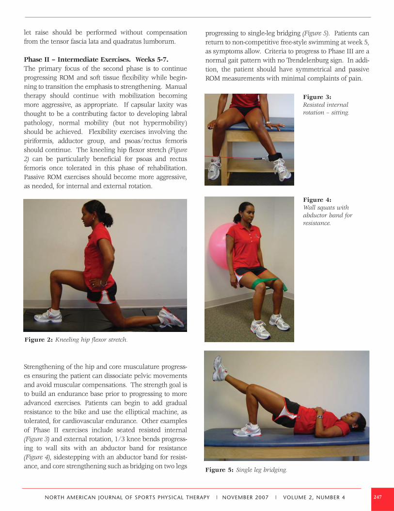
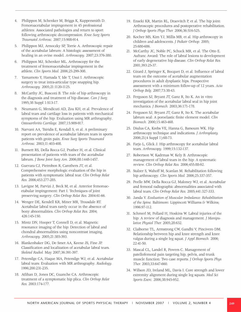
Strengthening of the hip and core musculature progress-es ensuring the patient can dissociate pelvic movementsand avoid muscular compensations. The strength goal isto build an endurance base prior to progressing to moreadvanced exercises. Patients can begin to add gradualresistance to the bike and use the elliptical machine, astolerated, for cardiovascular endurance. Other examplesof Phase II exercises include seated resisted internal(Figure 3) and external rotation, 1/3 knee bends progress-ing to wall sits with an abductor band for resistance(Figure 4), sidestepping with an abductor band for resist-ance, and core strengthening such as bridging on two legs
progressing to single-leg bridging (Figure 5). Patients canreturn to non-competitive free-style swimming at week 5,as symptoms allow. Criteria to progress to Phase III are anormal gait pattern with no Trendelenburg sign. In addi-tion, the patient should have symmetrical and passiveROM measurements with minimal complaints of pain.
247
Figure 2: Kneeling hip flexor stretch.
Figure 3: Resisted internal rotation – sitting.
Figure 5: Single leg bridging.
Figure 4: Wall squats with abductor band forresistance.
NORTH AMERICAN JOURNAL OF SPORTS PHYSICAL THERAPY | NOVEMBER 2007 | VOLUME 2, NUMBER 4
Phase III – Advanced Exercises. Weeks 8-12.The primary objectives for Phase III are for the patient tohave symmetrical ROM and begin integrated functionalstrengthening. Manual therapy should be performed asneeded. Flexibility and passive ROM interventionsshould become slightly more aggressive if limitations per-sist. If full ROM or flexibility has not been attained atweek 10, terminal stretches should be initiated, and mod-erate pain with stretching becomes acceptable.
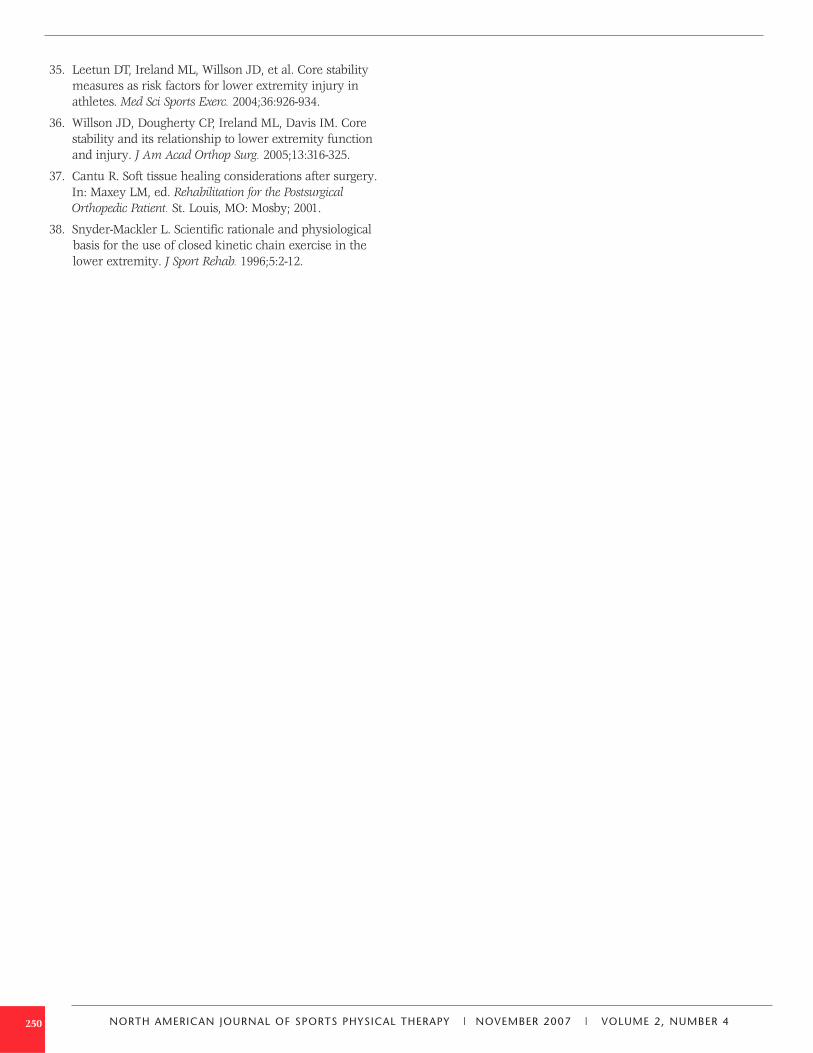
Strengthening exercises should now incorporate multi-planar movements involving multiple muscles groups.Single leg activities challenging proprioception andstrengthening of the hip muscles in a functional positionshould be performed. Examples of Phase III exercisesinclude standing resisted hip external rotation (Figure 6),walking lunges, lunges with trunk rotation, plyometricbounding in the water, resisted sportcord walking for-ward/backward/sideways, and a progressive exercise ballprogram for advanced core strengthening. Corestrengthening is an essential component to successfulrehabilitation of athletes with hip pathology. As the pro-gression to running and agility drills nears in the latterstages of the phase, athletes should be repetitivelyinstructed on the importance of shock absorption and
eccentric control during functional training. Criteria to progress to Phase IV are symmetrical ROM andsymmetrical flexibility of the psoas and piriformis. Nonoted Trendelenburg sign should exist with these higherlevel functional strengthening activities.
Phase IV – Return to Sports. Weeks 12+. The primary objective of this phase is a safe and effectivereturn to competition or previous activity level. Manualtherapy, flexibility, and ROM exercises can be continuedas deemed appropriate by the treating physical therapist.Careful attention and frequent re-assessment of theseareas should take place to prevent loss of mobility andflexibility as the activity level increases. Once patientscan demonstrate good muscular endurance, good eccen-tric control, and the ability to generate power, runningmay be progressed. Straight-ahead activities can be grad-ually progressed to lateral agilities. The completion of areturn-to-play assessment using a sportcord test (devel-oped by Steadman Hawkins Clinic and Howard HeadSports Medicine) prior to granted clearance for athleticcompetition. The athlete must perform a series of dynam-ic functional activities with resistance from a sportcordsuch as single-leg squats for 3 minutes, lateral boundingfor 80 seconds, and forward/backward jogging for 2minutes each. He or she is graded on the ability todemonstrate good neuromuscular control of the lowerextremity during multi-planar movements that simulateathletic activities.
Post-operative protocols following acetabular labraldebridement and repair will continue to develop as theseprocedures become more common. Current protocolsare based on basic science and clinical experience, whilefuture studies should include objective outcome meas-ures to determine the most appropriate post-operativeprogression.
REFERENCES1. Kelly BT, Weiland DE, Schenker ML, Philippon MJ.
Arthroscopic labral repair in the hip: Surgical technique and review of the literature. Arthroscopy. 2005; 21:1496-1504.
2. McCarthy J, Noble P, Aluisio FV, et al. Anatomy, pathologic features, and treatment of acetabular labral tears. Clin Orthop Relat Res. 2003;406:38-47.
3. Murphy KP, Ross AE, Javernick MA, Lehman RA, Jr. Repair of the adult acetabular labrum. Arthroscopy. 2006; 22:561-563.
248
Figure 6: Resisted hip externalrotation – standing.
NORTH AMERICAN JOURNAL OF SPORTS PHYSICAL THERAPY | NOVEMBER 2007 | VOLUME 2, NUMBER 4
4. Philippon M, Schenker M, Briggs K, Kuppersmith D. Femoroacetabular impingement in 45 professional athletes: Associated pathologies and return to sport following arthroscopic decompression. Knee Surg Sports Traumatol Arthrosc. 2007;15:908-914.
5. Philippon MJ, Arnoczky SP, Torrie A. Arthroscopic repair of the acetabular labrum: A histologic assessment of healing in an ovine model. Arthroscopy. 2007;23:376-380.
6. Philippon MJ, Schenker ML. Arthroscopy for the treatment of femoroacetabular impingement in the athlete. Clin Sports Med. 2006;25:299-308.
7. Yamamoto Y, Hamada Y, Ide T, Usui I. Arthroscopic surgery to treat intra-articular type snapping hip. Arthroscopy. 2005;21:1120-1125.
8. McCarthy JC, Busconi B. The role of hip arthroscopy in the diagnosis and treatment of hip disease. Can J Surg.1995;38 Suppl 1:S13-17.
9. Neumann G, Mendicuti AD, Zou KH, et al. Prevalence of labral tears and cartilage loss in patients with mechanical symptoms of the hip: Evaluation using MR arthrography. Osteoarthritis Cartilage. 2007;15:909-917.
10. Narvani AA, Tsiridis E, Kendall S, et al. A preliminary report on prevalence of acetabular labrum tears in sports patients with groin pain. Knee Surg Sports Traumatol Arthrosc. 2003;11:403-408.
11. Burnett RS, Della Rocca GJ, Prather H, et al. Clinical presentation of patients with tears of the acetabular labrum. J Bone Joint Surg Am. 2006;88:1448-1457.
12. Guevara CJ, Pietrobon R, Carothers JT, et al. Comprehensive morphologic evaluation of the hip in patients with symptomatic labral tear. Clin Orthop Relat Res. 2006;453:277-285.
13. Lavigne M, Parvizi J, Beck M, et al. Anterior femoroac-etabular impingement: Part I. Techniques of joint preserving surgery. Clin Orthop Relat Res. 2004:61-66.
14. Wenger DE, Kendell KR, Miner MR, Trousdale RT. Acetabular labral tears rarely occur in the absence of bony abnormalities. Clin Orthop Relat Res. 2004; 426:145-150.
15. Mintz DN, Hooper T, Connell D, et al. Magnetic resonance imaging of the hip: Detection of labral and chondral abnormalities using noncontrast imaging. Arthroscopy. 2005;21:385-393.
16. Blankenbaker DG, De Smet AA, Keene JS, Fine JP. Classification and localization of acetabular labral tears. Skeletal Radiol. May 2007;36:391-397.
17. Petersilge CA, Haque MA, Petersilge WJ, et al. Acetabular labral tears: Evaluation with MR arthrography. Radiology.1996;200:231-235.
18. Atlihan D, Jones DC, Guanche CA. Arthroscopic treatment of a symptomatic hip plica. Clin Orthop Relat Res. 2003:174-177.
19. Enseki KR, Martin RL, Draovitch P, et al. The hip joint: Arthroscopic procedures and postoperative rehabilitation. J Orthop Sports Phys Ther. 2006;36:516-525.
20. Kocher MS, Kim YJ, Millis MB, et al. Hip arthroscopy in children and adolescents. J Pediatr Orthop. 2005;25:680-686.
21. McCarthy JC, Noble PC, Schuck MR, et al. The Otto E.Aufranc Award: The role of labral lesions to development of early degenerative hip disease. Clin Orthop Relat Res.2001;393:25-37.
22. Girard J, Springer K, Bocquet D, et al. Influence of labral tears on the outcome of acetabular augmentation procedures in adult dysplastic hips. Prospective assessment with a minimum follow-up of 12 years. Acta Orthop Belg. 2007;73:38-43.
23. Ferguson SJ, Bryant JT, Ganz R, Ito K. An in vitro investigation of the acetabular labral seal in hip joint mechanics. J Biomech. 2003;36:171-178.
24. Ferguson SJ, Bryant JT, Ganz R, Ito K. The acetabular labrum seal: A poroelastic finite element model. Clin Biomech. 2000;15:463-468.
25. Diulus CA, Krebs VE, Hanna G, Barsoum WK. Hip arthroscopy technique and indications. J Arthroplasty.2006;21(4 Suppl 1):68-73.
26. Farjo L, Glick J. Hip arthroscopy for acetabular labral tears. Arthroscopy. 1999;15:132-137.
27. Robertson W, Kadrmas W, Kely B. Arthroscopic management of labral tears in the hip: A systematic review. Clin Orthop Relat Res. 2006;455:88-92.
28. Stalzer S, Wahoff M, Scanlan M. Rehabilitation following hip arthroscopy. Clin Sports Med. 2006;25:337-357.
29. Peelle MW, Della Rocca GJ, Maloney WJ, et al. Acetabular and femoral radiographic abnormalities associated with labral tears. Clin Orthop Relat Res. 2005;441:327-333.
30. Janda V. Evaluation of Muscular Imbalance: Rehabilitation of the Spine. Baltimore: Lippincott Williams & Wilkins; 1996:97-112.
31. Schmerl M, Pollard H, Hoskins W. Labral injuries of the hip: A review of diagnosis and management. J Manipu- lative Physiol Ther. 2005;28:632.
32. Claiborne TL, Armstrong CW, Gandhi V, Pincivero DM. Relationship between hip and knee strength and knee valgus during a single leg squat. J Appl Biomech. 2006; 22:41-50.
33. Mascal CL, Landel R, Powers C. Management of patellofemoral pain targeting hip, pelvis, and trunk muscle function: Two case reports. J Orthop Sports Phys Ther. 2003;33:647-660.
34. Willson JD, Ireland ML, Davis I. Core strength and lower extremity alignment during single leg squats. Med Sci Sports Exerc. 2006;38:945-952.
249NORTH AMERICAN JOURNAL OF SPORTS PHYSICAL THERAPY | NOVEMBER 2007 | VOLUME 2, NUMBER 4
35. Leetun DT, Ireland ML, Willson JD, et al. Core stability measures as risk factors for lower extremity injury in athletes. Med Sci Sports Exerc. 2004;36:926-934.
36. Willson JD, Dougherty CP, Ireland ML, Davis IM. Core stability and its relationship to lower extremity function and injury. J Am Acad Orthop Surg. 2005;13:316-325.
37. Cantu R. Soft tissue healing considerations after surgery. In: Maxey LM, ed. Rehabilitation for the Postsurgical Orthopedic Patient. St. Louis, MO: Mosby; 2001.
38. Snyder-Mackler L. Scientific rationale and physiological basis for the use of closed kinetic chain exercise in the lower extremity. J Sport Rehab. 1996;5:2-12.
250 NORTH AMERICAN JOURNAL OF SPORTS PHYSICAL THERAPY | NOVEMBER 2007 | VOLUME 2, NUMBER 4