Embed Size (px)
Citation preview

CpG Oligodeoxynucleotides Downregulate Placental Adiponectinand Increase Embryo Loss in Non-Obese Diabetic MiceChuan-Mei Qin, Fu-Ju Tian, Xiao-Rui Liu, Fan Wu, Xiao-Ling Ma, Yi Lin
Institute of Embryo-Fetal Original Adult Disease, The International Peace Maternity & Child Health Hospital, Shanghai Jiao Tong University School of
Medicine, Shanghai, China
Keywords
Adipokine, immunodeficiency, infection,
trophoblast
Correspondence
Yi Lin, Institute of Embryo-Fetal Original Adult
Disease, The International Peace Maternity &
Child Health Hospital, Shanghai Jiao Tong
University School of Medicine, Shanghai
200030, China.
E-mail: [email protected]
Chuan-Mei Qin and Fu-Ju Tian contributed
equally to this work.
The copyright line for this article was changed
on 26th August, 2016 after original online
publication
Submission January 15, 2016;
accepted March 23, 2016.
Citation
Qin C-M, Tian F-J, Liu X-R, Wu F, Ma X-L, Lin Y.
CpG oligodeoxynucleotides downregulate
placental adiponectin and increase embryo
loss in non-obese diabetic mice. Am J Reprod
Immunol 2016; 76: 38–49
doi:10.1111/aji.12515
Problem
CpG oligodeoxynucleotides (ODNs) can induce immunological changes
in non-obese diabetic (NOD) mice and increase embryo loss, but little is
known about the mechanism. This study aimed to determine the role of
adiponectin in CpG ODN-induced pregnancy failure.
Method of study
Oligodeoxynucleotide 1826 was intraperitoneally injected to NOD mice,
and ODN 2216, ODN 2006, and ODN 2395 were used to stimulate
human trophoblast cell lines to investigate adiponectin expression pat-
terns and its possible effects on trophoblast function.
Results
CpG ODNs downregulated adiponectin via the cJun N-terminal kinase
signaling pathway and led to increased embryo loss (from 6.9 to
33.3%). ODN 2006 impaired human trophoblast cell migration, which
was successfully rescued by adiponectin treatment.
Conclusion
CpG ODNs decreased placental adiponectin expression in NOD mice and
impaired human trophoblast function and was associated with increased
embryo loss. Adiponectin may therefore play an important protective
role in the prevention of bacteria-induced pregnancy failure.
Introduction
The maternal–fetal interface refers to an area of
direct contact between maternal (decidua) and fetal
(trophoblast) tissues,1 and effective maternal–fetalcross talk promotes successful fetal antigen exposure
to the maternal environment, thus influencing
normal fetal growth.2 During pregnancy, bacterial
infections may damage the local molecular and cel-
lular microenvironment, and aberrant cellular inter-
actions at the maternal–fetal interface can lead to
serious complications of pregnancy, such as implan-
tation failure, embryo loss, spontaneous abortion,
premature delivery, pre-eclampsia, and intrauterine
growth restriction.3–5
Toll-like receptor 9 (TLR9) recognizes unmethy-
lated CpG dinucleotides, a characteristic feature of
microbial DNA. CpG oligodeoxynucleotides (ODNs)
are short single-stranded synthetic DNA molecules
containing unmethylated CpG motifs that mimic
microbial DNA and can therefore produce an
immunostimulatory response. We have previously
demonstrated that intraperitoneal ODN injection in
non-obese diabetic (NOD) mice caused a series of
immunological changes at the maternal–fetal inter-
face. The NOD mouse strain is a model of type 1
American Journal of Reproductive Immunology 76 (2016) 38–49
ª 2016 The Authors. American Journal of Reproductive Immunology Published by John Wiley & Sons Ltd.
This is an open access article under the terms of the Creative Commons Attribution-NonCommercial-NoDerivs License, which permits use and
distribution in any medium, provided the original work is properly cited, the use is non-commercial and no modifications or adaptations are made.38
ORIGINAL ARTICLE

diabetes, which has natural killer (NK) cell deficits,
and is prone to embryo loss. Our studies identified
the ODN-induced immunological changes to be tak-
ing place via the TLR9 pathway. These changes
included abnormal proliferation of uterine macro-
phages and neutrophils, and tumor necrosis factor-a(TNF-a) and mouse keratinocyte-derived cytokine
(mKC) production by uterine CD11b+ F4/80+ cells,
which led to increased fetal loss and premature
delivery. Depletion of F4/80+ cells or transplantation
of induced Treg (iTreg) cells rescued CpG-mediated
pregnancy failure.6,7
In addition to immunological factors, metabolism
at the maternal–fetal interface is emerging as an
important factor to ensure normal pregnancy. As the
most abundant circulating adipokine, adiponectin
plays a key role in metabolic disorders. Adiponectin
is a 30-kDa protein, which exists as two forms (glob-
ular adiponectin and full-length adiponectin), and
mediates its actions mainly via two molecules; adipo-
nectin receptor 1 and adiponectin receptor 2.8 Adipo-
nectin is involved in various physiological activities,
including energy homeostasis, insulin sensitivity, glu-
cose and lipid metabolism, inflammation, immunity,
and angiogenesis, and can act as an antidiabetic,
anti-atherogenic, or anti-inflammatory adipokine.9
Furthermore, adiponectin also plays an important
role in maintaining normal reproductive function,
and its receptors are expressed in several reproduc-
tion-related organs, including the pituitary gland,
hypothalamus, testis, ovary, oviduct, uterus, endo-
metrium, and placenta.10–12 Circulating adiponectin
levels have been widely documented as being mark-
edly lower in patients with obesity, diabetes mellitus,
and metabolic syndrome compared to healthy indi-
viduals (in both pregnant and non-pregnant
women), and abnormal levels of adiponectin are
associated with a series of pregnancy complications,
including gestational diabetes mellitus and pre-
eclampsia. For example, low circulating adiponectin
levels in pregnant women have been associated with
an increased risk of gestational diabetes mellitus,10,13
and in pregnant women with normal weight, circu-
lating adiponectin has been correlated negatively
with the gestational age.14 In addition, it has been
suggested that pregnant women with a lower level of
serum adiponectin are more likely to develop pre-
eclampsia.15,16 However, some studies have reported
higher serum adiponectin concentrations in pre-
eclamptic patients compared to women with normal
pregnancies.17,18 While metabolic dysfunctions are
frequently linked to reproductive abnormalities, the
relationship between the anti-inflammatory role of
adiponectin and CpG ODN-induced embryo loss in
NOD mice remains unclear.
In this study, we hypothesized that adiponectin may
play a key role in resisting CpG-induced damage to
trophoblast function. CpG ODNs were used to mimic
bacterial infections during pregnancy. Placental adipo-
nectin expression was significantly decreased after
treatment with ODN, and this decreased adiponectin
expression was correlated with increased embryo loss
in NOD compared to wild-type (WT) mice. We also
investigated adiponectin expression patterns and its
potential effects on trophoblast function and preg-
nancy outcome using human trophoblast cell lines, to
explore the relationship between CpG ODN-induced
embryo loss and adiponectin function.
Materials and methods
Treatment of Pregnant Mice
Mouse treatment methods are shown in Figure S1.
Healthy WT female BALB/c and female NOD mice
with BALB/c background and male C57BL/6 mice
aged 8–10 weeks were purchased from Beijing HFK
Bioscience Co. Ltd. (Beijing, China). All mice were
housed in a pathogen-free facility. All animal proce-
dures followed national animal care guidelines and
were approved by the Institutional Animal Care and
Use Committee of Shanghai Jiao Tong University. The
day on which the vaginal plug was detected was des-
ignated as gestational day 0.5 (E0.5). ODN 1826 is
specific for mouse tissues and cells19,20 and was there-
fore used for all animal experiments conducted in this
study. Pregnant mice were intraperitoneally injected
with ODN 1826 control or ODN 1826 (InvivoGen,
San Diego, CA, USA) at the dose of 25 lg/dam dis-
solved in 200 lL phosphate-buffered saline (PBS)
(Gibco BRL Co.Ltd., Gaithersburg, MD, USA) on E6.5.
Each group consisted of at least 6 mice. Pregnant mice
were killed on E10.5.6,7,21 Embryos with a smaller size
(20% smaller than the average size), hemorrhage (at
the implantation site), and necrosis were identified as
resorbed embryos.22,23 Placentas were immediately
collected and frozen in liquid nitrogen.
Cell Culture
BeWo (cell model to mimic syncytialization of pla-
cental villous trophoblast in vivo) and JAR (human
American Journal of Reproductive Immunology 76 (2016) 38–49
ª 2016 The Authors. American Journal of Reproductive Immunology Published by John Wiley & Sons Ltd. 39
ADIPONECTIN AND EMBRYO LOSS

choriocarcinoma cell line used as in vitro model of
cytotrophoblasts) cell lines (China Infrastructure
of Cell Line Resources, Beijing, China) were rou-
tinely grown at 37°C with 5% CO2 in phenol-red
DMEM/F12 medium (Gibco) supplemented with
15% fetal bovine serum (FBS) (Gibco) and phenol-
red DMEM medium (Gibco) with 10% FBS, respec-
tively, along with streptomycin (10 lg/mL) and
penicillin (100 U/mL) (Gibco). HTR-8, a human
extravillous trophoblast cell line, was kindly pro-
vided by P. K. Lala (University of Western Ontario,
Ontario, Canada) and cultured in phenol-red
DMEM/F12 medium supplemented with 10% FBS,
streptomycin (10 lg/mL), and penicillin (100 U/mL)
at 37°C with 5% CO2.
Cells were seeded onto plates and incubated over-
night at 37°C with 5% CO2. The following day,
BeWo and HTR8 cells were cultured in DMEM/F12
medium, and JAR cells were cultured in DMEM
medium, all supplemented with 1% FBS in the pres-
ence of various agents, such as forskolin (25 nM;
Sigma Aldrich, St. Louis, MO, USA), adiponectin
(400 ng/mL; Peprotech, Rocky Hill, NJ, USA), TLR9
agonists (5 lM), SP600125, PD98059 (Cell Signaling
Technology, Danvers, MA, USA), or corresponding
controls.
Quantitative Real-time PCR (qRT-PCR)
Total RNA was extracted using TRIzol reagent
(TaKaRa Bio Inc., Tokyo, Japan), and 1 lg of total
RNA was used to synthesize first-strand cDNA with
the PrimeScriptTM II 1st Strand cDNA Synthesis Kit
(TaKaRa Bio Inc.) using random or oligo-dT primers.
Thereafter, qRT-PCR was performed using a SYBR
Green kit (TaKaRa Bio Inc.) according to the manu-
facturer’s instructions. Primer sequences for all genes
are listed in Table I. All samples were amplified in
triplicate, and the mean was used for analysis. The
2�DDCt method was applied to calculate the relative
expression normalized to the internal controls b-actinor GAPDH.
Western Blotting
Total protein from WT or NOD mouse placenta or
BeWo cells was isolated using radioimmunoprecipi-
tation assay buffer (Pierce, Waltham, MA, USA) and
then centrifuged at 12,000 9 g for 15 min at 4°C.Equal amounts of protein samples were subjected
to 10% polyacrylamide gel electrophoresis under
denaturing conditions (SDS-PAGE) and transferred
onto polyvinylidene difluoride (PVDF; Bio-Rad Labo-
ratories, Richmond, CA, USA) membranes. After
transfer, the PVDF membranes were blocked with
5% non-fat milk in TBST for 1 hr at room tempera-
ture and then incubated overnight at 4°C with
primary antibodies against adiponectin (1:1000 dilu-
tion; Abcam, Cambridge, UK) or a-Tubulin (1:500
dilution; Boster, Wuhan, China). At room tempera-
ture, the membranes were washed three times with
TBST for 5 min each time and then probed with a
secondary antibody for 1 hr. Signals were detected
using the Pro-light HRP Chemiluminescent Kit
(Tiangen Biotech, Beijing, China) according to the
manufacturer’s instructions. Gel Pro software was
used to obtain quantitative data from Western blots.
Detection of a-Tubulin was used as a loading
control.
Immunofluorescence
Bicolor immunofluorescence was used to identify
and localize proteins in sections of WT and NOD
Table I Primers Used in This Study
Primer sets Sequence 50 to 30
Tlr 9 (mouse)
Forward TTTCAGAACCTAACCCGCCT
Reverse GCCATCTGAGCGTGTACTTG
Adiponectin (mouse)
Forward GCCACTTTCTCCTCATTTCTGTCT
Reverse GCCATCCAACCTGCACAAG
b-actin (mouse)
Forward TGGCTCCTAGCACCATGAAG
Reverse AACGCAGCTCAGTAACAGTCC
b-HCG (human)
Forward CACCCCAGCATCCTATCACC
Reverse GCTCCTTGGATGCCCATGTC
Adiponectin (human)
Forward ATGACCAGGAAACCACGACTCA
Reverse ACCGATGTCTCCCTTAGGACCA
Leptin (human)
Forward ATCCTCACCAGTATGCCTTCC
Reverse ACCTCTGTGGAGTAGCCTGAA
Syncytin-2 (human)
Forward GCAGCTCGTTTTGTGACCAG
Reverse CCGCCTCTATGCTTGTCCAT
GAPDH (human)
Forward TCAAGGCTGAGAACGGGAAG
Reverse TGGACTCCACGACGTACTCA
American Journal of Reproductive Immunology 76 (2016) 38–49
40 ª 2016 The Authors. American Journal of Reproductive Immunology Published by John Wiley & Sons Ltd.
QIN ET AL.

mouse placenta tissues. Placentas were fixed in 4%
paraformaldehyde, embedded in optimal cutting
temperature compound, and sectioned at 5 lm.
After deparaffinization, rehydration, and unmasking,
slides were blocked with 5% FBS for 1 hr at room
temperature. After the blocking buffer was aspirated,
sections were incubated with a primary antibody
[rabbit anti-adiponectin polyclonal antibody (1:50
dilution; Abcam) or mouse anticytokeratin 7 mono-
clonal antibody (1:100 dilution; Abcam)] overnight
at 4°C. Cytokeratin 7 was used as a trophoblast mar-
ker as previously described.24 After being washed
three times with PBS, specimens were incubated
with secondary fluorescent antibodies for 1 hr at
room temperature in the dark. Sections were finally
mounted with fluoroshield mounting medium with
40,6-diamidino-2-phenylindole (DAPI; Abcam) and
then observed using a fluorescence microscope. For
the BeWo cells, identification and localization of
proteins were conducted using an immunofluores-
cence assay as previously reported.25 Rabbit anti-adi-
ponectin polyclonal antibody (1:50 dilution; Abcam)
and E-Cadherin rabbit monoclonal antibody (1:200
dilution; Cell Signaling Technology) were used as
primary antibodies.
Human Sample Characteristics
Ten women aged 22–35 years (mean age,
28.2 � 2.9) with normal pregnancies were recruited.
All of these women had had at least one success-
ful pregnancy, without chromosomal abnormal-
ity, obesity, spontaneous abortion, preterm labor, or
pre-eclampsia in any pregnancy. These patients
underwent artificial abortion (dilatation and curet-
tage) to their unwanted pregnancies at 8–12 gesta-
tional weeks and samples of villus and decidual
tissues were collected during pregnancy termination
and stored in liquid nitrogen. The protocol of this
study was approved by the Medical Ethics Commit-
tee of the International Peace Maternity & Child
Health Hospital of China Welfare Institute, Shang-
hai. Informed written consent was obtained from all
participants before enrollment.
Immunohistochemistry
Placenta tissues of WT and NOD mice, as well as
human villus and decidual tissues were collected
from terminations (at 8–12 gestational weeks) of
normal pregnancies, and subsequently embedded,
sliced, deparaffinized, rehydrated, and unmasked
using standard immunohistochemical techniques,
using the BB-SA-1021 detection kit (Boster) accord-
ing to the manufacturer’s instructions, with mouse
anti-adiponectin as the primary antibody (1:500
dilution; Abcam).
Transfection
Small interfering RNA (siAdipoq; GenePharma,
Shanghai, China) was transfected into cells at a final
concentration of 100 nmol/L using Oligofectamine
reagent (Invitrogen, Carlsbad, CA, USA). To generate
the adiponectin overexpression construct, the coding
region sequence of human adiponectin was cloned
into pEX-2 vector (GenePharma) and transfected
into the cells using Lipofectamine 3000 (Invitrogen).
Migration Assay
Migration of JAR or HTR-8 cells was measured using
a Transwell assay. JAR or HTR-8 cells (1 9 105 cells)
in DMEM/F12 (200 lL) with ODN 2006 control
(5 lM), ODN 2006 (5 lM) or ODN 2006 (5 lM) plus
adiponectin (400 ng/mL) were placed into the upper
chambers of a 24-well cell culture chamber (0.8 lmpore size; Corning, New York, NY, USA). The lower
chambers were filled with 800 lL DMEM/F12 con-
taining 15% FBS. Following incubation at 37°C for
24 hr, the inserts were removed, washed in ice-cold
PBS, fixed in 4% paraformaldehyde, stained with
crystal violet, wiped with a cotton bud, and observed
using an inverted phase-contrast microscope.
Gelatin Zymography
Matrix metalloproteinase-2 (MMP-2) enzyme activ-
ity in JAR cell culture medium was tested by stan-
dard gelatin zymography techniques as previously
reported.26 Briefly, cells were grown to approxi-
mately 80% confluency in complete growth media
and then washed twice with sterile PBS to remove
the serum completely. The cells were then incubated
in serum-free Opti-MEM medium with ODN 2006
control, ODN 2006, or ODN 2006 plus adiponectin.
After 16 hr, cell culture media were standardized
according to standard procedures and subjected to
10% SDS-PAGE with 0.1% w/v gelatin. After elec-
trophoresis, the gel was incubated at room tempera-
ture for 30 min in 100 ml diluted renaturing
solution and then rinsed at least once with 300 mL
American Journal of Reproductive Immunology 76 (2016) 38–49
ª 2016 The Authors. American Journal of Reproductive Immunology Published by John Wiley & Sons Ltd. 41
ADIPONECTIN AND EMBRYO LOSS

dH2O to remove the SDS. The gel was incubated
with gentle agitation at room temperature for 30 min
in 100 mL developing buffer, which was exchanged
with 100 mL fresh developing buffer for further
incubation at 37°C for approximately 16 hr. After
the incubation, the gel was immersed in staining
solution for 1 hr, followed by destaining solution.
Results were observed under a UV transilluminator.
Statistical Analyses
Experiments were performed in technical duplicates
of at least three biological replicates. Data are repre-
sented as the mean � standard error of the mean
(S.E.M.).23 Two-way ANOVA followed by post hoc Bon-
ferroni test was used to compare drug treatments
with corresponding vehicle controls in animal exper-
iments (Figs 1a,b,d and S2). One-way ANOVA fol-
lowed by post hoc Tukey’s test was used to compare
treatment groups with corresponding control groups
in cell experiments when more than two groups
were compared (Figs 2c,d,f, 3h, 4a–c,e,h, and 5b,c),
and student’s t-test was used when two groups were
compared (Figs 3d–f, and 4g). Differences were con-
sidered to be statistically significant when P-values
were <0.05, and sufficient statistically significant
when P-values were <0.01.
Results
ODN 1826 Increases Embryo Loss in NOD Mice
To investigate whether adiponectin could rescue the
CpG ODN-induced damage during pregnancy, NOD
mice were chosen as mouse models. qRT-PCR analysis
of mouse placentas showed that placental Tlr9 (encod-
ing the ligand of CpG ODN) expression levels were sig-
nificantly increased inWTmice that were injected with
ODN 1826 compared with control mouse groups
(Fig. 1a). When only the ODN 1826 control was
intraperitoneally injected, the embryo loss rate was
higher in NODmice than inWTmice, but the difference
was not statistically significant [6.9% (7/102) versus
3.8% (4/106)]. ODN 1826 caused a further increase in
the rate of fetal resorption in NODmice [33.3% (33/99)
versus 6.9% (7/ 102); P < 0.05], but did not impair
pregnancy in WT mice [5.3% (5/99) versus 3.8%
(4/106); Figure S2]. ODN 1826 thus increased the rate
of embryo loss in NOD but notWTmice (Figure S2).
ODN 1826 Decreases Adiponectin Expression in
NOD Mouse Placenta
To investigate the changes of adiponectin expression
in ODN 1826-injected NOD mice, qRT-PCR and
Fig. 1 ODN 1826-mediated increase of Tlr9
mRNA in NOD and WT mouse placenta and
decrease of adiponectin expression in NOD
mouse placenta. (a) Mouse placental TLR9
and (b) adiponectin mRNA expression (n = 6).
(c, d) Western blot and (e)
immunohistochemistry analyses of decreased
placental adiponectin protein expression in
NOD but not in WT mice (n = 6). *P < 0.05;
**P < 0.01. NOD mouse, non-obese diabetic
mouse; WT mouse, wild-type mouse; DE,
decidua; TG, trophoblast giant cells; LA,
labyrinth.
American Journal of Reproductive Immunology 76 (2016) 38–49
42 ª 2016 The Authors. American Journal of Reproductive Immunology Published by John Wiley & Sons Ltd.
QIN ET AL.

Western blot analyses of mouse placentas were
performed. qRT-PCR results demonstrated that adi-
ponectin expression was downregulated in ODN
1826-injected mice but did not reach statistical
significance (Fig. 1b). Western blot and immunohis-
tochemical analyses showed that adiponectin was
significantly downregulated in NOD mice but not in
WT mice after injection with ODN 1826, while in
control groups, placental adiponectin expression was
significantly higher in the NOD compared to WT
mice (Fig. 1c–e).
Adiponectin Is Expressed by Syncytiotrophoblasts
Immunohistochemical analyses of mouse placental
tissue, and human first-trimester villus and decidual
tissues, and bicolor immunofluorescence analysis of
mouse placental tissue were used to determine the
adiponectin expression patterns in both mouse and
human placentas. Mouse immunohistochemistry
results showed that adiponectin was expressed in
the decidua (DE), trophoblast giant cells (TG), and
labyrinth (LA; Fig. 1e). Cytokeratin 7 was used as a
trophoblast marker, and bicolor immunofluorescence
assay also showed that adiponectin was expressed in
the mouse trophoblast cells (Fig. 2a). Immunohisto-
chemical analysis of decidual and villus tissues col-
lected from terminated pregnancies demonstrated
that adiponectin expression was localized mainly in
syncytiotrophoblast and decidual glands (Fig. 2b).
BeWo cells can be fused by forskolin and were
therefore chosen as a model of syncytiotrophoblast
formation to study adiponectin expression and
function. qRT-PCR (Fig. 2c,d) and Western blot
(Fig. 2e,f) analyses showed that increased b-HCGsecretion (a marker of syncytialization) and adipo-
nectin expression was consistent with increased for-
skolin concentration, demonstrating that adiponectin
expression rises with increasing degree of syncytial-
ization.
ODN 2006 Reduces Expression of Adiponectin in
Fused BeWo Cells via the JNK Signaling Pathway
CpG ODNs (also known as human TLR9 agonists)
were used to investigate adiponectin expression.
Fig. 2 Adiponectin expression in
syncytiotrophoblasts. (a) Cytokeratin 7 was
used to identify trophoblasts, which
expressed adiponectin (n = 6). (b)
Adiponectin was expressed in the
syncytiotrophoblast and decidual in early
pregnancy (8–12 gestational weeks). Left
panel, magnification 1009; right panel,
magnification 2009 (n = 10). (c) Forskolin
promoted syncytialization in BeWo cells. (d)
qRT-PCR and (e, f) Western blot analyses
showed that adiponectin expression was
consistent with degree of syncytialization of
BeWo cells. *P < 0.05; **P < 0.01. CK7,
cytokeratin 7; DAPI, 40,6-diamidino-2-
phenylindole; adipoq, adiponectin; FSK,
forskolin.
American Journal of Reproductive Immunology 76 (2016) 38–49
ª 2016 The Authors. American Journal of Reproductive Immunology Published by John Wiley & Sons Ltd. 43
ADIPONECTIN AND EMBRYO LOSS
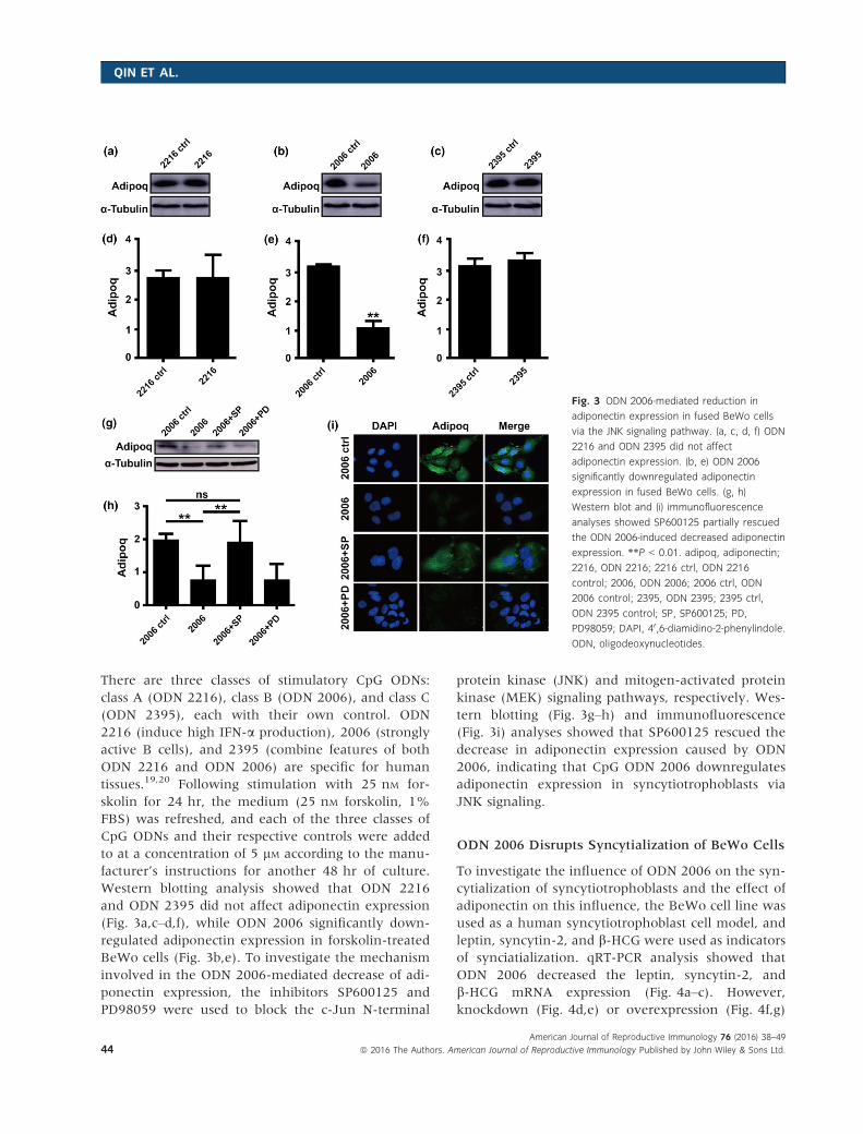
There are three classes of stimulatory CpG ODNs:
class A (ODN 2216), class B (ODN 2006), and class C
(ODN 2395), each with their own control. ODN
2216 (induce high IFN-a production), 2006 (strongly
active B cells), and 2395 (combine features of both
ODN 2216 and ODN 2006) are specific for human
tissues.19,20 Following stimulation with 25 nM for-
skolin for 24 hr, the medium (25 nM forskolin, 1%
FBS) was refreshed, and each of the three classes of
CpG ODNs and their respective controls were added
to at a concentration of 5 lM according to the manu-
facturer’s instructions for another 48 hr of culture.
Western blotting analysis showed that ODN 2216
and ODN 2395 did not affect adiponectin expression
(Fig. 3a,c–d,f), while ODN 2006 significantly down-
regulated adiponectin expression in forskolin-treated
BeWo cells (Fig. 3b,e). To investigate the mechanism
involved in the ODN 2006-mediated decrease of adi-
ponectin expression, the inhibitors SP600125 and
PD98059 were used to block the c-Jun N-terminal
protein kinase (JNK) and mitogen-activated protein
kinase (MEK) signaling pathways, respectively. Wes-
tern blotting (Fig. 3g–h) and immunofluorescence
(Fig. 3i) analyses showed that SP600125 rescued the
decrease in adiponectin expression caused by ODN
2006, indicating that CpG ODN 2006 downregulates
adiponectin expression in syncytiotrophoblasts via
JNK signaling.
ODN 2006 Disrupts Syncytialization of BeWo Cells
To investigate the influence of ODN 2006 on the syn-
cytialization of syncytiotrophoblasts and the effect of
adiponectin on this influence, the BeWo cell line was
used as a human syncytiotrophoblast cell model, and
leptin, syncytin-2, and b-HCG were used as indicators
of synciatialization. qRT-PCR analysis showed that
ODN 2006 decreased the leptin, syncytin-2, and
b-HCG mRNA expression (Fig. 4a–c). However,
knockdown (Fig. 4d,e) or overexpression (Fig. 4f,g)
Fig. 3 ODN 2006-mediated reduction in
adiponectin expression in fused BeWo cells
via the JNK signaling pathway. (a, c, d, f) ODN
2216 and ODN 2395 did not affect
adiponectin expression. (b, e) ODN 2006
significantly downregulated adiponectin
expression in fused BeWo cells. (g, h)
Western blot and (i) immunofluorescence
analyses showed SP600125 partially rescued
the ODN 2006-induced decreased adiponectin
expression. **P < 0.01. adipoq, adiponectin;
2216, ODN 2216; 2216 ctrl, ODN 2216
control; 2006, ODN 2006; 2006 ctrl, ODN
2006 control; 2395, ODN 2395; 2395 ctrl,
ODN 2395 control; SP, SP600125; PD,
PD98059; DAPI, 40,6-diamidino-2-phenylindole.
ODN, oligodeoxynucleotides.
American Journal of Reproductive Immunology 76 (2016) 38–49
44 ª 2016 The Authors. American Journal of Reproductive Immunology Published by John Wiley & Sons Ltd.
QIN ET AL.

of adiponectin did not alter ODN 2006-induced syn-
cytiotrophoblast endocrine dysfunction, and b-HCGexpression was not rescued (Fig. 4h). Similar results
were observed by immunofluorescence analysis. Fol-
lowing incubation of BeWo cells with forskolin and
ODN 2006 control, E-cadherin distribution indicated
the syncytial formation of BeWo cells, while incuba-
tion with forskolin and ODN 2006, resulted in the
E-cadherin being localized on the BeWo cell borders
indicating that they were not fused. Following knock-
down or overexpression of adiponectin, the localiza-
tion of E-cadherin was not significantly different
from that in cells only incubated with forskolin and
ODN 2006 (Fig. 4i).
Adiponectin Rescues ODN 2006-Induced
Impairment of Trophoblast Cell Migration
Transwell assays were used to investigate the migra-
tion of JAR and HTR-8 cells, as migration is a key
component of trophoblast invasion. ODN 2006 dis-
rupted HTR-8 and JAR cell migration, and adiponec-
tin rescued this impairment (Fig. 5a). The numbers
of migrating JAR (Fig. 5b) and HTR-8 (Fig. 5c) cells
Fig. 4 ODN 2006-mediated impairment of syncytialization of BeWo cells. (a–c) The markers leptin, syncytin-2 and b-HCG identified
syncytiotrophoblasts. ODN 2006 impaired syncytiotrophoblast endocrine function. (d, e) Knockdown of adiponectin (f, g) or overexpression of
adiponectin (h) did not alter ODN 2006-induced syncytiotrophoblast endocrine dysfunction. (i) Immunofluorescence analysis of the effect of ODN
2006 on syncytialization of BeWo cells and the effect of interaction between adiponectin and ODN 2006 on BeWo syncytialization. *P < 0.05;
**P < 0.01; adipoq, adiponectin; FSK, forskolin; 2006, ODN 2006; 2006 ctrl, ODN 2006 control; DAPI, 40,6-diamidino-2-phenylindole. ODN,
oligodeoxynucleotides.
American Journal of Reproductive Immunology 76 (2016) 38–49
ª 2016 The Authors. American Journal of Reproductive Immunology Published by John Wiley & Sons Ltd. 45
ADIPONECTIN AND EMBRYO LOSS

were significantly decreased when incubated with
ODN 2006 compared to ODN 2006 control or with
ODN 2006 plus adiponectin. ODN 2006 reduced
latent MMP-2 and active MMP-2 enzyme activity,
but this could be rescued by addition of adiponectin
(Fig. 5d), indicating that CpG ODN-induced impair-
ment of MMP-2-mediated cytotrophoblast migration
can be neutralized by adiponectin.
Discussion
Adiponectin is an important modulator of insulin
action and glucose metabolism, influences gonado-
tropin release and fetal growth, and maintains nor-
mal pregnancy. However, its anti-inflammatory role
in bacterial infections during pregnancy is not well
understood. In this study, we demonstrated, for the
first time, that CpG downregulate adiponectin
expression in mouse placenta and increase embryo
loss in NOD mice.
NOD mice are known to be prone to embryo
loss.27 The embryo was implanted on E4.5 or E5.5
and is finished on E6.5. On E10.5, the number of
immune cells reached to the highest level. So we
injected the pregnancy mice with ODN 1826 on
E6.5 and killed the mice on E10.5 to investigate the
influence of ODN 1826 on embryo loss, referenced
to previous experimental methods.21 In control
groups, the absolute number of lost embryos in NOD
mice was only slightly but not significantly higher
than that in WT mice. By contrast, when injected
with ODN 1826, the fetal resorption rate in NOD
mice was statistically higher than that in WT mice
with the same treatment, and Tlr9 was upregulated
in both WT and NOD mice. The NOD mouse is an
immunodeficiency model mouse characterized by a
functional deficit in NK cells but not in T and B lym-
phocytes and other immune cells.28,29 The results
further confirmed that ODN 1826 may activate the
immune system to disrupt the pregnancy outcome.
Thus, we selected ODN 1826 to challenge NOD mice
as a mouse model of the infection-induced increase
of embryo loss rate to investigate the anti-inflamma-
tory role of adiponectin during pregnancy.
In women, metabolic disorders increase the risk of
menstrual cycle abnormalities, ovulatory dysfunction,
and decreased fecundity, which can cause a number
of pregnancy complications, including hypertension,
pre-eclampsia, gestational diabetes, and fetal dis-
tress.30,31 Adiponectin is the most abundant adipokine
expressed in reproductive organs. It can suppress
macrophage production of pro-inflammatory cytoki-
nes, inhibit the phagocytic activity of macrophages,
inhibit the activation of NF-jB, and resist TNF-a-
Fig. 5 Counteractive effects of adiponectin
on ODN 2006-mediated impairment of
migration ability of JAR and HTR-8 cells. (a)
Migration abilities of JAR and HTR-8 cells
were impaired by ODN 2006, but could be
rescued by adiponectin. Numbers of
migrating (b) JAR and (c) HTR-8 cells were
both significantly decreased by ODN 2006. (d)
ODN 2006 impaired both latent MMP-2 and
active MMP-2 enzyme activity in JAR cells,
but this could be rescued by adiponectin.
**P < 0.01. adipoq, adiponectin; 2006, ODN
2006; 2006 ctrl, ODN 2006 control. ODN,
oligodeoxynucleotides.
American Journal of Reproductive Immunology 76 (2016) 38–49
46 ª 2016 The Authors. American Journal of Reproductive Immunology Published by John Wiley & Sons Ltd.
QIN ET AL.

induced inflammation.32–34 Adiponectin is thought to
function at the intricate interface between metabo-
lism and inflammation in pregnant woman,10 being
broadly associated with various obesity-related dis-
eases including infertility.35,36
The present study demonstrated that intraperi-
toneal injection of ODN 1826 reduced placental
adiponectin expression in NOD mice but not signifi-
cantly in WT mice. In line with previous reports of
increased adiponectin in NOD mice,37 placental adi-
ponectin in the NOD mouse was significantly higher
than that in the WT mouse placenta in our control
groups. As adiponectin promotes insulin sensitivity,
a compensatory increase of expression of adiponectin
may occur in the early stage of low insulin sensitiv-
ity of NOD mice. With development of autoimmune
sialadenitis, the serum adiponectin level has been
shown to decrease in NOD mice.38 Our results
showed that, in response to ODN 1826 stimulation,
placental adiponectin expression was downregulated
in the NOD mouse but not in the WT mouse
placenta. The reasons for the stable adiponectin
expression in the placenta of WT mice remain to be
explored.
While the presence of syncytiotrophoblast adipo-
nectin expression has been previously debated,39,40
the present study demonstrated adiponectin expres-
sion in both syncytiotrophoblasts and decidual
glands. In the in vitro model cell experiments, we
also found that adiponectin was expressed in syncy-
tial BeWo cells. Moreover, TLR9 agonists (ODN
2216, ODN 2006, and ODN 2395) and their controls
were given to fused BeWo cells to investigate
whether CpG ODN could influence adiponectin
expression in human syncytiotrophoblasts. The
expression of adiponectin decreased only after ODN
2006 stimulation for 48 hr. Then, we used ODN
2006 to stimulate cells in the following experiments.
Furthermore, JNK and MEK signaling pathways
have been previously reported as two signaling path-
ways that could reduce adiponectin expression dur-
ing inflammation.41,42 In our current study, only
specific inhibitors of the JNK signaling pathway
could rescue the ODN 2006-mediated decrease of
adiponectin expression, indicating JNK as the signal-
ing pathway regulating adiponectin expression in
syncytiotrophoblasts in response to ODN 2006 (or
bacterial) stimulation.
In the current study, pregnant women underwent
artificial abortion (dilatation and curettage) to their
unwanted pregnancies at gestational week 8–12
were defined as normal pregnancies, and samples of
villus and decidual tissues were collected during
pregnancy termination. All of these women had had
at least one successful pregnancy, without chromo-
somal abnormality, obesity, spontaneous abortion,
preterm labor, or pre-eclampsia in any pregnancy.
However, there are some potential limitations of
using tissue from these subjects. Potential limitation
may include the following: (i) As uterus ultrasonic
examination is not performed, we have no idea
whether the fetus is alive or dead. Thus, it is possible
that the fetus is dead, but we may still consider it a
normal pregnancy by mistake. (ii) Fetal chromosome
examination is not performed at the present gesta-
tion. Thus, it is possible that we may define some
patients with abnormal fetal chromosome as normal
pregnancies by mistake. (iii) Other limitation: Taken
together, the effect of adiponectin in protecting preg-
nancy may be slightly overestimated when these
samples are used. In future researches, it is better to
perform more detections including but not limited to
uterus ultrasonic examination before pregnancy ter-
mination and fetal chromosome examination right
at the end of pregnancy termination.
Trophoblast proliferation, migration, invasion, and
endocrine secretion play important roles in a suc-
cessful pregnancy, and it has been reported that adi-
ponectin promotes the syncytialization of BeWo cells
and primary trophoblast cells.43 However, no reports
have been made about the effect of adiponectin on
trophoblast syncytialization under infectious condi-
tions. In the current study, ODN 2006 impaired the
syncytialization of BeWo cells, but neither knock-
down nor overexpression of adiponectin could neu-
tralize this effect, indicating that while it can
promote trophoblast syncytialization, adiponectin
cannot rescue impairment of syncytialization caused
by CpG ODNs or bacterial infection. Furthermore, in
addition to supporting syncytialization, adiponectin
can also promote the human trophoblast migra-
tion.40 However, little is known about the effect of
adiponectin on trophoblast migration under infec-
tious conditions. Our results showed that ODN 2006
impaired the migration of both JAR and HTR-8 cells,
and adiponectin could mitigate this effect by regulat-
ing MMP-2 activity.
In conclusion, our findings provide evidence to
demonstrate the anti-inflammatory function of adi-
ponectin in countering the effect of CpG ODN (and
bacterial infections) during pregnancy. CpG ODNs
downregulated syncytiotrophoblast adiponectin
American Journal of Reproductive Immunology 76 (2016) 38–49
ª 2016 The Authors. American Journal of Reproductive Immunology Published by John Wiley & Sons Ltd. 47
ADIPONECTIN AND EMBRYO LOSS

expression via the JNK signaling pathway and
induced embryo loss in NOD mice. CpG ODNs also
impaired trophoblast syncytialization of syncytiotro-
phoblasts and cytotrophoblast migration. Although
adiponectin could not prevent the CpG ODN-
induced syncytiotrophoblast dysfunction, it was able
to rescue the CpG ODN-induced impairment of
cytotrophoblast migration. Our study provides fur-
ther understanding of the anti-inflammatory role of
adiponectin in response to specific unmethylated
CpG dinucleotides (infectious bacterial unmethylated
dinucleotides) during placental development. These
results indicate that adiponectin may be a beneficial
factor in providing protection in bacteria-induced
pregnancy failure and providing a new direction for
the clinical treatment of pregnancy failure caused by
bacterial infections.
Acknowledgments
This work was supported by the National Basic
Research Program of China (2013CB967401 and
2013CB967404), the National Natural Science Foun-
dation of China (81401218, 81125004 and 31171439),
the Fund for Outstanding Academic Leaders in Shang-
hai, China (12XD1406600 and 2013-049), Shanghai
Natural Science Fund Project (14ZR1443800), and
Shanghai Jiao Tong University Medicine-Engineering
Fund (YG2013ZD04).
Conflict of interest
All authors declared that they have no conflict of
interests.
References
1 Xu Y, Plazyo O, Romero R, Hassan SS, Gomez-Lopez N: Isolation of
leukocytes from the human maternal-fetal interface. J Vis Exp 2015;
99:e52863.
2 Erlebacher A: Immunology of the maternal-fetal interface. Annu Rev
Immunol 2013; 31:387–411.
3 Lin Y, Xie M, Chen Y, Di J, Zeng Y: Preterm delivery induced by
LPS in syngeneically impregnated BALB/c and NOD/SCID mice. J
Reprod Immunol 2006; 71:87–101.
4 Li L, Yang J, Ren L, Su N, Fang Y, Lin Y: Invariant NKT cells
increase lipopolysacchride-induced pregnancy loss by a mechanism
involving Th1 and Th17 responses. J Matern Fetal Neonatal Med
2013; 26:1212–1218.
5 Romero R, Espinoza J, Mazor M: Can endometrial infection/
inflammation explain implantation failure, spontaneous abortion,
and preterm birth after in vitro fertilization? Fertil Steril 2004;
82:799–804.
6 Lin Y, Liu X, Shan B, Wu J, Sharma S, Sun Y: Prevention of CpG-
induced pregnancy disruption by adoptive transfer of in vitro-
induced regulatory T cells. PLoS One 2014; 9:e94702.
7 Sun Y, Qin X, Shan B, Wang W, Zhu Q, Sharma S, Wu J, Lin Y:
Differential effects of the CpG-Toll-like receptor 9 axis on
pregnancy outcome in nonobese diabetic mice and wild-type
controls. Fertil Steril 2013; 99:1759–1767.
8 Yamauchi T, Kamon J, Ito Y, Tsuchida A, Yokomizo T, Kita S,
Sugiyama T, Miyagishi M, Hara K, Tsunoda M, Murakami K,
Ohteki T, Uchida S, Takekawa S, Waki H, Tsuno NH, Shibata Y,
Terauchi Y, Froguel P, Tobe K, Koyasu S, Taira K, Kitamura T,
Shimizu T, Nagai R, Kadowaki T: Cloning of adiponectin receptors
that mediate antidiabetic metabolic effects. Nature 2003; 423:762–
769.
9 Smitka K, Maresova D: Adipose tissue as an endocrine organ: an
update on pro-inflammatory and anti-inflammatory
microenvironment. Prague Med Rep 2015; 116:87–111.
10 Dupont J, Reverchon M, Bertoldo MJ, Froment P: Nutritional
signals and reproduction. Mol Cell Endocrinol 2014; 382:527–537.
11 Michalakis KG, Segars JH: The role of adiponectin in reproduction:
from polycystic ovary syndrome to assisted reproduction. Fertil Steril
2010; 94:1949–1957.
12 Klenke U, Taylor-Burds C, Wray S: Metabolic influences on
reproduction: adiponectin attenuates GnRH neuronal activity in
female mice. Endocrinology 2014; 155:1851–1863.
13 Waki H, Yamauchi T, Kamon J, Ito Y, Uchida S, Kita S, Hara K,
Hada Y, Vasseur F, Froguel P, Kimura S, Nagai R, Kadowaki T:
Impaired multimerization of human adiponectin mutants associated
with diabetes. Molecular structure and multimer formation of
adiponectin. J Biol Chem 2003; 278:40352–40363.
14 Nien JK, Mazaki-Tovi S, Romero R, Erez O, Kusanovic JP, Gotsch
F, Pineles BL, Gomez R, Edwin S, Mazor M, Espinoza J, Yoon BH,
Hassan SS: Plasma adiponectin concentrations in non-pregnant,
normal and overweight pregnant women. J Perinat Med 2007;
35:522–531.
15 Ranheim T, Haugen F, Staff AC, Braekke K, Harsem NK, Drevon
CA: Adiponectin is reduced in gestational diabetes mellitus in
normal weight women. Acta Obstet Gynecol Scand 2004; 83:341–347.
16 Suwaki N, Masuyama H, Nakatsukasa H, Masumoto A, Sumida Y,
Takamoto N, Hiramatrsu Y: Hypoadiponectinemia and circulating
angiogenic factors in overweight patients complicated with pre-
eclampsia. Am J Obstet Gynecol 2006; 195:1687–1692.
17 Haugen F, Ranheim T, Harsem NK, Lips E, Staff AC, Drevon CA:
Increased plasma levels of adipokines in preeclampsia: relationship
to placenta and adipose tissue gene expression. Am J Physiol
Endocrinol Metab 2006; 290:E326–E333.
18 Kajantie E, Kaaja R, Ylikorkala O, Andersson S, Laivuori H:
Adiponectin concentrations in maternal serum: elevated in
preeclampsia but unrelated to insulin sensitivity. J Soc Gynecol
Investig 2005; 12:433–439.
19 Ballas ZK, Krieg AM, Warren T, Rasmussen W, Davis HL,
Waldschmidt M, Weiner GJ: Divergent therapeutic and
immunologic effects of oligodeoxynucleotides with distinct CpG
motifs. J Immunol 2001; 167:4878–4886.
20 Bauer S, Kirschning CJ, Hacker H, Redecke V, Hausmann S, Akira
S, Wagner H, Lipford GB: Human TLR9 confers responsiveness to
bacterial DNA via species-specific CpG motif recognition. Proc Natl
Acad Sci USA 2005; 98:9237–9242.
21 Thaxton JE, Romero R, Sharma S: TLR9 activation coupled to IL-10
deficiency induces adverse pregnancy outcomes. J Immunol 2009;
183:1144–1154.
American Journal of Reproductive Immunology 76 (2016) 38–49
48 ª 2016 The Authors. American Journal of Reproductive Immunology Published by John Wiley & Sons Ltd.
QIN ET AL.

22 Lin Y, Xu L, Jin H, Zhong Y, Di J, Lin QD: CXCL12 enhances
exogenous CD4+CD25+ T cell migration and prevents embryo loss
in non-obese diabetic mice. Fertil Steril 2009; 91:2687–2696.
23 Lin Y, Liang Z, Chen Y, Zeng Y: TLR3-involved modulation of
pregnancy tolerance in double-stranded RNA-stimulated NOD/SCID
mice. J Immunol 2006; 176:4147–4154.
24 Arce RM, Barros SP, Wacker B, Peters B, Moss K, Offenbacher S:
Increased TLR4 expression in murine placentas after oral infection
with periodontal pathogens. Placenta 2009; 30:156–162.
25 Tian FJ, Qin CM, Li XC, Wu F, Liu XR, Xu WM, Lin Y: Decreased
stathmin-1 expression inhibits trophoblast proliferation and
invasion and is associated with recurrent miscarriage. Am J Pathol
2015; 185:2709–2721.
26 Toth M, Sohail A, Fridman R: Assessment of gelatinases (MMP-2
and MMP-9) by gelatin zymography. Methods Mol Biol 2012;
878:121–135.
27 Sun Y, Wang W, Shan B, Di J, Chen L, Ren L, Li W, Li DJ, Lin Y:
FTY720-induced conversion of conventional Foxp3- CD4+ T cells to
Foxp3+ regulatory T cells in NOD mice. Am J Reprod Immunol 2011;
66:349–362.
28 Lin Y, Chen Y, Zeng Y, Wang T, Zeng S: Lymphocyte phenotyping
and NK cell activity analysis in pregnant NOD/SCID mice. J Reprod
Immunol 2005; 68:39–51.
29 Lin Y, Zhong Y, Saito S, Chen Y, Shen W, Di J, Zeng S:
Characterization of natural killer cells in nonobese diabetic/severely
compromised immunodeficient mice during pregnancy. Fertil Steril
2009; 91:2676–2686.
30 Practice Committee of American Society for Reproductive Medicine:
Obesity and reproduction: a committee opinion. Fertil Steril 2015;
104:1116–1126.
31 Cedergren MI: Maternal morbid obesity and the risk of adverse
pregnancy outcome. Obstet Gynecol 2004; 103:219–224.
32 Ouchi N, Kihara S, Arita Y, Okamoto Y, Maeda K, Kuriyama H,
Hotta K, Nishida M, Takahashi M, Muraguchi M, Ohmoto Y,
Nakamura T, Yamashita S, Funahashi T, Matsuzawa Y: Adiponectin,
an adipocyte-derived plasma protein, inhibits endothelial NF-
kappaB signaling through a cAMP-dependent pathway. Circulation
2000; 102:1296–1301.
33 Yokota T, Oritani K, Takahashi I, Ishikawa J, Matsuyama A, Ouchi
N, Kihara S, Funahashi T, Tenner AJ, Tomiyama Y, Matsuzawa Y:
Adiponectin, a new member of the family of soluble defense
collagens, negatively regulates the growth of myelomonocytic
progenitors and the functions of macrophages. Blood 2000;
96:1723–1732.
34 Wang Y, Wang X, Lau WB, Yuan Y, Booth D, Li JJ, Scalia R,
Preston K, Gao E, Koch W, Ma XL: Adiponectin inhibits tumor
necrosis factor-alpha-induced vascular inflammatory response via
caveolin-mediated ceramidase recruitment and activation. Circ Res
2014; 114:792–805.
35 Ohashi K, Yuasa D, Shibata R, Murohara T, Ouchi N: Adiponectin
as a target in obesity-related inflammatory state. Endocr Metab
Immune Disord Drug Targets 2015; 15:145–150.
36 Palin MF, Bordignon VV, Murphy BD: Adiponectin and the
control of female reproductive functions. Vitam Horm 2012;
90:239–287.
37 Combs TP, Snell-Bergeon JK, Maahs DM, Bergman BC, Lamarche
M, Iberkleid L, AbdelBaky O, Tisch R, Scherer PE, Marliss EB:
Adiponectin-SOGA dissociation in type 1 diabetes. J Clin Endocrinol
Metab 2015; 100:E1065–E1073.
38 Su YC, Xiang RL, Zhang Y, Ding C, Cong X, Guo XH, Yang NY, Hua
H, Wu LL, Yu GY: Decreased submandibular adiponectin is involved
in the progression of autoimmune sialoadenitis in non-obese
diabetic mice. Oral Dis 2014; 20:744–755.
39 Chen J, Tan B, Karteris E, Zervou S, Digby J, Hillhouse EW, Vatish
M, Randeva HS: Secretion of adiponectin by human placenta:
differential modulation of adiponectin and its receptors by
cytokines. Diabetologia 2006; 49:1292–1302.
40 Benaitreau D, Dos Santos E, Leneveu MC, Alfaidy N, Feige JJ, de
Mazancourt P, Pecquery R, Dieudonne MN: Effects of adiponectin
on human trophoblast invasion. J Endocrinol 2010; 207:45–53.
41 Martinez K, Kennedy A, McIntosh MK: JNK inhibition by
SP600125 attenuates trans-10, cis-12 conjugated linoleic acid-
mediated regulation of inflammatory and lipogenic gene expression.
Lipids 2011; 46:885–892.
42 Song HY, Kim MR, Lee MJ, Jeon ES, Bae YC, Jung JS, Kim JH:
Oncostatin M decreases adiponectin expression and induces
dedifferentiation of adipocytes by JAK3- and MEK-dependent
pathways. Int J Biochem Cell Biol 2007; 39:439–449.
43 Benaitreau D, Dos Santos E, Leneveu MC, De Mazancourt P,
Pecquery R, Dieudonne MN: Adiponectin promotes syncytialisation
of BeWo cell line and primary trophoblast cells. Reprod Biol
Endocrinol 2010; 8:128.
Supporting Information
Additional Supporting Information may be found
online in the supporting information tab for this
article:
Figure S1. Animal treatment.
Figure S2. Increase of embryo loss in NOD mice
by ODN 1826.
American Journal of Reproductive Immunology 76 (2016) 38–49
ª 2016 The Authors. American Journal of Reproductive Immunology Published by John Wiley & Sons Ltd. 49
ADIPONECTIN AND EMBRYO LOSS