Embed Size (px)
Citation preview
REVIEW
SCIENCE INSIGHTS 138
Medicine
Review (Narrative)
COVID-19 Facts and Recommendations from A to Z
BASE Medicine Task Force
SUMMARY
Since the first case of COVID-19 was reported in late 2019, the virus has been filling almost every corner of the earth. The world suddenly woke up in a hurry, and began to surround and control SARS-CoV-2 on a large scale. Since then, the world has entered a state of lockdown. This task force reviewed from every as-pect involved in the occurrence, development and treatment of COVID-19. Alt-hough no effective treatments exist and many areas require further study at length regarding the virus and the diseases caused by it, all the medical care im-plemented currently reflects the compassionate control of the disease. Whether it is the development of effective drugs or vaccines, the occurrence of COVID-19 pandemic sounded the alarm for humans. It is time to seriously think about the disease prevention system we have built so far and the next catastrophic epi-demic that may come in the near future. While the virus is rapidly mutating, whether the development of medical science is catching up fast enough and find-ing effective countermeasures is a question worthy of serious consideration.■
KEYWORDS
SARS-CoV-2; COVID-19; Human catastrophe; Medical system; Future
Sci Insigt. 2020; 33(1):138-158. doi:10.15354/si.20.re061.
Author Affiliations: Author affili-ations are listed at the end of this article.
Correspondence to: BASE Medi-cine Task Force Committee, The Bonoi Academy of Science and Education, Chapel Hill, NC 27510, USA Email: [email protected]
Copyright © 2020 The BASE. This is an open access article distributed under the Creative Commons Attribution License,
which permits unrestricted use, distribution, and reproduction in any medium, provided the original work is properly cited.
BASE: COVID-19 Facts and Recommendations Review
SI 2020; Vol. 33, No. 1 www.bonoi.org 139
OUTLINE Introduction Origin of SARS-CoV-2 Structure of SARS-CoV-2 Epidemiology of COVID-19 Clinical Manifestations of COVID-19 Diagnosis of COVID-19 Virus Detection of SARS-CoV-2
Nucleic Acid Specimen Collection Nucleic Acid Test Procedures Serum Virus Antibody Detection Virus Isolation and Culture
Cytokine Storm of COVID-19 Imaging Study of COVID-19 Bronchoscopy in COVID-19 Classification of COVID-19 Treatment of COVID-19
General Management Anti-viral Therapeutics
Lopinavir-Ritonavir (Kaletra®)
Arbidol Chloroquine or Hydroxychloroquine Favipiravir (Avigan
®)
Remdesivir Darunavir/Cobicistat (Prezcobix; Rezolsta) Interferon
Anti-Shock Therapies of COVID-19 Indications for Corticosteroids Use Application of Corticosteroids Special Consideration during Corticosteroids Treatment
Oxygen Therapy for COVID-19 Oxygen Therapy Mechanical Ventilation Prone Position Ventilation
Extracorporeal Membrane Oxygenation Support Timing of ECMO Methods of Catheterization Mode Selection Flux Set-value and Target Oxygen Supply Ventilation Setting Anti-Coagulation and Bleeding Prevention
Antioxidant Treatment for COVID-19 Vitamin C Vitamin E Glutathione N-acetyl-L-cysteine (NAC) Melatonin
Traditional Chinese Medicine for COVID-19 Single Herbs Chinese Patent Formulas Chinese Herbal Compounds
Vaccine for COVID-19 Fluid Management Food Therapy for COVID-19
Discharge Criteria of COVID-19 Perspectives References
INTRODUCTION
INCE the outbreak of the severe acute respiratory
syndrome coronavirus 2 (SARS-CoV-2) in Wuhan,
Hubei Province of China (1), the pneumonia
caused by the virus was designated as COVID-19 by the
World Health Organization (2). With the rapid spread-
ing of the virus, the COVID-19 confirmed cases have
been overwhelmingly reach each corner of the world
and reach 1,282,931 until April 7, 2020 (3). The global
lockdown by the virus has been causing the world deep
into a worrisome situation. All the people are asking the
same questions as: Where did the virus come from?
How can we prevent or control it? Do we have effective
therapeutics to COVID-19? Will the virus disappear or
stay with our human being all the time to come? Can
we make efficient vaccine to conquer it? etc.
With evidence emerging quickly, this Task Force
will review and update the recognition of the virus and
COVID-19, and give recommendations for medical care
and individual prevention and control.
ORIGIN OF SARS-COV-2
Since the first report of the infectious disease, the origin
of the virus has been becoming an attractive topic. Ini-
tially, it was considered as an animal-derived virus, es-
pecially bats and pangolins are the intermediate hosts
(4). But cumulating evidence did not prove this link be-
cause the viruses from both types of animals did not
show a big identity with less than 85% (5).
However, a natural evolution is regarded as the
most likely possibility of the virus origin. Through ana-
lyzing the genomic sequence of SARS-CoV-2 and poten-
tially related viruses, no solid evidence supports the dec-
laration of the virus’s origin as an engineered one for
any reasons (6). Given the big disease spectrum caused
by the Coronaviruses such as 2003 Severe Acute Respir-
atory Syndrome (SARS) in China and 2012 Middle East
Respiratory Syndrome (MERS) in Saudi Arabia, this
time, SARS-CoV-2 emerged as a new one causing seri-
ous illness with different severity (7). So far, two fea-
tures of the virus suggest that it came from a natural
selection, but not genetic engineering.
First, the receptor binding domain (RBD) of the vi-
rus spike glycoprotein has evolved to bind to a molecule
called angiotensin-converting enzyme 2 (ACE2) that is
S
BASE: COVID-19 Facts and Recommendations Review
SI 2020; Vol. 33, No. 1 www.bonoi.org 140
located on the human cell membranes with regulating
function of blood pressure (8). This RBD mutation is
exactly an indicator for the natural evolution. Second,
generally, the engineered pathologic virus would have
an identical backbone with the mother virus. Neverthe-
less, SARS-CoV-2 has a totally different backbone from
those of already known coronaviruses such as SARS-
CoV, MERS-CoV, and viruses found in bats and pango-
lins (9). Although the viral originality was concluded as
a result of natural selection, how human being was in-
fected becomes the key question.
As SARS-CoV and MERS-CoV had their interme-
diate hosts as civets and camels, respectively, no docu-
mented cases exist to indicate a bat-human transmission.
However, theoretically, SARS-CoV-2 would have
evolved in two parts to realize its animal-human and
human-human transmission ability: the RBD portion of
spike protein and cleavage site to open the virus up. In
another situation, the virus evolved directly within hu-
man host to be pathogenic after getting into human
body. No matter the RBD or cleavage site, SARS-CoV-2
might quickly get evolved to be a virulent one in human
cells and then kicked off the current pandemic (10). For
these two possibilities, the first one is much more dan-
gerous than the second one because if the virus entered
human host with its current pathogenic property from
an intermediate animal host, it still would be possible
for a future outbreak; but if the virus first got into hu-
mans without pathogenic ability, so the chance of a fu-
ture outbreak is lower because the circulating viruses
are non-illness-causing strain.
STRUCTURE OF SARS-COV-2
In general, coronaviruses are a large family of viruses.
Because this type of virus shapes spherically with pro-
trusions like a spiky crown, so they are collectively
named as coronaviruses. The diameter of coronaviruses
are ranging from 75 to 160 nm, and the virus genome is
a continuous linear single-stranded RNA ((ss)-RNA).
The coronavirus genome can encode spike glycoprotein
(S), envelope protein (E), membrane protein (M), and
nucleoprotein (N protein) (11).
SARS-CoV-2 belongs to the beta genera of the
Coronaviridae family. It has ~70% sequence identity
with SARS-CoV and ~40% sequence identity with
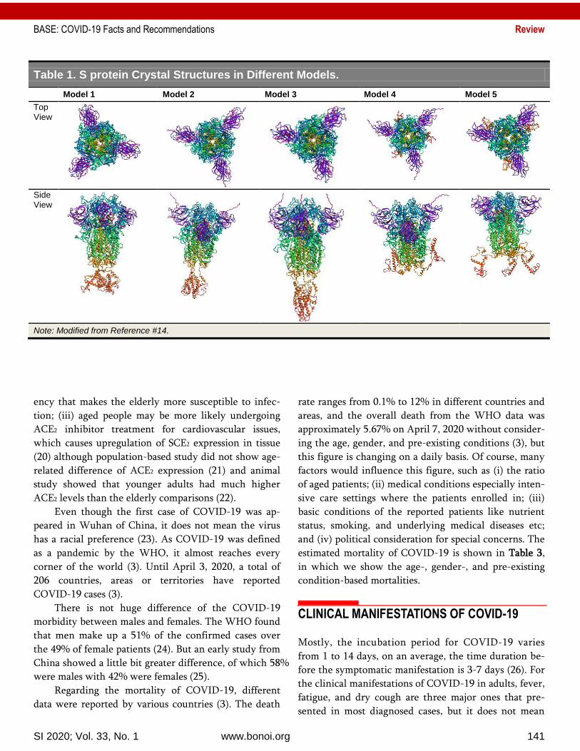
MERS CoV (12). Among the encoded viral proteins, S
protein is the most pivotal surface membrane protein of
coronavirus. Given the binding of S proteins to cellular
membrane receptors is the first step for the virus’ vigi-
lance, so S protein has been becoming the target for
most studies to find corresponding therapeutic drugs
and neutralizing vaccines. Primarily, S protein has two
tasks that assist host infection: (i) aid in the attachment
between the virus and host cell surface receptor ACE2,
and (ii) facilitate virus enter into the host cell through
helping the fusion process of the viral and host cell
membranes (13). S protein structure is being extensively
studies and different models were used to predict its
crystal structure (14) (Table 1).
The N protein, as a structural protein, binds to the
RNA genome so as to create capsid that encloses the nu-
cleic acid. Furthermore, N protein also plays an essential
role in: (i) viral assembly by interacting with the viral
membrane protein; (ii) RNA synthesis and folding; (iii)
virus budding, and (iv) host cell cycle and translation
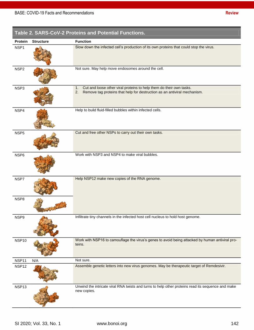
(15). Beside these two structural proteins, SARS-CoV-2
also encodes several non-structural proteins as showed
in Table 2.
EPIDEMIOLOGY OF COVID-19
The underlying cause of COVID-19 is the pathogenic
coronavirus designated as SARS-CoV-2. Patients with
symptoms after the infection are the major source of
transmission. However, asymptomatic patients are also
contagious and more dangerous than the symptom-
positive patients in spreading the virus (16) because you
don’t know the person next to you is a virus carrier.
Transmission of SARS-CoV-2 is majorly via respira-
tory droplets and close contact. There is the possibility
of aerosol transmission in a relatively closed environ-
ment for a long-time exposure to high concentrations of
virus (17). As the virus was isolated in feces and urine,
so special attention need to be paid to feces- or urine-
contaminated items that may result in feces-oral trans-
mission (18).
The susceptible people for SARS-CoV-2 include all
aged population. However, the COVID-19 morbidity
does some age-related difference. Although the young-
est confirmed case was an infant and who died in Illi-
nois (19), the virus looks like have a preference to senior
population due to several reasons: (i) older populations
generally have more underlying health conditions like
diabetes, heart disease, and other chronic illnesses; (ii)
with aging, the immune system gradually loses its resili-
BASE: COVID-19 Facts and Recommendations Review
SI 2020; Vol. 33, No. 1 www.bonoi.org 141
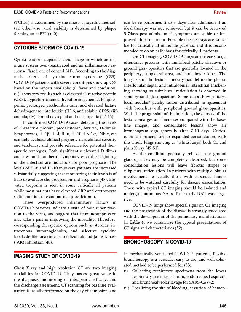
Table 1. S protein Crystal Structures in Different Models.
Model 1 Model 2 Model 3 Model 4 Model 5
Top View
Side View
Note: Modified from Reference #14.
ency that makes the elderly more susceptible to infec-
tion; (iii) aged people may be more likely undergoing
ACE2 inhibitor treatment for cardiovascular issues,
which causes upregulation of SCE2 expression in tissue
(20) although population-based study did not show age-
related difference of ACE2 expression (21) and animal
study showed that younger adults had much higher
ACE2 levels than the elderly comparisons (22).
Even though the first case of COVID-19 was ap-
peared in Wuhan of China, it does not mean the virus
has a racial preference (23). As COVID-19 was defined
as a pandemic by the WHO, it almost reaches every
corner of the world (3). Until April 3, 2020, a total of
206 countries, areas or territories have reported
COVID-19 cases (3).
There is not huge difference of the COVID-19
morbidity between males and females. The WHO found
that men make up a 51% of the confirmed cases over
the 49% of female patients (24). But an early study from
China showed a little bit greater difference, of which 58%
were males with 42% were females (25).
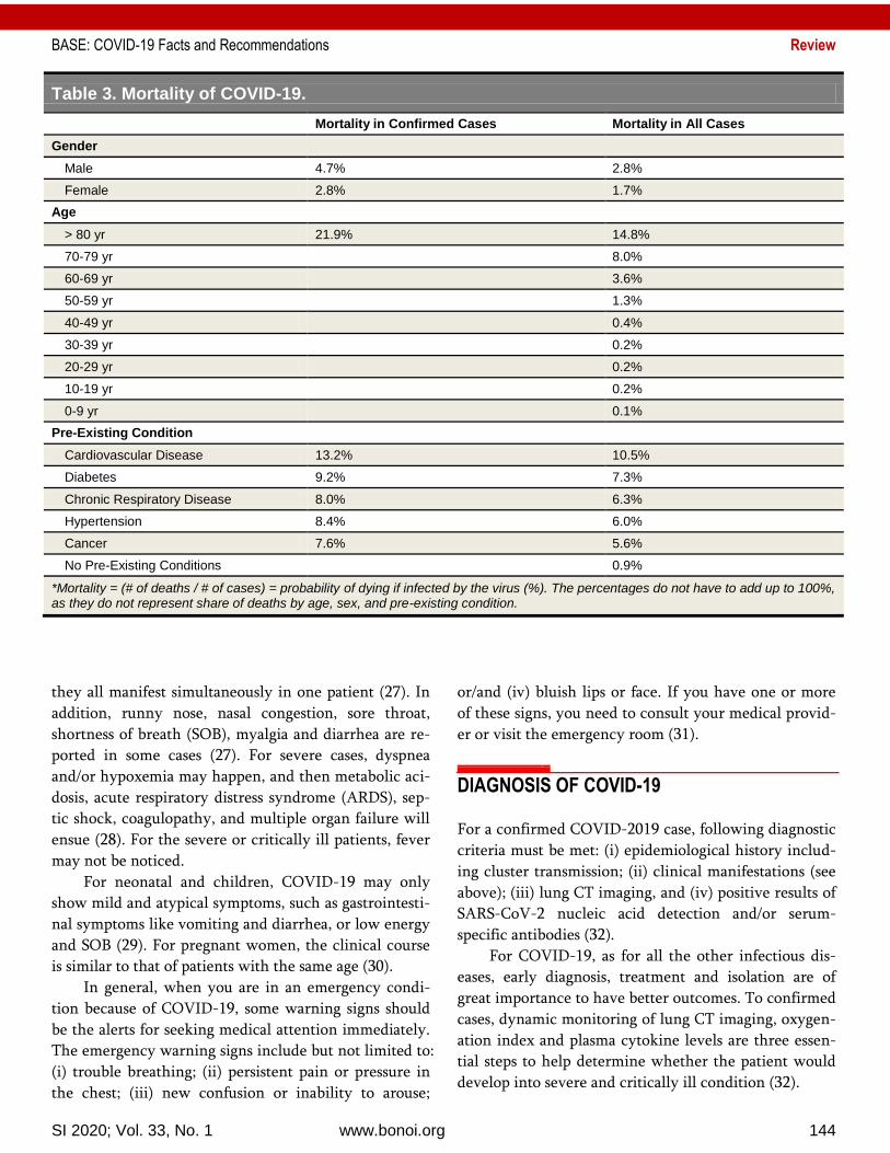
Regarding the mortality of COVID-19, different
data were reported by various countries (3). The death
rate ranges from 0.1% to 12% in different countries and
areas, and the overall death from the WHO data was
approximately 5.67% on April 7, 2020 without consider-
ing the age, gender, and pre-existing conditions (3), but
this figure is changing on a daily basis. Of course, many
factors would influence this figure, such as (i) the ratio
of aged patients; (ii) medical conditions especially inten-
sive care settings where the patients enrolled in; (iii)
basic conditions of the reported patients like nutrient
status, smoking, and underlying medical diseases etc;
and (iv) political consideration for special concerns. The
estimated mortality of COVID-19 is shown in Table 3,
in which we show the age-, gender-, and pre-existing
condition-based mortalities.
CLINICAL MANIFESTATIONS OF COVID-19
Mostly, the incubation period for COVID-19 varies
from 1 to 14 days, on an average, the time duration be-
fore the symptomatic manifestation is 3-7 days (26). For
the clinical manifestations of COVID-19 in adults, fever,
fatigue, and dry cough are three major ones that pre-
sented in most diagnosed cases, but it does not mean
BASE: COVID-19 Facts and Recommendations Review
SI 2020; Vol. 33, No. 1 www.bonoi.org 142
Table 2. SARS-CoV-2 Proteins and Potential Functions.
Protein Structure Function
NSP1
Slow down the infected cell’s production of its own proteins that could stop the virus.
NSP2
Not sure. May help move endosomes around the cell.
NSP3
1. Cut and loose other viral proteins to help them do their own tasks. 2. Remove tag proteins that help for destruction as an antiviral mechanism.
NSP4
Help to build fluid-filled bubbles within infected cells.
NSP5
Cut and free other NSPs to carry out their own tasks.
NSP6
Work with NSP3 and NSP4 to make viral bubbles.
NSP7
Help NSP12 make new copies of the RNA genome.
NSP8
NSP9
Infiltrate tiny channels in the infected host cell nucleus to hold host genome.
NSP10
Work with NSP16 to camouflage the virus’s genes to avoid being attacked by human antiviral pro-teins.
NSP11 N/A Not sure.
NSP12
Assemble genetic letters into new virus genomes. May be therapeutic target of Remdesivir.
NSP13
Unwind the intricate viral RNA twists and turns to help other proteins read its sequence and make new copies.
BASE: COVID-19 Facts and Recommendations Review
SI 2020; Vol. 33, No. 1 www.bonoi.org 143
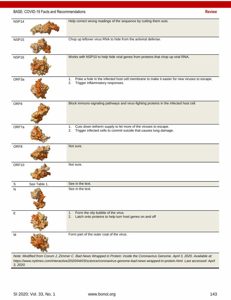
NSP14
Help correct wrong readings of the sequence by cutting them outs.
NSP15
Chop up leftover virus RNA to hide from the antiviral defense.
NSP16
Works with NSP10 to help hide viral genes from proteins that chop up viral RNA.
ORF3a
1. Poke a hole in the infected host cell membrane to make it easier for new viruses to escape. 2. Trigger inflammatory responses.
ORF6
Block immuno-signaling pathways and virus-fighting proteins in the infected host cell.
ORF7a
1. Cuts down tetherin supply to let more of the viruses to escape. 2. Trigger infected cells to commit suicide that causes lung damage.
ORF8
Not sure.
ORF10
Not sure.
S See Table 1. See in the text.
N
See in the text.
E
1. Form the oily bubble of the virus 2. Latch onto proteins to help turn host genes on and off
M
Form part of the outer coat of the virus.
Note: Modified from Corum J, Zimmer C. Bad News Wrapped in Protein: Inside the Coronavirus Genome. April 3, 2020. Available at:
https://www.nytimes.com/interactive/2020/04/03/science/coronavirus-genome-bad-news-wrapped-in-protein.html. Last accessed: April 3, 2020.
BASE: COVID-19 Facts and Recommendations Review
SI 2020; Vol. 33, No. 1 www.bonoi.org 144
Table 3. Mortality of COVID-19.
Mortality in Confirmed Cases Mortality in All Cases
Gender
Male 4.7% 2.8%
Female 2.8% 1.7%
Age
> 80 yr 21.9% 14.8%
70-79 yr
8.0%
60-69 yr
3.6%
50-59 yr
1.3%
40-49 yr
0.4%
30-39 yr
0.2%
20-29 yr
0.2%
10-19 yr
0.2%
0-9 yr
0.1%
Pre-Existing Condition
Cardiovascular Disease 13.2% 10.5%
Diabetes 9.2% 7.3%
Chronic Respiratory Disease 8.0% 6.3%
Hypertension 8.4% 6.0%
Cancer 7.6% 5.6%
No Pre-Existing Conditions
0.9%
*Mortality = (# of deaths / # of cases) = probability of dying if infected by the virus (%). The percentages do not have to add up to 100%, as they do not represent share of deaths by age, sex, and pre-existing condition.
they all manifest simultaneously in one patient (27). In
addition, runny nose, nasal congestion, sore throat,
shortness of breath (SOB), myalgia and diarrhea are re-
ported in some cases (27). For severe cases, dyspnea
and/or hypoxemia may happen, and then metabolic aci-
dosis, acute respiratory distress syndrome (ARDS), sep-
tic shock, coagulopathy, and multiple organ failure will
ensue (28). For the severe or critically ill patients, fever
may not be noticed.
For neonatal and children, COVID-19 may only
show mild and atypical symptoms, such as gastrointesti-
nal symptoms like vomiting and diarrhea, or low energy
and SOB (29). For pregnant women, the clinical course
is similar to that of patients with the same age (30).
In general, when you are in an emergency condi-
tion because of COVID-19, some warning signs should
be the alerts for seeking medical attention immediately.
The emergency warning signs include but not limited to:
(i) trouble breathing; (ii) persistent pain or pressure in
the chest; (iii) new confusion or inability to arouse;
or/and (iv) bluish lips or face. If you have one or more
of these signs, you need to consult your medical provid-
er or visit the emergency room (31).
DIAGNOSIS OF COVID-19
For a confirmed COVID-2019 case, following diagnostic
criteria must be met: (i) epidemiological history includ-
ing cluster transmission; (ii) clinical manifestations (see
above); (iii) lung CT imaging, and (iv) positive results of
SARS-CoV-2 nucleic acid detection and/or serum-
specific antibodies (32).
For COVID-19, as for all the other infectious dis-
eases, early diagnosis, treatment and isolation are of
great importance to have better outcomes. To confirmed
cases, dynamic monitoring of lung CT imaging, oxygen-
ation index and plasma cytokine levels are three essen-
tial steps to help determine whether the patient would
develop into severe and critically ill condition (32).
BASE: COVID-19 Facts and Recommendations Review
SI 2020; Vol. 33, No. 1 www.bonoi.org 145
Currently, a positive result of the nucleic acid of
SARS-CoV-2 is still the gold standard for COVID-19
diagnosis, whereas the characteristic signs in CT imag-
ing for those suspected cases can be treated as confirmed
cases even if the nucleic acid test is negative because of
the possibility of false negative in nucleic acid detection.
So, isolation and continuous tests of multiple specimens
should be carried out in such cases (33).
COVID-19 needs to be differentiated from upper
respiratory tract infections (URI) caused by other virus-
es such as influenza virus, adenovirus, and respiratory
syncytial virus. Generally, methods such as rapid anti-
gen detection and multiplex PCR nucleic acid detection
can be adopted for excluding common respiratory path-
ogens.
VIRUS DETECTION OF SARS-COV-2
Nucleic Acid Specimen Collection
The specimen quality is critical for improving the posi-
tive detection of viral nucleic acid test (NAT). The types
of specimen for SARS-CoV-2 include: (i) upper airway
samples such as pharyngeal swabs, nasal swabs, and na-
sopharyngeal secretions; (ii) lower airway samples like
sputum, airway secretions, and bronchoalveolar lavage
fluid; (iii) blood; (iv) feces; (v) urine; and (vi) conjunct-
tival secretions.
SARS-CoV-2 preferentially proliferates in type II
alveolar cells and the viral shedding peaks at the 3rd to
5th day after the onset of disease (34). Therefore, re-
peated sample collections and tests on the subsequent
days are necessary if the NAT is negative at the begin-
ning. In comparison, lower respiratory tract samples
have a high positive rate of NATs and are preferred
specimens.
Nucleic Acid Test Procedures
NAT is the preferred means for diagnosing COVID-19.
Generally, the testing procedures are follows with a lit-
tle bit difference in different detection kits: (i) pre-
process specimens, and lyse the virus to extract nucleic
acids; (ii) amplify the three specific genes of SARS-CoV-
2, i.e. ORF1a/b, N, and E genes using real-time quantita-
tive PCR; (iii) detect the amplified genes based on fluo-
rescence intensity. Criteria of positive NAT are: positive
ORF1a/b gene, and/or positive N gene/E genes (35).
The dual or triple detection of nucleic acids from
multiple types of specimens can substantially improve
the diagnostic sensitivity. In patients with NAT positive
respiratory tract, ~30%-40% of them have NAT positive
blood and ~50%-60% of NAT positive feces. However,
the NAT positive urine is quite low (36). Therefore, it is
helpful for improving the diagnostic accuracy, monitor-
ing treatment efficacy, and providing reference for post-
discharge isolation when the combined NATs were used.
Serum Virus Antibody Detection
As an invader, SARS-CoV-2 infection will evoke host
immune system to produce antibodies, so serum anti-
body detection plays an essential role in diagnosing
COVID-19. However, this method will delay the diag-
nosis to some contents because the production of special
antibodies needs time. Of course, this method may be
inversely used at least in part as an indicator of the
emergence of individual immunity to the virus even it is
still under research and discussion because we do not
know how long this immunity will last. In general, the
detection means for serum antibodies includes: enzyme-
linked immunosorbent assay (ELISA), colloidal gold-
based immunochromatography assay (GICA), and
chemiluminescence immunoassay (CLIA), etc. Positive
serum-specific IgM or specific IgG antibody titer in the
recovery phase is ≥ 4 times higher than that in the acute
phase. During the follow-up monitoring, IgM is detect-
able 10 days and IgG is detectable 12 days after the on-
set of the symptoms (37). With the increase of serum
antibody levels, the viral load gradually decreases (38).
On April 2, 2020, FDA approved the 1st SARS-CoV-2
antibody test kit (39), this will help to confirm the in-
fectious status of the suspected cases.
Virus Isolation and Culture If a laboratory wants to isolate and culture the SARS-
CoV-2, they need to be qualified with requirements of
Biosafety Level 3 (BSL-3). The procedure is briefly de-
scribed below: (i) obtain fresh specimens (sputum, feces,
etc.); (ii) inoculate on Vero-E6 cells; (iii) measure the
cytopathic effect (CPE) after 96 hours; (iv) detect viral
nucleic acid in the culture medium as an indicator of
successful culture; (v) measure virus titer by diluting the
virus stock concentration with a factor of 10 in series,
and then the median tissue culture infectious dose
BASE: COVID-19 Facts and Recommendations Review
SI 2020; Vol. 33, No. 1 www.bonoi.org 146
(TCID50) is determined by the micro-cytopathic method;
(vi) otherwise, viral viability is determined by plaque
forming unit (PFU) (40).
CYTOKINE STORM OF COVID-19
Cytokine storm depicts a vivid image in which an im-
mune system over-reactivated and an inflammatory re-
sponse flared out of control (41). According to the diag-
nosis criteria of cytokine storm syndrome (CSS),
COVID-19 patients with severe conditions show up CSS
based on the reports available: (i) fever and confusion;
(ii) laboratory results such as elevated C-reactive protein
(CRP), hyperferritinemia, hypofibrinogenemia, lympho-
penia, prolonged prothombin time, and elevated lactate
dehydrogenase, interleukin (IL) 6, and soluble CD25; (iii)
anemia; (iv) thrombocytopeni and neutropenia (42-46).
In confirmed COVID-19 cases, detecting the levels
of C-reactive protein, procalcitonin, ferritin, D-dimer,
lymphocytes, IL-1β, IL-4, IL-6, IL-10, TNF-α, INF-γ, etc,
can help evaluate clinical progress, alert clinical severity
and tendency, and provide reference for potential ther-
apeutic strategies. Both significantly elevated D-dimer
and low total number of lymphocytes at the beginning
of the infection are indicators for poor prognosis. The
levels of IL-6 and IL-10 in severe patients are increased
substantially suggesting that monitoring their levels is of
help to evaluate the progression and prognosis (47). Ele-
vated troponin is seen in some critically ill patients
while most patients have elevated CRP and erythrocyte
sedimentation rate and normal procalcitonin.
These overproduced inflammatory factors in
COVID-19 patients indicate a state of host super reac-
tion to the virus, and suggest that immunosuppression
may take a part in improving the mortality. Therefore,
corresponding therapeutic options such as steroids, in-
travenous immunoglobulin, and selective cytokine
blockade like anakinra or tocilizumab and Janus kinase
(JAK) inhibition (48).
IMAGING STUDY OF COVID-19
Chest X-ray and high-resolution CT are two imaging
modalities for COVID-19. They possess great value in
the diagnosis, monitoring of therapeutic efficacy, and
the discharge assessment. CT scanning for baseline eval-
uation is usually performed on the day of admission, and
can be re-performed 2 to 3 days after admission if an
ideal therapy was not achieved, but it can be reviewed
5-7days post admission if symptoms are stable or im-
proved after treatment. Portable chest X-rays are valua-
ble for critically ill immobile patients, and it is recom-
mended to do on daily basis for critically ill patients.
On CT imaging, COVID-19 lungs at the early stage
oftentimes presents with multifocal patchy shadows or
ground glass opacities that are generally located in the
periphery, subpleural area, and both lower lobes. The
long axis of the lesion is mostly parallel to the pleura.
Interlobular septal and intralobular interstitial thicken-
ing showing as subpleural reticulation is observed in
some ground glass opacities. Some cases show solitary,
local nodular/ patchy lesion distributed in agreement
with bronchus with peripheral ground glass opacities.
With the progression of the infection, the density of the
lesions enlarges and increases compared with the base-
line images, and consolidated lesions show air
bronchogram sign generally after 7-10 days. Critical
cases can present further expanded consolidation, with
the whole lungs showing as “white lungs” both CT and
plain X-ray (49-51).
As the condition gradually relieves, the ground
glass opacities may be completely absorbed, but some
consolidation lesions will leave fibrotic stripes or
subpleural reticulation. In patients with multiple lobular
involvements, especially those with expanded lesions
need to be watched carefully for disease exacerbation.
Those with typical CT imaging should be isolated and
undergo continuous NATs if the early NAT was nega-
tive.
COVID-19 lungs show special signs on CT imaging
and the progression of the disease is strongly associated
with the development of the pulmonary manifestations.
In Table 4, we summarize the typical presentations of
CT signs and characteristics (52).
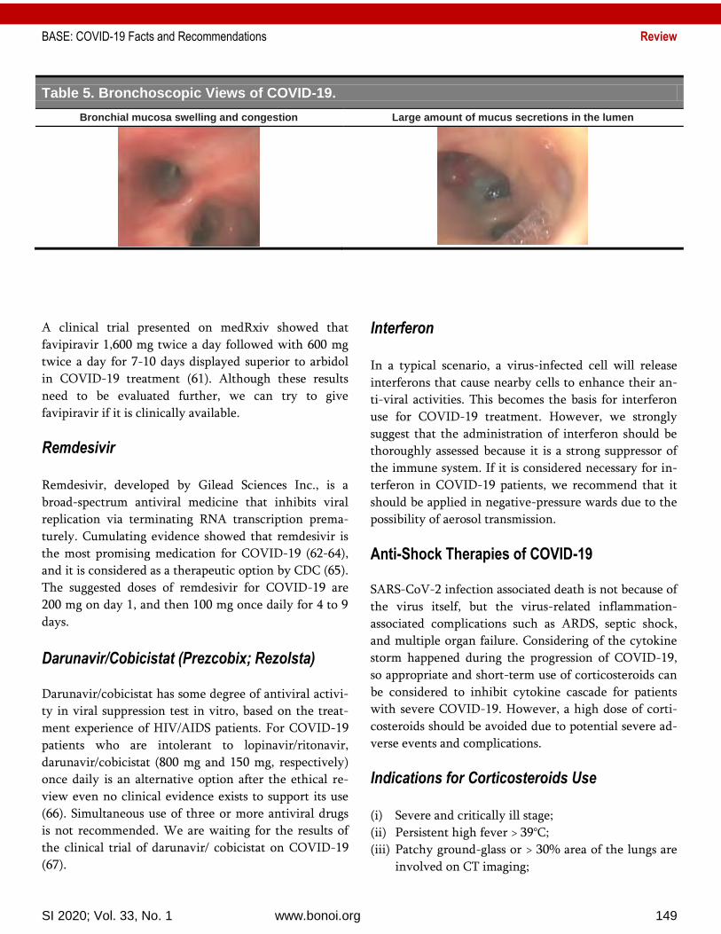
BRONCHOSCOPY IN COVID-19
In mechanically ventilated COVID-19 patients, flexible
bronchoscopy is a versatile, easy to use, and well toler-
ated method to be performed for (53):
(i) Collecting respiratory specimens from the lower
respiratory tract, i.e. sputum, endotracheal aspirate,
and bronchoalveolar lavage for SARS-CoV-2;
(ii) Localizing the site of bleeding, cessation of hemop-
BASE: COVID-19 Facts and Recommendations Review
SI 2020; Vol. 33, No. 1 www.bonoi.org 147
Table 4. CT Signs and Characteristics of COVID-19.
Characteristic
Distribution Subpleura, Subsegment, Segment, Lobar, Multi-Lobar, Inter-Lobar, Bilateral
Development The lesion develops from the lung parenchyma in the lower periphery to the lung interstitium in the center, and the virus directly reaches the alveoli and lung lobules and completely occupies them indicating that it is extreme-ly contagious and sinister.
Shape Triangular, Spherical, Trapezoidal, Rectangular, Fan-Shaped, Ring-Shaped Distribution, Ground-Glass Opacity, Grid-Like, Strip-Shaped, and Vascular Bundle.
Stage Early: Prickly Pear Sign, Hale Nodule Sign Middle: Grey Snow Sign, Gypsum Sign Late: Bat Wing Sign
Special Sign
Rosa Roxburghii Sign
Hale Nodule Sign
Grey Snow Sign
Gypsum Sign
Bat Wing Sign
White Lung Sign
Bronchus & Vascu-lar Bundle Sign
BASE: COVID-19 Facts and Recommendations Review
SI 2020; Vol. 33, No. 1 www.bonoi.org 148
tysis, sputum or blood clots removal;
(iii) Injecting cold saline, epinephrine, vasopressin, or
fibrin as well as laser treatment locally;
(iv) Assisting the establishment of artificial airways by
guiding tracheal intubation or percutaneous tra-
cheotomy;
(v) Administering medicines such as α-interferon and
N-acetylcysteine.
Through bronchoscopy, we can view the extensive
bronchial mucosal hyperemia, swelling, mucus-like se-
cretions in the lumen and jelly-like sputum blocking the
airway in critically ill COVID-19 patients (Table 5).
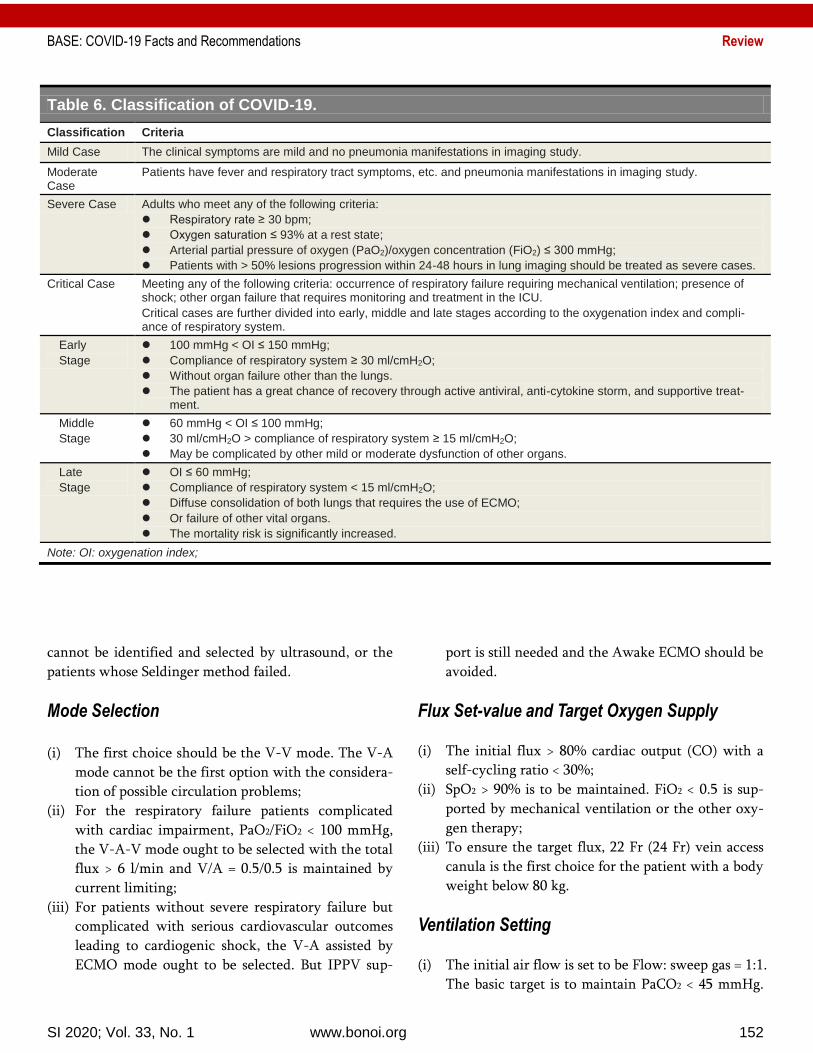
CLASSIFICATION OF COVID-19
The classification of COVID-19 majorly depends on the
severity, and correspondingly divided into four types as
showed in Table 6.
Treatment of COVID-19
So far, no effective therapeutic methods are available
clinically to COVID-19. All listed therapeutic strategies
and maneuvers only can be used with cautiousness at
this stage because no solid clinical evidence exists to
support their viability and reliability. All our listed
therapeutic methods are only based on sporadic cases
with successful therapies. No matter whatever treat-
ment will be used, the eventual outcome of the individ-
ual patient will strongly depend on patient’s personal
immune status and potential response to the virus. We
do not recommend, but suggest that healthcare profes-
sionals consider these potentially useful therapeutic
strategies if conditions permit and are feasible.
General Management
For suspected and mild confirmed cases, isolation in
designated areas is required. Close observation is ex-
tremely important. Basically, thermometer, finger
oximeter, and oxygen compressor should be available in
the isolation environment to guarantee continuous
measuring of temperature, pulse oxygen saturation,
providing O2 once in need.
Anti-viral Therapeutics
For moderate to critically ill patients, anti-viral thera-
pies can be administered as early as possible even no
strong evidence exists.
Lopinavir-Ritonavir (Kaletra®)
Controversial results exist regarding lopinavir-ritonavir
(400 mg and 100 mg, respectively) in treating severe
COVDI-19 patients (54-56). However, this combined
medication can be given to patients as a basic regimen.
It can be applied twice a day for 14 days. Even we do
not know whether it has an optimal dosage for COVID-
19, it is completely acceptable for a trial with higher
doses.
Arbidol
The therapeutic efficacy of single use of arbidol, one of
Russia’s most popular OTC flu medicines, for COVID-19
has not been thoroughly studied. There were case re-
ports on its combined use with lopinavir-ritonavir and
some traditional Chinese medicine in COVID-19 pa-
tients, and showed favorable results (57, 58). Therefore,
arbidol 200 mg can be applied twice day for 14 days.
Chloroquine or Hydroxychloroquine Chloroquine phosphate can be used on adults between
18-65 years old based on the weight: (i) if wt ≥ 50 kg,
500 mg bid for less than 7 days; (ii) weight < 50 kg, 500
mg bid for first two days, and then 500 mg once daily
for following five days (53). Given the severe side effects
of chloroquine (59), we strongly suggest that it can be
given carefully with weighing its benefits over the risks.
Hydroxychloroquine sulfate was observed and
found that hydroxychloroquine 200 mg three times a
day produced an effective therapeutic role in the viral
load reduction/disappearance in COVID-19 patients
with an enforced effect by azithromycin (500 mg on day
1 followed by 250 mg per day for 4 days) (60). So we
suggest that hydroxychloroquine plus azithromycin can
be an alternative to chloroquine.
Favipiravir (Avigan®) Favipiravir, marketed as an anti-influenza drug by Fuji-
film, have shown “obvious efficacy” against COVID-19.
BASE: COVID-19 Facts and Recommendations Review
SI 2020; Vol. 33, No. 1 www.bonoi.org 149
Table 5. Bronchoscopic Views of COVID-19.
Bronchial mucosa swelling and congestion Large amount of mucus secretions in the lumen
A clinical trial presented on medRxiv showed that
favipiravir 1,600 mg twice a day followed with 600 mg
twice a day for 7-10 days displayed superior to arbidol
in COVID-19 treatment (61). Although these results
need to be evaluated further, we can try to give
favipiravir if it is clinically available.
Remdesivir Remdesivir, developed by Gilead Sciences Inc., is a
broad-spectrum antiviral medicine that inhibits viral
replication via terminating RNA transcription prema-
turely. Cumulating evidence showed that remdesivir is
the most promising medication for COVID-19 (62-64),
and it is considered as a therapeutic option by CDC (65).
The suggested doses of remdesivir for COVID-19 are
200 mg on day 1, and then 100 mg once daily for 4 to 9
days.
Darunavir/Cobicistat (Prezcobix; Rezolsta)
Darunavir/cobicistat has some degree of antiviral activi-
ty in viral suppression test in vitro, based on the treat-
ment experience of HIV/AIDS patients. For COVID-19
patients who are intolerant to lopinavir/ritonavir,
darunavir/cobicistat (800 mg and 150 mg, respectively)
once daily is an alternative option after the ethical re-
view even no clinical evidence exists to support its use
(66). Simultaneous use of three or more antiviral drugs
is not recommended. We are waiting for the results of
the clinical trial of darunavir/ cobicistat on COVID-19
(67).
Interferon In a typical scenario, a virus-infected cell will release
interferons that cause nearby cells to enhance their an-
ti-viral activities. This becomes the basis for interferon
use for COVID-19 treatment. However, we strongly
suggest that the administration of interferon should be
thoroughly assessed because it is a strong suppressor of
the immune system. If it is considered necessary for in-
terferon in COVID-19 patients, we recommend that it
should be applied in negative-pressure wards due to the
possibility of aerosol transmission.
Anti-Shock Therapies of COVID-19
SARS-CoV-2 infection associated death is not because of
the virus itself, but the virus-related inflammation-
associated complications such as ARDS, septic shock,
and multiple organ failure. Considering of the cytokine
storm happened during the progression of COVID-19,
so appropriate and short-term use of corticosteroids can
be considered to inhibit cytokine cascade for patients
with severe COVID-19. However, a high dose of corti-
costeroids should be avoided due to potential severe ad-
verse events and complications.
Indications for Corticosteroids Use (i) Severe and critically ill stage;
(ii) Persistent high fever > 39°C;
(iii) Patchy ground-glass or > 30% area of the lungs are
involved on CT imaging;
BASE: COVID-19 Facts and Recommendations Review
SI 2020; Vol. 33, No. 1 www.bonoi.org 150
(iv) Rapid progression with > 50% area involved in
chest CT images within 48 hours;
(v) IL-6 ≥ 5 ULN.
Application of Corticosteroids (i) Initially, methylprednisolone 0.75-1.5 mg/kg i.v.
once a day is recommended;
(ii) Methylprednisolone 40 mg every 12 hours can be
considered for patients with falling body tempera-
ture or for patients with significantly increased cy-
tokines under routine doses of steroid;
(iii) Methylprednisolone 40-80 mg every 12 hours can
be considered for critical cases;
(iv) Closely monitor body temperature, OI, blood rou-
tine, CRP, cytokines, biochemical profile and lung
CT every 2 to 3 days during the treatment;
(v) Methylprednisolone should be halved every 3-5
days if medical conditions are improved, the body
temperature normalized, or involved lesions on CT
are significantly absorbed;
(vi) Oral methylprednisolone once a day is recom-
mended when the i.v. dose is reduced to 20 mg per
day. The time course of corticosteroids in not de-
fined, and it should be used on an individual basis.
Special Consideration during Corticosteroids Treatment (i) Screen TB by T-SPOT assay, HBV and HCV by an-
tibody assay before corticosteroid therapy;
(ii) Consider proton pump inhibitors (PPIs) to prevent
complications;
(iii) Monitor blood glucose and treat high blood glucose
with insulin if needed;
(iv) Correct low serum potassium;
(v) Monitor liver function closely;
(vi) Give sedative-hypnotics temporarily for sleep diffi-
culty.
Oxygen Therapy for COVID-19
Frequently, COVID-19 causes hypoxemia due to im-
paired respiratory functions. Therefore, O2 supplemen-
tation is necessary to correct hypoxemia, and relieve
secondary organ damage resulted from respiratory dis-
tress and hypoxemia (53).
Oxygen Therapy (i) Continual oxygen saturation monitoring during
oxygen therapy to make sure SpO2 > 92%;
(ii) Oxygen therapy should be delivered as soon as pos-
sible if PaO2/FiO2 < 300 mmHg;
(iii) High-flow nasal cannula (HFNC) oxygen therapy is
recommended if COVID-19 patients had:
SpO2 < 93%;
PaO2/FiO2 < 300 mmHg;
Respiratory rate > 25 bpm at bed;
Remarkable progression on chest X-ray;
Wear a surgical mask during HFNC treatment;
The airflow of HFNC oxygen therapy should
start at a low level and gradually increased up
to 40-60 l/min when PaO2/FiO2 is between
200-300 mmHg;
An initial flow of at least 60 l/min should be
given immediately for patients with obvious
respiratory distress.
(iv) Tracheal intubation for patients is dependent on
disease progression, systemic status and complica-
tion of patients for those with stable situation but
with a low OI < 100 mmHg (53).
Tracheal intubation should be performed as
early as possible for patients with an OI < 150
mmHg;
Worsening symptoms of respiratory distress;
Multiple organ dysfunction within 1-2 hours
after high-flow (60 l/min) and high-concen-
tration (> 60%) HFNC oxygen therapy.
(v) Patients > 60 years with more complications or
PaO2/FiO2 < 200 mmHg should be treated in ICU.
Mechanical Ventilation (i) Noninvasive Ventilation (NIV)
NIV is not recommended in COVID-19 patients who
fail to HFNC treatment. It can worsen ARDS, and cause
intolerance to aspiration and worsen lung injury.
(ii) Invasive Mechanical Ventilation
It is extremely critical to balance the benefits of ventila-
tion and the risk of mechanical ventilation-related lung
injury (53).
Tidal volume to 4-8 ml/kg;
Platform pressure < 30 cmH2O;
BASE: COVID-19 Facts and Recommendations Review
SI 2020; Vol. 33, No. 1 www.bonoi.org 151
Driving pressure <15 cmH2O;
Set PEEP according to the institutional ARDS’s
protocol;
Ventilation frequency: 18-25 times per minute;
Moderate hypercapnia is allowed;
Administer sedation, analgesia, or muscle re-
laxant if the variables like tidal volume, plat-
form pressure and driving pressure are too high.
(iii) Weaning of Ventilation
Sedatives is reduced and discontinued before awakening
when PaO2/FiO2 > 150 mmHg. The patient should be
extubated as earlier as possible if the condition is per-
mitted. HFNC or NIV is used for sequential respiratory
support after extubation.
Prone Position Ventilation
With a rapid improvement of oxygenation and lung me-
chanics, most critically ill patients with COVID-19 re-
spond well to prone ventilation. Prone ventilation is
recommended as a routine strategy for patients with
PaO2/FiO2 < 150 mmHg or with obvious imaging mani-
festations without contraindications. Time course rec-
ommended for prone ventilation is more than 16 hours
each time. The prone ventilation can be ceased once
PaO2/FiO2 > 150 mmHg for more than 4 hours in the
supine position (53).
Prone ventilation while awake may be attempted
for patients who have not been intubated or have no
obvious respiratory distress but with impaired oxygena-
tion or have consolidation in gravity-dependent lung
zones on lung images. Procedures for at least 4 hours
each time is recommended. Prone position can be con-
sidered several times per day depending on the effects
and tolerance.
Extracorporeal Membrane Oxygenation Sup-port SARS-CoV-2 is a highly contagious virus primarily tar-
geting pulmonary alveoli that results in respiratory fail-
ure. Extracorporeal membrane oxygenation (ECMO) is
an alternative means for COVID-19 patients. When do-
ing this, following attentions need to be paid to (53):
Timing of ECMO
(i) Salvage ECMO: salvage ECMO intervention needs
to be considered with the onset of one of the fol-
lowing conditions:
PaO2/FiO2 < 80 mmHg, regardless of PEEP lev-
el;
Pplat ≤ 30 mmHg, PaCO2 > 55 mmHg;
The onset of pneumothorax, air leakage > 1/3
tidal volume, duration > 48 hours;
Circulation deterioration, the dosage of nore-
pinephrine > 1 μg/(kg×min);
Cardio-pulmonary resuscitation.
(ii) Replacement ECMO: ECMO replacement needs to
be considered with the onset of one of the follow-
ing conditions:
Decreased lung compliance. After the pulmo-
nary recruitment maneuver, the compliance of
the respiratory system < 10 ml/cmH2O;
Persistent exacerbation of pneumomediasti-
num or subcutaneous emphysema, and the pa-
rameters of mechanical ventilation support
cannot be reduced within 48 hours;
PaO2/FiO2 < 100 mmHg, and it cannot be im-
proved by routine methods in 72 hours.
(iii) Early Awake ECMO: For early awake ECMO, all
the following conditions must be met:
Patient must be in a clear state of conscious-
ness and is fully compliant;
Patient is not complicated with neuromuscular
diseases;
Pulmonary damage score Murry > 2.5;
Few pulmonary secretions. The time interval
between the two airway suction procedures > 4
hours;
Stable hemodynamics without assistance of
vasoactive agents.
Methods of Catheterization Because the ECMO supporting time for most COVID-19
patients will be > 7 days, the Seldinger wire technique
should be used under the guidance of ultrasound, which
reduces the bleeding damages and infection risks
brought about by intravascular cathterization by venous
angiotomy.
Intravascular catheterization by venous angiotomy
may be considered only for the patients with bad blood
vessel conditions, or the patients whose catheterization
BASE: COVID-19 Facts and Recommendations Review
SI 2020; Vol. 33, No. 1 www.bonoi.org 152
Table 6. Classification of COVID-19.
Classification Criteria
Mild Case The clinical symptoms are mild and no pneumonia manifestations in imaging study.
Moderate Case
Patients have fever and respiratory tract symptoms, etc. and pneumonia manifestations in imaging study.
Severe Case Adults who meet any of the following criteria:
Respiratory rate ≥ 30 bpm;
Oxygen saturation ≤ 93% at a rest state;
Arterial partial pressure of oxygen (PaO2)/oxygen concentration (FiO2) ≤ 300 mmHg;
Patients with > 50% lesions progression within 24-48 hours in lung imaging should be treated as severe cases.
Critical Case Meeting any of the following criteria: occurrence of respiratory failure requiring mechanical ventilation; presence of shock; other organ failure that requires monitoring and treatment in the ICU.
Critical cases are further divided into early, middle and late stages according to the oxygenation index and compli-ance of respiratory system.
Early
Stage
100 mmHg < OI ≤ 150 mmHg;
Compliance of respiratory system ≥ 30 ml/cmH2O;
Without organ failure other than the lungs.
The patient has a great chance of recovery through active antiviral, anti-cytokine storm, and supportive treat-ment.
Middle
Stage
60 mmHg < OI ≤ 100 mmHg;
30 ml/cmH2O > compliance of respiratory system ≥ 15 ml/cmH2O;
May be complicated by other mild or moderate dysfunction of other organs.
Late
Stage
OI ≤ 60 mmHg;
Compliance of respiratory system < 15 ml/cmH2O;
Diffuse consolidation of both lungs that requires the use of ECMO;
Or failure of other vital organs.
The mortality risk is significantly increased.
Note: OI: oxygenation index;
cannot be identified and selected by ultrasound, or the
patients whose Seldinger method failed.
Mode Selection (i) The first choice should be the V-V mode. The V-A
mode cannot be the first option with the considera-
tion of possible circulation problems;
(ii) For the respiratory failure patients complicated
with cardiac impairment, PaO2/FiO2 < 100 mmHg,
the V-A-V mode ought to be selected with the total
flux > 6 l/min and V/A = 0.5/0.5 is maintained by
current limiting;
(iii) For patients without severe respiratory failure but
complicated with serious cardiovascular outcomes
leading to cardiogenic shock, the V-A assisted by
ECMO mode ought to be selected. But IPPV sup-
port is still needed and the Awake ECMO should be
avoided.
Flux Set-value and Target Oxygen Supply
(i) The initial flux > 80% cardiac output (CO) with a
self-cycling ratio < 30%;
(ii) SpO2 > 90% is to be maintained. FiO2 < 0.5 is sup-
ported by mechanical ventilation or the other oxy-
gen therapy;
(iii) To ensure the target flux, 22 Fr (24 Fr) vein access
canula is the first choice for the patient with a body
weight below 80 kg.
Ventilation Setting
(i) The initial air flow is set to be Flow: sweep gas = 1:1.
The basic target is to maintain PaCO2 < 45 mmHg.
BASE: COVID-19 Facts and Recommendations Review
SI 2020; Vol. 33, No. 1 www.bonoi.org 153
For the patients complicated with COPD, PaCO2 <
80% basal level;
(ii) The patient’s spontaneous respiratory strength and
respiratory rate (RR) should be maintained, with 10
< RR < 20 and without chief complaint of breathing
difficulty from the patient;
(iii) The sweep gas setup of the V-A mode needs to en-
sure the 7.35-7.45 pH value of the bloodstream out
of the oxygenator membrane.
Anti-Coagulation and Bleeding Prevention (i) For the patients without active bleeding, without
visceral bleeding, and with platelet count > 50×109/l,
the recommended initial heparin dosage is 50 U/kg;
(ii) For the patients complicated with bleeding or with
platelet count < 50×109/l, the recommended initial
heparin dosage is 25 U/kg;
(iii) Maintain the activated partial thromboplastin time
(aPPT) at 40-60 seconds. The trend of D-dimer
change should be considered at the same time.
Antioxidant Treatment for COVID-19
Free radicals refer to any molecules capable of inde-
pendent existence and contain unpaired electrons. They
behave as oxidants or reductants by either donating an
electron to or accepting an electron from other mole-
cules. In many pathological conditions, especially infec-
tion-related inflammatory states, oxygen-containing
free radicals are the major underlying mechanism for
cellular injury (68). These O2-related free radicals in-
clude hydroxyl radical, hydrogen peroxide, superoxide
anion radical, hypochlorite, oxygen singlet, nitric oxide
radical, and peroxynitrite radical. They are highly reac-
tive species and can react to and damage DNA, proteins,
carbohydrates, and lipids (68, 69).
SARS-CoV-2 infection would, theoretically, also
evoke free radical-associated damage in the body via
targeting to all kinds of molecules. Therefore, all thera-
peutic means that can alleviate free radicals can be ap-
plied to COVID-19 patients to conquer the inflamma-
tion-induced burst of free radicals. Furthermore, such
potential therapeutics should be used as early as possible
to prevent the disease from developing into late stage.
For this, an antioxidant, a molecule that is stable enough
to donate an electron to a rampaging free radical and
neutralize it, can be applied to reduce the damage.
Vitamin C
Vitamin C (Ascorbic acid) is a monosaccharide antioxi-
dant. It is a reducing agent and can reduce and neutral-
ize reactive oxygen species such as hydrogen peroxide.
So a therapeutic dose of vitamin C of 3,000 mg once dai-
ly can be applied for COVID-19 patients.
Vitamin E Vitamin E, acollective name for a set of eight related
tocopherols and tocotrienols, is a fat-soluble vitamin
with strong antioxidant properties. It can remove free
radical intermediates and prevent the propagation reac-
tion from continuing. Therefore, a therapeutic dose of
vitamin E of 1,000 IU once daily can be used for
COVID-19 patients.
Glutathione Due to the thiol group in its cysteine moiety, glutathi-
one possesses antioxidant properties. It is a reducing
agent and can be reversibly oxidized and reduced. It has
a high concentration and plays a central role in main-
taining cell’s redox state, as thus glutathione becomes
one of the most pivotal cellular antioxidants. The possi-
ble dosage of glutathione in COVID-19 patients can
reach 70 mg/kg per day.
N-acetyl-L-cysteine (NAC) NAC is a precursor of L-cysteine that increases biosyn-
thesis of glutathione. It acts directly as a scavenger of
free radicals, especially oxygen radicals. With combined
administration of NAC and glutathione, the peroxi-
dative stress of patients with septic shock was signifi-
cantly decreased (70). From this, the potential therapeu-
tic dose of NAC for COVID-19 patients can be 75 mg/kg
per day.
Melatonin Melatonin, N-acetyl-5-methoxytryptamine, is a natural-
ly occurring hormone. It is a powerful antioxidant that
can easily cross cell membranes and the blood-brain
barrier. Melatonin can form several stable end-products
BASE: COVID-19 Facts and Recommendations Review
SI 2020; Vol. 33, No. 1 www.bonoi.org 154
upon reacting with free radicals. In clinical setting, mel-
atonin has been proposed to treat sepsis or septic shock
(71, 72). Even the optimal dose in this setting has not
been established (73), we suggest that it can be given at
less than 50 mg orally per day for COVID-19 patients.
Traditional Chinese Medicine for COVID-19
Traditional Chinese medicine (TCM) is an essential al-
ternative means for western medicine in China. Particu-
larly, herbal compounds are the major part of TCM.
Cumulating evidence is becoming increasing in academ-
ic field on its potential effects in disease prevention and
therapy. Given its property of multi-target and multi-
signaling pathway intervention including anti-oxidative
effect, herbal formulas may play a critical role in miti-
gating COVID-19-associated pathophysiological altera-
tions (74, 75), and it can become a source of drug dis-
covery against COVID-19 (76). We herein present some
TCM herbs and herbal formulas for reference (77).
Single Herbs
Radix Isatidis
Banlan Gen
Small Bupleurum
Coptis
Chinese Patent Formulas
Huoxiang Zhengqi capsules (pills, liquid, or
oral solution)
Jinhua Qinggan granules
Lianhua Qingwen capsules (granules)
Shufeng Jiedu capsules (granules)
Fangfeng Tongsheng pills (granules)
Chinese Herbal Compounds Ephedra 9 g, Zhigancao 6 g, Almond 9 g, Gypsum
15-30 g (fried first), Guizhi 9 g, Zixie 9 g, Zhuling 9
g, Baizhu 9 g, Zhiling 15 g, Bupleurum 16 g,
Scutellaria baicalensis 6 g, and Pinellia 9 g , Ginger
9 g, aster 9 g, winter flower 9 g, shoot dry 9 g,
asarum 6 g, yam 12 g, coriander fruit 6 g, tangerine
peel 6 g, aquilegia 9 g. (One dose per day, twice in
the morning and evening (forty minutes after a
meal), take with warm water, and three doses a
course.)
Raw ephedra 6 g, raw gypsum 15 g, almond 9 g,
loquat 15 g, gardenia 15 g, Guanzhong 9 g, Dilon g
15 g, Xu Changqing 15 g, Huoxiang 15 g, Peilan 9g,
Cangzhu 15 g, Yunling 45 g, Atractylodes 30 g, Jiao
Sanxian 9 g each , Magnolia officinalis 15 g, betel
coconut 9 g, yarrow fruit 9 g, ginger 15 g. (One
dose daily, boiled with 600ml water, take it three
times at morning, noon and evening before meal.)
Betel nut 10 g, apple 10 g, Magnolia 10 g, Zhimu 10
g, scutellaria baicalensis 10 g, Bupleurum 10 g, red
peony 10 g, forsythia 15 g, artemisia annua 10 g
(decocted later), 10 g of green leaves, 10 g of green
leaves, 5 g of raw licorice. (One dose daily, boiled
with 400 ml water, take it twice at morning and
evening.)
Raw ephedra 6 g, bitter almond 15 g, raw gypsum
30 g, raw coix seed 30 g, grass root 10 g, patchouli
15 g, artemisia annua 12 g, Polygonum cuspidatum
20 g, verbena 30 g, dried reed root 30 g, gardenia 15
g 15 g of orange red, 10 g of raw licorice. (One dose
daily, boiled with 400 ml water, take it twice at
morning and evening.)
Atractylodes lancea 15 g, Chenpi 10 g, Magnolia 10
g, Aquilegia 10 g, grass fruit 6 g, raw ephedra 6 g,
Zhihuo 10 g, ginger 10 g, betel nut 10 g. (One dose
daily, boiled with 400 ml water, take it twice at
morning and evening.)
Raw ephedra 6 g, almond 9 g, raw gypsum 15 g,
licorice 3 g, fragrant fragrant 10 g (back), Magnolia
10 g, atractylodes 15 g, grass fruit 10 g, pinellia 9 g,
Poria 15 g, raw rhubarb 5 g (back) 10 g, gardenia 10
g, red peony 10 g. (One or two doses daily, boiled
with 100-200 ml water, take it 2-4 times, oral or
nasal feeding.)
30-60 g gypsum (fried first), 30 g of Zhimu, 30-60 g
of raw land, 30 g of buffalo horn (fried first), 30 g of
red sage, 30 g of black ginseng, 15 g of forsythia, 15
g of paeonia, 6 g of peony 12 g, gardenia 15 g, raw
licorice 6 g. (One dose per day, decoction, first de-
coct gypsum and buffalo horn, then apply other
pieces, 100-200 ml each time, 2-4 times a day, oral-
ly or nasally.)
Vaccine for COVID-19
BASE: COVID-19 Facts and Recommendations Review
SI 2020; Vol. 33, No. 1 www.bonoi.org 155
Although vaccine development needs time, we compas-
sionate with the potential COVID-19 vaccine, because
this is the only means that can effectively prevent and
control even eradiate the virus. With the first mRNA
vaccine mRNA-1273 was injected into the volunteers
(78), we put the hope of conquering the virus on the
vaccine, and we believe effective vaccine will be at the
corner.
Fluid Management Pulmonary function is the key of COVID-19 patients.
Excessive fluid burden will worsen the hypoxemia. In
order to reduce pulmonary exudation and improve oxy-
genation, the amount of fluid should be strictly con-
trolled while ensuring the patient’s basic perfusion.
Food Therapy for COVID-19
Food therapy is a supplementary method for COVID-19
treatment that should be on the basis of balanced nutri-
tion supply. This is suitable for everyone including
those sheltered in place due to the pandemic, and sus-
pected and confirmed cases at different stages. When
preparing foods, mostly, we can add foods with anti-
oxidative ingredients as much as possible ensuring the
nutrition balance.
We herein list the top foods with anti-oxidative
role: Tomatoes, Oats, Green Tea, Ginseng, Blueberries,
Dark Chocolate, Raspberries, Strawberries, Spinach, Or-
anges, Beans, Blackberries, Kale, Cranberries, Beets, Red
Cabbage, Goji Berries, Artichokes, and Pecans.
DISCHARGE CRITERIA OF COVID-19 If COVID-19 patients meet following criteria, they can
be discharged home.
(i) No fever ≥ 3 days;
(ii) No need for O2 > 48 hours;
(iii) Negative NAT twice consecutively with sampling
interval at least 24 hours;
(iv) Respiratory symptoms improve obviously;
(v) Pulmonary imaging shows obvious absorption of
inflammation;
(vi) 14 days isolation and observation after discharge.
PERSPECTIVES
The occurrence of COVID-19 is unavoidable. As the
virus mutates, we cannot predict the next potentially
more deadly virus. Even today, with the development of
medical science to a certain degree, we are still suddenly
at a loss when faced with tiny viruses that cannot be
seen by the naked eye. Since the advent of penicillin,
we have developed many antibacterial drugs. However,
in an era when humans hurriedly mapped the entire
human genome sequence, whereas we could not find a
broad-spectral drug that can effectively fight against
viruses. As we humans continue to move forward,
should we pause for a moment to reexamine what we
have done in science today. When the world shuts
down because of COVID-19, should we slow down and
review the path we have traveled. We seem to have
enough power to leap into the vast universe to find the
next so-called human resting place, but have we learned
a little lesson from this global pandemic of COVID-19?
We look up at the sky on earth, always thinking of
printing our footprints on the surface of other planets.
However, do we think about what we really should do?
Obviously, the seemingly highly developed science and
technology have numerous fatal flaws and loopholes.
We believe that everyone who has experienced this
COVID-19 disaster will seriously reflect on themselves
and reposition themselves in the next step. We hope
that after a certain period of time, we human being will
no longer be that blind confident, but will be prepared
for the next human crisis.■
ARTICLE INFORMATION
Author Affiliations: BASE Medicine Task Force, The Bonoi Academy of Science and Education, Chapel Hill, NC 27510, USA
Author Contributions: BASE has full access to all of the data in the study and takes respon-
sibility for the integrity of the data and the accuracy of the data analysis. Study concept and design: BASE. Acquisition, analysis, or interpretation of data: BASE. Drafting of the manuscript: BASE.
Critical revision of the manuscript for im-portant intellectual content: BASE. Statistical analysis: N/A. Obtained funding: N/A. Administrative, technical, or material support: BASE.
BASE: COVID-19 Facts and Recommendations Review
SI 2020; Vol. 33, No. 1 www.bonoi.org 156
Study supervision: BASE.
Conflict of Interest Disclosures: BASE de-clared no competing interests of this manu-script submitted for publication.
Funding/Support: N/A.
Role of the Funder/Sponsor: N/A.
How to Cite This Paper: BASE Medicine Task Force. COVID-19: Facts and Recommendations from A to Z. Sci Insigt. 2020; 33(1):138-158.
Digital Object Identifier (DOI): http://dx.doi.org/ 10.15354/si.20.re061.
Article Submission Information: Received, March 21, 2020; Revised: April 07, 2020; Ac-cepted: April 07, 2020.
REFERENCES
1. Velavan TP, Meyer CG. The COVID-19 epidemic. Trop Med Int Health 2020;25(3):278-280.
2. World Health Organization. Naming the coronavirus disease (COVID-19) and the virus that causes it. Available at: https://www.who.int/emergencies/diseases/novel-coronavirus-2019/technical-guidance/naming-the-coronavirus-disease-(covid-2019)-and-the-virus-that-causes-it. Last ac-cessed: April 7, 2020.
3. Coronavirus disease (COVID-19) Situation Dashboard. Available at: https://experience.arcgis.com/experience/685d0ace521648f8a5beeeee1b9125cd. Last accessed: April 7, 2020.
4. de Jesus EG. There’s no evidence the coronavirus jumped from pango-lins to people, but the animals do host viruses similar to SARS-CoV-2. MARCH 26, 2020. Available at: https://www.sciencenews.org/article/covid-19-no-evidence-coronavirus-jumped-pangolin-people. Last ac-cessed: April 7, 2020.
5. Zhang C, Zheng W, Huang X, Bell EW, Zhou X, Zhang Y. Protein struc-ture and sequence reanalysis of 2019-nCoV genome refutes snakes as its intermediate host and the unique similarity between its spike protein insertions and HIV-1. J Proteom Res 2020; DOI: 10.1021/acs.jproteome.0c00129.
6. Science Daily. COVID-19 coronavirus epidemic has a natural origin. March 17, 2020. Available at: https://www.sciencedaily.com/releases/2020/03/200317175442.htm. Last accessed: April 7, 2020.
7. de Wilde AH, Snijder EJ, Kikkert M, van Hemert MJ. Host Factors in Coronavirus Replication. Curr Top Microbiol Immunol 2018;419:1-42.
8. Imai Y, Kuba K, Ohto-Nakanishi T, Penninger JM. Angiotensin-converting enzyme 2 (ACE2) in disease patho-genesis. Circ J 2010; 74(3):405-410.
9. Cascella M, Rajnik M, Cuomo A, Dulebohn SC, Di Napoli R. Features, Evaluation and Treatment Corona-
virus (COVID-19). 2020 Mar 20. In: StatPearls [Internet]. Treasure Island (FL): StatPearls Publishing; 2020 Jan.
10. Andersen KG, Rambaut A, Lipkin WI, Holmes EC, Garry RF. The proximal origin of SARS-CoV-2. Nat Med 2020; DOI: 10.1038/s41591-020-0820-9.
11. Harrison SC. Viral membrane fusion. Nat Struct Mol Biol 2008; 15(7):690-698.
12. Ahmed SF, Quadeer AA, McKay MR. Preliminary identification of potential vaccine targets for the COVID-19 Coronavirus (SARS-CoV-2) Based on SARS-CoV immunological studies. Vi-ruses 2020; 12(3):E254.
13. Yan R, Zhang Y, Li Y, Xia L, Guo Y, Zhou Q. Structural basis for the recognition of SARS-CoV-2 by full-length human ACE2. Science 2020; 367(6485):1444-1448.
14. Spike glycoprotein S. Available at: http://new.robetta.org/results.php?id=15652#pdb_download1. Last ac-cessed: April 7, 2020.
15. McBride R, van Zyl M, Fielding BC. The coronavirus nucleocapsid is a multifunctional protein. Viruses 2014; 6(8):2991-3018.
16. Bai Y, Yao L, Wei T, Tian F, Jin DY, Chen L, Wang M. Presumed asymp-tomatic carrier transmission of COVID-19. JAMA 2020; e202565.
17. Wang J, Du G. COVID-19 may transmit through aerosol. Ir J Med Sci 2020 Mar 24:1-2.
18. van Doremalen N, Bushmaker T, Mor-ris DH, Holbrook MG, Gamble A, Wil-liamson BN, Tamin A, Harcourt JL, Thornburg NJ, Gerber SI, Lloyd-Smith JO, de Wit E, Munster VJ. Aerosol and surface stability of SARS-CoV-2 as compared with SARS-CoV-1. N Engl J Med 2020; NEJMc2004973.
19. Quinn A. ‘We Should Grieve’: Infant Becomes Youngest COVID-19 Death in Illinois. Available at: https://www.thedailybeast.com/infant-becomes-youngest-coronavirus-death-in-illinois. Last accessed: April 7, 2020.
20. Vuille-dit-Bille RN, Camargo SM,
Emmenegger L, Sasse T, Kummer E, Jando J, Hamie QM, Meier CF, Hunziker S, Forras-Kaufmann Z, Kuyumcu S, Fox M, Schwizer W, Fried M, Lindenmeyer M, Götze O, Verrey F. Human intestine luminal ACE2 and amino acid transporter ex-pression increased by ACE-inhibitors. Amino Acids 2015; 47(4):693-705.
21. Schouten LR, van Kaam AH, Kohse F, Veltkamp F, Bos LD, de Beer FM, van Hooijdonk RT, Horn J, Straat M, Witteveen E, Glas GJ, Wieske L, van Vught LA, Wiewel MA, Ingelse SA, Cortjens B, van Woensel JB, Bos AP, Walther T, Schultz MJ, Wösten-van Asperen RM for the MARS consorti-um. Age-dependent differences in pulmonary host responses in ARDS: a prospective observational cohort study. Ann Intensive Care 2019; 9(1):55.
22. Xie X, Chen J, Wang X, Zhang F, Liu Y. Age- And Gender-Related Differ-ence of ACE2 Expression in Rat Lung. Life Sci 2006; 78(19):2166-2171.
23. COVID-19: Racial Equity & Social Justice Resources. https://www.racialequitytools.org/fundamentals/resource-lists/resources-addressing-covid-19-with-racial-equity-lens. Last accessed: April 7, 2020.
24. Age, Sex, Existing Conditions of COVID-19 Cases and Deaths. Febru-ary 29, 2020. Available at: https://www.worldometers.info/coronavirus/coronavirus-age-sex-demographics/. Last accessed: April 7, 2020.
25. Guan WJ, Ni ZY, Hu Y, Liang WH, Ou CQ, He JX, Liu L, Shan H, Lei CL, Hui DSC, Du B, Li LJ, Zeng G, Yuen KY, Chen RC, Tang CL, Wang T, Chen PY, Xiang J, Li SY, Wang JL, Liang ZJ, Peng YX, Wei L, Liu Y, Hu YH, Peng P, Wang JM, Liu JY, Chen Z, Li G, Zheng ZJ, Qiu SQ, Luo J, Ye CJ, Zhu SY, Zhong NS; China Medical Treatment Expert Group for Covid-19. Clinical characteristics of coronavirus disease 2019 in China. N Engl J Med 2020; 0:NEJMoa2002032. doi: 10.1056/NEJMoa2002032.
BASE: COVID-19 Facts and Recommendations Review
SI 2020; Vol. 33, No. 1 www.bonoi.org 157
26. Lauer SA, Grantz KH, Bi Q, Jones FK, Zheng Q, Meredith HR, Azman AS, Reich NG, Lessler J. The incubation period of coronavirus disease 2019 (COVID-19) from publicly reported confirmed cases: Estimation and ap-plication. Ann Intern Med 2020; M20-0504.
27. Han R, Huang L, Jiang H, Dong J, Peng H, Zhang D. Early clinical and CT manifestations of coronavirus dis-ease 2019 (COVID-19) Pneumonia. Am J Roentgenol 2020; 1-6.
28. Thomas-Rüddel D, Winning J, Dickmann P, Ouart D, Kortgen A, Janssens U, Bauer M. Coronavirus disease 2019 (COVID-19): update for anesthesiologists and intensivists March 2020. Anaesthesist 2020; doi: 10.1007/s00101-020-00760-3.
29. Wang S, Guo L, Chen L, Liu W, Cao Y, Zhang J, Feng L. A case report of neonatal COVID-19 infection in China. Clin Infect Dis 2020; ciaa225.
30. Rasmussen SA, Smulian JC, Lednicky JA, Wen TS, Jamieson DJ. Coronavirus disease 2019 (COVID-19) and pregnancy: What obstetricians need to know. Am J Obstet Gynecol 2020; doi: 10.1016/j.ajog.2020.02.017.
31. Symptoms of Coronavirus. Corona-virus Disease 2019 (COVID-19). Available at: https://www.cdc.gov/coronavirus/2019-ncov/symptoms-testing/symptoms.html. Last accessed: April 7, 2020.
32. Xie C, Jiang L, Huang G, Pu H, Gong B, Lin H, Ma S, Chen X, Long B, Si G, Yu H, Jiang L, Yang X, Shi Y, Yang Z. Comparison of different samples for 2019 novel coronavirus detection by nucleic acid amplification tests. Int J Infect Dis 2020; 93:264-267.
33. Xiong Z, Fu L, Zhou H, Liu JK, Wang AM, Huang Y, Huang X, Yi B, Wu J, Li CH, Quan J, Li M, Leng YS, Luo WJ, Hu CP, Liao WH. Construction and evaluation of a novel diagnosis process for 2019-Corona Virus Dis-ease. Zhonghua Yi Xue Za Zhi 2020; 100(0):E019. [In Chinese]
34. Cheng H, Wang Y, Wang GQ. Organ-protective effect of angiotensin-converting enzyme 2 and its effect on the prognosis of COVID-19. J Med Virol 2020; doi: 10.1002/jmv.25785.
35. Testing for COVID-19. Coronavirus Disease 2019 (COVID-19). Available at: https://www.cdc.gov/coronavirus/2019-ncov/symptoms-testing/testing.html. Last accessed: April 7, 2020.
36. Wang W, Xu Y, Gao R, Lu R, Han K, Wu G, Tan W. Detection of SARS-
CoV-2 in Different Types of Clinical Specimens. JAMA 2020; e203786.
37. Dong X, Cao YY, Lu XX, Zhang JJ, Du H, Yan YQ, Akdis CA, Gao YD. Eleven faces of coronavirus disease 2019. Allergy 2020. doi: 10.1111/all.14289.
38. Zou L, Ruan F, Huang M , Liang L, Huang H, Hong Z, Yu J, Kang M, Song Y, Xia J, Guo Q, Song T, He J, Yen HL, Peiris M, Wu J. SARS-CoV-2 viral load in upper respiratory speci-mens of infected patients. N Engl J Med 2020; 382(12):1177-1179.
39. The Food and Drug Administration. Cellex qSARS-CoV-2 IgG/IgM Rapid Test. Available at: https://www.fda.gov/media/136625/download. Last accessed: April 7, 2020.
40. Kiernan RE, McPhee DA, Doherty RR. Quantitation of infectious human im-munodeficiency virus using a modified plaque forming assay. J Virol Meth-ods 1988; 22(2-3):303-308.
41. Jennifer R. Tisoncik,a Marcus J. Korth,a Cameron P. Simmons,b Jer-emy Farrar,b Thomas R. Martin,c and Michael G. Katze. Into the Eye of the Cytokine Storm. Microbiol Mol Biol Rev 2012; 76(1):16-32.
42. Chen N, Zhou M, Dong X, Qu J, Gong F, Han Y, Qiu Y, Wang J, Liu Y, Wei Y, Xia J, Yu T, Zhang X, Zhang L. Ep-idemiological and clinical characteris-tics of 99 cases of 2019 novel coro-navirus pneumonia in Wuhan, China: A descriptive study. Lancet 2020; 395(10223):507-513.
43. Chen L, Liu HG, Liu W, Liu J, Liu K, Shang J, Deng Y, Wei S. Analysis of clinical features of 29 patients with 2019 novel coronavirus pneumonia. Zhonghua Jie He He Hu Xi Za Zhi 2020; 43(0):E005. [In Chinese]
44. Wang D, Hu B, Hu C, Zhu F, Liu X, Zhang J, Wang B, Xiang H, Cheng Z, Xiong Y, Zhao Y, Li Y, Wang X, Peng Z. Clinical characteristics of 138 hos-pitalized patients with 2019 novel coronavirus-infected pneumonia in Wuhan, China. JAMA 2020; e201585.
45. Huang C, Wang Y, Li X, Ren L, Zhao J, Hu Y, Zhang L, Fan G, Xu J, Gu X, Cheng Z, Yu T, Xia J, Wei Y, Wu W, Xie X, Yin W, Li H, Liu M, Xiao Y, Gao H, Guo L, Xie J, Wang G, Jiang R, Gao Z, Jin Q, Wang J, Cao B. Clinical features of patients infected with 2019 novel coronavirus in Wuhan, China. Lancet 2020; 395(10223):P497-P506.
46. Shi H, Han X, Jiang N, Cao Y, Alwalid O, Gu J, Fan Y, Zheng C. Radiologi-cal findings from 81 patients with COVID-19 pneumonia in Wuhan, China: A descriptive study. Lancet In-
fect Dis 2020; 20(4):425-434.
47. Mehta P, McAuley DF, Brown M, Sanchez E, Tattersall RS, Manson JJ, on behalf of the HLH Across Speciali-ty Collaboration, UK. COVID-19: Consider cytokine storm syndromes and immunosuppression. Lancet 2020; 395(10229):1033-1034.
48. Randy Q. Cron, W. Winn Chatham. Don’t Forget the Host: COVID-19 Cy-tokine Storm. March 16, 2020. Avail-able at: https://www.the-rheumatologist.org/article/dont-forget-the-host-covid-19-cytokine-storm/. Last accessed: April 5, 2020.
49. Dai WC, Zhang HW, Yu J, Xu HJ, Chen H, Luo SP, Zhang H, Liang LH, Wu XL, Lei Y, Lin F. CT Imaging and differential diagnosis of COVID-19. Can Assoc Radiol J 2020; 71(2):195-200.
50. Li Y, Xia L. Coronavirus disease 2019 (COVID-19): Role of chest CT in di-agnosis and management. Am J Roentgenol 2020; 1-7.
51. Zhou S, Wang Y, Zhu T, Xia L. CT Features of Coronavirus Disease 2019 (COVID-19) Pneumonia in 62 Patients in Wuhan, China. AJR Am J Roentgenol. 2020 Mar 5:1-8.
52. CT Sign Recognition and Rapid Diag-nosis of New Coronary Pneumonia. Available at: https://mp.weixin.qq.com/s/DEvF3RL0lJTize6C09z0Zw. Last accessed: April 7, 2020.
53. The First Affiliated Hospital, Zhejiang University School of Medicine. Com-piled According to Clinical Experience. Handbook of COVID-19 Prevention and Treatment.
54. Lim J, Jeon S, Shin HY, Kim MJ, Seong YM, Lee WJ, Choe KW, Kang YM, Lee B, Park SJ. Case of the in-dex patient who caused tertiary transmission of COVID-19 infection in Korea: The application of lopinavir/ritonavir for the treatment of COVID-19 infected pneumonia moni-tored by quantitative RT-PCR. J Ko-rean Med Sci 2020; 35(6):e79.
55. Cao B, Wang Y, Wen D, Liu W, Wang J, Fan G, Ruan L, Song B, Cai Y, Wei M, Li X, Xia J, Chen N, Xiang J, Yu T, Bai T, Xie X, Zhang L, Li C, Yuan Y, Chen H, Li H, Huang H, Tu S, Gong F, Liu Y, Wei Y, Dong C, Zhou F, Gu X, Xu J, Liu Z, Zhang Y, Li H, Shang L, Wang K, Li K, Zhou X, Dong X, Qu Z, Lu S, Hu X, Ruan S, Luo S, Wu J, Peng L, Cheng F, Pan L, Zou J, Jia C, Wang J, Liu X, Wang S, Wu X, Ge Q, He J, Zhan H, Qiu F, Guo L, Huang C, Jaki T, Hayden FG, Horby PW, Zhang D, Wang C. A trial of lopinavir-
BASE: COVID-19 Facts and Recommendations Review
SI 2020; Vol. 33, No. 1 www.bonoi.org 158
ritonavir in adults hospitalized with severe COVID-19. N Engl J Med 2020; doi: 10.1056/NEJMoa2001282.
56. Xu K, Cai H, Shen Y, Ni Q, Chen Y, Hu S, Li J, Wang H, Yu L, Huang H, Qiu Y, Wei G, Fang Q, Zhou J, Sheng J, Liang T, Li L. Management of coro-na virus disease-19 (COVID-19): the Zhejiang experience. Zhejiang Da Xue Xue Bao Yi Xue Ban 2020; 49(1):0. [In Chinese]
57. Deng L, Li C, Zeng Q, Liu X, Li X, Zhang H, Hong Z, Xia J. Arbidol com-bined with LPV/r versus LPV/r alone against Corona Virus Disease 2019: A retrospective cohort study. J Infect 2020; S0163-4453(20)30113-4.
58. Wang Z, Chen X, Lu Y, Chen F, Zhang W. Clinical characteristics and therapeutic procedure for four cases with 2019 novel coronavirus pneumo-nia receiving combined Chinese and Western medicine treatment. Biosci Trends 2020; 14(1):64-68.
59. Wong YK, Yang J, He Y. Caution and clarity required in the use of chloroquine for COVID-19. Lancet Rheumotol 2020; DOI:https://doi.org/10.1016/S2665-9913(20)30093-X
60. Gautret P, Lagier JC, Parola P, Ho-ang VT, Meddeb L, Mailhe M, Doudier B, Courjon J, Giordanengo V, Vieira VE, Dupont HT, Honoré S, Colson P, Chabrière E, La Scola B, Rolain JM, Brouqui P, Raoult D. Hydroxychloroquine and azithromycin as a treatment of COVID-19: results of an open-label non-randomized clin-ical trial. Int J Antimicrob Agents 2020; 105949. doi: 10.1016/j.ijantimicag.2020.105949.
61. Chen C, Huang J, Cheng Z, Wu J, Chen S, Zhang Y, Chen B, Lu M, Luo Y, Zhang J, Yin P, Wang X. Favipiravir versus Arbidol for COVID-19: A randomized clinical trial. medRxiv 2020.03.17.20037432; doi: https://doi.org/10.1101/2020.03.17.20037432
62. Ko WC, Rolain JM, Lee NY, Chen PL, Huang CT, Lee PI, Hsueh PR. Argu-ments in favour of remdesivir for treat-
ing SARS-CoV-2 infections. Int J Antimicrob Agents 2020; 105933.
63. Al-Tawfiq JA, Al-Homoud AH, Memish ZA. Remdesivir as a possible thera-peutic option for the COVID-19. Trav-el Med Infect Dis 2020; 101615.
64. Wang M, Cao R, Zhang L, Yang X, Liu J, Xu M, Shi Z, Hu Z, Zhong W, Xiao G. Remdesivir and chloroquine effectively inhibit the recently emerged novel coronavirus (2019-nCoV) in vitro. Cell Res 2020; 30(3):269-271.
65. Centers of Disease Control and Pre-vention. Information for Clinicians on Therapeutic Options for COVID-19 Patients. Coronavirus Disease 2019 (COVID-19). Availabel at: https://www.cdc.gov/coronavirus/2019-ncov/hcp/therapeutic-options.html. Last accessed: April 7, 2020.
66. Johnson & Johnson Services, Inc. Lack of evidence to support use of darunavir-based treatments for SARS-CoV-2. How We’re Mobilizing Our Resources to Help Find Solutions for Covid-19. Availabel at: https://www.jnj.com/lack-of-evidence-to-support-darunavir-based-hiv-treatments-for-coronavirus. Last ac-cessed: April 7, 2020.
67. ClinicalTrials.gov. Efficacy and Safety of Darunavir and Cobicistat for Treatment of Pneumonia Caused by 2019-nCoV (DACO-nCoV). NCT04252274. Availabel at: https://clinicaltrials.gov/ct2/show/NCT04252274. Last accessed: April 7, 2020.
68. Akaike T. Role of free radicals in viral pathogenesis and mutation. Rev Med Virol 2001; 11(2):87-101.
69. Lobo V, Patil A, Phatak A, Chandra N. Free radicals, antioxidants and func-tional foods: Impact on human health. Pharmacogn Rev 2010; 4(8):118-126.
70. Ortolani O, Conti A, De Gaudio AR, Moraldi E, Cantini Q, Novelli G. The effect of glutathione and N-acetylcysteine on lipoperoxidative damage in patients with early septic shock. Am J Respir Crit Care Med
2000; 161(6):1907-1911.
71. Galley HF, Lowes DA, Allen L, Cam-eron G, Aucott LS, Webster NR. Mel-atonin as a potential therapy for sep-sis: a phase I dose escalation study and an ex vivo whole blood model under conditions of sepsis. J Pineal Res 2014; 56(4):427-438.
72. Biancatelli RMLC, Berrill M, Moham-med YH, Paul E. Marik. Melatonin for the treatment of sepsis: the scientific rationale. J Thorac Dis 2020; 12(Suppl 1):S54-S65.
73. Zhang R, Wang X, Ni L, Di X, Ma B, Niu S, Liu C, Reiter RJ. COVID-19: Melatonin as a potential adjuvant treatment. Life Sci 2020; 117583.
74. Luo H, Tang QL, Shang YX, Liang SB, Yang M, Robinson N, Liu JP. Can Chinese Medicine Be Used for Pre-vention of Corona Virus Disease 2019 (COVID-19)? A Review of Historical Classics, Research Evidence and Current Prevention Programs. Chin J Integr Med 2020; 26(4):243-250.
75. Yang Y, Islam MS, Wang J, Li Y, Chen X. Traditional Chinese Medicine in the Treatment of Patients Infected with 2019-New Coronavirus (SARS-CoV-2): A Review and Perspective. Int J Biol Sci 2020; 16(10):1708-1717.
76. Ling CQ. Traditional Chinese medi-cine is a resource for drug discovery against 2019 novel coronavirus (SARS-CoV-2). J Integr Med 2020; 18(2):87-88.
77. National Health Commission & State Administration of Traditional Chinese Medicine on March 3, 2020. Diagno-sis and Treatment Protocol for Novel Coronavirus Pneumonia. Available at: https://www.chinalawtranslate.com/wp-content/uploads/2020/03/Who-translation.pdf. Last accessed: April 7, 2020.
78. Park A. COVID-19 vaccine shipped, and drug trials start. February 25, 2020. Available at: https://time.com/5790545/first-covid-19-vaccine/. Last accessed: April 7, 2020.■