Embed Size (px)
Citation preview
Proc. Nati. Acad. Sci. USAVol. 89, pp. 10807-10811, November 1992Medical Sciences
Covalent modification of proteins by ligands of steroidhormone receptors
HLCO ceils/retiok add)
NORIKo TAKAHASHI AND THEODORE R. BREITMANLaboratory of Biological Chemistry, Developmental Therapeutics Program, Division of Cancer Treatment, National Cancer Institute, National Institutes ofHealth, Bethesda, MD 20892
Communicated by Gordon H. Sato, August 7, 1992 (received for review May 4, 1992)
ABSTRACT Retinoylation, acylation with retinoic acid(RA), is a covalent modification of proteins occurring in avariety of eukaryotic cell lines. In this study, we found thatproteins in HL-60 cells were labeled by 17p-[3Hlestradiol (E2),[3H]progesterone (Pg), la,25-dihydroxy[3H]vitamin D3[1,25(OH)2D3I, [12'Iltriiodothyronine (T3), [12iIlthyroxine(T4), and [3Hlprostaglandin E2 (PGE2). All of these hormones,except PGE2, are ligands of the steroid hormone receptorfamily. Addition to the growth medium of 5 FM ketoconazole,an inhibitor of cytochrome P450-dependent enzymes, in-creased about 2-fold the labeling of proteins by T3, T4,1,25(0H)2D3, and PGE2. In contrast, ketoconazole did notchange markedly the extent of labeling by RA, E2, or Pg.Alkaline methanolysis, which cleaves ester bonds, e dvariable percentages of the radioactive ligands bound to pro-tein. These values were about 80% for RA and PGE2; 50% forT3, T4, and Pg; and 20% for E2 and 1,25(OH)2D3. Treatmentwith thioether-cleavage reagents, iodomethane or Raney nickelcatalyst, released <2% of the covalently bound ligands. Two-dimensional polyacrylamide gel electrophoresis patterns oflabeled proteins were unique for each ligand. Proteins of Mr47,000 and 51,000 were labeled by RA, E2, T3, and T4. Theseproteins had the same mobilities as RI and RH, the cAMP-binding regulatory subunits of type I and type II cAMP-dependent protein kiases. 1,25(OH)2D3 also bound to proteinsofMr 47,000 and 51,000. However, these proteins had pI valuesdifferent from those of RI or RII. These results suggest thatsome activities of ligands of the steroid hormone receptorfamily and of PGE2 may be mediated by their covalent mod-ification of proteins.
All-trans-retinoic acid (RA) induces terminal differentiationof the human acute myeloid leukemia cell line HL-60 (1) andof cells from patients with acute promyelocytic leukemia (2).As a sole agent, RA induces complete remissions of patientswith acute promyelocytic leukemia (3-5).
Recently, we reported evidence that a nuclear protein inHL-60 cells is retinoylated via a thioester bond in a dose-dependent manner (6). Furthermore, the dose-responsecurves for RA-induced differentiation and for retinoylationare similar (6, 7). Retinoylation is not restricted to HL-60cells and occurs in other cell types (7-9). These resultsshowed that retinoylation is widespread and that the responseto RA of different cell types may depend on the retinoylationof specific proteins. Thus, some effects of RA on cells maybe independent of RA nuclear receptors (10-12).The RA receptors are members of the steroid hormone
receptor multigene family (10). Some biologic effects ofligands for the steroid hormone receptor family may notinvolve activation of a nuclear receptor and its interactionwith specific DNA sequences (13-25).
The covalent attachment to proteins may be a mechanismfor the action of molecules that also bind to specific recep-tors. In experiments described almost 40 years ago, enzymesin rat liver homogenates catalyzed the covalent binding ofestrogens to protein (26, 27). The activation of 17.B-estradiol(E2) to a quinone, which can damage or form an adduct witheither protein or DNA, may be involved in kidney tumorformation (28). The proestrogen/proantiestrogen activities oftamoxifen and chlorotriansene may entail activation by cy-tochrome P450-dependent enzymes to intermediates thatcovalently attach to the E2 receptor and the activatingenzymes (29, 30). In MCF7 human breast cancer cells, the E2metabolite 16a-hydroxyestrone covalently binds to nuclearmatrix proteins and the E2 receptor (31, 32). We showed thatE2 and RA bind covalently to proteins in MCF7 cells (9).These results prompted us to see whether other ligands of
the steroid hormone receptor family also bind covalently tocellular proteins. We have examined this question with HL-60cells, which respond to many ligands of the steroid hormonereceptor family, including RA (1), la,25-dihydroxyvitamin D3[1,25(OH)ID31 (33), 3,3',5-triiodo-L-thyronine (73) (34), E2(34-36), and dexamethasone (35, 37-42). HL-60 cells containreceptors for RA (43), 1,25(OHj)2D3 (44, 45), dexamethasone(38), and E2 (46). They do not have androgen receptors (46)and we are unaware ofany reports that they contain receptorsfor T3 or progesterone (Pg).
In this study we show that some ligands of the steroidhormone receptor family covalently bind to HL-60 cellularproteins.*
MATERIALS AND METHODSCells. We maintained the human myeloid leukemia cell line
HL-60 in RPMI 1640 medium (GIBCO) supplemented with 10mM 4-(2-hydroxyethyl)-1-piperazinethanesulfonate (pH 7.3)and 10%o fetal bovine serum (GIBCO). Cell cultures wereincubated at 370C in a humidified atmosphere of 5% CO2 inair. We estimated cell number with an electronic particlecounter (Coulter) and assessed cell viability by trypan bluedye exclusion.
Labeling of HL-60 Proteins. Exponentially growing cellswere harvested by centrifugation and suspended at a con-centration of 2 x 10⁶ cells per ml in serum-free mediumconsisting of RPMI 1640 supplemented with 10 mM 4-(2-hydroxyethyl)-l-piperazinethanesulfonate (pH 7.3), 5 ;g ofinsulin per ml, and 5 ug of transferrin per ml (47). All
Abbreviations: RA, all-trans-retinoic acid; 2D-PAGE, two-dimensional polyacrylamide gel electrophoresis; RI and RJI, cAMP-binding regulatory subunits of type I and type II cAMP-dependentprotein kinase; E2, 17#-estradiol; Pg, progesterone; 1,25(OH)2D3,lca,25-dihydroxyvitamin D3; T3, 3,3',5-triiodo-L-thyronine; T4,L-thyroxine; PGE2, prostaglandin E2.*The data in this paper apply to the overall formation of protein-bound radioactive ligands. Since the structure of the bound moietyis not known for all ligands, binding merely reflects an approxima-tion of ligand equivalents.
10807
The publication costs of this article were defrayed in part by page chargepayment. This article must therefore be hereby marked "advertisement"in accordance with 18 U.S.C. §1734 solely to indicate this fact.
10808 Medical Sciences: Takahashi and Breitman
radioactive compounds were purchased from DuPont/NEN.Unlabeled RA (Sigma) and radioactive RA ([11,12-3H], 51Ci/mmol; 1 Ci = 37 GBq), 1,25(OH)2D3 ([26,27-3H], 160Ci/mmol), prostaglandin E2 (PGE2) ([5,6,8,11,14,15-3H], 200Ci/mmol), E2 ([2,4,6,7,16,17-3H(N)], 150 Ci/mmol), Pg([1,2,6,7,21-3H(N)], 193 Ci/mmol), L-thyroxine (T4) ([1251],4400 Ci/mmol), and T3 ([3,3',5-125I], 648 Ci/mmol) weredissolved in absolute ethanol and diluted into the growthmedium. The final concentration of ethanol was <0.1%.After incubation for 24 hr, cells were harvested by centrifu-gation (200 x g, 5 min) and washed with phosphate-bufferedsaline (1.5 mM KH2PO4/8.1 mM Na2HPO4/136.9 mM NaCl,pH 7.2). A dried delipidated cell pellet was prepared by theBligh-Dyer procedure (48) as described (49) and dissolved invarious solutions depending on the test system.
Stability ofBound Ligands. Alkaline methanolysis to cleaveester bonds was as described (49). Treatment of labeledproteins to detect thioethers was carried out either withRaney nickel catalyst (Sigma) (50) or with CH3I (51-53).Two-Dimensional Polyacrylamide Gel Electrophoresis (2D-
PAGE). 2D-PAGE was according to O'Farrell (54) with minormodifications (7). Gels were fixed, stained with Coomassieblue R-250, and prepared for fluorography with Entensify(DuPont/NEN) according to the manufacturer's instructions.
RESULTSLabeling of Total HL-60 Proteins by Hormones. We saw
radioactivity in the delipidated residues prepared by the Bligh-Dyer extraction procedure after incubation of HL-60 cellswith [3H]RA, [3H11,25(OH)2D3, [3H]E2, [3H]Pg, [1251]T3, and[125I]T4 (Table 1). Hydroxylated steroids could be intermedi-ates in the binding of steroids to proteins. Ketoconazoleinhibits cytochrome P450-dependent enzymes that catalyzemany of these hydroxylations (55-57). We measured thelabeling of HL-60 proteins in the presence of 5 ,uM ketoco-
RA
Table 1. Modulation by ketoconazole of labeling ofHL-60 proteins
Total bound, fmol per 106 cells
Labeled compound - ketoconazole + ketoconazole Ratio*RA (100 nM) 330 250 0.76E2 (100 nM) 250 300 1.2Pg (100 nM) 27 31 1.2T4 (0.75 nM) 0.17 0.33 2T3 (10 nM) 6 12.8 2.11,25(OH)2D3 (2.5 nM) 40 94 2.4HL-60 cells were grown for 24 hr in medium containing the radiola-
beled ligand, without and with 5 uM ketoconazole. Cells were har-vested by centrifugation and extracted by the Bligh-Dyer procedure.*Total bound with ketoconazole divided by the total bound withoutketoconazole.
nazole, which effectively inhibits cytochrome P450-dependentenzymes in intact cells (57, 58). In the presence of ketocona-zole the labeling of total proteins by T3, T4, and 1,25(OH)2D3increased about 2-fold (Table 1) and the labeling by RA, E2,and Pg was about ±20% of the control. In the presence ofketoconazole there was a slight decrease ofcell growth and cellviability was >95% (data not shown).Labeing of Individual HL-60 Proteins. With 2D-PAGE and
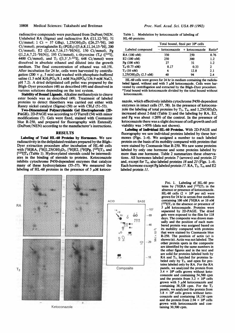
fluorography we saw individual proteins labeled by these hor-mones (Figs. 1-4). We assigned a number to each labeledprotein on the basis of its mobility compared with proteins thatwere stained by Coomassie blue R-250. We saw some proteinslabeled by only one hormone and some proteins labeled bymore than one hormone. Table 2 summarizes these observa-tions. All hormones labeled protein 7 (arrows) and protein 22and, except for T4, also labeled proteins 18 and 23 (Figs. 1-4).Each hormone except Pg labeled protein 17. RA, T3, T4, and E2labeled protein 11.
ACID BASE
Composite
Ketoconazole
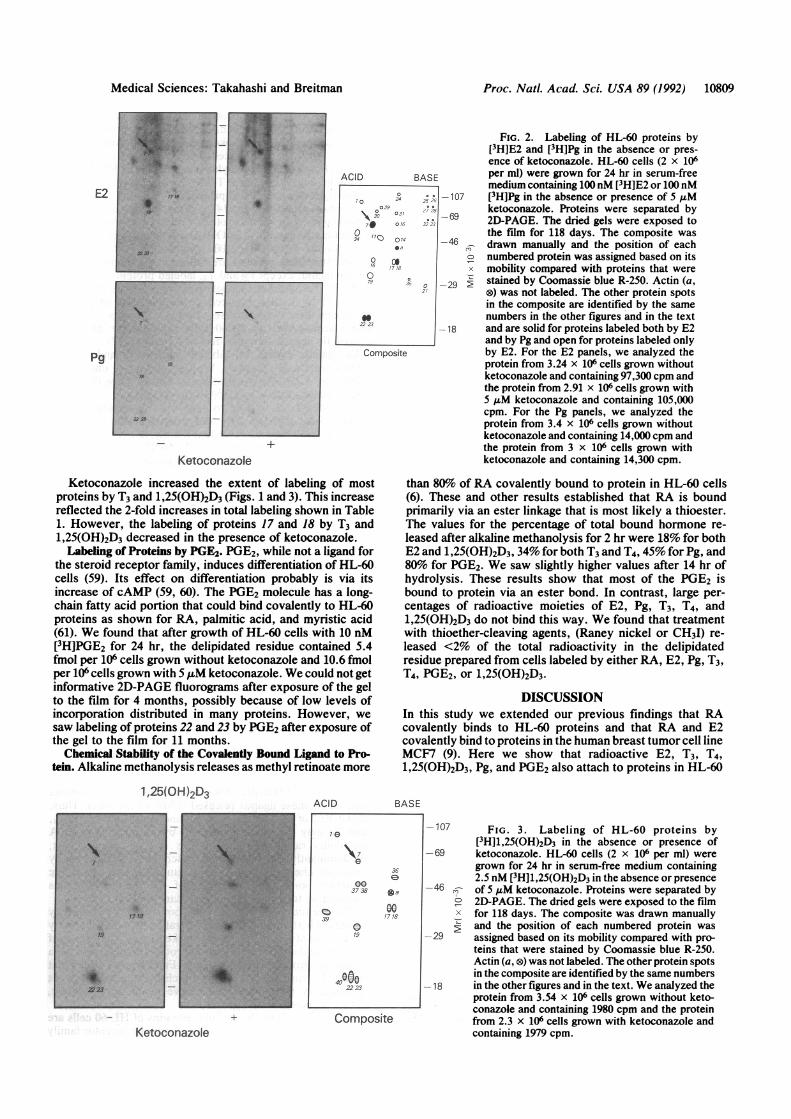
FIG. 1. Labeling of HL-60 pro-teins by [3H]RA and [125I]T3 in theabsence or presence of ketoconazole.HL-60 cells (2 x 10⁶ per ml) weregrown for 24 hr in serum-free mediumcontaining 100 nM [3H]RA or 10 nM[125IJT3 in the absence or presence of
-107 5ASM ketoconazole. Proteins wereseparated by 2D-PAGE. The dried
-69 gels were exposed to the film for 118days. The composite was drawn man-ually and the position of each num-
-46 bered protein was assigned based onr- its mobility compared with proteins
that were stained by Coomassie blueX R-250. The position of actin (a) is
2shown (®). Actin was not labeled. The
-29 other protein spots in the compositeare identified by the same numbers inthe other figures and in the text andare solid for proteins labeled both by
-18 RA and T3, hatched for proteins la-beled only by T3, and open for pro-teins labeled only by RA. For the RApanels, we analyzed the protein from3.4 x 106 cells grown without keto-conazole and containing 54,500 cpmand the protein from 3.2 x 10⁶ cellsgrown with 5 ,uM ketoconazole andcontaining 38,528 cpm. For the T3panels, we analyzed the protein from3.8 x 106 cells grown without keto-conazole and containing 18,130 cpmand the protein from 2.94 x 10⁶ cellsgrown with ketoconazole and con-taining 30,500 cpm.
17AL,;..
Ax-. ..~A
I.;w22i
10
20 4 5
\03 5s
_0 0008 9 10
1213
15
160 a17 18
19 20
22 23
C)2
L
Proc. NatL Acad. Sci. USA 89 (1992)
AlWrl-
3-s;
Proc. Natl. Acad. Sci. USA 89 (1992) 10809
E2
Pg
?
to
to
#vI
ACID BASE
Composite
_LKetoconazole
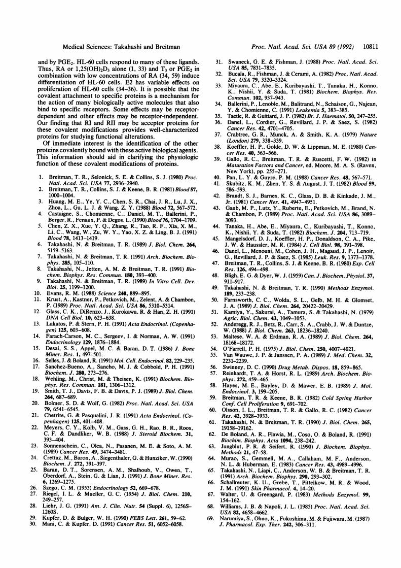
Ketoconazole increased the extent of labeling of mostproteins by T3 and 1,25(OH)2D3 (Figs. 1 and 3). This increasereflected the 2-fold increases in total labeling shown in Table1. However, the labeling of proteins 17 and 18 by T3 and1,25(OH)2D3 decreased in the presence of ketoconazole.Labeling of Proteins by PGE2. PGE2, while not a ligand for
the steroid receptor family, induces differentiation of HL-60cells (59). Its effect on differentiation probably is via itsincrease of cAMP (59, 60). The PGE2 molecule has a long-chain fatty acid portion that could bind covalently to HL-60proteins as shown for RA, palmitic acid, and myristic acid(61). We found that after growth of HL-60 cells with 10 nM[3H]PGE2 for 24 hr, the delipidated residue contained 5.4fmol per 106 cells grown without ketoconazole and 10.6 fmolper 106 cells grown with 5 ,uM ketoconazole. We could not getinformative 2D-PAGE fluorograms after exposure of the gelto the film for 4 months, possibly because of low levels ofincorporation distributed in many proteins. However, wesaw labeling of proteins 22 and 23 by PGE2 after exposure ofthe gel to the film for 11 months.Chemical Stability of the Covalently Bound Ligand to Pro-
tein. Alkaline methanolysis releases as methyl retinoate more
1 ,25(OH)2D3ACID
Composite+
Ketoconazole
FIG. 2. Labeling of HL-60 proteins by[3H]E2 and [3H]Pg in the absence or pres-ence of ketoconazole. HL-60 cells (2 x 10⁶per ml) were grown for 24 hr in serum-freemedium containing 100nM [3H]E2 or 100 nM[3H]Pg in the absence or presence of 5 1Mketoconazole. Proteins were separated by2D-PAGE. The dried gels were exposed tothe film for 118 days. The composite was
,, drawn manually and the position of eacho numbered protein was assigned based on its
mobility compared with proteins that were> stained by Coomassie blue R-250. Actin (a,
®) was not labeled. The other protein spotsin the composite are identified by the samenumbers in the other figures and in the textand are solid for proteins labeled both by E2and by Pg and open for proteins labeled onlyby E2. For the E2 panels, we analyzed theprotein from 3.24 x 10⁶ cells grown withoutketoconazole and containing 97,300 cpm andthe protein from 2.91 x 10⁶ cells grown with5 AuM ketoconazole and containing 105,000cpm. For the Pg panels, we analyzed theprotein from 3.4 x 10⁶ cells grown withoutketoconazole and containing 14,000 cpm andthe protein from 3 x 10⁶ cells grown withketoconazole and containing 14,300 cpm.
than 8%o of RA covalently bound to protein in HL-60 cells(6). These and other results established that RA is boundprimarily via an ester linkage that is most likely a thioester.The values for the percentage of total bound hormone re-leased after alkaline methanolysis for 2 hr were 18% for bothE2 and 1,25(OH)2D3, 34% for both T3 and T4, 45% for Pg, and80% for PGE2. We saw slightly higher values after 14 hr ofhydrolysis. These results show that most of the PGE2 isbound to protein via an ester bond. In contrast, large per-centages of radioactive moieties of E2, Pg, T3, T4, and1,25(OH)2D3 do not bind this way. We found that treatmentwith thioether-cleaving agents, (Raney nickel or CH3I) re-leased <2% of the total radioactivity in the delipidatedresidue prepared from cells labeled by either RA, E2, Pg, T3,T4, PGE2, or 1,25(OH)2D3.
DISCUSSIONIn this study we extended our previous findings that RAcovalently binds to HL-60 proteins and that RA and E2covalently bind to proteins in the human breast tumor cell lineMCF7 (9). Here we show that radioactive E2, T3, T4,1,25(OH)2D3, Pg, and PGE2 also attach to proteins in HL-60
BASE
-107 FIG. 3. Labeling of HL-60 proteins by[3H]1,25(OH)2D3 in the absence or presence of
-69 ketoconazole. HL-60 cells (2 x 10⁶ per ml) weregrown for 24 hr in serum-free medium containing2.5 nM [3H]1,25(OH)2D3 in the absence or presence
-46 n of 5 ,uM ketoconazole. Proteins were separated by° 2D-PAGE. The dried gels were exposed to the filmX for 118 days. The composite was drawn manuallyE and the position of each numbered protein was
-29 assigned based on its mobility compared with pro-teins that were stained by Coomassie blue R-250.Actin (a, ®) was not labeled. The other protein spotsin the composite are identified by the same numbers
-18 in the other figures and in the text. We analyzed theprotein from 3.54 x 10⁶ cells grown without keto-conazole and containing 1980 cpm and the proteinfrom 2.3 x 10⁶ cells grown with ketoconazole andcontaining 1979 cpm.
36
eo37 38 fo a
Q~~~ 0039 177'
4000022 23
Medical Sciences: Takahashi and Breitman

10810 Medical Sciences: Takahashi and Breitman
AC-ID RARF
109-
72-
;-
x
46-
29-
18-
FIG. 4. Labeling of HL-60 proteins by [125I]T4. HL-60 cells (2 x
106 per ml) were grown for 24 hr in serum-free medium containing 2.5nM [1251]T4. Proteins were separated by 2D-PAGE. The dried gel wasexposed to the film for 30 days. The protein spots are identified bythe same numbers in the other figures and in the text. We analyzedthe protein from 1 x 10⁷ cells containing 6660 cpm.
cells. All of these hormones, except PGE2, are ligands formembers of the steroid hormone receptor family.The steroid hormone receptors function as regulators of
gene expression (10). It is likely that these receptors mediatemost of the biological effects of their ligands. However, some
effects of T3 (13), 1,25(OH)2D3 (14, 25, 62), RA (20, 24), E2(22, 23), and Pg (23) may be non-receptor-mediated or
nongenomic or both. The mechanisms for these effects are
unknown.Our results raise the possibility that some effects of T3, T4,
1,25(0H)2D3, RA, E2, and Pg are mediated by their covalentbinding to proteins. We saw 40 proteins of HL-60 cellslabeled by at least one of these six ligands (Figs. 1-4; Table2). Twelve of these proteins were labeled by more than one
ligand. More than 600 HL-60 proteins (silver-stained or
labeled with [35S]methionine) are seen by 2D-PAGE underconditions similar to those we used (63, 64). Thus, theprobability is very high that some proteins are modified bymore than one hormone. It will be of interest to determinewhether more than one hormone can bind to one proteinmolecule.
Table 2. Summary of HL-60 proteins labeled by RA, T3, T4, E2,
No. RA T3 T4 E2 Pg D3 No. RA T3 T4 E2 Pg D30 0 0 21 0 0
2 0 22 0 0 0 0
3 0 23 0 0
4 0 24 0
5 0 25 0 0
6 0 26 0 0
7 0 0 0 0 0 27 08 0 28 0
9 0 29 0
10 0 30 0
1 00 - 31 0
12 0 32 0
13 0 33 0
14 0 0 0 0 34 0
0 35 0
16 0 36 0
17 0 0 00 0 37 0
18 00 0 0 38 0
19 0 0 0 39 0
20 0 40 0
Proteins are identified by number as in Figs. 1-4. D3, 1,25(OH)2D3;0, protein labeled by more than one ligand; 0, protein labeled byonly one ligand.
The list of retinoylated proteins includes RI and RII, thecAMP-regulatory subunits ofcAMP-dependent protein kinasetype I and type II (65); cytokeratins (8); and ribonucleotidereductase (66). In HL-60 cells we identified retinoylatedprotein 11 as RII and retinoylated protein 14 as RI (65). Asshown in Figs. 1-4 and Table 2, all ligands, except1,25(OH)2D3 and Pg, labeled proteins 11 and 14. The absenceof labeling of proteins 11 and 14 by Pg may reflect the low levelof binding of Pg to HL-60 proteins (Table 1 and Fig. 2). Thismay not be the case for 1,25(OH)2D3, which uniquely labeledproteins ofMr 47,000 and 51,000 (proteins 36-38, Fig. 3). It ispossible that these proteins are isoforms of RII and RI.Each ligand, except Pg and T4, labeled proteins 17 and 18
(Table 2). The absence of labeling of protein 17 by Pg and ofprotein 18 by T4 also may reflect the low level of proteinlabeling by these two hormones (Table 1). Proteins 17 and 18bind 8-azido-cAMP in HL60 cells (65) and are probablyproteolytic degradation products of RI or RII (67).
All ligands, except T4, labeled proteins 22 and 23. T4labeled protein 23 (Table 2). We saw protein 40 only afterlabeling by 1,25(OH)2D3. Proteins 22 and 23 were the prin-cipal proteins labeled by 1,25(OH)2D3 (Fig. 3).We added ketoconazole to suppress the metabolism of
steroid, RA, 1,25(OH)2D3, and prostaglandin by a cy-tochrome P450-dependent system (56-58, 68). The covalentbinding to total HL-60 protein by 1,25(OH)2D3, T3, T4, andPGE2 was about 2-fold greater in the presence of ketocona-zole (Table 1). In comparison, ketoconazole affected slightlythe covalent binding to protein of RA, E2, and Pg. Theseresults show that the covalent labeling ofproteins by RA, E2,and Pg may not require their conversion to an intermediate ofa cytochrome P450-dependent enzyme reaction. We do nothave an explanation for the increased binding of T3 and T4 inthe presence of ketoconazole.We showed that about 80% of RA covalently binds to
HL-60 protein via a thioester linkage (6). However, thislinkage of RA to protein is not universal. RA is not attachedby a thioester bond to cytokeratins of normal keratinocytes(8). PGE2 is the only other ligand we investigated that maybind to HL.60 protein primarily via an ester bond. Alkalinemethanolysis released about 80% of the PGE2 bound toprotein. This procedure hydrolyzes thioesters and most ox-oesters. Therefore, it is possible that PGE2 binds by an esterbond between its carboxyl group and either a hydroxy aminoacid or a cysteine of a protein. Our results with PGE2 may besimilar to those showing an alkali-labile covalent binding ofA12-prostaglandin J2 to proteins in the nuclear matrix andchromatin of mouse leukemia L1210 cells (69).Compared to our knowledge of the chemical bond between
protein and either RA or PGE2 we have less informationabout the chemical bond between protein and either E2, T3,T4, Pg, or 1,25(OH)2D3. Alkaline methanolysis of proteinslabeled by these ligands released <50% of the label. Thus,about 20%6 of E2 and 1,25(OH)2D3 and about 40%o of T3, T4,and Pg may be bound to protein by an ester linkage. Apossibility raised by these results is that some ligands,predominantly Pg, T3, and T4, each bind to proteins bydifferent chemical bonds. It is unlikely that there arethioether bonds, because <2% of the radioactivity wasreleased by treatment with either Raney nickel or CH3I.The chemical stability of the E2 bound to protein seen by
us agrees with findings reported in rat liver homogenates byRiegel and Mueller (27). Those workers found that the chiefreactant in the binding process to proteins was E2, and not ametabolic product such as estrone. Further, they found thatbound E2 was not attached to nucleic acids and was notreleased by procedures that cleave disulfide, glycosidic,ester, or thioether linkages.Our results show that cellular proteins of HL-60 cells are
modified by ligands for the steroid hormone receptor family
Proc. Natl. Acad. Sci. USA 89 (1992)
Proc. Natl. Acad. Sci. USA 89 (1992) 10811
and by PGE2. HL-60 cells respond to many of these ligands.Thus, RA or 1,25(OH)2D3 alone (1, 33) and T3 or PGE2 incombination with low concentrations of RA (34, 59) inducedifferentiation of HL-60 cells. E2 has variable effects onproliferation of HL-60 cells (34-36). It is possible that thecovalent attachment to specific proteins is a mechanism forthe action of many biologically active molecules that alsobind to specific receptors. Some effects may be receptor-dependent and other effects may be receptor-independent.Our finding that RI and RII may be acceptor proteins forthese covalent modifications provides well-characterizedproteins for studying functional alterations.Of immediate interest is the identification of the other
proteins covalently bound with these active biological agents.This information should aid in clarifying the physiologicfunction of these covalent modifications of proteins.
1. Breitman, T. R., Selonick, S. E. & Collins, S. J. (1980) Proc.Nati. Acad. Sci. USA 77, 2936-2940.
2. Breitman, T. R., Collins, S. J. & Keene, B. R. (1981) Blood 57,1000-1004.
3. Huang, M. E., Ye, Y. C., Chen, S. R., Chai, J. R., Lu, J. X.,Zhoa, L., Gu, L. J. & Wang, Z. Y. (1988) Blood 72, 567-572.
4. Castaigne, S., Chomienne, C., Daniel, M. T., Ballerini, P.,Berger, R., Fenaux, P. & Degos, L. (1990) Blood 76, 1704-1709.
5. Chen, Z. X., Xue, Y. Q., Zhang, R., Tao, R. F., Xia, X. M.,Li, C., Wang, W., Zu, W. Y., Yao, X. Z. & Ling, B. J. (1991)Blood 78, 1413-1419.
6. Takahashi, N. & Breitman, T. R. (1989) J. Biol. Chem. 264,5159-5163.
7. Takahashi, N. & Breitman, T. R. (1991) Arch. Biochem. Bio-phys. 285, 105-110.
8. Takahashi, N., Jetten, A. M. & Breitman, T. R. (1991) Bio-chem. Biophys. Res. Commun. 180, 393-400.
9. Takahashi, N. & Breitman, T. R. (1989) In Vitro Cell. Dev.Biol. 25, 1199-1200.
10. Evans, R. M. (1988) Science 240, 889-895.11. Krust, A., Kastner, P., Petkovich, M., Zelent, A. & Chambon,
P. (1989) Proc. Natl. Acad. Sci. USA 86, 5310-5314.12. Glass, C. K., DiRenzo, J., Kurokawa, R. & Han, Z. H. (1991)
DNA Cell Biol. 10, 623-638.13. Lakatos, P. & Stern, P. H. (1991) Acta Endocrinol. (Copenha-
gen) 125, 603-608.14. Farach-Carson, M. C., Sergeev, I. & Norman, A. W. (1991)
Endocrinology 129, 1876-1884.15. Desai, S. S., Appel, M. C. & Baran, D. T. (1986) J. Bone
Miner. Res. 1, 497-501.16. Selles, J. & Boland, R. (1991) Mol. Cell. Endocrinol. 82,229-235.17. Sanchez-Bueno, A., Sancho, M. J. & Cobbold, P. H. (1991)
Biochem. J. 280, 273-276.18. Wehling, M., Christ, M. & Theisen, K. (1991) Biochem. Bio-
phys. Res. Commun. 181, 1306-1312.19. Smith, T. J., Davis, F. B. & Davis, P. J. (1989) J. Biol. Chem.
264, 687-689.20. Bolmer, S. D. & Wolf, G. (1982) Proc. Natl. Acad. Sci. USA
79, 6541-6545.21. Chetrite, G. & Pasqualini, J. R. (1991) Acta Endocrinol. (Co-
penhagen) 125, 401-408.22. Meyers, C. Y., Kolb, V. M., Gass, G. H., Rao, B. R., Roos,
C. F. & Dandliker, W. B. (1988) J. Steroid Biochem. 31,393-404.
23. Sonnenschein, C., Olea, N., Pasanen, M. E. & Soto, A. M.(1989) Cancer Res. 49, 3474-3481.
24. Crettaz, M., Baron, A., Siegenthaler, G. & Hunziker, W. (1990)Biochem. J. 272, 391-397.
25. Baran, D. T., Sorensen, A. M., Shalhoub, V., Owen, T.,Oberdorf, A., Stein, G. & Lian, J. (1991) J. Bone Miner. Res.6, 1269-1275.
26. Szego, C. M. (1953) Endocrinology 52, 669-678.27. Riegel, I. L. & Mueller, G. C. (1954) J. Biol. Chem. 210,
249-257.28. Liehr, J. G. (1991) Am. J. Clin. Nutr. 54 (Suppl. 6), 1256S-
1260S.29. Kupfer, D. & Bulger, W. H. (1990) FEBS Lett. 261, 59-62.30. Mani, C. & Kupfer, D. (1991) Cancer Res. 51, 6052-6058.
31. Swaneck, G. E. & Fishman, J. (1988) Proc. Natl. Acad. Sci.USA 85, 7831-7835.
32. Bucala, R., Fishman, J. & Cerami, A. (1982) Proc. Natl. Acad.Sci. USA 79, 3320-3324.
33. Miyaura, C., Abe, E., Kuribayashi, T., Tanaka, H., Konno,K., Nishii, Y. & Suda, T. (1981) Biochem. Biophys. Res.Commun. 102, 937-943.
34. Ballerini, P., Lenoble, M., Balitrand, N., Schaison, G., Najean,Y. & Chomienne, C. (1991) Leukemia 5, 383-385.
35. Taetle, R. & Guittard, J. P. (1982) Br. J. Haematol. 50, 247-255.36. Danel, L., Cordier, G., Revillard, J. P. & Saez, S. (1982)
Cancer Res. 42, 4701-4705.37. Crabtree, G. R., Munck, A. & Smith, K. A. (1979) Nature
(London) 279, 338-339.38. Koeffler, H. P., Golde, D. W. & Lippman, M. E. (1980) Can-
cer Res. 40, 563-566.39. Gallo, R. C., Breitman, T. R. & Ruscetti, F. W. (1982) in
Maturation Factors and Cancer, ed. Moore, M. A. S. (Raven,New York), pp. 255-271.
40. Pan, L. Y. & Guyre, P. M. (1988) Cancer Res. 48, 567-571.41. Skubitz, K. M., Zhen, Y. S. & August, J. T. (1982) Blood 59,
586-593.42. Brandt, S. J., Barnes, K. C., Glass, D. B. & Kinkade, J. M.,
Jr. (1981) Cancer Res. 41, 4947-4951.43. Gaub, M. P., Lutz, Y., Ruberte, E., Petkovich, M., Brand, N.
& Chambon, P. (1989) Proc. Natl. Acad. Sci. USA 86, 3089-3093.
44. Tanaka, H., Abe, E., Miyaura, C., Kuribayashi, T., Konno,K., Nishii, Y. & Suda, T. (1982) Biochem. J. 204, 713-719.
45. Mangelsdorf, D. J., Koeffler, H. P., Donaldson, C. A., Pike,J. W. & Haussler, M. R. (1984) J. Cell Biol. 98, 391-398.
46. Danel, L., Menouni, M., Cohen, J. H., Magaud, J. P., Lenoir,G., Revillard, J. P. & Saez, S. (1985) Leuk. Res. 9, 1373-1378.
47. Breitman, T. R., Collins, S. J. & Keene, B. R. (1980) Exp. CellRes. 126, 494-498.
48. Bligh, E. G. & Dyer, W. J. (1959) Can. J. Biochem. Physiol. 37,911-917.
49. Takahashi, N. & Breitman, T. R. (1990) Methods Enzymol.189, 233-238.
50. Farnsworth, C. C., Wolda, S. L., Gelb, M. H. & Glomset,J. A. (1989) J. Biol. Chem. 264, 20422-20429.
51. Kamiya, Y., Sakurai, A., Tamura, S. & Takahashi, N. (1979)Agric. Biol. Chem. 43, 1049-1053.
52. Anderegg, R. J., Betz, R., Carr, S. A., Crabb, J. W. & Duntze,W. (1988) J. Biol. Chem. 263, 18236-18240.
53. Maltese, W. A. & Erdman, R. A. (1989) J. Biol. Chem. 264,18168-18172.
54. O'Farrell, P. H. (1975) J. Biol. Chem. 250, 4007-4021.55. Van Wauwe, J. P. & Janssen, P. A. (1989) J. Med. Chem. 32,
2231-2239.56. Swinney, D. C. (1990) Drug Metab. Dispos. 18, 859-865.57. Reinhardt, T. A. & Horst, R. L. (1989) Arch. Biochem. Bio-
phys. 272, 459-465.58. Hayes, M. E., Bayley, D. & Mawer, E. B. (1989) J. Mol.
Endocrinol. 3, 199-205.59. Breitman, T. R. & Keene, B. R. (1982) Cold Spring Harbor
Conf. Cell Proliferation 9, 691-702.60. Olsson, I. L., Breitman, T. R. & Gallo, R. C. (1982) Cancer
Res. 42, 3928-3933.61. Takahashi, N. & Breitman, T. R. (1990) J. Biol. Chem. 265,
19158-19162.62. De Boland, A. R., Flawia, M., Coso, 0. & Boland, R. (1991)
Biochim. Biophys. Acta 1094, 238-242.63. Jungblut, P. R. & Seifert, R. (1990) J. Biochem. Biophys.
Methods 21, 47-58.64. Murao, S., Gemmell, M. A., Callaham, M. F., Anderson,
N. L. & Huberman, E. (1983) Cancer Res. 43, 4989-4996.65. Takahashi, N., Liapi, C., Anderson, W. B. & Breitman, T. R.
(1991) Arch. Biochem. Biophys. 290, 293-302.66. Schallreuter, K. U., Grebe, T., Pittelkow, M. R. & Wood,
J. M. (1991) Skin Pharmacol. 4, 14-20.67. Walter, U. & Greengard, P. (1983) Methods Enzymol. 99,
154-162.68. Williams, J. B. & Napoli, J. L. (1985) Proc. Natl. Acad. Sci.
USA 82, 4658-4662.69. Narumiya, S., Ohno, K., Fukushima, M. & Fujiwara, M. (1987)
J. Pharmacol. Exp. Ther. 242, 306-311.
Medical Sciences: Takahashi and Breitman


![Stable Non-Covalent Co(Salphen)-Based Polymeric Catalyst ......designed catalysts exhibited excellent conversion (>99%) and high selectivity (>99%) in oxidizing TMP to TMBQ [18]. It](https://img.pdfslide.us/doc/110x75/5ffe48b929d65011d54956a3/stable-non-covalent-cosalphen-based-polymeric-catalyst-designed-catalysts.jpg)