Embed Size (px)
Citation preview
BioOne Complete (complete.BioOne.org) is a full-text database of 200 subscribed and open-access titles in the biological, ecological, and environmental sciences published by nonprofit societies, associations, museums, institutions, and presses.
Your use of this PDF, the BioOne Complete website, and all posted and associated content indicates your acceptance of BioOne’s Terms of Use, available at www.bioone.org/terms-of-use.
Usage of BioOne Complete content is strictly limited to personal, educational, and non-commercial use. Commercial inquiries or rights and permissions requests should be directed to the individual publisher as copyright holder.
BioOne sees sustainable scholarly publishing as an inherently collaborative enterprise connecting authors, nonprofit publishers, academic institutions, research libraries, and research funders in the common goal of maximizing access to critical research.
Covalent Modification of Amino Acids and Peptides Induced by IonizingRadiation from an Electron Beam Linear Accelerator Used inRadiotherapyAuthors: Benjamin B. Minkoff, Steven T. Bruckbauer, Grzegorz Sabat, Michael M. Cox, andMichael R. SussmanSource: Radiation Research, 191(5) : 447-459Published By: Radiation Research SocietyURL: https://doi.org/10.1667/RR15288.1
Downloaded From: https://bioone.org/journals/Radiation-Research on 11 Jul 2019Terms of Use: https://bioone.org/terms-of-use Access provided by University of Wisconsin Madison
RADIATION RESEARCH 191, 447–459 (2019)0033-7587/19 $15.00�2019 by Radiation Research Society.All rights of reproduction in any form reserved.DOI: 10.1667/RR15288.1
Covalent Modification of Amino Acids and Peptides Induced by IonizingRadiation from an Electron Beam Linear Accelerator Used in Radiotherapy
Benjamin B. Minkoff,a Steven T. Bruckbauer,b Grzegorz Sabat,a Michael M. Coxb and Michael R. Sussmana,1
a Biotechnology Center and b Department of Biochemistry, University of Wisconsin-Madison, Madison, Wisconsin 53706
Minkoff, B. B., Bruckbauer, S. T., Sabat, G., Cox, M. M. andSussman, M. R. Covalent Modification of Amino Acids andPeptides Induced by Ionizing Radiation from an ElectronBeam Linear Accelerator Used in Radiotherapy. Radiat. Res.191, 447–459 (2019).
To identify modifications to amino acids that are directlyinduced by ionizing radiation, free amino acids and 3-residuepeptides were irradiated using a linear accelerator (Linac)radiotherapy device. Mass spectrometry was performed todetail the relative sensitivity to radiation as well as identifycovalent, radiation-dependent adducts. The order of reactiv-ity of the 20 common amino acids was generally in agreementwith published literature except for His (most reactive of the20) and Cys (less reactive). Novel and previously identifiedmodifications on the free amino acids were detected. Aminoacids were far less reactive when flanked by glycine residuesin a tripeptide. Order of reactivity, with GVG most and GEGleast, was substantially altered, as were patterns of modifi-cation. Radiation reactivity of amino acids is clearly andstrongly affected by conversion of the a-amino and a-carboxyl groups to peptide bonds, and the presence ofneighboring amino acid residues. � 2019 by Radiation Research Society
INTRODUCTION
Biological systems have evolved multiple mechanisms tocombat oxidative damage induced by free radicals (1–8).Acute exposure to ionizing radiation produces higher levelsof reactive oxygen species than most organisms canameliorate, although tolerance varies significantly amongorganisms. For example, a dose of ,10 Gy is lethal tohumans (9), whereas the radiation-resistant bacteriumDeinococcus radiodurans can survive doses in excess of10,000 Gy (2). All cellular macromolecules are targets ofradiation-generated free radicals. The in vivo effect on the
genome and transcriptome (e.g., DNA or RNA strandbreakage) is substantial (10). However, the proteome maybe the primary target of free radicals due to proteinabundance and reactivity (11, 12).
In aqueous systems, radiation treatment produces manyreactive species, including solvated free electrons (eaq
–),hydroxyl radicals (OH
�), superoxides (O2
–), hydroperoxyls(HO2
�) and hydrogen peroxide (H2O2). The effects of these
species on free amino acids in solution have been studiedusing synchrotron-generated X rays or elemental decay-generated gamma rays (13–17). Major products of exposureto radiation involve classical oxidation, such as þ16(oxidation/hydroxylation; –CH2
– ! –CH–OH) and þ14(carbonylation; –CH2
– ! C¼O) (13–15). Other productshave been noted as well, such as side chain ring opening orisobaric conversion between amino acids via loss of sidechain groups (17). In addition, rate constants have beenestablished for the reaction of eaq
– and OH�
with amino acids(18, 19). Relative reactivities of common amino acids,encompassing many possible products, have also beenempirically determined by monitoring the loss of theunmodified amino acid reactant using mass spectrometry(17, 20).
The effects of radiation on particular amino acids havebeen examined within a peptide context as well. In general,the peptides used have contained sequences of biologicalinterest or have focused on specific chemistries (16, 17, 21–24). In a study by Morgan et al. (24), peptides containingGly/Ala/Val/Pro residues were used to examine a reportedpreference for these residues in radical-dependent peptidecleavage. In their published study, Saludino et al. examinedthe effect of aliphatic side group radicals within a tripeptidecontext as a source of proximal oxidation (23). In theirpublished work, Xu et al. have broadly defined acidic,basic, and sulfur-containing side chain chemistries afterirradiation in a variety of peptide contexts (16, 21, 22). Tobuild on and expand this base, we now provide acomprehensive comparison of relative sensitivity to radia-tion and modifications among all 20 of the common aminoacids free in solution or within a standardized tripeptidecontext.
The effect of radiation on amino acid chemistry is highlyrelevant to fields ranging from cancer radiotherapy to space
Editor’s note. The online version of this article (DOI: 10.1667/RR15288.1) contains supplementary information that is available toall authorized users.
1 Address for correspondence: University of Wisconsin-Madison,Biochemistry and Biotechnology Center, 425 Henry Mall, Madison,WI 53706; email address: [email protected].
447
Downloaded From: https://bioone.org/journals/Radiation-Research on 11 Jul 2019Terms of Use: https://bioone.org/terms-of-use Access provided by University of Wisconsin Madison
travel to national defense (25–29). The first step in detailingthese chemistries is to bridge the gaps in our understandingof the differences between radiation effects on free aminoacids and amino acids participating in peptide bonds.Towards that end, we have used mass spectrometry toempirically analyze relative sensitivity to radiation andmodifications induced by radiation for both free aminoacids and amino acids in tripeptides. This work makes useof a clinical linear accelerator (Linac) commonly used intumor radiotherapy. The Linac-produced electron beam ismaintained by the University of Wisconsin (UW)-MadisonMedical Radiation Research Center (Madison, WI), whichprovides a highly controlled and reproducible source ofradiation for building a strong foundational understandingof radiation-induced amino acid modification (30).
MATERIALS AND METHODS
Linac Irradiation Protocol
Stocks of amino acids were made at a concentration of 500 lM in10 ml dH2O. Stocks were kept at –208C and thawed when necessary.The 500-lM amino acid stocks were diluted 1:10 in 1 ml total dH2O in1.8-ml Eppendorf tubes for each sample to be irradiated. Theremaining volume in each tube was comprised of air. Stocks oftripeptides were made at a standard concentration of 10 mg/ml, anddiluted to 50 lM using dH2O immediately prior to irradiation. Sampleswere maintained at 48C and taken to a Varian 21EX Linac forirradiation (;15 min duration each way). For each irradiation, theLinac was set to deliver a beam of electrons with 6 MeV of energy touniformly irradiate all samples (a total of 14) at once. To accomplishthis, a special high-dose total-skin electron mode (HDTSe–) wasutilized, which resulted in a dose rate to the samples of approximately72 Gy/min.
The sample tubes were placed horizontally and submerged at adepth of 1.3 cm (measured to the center of the tube’s volume) in anice-water-filled plastic tank and set to a source-to-surface distance(SSD) of 61.7 cm. A 30 3 30 cm2 square-field size was set at the Linacconsole, which gave an effective field size at this SSD of 18.5 3 18.5cm2. This is ample coverage to provide a uniform dose to all of the1.5-ml sample tubes. The monitor unit calculations (determination ofthe amount of time to leave the Linac on) were based on the AmericanAssociation of Physicists in Medicine (AAPM) Task Group 51protocol for reference dosimetry (62). This is the standard method fordetermining dose per monitor unit in water for radiation therapycalculations. Once the dose was determined in the AAPM Task Group51 reference protocol conditions (SSD¼ 100 cm and depth¼ 10 cm),an ion chamber and water-equivalent plastic slabs were used totranslate this dose to the specific conditions used in this project.Irradiation was performed while the samples remained submerged at adepth of 1.3 cm in water. After irradiation, samples were frozen at–208C until needed. The following amino acids were used: L-Isoleucine, L-Proline and the remaining amino acids came from a L-amino acids kit (all from (Sigma-Aldricht LLC, St. Louis, MO).
High-Resolution Liquid Chromatography/Electrospray IonizationTime-of-Flight Mass Spectrometry (LC/ESI-TOF-MS) Analysis ofAmino Acids
LC/ESI-TOF-MS analysis of radiation-dosed amino acids in waterwas performed using a liquid chromatograph/mass selective detectortime (LC/MSD TOF) Agilent system equipped with a 1200 seriesHPLC liquid handling system (Agilent Technologies Inc., Palo Alto,CA). Compounds were measured under positive- or negative-ion
polarity, whereby 2 ll of control or irradiated sample was injectedfrom an autosampler vial sitting at 68C into an isocratic 0.05 ml/minflow of 50:50 (water/0.1% formic acid):(acetonitrile/0.1% formicacid). The following instrumental parameters were used to generatethe most optimum protonated (MþHþ) or deprotonated (M–H–) ionsunder their respective acquisition polarity: Capillary voltage (3,000V); drying gas (7 l/min); gas temperature (3008C); nebulizer (15 psig);oct DC1 (35 V) for positive and (–34 V) for negative ionization;fragmentor (140 V); oct RF (200 V); skimmer (60 V). Internalcalibration was achieved with assisted spray of two reference masses,112.9856 m/z and 1033.9881 m/z. Data were acquired in profile modescanning from 50–1,600 AMU at 0.89 cycles per s and 10,000transients per scan.
Liquid Chromatography-Assisted Ultra-High-Resolution ElectrosprayIonization (LC-UHR-ES) Orbitrap Mass Spectrometry Analysis ofTripeptides
LC-UHR-ESI-Orbitrap analysis of irradiated tripeptides in waterwas done on Thermo Scientific LTQ-Orbitrap EliteTM system equippedwith an Ion Max electrospray source. Compounds were measuredunder positive ion polarity in profile mode either as a one-scan or five-scan event, where 2 ll of control or irradiated sample was injectedfrom an autosampler vial sitting at 68C into an isocratic 0.05 ml/minflow of 50:50 (water/0.1% formic acid:acetonitrile/0.1% formic acid).The one-scan event consisted of a continuous MS1 scanning at120,000 resolution over 50–1,000 m/z mass range. The five-scanevent consisted of continuous cycle of a single MS1 scan at 120,000resolution over 50–400 m/z followed by four MS/MS scans on thefour most abundant precursors observed in the preceding MS1 event.Higher energy collisional (HCD)-based fragmentation with a normal-ized collision energy of 35% and isolation width of 1.2 Da wasscanned into the Orbitrap under 15,000 resolution. The followinginstrumental parameters were used to generate the most optimumprotonated (MþHþ) ions: Source voltage (3,800 V); source heater(1008C); capillary temperature (3208C); sheath gas flow (15); auxiliarygas flow (5); S-lens RF level (35%); multipole RF amplifier (600).
MS Data Analysis
Data processing of the single amino acids was executed usingAnalyst QS version 1.1 (build: 9865) software (Agilent Technologies)to extract unique masses observed in the spectrum. This semi-manualanalysis strategy was further complemented and verified with fully-automatic processing using Agilent’s MassHunter WorkstationQualitative Analysis software, version B.01.03 (build: 1.3.157.0),where centroided data (filtered based on absolute peak height of 200counts per s, minimum to differentiate peaks from the baseline on ourinstrument) was used to assign compounds from their molecularfeatures and ultimately generate extracted ion abundance chromato-grams (using original profile data) to aid in quantification anddistribution over the entire radiation dosage spectrum.
Data processing of the tripeptides was executed using theXcalibure Qual Browser (Thermo Fisher Scientifice Inc., Waltham,MA). A boxcar smoothing of 7 and ppm window of 4 amu was used toextract ion chromatograms for the unmodified version of the tripeptideand all modifications observed. Modifications were identified byexamining spectra averaged over the injection curve per sample, witha S/N threshold of .1.0% for peak identification. Whether the 1þ orthe 2þ variant of the tripeptide was used for quantification is specifiedin Supplementary Table S1 (http://dx.doi.org/10.1667/RR15288.1.S1), when both forms were identified. Area under extracted ionchromatograms was calculated using Qual Browser’s automatic peakpicking algorithm.
Relative reactivity rates were calculated by fitting linear trendlinesto the obtained data as specified. For the amino acids, the doses usedincluded 0, 100 and 200 Gy. For tripeptides, radiation doses across thefull range (0–1,000 Gy) were used. No fitting was performed for
448 MINKOFF ET AL.
Downloaded From: https://bioone.org/journals/Radiation-Research on 11 Jul 2019Terms of Use: https://bioone.org/terms-of-use Access provided by University of Wisconsin Madison
modification appearance rates. Tripeptide volumes were determinedusing the online tool, Peptide Property Calculator (https://bit.ly/2IkH9yY) (53).
RESULTS
Overview of Experimental Design and Rationale
Free amino acids in solution were first analyzed via massspectrometry after irradiation at a range of doses using theLinac. Relative sensitivity to radiation and dose-dependentproduct formation were documented. Next, tripeptides weresynthesized with the composition G-X-G, where everyamino acid was separately placed in the X position. Glycinewas chosen because it is relatively nonreactive compared tothe remaining 19 amino acids free in solution. With theamino acids thus placed in a more protein-relevant context,we both determined radiation sensitivity and identifiedcovalent modifications on the tripeptides using massspectrometry. A goal of this work was to catalog themodifications induced by UW-Madison’s radiation sourcefor this and future studies and provide a benchmarkcomparison with other previously reported reactivities ofamino acids with radiation-generated radicals.
Radiation Response and Modification of Free Amino Acidsin Aqueous Solution
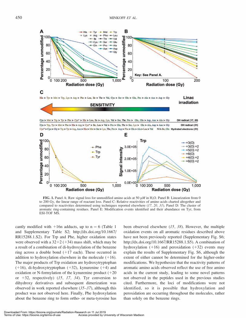
To begin with the simplest possible system, 50-lMsolutions were made of each of the common 20 amino acidsin water. Each solution was irradiated with 0, 100, 200, 500or 1,000 Gy. The products were analyzed using ESI-TOFmass spectrometry with both positive and negative mode(Supplementary Table S2; http://dx.doi.org/10.1667/RR15288.1.S2), and the loss of the unmodified form ofthe amino acids was monitored. This is shown here as apercentage of the control signal per amino acid remainingafter each dose of radiation (Fig. 1A).
For the majority of amino acids, a significant portion ofsignal from the unmodified forms was lost after the 200 Gydose, suggesting their complete conversion to products at orbefore the subsequent 500 Gy dose (Fig. 1A). For themajority of amino acids with unmodified signal remainingafter receiving the 200 Gy dose, the linear rate of lossdisplayed from 0 to 200 Gy then slowed significantly forhigher doses. Concurrent with this, increased production ofmany modifications was lost after exposure to 200 Gy(Supplementary Figs. S1–S3; http://dx.doi.org/10.1667/RR15288.1.S5). Furthermore, in the cases where modifica-tion signals still increased beyond the 200 Gy dose, the ratewas significantly slower (Supplementary Figs. S1–S3).Thus, linear relative rates of dose response, correspondingto their respective radiation sensitivities, were built usingthe data from 0 to 200 Gy for each amino acid, and plottedover this range (Fig. 1B, based on data from SupplementaryTable S2; http://dx.doi.org/10.1667/RR15288.1.S2). Usingthese relative sensitivities, a scale of response was
constructed and compared to published data from otherROS-producing techniques (17, 18, 31) (Fig. 1C). The mostsensitive amino acid was His. Relative to previouslypublished studies (16, 17, 31), the significant response ofHis observed here is unusual and may be specific to thehigh-energy electron beam used. Otherwise, the relativeamino acid dose responses are generally consistent withpreviously reported data (17).
Correlations of sensitivity with side group chemistries andphysicochemical properties of amino acids, includingvolume (32), hydropathy (33) and pKa/pI values, werecalculated (Supplementary Figs. S4 and S5; http://dx.doi.org/10.1667/RR15288.1.S5). The response of the aminoacids containing aromatic R groups produced the mostnotable grouping (Fig. 1D). The strongest correlation ofradiosensitivity level with a physicochemical property waswith molecular volume (R2 ¼ 0.5672; Supplementary Fig.S5), suggesting there may be some dependence on volumefor the relative sensitivities to radiation we observe. Thearomatic ring-containing residues were all highly respon-sive, and exhibited similar dose responses of modification(Fig. 1D). They also all exhibited multiple oxidation events(Fig. 1E and Supplementary Fig. S6; http://dx.doi.org/10.1667/RR15288.1.S5), which we hypothesize to be due toresonance associated with the aromatic rings. No othersignificant correlations between responses or modificationsand chemical properties of amino acids were observed(Supplementary Figs. S4 and S5).
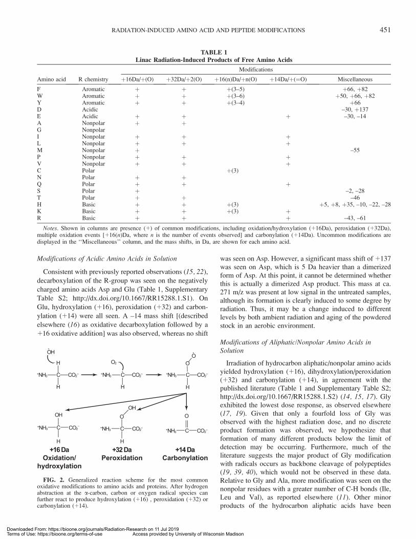
Classical hydroxylation (þ16)(þO) was observed on eachamino acid except for Asp, Cys and Gly (Table 1).Dihydroxylation/peroxidation (þ32)(þ2O) events wereseen on the vast majority of amino acids. Carbonylation(þ14) was observed on less than one half of the aminoacids. For His, Lys and Cys only, adducts of þ48 wereobserved (Table 1 and Supplementary Table S2; http://dx.doi.org/10.1667/RR15288.1.S2). This is likely a combina-tion of þ16 hydroxyl and þ32 dihydroxyl/peroxy productmass shifts. A generalized reaction scheme for thesecommon oxidative modifications is shown in Fig. 2.
With radiation treatment, a number of products of verylow signal with less mass than the unmodified amino acidwere observed for the majority of amino acids (Supple-mentary Table S2; http://dx.doi.org/10.1667/RR15288.1.S2). Despite the abundance of many products, includingclassical oxidation and others with known atomic compo-sition, there was not a strong correlation between thedisappearance of unmodified amino acid variants (orders ofmagnitude) and the appearance of particular products. Thus,the reaction pathways leading to products caused byradiation are likely to be complex, an issue that must beaddressed in future work.
Modifications of Aromatic Amino Acids in Solution
The aromatic ring-containing residues were the mosthighly modified subset of amino acids. Each was signifi-
RADIATION-INDUCED AMINO ACID AND PEPTIDE MODIFICATIONS 449
Downloaded From: https://bioone.org/journals/Radiation-Research on 11 Jul 2019Terms of Use: https://bioone.org/terms-of-use Access provided by University of Wisconsin Madison
cantly modified with þ16n adducts, up to n ¼ 6 (Table 1
and Supplementary Table S2; http://dx.doi.org/10.1667/
RR15288.1.S2). For Trp and Phe, higher oxidation states
were observed with a 32þ2 (þ34) mass shift, which may be
a result of a combination of di-hydroxylation of the benzene
ring across a double bond (þ17 each). These occurred in
addition to hydroxylation elsewhere in the molecule (þ16).
The major products of Trp oxidation are hydroxytryptophan
(þ16), di-hydroxytryptophan (þ32), kynurenine (þ4) and
oxidation or N-formylation of the kynurenine product (þ20
or þ32, respectively) (15, 17, 34). Tyr conversion to
dihydroxy derivatives and subsequent dimerization was
observed in work reported elsewhere (35–37), although this
product was not observed here. Finally, Phe hydroxylation
about the benzene ring to form ortho- or meta-tyrosine has
been observed elsewhere (15, 38). However, the multiple
oxidation events on all aromatic residues described above
have not been previously reported (Supplementary Fig. S6;
http://dx.doi.org/10.1667/RR15288.1.S5). A combination of
hydroxylation (þ16) and peroxidation (þ32) events may
explain the results of Supplementary Fig. S6, although the
extent of either cannot be determined for the higher-order
modifications. We hypothesize that the reactivity patterns of
aromatic amino acids observed reflect the use of free amino
acids in the current study, leading to some novel patterns
not observed in the peptides used in the previous studies
cited. Furthermore, the loci of modifications were not
identified, so it is possible that hydroxylation and
peroxidation are occurring throughout the molecules, rather
than solely on the benzene rings.
FIG. 1. Panel A: Raw signal loss for unmodified amino acids at 50 lM in H2O. Panel B: Linearization from 0to 200 Gy, the linear range of reactant loss. Panel C: Relative reactivities of amino acids charted altogether andcompared to reactivities determined using techniques reported elsewhere (17, 20, 31). Panel D: The cluster ofaromatic ring-containing residues. Panel E: Modification events identified and their abundance on Tyr, fromESI-TOF MS.
450 MINKOFF ET AL.
Downloaded From: https://bioone.org/journals/Radiation-Research on 11 Jul 2019Terms of Use: https://bioone.org/terms-of-use Access provided by University of Wisconsin Madison
Modifications of Acidic Amino Acids in Solution
Consistent with previously reported observations (15, 22),
decarboxylation of the R-group was seen on the negatively
charged amino acids Asp and Glu (Table 1, Supplementary
Table S2; http://dx.doi.org/10.1667/RR15288.1.S1). On
Glu, hydroxylation (þ16), peroxidation (þ32) and carbon-
ylation (þ14) were all seen. A –14 mass shift [(described
elsewhere (16) as oxidative decarboxylation followed by aþ16 oxidative addition] was also observed, whereas no shift
was seen on Asp. However, a significant mass shift of þ137was seen on Asp, which is 5 Da heavier than a dimerized
form of Asp. At this point, it cannot be determined whetherthis is actually a dimerized Asp product. This mass at ca.271 m/z was present at low signal in the untreated samples,although its formation is clearly induced to some degree byradiation. Thus, it may be a change induced to differentlevels by both ambient radiation and aging of the powderedstock in an aerobic environment.
Modifications of Aliphatic/Nonpolar Amino Acids inSolution
Irradiation of hydrocarbon aliphatic/nonpolar amino acidsyielded hydroxylation (þ16), dihydroxylation/peroxidation(þ32) and carbonylation (þ14), in agreement with thepublished literature (Table 1 and Supplementary Table S2;http://dx.doi.org/10.1667/RR15288.1.S2) (14, 15, 17). Glyexhibited the lowest dose response, as observed elsewhere
(17, 19). Given that only a fourfold loss of Gly wasobserved with the highest radiation dose, and no discreteproduct formation was observed, we hypothesize thatformation of many different products below the limit ofdetection may be occurring. Furthermore, much of theliterature suggests the major product of Gly modificationwith radicals occurs as backbone cleavage of polypeptides
(19, 39, 40), which would not be observed in these data.Relative to Gly and Ala, more modification was seen on thenonpolar residues with a greater number of C-H bonds (Ile,Leu and Val), as reported elsewhere (11). Other minorproducts of the hydrocarbon aliphatic acids have been
TABLE 1Linac Radiation-Induced Products of Free Amino Acids
Amino acid R chemistry
Modifications
þ16Da/þ(O) þ32Da/þ2(O) þ16(n)Da/þn(O) þ14Da/þ(¼O) Miscellaneous
F Aromatic þ þ þ(3–5) þ66, þ82W Aromatic þ þ þ(3–6) þ50, þ66, þ82Y Aromatic þ þ þ(3–4) þ66D Acidic –30, þ137E Acidic þ þ þ –30, –14A Nonpolar þ þG NonpolarI Nonpolar þ þ þL Nonpolar þ þ þM Nonpolar þ –55P Nonpolar þ þ þV Nonpolar þ þ þC Polar þ(3)N Polar þ þQ Polar þ þ þS Polar þ –2, –28T Polar þ þ –46H Basic þ þ þ(3) þ5, þ8, þ35, –10, –22, –28K Basic þ þ þ(3) þR Basic þ þ þ –43, –61
Notes. Shown in columns are presence (þ) of common modifications, including oxidation/hydroxylation (þ16Da), peroxidation (þ32Da),multiple oxidation events [þ16(n)Da, where n is the number of events observed] and carbonylation (þ14Da). Uncommon modifications aredisplayed in the ‘‘Miscellaneous’’ column, and the mass shifts, in Da, are shown for each amino acid.
FIG. 2. Generalized reaction scheme for the most commonoxidative modifications to amino acids and proteins. After hydrogenabstraction at the a-carbon, carbon or oxygen radical species canfurther react to produce hydroxylation (þ16) , peroxidation (þ32) orcarbonylation (þ14).
RADIATION-INDUCED AMINO ACID AND PEPTIDE MODIFICATIONS 451
Downloaded From: https://bioone.org/journals/Radiation-Research on 11 Jul 2019Terms of Use: https://bioone.org/terms-of-use Access provided by University of Wisconsin Madison
observed under a variety of conditions, but none were foundhere (31). Modification of þ16, þ32 and þ14 was seen onPro. The þ16 mass shift on Pro could represent either 5-hydroxyproline or glutamyl semialdehyde, as the twospecies are reportedly in equilibrium (40–43). Similarly, amass shift of þ32 corresponds to either dihydroxylation,peroxidation or conversion to Glu. The þ14 product isconversion to pyroglutamic acid via carbonylation (15, 17).
Modifications of Polar Amino Acids in Solution
Amino acids containing polar side chains yielded a widervariety of products than those in the nonpolar group (Table1 and Supplementary Table S2; http://dx.doi.org/10.1667/RR15288.1.S2). Asn, Gln, Ser and Thr have not been asextensively studied as the other amino acids. Hydroxylationand carbonylation have been reported for Asn and Gln,although product formation is low in signal (16). In additionto hydroxylation on both Asn and Gln, we observeddihydroxylation/peroxidation (þ32), the first report of such,as well as carbonylation (þ14) on Gln. No other Asn or Glnproducts were observed. Both Ser and Thr were hydroxyl-ated (þ16), whereas radiation treatment of Thr also yieldeda dihydroxylation/peroxidation (þ32) product. Consistentwith previously published data, a mass shift of –2 wasobserved on Ser, corresponding to hydroxyl side chainconversion to a carbonyl group (16). The strongest signalswere mass shifts of –28 for Ser and –46 for Thr,corresponding to the loss of CO and C2OH6, respectively.We hypothesize that these alterations correspond to loss ofthe respective side chains, via an unknown mechanism.
Modifications of Sulfur-Containing Amino Acids in Solution
In contrast to previously reported studies (16, 17, 31), thesulfur side chain-containing amino acids Met and Cys werenot observed to be the most responsive. Whereas Met wasone of the more sensitive residues, Cys was close to themiddle of all of the amino acids with respect to sensitivity(Fig. 1C). We identified a mass shift of þ16 on Met to formmethionine sulfoxide, corresponding to the classical oxida-tive product, which has been previously reported manytimes (16, 17, 31, 44, 45). Sulfoxide formation is stronglyinduced at a radiation dose of 100 Gy, after which the signalremains stable at 200 Gy but drops significantly thereafter.In negative mode, a strong, dose-dependent signal thatpersisted to 1,000 Gy was observed for methanesulfonicacid (–55; Supplementary Table S2). Given the highlyoxidative environment created upon irradiation, we positthis is a cleavage product of unmodified Met andmethionine sulfoxide. We hypothesize that radiationtriggers a quick conversion of Met to methionine sulfoxide,after which the main byproduct is methanesulfonic acid.
Cys was less sensitive than previously reported (17, 31)(Fig. 1A). With Cys, the major product formed is cysteinesulfonic acid, the triply oxidized Cys species (þ48). Thismodification has been observed on the tripeptide GCG and
a much longer peptide (16) as well as free Cys (46, 47).Additionally, we observed formation of cystine (Cys*), thedisulfide-linked, dimeric form of Cys (Table 1 andSupplementary Table S2; http://dx.doi.org/10.1667/RR15288.1.S2). The proposed mechanism for Cys* forma-tion is hydrogen abstraction from the sulfhydryl groupfollowed by subsequent radical dimerization (31, 46, 47).The abundance of Cys* initially increases at 100 Gy doseand decreases at higher doses, presumably as other productsare formed. Xu and Chance noted this phenomenonpreviously in experiments to irradiate a disulfide linkeddipeptide (GC)2. They observed free dipeptide produced viadisulfide cleavage (16). The major product was monomericGC-sulfonic acid, as observed here. A number of additionalproducts, including þ16 and þ32, were produced from thedipeptide as reported but not observed here with the freeamino acid.
Modifications of Basic Amino Acids in Solution
Finally, the basic amino acids were all among the mostsensitive (Fig. 1C). Their modification profiles also followthis trend. Mass adducts up to þ48 were observed on bothLys and His. Arg produced unique products, and manyproducts of His were seen (Tables 1 and SupplementaryTable S2; http://dx.doi.org/10.1667/RR15288.1.S2). Con-version of Arg to glutamic-semialdehyde (–43) has beennoted multiple times, both in radiolysis and metal-catalyzedoxidation (21, 41, 43), and was also observed here. We alsoidentified a –61 product of Arg, the molecular compositionof which is unknown. On His, mass shifts of þ5, þ8, þ35,–10, –22 and –28 were observed, in addition to mass shiftsof þ16n up to n¼ 3. Oxidation to form 2-oxohistidine andthe –22 conversion to Asp were noted many years agoelsewhere (48), and the þ5 and –10 products have beenreported more recently (17, 21). Mass shifts within thisdataset of þ8, þ35 and –28 are also present (SupplementaryTable S2). While studies have been published examiningoxidation pathways of His (49–52), none of the reportedintermediates are consistent with the masses of unknowncomposition identified here.
Radiation Response of Amino Acids in a Peptide Context
A second goal of this study was to examine the effect ofradiation on amino acids when the a-amino and a-carboxylgroups are incorporated into peptide bonds. Thus, we nextturned to tripeptides. Gly, the least reactive residue, waschosen to provide the peptide linkages. This allowed us toisolate, as much as possible, the effects of the peptide bondsthemselves. All 20 tripeptides were synthesized as G-X-G,where X is an amino acid. For 15 of the 20 tripeptides,radiation treatment from 100 to 1,000 Gy was performed,identical to the procedure used for the free amino acids, inwater at a concentration of 50 lM. Both technical(analyzing the same irradiated sample multiple times on amass spectrometer) and experimental (re-irradiating with the
452 MINKOFF ET AL.
Downloaded From: https://bioone.org/journals/Radiation-Research on 11 Jul 2019Terms of Use: https://bioone.org/terms-of-use Access provided by University of Wisconsin Madison
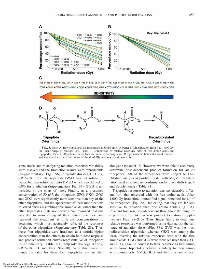
same stocks and re-analyzing radiation response) variabilitywere assayed and the irradiation results were reproducible(Supplementary Fig. S6; http://dx.doi.org/10.1667/RR15288.1.S5). The tripeptide GWG was not soluble inwater, but was solubilized into DMSO which was diluted to0.8% for irradiation (Supplementary Fig. S7). GWG is notincluded in the chart of rates. Finally, at a presumedconcentration of 50 lM, the tripeptides GPG, GKG, GQGand GDG were significantly more sensitive than any of theother tripeptides, and the appearance of their modificationsfollowed curves resembling free amino acids, rather than theother tripeptides (data not shown). We reasoned that thiswas due to misreporting of their initial quantities, andrepeated the treatment at different concentrations todetermine which most accurately reflected the responsesof the other tripeptides (Supplementary Table S3). Thus,these four tripeptides were irradiated at a tenfold higherconcentration than the others, to obtain both dose responseand product formation curves representative of tripeptides(Supplementary Table S1; http://dx.doi.org/10.1667/RR15288.1.S1 and Figs. S8–S10). With this caveat inmind, the rates for these four tripeptides are included
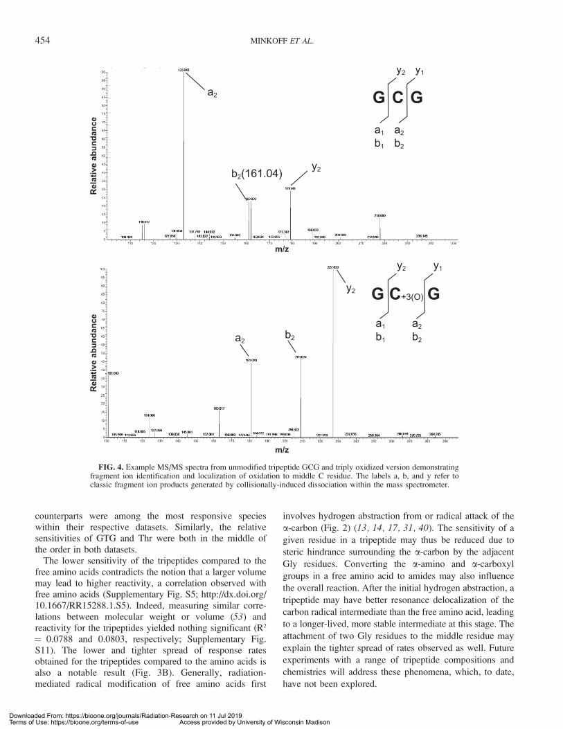
alongside the other 15. However, we were able to accuratelydetermine dose-dependent product formation for all 20tripeptides. All of the tripeptides were subject to ESI-Orbitrap analysis in positive mode, with MS/MS fragmen-tation used as secondary confirmation for mass shifts (Fig. 4and Supplementary Table S1).
Tripeptide response to radiation was considerably differ-ent from that observed with the free amino acids. After1,000 Gy irradiation, unmodified signal remained for all ofthe tripeptides (Fig. 2A), indicating that they are far lesssensitive to radiation than free amino acids (Fig. 1A).Reactant loss was dose-dependent throughout the range ofexposures (Fig. 3A), as was product formation (Supple-mentary Figs. S8–S10). Thus, linear fitting to determinerelative responses was performed using data across the fullrange of radiation doses (Fig. 3B). GVG was the mostradiosensitive tripeptide, whereas GKG was among theleast, inverting the relative responses seen with the freeamino acids. GAG and GGG were more sensitive than GYGand GFG, again in contrast to their behavior as free aminoacids. Some tripeptides behaved similarly to their aminoacid counterparts. GMG, GHG and their free amino acid
FIG. 3. Panel A: Raw signal loss for tripeptides at 50 lM in H2O. Panel B: Linearization from 0 to 1,000 Gy,the linear range of reactant loss. Panel C: Comparison of relative reactivity rates of free amino acids andtripeptides. Panel D: Reaction scheme for C-terminal decarboxylation. R represents the first and second residues,and the sidechain and C-terminus of the third Gly residues are shown in full.
RADIATION-INDUCED AMINO ACID AND PEPTIDE MODIFICATIONS 453
Downloaded From: https://bioone.org/journals/Radiation-Research on 11 Jul 2019Terms of Use: https://bioone.org/terms-of-use Access provided by University of Wisconsin Madison
counterparts were among the most responsive specieswithin their respective datasets. Similarly, the relativesensitivities of GTG and Thr were both in the middle ofthe order in both datasets.
The lower sensitivity of the tripeptides compared to thefree amino acids contradicts the notion that a larger volumemay lead to higher reactivity, a correlation observed withfree amino acids (Supplementary Fig. S5; http://dx.doi.org/10.1667/RR15288.1.S5). Indeed, measuring similar corre-lations between molecular weight or volume (53) andreactivity for the tripeptides yielded nothing significant (R2
¼ 0.0788 and 0.0803, respectively; Supplementary Fig.S11). The lower and tighter spread of response ratesobtained for the tripeptides compared to the amino acids isalso a notable result (Fig. 3B). Generally, radiation-mediated radical modification of free amino acids first
involves hydrogen abstraction from or radical attack of the
a-carbon (Fig. 2) (13, 14, 17, 31, 40). The sensitivity of a
given residue in a tripeptide may thus be reduced due to
steric hindrance surrounding the a-carbon by the adjacent
Gly residues. Converting the a-amino and a-carboxyl
groups in a free amino acid to amides may also influence
the overall reaction. After the initial hydrogen abstraction, a
tripeptide may have better resonance delocalization of the
carbon radical intermediate than the free amino acid, leading
to a longer-lived, more stable intermediate at this stage. The
attachment of two Gly residues to the middle residue may
explain the tighter spread of rates observed as well. Future
experiments with a range of tripeptide compositions and
chemistries will address these phenomena, which, to date,
have not been explored.
FIG. 4. Example MS/MS spectra from unmodified tripeptide GCG and triply oxidized version demonstratingfragment ion identification and localization of oxidation to middle C residue. The labels a, b, and y refer toclassic fragment ion products generated by collisionally-induced dissociation within the mass spectrometer.
454 MINKOFF ET AL.
Downloaded From: https://bioone.org/journals/Radiation-Research on 11 Jul 2019Terms of Use: https://bioone.org/terms-of-use Access provided by University of Wisconsin Madison
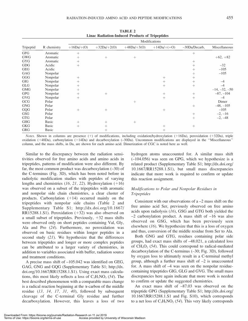
Similar to the discrepancy between the radiation sensi-tivities observed for free amino acids and amino acids intripeptides, patterns of modification were also different. Byfar, the most common product was decarboxylation (–30) ofthe C-terminus (Fig. 3D), which has been noted before inradiolytic modification studies with peptides of varyinglengths and chemistries (16, 21, 22). Hydroxylation (þ16)was observed on a subset of the tripeptides with aromaticand nonpolar side chain chemistries, a clear cluster ofproducts. Carbonylation (þ14) occurred mainly on thetripeptides with nonpolar side chains (Table 2 andSupplementary Table S1; http://dx.doi.org/10.1667/RR15288.1.S1). Peroxidation (þ32) was also observed ona small subset of tripeptides. Previously, þ32 mass shiftswere observed only on short peptides containing Val, Gly,Ala and Pro (24). Furthermore, no peroxidation wasobserved on basic residues within longer peptides in asecond study (21). We hypothesize that the differencesbetween tripeptides and longer or more complex peptidescan be attributed to a larger variety of chemistries, inaddition to variables associated with buffer, radiation sourceand treatment conditions.
A precise mass shift of –105.042 was identified on GEG,GAG, GNG and GQG (Supplementary Table S1; http://dx.doi.org/10.1667/RR15288.1.S1). Using exact mass calcula-tions, this most likely reflects a loss of C3H7NO3 (54). Thebest described phenomenon with a comparable mass changeis a radical reaction beginning at the a-carbon of the middleresidue (13, 14, 17, 31, 40), followed by subsequentcleavage of the C-terminal Gly residue and furtherdecarboxylation. However, this leaves a loss of two
hydrogen atoms unaccounted for. A similar mass shift(–104.056) was seen on GPG, which we hypothesize is arelated product (Supplementary Table S1; http://dx.doi.org/10.1667/RR15288.1.S1), but small mass discrepanciesindicate that more work is required to confirm or updatethis reaction assignment.
Modifications to Polar and Nonpolar Residues inTripeptides
Consistent with our observations of a –2 mass shift on thefree amino acid Ser, previously observed on free aminoacids upon radiolysis (16), GSG and GTG both yielded the–2 carbonylation product. A mass shift of –16 was alsoobserved on GSG, which has been previously seenelsewhere (16). We hypothesize that this is a loss of oxygenand thus, conversion of the middle residue from Ser to Ala.
Both GNG and GTG, residues containing polar sidegroups, had exact mass shifts of –48.021, a calculated lossof CH4O2 (54). This could correspond to radical-mediateddecarboxylation of the C-terminus (–30; Fig. 3D), followedby oxygen loss to ultimately result in a C-terminal methylgroup, although a further mass shift of –2 is unaccountedfor. A mass shift of –4 was seen on the nonpolar residue-containing tripeptides GIG, GLG and GVG. The small massdiscrepancies here again indicate that more work is neededto confirm or update the suggested chemistries.
An exact mass shift of –87.03 was observed on thetripeptide GPG (Supplementary Table S1; http://dx.doi.org/10.1667/RR15288.1.S1 and Fig. S10), which correspondsto a net loss of C3H5NO2 (54). This very likely corresponds
TABLE 2Linac Radiation-Induced Products of Tripeptides
Tripeptid R chemistry
Modifications
þ16Da/þ(O) þ32Da/þ2(O) þ48Da/þ3(O) þ14Da/þ(¼O) –30Da/Decarb, Miscellaneous
GFG Aromatic þ þGWG Aromatic þ þ þ þ62, þ82GYG Aromatic þ þGDG Acidic þ þ –32GEG Acidic þ –105GAG Nonpolar þ –105GGG Nonpolar þ þ þGIG Nonpolar þ þ þ –4GLG Nonpolar þ þ þ –4GMG Nonpolar þ –14, –32, –50GPG Nonpolar þ þ þ þ –87, –104GVG Nonpolar þ þ þ –4GCG Polar þ DimerGNG Polar þ –48, –105GQG Polar þ þ þ –105GSG Polar þ –2, –16GTG Polar þ –2, –48GHG Basic þ þ þGKG Basic þ þ þ þGRG Basic þ þ
Notes. Shown in columns are presence (þ) of modifications, including oxidation/hydroxylation (þ16Da), peroxidation (þ32Da), tripleoxidation (þ48Da), carbonylation (þ14Da) and decarboxylation (–30Da). Uncommon modifications are displayed in the ‘‘Miscellaneous’’column, and the mass shifts, in Da, are shown for each amino acid. Dimerization of CGC is noted here as well.
RADIATION-INDUCED AMINO ACID AND PEPTIDE MODIFICATIONS 455
Downloaded From: https://bioone.org/journals/Radiation-Research on 11 Jul 2019Terms of Use: https://bioone.org/terms-of-use Access provided by University of Wisconsin Madison
to backbone cleavage and loss of all the atoms C-terminal tothe Pro ring, via oxidation of the tripeptide to the 2-pyrrolidone derivative. Backbone cleavage via oxidation ofPro residues has been observed previously elsewhere (42,55), and a mechanism has been defined (56). In this case,such cleavage would result in a mass shift of –86, leaving ahydrogen loss unaccounted for and the need for futureexperiments to confirm this possible similar product.
Modifications to Sulfur-Containing Residues in Tripeptides
Cys has been reported to dimerize to form cystine uponradiolysis via: 1. Thiyl radical formation and reaction withoxygen; 2. Hydroxylated or peroxylated side chaincombination via loss of an H2O2; or 3. Direct combinationof thiyl radicals in conditions lacking oxygen (46, 47, 57–59). Consistent with this, we observed a strong signal at m/z235.06, corresponding to þ2 form of the dimerized cystine-containing tripeptide. Although it was present in the controlsample, radiolysis strongly induced more cystine formationin a dose-dependent fashion. Concurrent with GCGdisulfide dimerization, we identified sulfonic acid (þ48)formation, addition of three oxygens to the sulfur atom.These results are consistent with those reported previouslyby Xu and Chance, in which an identical tripeptide and137Cs irradiation were used (60). In another published study,Xu and Chance identified more oxidative modifications onGCG using MS in negative-mode; specifically, mass shiftsof –16, –34, þ32, þ46, þ64 and þ80 (16). We posit thatthese modifications were not observed in the current studydue to our use of only positive-mode MS analysis for thetripeptides, given that a secondary positive-mode MSanalysis in the same publication identified only the –16mass shift and cystine product formation (16).
The tripeptide GMG was highly modified (Table 2 andSupplementary Table S1; http://dx.doi.org/10.1667/RR15288.1.S1). Mass shifts of þ16, –14, –32, and –50were observed. The sulfoxide product, þ16, was by far thestrongest signal (Supplementary Table S1). The –32 and–14 mass shifts were identified in the previous study by Xuand Chance in which GMG and 137Cs were used (16). The–14 product corresponds to decarboxylation (–30) followedby oxidation (þ16). The –32 product results from loss of amethanesulfinyl group followed by aldehyde formation atthe gamma carbon (16). Exact mass measurements usinghigh-resolution MS confirm that the mass shift of –32.008corresponds precisely to this structural difference. Despitethe reactivity of GMG, decarboxylation of the C-terminuswas not identified. This is likely due to the strong formationof the –14 product, for which the decarboxylation is areaction intermediate. Finally, a product with a mass shift of–50.019 was identified (Supplementary Table S3; http://dx.doi.org/10.1667/RR15288.1.S3), which has not been re-ported before. Reported elemental composition for this massshift is CH6S (54), which we propose is a loss of theseatoms from the Met side chain.
Modifications to Aromatic Residues in Tripeptides
On GYG and GFG, only hydroxylation (þ16) anddecarboxylation (–30) were identified (Table 2). Thetripeptide GWG could only be solubilized in 100%dimethyl sulfoxide (DMSO) due to its strong hydrophobic-ity. When diluted in dH2O to a working concentration of 50lM, this resulted in 0.8% DMSO being present duringirradiation. To benchmark possible DMSO-dependentdifferences in response rate, GIG and GTG were alsoirradiated in 0.8% DMSO and the response curves werecompared to their respective curves without DMSO(Supplementary Table S4; http://dx.doi.org/10.1667/RR15288.1.S4 and Fig. S7; http://dx.doi.org/10.1667/RR15288.1.S5). Under these conditions, GIG and GTGwere similar in reactivity rate to their rate without addedDMSO. DMSO can modify Trp to form 2-hydroxy-Trp inthe presence of HCl as well as DMSO (61). With thesecaveats in mind, the sensitivity of GWG in DMSO wasmuch greater than that of the remaining tripeptides. Signalwas lost at 200 Gy irradiation, a result more similar to themore highly-reactive free amino acids than to the tripeptides(Table 2, Supplementary Table S1; http://dx.doi.org/10.1667/RR15288.1.S1 and Fig. S7). Mass shifts of þ16n wereidentified on GWG, up to n¼ 3, similar to that observed onfree Trp. These likely include the products hydroxytrypto-phan (þ16), dihydroxytryptophan (þ32) and N-formylky-nurenine (þ32) (34, 48). Two unique products wereidentified on GWG: mass shifts of þ62 and þ82. Wehypothesize that þ62 is a result of four þ16n oxidativeevents concurrent with carbon-carbon double-bond forma-tion somewhere in the molecule (–2). In contrast, þ82 maycorrespond to a product in which there are five þ16 eventsin total as well as hydrogen atom addition across a doublebond (þ2). Xu and Chance also identified mass shifts ofþ16n up to n ¼ 5 when the tripeptide GWG underwentradiolysis (17). Our identification of þ16n mass shifts (withminor adducts or losses) up to n¼ 5 here corroborates thisdata. In general, our data highlight the fact that one mustpay attention to all chemical species present in the mixturewhen irradiation is performed, as even small amounts ofsolvents or buffers may make a large difference in theresults obtained.
CONCLUSIONS
We have begun to study, in a methodical fashion, theeffect of Linac-produced radiation on amino acids andtripeptides, with the goal of elucidating the differencesbetween simplified systems and systems of more complex-ity when exposed to ionizing radiation. The ultimate goalwas to infer testable biological implications from datasetssuch as that produced here. However, there is a need forfundamental groundwork as the basis for future studies. Inthis study, novel mass shifts were observed. In contrast,modifications observed in previously published studies
456 MINKOFF ET AL.
Downloaded From: https://bioone.org/journals/Radiation-Research on 11 Jul 2019Terms of Use: https://bioone.org/terms-of-use Access provided by University of Wisconsin Madison
were not seen here. The results described herein underscorethe notion that radiosensitivity of amino acids is highlycontextual. Incorporating amino acids into a simpletripeptide substantially reduces their sensitivities, a resultobserved here as well as in comparisons of these data tomany other studies. Additionally, modification patternsobtained by exposing free amino acids to radiation may notreadily be extrapolated to irradiation of the same amino acidresidues within peptides. This claim is supported by bothquantitative (dose-response rates) and qualitative (chemicalmodifications) variability observed between even the simplesystems used in these experiments. Furthermore, variabilityin amino acid response is also evident when comparingpublished studies, likely due to differences in solutionconditions, radiation source or the exact amino acidsequence of the peptides used. Future studies will beundertaken to further examine these differences, andelucidate the effect of radiation on biological molecules inhighly controlled and reproducible conditions. Overall, theresults begin to provide a basis for identifying radiation-related protein damage on a proteomic scale.
SUPPLEMENTARY INFORMATION
Table S1. Tripeptide radiation sensitivity and modifica-tion patterns, collected using electrospray ionization-orbi-trap mass spectrometry. Each tripeptide is shown on adifferent sheet. Charts are extracted ion chromatograms forthe specified species for each dosage, used for thequantification tables shown below them. The extractedmass is displayed above the respective table per species.The data here were used to build the charts forSupplementary Figs. S8–10. All masses displayed are m/z(Da/charge state).
Table S2. Free amino acid radiosensitivity and modifi-cation patterns, collected using electrospray ionization-timeof flight mass spectrometry. Each amino acid is shown bothfor positive and negative mode on individual sheets. Beloware the automatically extracted masses, their retention times,peak heights, and identified adducts. Above, values forquantification were built on extracted ion chromatograms ofthe specified masses (the example of the unmodified isgiven in every case). In many cases, shown in bolded red,the automatic peak picking informed the masses tomanually extract. Masses that were extracted and quantifiedabove but not bolded in red below were chosen based onliterature reports, cited in the main text. The data here wereused to build charts for Supplementary Figs. S1–S3. Allmasses displayed are m/z (Da/charge state), though chargestates of only þ1 or –1 were observed.
Table S3. Data used for low abundance tripeptides,including GQG, GDG, GPG and GKG. Each was tested byconcentration from stock exposure 23, 53 and 103. For allof these, the tenfold concentration was used as representa-tive, and the data from those exposures are used in Figs.3A–C. All masses displayed are m/z (Da/charge state).
Table S4. Data used for tripeptides in DMSO figure. Eachof the three tripeptides is shown on a separate sheet.
Figs. S1–S3. Product formation over a range of 0–1,000Gy for the free amino acids. Known modifications areshown as atomic adducts, whereas unknown modificationsare shown as mass shifts. The y-axes are unitless, and areraw signal intensities for the listed modifications.
Fig. S4. Clustering and ESI-TOF data demonstratingsensitivities of nonpolar, polar, charged and aromatic freeamino acids.
Fig. S5. Correlations of sensitivities (slope over linearrange of signal loss for unmodified versions, all x-axes)with physicochemical properties for free amino acids.
Fig. S6. Reproducibility of experiments. Top left: Threetechnical replicates (same sample, analyzed thrice withsame instrumental method, processing, etc. postirradiation)of GKG at low concentration. Top right and bottom:Experimental replicates (samples independently made up,exposed and analyzed). Top right: GMG twice indepen-dently diluted to 50 lM, irradiated and analyzed, approx-imately one month apart. Bottom left: GVG twiceindependently diluted to 50 lM, irradiated and analyzed,approximately one month apart.
Fig. S7. Comparison of sensitivity for two tripeptides indH2O and 0.8% DMSO. All tripeptides are 50 lM.
Figs. S8–S10. Product formation over a range of 0–1,000Gy for the tripeptides. Known modifications are shown asatomic adducts, whereas unknown modifications are shownas mass shifts. The y-axes are unitless, and are raw signalintensities for the listed modifications.
Fig. S11. Correlations of relative sensitivities (slope overlinear range of signal loss for unmodified versions) withphysicochemical properties for free amino acids.
ACKNOWLEDGMENTS
We gratefully acknowledge funding provided to BBM, GS, STB, MMC
and MRS by the U.S. Department of Defense (Homeland DTRA grant no.
HDTRA1-16-1-0049) and to STB and MMC by the National Institutes of
Health (NIH grant no. GM112575). Funding was also provided to STB by
the Morgridge Biotechnology Fellowship from the Vice Chancellor’s
Office for Research and Graduate Education and the UW Biotechnology
Center.
Received: November 12, 2018; accepted: February 13, 2019; published
online: March 8, 2019
REFERENCES
1. Daly MJ. A new perspective on radiation resistance based onDeinococcus radiodurans. Nat Rev Microbiol 2009; 7:237–45.
2. Krisko A, Radman M. Biology of extreme radiation resistance: theway of Deinococcus radiodurans. Cold Spring Harb Perspect Biol2013; 5.
3. Morgan MA, Lawrence TS. Molecular pathways: overcomingradiation resistance by targeting DNA damage response pathways.Clin Cancer Res 2015; 21:2898–904.
4. Hashimoto T, Kunieda T. DNA protection protein, a novelmechanism of radiation tolerance: lessons from tardigrades. Life(Basel) 2017; 7.
RADIATION-INDUCED AMINO ACID AND PEPTIDE MODIFICATIONS 457
Downloaded From: https://bioone.org/journals/Radiation-Research on 11 Jul 2019Terms of Use: https://bioone.org/terms-of-use Access provided by University of Wisconsin Madison
5. Jung KW, Lim S, Bahn YS. Microbial radiation-resistancemechanisms. J Microbiol 2017; 55:499–507.
6. Ranawat P, Rawat S. Radiation resistance in thermophiles:mechanisms and applications. World J Microbiol Biotechnol2017; 33:112.
7. Shuryak I, Matrosova VY, Gaidamakova EK, Tkavc R, GrichenkoO, Klimenkova P, et al. Microbial cells can cooperate to resisthigh-level chronic ionizing radiation. PLoS One 2017;12:e0189261.
8. Jung KW, Yang DH, Kim MK, Seo HS, Lim S, Bahn YS.Unraveling fungal radiation resistance regulatory networks throughthe genome-wide transcriptome and genetic analyses of crypto-coccus neoformans. MBio 2016; 7.
9. Anno GH, Young RW, Bloom RM, Mercier JR. Dose responserelationships for acute ionizing-radiation lethality. Health Phys2003; 84:565–75.
10. Islam MT. Radiation interactions with biological systems. Int JRadiat Biol 2017; 93:487–93.
11. Davies MJ. The oxidative environment and protein damage.Biochim Biophys Acta 2005; 1703:93–109.
12. Davies MJ. Reactive species formed on proteins exposed to singletoxygen. Photochem Photobiol Sci 2004; 3:17–25.
13. Davies KJ, Delsignore ME, Lin SW. Protein damage anddegradation by oxygen radicals. II. Modification of amino acids.J Biol Chem 1987; 262:9902–7.
14. Stadtman ER. Oxidation of free amino acids and amino acidresidues in proteins by radiolysis and by metal-catalyzed reactions.Annu Rev Biochem 1993; 62:797–821.
15. Stadtman ER, Levine RL. Free radical-mediated oxidation of freeamino acids and amino acid residues in proteins. Amino Acids2003; 25:207–18.
16. Xu G, Chance MR. Radiolytic modification of sulfur-containingamino acid residues in model peptides: fundamental studies forprotein footprinting. Anal Chem 2005; 77:2437–49.
17. Xu G, Chance MR. Hydroxyl radical-mediated modification ofproteins as probes for structural proteomics. Chem Rev 2007;107:3514–43.
18. Benon H.J. Bielski DEC, Alberta B. Ross, Ravindra L. Arudi.Reactivity of HO2/O�2 radicals in aqueous solution. J Phys ChemRef Data 1985; 14:1041–100.
19. George V. Buxton CLG, W. Phillips Helman, Alberta B. Ross.Critical review of rate constants for reactions of hydrated electrons,hydrogen atoms and hydroxyl radicals (�OH/�O�) in aqueoussolution. J Phys Chem Ref Data 1988; 17:513–886.
20. Kaur P, Kiselar JG, Chance MR. Integrated algorithms for high-throughput examination of covalently labeled biomolecules bystructural mass spectrometry. Anal Chem 2009; 81:8141–9.
21. Xu G, Takamoto K, Chance MR. Radiolytic modification of basicamino acid residues in peptides: probes for examining protein-protein interactions. Anal Chem 2003; 75:6995–7007.
22. Xu G, Chance MR. Radiolytic modification of acidic amino acidresidues in peptides: probes for examining protein-proteininteractions. Anal Chem 2004; 76:1213–21.
23. Saladino J, Liu M, Live D, Sharp JS. Aliphatic peptidylhydroperoxides as a source of secondary oxidation in hydroxylradical protein footprinting. J Am Soc Mass Spectrom 2009;20:1123–6.
24. Morgan PE, Pattison DI, Davies MJ. Quantification of hydroxylradical-derived oxidation products in peptides containing glycine,alanine, valine, and proline. Free Radic Biol Med 2012; 52:328–39.
25. De Meutter P, Camps J, Delcloo A, Termonia P. Sourcelocalisation and its uncertainty quantification after the third DPRKnuclear test. Sci Rep 2018; 8:10155.
26. Williams M, Sizemore DC. Biologic, chemical, and radiationterrorism review. Treasure Island, FL: StatPearls; 2018.
27. Jorgensen TJ. Predicting the public health consequences of anuclear terrorism attack: drawing on the experiences of Hiroshimaand Fukushima. Health Phys 2018; 115:121–5.
28. Smirnova OA, Cucinotta FA. Dynamical modeling approach torisk assessment for radiogenic leukemia among astronauts engagedin interplanetary space missions. Life Sci Space Res (Amst) 2018;16:76–83.
29. Jandial R, Hoshide R, Waters JD, Limoli CL. Space-brain: Thenegative effects of space exposure on the central nervous system.Surg Neurol Int 2018; 9:9.
30. Bruckbauer ST, Trimarco JD, Martin J, Senn KA, Schackwitz W,Lipzen A, et al. Experimental evolution of extreme resistance toionizing radiation in Escherichia coli after 50 cycles of selection. JBacteriol 2019. (DOI: 10.1128/JB.00784-18)
31. Davies MJ, Dean RT. Radical-mediated protein oxidation. NewYork: Oxford University Press; 1997.
32. Zamyatnin AA. Protein volume in solution. Prog Biophys MolBiol 1972; 24:107–23.
33. Kyte J, Doolittle RF. A simple method for displaying thehydropathic character of a protein. J Mol Biol 1982; 157:105–32.
34. Finley EL, Dillon J, Crouch RK, Schey KL. Identification oftryptophan oxidation products in bovine a-crystallin. Protein Sci1998; 7:2391–7.
35. Heinecke JW, Li W, Daehnke HL 3rd, Goldstein JA. Dityrosine, aspecific marker of oxidation, is synthesized by the myeloperox-idase-hydrogen peroxide system of human neutrophils andmacrophages. J Biol Chem 1993; 268:4069–77.
36. Huggins TG, Wells-Knecht MC, Detorie NA, Baynes JW, ThorpeSR. Formation of o-tyrosine and dityrosine in proteins duringradiolytic and metal-catalyzed oxidation. J Biol Chem 1993;268:12341–7.
37. Marquez LA, Dunford HB. Kinetics of oxidation of tyrosine anddityrosine by myeloperoxidase compounds I and II. Implicationsfor lipoprotein peroxidation studies. J Biol Chem 1995;270:30434–40.
38. Maskos Z, Rush JD, Koppenol WH. The hydroxylation ofphenylalanine and tyrosine: a comparison with salicylate andtryptophan. Arch Biochem Biophys 1992; 296:521–9.
39. Easton CJ. Free-radical reactions in the synthesis of alpha-aminoacids and derivatives. Chem Rev 1997; 97:53–82.
40. Garrison WM. Reaction mechanisms in the radiolysis of peptides,polypeptides, and proteins. Chem Rev 1987; 87:381–98.
41. Amici A, Levine RL, Tsai L, Stadtman ER. Conversion of aminoacid residues in proteins and amino acid homopolymers tocarbonyl derivatives by metal-catalyzed oxidation reactions. JBiol Chem 1989; 264:3341–6.
42. Dean RT, Wolff SP, McElligott MA. Histidine and proline areimportant sites of free radical damage to proteins. Free Radic ResCommun 1989; 7:97–103.
43. Requena JR, Chao CC, Levine RL, Stadtman ER. Glutamic andaminoadipic semialdehydes are the main carbonyl products ofmetal-catalyzed oxidation of proteins. Proc Natl Acad Sci U S A2001; 98:69–74.
44. Li S, Schoneich C, Borchardt RT. Chemical pathways of peptidedegradation. VIII. Oxidation of methionine in small modelpeptides by prooxidant/transition metal ion systems: influence ofselective scavengers for reactive oxygen intermediates. Pharm Res1995; 12:348–55.
45. Vogt W. Oxidation of methionyl residues in proteins: tools,targets, and reversal. Free Radic Biol Med 1995; 18:93–105.
46. Armstrong DA. Applications of pulse radiolysis for the study ofshort-lived sulphur species. In: Chatgilialoglu C, Asmus, K-D,editors. Sulfur-centered reactive intermediates in chemistry andbiology. New York: Plenum Press; 1990. p. 121–34.
47. von Sonntag C. Free-radical reactions involving thiols anddisulphides. In: Chatgilialoglu C, Asmus, K-D, editors. Sulfur-
458 MINKOFF ET AL.
Downloaded From: https://bioone.org/journals/Radiation-Research on 11 Jul 2019Terms of Use: https://bioone.org/terms-of-use Access provided by University of Wisconsin Madison
centered reactive intermediates in chemistry and biology. NewYork: Plenum Press; 1990. p. 359–66.
48. Stadtman ER. Role of oxidized amino acids in protein breakdownand stability. In: Klinman JP, editor. Methods in enzymology.Redox-active amino acids in biology, Volume 258. Elsevier; 1995.
49. Hawkins CL, Davies, M.J. Generation and propagation of radicalreactions on proteins. Biochim Biophys Acta 2001; 1504:196–219.
50. Uchida K, Kawakishi S. Selective oxidation of imidazole ring inhistidine residues by the ascorbic acid-copper ion system. BiochemBiophys Res Commun 1986; 138:659–65.
51. Rao PS, Simic, M., Hayon, E. Pulse radiolysis study of imidazoleand histidine in water. J Phys Chem 1975; 79.
52. Tomita M, Masachika I, Tyunosin U. Sensitized photooxidation ofhistidine and its derivatives. Products and mechanism of thereaction. Biochemistry 1969; 8:5149–60.
53. Harpaz Y, Gerstein M, Chothia C. Volume changes on proteinfolding. Structure 1994; 2:641–9.
54. Patiny L, Borel A. ChemCalc: a building block for tomorrow’schemical infrastructure. J Chem Inf Model 2013; 53:1223–8.
55. Schuessler H, Schilling K. Oxygen effect in the radiolysis ofproteins. Part 2. Bovine serum albumin. Int J Radiat Biol RelatStud Phys Chem Med 1984; 45:267–81.
56. Uchida K, Kato Y, Kawakishi S. A novel mechanism for oxidativecleavage of prolyl peptides induced by the hydroxyl radical.Biochem Biophys Res Commun 1990; 169:265–71.
57. Lal M. Radiation induced oxidation of sulphydryl molecules inaqueous solutions. A comprehensive review. Radiat Phys Chem1994; 43:595–611.
58. Dewey DL, Beecher J. Interconversion of cystine and cysteineinduced by x-rays. Nature 1965; 206:1369–70.
59. Owen TC, Brown MT. Radiolytic oxidation of cysteine. J OrgChem 1969; 34:1161–2.
60. Xu G, Chance MR. Radiolytic modification and reactivity ofamino acid residues serving as structural probes for proteinfootprinting. Anal Chem 2005; 77:4549–55.
61. Savige WE, Fontana A. Oxidation of tryptophan to oxindolylala-nine by dimethyl sulfoxide-hydrochloric acid. Selective modifica-tion of tryptophan containing peptides. Int J Pept Protein Res 1980;15:285–97.
62. Almond PR, Biggs PJ, Coursey BM, Hanson WF, Huq MS, NathR, et al. AAPM’s TG-51 protocol for clinical reference dosimetryof high-energy photon and electron beams. Med Phys 1999;26:1847–70.
RADIATION-INDUCED AMINO ACID AND PEPTIDE MODIFICATIONS 459
Downloaded From: https://bioone.org/journals/Radiation-Research on 11 Jul 2019Terms of Use: https://bioone.org/terms-of-use Access provided by University of Wisconsin Madison