Embed Size (px)
Citation preview

1

2

3

4
FELLOWSHIP COURSE IN BASIC ECHOCARDIOGRAPHY
1. Proper name of the certificate course: Fellowship Course in Basic Echocardiography
2. Duration of the course: 12 months
3. Commencement of Course: 17th January 2016
4. Intake capacity: 50 - 75 candidates per year
5. Complete curriculum of the course Appendix I
6. Teaching scheme: Total Time & periods Through 15 modules throughout the year followed by an internal assessment combined 100 marks for internal assessment
7. Eligibility criteria for admission:
Post one year training in:-
o DM/FNB/PDCC/FIACTA/FTEE
o Critical care – DM/FNB/FICCM
o DM/FNB: Pediatrics/neonatology
o DM/FNB: Pulmonary medicine
o DM/FNB: Cardiac Surgery
Post 2 year MD Anaesthesia with atleast one year experience in a cardiac setup is also eligible.
8. FACULTY REQUIRED WITH THEIR QUALIFICATIONS AND EXPERIENCE:
Faculty should have DM/DNB Cardiology degree or similar degree in cardiac anaesthesia
At least 15 years of teaching experience as faculty.
9. SCHEME OF EXAMINATION IN DETAILS:
(Number of question papers, Number of marks to each question paper, Duration of question paper,
practical examination etc.)
Theory paper: See Point No 16 below
Practical examination: See Point No 16 below
10. Infrastructure required for conducting the course.
INTENSIVE CARE UNITS:
Medical Neuro Intensive Care Unit
Intensive Coronary Care Unit
Dialysis Unit
Surgical ICU
Emergency Medicine ICU
5
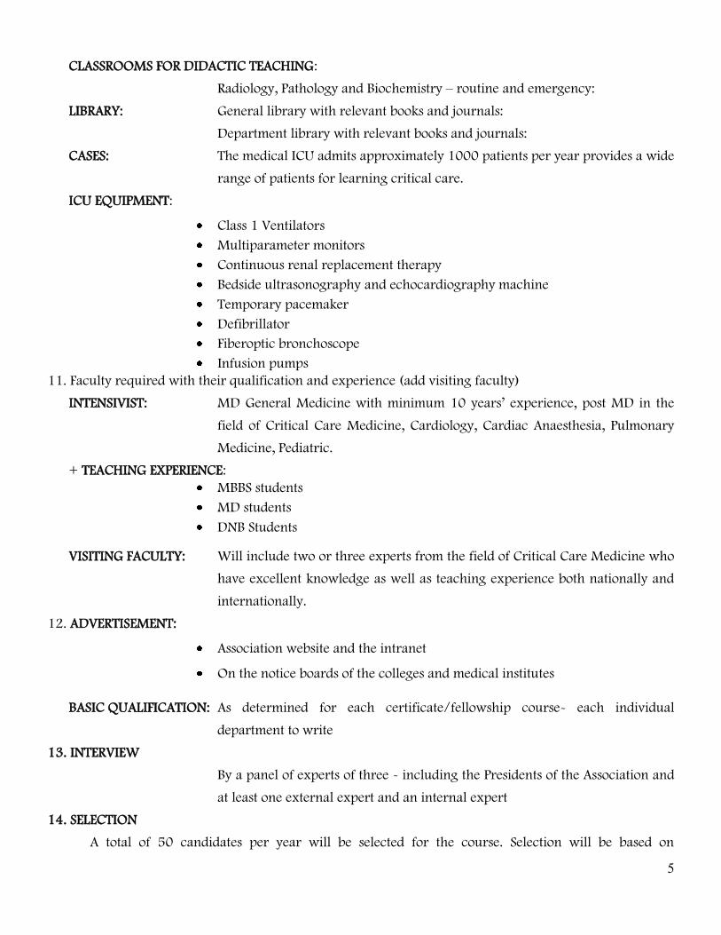
CLASSROOMS FOR DIDACTIC TEACHING:
Radiology, Pathology and Biochemistry – routine and emergency:
LIBRARY: General library with relevant books and journals:
Department library with relevant books and journals:
CASES: The medical ICU admits approximately 1000 patients per year provides a wide
range of patients for learning critical care.
ICU EQUIPMENT:
Class 1 Ventilators
Multiparameter monitors
Continuous renal replacement therapy
Bedside ultrasonography and echocardiography machine
Temporary pacemaker
Defibrillator
Fiberoptic bronchoscope
Infusion pumps
11. Faculty required with their qualification and experience (add visiting faculty)
INTENSIVIST: MD General Medicine with minimum 10 years’ experience, post MD in the
field of Critical Care Medicine, Cardiology, Cardiac Anaesthesia, Pulmonary
Medicine, Pediatric.
+ TEACHING EXPERIENCE:
MBBS students
MD students
DNB Students
VISITING FACULTY: Will include two or three experts from the field of Critical Care Medicine who
have excellent knowledge as well as teaching experience both nationally and
internationally.
12. ADVERTISEMENT:
Association website and the intranet
On the notice boards of the colleges and medical institutes
BASIC QUALIFICATION: As determined for each certificate/fellowship course- each individual
department to write
13. INTERVIEW
By a panel of experts of three - including the Presidents of the Association and
at least one external expert and an internal expert
14. SELECTION
A total of 50 candidates per year will be selected for the course. Selection will be based on
6
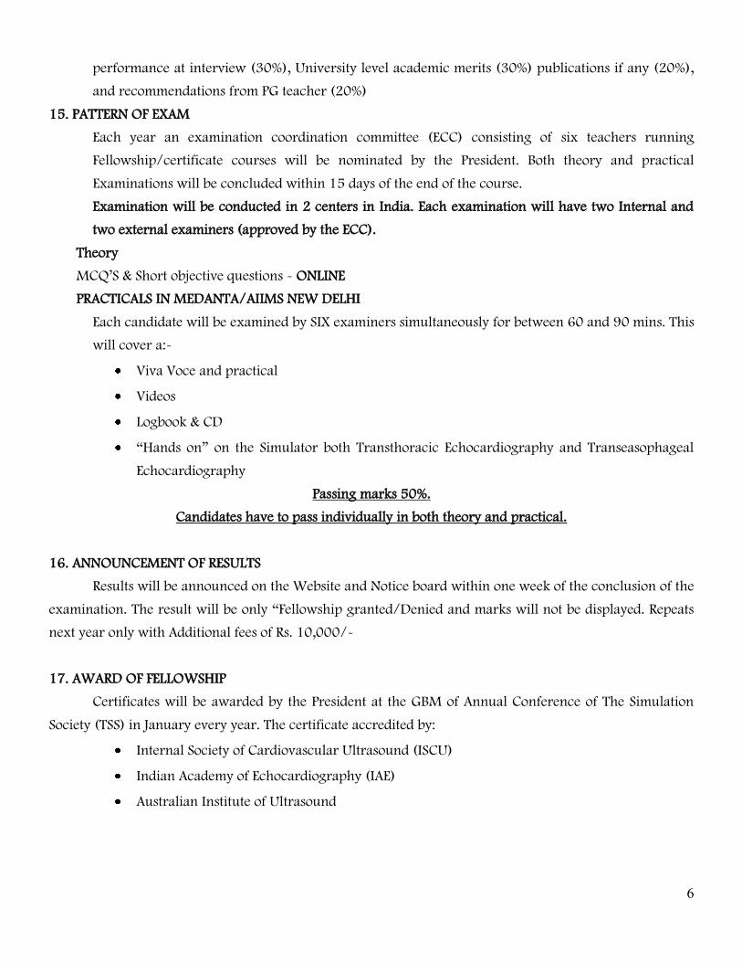
performance at interview (30%), University level academic merits (30%) publications if any (20%),
and recommendations from PG teacher (20%)
15. PATTERN OF EXAM
Each year an examination coordination committee (ECC) consisting of six teachers running
Fellowship/certificate courses will be nominated by the President. Both theory and practical
Examinations will be concluded within 15 days of the end of the course.
Examination will be conducted in 2 centers in India. Each examination will have two Internal and
two external examiners (approved by the ECC).
Theory
MCQ’S & Short objective questions - ONLINE
PRACTICALS IN MEDANTA/AIIMS NEW DELHI
Each candidate will be examined by SIX examiners simultaneously for between 60 and 90 mins. This
will cover a:-
Viva Voce and practical
Videos
Logbook & CD
“Hands on” on the Simulator both Transthoracic Echocardiography and Transeasophageal
Echocardiography
Passing marks 50%.
Candidates have to pass individually in both theory and practical.
16. ANNOUNCEMENT OF RESULTS
Results will be announced on the Website and Notice board within one week of the conclusion of the
examination. The result will be only “Fellowship granted/Denied and marks will not be displayed. Repeats
next year only with Additional fees of Rs. 10,000/-
17. AWARD OF FELLOWSHIP
Certificates will be awarded by the President at the GBM of Annual Conference of The Simulation
Society (TSS) in January every year. The certificate accredited by:
Internal Society of Cardiovascular Ultrasound (ISCU)
Indian Academy of Echocardiography (IAE)
Australian Institute of Ultrasound

7
The decision of the Examination Co-ordination Committee will be binding on all. On all matters pertaining
to the examinations
Clinical rotation:
Intensive Cardiac Care Unit (ICCU) 6 months.
CCU and Cath Lab :- 6 months
MAINTENANCE OF LOG BOOKS:
Every fellow shall maintain a record of skills he has acquired during the training period certified by
the Head of Department in which he/she has undergone training.
The candidates should also be required to participate in the teaching and training programme of
undergraduate and post graduate (MD) students.
In addition, the Head of the Department shall involve their Post-Graduate CCM candidates in
Seminars, Journal Clubs, Group Discussions and participation in clinical, clinicopathological
conferences.
Candidates are required to attend at least 2 Regional/National/ International Conferences and make
at least one presentation at any of these conferences during the course on relevant subjects. These
should be entered in the Log Book.
At the end of the course, the candidate should summarise the contents and get the Log Book Certified
by the Head of the Department.
The Log book should be submitted at the time of practical Examination for the scrutiny of the Board
of Examiners.

8

9
9
___________________________
SIGNATURE OF CANDIDATE

10
10
LOG OF CASES DONE MONTHLY IN ICU

11
11
LOG OF CASES DONE MONTHLY IN ICU

12
12
LOG OF CASES DONE MONTHLY IN CCU

13
13
LOG OF CASES DONE MONTHLY IN CCU

14
14
LOG OF CASES DONE MONTHLY IN OPERATING ROOM

15
15
LOG OF CASES DONE MONTHLY IN OPERATING ROOM

16
16
NUMBER OF CASES & PROCEDURES DONE MONTHLY
CASES JAN FEB MAR APR MAY JUNE JULY AUG SEPT OCT NOV DEC TOTAL
ROUTINE /
EMERGENCY
OPEN /
CLOSED
CABG
VALVE
CHD
OTHERS
CENTRAL
VENOUS LINES
TEE
MRI
CT ANGIO
GRAPHY
CATH LAB
PROCEDURES
- AICD
- RFA
- PTCA
- OTHERS like
- CCU CARE
-
RESUSCITATION
TOTAL NO OF
CASES

17
17
POSTINGS IN OR
S.NO OPERATION THEATRE DURATION

18
18
POSTINGS IN OR
INCHARGE SIGNATURE REMARKS

19
19
POSTINGS IN ICU
INCHARGE SIGNATURE REMARKS

20
20
POSTINGS IN ICU
INCHARGE SIGNATURE REMARKS

21
21
POSTINGS IN CATH LAB
INCHARGE SIGNATURE REMARKS

22
22
POSTINGS IN CATH LAB
INCHARGE SIGNATURE REMARKS

23
23
POSTINGS IN CCU
INCHARGE SIGNATURE REMARKS

24
24
POSTINGS IN CCU
INCHARGE SIGNATURE REMARKS

25
25
RESEARCH WORK UNDERTAKER
S.NO TOPIC GUIDE /CO-
GUIDES
DATE OF
SUBMISSION

26
26
CLINICAL PRESENTATIONS
S.NO TITLE PRESENTED AT

27
27
PUBLICATIONS CURRENT YEAR (2017)
S.NO TITLE PUBLISHED IN

28
28
SYLLABUS
APPENDIX I
Complete curriculum of the course
COURSE OBJECTIVES
At the end of the course, the candidate should be able to:
Understand the theoretical basis of organ dysfunction and critical illness in
Echocardiography
Apply these principles in Echocardiographic techniques, both Transeasophageal
Echocardiography (TEE) and Transthoracic Echocardiography (TTE)
Critically evaluate published literature
Apply the highest ethical standards in the practice of medicine
Transeasophageal Echocardiography (TEE) and Transthoracic Echocardiography (TTE)
views as per the American Society Guidelines
TEE Probe manipulation
Knobology of the latest ECHO machine
Left ventricle
Acquisition and display techniques - Volumes - Mass - Shape - 4D strain - Regional
and global function (ischemic heart disease, cardiomyopathy)
Right ventricle
Acquisition and display techniques - Regional and global function
Atria
Acquisition and display - Echo anatomy: left atrial appendage, pulmonary veins -
Global geometry and phasic function
Mitral valve
Acquisition and display techniques - Quantification - Mitral valve diseases - Mitral
annulus quantification in normal and diseases
Aortic valve
Acquisition and display techniques - Aortic valve diseases
Tricuspid valve
Acquisition and display techniques - Tricuspid valve diseases

29
29
How to implement 4D echo in the routine of the echo lab?
Congenital heart diseases
Atrial septal defects - Patent foramen ovale - Ventricular septal defects - Uni-/ Bi-
and Quadri-cuspid aortic valve - Mitral cleft - Parachute mitral valve - Cor
triatriatum - Univentricular heart - Ebstein disease - Pulmonary valve
Masses
Tumors - Thrombi
Pharmacological stress echo
3rd generation 3D transoesophageal echo
Introduction to Basic/Advanced Echocardiography
Anatomy/Imaging Planes
Muscular construct
Atria
Ventricles
Papillary Muscles
Electrical -Purkinje system
Great vessels (arteries & vein construct)
Hemodynamic Monitoring
Flow pattern of cardiovascular system
Normal blood flow
Chamber pressures normal range
Inflow and outflow
Frank Starling law
IMAGING TECHNIQUES
Knobology
Gain (B-Mode)
Time gain compensation (signal strength)
Focus zone
Transducer construct
Transducer function
Analog versus digital

30
30
Artifacts
Hardcopy units
M-Mode
Value
Technique
Timing parameters
Chamber sizes
3/4 Dimensional
Value
Technique/windows
Physics & physical limitations
Complete echo examination
Limited echo, when is it used
Color Basics/Advanced
Limitations
Benefits
Nyquist limitations
Gain settings
Doppler Echocardiography
Continuous Wave Motion
Pulsed Wave Motion
Measurements
M-Mode
2D/3D
Doppler
PATHOPHYSIOLOGY OF DISEASES
Mitral/Tricuspid Disease on ECHO
o Review normal anatomy
o Stenosis (mild, moderate, severe)
o Hemodynamic changes
o Valve regurgitation

31
31
o Chordae tendinae
Aortic/Pulmonic Valves on ECHO
o Review normal anatomy
o Stenosis (mild, moderate, severe)
o Hemodynamic changes
o Valve regurgitation
o Outflow abnormalities
Cardiomyopathy
o Hypertrophic CM (obstructive/non-obstructive)
o Dilated CM
o Restrictive CM
Coronary Artery Disease/Ischemia
o Anatomy
o Flow distribution
o Heart attack
o Chemistry changes
o EKG changes
o Arrythmia
o Acute, chronic
o Discussion of stress echocardiograms & value
Aortic Diseases
Pericardial Disease
THEORETICAL KNOWLEDGE
The Echocardiography specialist must understand the physiology, diagnosis, prevention
and management of the following disorders:
CARDIOVASCULAR DISEASES AND ECHO
Hemodynamic instability and shock
Acute myocardial infarction
Intra-aortic balloon pump and left ventricular assist devices
Severe heart failure
Cardiomyopathies

32
32
Valvular heart disease
Myocarditis
Cardiac tamponade
Pulmonary embolism
Aortic dissection
Peripheral vascular diseases
Cardiovascular surgery post-operative care
INTRAOPERATIVE TRANSESOPHAGEAL ECHOCARDIOGRAPHY
Principles of echocardiography:
Physics, digital echocardiography, imaging artifacts and pitfalls, optimizing 2D echo
Intraoperative examination:
Surgical anatomy correlated with echocardiographic imaging planes,
Assessment of global ventricular function, right ventricular function,
Regional ventricular function,
Mitral valve,
Aortic valve ,
Tricuspid and pulmonic valves thoracic aorta and
Prosthetic valves.
Assessment of congenital heart disease in adults
Decision making in critical care:
TEE in critical care setting,
Assessment of perioperative hemodynamic
Surgical decision making in coronary artery disease:
Assessment of myocardial viability,
Assessment in higher risk myocardial revascularization,
Assessment of mitral valve in ischemic heart disease,
Assessment in off pump myocardial revascularization.
Surgical decision making in valvular heart disease:
Surgical consideration in mitral and tricuspid valve surgery,
Assessment in mitral valve surgery,

33
33
Surgical consideration in aortic valve surgery,
Assessment in aortic valve surgery.
Surgical decision making in major vascular surgery:
Assessment of surgery of aorta.
Surgical decision making in congestive cardiac failure:
Pathophysiologic,
Assessment of cardiomyopathy,
Surgical considerations in nontransplant surgery for congestive cardiac failure,
Assessment in surgical procedures for CHF,
Assessment of cardiac transplantation
Surgical decision making in Interventional Cardiovascular Medicine and Non
cardiac surgery :
Assessment in cardiac intervention, assessment in noncardiac surgery.
Elective: Echocardiography
Identify the structures appreciated in the parasternal (long & short axis), subcostal &
apical views
The assessment of LV systolic function using the modified Simpson rule method, &
it’s limitations
The assessment of RV systolic function
Identify pericardial effusion & the criteria for tamponade physiology
Identify normal chamber size for the right & left ventricles
Identify normal thickness for the RV free wall, IVS & LV free wall
The principles of Doppler in measuring the velocity of fluid, & therefore pressure
gradient (prograde & retrograde)
Identify findings consistent with moderate & severe MR
The assessment of aorta pulmonary shunt function
The determination of RV systolic pressure based on the TR jet
The estimation of RV systolic pressure based on the position of IVS during systole
The normal orientation & position of the IVS during diastole & systole
The abnormal orientation & position of IVS during diastole & systole

34
34
SCHEME OF EVALUATION:-
Evaluation will be by Credit Based System
ALLOTMENT OF CREDITS:-
Total Credit Points 300
Minimum required for passing 240 (80%)
A) Didactic Credits :( 25 credit points)
Didactic on various topics as per syllabus spread over One Hour lecture. Total 10-
20 Lectures depending upon specified course material and depth of theory.
B) Presentation, Publications & Project Work (25 credit point)
Under faculty guidance, presentations for Local, Regional & national conferences.
C) Video Learning, Grand Rounds, Faculty Discussions (10 credit Points)
Review of Recorded Cases/ Procedures/Surgeries with Faculty Input.
Specific/specialized Surgery Grand Round/Clinical Case Conference.
Disease/Journal Club, Round Table Discussions.
D) Sessions in Imaging Clinics (15 credit points)
Learning of Ultrasound, CT scan, and MRI & Various Procedures required for the
particular course/module (e.g. Doppler etc.)
E) INTENSIVE CARDIAC CARE MANAGEMENT CREDITS. (TOTAL 175)
A Candidate is expected to maintain certified Log book indicating number of cases
assisted or individually managed and procedures performed under the guidance of
faculty for each Module.
F) Fellowship Examination 50 Credit Points (Terminal Exam by MCQs)
The examination for a particular course may be conducted according to the
requirement of a course.
TEXT BOOKS:
ASE’s Comprehensive Echocardiography
Cardiovascular Sonographer Registry Review Guide
Textbook of Clinical Echocardiography by Catherine M Otto, MD

35
35
Echocardiography Review Guide: Companion to the Textbook of Clinical
Echocardiography BY Catherine M. Otto MD (Author), Rebecca Gibbons
Schwaegler BS RDCS (Author), Rosario V. Freeman MD MS (Author) Clinical
Sonography: A Practical Guide by Roger C Sanders
Diagnostic Ultrasound: Principles and Instruments by Frederick W Kremkau,
The Echo Manual by Jae K Oh, MD, James B Seward, A Jamil Tajik, MD
Echo Made Easy by Sam Kaddoura
Practice of Clinical Echocardiography by Catherine Otto
Echocardiography – a practical guide for reporting and interpretation by Priya
Margaret Joseph
Successful Accreditation in Echocardiography - A Self-Assessment Guide by Sanjay
Banypersad (Author)
INDIAN BOOKS:-
Problem Based Transesophageal Echocardiography by Deepak K.Tempe /
Balachundhar Subramaniam / Kathirvel Subramaniam / Harish Ramakrishna
(Author)
Review of Cardiac Anesthesia with 2100 MCQs by Poonam Malhotra Kapoor (1st
Edition)
Review of Cardiac Anesthesia with 2100 MCQs by Poonam Malhotra Kapoor (2nd
Edition)
Clinical Simulation in Medicine (For all Examinations in Cardiology, Critical Care,
Anesthesia and Pulmonary Medicine) by Poonam Malhotra Kapoor
Manual of Extracorporeal Membrane Oxygenation (ECMO) in the ICU: by Poonam
Malhotra Kapoor
Transesophageal Echocardiography of the Tricuspid and Pulmonary Valves by
Poonam Malhotra Kapoor
Journals
International Journal of Cardiovascular Research
Journal of the American College of Cardiology
American Journal of Cardiology
Journal of Molecular and Cellular Cardiology
European Journal of Preventive Cardiology
Cardiology
Cardiology Clinics