Embed Size (px)
Citation preview
COUNCIL ON PHARMACYAND CHEMISTRY
REPORT TO THE COUNCILThe Council has authorized publication of the following re-
port from its Committee on Research.Robert T. Stormont, M.D., Secretary.
The Committee on Research, through its Subcommittee on
Steroids and Cancer, is sponsoring a collaborative study on
steroids and mammary cancer. Reports summarizing this workhave been published (Estrogens and Androgens in MammaryCancer, Report to the Council on Pharmacy and Chemistry,J. A. M. A. 140:1214 [Aug. 13] 1949. Proceedings of the FirstConference on Steroid Hormones and Mammary Cancer, Chi-cago, A.M. A. [April] 1949. Current Status of Hormone Therapyof Advanced Mammary Cancer, Report to the Council on
Pharmacy and Chemistry, J. A. M. A. 146:47/ [June 2] 1951).In the following report, the conclusions expressed are those
of the authors and final conclusions of the Subcommittee mustawait evaluation of the studies now in progress.
Paul L. Wermer, M.D., Secretary.
EFFECT OF TESTOSTERONE ON PATIENTSWITH BONE METASTASES
A METABOLIC STUDY, PARTICULARLY OFBREAST CARCINOMA
Daniel Laszlo, M.D., Albert Schilling, M.D.Judith Bellin, M.S., Estelle D. Gottesman, B.S.andCyril A. Schulman, M.D., New York
In the past decade, hormonal therapy has been advocated inthe palliative management of patients with metastatic breastcarcinoma 1; reports of temporary palliation have been publishedfrom many centers,2 and a wide discrepancy between the inci-dence of subjective and objective improvement has been noted.In a preceding paper,3a it was pointed out that metabolic
studies are an important aid in defining the course of malig-nant disease, in gaging the effect of therapeutic agents, andin studying their mode of action. The mineral metabolism ofpatients with active osteolysis has been characterized by elevatedurinary calcium and phosphorus excretions and negative bal-ances.3 At times, excessive rates of demineralization exceed theability of the kidneys to excrete calcium, resulting in hyper-calcemia.3a Conversely, patients with osteoblastic metastaseshave characteristically subnormal urinary calcium excretion and
a tendency to maximal calcium retention.4 On occasion, patientswith osteolytic metastases may spontaneously have temporaryphases of bone repair, evidenced by a metabolic behavior thatapproaches the calcium-retentive traits seen in osteoblastic cases.
The purpose of the present study is to observe the effect oftestosterone on the metabolism of patients with osteolytic metas-tases secondary to breast carcinoma. If the hormone were toencourage the remineralization of osteolytic areas, the processwould be evidenced by improvement in the mineral balances.After metabolic studies in the pretreatment phase, testos-
terone was administered to six patients 5 with osteolytic metas-tases secondary to breast carcinoma. To assess whether the resultsattained were specific for breast carcinoma, a tumor that is pre-sumed to be influenced by hormones, control studies were per-formed with patients having other types of malignancy. A patientwith extensive osteolytic métastases secondary to leiomyosar-coma of the vulva was given testosterone. A male patient withmultiple myeloma was given estrogen therapy initially and latertestosterone. Finally, the effect of radiation castration was ob-served in a premenopausal patient with osteolytic métastasessecondary to breast carcinoma.
The various effects observed are illustrated by the cases re-
ported in this paper.nitrogen retention induced by testosterone with minimal
improvement in calcium balance
Case 1, a 50-year-old white woman, had extensive osteolyticand pulmonary métastases secondary to leiomyosarcoma of thevulva that had been resected three years previously. Métastases,which progressed slowly, were noted a year later. At no time dur-ing the course of the disease were major complications, such as
severe anemia, pathologic fracture, or hypercalcemia, noted.Metabolic studies were conducted for a total of 153 days
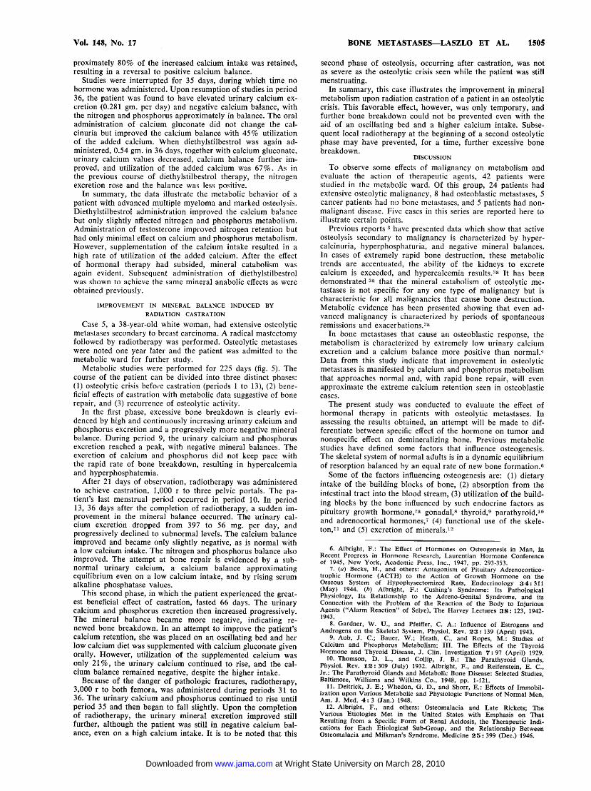
(fig. 1). At that time, the patient was ambulatory and in a fairlygood nutritional state. The pretreatment phase lasted for 21 days,during which the patient was in slightly positive nitrogen andphosphorus balance and in negative calcium balance with a
moderately elevated urinary calcium excretion.Upon the administration of testosterone, a total of 2.25 gm.
in 45 days, urinary nitrogen excretion dropped immediately,resulting in a highly positive nitrogen balance. The calcinuriawas somewhat decreased, so that the calcium balance was slightlyless negative.The studies were then discontinued for 62 days during which
time testosterone was not administered. In the first 15 days afterthe resumption of studies, the nitrogen and phosphorus balanceswere similar to those observed in the first pretreatment phase,but the urinary and fecal calcium values were lower than previ-ously. Upon the additional administration of 1.8 gm. of testos-terone in 36 days, an immediate drop in urinary nitrogen andphosphorus excretion was again noted, resulting in a positivenitrogen and phosphorus balance. The calcium excretion, how-ever, showed no further change.To observe the possible favorable effect of testosterone on
the mineral balance if an adequate supply of calcium were fur-nished, the daily administration of testosterone was continued inconjunction with an increased calcium intake. In this patient thepresumption did not prove to be correct. Although the anaboliceffect of testosterone on the nitrogen metabolism was still clearlydemonstrable, the urinary calcium remained uninfluenced. Thecalcium balance became positive but to an extent no greater thanthat observed when calcium intake was increased without theadditional administration of testosterone.3" With increased fecalcalcium elimination, more phosphorus was now excreted throughthe stool and the phosphorus balance became somewhat less posi-tive. These changes are similar to those seen in patients on a
similar dietary calciumrphosphorus ratio who did not receivetestosterone.This case illustrates a definite anabolic effect of testosterone
on protein metabolism. It induced a total retention of 194 gm.of nitrogen corresponding to 1,213 gm. of protein. The patientgained only 1.2 kg. of body weight in this phase. No majorchanges were noted in the patient's clinical condition and labora-tory data with the exception of roentgenograms which showedslow progression of osteolysis. No major improvement of thecalcium metabolism was induced by testosterone.
Dr. Schilling is a trainee of the National Cancer Institute.From the Division of Neoplastic Diseases, Montefiore Hospital.This project has been supported in part by a grant from the National
Cancer Institute of the National Institutes of Health, United States PublicHealth Service and by a grant from the American Cancer Society.
1. (a) Haddow, A.; Watkinson, J. M., and Patterson, E.: Influence ofSynthetic Oestrogens upon Advanced Malignant Disease, Brit. M. J2:393 (Sept. 23) 1944. (b) Hermann, J. B.; Adair, F. E., and Woodard,H. Q.: The Use of Testosterone Propionate in the Treatment of AdvancedCarcinoma of the Breast. II. The Treatment of Osseous Metastases, Sur-gery 22: 101 (July) 1947. (c) Nathanson, I. T.; Adair, F. E.; Allen, W.,and Engle, E. T.: Estrogens and Androgens in Mammary Cancer: (AProgress Report of a Sub-Committee of the Therapeutic Trials Committeeof the Council on Pharmacy and Chemistry), J. A. M. A. 140: 1214(Aug. 13) 1949.
2. Proceedings of the First Conference on Steroid Hormones andMammary Cancer: The Therapeutic Trials Committee of the Council on
Pharmacy and Chemistry of the American Medical Association, Chicago,1949.
3. (a) Laszlo, D.; Schulman, C. A.; Bellin, J.; Gottesman, E. D., andSchilling, A.: Mineral and Protein Metabolism in Osteolytic Metastases,J. A. M. A. 148:1027 (March 22) 1952. (b) Laszlo, D.: Mineral Metab-olism in Metastatic Bone Cancer, Cancer Res. 9:614 (Oct.) 1949. (c)Laszlo, D.; Schulman, C. A.; Bellin, J.; Gottesman, E. D., and Schil-ling, A.: Metabolic Studies of Patients with Carcinoma of the Breast andthe Effects of Testosterone Therapy, ibid. 10: 230 (April) 1950.
at Wright State University on March 28, 2010 www.jama.comDownloaded from

MINERAL LOSS INDUCED BY TESTOSTERONE IN SPITE OFNITROGEN RETENTION
Case 2, a 57-year-old postmenopausal woman, had carcinomaof the left breast. Radical mastectomy, followed by radiother-apy, was performed and the patient was asymptomatic for fouryears, when extensive osteolytic métastases with spontaneoushypercalcemia developed. Testosterone was then administeredbut resulted only in temporary symptomatic improvement fol-lowed by progression of symptoms and findings. When admittedto the hospital, the patient was in hypercalcémie crisis. Testoste-rone was discontinued and intravenous fluid administration be-gun, resulting in a return of serum calcium and blood urea nitro-gen values to normal levels, decrease in pain, and ambulation. Atthis time, the patient showed evidence of weight loss, masculini-zation, extensive osteolytic métastases with rib and vertebralfractures, diplopia, and blurred vision. The renal function, asmeasured by urinary concentration and urea clearance tests, wasunimpaired.Metabolic studies began two and one-half months after the
discontinuation of testosterone and were conducted for nineperiods totaling 54 days. The data are illustrated in figure 2.The pretreatment phase consisted of six periods, three on a lowcalcium intake and the next three on a higher calcium intake.In this phase, the nitrogen balance was in equilibrium. Thephosphorus balances showed a retention on a low calcium intakewhich changed toward equilibrium on increased calcium intake.This change was mainly caused by an increase in fecal phos-phorus and followed the trend which has already been de-
Fig. 1.—Metabolie data of a patient with leiomyosarcoma of the vulvaand osteolytic métastases (case 1). All periods are for six days exceptperiods 11 and 12 which are nine-day periods. Testosterone, 50 mg. perday, was administered in periods 5 to 11 and in periods 14 to 25, inducingnitrogen retention.The metabolic data graph is charted according to Reifenstein, Albright,
and Wells. Horizontal lines at zero in the balance graphs indicate theequilibrium between intake and output. Intake is charted from the zeroline downward. Output is charted from the intake line upward toward thezero line. Interrupted lines in the phosphorus graph represent areas cor-responding to the theoretical phosphorus retention or loss, calculated onthe basis of theoretical N:P and Ca:P ratios. One unit of the phosphorusscale equals 15 units of the nitrogen scale and 2 units of the calciumscale.
scribed.3" The urinary calcium excretion was subnormal in thefirst three periods, indicating attempt at bone repair. In periods4 to 6, the urinary calcium excretion tended to rise, but re-mained within normal limits. The calcium balance was onlyslightly negative on a low calcium intake. Minimal utilization(10%) of calcium gluconate was achieved in periods 4 to 6,resulting in only slight improvement of the calcium balance.The testosterone phase was clearly characterized by gradual de-crease in urinary nitrogen and improvement in nitrogen bal-ance. In contrast to the nitrogen retention was the mineralloss occurring during testosterone therapy. Hypercalcinuria oc-curred in the initial period of testosterone and increased pro-gressively to excessive values accompanied by hyperphosphaturia.The excess urinary calcium and phosphorus loss approximatedthe ratio of these minerals in bone. The rate of mineral break-down exceeded the urinary excretion, resulting in hypercalcemia.The calcium and phosphorus balances became progressivelynegative.Metabolic studies had to be discontinued for 11 days, at which
time the serum calcium rose to 18.9 mg. per 100 cc. and the blood
urea nitrogen to 74.8 mg. per 100 cc. Urinary collections wereresumed for two periods when only intravenous feeding wasfeasible. Although a trend toward improvement in serum cal-cium and blood urea nitrogen levels was achieved, the patientdied several days later of uremia. In the last period, almost 0.9gm. of urinary calcium was excreted daily. At autopsy, markednephrocalcinosis was found.
MINERAL AND NITROGEN LOSS INDUCED BY TESTOSTERONE
Case 3, a 47-year-old woman, underwent a left radical mas-tectomy two years prior to metabolic studies. Soon after theoperation, osseous métastases were noted and the patient be-came bedridden with pain. The administration of 0.8 gm. oftestosterone resulted in a cessation of menses but produced nosymptomatic improvement. Subsequent radiotherapy to the lum-bar spine and pelvis did not produce relief. Five months later,after a hypercalcémie crisis which improved after parenteralfluid was administered, testosterone therapy was reinstituted,the patient receiving a total of 4.1 gm. over a nine-month period.This therapy provoked hypercalcemia (15.1 mg. per 100 cc),
Fig. 2.—Metabolie data of a patient with breast carcinoma and osteo-lytic métastases (case 2). All periods are for six days. Testosterone, 50 mg.per day, was administered in periods 7 to 9, inducing hypercalcemia, nitro-gen retention, and negativity of mineral balances.
hypopotassemia (6.7 mg. per 100 cc), nausea, vomiting, and de-hydration. Testosterone was discontinued, and parenteral fluidand potassium were administered, resulting in a return to nor-mal of the serum calcium and potassium within three weeks.However, anorexia and vomiting persisted, the blood urea nitro-gen was 24.5 mg. per 100 cc, and the patient sustained a patho-logic fracture of the left femur.
Ten days later, at the time metabolic studies began, the pa-tient had been bedridden for 22 months and was in poor nutri-
4. Aub, J. C.: Personal Communication to the author. Aub, J. C.;Tibbets, D. M., and Nathanson, I. T.: Metabolic Effects of Treatment ofCarcinoma of the Prostate, Cancer Res. 7: 723 (Nov.) 1947. Schilling, A.;Bellin, J.; Gottesman, E. D., and Laszlo, D.: Metabolic Studies of Patientswith Carcinoma of the Prostate and the Effects of Stilbestrol Therapy,ibid. 10:239 (April) 1950. Schilling, A., and Laszlo, D.: The Effect ofDiethylstilbestrol on the Calcium, Phosphorus and Nitrogen Metabolism ofProstatic Carcinoma, J. Clin. Investigation 29:918 (July) 1950.
5. These cases have been mentioned in a previous report as cases 3,4, 6, 7, 8, and 9 (see footnote 3a). Cases 2 and 3 of this paper correspondto cases 8 and 9 of the previous report and are now described in detailas illustrative of the results obtained in this series.
at Wright State University on March 28, 2010 www.jama.comDownloaded from

tional state. The studies totaled 155 days, and are shown infigure 3. During the first five days of study, a moderately ele-vated calcinuria was noted. Later, urinary calcium excretiondropped toward subnormal values (as low as 20 mg. per day),calcium balance improved and was only slightly negative ona low calcium intake, and the patient was maintained in a stateof nitrogen and phosphorus balance. The serum calcium wasnormal and the alkaline phosphatase elevated.After 53 days of study, a total of 1.2 gm. of testosterone was
administered intramuscularly in 24 days. Nine days after the in-stitution of the hormone therapy, nausea and vomiting recurred,and the food and fluid intake decreased. Urinary calcium andphosphorus excretions rose rapidly. In the second period of hor-mone administration, the serum calcium rose to hypercalcémielevels and the alkaline phosphatase fell, indicating acceleratedosteolysis. The average calcium balance during testosterone ad-ministration (-628) was five times that of the pretreatmentperiods (-129). The phosphorus balance, which was in equilib-rium during the pretreatment phase, became negative because
Fig. 3.—Metabolie data of a patient with breast carcinoma and osteo-lytic métastases (case 3) All periods are for six days except periods 1, 2,7, and 8, which total 3, 2, 9, and 9 days respectively. Testosterone, 50 mg.per day, was administered in periods 10 to 13, inducing osteolysis andhypercalcemia.
of the increased urinary phosphorus. The nitrogen balance be-came negative due to nausea, vomiting, and the decrease in foodintake.With the discontinuation of testosterone, vomiting became less
severe. However, the urinary calcium and phosphorus excretionsincreased still further, reaching a peak of 0.94 gm. and 1.043gm. per day, respectively, six weeks after the discontinuation oftestosterone. Clinically, during this phase, the patient showed a
progressive downhill course. Accelerated tumor growth was evi-denced by a rapidly enlarging nodular liver. Hypercalcemia,azotemia, and negative mineral balances persisted until deathwhich occurred 12 weeks after the discontinuation of the hor-monal therapy.
In summary, this case illustrates the metabolic behavior of a
patient recovering from a previous hypercalcémie crisis inducedby testosterone and in whom definite attempts at bone repairwere observable. Reinstitution of testosterone induced anotherhypercalcémie crisis with excessive bone breakdown which per-sisted even after hormone therapy was stopped.IMPROVEMENT IN CALCIUM BALANCE INDUCED BY DIETHYLSTIL-
BESTROL; IMPROVEMENT IN NITROGEN BALANCE INDUCEDBY TESTOSTERONE
Case 4, a 67-year-old white man, had multiple myeloma withextensive osteolysis. Ten weeks prior to the onset of metabolicstudies, a compression fracture of the seventh thoracic vertebracaused paraplegia. A course of radiotherapy to the dorsal spinedid not relieve pain nor did it improve the paraplegia.
Metabolie studies were performed for 288 days (fig. 4). In thefour periods prior to treatment, the patient was in a state of posi-tive nitrogen balance and of phosphorus equilibrium. The uri-nary calcium excretion was moderately elevated, averaging0.212 gm. per day, and the balance was negative.During the following 54 days, 15 mg. per day of diethylstil-
bestrol ("stilbestrol") was administered intramuscularly, totaling0.81 gm. Urinary nitrogen excretion increased slightly and the
Fig. 4.—Metabolie data of a patient with multiple myeloma (case 4).All periods are for six days. Dicthylstilbestrol, 15 mg. per day, was ad-ministered in periods 5 to 13 and improved the calcium balance. Testos-terone, 50 mg, per day, was administered in periods 23 to 35, inducingnitrogen retention with minimal improvement in the mineral balances.Diethylstilbcstrol, 15 mg. per day, was administered in periods 45 to 48and improved the mineral balances.
balance became less positive. The phosphorus balance did notchange. The urinary calcium excretion dropped promptly andreached values at the lowest limits of normal, resulting in im-provement of the calcium balance.Therapy was then discontinued during the following 54 days.
The nitrogen balance returned to the previous pretreatment level.The phosphorus balance remained unchanged. Urinary calciumexcretion rose slightly and the balance was slightly morenegative.For the following 60 days 50 mg. of testosterone was ad-
ministered daily, totaling 3 gm. Urinary nitrogen excretion de-creased promptly, greatly improving the nitrogen balance.Urinary calcium and phosphorus decreased slightly, resultingin minimal improvement of the mineral balance.
Fig. 5.—Metabolie data of a premenopausal patient with breast carci-noma (case 5). All periods are for six days except period 1, which was fornine days. Radiation castration induced an artificial menopause with im-provement in the mineral balances. Subsequent radiotherapy to osteolyticareas in both femora achieved minimal improvement in the calciumbalance.
In the succeeding 18 days of study, calcium gluconate wasadministered orally to increase the calcium intake, and testos-terone administration was continued. The urinary nitrogen in-creased slightly, resulting in a somewhat less positive nitrogenbalance. The phosphorus balance did not change significantly.The urinary calcium excretion did not change. However, ap-
at Wright State University on March 28, 2010 www.jama.comDownloaded from
proximately 80% of the increased calcium intake was retained,resulting in a reversal to positive calcium balance.
Studies were interrupted for 35 days, during which time nohormone was administered. Upon resumption of studies in period36, the patient was found to have elevated urinary calcium ex-cretion (0.281 gm. per day) and negative calcium balance, withthe nitrogen and phosphorus approximately in balance. The oraladministration of calcium gluconate did not change the cal-cinuria but improved the calcium balance with 45% utilizationof the added calcium. When diethylstilbestrol was again ad-ministered, 0.54 gm. in 36 days, together with calcium gluconate,urinary calcium values decreased, calcium balance further im-proved, and utilization of the added calcium was 67%. As inthe previous course of diethylstilbestrol therapy, the nitrogenexcretion rose and the balance was less positive.In summary, the data illustrate the metabolic behavior of a
patient with advanced multiple myeloma and marked osteolysis.Diethylstilbestrol administration improved the calcium balancebut only slightly affected nitrogen and phosphorus metabolism.Administration of testosterone improved nitrogen retention buthad only minimal effect on calcium and phosphorus metabolism.However, supplementation of the calcium intake resulted in ahigh rate of utilization of the added calcium. After the effectof hormonal therapy had subsided, mineral catabolism was
again evident. Subsequent administration of diethylstilbestrolwas shown to achieve the same mineral anabolic effects as wereobtained previously.
IMPROVEMENT IN MINERAL BALANCE INDUCED BYRADIATION CASTRATION
Case 5, a 38-year-old white woman, had extensive osteolyticmétastases secondary to breast carcinoma. A radical mastectomyfollowed by radiotherapy was performed. Osteolytic métastaseswere noted one year later and the patient was admitted to themetabolic ward for further study.Metabolic studies were performed for 225 days (fig. 5). The
course of the patient can be divided into three distinct phases:(1) osteolytic crisis before castration (periods 1 to 13), (2) bene-ficial effects of castration with metabolic data suggestive of bonerepair, and (3) recurrence of osteolytic activity.In the first phase, excessive bone breakdown is clearly evi-
denced by high and continuously increasing urinary calcium andphosphorus excretion and a progressively more negative mineralbalance. During period 9, the urinary calcium and phosphorusexcretion reached a peak, with negative mineral balances. Theexcretion of calcium and phosphorus did not keep pace withthe rapid rate of bone breakdown, resulting in hypercalcemiaand hyperphosphatemia.After 21 days of observation, radiotherapy was administered
to achieve castration, 1,000 r to three pelvic portals. The pa-tient's last menstrual period occurred in period 10. In period13, 36 days after the completion of radiotherapy, a sudden im-provement in the mineral balance occurred. The urinary cal-cium excretion dropped from 397 to 56 mg. per day, andprogressively declined to subnormal levels. The calcium balanceimproved and became only slightly negative, as is normal witha low calcium intake. The nitrogen and phosphorus balance alsoimproved. The attempt at bone repair is evidenced by a sub-normal urinary calcium, a calcium balance approximatingequilibrium even on a low calcium intake, and by rising serumalkaline phosphatase values.This second phase, in which the patient experienced the great-
est beneficial effect of castration, lasted 66 days. The urinarycalcium and phosphorus excretion then increased progressively.The mineral balance became more negative, indicating re-newed bone breakdown. In an attempt to improve the patient'scalcium retention, she was placed on an oscillating bed and herlow calcium diet was supplemented with calcium gluconate givenorally. However, utilization of the supplemented calcium wasonly 21%, the urinary calcium continued to rise, and the cal-cium balance remained negative, despite the higher intake.
Because of the danger of pathologic fractures, radiotherapy,3,000 r to both femora, was administered during periods 31 to36. The urinary calcium and phosphorus continued to rise untilperiod 35 and then began to fall slightly. Upon the completionof radiotherapy, the urinary mineral excretion improved stillfurther, although the patient was still in negative calcium bal-ance, even on a high calcium intake. It is to be noted that this
second phase of osteolysis, occurring after castration, was notas severe as the osteolytic crisis seen while the patient was stillmenstruating.In summary, this case illustrates the improvement in mineral
metabolism upon radiation castration of a patient in an osteolyticcrisis. This favorable effect, however, was only temporary, andfurther bone breakdown could not be prevented even with theaid of an oscillating bed and a higher calcium intake. Subse-quent local radiotherapy at the beginning of a second osteolyticphase may have prevented, for a time, further excessive bonebreakdown.
DISCUSSION
To observe some effects of malignancy on metabolism andevaluate the action of therapeutic agents, 42 patients werestudied in the metabolic ward. Of this group, 24 patients hadextensive osteolytic malignancy, 8 had osteoblastic métastases, 5cancer patients had no bone métastases, and 5 patients had non-malignant disease. Five cases in this series are reported here toillustrate certain points.
Previous reports 3 have presented data which show that activeosteolysis secondary to malignancy is characterized by hyper-calcinuria, hyperphosphaturia, and negative mineral balances.In cases of extremely rapid bone destruction, these metabolictrends are accentuated, the ability of the kidneys to excretecalcium is exceeded, and hypercalcemia results.33 It has beendemonstrated 3a that the mineral catabolism of osteolytic mé-tastases is not specific for any one type of malignancy but ischaracteristic for all malignancies that cause bone destruction.Metabolic evidence has been presented showing that even ad-vanced malignancy is characterized by periods of spontaneousremissions and exacerbations.3"In bone métastases that cause an osteoblastic response, the
metabolism is characterized by extremely low urinary calciumexcretion and a calcium balance more positive than normal.1Data from this study indicate that improvement in osteolyticmétastases is manifested by calcium and phosphorus metabolismthat approaches normal and, with rapid bone repair, will evenapproximate the extreme calcium retention seen in osteoblasticcases.The present study was conducted to evaluate the effect of
hormonal therapy in patients with osteolytic métastases. Inassessing the results obtained, an attempt will be made to dif-ferentiate between specific effect of the hormone on tumor andnonspecific effect on demineralizing bone. Previous metabolicstudies have defined some factors that influence osteogenesis.The skeletal system of normal adults is in a dynamic equilibriumof résorption balanced by an equal rate of new bone formation."
Some of the factors influencing osteogenesis are: (1) dietaryintake of the building blocks of bone, (2) absorption from theintestinal tract into the blood stream, (3) utilization of the build-ing blocks by the bone influenced by such endocrine factors aspituitary growth hormone,78 gonadal,8 thyroid,9 parathyroid,10and adrenocortical hormones,7 (4) functional use of the skele-ton,11 and (5) excretion of minerals.12
6. Albright, F.: The Effect of Hormones on Osteogenesis in Man, inRecent Progress in Hormone Research, Laurentian Hormone Conferenceof 1945, New York, Academic Press, Inc., 1947, pp. 293-353.
7. (a) Becks, H., and others: Antagonism of Pituitary Adrenocortico-trophic Hormone (ACTH) to the Action of Growth Hormone on theOsseous System of Hypophysectomizcd Rats, Endocrinology 34:311(May) 1944. (b) Albright, F.: Cushing's Syndrome: Its PathologicalPhysiology, Its Relationship to the Adreno-Genital Syndrome, and itsConnection with the Problem of the Reaction of the Body to InjuriousAgents ("Alarm Reaction" of Selye), The Harvey Lectures 38: 123, 1942\x=req-\1943.
8. Gardner, W. U., and Pfeiffer, C. A.: Influence of Estrogens andAndrogens on the Skeletal System, Physiol. Rev. 23: 139 (April) 1943.
9. Aub, J. C.; Bauer, W.; Heath, C., and Ropes, M.: Studies ofCalcium and Phosphorus Metabolism; III. The Effects of the ThyroidHormone and Thyroid Disease, J. Clin. Investigation 7: 97 (April) 1929.
10. Thomson, D. L., and Collip, J. B.: The Parathyroid Glands,Physiol. Rev. 12: 309 (July) 1932. Albright, F., and Reifenstein, E. C.,Jr.: The Parathyroid Glands and Metabolic Bone Disease: Selected Studies,Baltimore, Williams and Wilkins Co., 1948, pp. 1-121.
11. Deitrick, J. E.; Whedon, G. D., and Shorr, E.: Effects of Immobil-ization upon Various Metabolic and Physiologic Functions of Normal Men,Am. J. Med. 4: 3 (Jan.) 1948.
12. Albright, F., and others: Osteomalacia and Late Rickets; TheVarious Etiologies Met in the United States with Emphasis on ThatResulting from a Specific Form of Renal Acidosis, the Therapeutic Indi-cations for Each Etiological Sub-Group, and the Relationship BetweenOsteomalacia and Milkman's Syndrome, Medicine 25: 399 (Dec.) 1946.
at Wright State University on March 28, 2010 www.jama.comDownloaded from

Impairment of any of these factors can cause osteolytic bonedisease characterized metabolically by hypercalcinuria, hyper-phosphaturia, and negative mineral balances, similar to the dataobtained in osteolytic malignancies. Metabolic studies have beenreported for osteomalacia,12 steatorrhea,13 postmenopausal andsenile osteoporosis,11 hyperparathyroidism,10 Cushing's dis-ease,1311 immobilization,11 and renal rickets.12In a number of these diseases, particularly in postmenopausal
osteoporosis, androgen and estrogen therapy has induced im-provement in calcium, phosphorus, and nitrogen metabolism.14bIt has been noted that androgen has a significant protein ana-bolic effect, whereas estrogen induces predominantly mineralanabolism.In patients with malignant bone disease, severe pain may
cause immobilization, and osteoporosis from disuse is often a
prominent feature. When gonadal hormones are administeredto these patients, a favorable effect may be achieved on theosteoporosis, one that may be entirely nonspecific with respectto tumor growth.For the purpose of this discussion, a specific effect on tumor
shall be considered as one that causes inhibition or accelerationof tumor growth.Experimental evidence has been advanced indicating that the
incidence of mammary carcinoma in susceptible strains of miceis influenced by hormonal factors. This subject has been re-
cently reviewed.15 It has been demonstrated that ovariectomydecreased the incidence and delayed the appearance of mammarycarcinoma in female mice of a susceptible strain ie; similar re-sults have been achieved by implantation of testes,17 adminis-tration of testosterone,18 and ovarian irradiation.111 Ovariangrafts induced mammary cancer in castrated male mice of a sus-
ceptible strain and the incidence approximated that in femalesof that strain 20; similar results have been achieved by admin-istration of estrogen.21 However, it must be stressed that theseresults are attained in mice of high mammary cancer strains.Androgens and estrogens apparently have no effect on the growthof a mammary tumor once it is established in a mouse.1""'
Numerous clinical investigations have been conducted on thetherapeutic effect of hormones in human breast cancer. Surgicalor x-ray castration has resulted in clinical improvement of 20to 56% of premenopausal women with breast carcinoma.22Castration has also resulted in improvement of some male pa-tients with breast cancer.23 Haddow and his collaborators re-ported great improvement in patients with advanced breastcancer receiving estrogen therapy, and serial biopsies of a fewpatients showed histological alterations suggesting regression oftumor cells.1»
The Committee on Research of the American Medical Asso-ciation has undertaken to assemble the data of its cooperatinginvestigators on the effects of steroid hormones in the treatmentof breast cancer.24 In a total of 285 patients with métastasestreated with androgen, subjective improvement was noted in62%. Objective improvement in soft tissue lesions was noted inabout 20%. Objective improvement in osseous lesions was re-ported in 18%. In a total of 144 patients treated with estrogen,subjective improvement was reported in 60%. Objective im-provement in soft tissue lesions was noted in 43%; a number ofosseous lesions showed improvement.
However, there have also been clinical reports suggestingtumor promotion by hormones. Cancer of the breast has ap-peared in male 2r> and female 26 patients receiving estrogens.Apparent acceleration of tumor growth was noted in patientswith metastatic breast cancer receiving estrogen or androgentherapy.27
Case 1 is a study of the effect of testosterone in a patientwith extensive osteolytic métastases secondary to leiomyosar-coma of the vulva. The androgen induced retention of proteinand had a slight mineral-anabolic effect. Since this effect is sim-ilar to the one expected in a patient with postmenopausal osteo-porosis, and since leiomyosarcoma is assumed to be a tumorthat is not controlled by hormones, it may be assumed that thefavorable effect of testosterone in this case is not a specifictumor-inhibiting effect.
Case 2 is a study of testosterone therapy in a postmenopausalpatient with extensive osteolytic métastases secondary to breastcarcinoma. Testosterone had the seemingly paradoxical effectof inducing nitrogen retention together with mineral loss caus-ing hypercalcemia. If it were assumed that testosterone inducedprotein anabolism in tumor tissue, the accelerated rate of tumorgrowth in the osseous métastases would account for the in-creased bone breakdown.
Case 3 is a study of the effect of testosterone in a postmeno-pausal patient with osteolytic métastases secondary to breastcarcinoma. The pretreatment phase illustrates a trend towardspontaneous bone repair in a patient with advanced malignancy.This favorable phase was interrupted by the administration oftestosterone which promptly induced accelerated osteolysis andhypercalcemia. The accompanying protein catabolism was dueto anorexia; the decreased food intake, nausea, and vomitingresulted from hypercalcemia. These catabolic factors outweighedwhatever protein-anabolic effect the androgen might have had.As in the previous case, the osteolysis may have been causedby a tumor-promoting effect of testosterone. However, it mustalso be considered that the hormone may have had a directmineral-catabolic -effect as reported in mice by Gardner andPfeiffer.8
Case 4 is a study of the effects of estrogen, and later andro-gen, in a patient with multiple myeloma. Estrogen inducedmineral anabolism, but did not improve the nitrogen balance.Androgen promoted protein retention, but improved the calciumbalance only slightly. This patient was paraplegic and bedriddenat the onset of studies and considerable mineral catabolismcould be attributable to osteoporosis of disuse. Since multiplemyeloma is not considered to be a tumor that is hormonallycontrolled, the beneficial effects of hormone therapy cannot beconsidered to be due to tumor inhibition. As in case 1, the favor-able effect achieved may not be assumed to be specific for tumor,but rather one of remineralization of osteoporotic areas.It was reported previously 3a that 30 to 40% of the ingested
calcium gluconate was utilized in patients with osteolytic breastcancer métastases, a rate similar to that observed in normalpersons.28 In case 4, when calcium gluconate was administered
13. Albright, F., and Stewart, J. D.: Hypovitaminosis of All Fat-Soluble Vitamins due to Steatorrhea; Report of a Case, New England J.Med. 223:239 (Aug. 15) 1940.
14. (a) Adams, M.; Boothby, W. M., and Snell, A. M.: MetabolicStudies in Osteoporosis, Am. J. Physiol. 114: 383 (Jan.) 1936. (b) Reifen-stein, E. C., Jr., and Albright, F.: The Metabolic Effects of SteroidHormones in Osteoporosis, J. Clin. Investigation 26:24 (Jan.) 1947.
15. (a) Gardner, W. U.: Tumors in Experimental Animals ReceivingSteroid Hormones, Surgery 16:8 (July) 1944. (b) Nathanson, I. T.:Endocrine Aspects of Cancer, New England J. Med. 231:764 (Dec. 7)1944; 231:795 (Dec. 14) 1944. (c) Greenstein, J. P.: Biochemistry ofCancer. New York, Academic Press Inc., 1947, pp. 104-113 and 156-158.
16. Lathrop, A. E. C., and Loeb, L.: Further Investigation on Originof Tumors in Mice. III. On Part Played by Internal Secretion in Spon-taneous Development of Tumors, J. Cancer Res. 1: 1, 1916. Cori, C. F.:Influence of Ovariectomy on Spontaneous Occurrence of Mammary Carci-nomas in Mice, J. Exper. Med. 45: 983 (June) 1927.
17. Murray, W. S.: Sex Hormones and Cancer; Some Effects of Inter-play of Sex Hormones Upon Incidence of Mammary Cancer in Mice,Am. J. Cancer 30:517 (July) 1937.
18. Nathanson, I. T., and Andervont, H. B.: Effect of TestosteronePropionate on Development and Growth of Mammary Carcinoma in Fe-male Mice, Proc. Soc. Exper. Biol. & Med. 40:421 (March) 1939.
19. Furth, J., and Butterworth, J. S.: Neoplastic Diseases Occurringamong Mice Subjected to General Irradiation with X-rays; Ovarian Tumorsand Associated Lesions, Am. J. Cancer 28: 66 (Sept.) 1936.20. Murray, W. S.: Ovarian Secretion and Tumor Incidence, J. Cancer
Res. 12: 18 (March) 1928.21. Lacassagne, A.: Relationship of Hormones and Mammary Adeno-
carcinoma in Mouse, Am. J. Cancer 37:414 (Nov.) 1939.22. Halberstaedter, L., and Hochman, A.: The Artificial Menopause and
Cancer of the Breast, J. A. M. A. 131: 810 (July 6) 1946.23. Treves, N.: Castration as a Therapeutic Measure in Cancer of the
Male Breast, Cancer 2: 191 (March) 1949.24. Footnotes 1e and 2.25. Abramson, W., and Warshawsky, H.: Cancer of the Breast in Male,
Secondary to Estrogenic Administration; Report of a Case, J. Urol. 59:76 (Jan.) 1948.
26. Auchinloss, H., and Haagensen, C. D.: Cancer of the Breast PossiblyInduced by Estrogenic Substance, J. A. M. A. 114: 1517 (April 20) 1940.27. Farrow, J. H., and Woodard, H. Q.: The Influence of Androgenic
and Estrogenic Substances on Serum Calcium in Cases of SkeletalMetastases from Mammary Cancer, J. A. M. A. 118: 339 (Jan. 31) 1942.Taylor, S. G., III, and others: The Effect of Sex Hormones on AdvancedCarcinoma of the Breast, Cancer 1: 604 (Nov.) 1948.
28. Steggerda, F. R., and Mitchell, H. H.: Variability in the CalciumMetabolism and Calcium Requirements of Adult Human Subjects, J.Nutrition 31:407 (April 10) 1946.
at Wright State University on March 28, 2010 www.jama.comDownloaded from

with testosterone, the rate of utilization was 80%, indicatingthe anabolic effect of testosterone. Tracer studies with radio-active calcium and phosphorus are planned to further elucidatethis point.
Case 5 is a study of the effect of radiation castration in apremenopausal patient with extensive osteolytic métastases sec-ondary to breast carcinoma. This artificial menopause induceda definite, but temporary, improvement in mineral and proteinbalance. It is likely that the palliation achieved was due to tumorinhibition caused by castration.In none of the six patients with breast cancer who received
testosterone during metabolic studies was a beneficial therapeuticeffect achieved. Testosterone accelerated osteolysis and inducedhypercalcemia in three cases. This small series cannot be usedfor a statistical evaluation of the efficacy of testosterone therapy.Since the purpose of the study was to define the mechanism whentestosterone had an unfavorable action, four of the cases selectedfor study had previously had adverse effects from androgen ther-apy. Statistics have been submitted from other centers on thebeneficial clinical effects of testosterone.24From the data presented, it appears that testosterone therapy
of advanced breast cancer may be most beneficial to those pa-tients in whom it slows down tumor growth and induces recalci-fication of demineralized areas. A favorable response may alsobe expected from patients in whom the hormone improves theosteoporosis but does not affect the tumor. However, clinicaland metabolic evidence is available to indicate that androgenor estrogen may accelerate tumor growth. In these instances,a more rapid downhill course may result from hormonal ad-ministration, with pain, anemia, hypercalcemia, tendency tofracture, and accelerated growth of métastases. There does notappear to be any specific criteria that will enable prediction ofthe therapeutic response to testosterone in a given patient withbreast cancer. Therefore, precaution in the administration ofhormonal therapy is indicated, with close clinical and laboratoryfollow-up.
SUMMARY
1. To study the action of testosterone under controlled con-ditions, metabolic experiments were performed in six patientswith breast cancer with osteolytic métastases.
2. Control studies were also performed in three patients withextensive osteolytic malignancy. Testosterone was administeredto a patient with Ieiomyosarcoma. Diethylstilbestrol, and latertestosterone, was administered to a patient with multiple mye-loma. A premenopausal breast cancer patient received x-raycastration.
3. Testosterone induced accelerated osteolysis and hypercal-cemia in three patients with breast cancer.
4. Testosterone induced nitrogen retention in the patient withIeiomyosarcoma, with minimal improvement in calcium balance.
5. Diethylstilbestrol improved the mineral balance of the pa-tient with multiple myeloma. Testosterone induced nitrogen re-tention, with slight improvement of the mineral balance.
6. X-ray castration of the patient with breast cancer tempor-arily improved the mineral balance.
7. The mechanism of these favorable and unfavorable re-sponses to alteration in the hormonal balance has been discussed.
Patient-Physician Relationship.—The essential ingredient of alasting patient-physician relationship is time, time skilfullyapportioned so that the garrulous are not allowed more northe inarticulate allotted less. When we begin to hurry our pa-tients the bond that is normally strengthened through listeningand understanding is weakened. And when we introduce pro-duction line methods we are no longer physicians, just licensedpill-peddlers.The sick sense being hurried and resent it. More than any-
thing else they want us to grant them the warm sympathetichearing they deserve. Denied this, they search elsewhere, any-where, for an attentive ear.To trim time solely to see more patients per day is to developin the direction of greater mechanical efficiency and lesser diag-
nostic proficiency and the net result is that patient satisfactionshrinks as time spent is reduced.—W. S. Reveno, R Time, q. s.,Detroit Medical News, March 17, 1952.
COUNCIL ON PHYSICAL MEDICINEAND REHABILITATION
The Council on Physical Medicine and Rehabilitation hasauthorized publication of the following reports.
Ralph E. De Forest, M.D., Secretary.
SANBORN ELECTROPHRENIC RESPIRATORACCEPTEDManufacturer: Sanborn Company, 39 Osborn St., Cam-
bridge 39, Mass.The Sanborn Electrophrenic Respirator is a generator of elec-
tric currents designed to stimulate the phrenic nerves and thusinduce contractions of the diaphragm in apneic patients. Itoperates by connection with a source of 60-cycle alternatingcurrent at 115 volts. It is provided with a dispersive electrodewhich is applied to the patient's back and an active electrodewhich must be applied with pressure over the phrenic nerve inthe neck. The electrical apparatusis housed in a portable case weigh-ing 5.5 kg. (12 lb.) and measuring25 by 24 by 20 cm. (10 by 9>/2by 8 in.). Packed for shipping itmeasures 30.5 by 38 by 38 cm.(12 by 15 by 15 in.) and weighs6.8 kg. (15 lb.). The foreign ship-ping weight is 15.9 kg.
Evidence from sources accept-able to the Council indicated thateven submaximal stimulation ofone phrenic nerve by this device Sanborn Electrophreniccould provide adequate pulmo- Respiratornary ventilation, and that in thehands of properly trained persons under the supervision of aphysician, especially under hospital conditions, the device wasa means of supplying artificial respiration. Although the Coun-cil recognized the existence of certain difficulties in applicationof the method, the apparatus itself was found to be technicallysound.The Council on Physical Medicine and Rehabilitation voted
to include the Sanborn Electrophrenic Respirator in its list ofaccepted devices with the express understanding that it shouldnot be used for first aid by lay groups (such as fire departments)nor for the prolonged care of apneic patients, but that it is asatisfactory means of resuscitation when used under the directsupervision of a physician.
HEALTH RESORT FACILITIES OF WHITE SULPHURCOMPANY OF SHARON SPRINGS ACCEPTEDOperating Agency: White Sulphur Company of Sharon
Springs, N. Y., Inc., Sharon Springs, N. Y.The White Sulphur Company of Sharon Springs, N. Y., Inc.,
operates an extensive system of bathing establishments in Scho-harie County. The distinctive natural resources is a mineral watercontaining a relatively high percentage of hydrogen sulfide. Themanagement considers this institution essentially as a bathingestablishment, there being no hospital connected with it. Thereis no laboratory and no roentgen ray department, and the physi-cal therapy includes primarily baths, Scotch douches, and mas-sages. Recreational facilities are offered both by the institutionitself and by the community in cooperation with the institution.The Council obtained evidence that the medical supervision
was adequate and that the baths were not being exploited bycultists. Most of the patrons come without any desire for medi-cal attention and without any expectation that the baths willgive anything more than an enhanced feeling of well-being. Thewaters are not being promoted with therapeutic claims. TheCouncil on Physical Medicine and Rehabilitation voted to in-clude the Health Resort Facilities of the White Sulphur Com-pany of Sharon Springs, N. Y., Inc., in its list of accepted healthresorts.
at Wright State University on March 28, 2010 www.jama.comDownloaded from