Embed Size (px)
Citation preview

Cite asTenore G, Palaia G, Mohsen A, et al. Could the super-pulsed CO2 laser be used for oral excisional biopsies? Adv Clin Exp Med. 2019;28(11):1513–1517. doi:10.17219/acem/104525
DOI10.17219/acem/104525
Copyright© 2019 by Wroclaw Medical University This is an article distributed under the terms of the Creative Commons Attribution Non-Commercial License(http://creativecommons.org/licenses/by-nc-nd/4.0/)
Address for correspondenceAhmed MohsenE-mail: [email protected]
Funding sourcesNone declared
Conflict of interestNone declared
Received on May 11, 2018Reviewed on May 13, 2018Accepted on February 18, 2019
Published online on October 7, 2019
AbstractBackground. The main purpose of a biopsy is microscopic examination and diagnosis. Keeping the margins of specimens safe and readable is always fundamental to detecting marginal infiltrations or malignant trans-formation. Numerous options and tools have been introduced for biopsy procedures. Lasers are one of these options that provide many enhancements to clinical and surgical biopsy procedures in comparison to scalpels.
Objectives. The aim of the present study is to quantify the thermal artefacts in histological specimens obtained using a CO2 laser from different oral mucosal lesions and to evaluate if the resulting thermal effect hinders the histological examination. This aim is accomplished through quantitatively and qualitatively as-sessing the thermal effect in both the epithelium and connective tissue.
Material and methods. A super-pulsed CO2 laser (10,600 nm) was used to obtain 10 excision biopsy samples. The parameters were a power of 4.2 W in focused mode and a frequency of 80 Hz in super-pulse mode. The histological analysis was performed with an optical microscope. Computerized imaging software was utilized to quantitatively evaluate the thermal effect in both the epithelium and connective tissue expressed in microns.
Results. The thermal effect of the CO2 laser was limited to the surgical resection margins in all the specimens and did not hinder the histological analysis. Thermal artefacts were observed in 3 specimens. The range of thermal effects in the epithelial tissue was between 184 μm and 2,292 μm, while in the connective tissue it was between 133 μm and 2,958 μm.
Conclusions. The resulting thermal effects of using a CO2 laser did not hamper the histological evaluation. Utilizing a laser in biopsy procedures should be tailored. Not only should laser parameters and safety margins be taken into consideration but also the working time, clinical accessibility, and the nature and water content of the tissue.
Key words: biopsy, artefacts, carbon dioxide laser (CO2)
Original papers
Could the super-pulsed CO2 laser be used for oral excisional biopsies?
Gianluca Tenore1,A–F, Gaspare Palaia1,A–F, Ahmed Mohsen1,A–F, Simone Ambrogiano1,A–F, Cira Rosaria Tiziana Di Gioia2,A–F, Marzena Dominiak3,A–F, Umberto Romeo1,A–F
1 Department of Oral and Maxillofacial Sciences, Sapienza University of Rome, Italy2 Department of Radiological, Oncological and Pathological Sciences, Sapienza University of Rome, Italy3 Department of Oral Surgery, Wroclaw Medical University, Poland
A – research concept and design; B – collection and/or assembly of data; C – data analysis and interpretation; D – writing the article; E – critical revision of the article; F – final approval of the article
Advances in Clinical and Experimental Medicine, ISSN 1899–5276 (print), ISSN 2451–2680 (online) Adv Clin Exp Med. 2019;28(11):1513–1517
G. Tenore, et al. CO2 laser for oral excisional biopsies1514
Introduction
The main purpose of a biopsy is microscopic examina-tion and diagnosis. Keeping the margins of specimens safe and readable, especially in suspected lesions or neoplas-tic lesions, is always fundamental to detecting marginal infiltrations or malignant transformation.1–6 Numerous options and tools have been introduced for biopsy proce-dures.7 Lasers are one of these options that provide many enhancements to clinical and surgical biopsy procedures in comparison to scalpels. A high degree of decontamina-tion of the surgical area, minimal postoperative bleed-ing, and reduction of inflammation and postoperative pain have been described in studies about lasers used for biopsies.8–14
There are more than 10 different laser devices for dental use.9,15 The carbon dioxide (CO2) laser is characterized by high affinity to water and has become one of the favorite instruments for the treatment of benign lesions, such as fi-bromas, papillomas, labial and lingual mucosal frenula and gingival hyperplasia, as well as for premalignant lesions such as oral leukoplakias.3,16–19 In general, cutting with a laser is accomplished through the photothermal effect, which is the conversion of light into thermal energy that heats the target tissue and eventually leads to the cutting action. Consequently, thermal effects occur at the periph-ery in the collected specimens.3,11 These thermal effects may result in creating tissue artefacts that lead to altera-tions in the histopathological evaluation and confusion for pathologists.1,9
Thus, it is important to evaluate the thermal effects of CO2 lasers on the peripheral margins of specimens in or-der to assess if the CO2 laser is a reliable tool for biopsy procedures. The aim of the present study was to quan-tify the thermal artefacts in histological specimens ob-tained by CO2 lasers from different oral mucosal lesions and to evaluate if the resulting thermal effect will hinder the histological examination. This aim was accomplished through quantitatively and qualitatively assessing the ther-mal effect in both the epithelium and connective tissue.
Material and methods
Ten oral lesions from 10 different patients, 5 males and 5 females, ranging in age from 23 to 72 years (mean: 48.5 years) were examined. The cases included 1 carcinoma in situ, 2 mucocele, 4 focal fibrous hyperplasia, 1 kaposiform hemangioendothelioma, 1 peripheral giant cell granuloma, and 1 granular cell tumor. The lesions were distributed as follows: 3 cases from buccal mucosa, 3 cases from the at-tached gingiva and 4 cases from the labial mucosa. The bi-opsy procedures were conducted at our outpatient clinic.
Before the biopsy procedures, all patients were informed about the advantages and disadvantages of laser surgery. They signed an informed consent form. The study was
conducted following the Declaration of Helsinki according to the local Ethical Committee guidelines. Exclusion crite-ria included systemic disease, degenerative bone disease, chemotherapy or radiotherapy to the head and neck region, pregnancy, smoking habit, and alcohol consumption.
All the cases were photographed pre- and postopera-tively. Two follow-up visits were performed. All biopsies were performed under local anesthesia using 1.8 mL of mepivacaine solution containing 1:100,000 epinephrine by the same surgeon under similar conditions.
A super-pulsed CO2 laser (Smart US20D; DEKA Laser, Florence, Italy) with the following characteristics was used to perform the biopsy: wavelength of 10,600 nm, frequency range between 5 Hz and 100 Hz, and pulse length range between 200 μs and 80 ms. The efficiency of power trans-fer was measured to be greater than 85%. The 15% power loss was balanced by a suitable calibration of the internal pump to avoid dust and particle deposition over the lens-es during operation.3 All the samples were excised using dental handpiece focal 2″ with non-contact tip (tip with a mirror to deflect the laser of 120°) with a power of 4.2 W in focused mode with spot diameter between 0.2 mm and 0.4 mm at a distance of 2 mm to 4 mm from the tip and a frequency of 80 Hz in super-pulse mode.
Both 0.2% chlorhexidine spray and 0.5 mL of amino acids and sodium hyaluronate gel were prescribed 3 times daily for 1 week. All excised specimens were immediately fixed in a 10% neutral buffered formalin solution. Then, they were embedded in paraffin and stained with hematoxylin and eosin (H&E) for the histological evaluation.
The histological analysis was performed with an opti-cal microscope (Leica Leitz Camera; Leica Camera AG, Wetzlar, Germany). A computerized digital camera (Olym-pus Camedia 5050; Olympus Inc., Tokyo, Japan) was used to capture 5 Mp (24-bit color depth) images (×100 mag-nification) of surgical resection margins (stored as JPG files). Computerized imaging software (ImageJ; National Institutes of Health, Bethesda, USA) was utilized to quan-titatively evaluate the thermal effect in both the epithelium and connective tissue, expressed in microns.
Results
The thermal effects of the CO2 laser were limited to the surgical resection margins in all the specimens and did not hinder the histological analysis. Thermal artefacts were found in 3 specimens: vacuolar degenera-tion at the basal keratinocytes in one of the labial mucosa specimens (Fig. 1) and diathermocautery artefacts in 2 specimens: 1 from the labial mucosa and the other from attached gingiva.
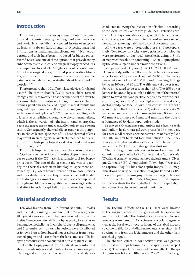
The thermal effect in connective tissue was greater than that in the epithelium in all the specimens except 1 (Fig. 2). The range of the measured thermal effect in the ep-ithelium was between 184 μm and 2,292 μm. The range

Adv Clin Exp Med. 2019;28(11):1513–1517 1515
of the thermal effect in the connective tissue was between 133 μm and 2,958 μm (Table 1). The mean of the thermal effect in the epithelium was 687 μm, while in connective tissue it was 1,407 μm. The mean total thermal effect was 2,094 μm (Fig. 3,4).
The most prominent thermal effect was observed in the specimens excised from attached gingiva. Only 1 specimen did not show any thermal effect.
Discussion
Specimens collected with a laser are usually compro-mised by thermal effects. It is often considered a common disadvantage that may cause tissue artefacts and marginal dysplastic changes.20,21 For this reason, many studies have
Table 1. The evaluated marginal thermal effects and thermal artefacts in all specimens in the study
Specimen No. Diagnosis Site of lesion Histologic artefact Thermal effect
in the epithelium [μm]
Thermal effect in connective
tissue [μm]
Total thermal effect [μm]
1kaposiform
hemangioendotheliomabuccal mucosa no ≃0 μm ≃0 μm ≃0 μm
2 granular cells tumor labial mucosa no 322.75 μm 133.4 μm 456.15 μm
3peripheral giant cell
granulomaattached gingiva no 184.24 μm 867.75 μm 1,052 μm
4 mucocele labial mucosa no 262 μm 968.26 μm 1,230.26 μm
5 focal fibrous hyperplasia labial mucosa
vacuolar degeneration at the basal
keratinocytes
429.62 μm 1,101.22 μm 1,530.83 μm
6squamous cell carcinoma
in situbuccal mucosa no 828.36 μm 1,151.1 μm 1,979.47 μm
7 focal epithelial hyperplasia buccal mucosa no 476.69 μm 1,646.86 μm 2,123.56 μm
8 focal fibrous hyperplasia attached gingiva no 1,245.19 μm 2,478.2 μm 3,723.39 μm
9 mucocele labial mucosadiathermocautery
artefacts831.74 μm 2,958.06 μm 3,789.8 μm
10 focal epithelial hyperplasia attached gingivadiathermocautery
artefacts2,292.94 μm 2,767.69 μm 5,060.63 μm
Fig. 1. A – representative photomicrograph of labial mucosa with focal fibrous hyperplasia. Original magnification ×5. B – high magnification of the lesion showing a hyperkeratotic epithelium with vacuolar degeneration of basal keratinocytes and a dense collagen matrix in the lamina propria (×20 magnification). C – surgical resection margin of the oral mucosa shows the thermal effects both in the epithelium and in the connective tissue (×10 magnification). The bars show the extension of tissue damage in the epithelium (thin bar) and in the connective tissue (thick bar). Hematoxylin and eosin (H&E) staining
Fig. 2. Bar chart of average values of the thermal effect obtained in epithelial and connective tissue
specimens1° 2° 3° 4° 5° 6° 7° 8° 9° 10°
thermal e�ects in connective tissuethermal e�ects in epithelial tissue
6,000
5,000
4,000
3,000
2,000
1,000
0
ther
mal
e�e
cts
[µm
]

G. Tenore, et al. CO2 laser for oral excisional biopsies1516
been carried out to assess this disadvantage and its im-pact on histological evaluation.7,17,19,22 In an experimental study performed on 25 Sprague Dawley rats, the influence of the thermal effect caused by different CO2 laser pow-ers (between 3 W and 12 W) was examined, and it was concluded that the CO2 laser, unrelated to the wattage, generates epithelial thermal damage similar to dysplastic changes. Thus, it was suggested that clinicians should take these changes into consideration.21
The control of power settings, spot diameter and pulse duration minimizes the thermal damage and enables achieving histologically acceptable specimens for diagno-sis. Many authors consider the thermal effect of lasers that impairs the histological evaluation to be caused by the op-erator rather than the laser itself.23
Therefore, many ex vivo and in vivo studies were car-ried out to find the ideal parameters for the laser that minimizes this thermal effect and consequently decreases the chance of thermal artefacts.5,9,21,24 In an ex vivo study, the histological analysis of specimens collected by differ-ent CO2 laser parameters were compared, and it was found that efficient cutting with minimal thermal effect can be achieved by a power of 3 W in continuous wave (CW) or in pulsed wave (PW) settings at a frequency of 50 Hz.3
The laser beam in PW has shown reduced thermal damage compared to CW in many animal studies.25–28
In a clinical study, the thermal damage outcomes follow-ing excision biopsy of 100 fibrous hyperplasia lesions using CO2 laser in PW and CW mode were compared. It was concluded that both laser modes produced similar thermal damage, and researchers recommended adding a 1 mm safety margin, especially in suspicious soft tissue lesions.25
Other studies were carried out to compare the thermal effect of lasers compared with other tools.7,11,16,22 Matsu-moto22 compared CO2 lasers with an electrotome. In his study, the optical microscopic examination of specimens excised by a CO2 laser, particularly in PW mode, produced less thermal damage than the electrotome. The thermal damage was estimated to be less than 500 µm and did not affect the pathological diagnosis.
In the present study, one of the collected specimens was carcinoma in situ, and histological evaluation was achieved without confusion. Utilizing a laser for excision biopsy of oral malignancy in an early stage has been re-ported.25,29–31 The nature of the lesion and water content appear to have an impact on the thermal effect during excision, as the most prominent thermal effect in our study was observed in a focal fibrous hyperplasia lesion.
Fig. 3. A – representative photomicrograph of the oral mucosa with peripheral giant cell granuloma. Original magnification ×5. B – high magnification of the lesion showing abundant multinucleated osteoclast-like giant cells in a fibroblastic stroma (×20 magnification). C – surgical resection margin of the oral mucosa shows the thermal effects both in the epithelium and in the connective tissue (×10 magnification). The bars show the extension of tissue damage in the epithelium (thin bar) and in the connective tissue (thick bar). Hematoxylin and eosin (H&E) staining
Fig. 4. A – representative photomicrograph of the oral mucosa with in situ squamous cell carcinoma arising on lichenoid keratosis. The lesion shows a pronounced hyperkeratosis and a papillary surface. Original magnification ×5. B – high magnification of the lesion showing a moderate inflammatory infiltrate in the lamina propria (×20 magnification). C – surgical resection margin of the oral mucosa shows the thermal effects both in the epithelium and in the connective tissue (×10 magnification). The bars show the extension of tissue damage in the epithelium (thin bar) and in the connective tissue (thick bar). Hematoxylin and eosin (H&E) staining

Adv Clin Exp Med. 2019;28(11):1513–1517 1517
In the sample with the lowest value, the thermal effect in both epithelium and connective tissue was so minimal that it was considered by the pathologist to be proximal to 0 (≃). Additionally, the working time (different depend-ing on the site of intervention) was reported to be a possible factor that affects the thermal effect.9
In fact, there is a difference in the laser parameters used in clinical and ex vivo studies. The parameters for this study were similar to the parameters recommended in the literature.22,25 In this study, the thermal effect was prominent in all the specimens, as the average of the to-tal thermal effect was approx. 2 mm (2,049 μm). It was generally higher in attached gingiva compared to other anatomical sites.
It is obvious that the thermal effect of the CO2 laser will occur and cannot be prevented but can be minimized. For that reason, the control of laser parameters and working time and adding laser safety margins are suggested.3,24 The resulting thermal effects of using a CO2 laser did not hamper the histological evaluation. Utilizing a laser in bi-opsy procedures should be tailored. Not only should laser parameters and safety margins be taken in consideration but also the working time, clinical accessibility, and the na-ture and water content of the tissue.
ORCID iDsGianluca Tenore https://orcid.org/0000-0001-9963-8052Gaspare Palaia https://orcid.org/0000-0001-8509-2255Ahmed Mohsen https://orcid.org/0000-0001-7857-4416Simone Ambrogiano https://orcid.org/0000-0003-0405-7189Cira Rosaria Tiziana Di Gioia https://orcid.org/0000-0003-4696-0560Marzena Dominiak https://orcid.org/0000-0001-8943-0549Umberto Romeo https://orcid.org/0000-0003-2439-2187
References1. Bernstein ML. Biopsy technique: The pathological considerations.
J Am Dent Assoc. 1978;96(3):438–443.2. Shafer WG, Hine MK, Levy BM. A Textbook of Oral Pathology. 4th ed.
Philadelphia, PA: W.B. Saunders Company; 1983.3. Palaia G, Del Vecchio A, Impellizzeri A, et al. Histological ex vivo eval-
uation of peri-incisional thermal effect created by a new-generation CO2 superpulsed laser. ScientificWorldJournal. 2014;2014:345685.
4. Romeo U, Palaia G, Del Vecchio A, et al. Effects of KTP laser on oral soft tissues: An in vitro study. Lasers Med Sci. 2010;25(4):539–543.
5. Spector N, Spector J, Ellis DL, Reinisch L. Reduction in lateral thermal damage using heat-conducting templates: A comparison of continu-ous wave and pulsed CO2 lasers. Lasers Surg Med. 2003;32(2):94–100.
6. Margarone JE, Natiella JR, Vaughan CD. Artifacts in oral biopsy spec-imens. J Oral Maxillofac Surg. 1985;43(3):163–172.
7. Tuncer I, Ozçakir-Tomruk C, Sencift K, Cöloğlu S. Comparison of con-ventional surgery and CO2 laser on intraoral soft tissue pathologies and evaluation of the collateral thermal damage. Photomed Laser Surg. 2010;28(1):75–79.
8. Pick RM, Colvard MD. Current status of lasers in soft tissue dental surgery. J Periodontol. 1993;64(7):589–602.
9. Cercadillo-Ibarguren I, España-Tost A, Arnabat-Domínguez J, Val-maseda-Castellón E, Berini-Aytés L, Gay-Escoda C. Histologic evalua-tion of thermal damage produced on soft tissues by CO2, Er,Cr:YSGG and diode lasers. Med Oral Patol Oral Cir Bucal. 2010;15(6):e912–918.
10. Romeo U, Del Vecchio A, Ripari F, Palaia G. Effects of different laser devices on oral soft tissues: An in vitro experience. J Laser Appl. 2007; 7(3):155–159.
11. Vescovi P, Corcione L, Meleti M, et al. Nd:YAG laser versus tradition-al scalpel: A preliminary histological analysis of specimens from the human oral mucosa. Lasers Med Sci. 2010;25(5):685–691.
12. Romeo U, Palaia G, Tenore G, Del Vecchio A, Nammour S. Excision of oral mucocele by different wavelength lasers. Indian J Dent Res. 2013;24(2):211–215.
13. Romeo U, Palaia G, Galanakis A, et al. Granular cell tumour of the tongue: Two clinical cases treated with laser. Italian Oral Surgery. 2012;11(5):s90–95.
14. Vescovi P, Del Vecchio A, Manfredi M, Fornaini C, Tenore G, Romeo U. The use of laser for treatment of oral mucosal diseases [in Italian]. Dental Cadmos. 2009;77(10):i–xvii.
15. Romeo U, Libotte F, Palaia G, et al. Histological in vitro evaluation of the effects of Er:YAG laser on oral soft tissues. Lasers Med Sci. 2012; 27(4):749–753.
16. Yagüe-García J, España-Tost AJ, Berini-Aytés L, Gay-Escoda C. Treat-ment of oral mucocele-scalpel versus CO2 laser. Med Oral Patol Oral Cir Bucal. 2009;14(9):e469–474.
17. Bornstein MM, Winzap-Kälin C, Cochran DL, Buser D. The CO2 laser for excisional biopsies of oral lesions: A case series study. Int J Peri-odontics Restorative Dent. 2005;25(3):221–229.
18. Huang IY, Chen CM, Kao YH, Worthington P. Treatment of mucocele of the lower lip with carbon dioxide laser. J Oral Maxillofac Surg. 2007; 65(5):855–858.
19. Suter VG, Altermatt HJ, Sendi P, Mettraux G, Bornstein MM. CO2 and diode laser for excisional biopsies of oral mucosal lesions: A pilot study evaluating clinical and histopathological parameters. Schweiz Monatsschr Zahnmed. 2010;120(8):664–671.
20. White JM, Chaudhry SI, Kudler JJ, Sekandari N, Schoelch ML, Silver-man S Jr. Nd:YAG and CO2 laser therapy of oral mucosal lesions. J Clin Laser Med Surg. 1998;16(6):299–304.
21. Seoane J, Caballero TG, Urizar JM, Almagro M, Mosquera AG, Varela- Centelles P. Pseudodysplastic epithelial artefacts associated with oral mucosa CO2 laser excision: An assessment of margin status. Int J Oral Maxillofac Surg. 2010;39(8):783–787.
22. Matsumoto K, Suzuki H, Usami Y, Hattori M, Komoro T. Histological evaluation of artifacts in tongue tissue produced by the CO2 laser and the electrotome. Photomed Laser Surg. 2008;26(6):573–577.
23. Convissar RA. Laser biopsy artifact. Oral Surg Oral Med Oral Pathol Oral Radiol Endod. 1997;84(5):458.
24. Merigo E, Clini F, Fornaini C, et al. Laser-assisted surgery with differ-ent wavelengths: A preliminary ex vivo study on thermal increase and histological evaluation. Lasers Med Sci. 2013;28(2):497–504.
25. Suter VG, Altermatt HJ, Dietrich T, Warnakulasuriya S, Bornstein MM. Pulsed versus continuous wave CO2 laser excisions of 100 oral fibrous hyperplasias: A randomized controlled clinical and histopathologi-cal study. Lasers Surg Med. 2014;46(5):396–404.
26. Lanzafame RJ, Naim JO, Rogers DW, Hinshaw JR. Comparison of con-tinuous-wave, chop-wave and super pulse laser wounds. Lasers Surg Med. 1988;8(2):119–124.
27. Walsh JT, Flotte TJ, Anderson RR, Deutsch TF. Pulsed CO2 laser tissue ablation: Effect of tissue type and pulse duration on thermal dam-age. Lasers Surg Med. 1988;8(2):108–118.
28. Sanders DL, Reinisch L. Wound healing and collagen thermal dam-age in 7.5-microsec pulsed CO2 laser skin incisions. Lasers Surg Med. 2000;26(1):22–32.
29. Frame JW. Removal of oral soft tissue pathology with the CO2 laser. J Oral Maxillofac Surg. 1985;43(11):850–855.
30. Azevedo LH, Galletta VC, De Paula Eduardo C, De Sousa SO, Migliari DA. Treatment of oral verrucous carcinoma with carbon dioxide laser. J Oral Maxillofac Surg. 2007;65(11):2361–2366.
31. Jerjes W, Upile T, Hamdoon Z, Mosse CA, Akram S, Hopper C. Pro-spective evaluation of outcome after transoral CO2 laser resection of T1/T2 oral squamous cell carcinoma. Oral Surg Oral Med Oral Pathol Oral Radiol Endod. 2011;112(2):180–187.