Embed Size (px)
Citation preview
Costorage and Corelease of Modulatory Peptide Cotransmitterswith Partially Antagonistic Actions on the Accessory Radula CloserMuscle of Aplysia californica
F. S. Vilim,1 D. A. Price,2 W. Lesser,2 I. Kupfermann,1 and K. R. Weiss3
1Center for Neurobiology and Behavior, College of Physicians and Surgeons, Columbia University, New York, New York10032, 2C. V. Whitney Laboratories, University of Florida, St. Augustine, Florida 32086, and 3Department of Physiologyand Biophysics, Mount Sinai School of Medicine, New York, New York 10029
Many neurons that contain a classical neurotransmitter alsocontain modulatory peptides, but it has been difficult toestablish unequivocally that these peptides are functionalcotransmitters. Here, we provide evidence for functionalcotransmission in a neuromuscular system of Aplysia. Usingimmunocytochemical techniques, we localize members oftwo peptide families, the small cardioactive peptides (SCPs)and the buccalins (BUCs), to a single subset of dense-corevesicles in the terminals of the cholinergic motorneuron B15.We describe a new preparation and method for the directdetection of released peptides and show that the SCPs andBUCs are released when neuron B15 is intracellularly stim-ulated. Consistent with their subcellular localization, theSCPs and BUCs are released in a stoichiometric ratio that isconstant across conditions that change the absolute amountof peptides released. Peptide release is calcium-dependent
but does not require muscle contractions. Thus, the releasecannot be attributed to a displacement of peptides that maybe present in the extracellular space. In previous studies, wecharacterized the physiological firing patterns of neuron B15.Here, we simulate these firing patterns and show that pep-tide release occurs. Additionally, we find that significantquantities of material are released under behaviorally rele-vant conditions. We find that concentrations of releasedpeptides in the muscle are in the concentration range inwhich exogenously applied peptides exert characterizedmodulatory actions on muscle contractions. Together, ourfindings provide strong support for the hypothesis that pep-tides contained in neuron B15 are functional cotransmitters.Key words: neuropeptides; dense-core vesicles; peptide re-
lease; Aplysia; cotransmission; EM immunocytochemistry; mo-tor neuron; modulation
When several putative transmitters are found in a single neuron,it is easy to assume that they function as cotransmitters. Thissupposition, however, has been difficult to verify. A recent reviewof cotransmission (Kupfermann, 1991) concluded that there wasno definitive evidence that cotransmission occurs under normalphysiological conditions. In general, the greatest obstacle to es-tablishing functional cotransmission has been the difficulty indemonstrating that putative cotransmitters are released whenneurons fire in physiological patterns.To overcome difficulties in the study of cotransmission, simpler,
more experimentally advantageous model systems were devel-oped. One such system consists of the accessory radula closer(ARC) muscle and its cholinergic motorneurons, B15 and B16.B15 and B16 synthesize peptides that potentiate contraction sizeand relaxation rate. B15 synthesizes (Cropper et al., 1987a) thesmall cardioactive peptides (SCPs), and B16 synthesizes the myo-modulins (MMs) (Cropper et al., 1987b, 1991; Brezina et al.,1995). In addition, both motorneurons synthesize the buccalins(BUCs), which depress muscle contractions (Cropper et al., 1988,1990a; Vilim et al., 1994).Studies of peptide release in the ARC (Whim and Lloyd, 1989,
1990; Cropper et al., 1990b; Hooper et al., 1994a,b; Probst et al.,1994) have taken two approaches. It has been demonstrated (1)that prolonged stimulation of B15 at physiological frequenciesdecreases SCP levels in nerve terminals, and (2) that physiologicalstimulation of B15 produces second messenger-mediated changesin ARC muscle that cannot be accounted for by ACh action butare produced by application of the SCPs. Although these findingssuggest that the SCPs are released under physiological conditions,alternative interpretations are possible. In depletion experiments,stimulation lasted up to 5 hr. Depletion could have resulted fromtoxic effects of increased calcium levels caused by the maintainedstimulation. When biochemical changes in muscles were observed,it could be argued that motor neurons release unknown sub-stances and that these could produce biochemical changes ob-served in the muscle.Additionally, previous experiments have sought only to demon-
strate the release of the peptides that exert potentiating actions.Because B15 also synthesizes the BUCs, fundamental questionsconcerning the physiological consequences of peptide release inthe ARC remain unanswered. Thus, are the BUCs and the SCPspackaged in the same vesicles and coreleased, or are they pack-aged in different vesicles and differentially released (Sossin et al.,1989; Wang and Scheller, 1991)? In either case, the characteristicsof motor neuron firing that result in the release of these peptideshave to be determined if one is to understand the functional roleof cotransmission. Therefore, in the studies presented in thispaper and its companion (this issue), we have undertaken (1) todetermine the subcellular distribution of neuropeptides in neuron
Received June 24, 1996; revised Sept. 20, 1996; accepted Sept. 24, 1996.This work was supported in part by National Institutes of Health Grants MH
36730, MH50235, and GM32009.Correspondence should be addressed to Klaudiusz R. Weiss, Department of
Physiology and Biophysics, Mt. Sinai School of Medicine, 1 Gustave L. Levy Place,New York, NY 10029.Copyright q 1996 Society for Neuroscience 0270-6474/96/168092-13$05.00/0
The Journal of Neuroscience, December 15, 1996, 16(24):8092–8104
B15, (2) to develop a preparation that can be used to investigatepeptide release under physiological conditions, and (3) to definehow behaviorally relevant changes in the B15 activity determinethe characteristics of peptide release. Taken together, the purposeof our studies was to provide evidence for the existence of co-transmission and to elucidate the behavioral function(s) of thismode of signaling.
MATERIALS AND METHODSAnimals. Specimens of Aplysia californica were obtained from Marinus.The animals were maintained at 14–168C on a 12 hr light/dark cycle andfed every 3 d. Animals in the range of 50–100 gm were used for themorphology experiments, and animals in the range of 300–600 gm wereused for release experiments. Isotonic MgCl2 (25–50% body weight) wasused to immobilize the animals, and all stages of the dissection werecarried out in the animals’ own hemolymph containing the added MgCl2.Backfills. A unilateral ARC preparation was pinned out on SYLGARD
(Dow Corning, Corning, NY) in a 3-inch-deep dish. A 3-inch-deepsubchamber was sealed around the distal portion of the ARC withsilicone grease. The space outside the subchamber was filled almost to thetop with L-15 (adjusted to Aplysia salinity and buffered with 10 mMHEPES to pH 7.6). The fluid inside the subchamber was evacuated, andthe viability of the seal was evaluated. If the seal was adequate, the fillingsolution (1 M CoCl2 or 1 M NiCl2) was added to the subchamber inamounts sufficient to completely submerge the ARC (;100 ml). Thehydrostatic back pressure prevented the nonspecific diffusion of fillingsolution. The preparation was then placed at 48C, and the filling solutionwas changed daily. After 2–3 d, the filling solution in the subchamber waswashed out with several changes of artificial seawater (ASW), and thepreparation was pinned out in a smaller dish for developing. The prep-aration was developed by the addition of a one-tenth volume of saturateddithio-oximide in 95% ethanol, which produces a blue–black precipitatewith NiCl2 and a red–brown precipitate with CoCl2 (Quicke and Brace,1979). After the filled cells stopped getting darker (;10 min), the prep-aration was washed with ASW and fixed with 4% paraformaldehydeovernight. The tissue was washed free of fixative, dehydrated in a seriesof ethanols, and viewed in methyl salycilate.Antibodies. The rabbit antibody to SCPb used for immunocytochemis-
try was a kind gift from Dr. Richard Scheller (Stanford University,Stanford, CA). The rabbit antibody to SCPb used for radioimmunoassay(RIA) was a kind gift from Dr. H. R. Morris (Imperial College). Therabbit antibody to buccalin used for immunocytochemistry and RIA wasraised against BUCa coupled to bovine serum albumin (BSA). The ratantibody to SCPb used for immunocytochemistry was raised againstpeptide coupled to bovine thyroglobulin (BTG). The peptides werecoupled to the carrier protein with 1-ethyl-3-(3-dimethylaminopropyl)-carbodiimide (EDC). The rabbit buccalin antibodies were prepared byBabco (Richmond, CA), and the rat SCP antibody was made in house. Allreagents were purchased from Sigma (St. Louis, MO) except the pep-tides, which were synthesized by Applied Biosystems (Foster City, CA).LM immunocytochemistry. A number of methods were used, but the
procedure that consistently gave superior results was one adapted fromLlewellyn-Smith et al. (1985). All of the procedures were carried out atroom temperature unless stated otherwise. The ganglia and muscles wereexcised, washed with a 1:1 mixture of ASW and isotonic MgCl2, andpinned out on SYLGARD slabs. The slabs were placed in 50 ml conicaltubes containing 25 ml of freshly prepared fixative (4% paraformalde-hyde, 0.2% picric acid, 25% sucrose, 0.1 M sodium phosphate, pH 7.6),shaken for 30 min, and left to fix overnight on a rocker. After fixation, thetissue was rinsed with several changes of distilled water until the yellowcolor dissipated. The tissue was then washed with several changes of 50%ethanol and transferred to 50% ethanol 3% H2O2 for 1 d to permeabilizethe tissue and reduce background fluorescence. After a wash with severalchanges of distilled water, nonspecific staining was blocked by overnightincubation with 10% normal donkey serum (NDS) in RIA buffer (seebelow). The tissue was exposed to the primary antibody in the same bufferfor 2–7 d, washed with RIA buffer with several changes for 1 d, and placedin fluorescently labeled (lissamine rhodamine and DTAF) secondaryantibody raised in donkey (Jackson ImmunoResearch, West Grove, PA)for 2 d. Secondary antibody was washed out with several changes of RIAbuffer for 1 d and viewed on a Leitz microscope equipped with epifluo-rescence and the appropriate filterpacks for rhodamine (N-2) and fluo-rescein (D). Selected specimens were photographed on T-Max ASA 400film (Eastman Kodak, Rochester, NY).
EM immunocytochemistry. The procedure was adapted from Reed et al.(1988) and Merighe et al. (1989). Tissue was pinned out on SYLGARDslabs, placed in a conical tube with 30 ml of fixative (4% glutaraldehyde,10% sucrose, 11 mM magnesium chloride, 0.2 M Na-HEPES, pH 7.6),shaken for 15 min, and placed on a rocker for 3 hr. The fixative waswashed away with several changes of buffer (fixative minus glutaralde-hyde) during 3 hr. The tissue was stained en bloc with buffer containing1% uranium acetate for 3 hr and then post-fixed with 1% osmiumtetroxide in buffer at 48C for 1 hr. The tissue was washed with severalchanges of distilled water, dehydrated in an ethanol series, washed withpropylene oxide, infiltrated with EMbed 812, and polymerized at 608C for2 d. Ultrathin sections were cut on a Sorvall ultratome, and pale goldsections were collected on 200-mesh, thin-bar hex grids. The grid wasthen floated sequentially on both sides on saturated sodium metaperio-date, which etched both sides of the section; immersion during etchingcaused section detachment. The grids were then immersed in distilledwater for 15 min, followed by Tris-buffered saline (TBS), pH 8.2, for 15min and then were blocked with 8% normal goat serum (NGS) in TBS for1 hr. A 1:100 dilution of primary antibody in 4% NGS–TBS was used tostain the sections overnight. The grids were washed 4 times for 30 mineach with 4% NGS–TBS, followed by 1 hr in 1:4 dilution of gold labeledsecondary antibodies (Amersham, Arlington Heights, IL) in 4% NGS–TBS. The grids were again washed 4 times for 30 min each with 4%NGS–TBS, followed by 2 times for 5 min each with 0.1 M PBS. Thegold-labeled antibodies were fixed to the sections with 2% glutaraldehydein 0.1 M PBS and washed with several changes of dH2O. The sectionswere lightly counterstained with 5% aqueous uranyl acetate for 30–60sec, followed by Reynolds lead citrate for 30–60 sec. Sections wereexamined and photographed with a JOEL 100 CX at 80 kV. Electronmicroscopy supplies and reagents were obtained from EMS (Fort Wash-ington, PA). Normal sera were obtained from Jackson ImmunoResearch.Release preparation. The ARC–buccal ganglion preparation was iso-
lated as described previously (Cohen et al., 1978) with some minordifferences. The buccal and cerebral ganglia were dissected free, but theconnection between nerve 3 and the buccal mass was kept intact. Thisnerve contains the axons of B15 and B16 that innervate the ARC. Theconnective tissue midway along the ARC was preserved because it housesthe major artery used for perfusion. The distal end of the ARC wasdissected free of the radular sac and ligated with 6-0 silk suture to preventloss of perfusate via the distal cut end.The ARC artery was cannulated with a 30 gauge blunt needle and
perfused with ASW at 20 ml/min with a multistaltic pump. The deadvolume of the perfusion system was ;100 ml so that solutions perfusingthe ARC could be changed while a constant flow rate was maintained.The perfused muscle was hung outside a 60 mm dish, and the nerveconnecting the ARC to the ganglion was passed through a slit in the sideof the dish, so that the buccal ganglion could be pinned inside the dish.The slit was filled with silicone grease, the ganglion was desheathed, andthe ARC was partially encased with parafilm coated with silicone grease.The parafilm formed a subchamber around the ARC, preventing it fromdrying out, and the silicone grease prevented nonspecific adsorption ofreleased peptides. The dish was clamped onto a platform with the ARChanging off the side, permitting drops of ARC perfusate to form and fallfreely. The drops were collected every 2.5 min into glass test tubes (12 375 mm) that were then processed for RIA. The solution bathing theganglion contained 25% isotonic MgCl2 to prevent spontaneous activityof the neurons in the ganglion. This arrangement ensured that only themotorneuron being fired could be responsible for the peptide releasedwithin the ARC. The motorneuron was impaled with two independentglass microelectrodes, one for recording voltage and one for injectingcurrent. The resulting configuration of the release preparation is illus-trated in Figure 1. The temperature of the preparation was maintained at15 6 0.58C (i.e., the temperature of the aquaria housing the animals) bycooling the room with an air conditioner.RIA. A 3-( p-hydroxyphenyl)-propionic acid (desamino-tyrosyl) deriv-
ative of buccalin A was prepared by reaction of 1 mmol of peptide, 1 mmolof triethylamine, and 3 mmol of Bolton–Hunter reagent [3-( p-hydroxyphenyl)-propionic acid N-hydroxy-succinimide ester] in 100 ml ofdimethylformamide for 6 hr at 48C followed by purification on RP-HPLCwith a 15–45% acetonitrile gradient with 0.01 M TFA as counterion.Peptides (the desaminotyrosinoyl-buccalin or SCPb) were iodinated
with 125I using chloramine-T as follows. To the peptide (;1 nmol in 10 mlof 0.5 M phosphate buffer, pH 7.0) was added the radioactive sodiumiodide solution (1.5 mCi in 1–3 ml) followed by 5 ml of chloramine-Tsolution (freshly prepared in the 0.5 M phosphate buffer, 2 mg/ml), and
Vilim et al. • Storage and Release of Peptide Cotransmitters J. Neurosci., December 15, 1996, 16(24):8092–8104 8093
the solution was mixed well by repeated pipetting with an aerosol barriertip. Sodium metabisulfite (100 ml of 5 mg/ml in the 0.5 M phosphatebuffer) was added 15–30 sec after the chloramine-T, and the solution wasmixed by repeated pipetting for 1 min. After another 4–5 min, theiodination mixture was applied to a Sep-Pak (C18, Waters Associates,Milford, MA) that had been wetted previously with acetonitrile andrinsed with distilled water. The Sep-Pak was washed with 20 ml of water(discarded as waste), and the peptide was eluted with 80% aqueousacetonitrile containing 0.01 M TFA (a total of 5 ml collected in 1 mlfractions). Usually, the first 1 ml fraction was kept and the othersdiscarded after counting revealed that the majority of the elutable radio-activity resided in the first fraction. The partially purified (by Sep-Pak)trace could be kept for several months in the freezer as a stock for HPLCpurification of monoiodinated peptide. Because some uniodinated andunoxidized peptide remained in the Sep-Pak purified trace, it was treatedwith 1% H2O2 for 1 hr to fully oxidize all methionine residues to thesulfoxides. After this treatment, the desired monoiodo form of thepeptide could be separated from both the uniodinated and the diiodi-nated forms on RP-HPLC with a gradient as above. Radioactive peakscorresponding to the monoiodinated form were lyophilized and resus-pended in RIA buffer (154 mM NaCl, 10 mM Na2HPO4, 50 mM EDTA,0.25 mM merthiolate, 1% BSA, pH 7.5) to a final activity of 10,000–15,000cpm per 100 ml. Antibodies were diluted in RIA buffer to a point at which100 ml would bind ;50% of the counts in 100 ml of iodinated peptidetrace. The sample volume (blanks, peptide standards, perfusate) was 50ml in each reaction. The reaction was carried out for 2–3 d at 48C and was
terminated by the addition of 2 ml of RIA charcoal (10 mM Na2HPO4,0.25 mM merthiolate, 0.25% activated charcoal, 0.025% 70,000 kDadextran, pH 7.5). After 15 min, the samples were spun to separate thecharcoal and supernatant. The supernatant, containing the bound pep-tide, was decanted and counted in a gamma counter. Standard curveswere generated from serial dilutions of peptide in ASW containing 1%BSA to prevent sticking of the peptide to the tubes and pipette tips. Thesample volume was 50 ml, which matched the volume of the drops in theexperiment, and each tube had half the peptide of the previous tube. Aspreadsheet program (Kaleidagraph 2.1) was used to plot the standardcurves, to convert counts bound to femtomoles of peptide in the un-knowns, and to graph the data. A statistical analysis program (StatView4.5) was used to perform a within-subjects repeated-measures ANOVAon relevant data to assess the overall level of statistical significance.Individual comparisons were performed using paired t tests. All reagentswere obtained from Sigma, except where noted otherwise.
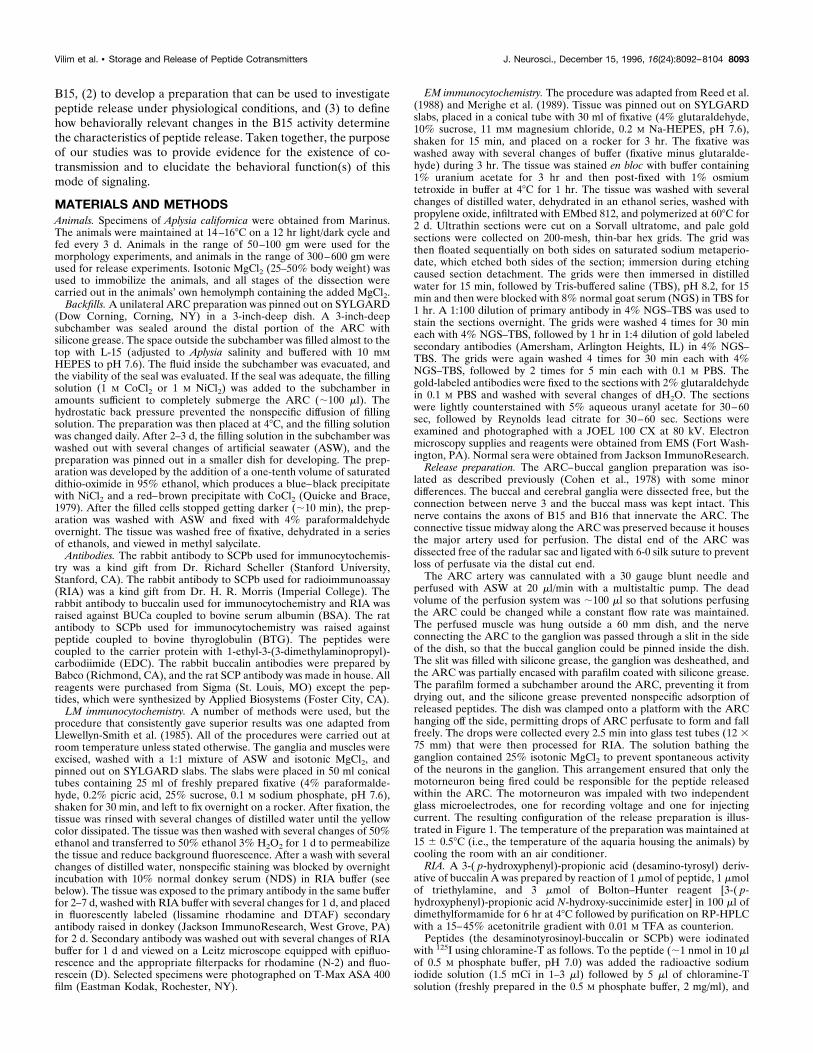
RESULTSThe ARC is innervated by a limited number of neuronsElectrophysiological evidence indicates that B15 and B16 are theonly motorneurons that innervate the ARC (Cohen et al., 1978).The results of ARC muscle backfills support this finding, becauseonly two motorneurons were retrogradely labeled and their posi-tion and size correspond to those of cells B15 and B16 (Fig. 2).Some backfills also resulted in retrograde labeling of small neu-rons in the sensory cluster (Fiore and Geppetti, 1980), whichapparently represent the sensory innervation of the ARC (datanot shown). Thus, only a small number of neurons are known toinnervate the ARC, thereby simplifying the identification of itsprocesses.
Antibody staining corresponds to thebiochemical localizationFigure 3, A1 and A2, shows the simultaneous staining of a buccalhemiganglion with a rabbit BUCa antibody and a rat SCPb anti-body. The buccal hemiganglion shows cells in the positions, re-spectively, of B15 (white arrowheads), which stains for both SCP(A1) and buccalin (A2), and B16 (black arrowheads), which stainsfor buccalin alone. These patterns match the previously reporteddistribution of staining and biochemical localization in the buccalganglion for SCP (Lloyd et al., 1987a; Church and Lloyd, 1991)
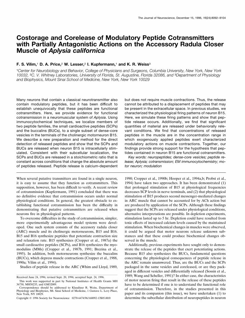
Figure 1. Diagram of the preparation used to measure peptide releasewithin the ARC. The buccal ganglion is bathed in high-Mg21 ASW toprevent spontaneous neuronal activity. The motorneuron is impaled withtwo microelectrodes (one to measure voltage, the other to inject current).These conditions enable precise control over the firing of the motorneu-ron and ensure that only the motorneuron being fired is responsible forthe peptide released within the ARC. The ARC (medium gray) is sus-pended outside the dish and encased in silicone grease (light gray) andparafilm to prevent dehydration. The nerve connecting the buccal gan-glion to the ARC runs through a slit in the side of the dish that issubsequently filled with silicone grease to prevent leakage of solutionsfrom the dish. The ARC is perfused via an artery midway along its length,and the perfusate is collected in drops directly into the tubes that aresubsequently used for the measurement of their peptide content. Thetemperature and length of the ARC were also measured with a temper-ature probe mounted on an isotonic force transducer (not shown). The tipof the temperature probe was inserted at the bottom of the loop betweenthe base muscle (dark gray) and the artery.
Figure 2. Cobalt backfill of a buccal ganglion from the ARC muscle. Thearrow points to two neurons in the rostral aspect of the ventral motorneu-ron cluster stained for cobalt. The size and position of the neurons areconsistent with the larger one being B15 and the smaller one being B16.Scale bar, 0.5 mm. This agrees with the electrophysiological data indicat-ing that the ARC is innervated by only two motorneurons.
8094 J. Neurosci., December 15, 1996, 16(24):8092–8104 Vilim et al. • Storage and Release of Peptide Cotransmitters
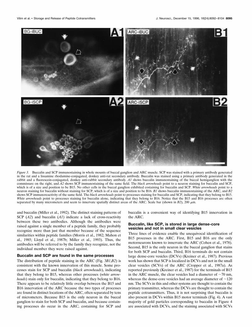
and buccalin (Miller et al., 1992). The distinct staining patterns ofSCP (A2) and buccalin (A1) indicate a lack of cross-reactivitybetween these two antibodies. Although the antibodies wereraised against a single member of a peptide family, they probablyrecognize more than just that member because of the sequencesimilarities within peptide families (Morris et al., 1982; Mahon etal., 1985; Lloyd et al., 1987b; Miller et al., 1993). Thus, theantibodies will be referred to by the family they recognize, not theindividual member they were raised against.
Buccalin and SCP are found in the same processesThe distribution of peptide staining in the ARC (Fig. 3B1,B2) isconsistent with the known innervation of this muscle. Some pro-cesses stain for SCP and buccalin (black arrowheads), indicatingthat they belong to B15, whereas other processes (white arrow-heads) stain only for buccalin, indicating that they belong to B16.There appears to be relatively little overlap between the B15 andB16 innervation of the ARC because the two types of processesare found in distinct locations of the ARC, often separated by tensof micrometers. Because B15 is the only neuron in the buccalganglion to stain for both SCP and buccalin, and because costain-ing processes do occur in the ARC, costaining for SCP and
buccalin is a convenient way of identifying B15 innervation inthe ARC.
Buccalin, like SCP, is stored in large dense-corevesicles and not in small clear vesiclesThree lines of evidence enable the unequivocal identification ofB15 processes in the ARC. First, B15 and B16 are the onlymotorneurons known to innervate the ARC (Cohen et al., 1978).Second, B15 is the only neuron in the buccal ganglion that stainsfor both SCP and buccalin. Third, B16 terminals do not containlarge dense-core vesicles (DCVs) (Kreiner et al., 1987). Previouswork has shown that SCP is localized in DCVs and not in the smallclear vesicles (SCVs) of the ARC (Cropper et al., 1987a). Asreported previously (Kreiner et al., 1987) for the terminals of B15in the ARC muscle, the clear vesicles had a diameter of ;70 nm,whereas the dense-core vesicles had an average diameter of ;120nm. The SCVs in this and other systems are thought to contain theprimary transmitter, whereas the DCVs are thought to contain thepeptide cotransmitters. Thus, it is not surprising that buccalin isalso present in DCVs within B15 motor terminals (Fig. 4). A vastmajority of gold particles corresponding to buccalin in Figure 4are associated with DCVs, and the staining associated with SCVs
Figure 3. Buccalin and SCP immunostaining in whole mounts of buccal ganglion and ARC muscle. SCP was stained with a primary antibody generatedin the rat and a lissamine rhodamine-conjugated, donkey anti-rat secondary antibody. Buccalin was stained using a primary antibody generated in therabbit and a fluorescein-conjugated, donkey anti-rabbit secondary antibody. A1 shows buccalin immunostaining of the buccal hemiganglion with thecommissure on the right, and A2 shows SCP immunostaining of the same field. The black arrowheads point to a neuron staining for buccalin and SCP,which is of a size and position to be B15. No other cells in the buccal ganglion exhibited costaining for buccalin and SCP. White arrowheads point to aneuron staining for buccalin without staining for SCP, which is of a size and position to be B16. B1 shows buccalin immunostaining of the ARC, and B2shows SCP immunoreactivity of the same field. The black arrowheads point to processes staining for buccalin and SCP, indicating that they belong to B15.White arrowheads point to processes staining for buccalin alone, indicating that they belong to B16. Notice that the B15 and B16 processes are oftenseparated by many micrometers and seem to innervate spatially distinct areas of the ARC. Scale bar (shown in B2), 200 mm.
Vilim et al. • Storage and Release of Peptide Cotransmitters J. Neurosci., December 15, 1996, 16(24):8092–8104 8095
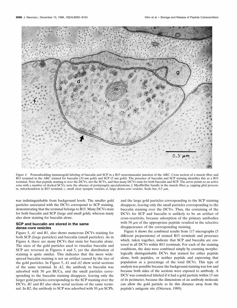
was indistinguishable from background levels. The smaller goldparticles associated with the DCVs correspond to SCP staining,demonstrating that the terminal belongs to B15. Many DCVs stainfor both buccalin and SCP (large and small gold), whereas manyalso show staining for buccalin alone.
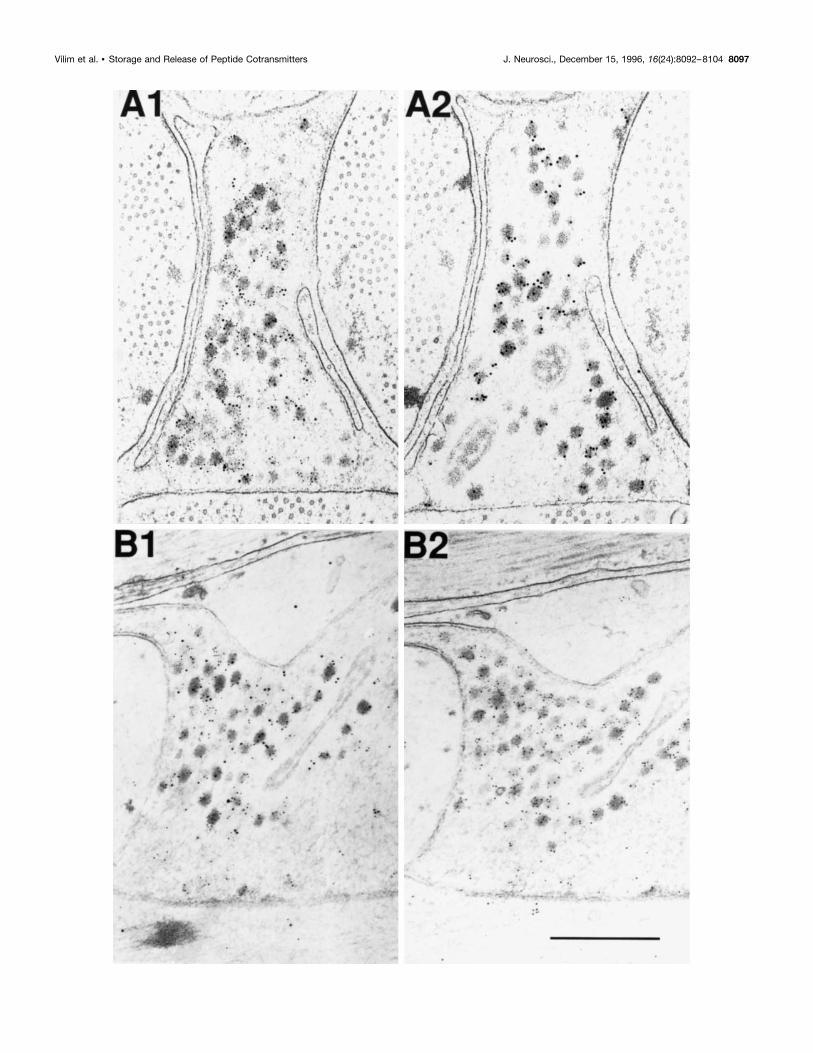
SCP and buccalin are stored in the samedense-core vesiclesFigure 5, A1 and B1, also shows numerous DCVs staining forboth SCP (large particles) and buccalin (small particles). As inFigure 4, there are many DCVs that stain for buccalin alone.The sizes of the gold particles used to visualize buccalin andSCP are reversed in Figures 4 and 5, yet the distribution ofstaining is quite similar. This indicates that the more wide-spread buccalin staining is not an artifact caused by the size ofthe gold particles. In Figure 5, A1 and A2 show serial sectionsof the same terminal. In A2, the antibody to buccalin wasadsorbed with 50 mM BUCa, and the small particles corre-sponding to the buccalin staining disappear, leaving only thelarger gold particles corresponding to the SCP staining over theDCVs. B1 and B2 also show serial sections of the same termi-nal. In B2, the antibody to SCP was adsorbed with 50 mM SCPb,
and the large gold particles corresponding to the SCP stainingdisappear, leaving only the small particles corresponding to thebuccalin staining over the DCVs. Thus, the costaining of theDCVs for SCP and buccalin is unlikely to be an artifact ofcross-reactivity, because adsorption of the primary antibodieswith 50 mM of the appropriate peptide resulted in the selectivedisappearance of the corresponding staining.Figure 6 shows the combined results from 117 micrographs (5
different preparations) of stained B15 terminals and processeswhich, taken together, indicate that SCP and buccalin are cos-tored in all DCVs within B15 terminals. For each of the stainingconditions, the data were combined simply by counting morpho-logically distinguishable DCVs that stained for either peptidealone, both peptides, or neither peptide and expressing thatpopulation as a percentage of the total DCVs. This type ofanalysis was possible because the background staining was low andbecause both sides of the sections were exposed to antibody. ADCV was considered labeled if it had a gold particle within 15 nmof its perimeter, because the dimensions of an antibody moleculecan allow the gold particle to lie this distance away from thepeptide’s antigenic site (Ottersen, 1989).
Figure 4. Postembedding immunogold labeling of buccalin and SCP in a B15 neuromuscular junction of the ARC. Cross section of a muscle fiber andB15 terminal in the ARC stained for buccalin (10 nm gold) and SCP (5 nm gold). The presence of buccalin and SCP staining identifies this as a B15terminal. Note that peptide staining is over the DCVs, not the SCVs, and that many DCVs stain for both buccalin and SCP. The arrow points to an activezone with a number of docked SCVs; note the absence of postsynaptic specializations. f, Myofibrillar bundle in the muscle fiber; g, capping glial process;m, mitochondrion in B15 terminal; c, small clear synaptic vesicles; d, large dense-core vesicles. Scale bar, 0.5 mm.
8096 J. Neurosci., December 15, 1996, 16(24):8092–8104 Vilim et al. • Storage and Release of Peptide Cotransmitters
Vilim et al. • Storage and Release of Peptide Cotransmitters J. Neurosci., December 15, 1996, 16(24):8092–8104 8097
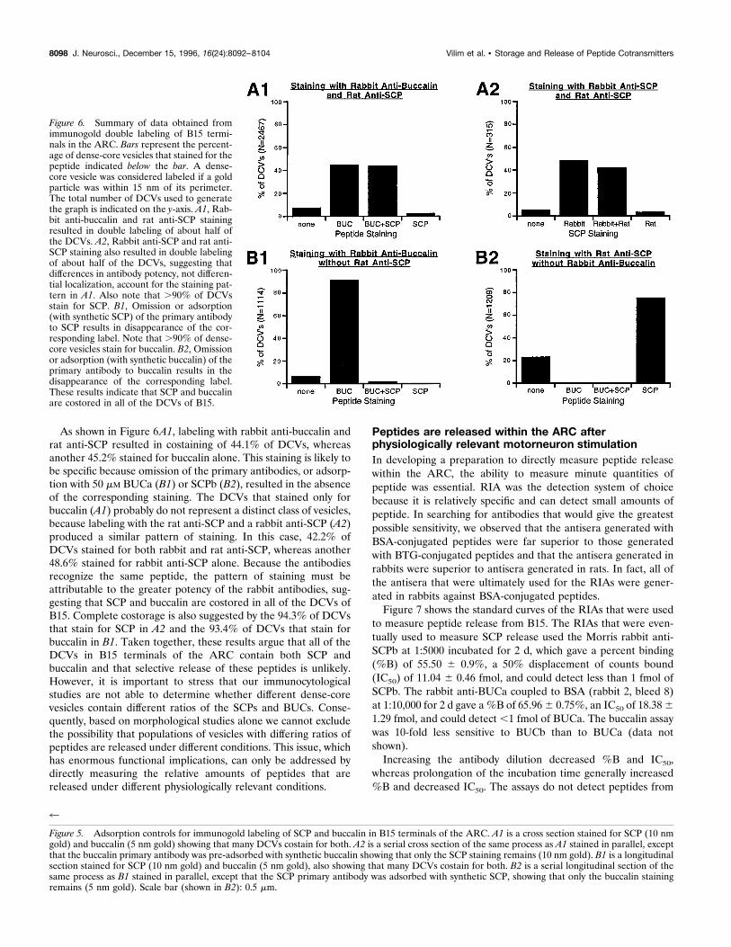
As shown in Figure 6A1, labeling with rabbit anti-buccalin andrat anti-SCP resulted in costaining of 44.1% of DCVs, whereasanother 45.2% stained for buccalin alone. This staining is likely tobe specific because omission of the primary antibodies, or adsorp-tion with 50 mM BUCa (B1) or SCPb (B2), resulted in the absenceof the corresponding staining. The DCVs that stained only forbuccalin (A1) probably do not represent a distinct class of vesicles,because labeling with the rat anti-SCP and a rabbit anti-SCP (A2)produced a similar pattern of staining. In this case, 42.2% ofDCVs stained for both rabbit and rat anti-SCP, whereas another48.6% stained for rabbit anti-SCP alone. Because the antibodiesrecognize the same peptide, the pattern of staining must beattributable to the greater potency of the rabbit antibodies, sug-gesting that SCP and buccalin are costored in all of the DCVs ofB15. Complete costorage is also suggested by the 94.3% of DCVsthat stain for SCP in A2 and the 93.4% of DCVs that stain forbuccalin in B1. Taken together, these results argue that all of theDCVs in B15 terminals of the ARC contain both SCP andbuccalin and that selective release of these peptides is unlikely.However, it is important to stress that our immunocytologicalstudies are not able to determine whether different dense-corevesicles contain different ratios of the SCPs and BUCs. Conse-quently, based on morphological studies alone we cannot excludethe possibility that populations of vesicles with differing ratios ofpeptides are released under different conditions. This issue, whichhas enormous functional implications, can only be addressed bydirectly measuring the relative amounts of peptides that arereleased under different physiologically relevant conditions.
Peptides are released within the ARC afterphysiologically relevant motorneuron stimulationIn developing a preparation to directly measure peptide releasewithin the ARC, the ability to measure minute quantities ofpeptide was essential. RIA was the detection system of choicebecause it is relatively specific and can detect small amounts ofpeptide. In searching for antibodies that would give the greatestpossible sensitivity, we observed that the antisera generated withBSA-conjugated peptides were far superior to those generatedwith BTG-conjugated peptides and that the antisera generated inrabbits were superior to antisera generated in rats. In fact, all ofthe antisera that were ultimately used for the RIAs were gener-ated in rabbits against BSA-conjugated peptides.Figure 7 shows the standard curves of the RIAs that were used
to measure peptide release from B15. The RIAs that were even-tually used to measure SCP release used the Morris rabbit anti-SCPb at 1:5000 incubated for 2 d, which gave a percent binding(%B) of 55.50 6 0.9%, a 50% displacement of counts bound(IC50) of 11.04 6 0.46 fmol, and could detect less than 1 fmol ofSCPb. The rabbit anti-BUCa coupled to BSA (rabbit 2, bleed 8)at 1:10,000 for 2 d gave a %B of 65.966 0.75%, an IC50 of 18.3861.29 fmol, and could detect ,1 fmol of BUCa. The buccalin assaywas 10-fold less sensitive to BUCb than to BUCa (data notshown).Increasing the antibody dilution decreased %B and IC50,
whereas prolongation of the incubation time generally increased%B and decreased IC50. The assays do not detect peptides from
4
Figure 5. Adsorption controls for immunogold labeling of SCP and buccalin in B15 terminals of the ARC. A1 is a cross section stained for SCP (10 nmgold) and buccalin (5 nm gold) showing that many DCVs costain for both. A2 is a serial cross section of the same process as A1 stained in parallel, exceptthat the buccalin primary antibody was pre-adsorbed with synthetic buccalin showing that only the SCP staining remains (10 nm gold). B1 is a longitudinalsection stained for SCP (10 nm gold) and buccalin (5 nm gold), also showing that many DCVs costain for both. B2 is a serial longitudinal section of thesame process as B1 stained in parallel, except that the SCP primary antibody was adsorbed with synthetic SCP, showing that only the buccalin stainingremains (5 nm gold). Scale bar (shown in B2): 0.5 mm.
Figure 6. Summary of data obtained fromimmunogold double labeling of B15 termi-nals in the ARC. Bars represent the percent-age of dense-core vesicles that stained for thepeptide indicated below the bar. A dense-core vesicle was considered labeled if a goldparticle was within 15 nm of its perimeter.The total number of DCVs used to generatethe graph is indicated on the y-axis. A1, Rab-bit anti-buccalin and rat anti-SCP stainingresulted in double labeling of about half ofthe DCVs. A2, Rabbit anti-SCP and rat anti-SCP staining also resulted in double labelingof about half of the DCVs, suggesting thatdifferences in antibody potency, not differen-tial localization, account for the staining pat-tern in A1. Also note that .90% of DCVsstain for SCP. B1, Omission or adsorption(with synthetic SCP) of the primary antibodyto SCP results in disappearance of the cor-responding label. Note that .90% of dense-core vesicles stain for buccalin. B2, Omissionor adsorption (with synthetic buccalin) of theprimary antibody to buccalin results in thedisappearance of the corresponding label.These results indicate that SCP and buccalinare costored in all of the DCVs of B15.
8098 J. Neurosci., December 15, 1996, 16(24):8092–8104 Vilim et al. • Storage and Release of Peptide Cotransmitters
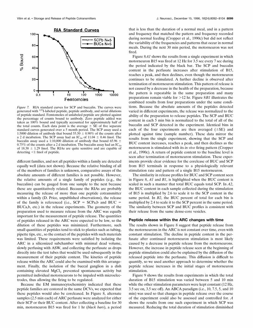
different families, and not all peptides within a family are detectedequally well (data not shown). Because the relative binding of allof the members of families is unknown, comparative assays of theabsolute amounts of different families is not possible. However,the relative amounts of a single family of peptides (e.g., thebuccalins) can be gauged from one sample to the next becausethese are quantitatively related. Because the RIAs are probablymeasuring the release of more than one peptide cotransmitterwithin a family (D. Price, unpublished observations), the releaseof the family is referenced (i.e., SCP 5 SCPa,b and BUC 5BUCa,b, etc.) in the release experiments. The geometry of thepreparation used to measure release from the ARC was equallyimportant for the measurement of peptide release. The quantitiesof peptides released in the ARC were expected to be low, so thedilution of these peptides was minimized. Furthermore, suchsmall quantities of peptides tend to stick to plastics such as tubing,pipette tips, etc., so the contact of the peptides with such materialswas limited. These requirements were satisfied by isolating theARC in a siliconized subchamber with minimal dead volume,slowly perfusing with ASW, and collecting the perfusate as dropsdirectly into the test tubes that would be used subsequently in themeasurement of their peptide content. The kinetics of peptiderelease within the ARC could also be examined with this arrange-ment. Finally, the isolation of the buccal ganglion in a dishcontaining elevated MgCl2 prevented spontaneous activity butpermitted individual motorneurons to be impaled with microelec-trodes, thus allowing the firing to be regulated.Because the EM immunocytochemistry indicated that these
peptide families are costored in the same DCVs, we expected thatthese peptides would also be coreleased. In Figure 8, alternatesamples (2.5 min each) of ARC perfusate were analyzed for eithertheir SCP or their BUC content. After collecting a baseline for 30min, motorneuron B15 was fired for 1 hr (black bars), a period
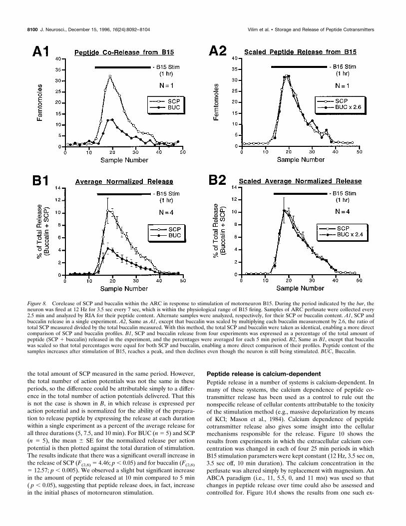
that is less than the duration of a normal meal, and in a patternand frequency that matched the pattern and frequency recordedduring normal feeding (Cropper et al., 1990c) but did not reflectthe variability of the frequencies and patterns that occur in normalmeals. During the next 30 min period, the motorneuron was notfired.Figure 8A1 shows the results from a single experiment in which
motorneuron B15 was fired at 12 Hz for 3.5 sec every 7 sec duringthe period indicated by the black bar. The SCP and buccalincontent in the perfusate increases after stimulation of B15,reaches a peak, and then declines, even though the motorneuroncontinues to be stimulated. A further decline is observed aftertermination of motorneuron stimulation. This pattern of release isnot caused by a decrease in the health of the preparation, becausethe pattern is repeatable in the same preparation and manypreparations remain viable for .12 hr. Figure 8B1 illustrates thecombined results from four preparations under the same condi-tions. Because the absolute amounts of the peptides detectedvaried in different experiments, the release was normalized to theability of the preparation to release peptides. The SCP and BUCcontent in each 5 min bin is normalized to the total of all of thebuccalin and SCP detected in the experiment. Identical bins ineach of the four experiments are then averaged (6SE) andplotted against time (sample number). These data mirror theresults from the single experiment, showing that the SCP andBUC content increases, reaches a peak, and then declines as themotorneuron is stimulated with its in vivo firing pattern (Cropperet al., 1990c). A return of peptide content to the baseline level isseen after termination of motorneuron stimulation. These exper-iments provide clear evidence for the corelease of BUC and SCPfrom B15 terminals in response to a physiologically relevantstimulation rate and pattern of a single B15 motorneuron.The similarity in release profiles for BUC and SCP content seen
in Figure 8, A1 and B1, is highlighted when the BUC content isscaled in such a manner that total BUC equals total SCP. In A2,the BUC content in each sample collected during the stimulationof B15 is multiplied by 2.6 to scale it to the SCP content in thesame period. In B2, the BUC percent of total for each bin ismultiplied by 2.4 to scale it to the SCP percent in the same period.The similarity between SCP and BUC profiles is consistent withtheir release from the same dense-core vesicles.
Peptide release within the ARC changes with timeThe results shown in Figure 8 suggest that peptide release fromthe motorneurons in the ARC is not constant over time, even withconstant stimulation. The decline in peptide content in the per-fusate after continued motorneuron stimulation is most likelycaused by a decrease in peptide release from the motorneurons.However, the increase in peptide release seen at the beginning ofpeptide stimulation could also be explained by the diffusion of thereleased peptide into the perfusate. This diffusion is difficult toquantify, so we used another approach to determine whether thepeptide release increases in the initial stages of motorneuronstimulation.Figure 9 shows the results from experiments in which the total
duration of B15 stimulation was varied between 5 and 10 minwhile the other stimulation parameters were kept constant (12 Hz,3.5 sec on, 3.5 sec off). An ABCA paradigm (i.e., 10, 7.5, 5, and 10min) was used so that changes in peptide release over the courseof the experiment could also be assessed and controlled for. Ashows the results from one such experiment in which SCP wasmeasured. Reducing the total duration of stimulation diminished
Figure 7. RIA standard curves for SCP and buccalin. The curves weregenerated with 125I-labeled peptide, peptide antibody, and serial dilutionsof peptide standard. Femtomoles of unlabeled peptide are plotted againstthe percentage of counts bound to antibody. Zero peptide added wastaken as 100% bound and typically accounted for approximately half ofthe total counts. Each data point is the average 6 SE of five separatestandard curves generated over a 5 month period. The SCP assay used a1:5000 dilution of antibody that bound 55.50 6 0.90% of the counts aftera 2 d incubation. The SCP assay had an IC50 of 11.04 6 0.46 fmol. Thebuccalin assay used a 1:10,000 dilution of antibody that bound 65.96 60.75% of the counts after a 2 d incubation. The buccalin assay had an IC50of 18.38 6 1.29 fmol. The RIAs are quite sensitive and are capable ofdetecting ,1 fmol of peptide.
Vilim et al. • Storage and Release of Peptide Cotransmitters J. Neurosci., December 15, 1996, 16(24):8092–8104 8099
the total amount of SCP measured in the same period. However,the total number of action potentials was not the same in theseperiods, so the difference could be attributable simply to a differ-ence in the total number of action potentials delivered. That thisis not the case is shown in B, in which release is expressed peraction potential and is normalized for the ability of the prepara-tion to release peptide by expressing the release at each durationwithin a single experiment as a percent of the average release forall three durations (5, 7.5, and 10 min). For BUC (n5 5) and SCP(n 5 5), the mean 6 SE for the normalized release per actionpotential is then plotted against the total duration of stimulation.The results indicate that there was a significant overall increase inthe release of SCP (F(2,8) 5 4.46; p, 0.05) and for buccalin (F(2,8)5 12.57; p , 0.005). We observed a slight but significant increasein the amount of peptide released at 10 min compared to 5 min( p , 0.05), suggesting that peptide release does, in fact, increasein the initial phases of motorneuron stimulation.
Peptide release is calcium-dependentPeptide release in a number of systems is calcium-dependent. Inmany of these systems, the calcium dependence of peptide co-transmitter release has been used as a control to rule out thenonspecific release of cellular contents attributable to the toxicityof the stimulation method (e.g., massive depolarization by meansof KCl; Mason et al., 1984). Calcium dependence of peptidecotransmitter release also gives some insight into the cellularmechanisms responsible for the release. Figure 10 shows theresults from experiments in which the extracellular calcium con-centration was changed in each of four 25 min periods in whichB15 stimulation parameters were kept constant (12 Hz, 3.5 sec on,3.5 sec off, 10 min duration). The calcium concentration in theperfusate was altered simply by replacement with magnesium. AnABCA paradigm (i.e., 11, 5.5, 0, and 11 mM) was used so thatchanges in peptide release over time could also be assessed andcontrolled for. Figure 10A shows the results from one such ex-
Figure 8. Corelease of SCP and buccalin within the ARC in response to stimulation of motorneuron B15. During the period indicated by the bar, theneuron was fired at 12 Hz for 3.5 sec every 7 sec, which is within the physiological range of B15 firing. Samples of ARC perfusate were collected every2.5 min and analyzed by RIA for their peptide content. Alternate samples were analyzed, respectively, for their SCP or buccalin content. A1, SCP andbuccalin release in a single experiment. A2, Same as A1, except that buccalin was scaled by multiplying each buccalin measurement by 2.6, the ratio oftotal SCP measured divided by the total buccalin measured. With this method, the total SCP and buccalin were taken as identical, enabling a more directcomparison of SCP and buccalin profiles. B1, SCP and buccalin release from four experiments was expressed as a percentage of the total amount ofpeptide (SCP 1 buccalin) released in the experiment, and the percentages were averaged for each 5 min period. B2, Same as B1, except that buccalinwas scaled so that total percentages were equal for both SCP and buccalin, enabling a more direct comparison of their profiles. Peptide content of thesamples increases after stimulation of B15, reaches a peak, and then declines even though the neuron is still being stimulated. BUC, Buccalin.
8100 J. Neurosci., December 15, 1996, 16(24):8092–8104 Vilim et al. • Storage and Release of Peptide Cotransmitters
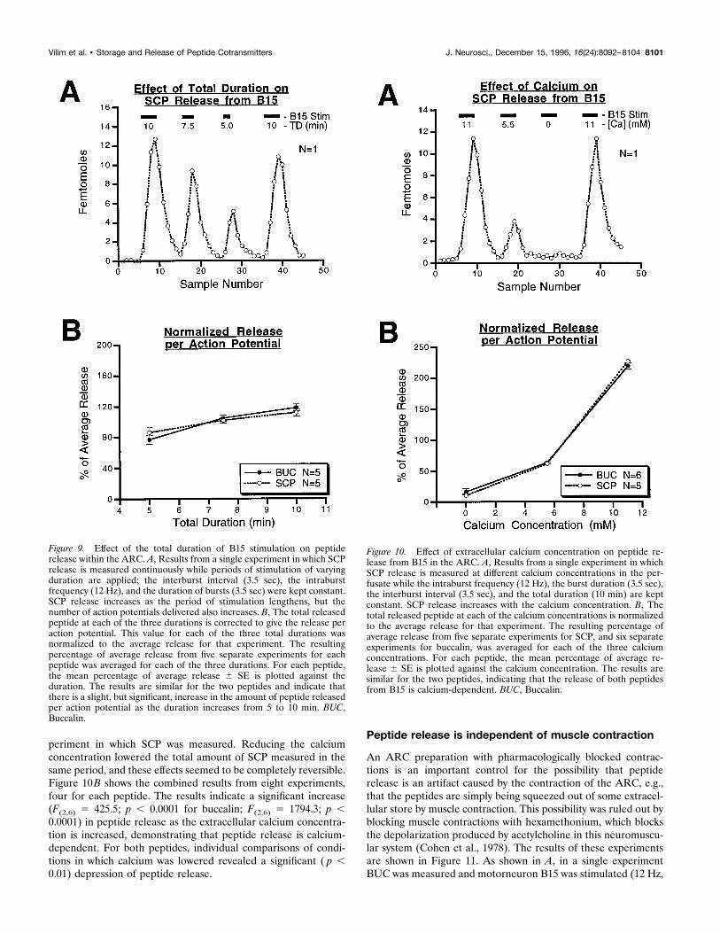
periment in which SCP was measured. Reducing the calciumconcentration lowered the total amount of SCP measured in thesame period, and these effects seemed to be completely reversible.Figure 10B shows the combined results from eight experiments,four for each peptide. The results indicate a significant increase(F(2,6) 5 425.5; p , 0.0001 for buccalin; F(2,6) 5 1794.3; p ,0.0001) in peptide release as the extracellular calcium concentra-tion is increased, demonstrating that peptide release is calcium-dependent. For both peptides, individual comparisons of condi-tions in which calcium was lowered revealed a significant ( p ,0.01) depression of peptide release.
Peptide release is independent of muscle contraction
An ARC preparation with pharmacologically blocked contrac-tions is an important control for the possibility that peptiderelease is an artifact caused by the contraction of the ARC, e.g.,that the peptides are simply being squeezed out of some extracel-lular store by muscle contraction. This possibility was ruled out byblocking muscle contractions with hexamethonium, which blocksthe depolarization produced by acetylcholine in this neuromuscu-lar system (Cohen et al., 1978). The results of these experimentsare shown in Figure 11. As shown in A, in a single experimentBUC was measured and motorneuron B15 was stimulated (12 Hz,
Figure 9. Effect of the total duration of B15 stimulation on peptiderelease within the ARC. A, Results from a single experiment in which SCPrelease is measured continuously while periods of stimulation of varyingduration are applied; the interburst interval (3.5 sec), the intraburstfrequency (12 Hz), and the duration of bursts (3.5 sec) were kept constant.SCP release increases as the period of stimulation lengthens, but thenumber of action potentials delivered also increases. B, The total releasedpeptide at each of the three durations is corrected to give the release peraction potential. This value for each of the three total durations wasnormalized to the average release for that experiment. The resultingpercentage of average release from five separate experiments for eachpeptide was averaged for each of the three durations. For each peptide,the mean percentage of average release 6 SE is plotted against theduration. The results are similar for the two peptides and indicate thatthere is a slight, but significant, increase in the amount of peptide releasedper action potential as the duration increases from 5 to 10 min. BUC,Buccalin.
Figure 10. Effect of extracellular calcium concentration on peptide re-lease from B15 in the ARC. A, Results from a single experiment in whichSCP release is measured at different calcium concentrations in the per-fusate while the intraburst frequency (12 Hz), the burst duration (3.5 sec),the interburst interval (3.5 sec), and the total duration (10 min) are keptconstant. SCP release increases with the calcium concentration. B, Thetotal released peptide at each of the calcium concentrations is normalizedto the average release for that experiment. The resulting percentage ofaverage release from five separate experiments for SCP, and six separateexperiments for buccalin, was averaged for each of the three calciumconcentrations. For each peptide, the mean percentage of average re-lease 6 SE is plotted against the calcium concentration. The results aresimilar for the two peptides, indicating that the release of both peptidesfrom B15 is calcium-dependent. BUC, Buccalin.
Vilim et al. • Storage and Release of Peptide Cotransmitters J. Neurosci., December 15, 1996, 16(24):8092–8104 8101
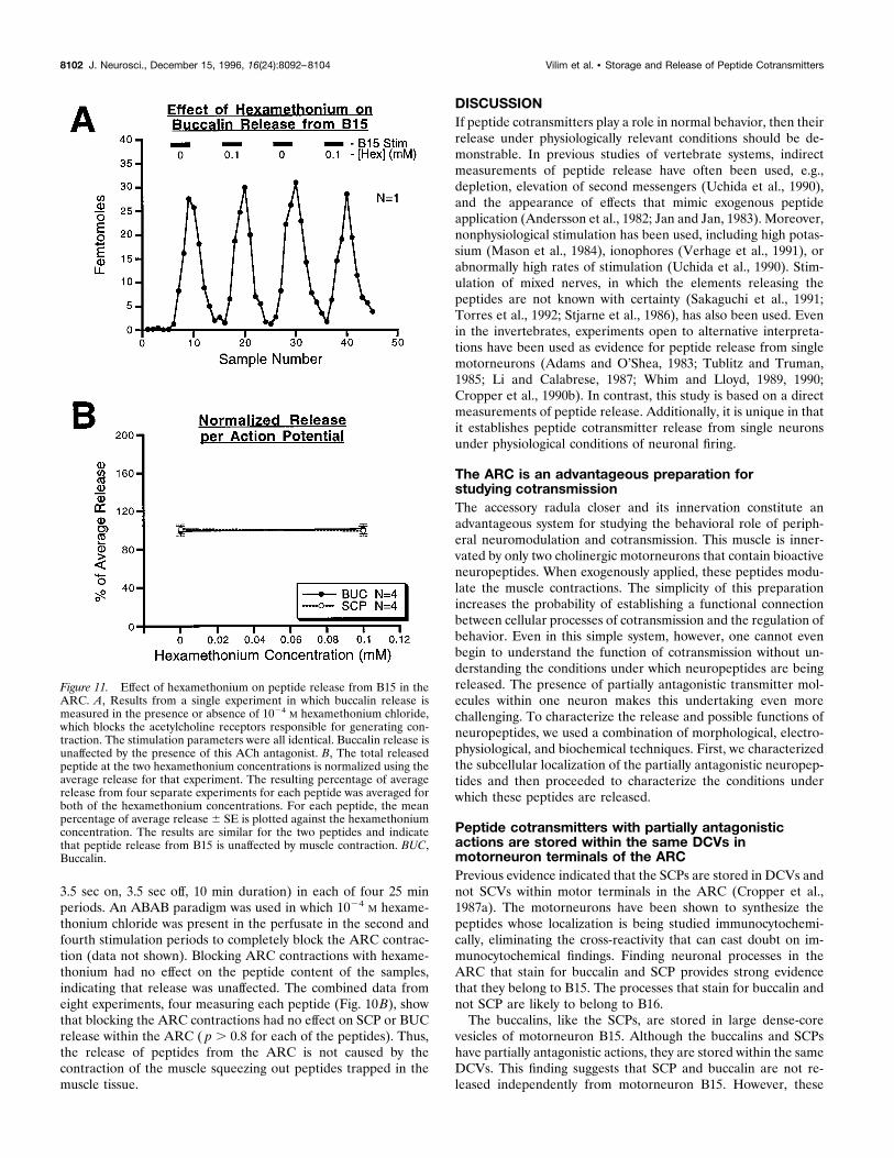
3.5 sec on, 3.5 sec off, 10 min duration) in each of four 25 minperiods. An ABAB paradigm was used in which 1024 M hexame-thonium chloride was present in the perfusate in the second andfourth stimulation periods to completely block the ARC contrac-tion (data not shown). Blocking ARC contractions with hexame-thonium had no effect on the peptide content of the samples,indicating that release was unaffected. The combined data fromeight experiments, four measuring each peptide (Fig. 10B), showthat blocking the ARC contractions had no effect on SCP or BUCrelease within the ARC ( p . 0.8 for each of the peptides). Thus,the release of peptides from the ARC is not caused by thecontraction of the muscle squeezing out peptides trapped in themuscle tissue.
DISCUSSIONIf peptide cotransmitters play a role in normal behavior, then theirrelease under physiologically relevant conditions should be de-monstrable. In previous studies of vertebrate systems, indirectmeasurements of peptide release have often been used, e.g.,depletion, elevation of second messengers (Uchida et al., 1990),and the appearance of effects that mimic exogenous peptideapplication (Andersson et al., 1982; Jan and Jan, 1983). Moreover,nonphysiological stimulation has been used, including high potas-sium (Mason et al., 1984), ionophores (Verhage et al., 1991), orabnormally high rates of stimulation (Uchida et al., 1990). Stim-ulation of mixed nerves, in which the elements releasing thepeptides are not known with certainty (Sakaguchi et al., 1991;Torres et al., 1992; Stjarne et al., 1986), has also been used. Evenin the invertebrates, experiments open to alternative interpreta-tions have been used as evidence for peptide release from singlemotorneurons (Adams and O’Shea, 1983; Tublitz and Truman,1985; Li and Calabrese, 1987; Whim and Lloyd, 1989, 1990;Cropper et al., 1990b). In contrast, this study is based on a directmeasurements of peptide release. Additionally, it is unique in thatit establishes peptide cotransmitter release from single neuronsunder physiological conditions of neuronal firing.
The ARC is an advantageous preparation forstudying cotransmissionThe accessory radula closer and its innervation constitute anadvantageous system for studying the behavioral role of periph-eral neuromodulation and cotransmission. This muscle is inner-vated by only two cholinergic motorneurons that contain bioactiveneuropeptides. When exogenously applied, these peptides modu-late the muscle contractions. The simplicity of this preparationincreases the probability of establishing a functional connectionbetween cellular processes of cotransmission and the regulation ofbehavior. Even in this simple system, however, one cannot evenbegin to understand the function of cotransmission without un-derstanding the conditions under which neuropeptides are beingreleased. The presence of partially antagonistic transmitter mol-ecules within one neuron makes this undertaking even morechallenging. To characterize the release and possible functions ofneuropeptides, we used a combination of morphological, electro-physiological, and biochemical techniques. First, we characterizedthe subcellular localization of the partially antagonistic neuropep-tides and then proceeded to characterize the conditions underwhich these peptides are released.
Peptide cotransmitters with partially antagonisticactions are stored within the same DCVs inmotorneuron terminals of the ARCPrevious evidence indicated that the SCPs are stored in DCVs andnot SCVs within motor terminals in the ARC (Cropper et al.,1987a). The motorneurons have been shown to synthesize thepeptides whose localization is being studied immunocytochemi-cally, eliminating the cross-reactivity that can cast doubt on im-munocytochemical findings. Finding neuronal processes in theARC that stain for buccalin and SCP provides strong evidencethat they belong to B15. The processes that stain for buccalin andnot SCP are likely to belong to B16.The buccalins, like the SCPs, are stored in large dense-core
vesicles of motorneuron B15. Although the buccalins and SCPshave partially antagonistic actions, they are stored within the sameDCVs. This finding suggests that SCP and buccalin are not re-leased independently from motorneuron B15. However, these
Figure 11. Effect of hexamethonium on peptide release from B15 in theARC. A, Results from a single experiment in which buccalin release ismeasured in the presence or absence of 1024 M hexamethonium chloride,which blocks the acetylcholine receptors responsible for generating con-traction. The stimulation parameters were all identical. Buccalin release isunaffected by the presence of this ACh antagonist. B, The total releasedpeptide at the two hexamethonium concentrations is normalized using theaverage release for that experiment. The resulting percentage of averagerelease from four separate experiments for each peptide was averaged forboth of the hexamethonium concentrations. For each peptide, the meanpercentage of average release 6 SE is plotted against the hexamethoniumconcentration. The results are similar for the two peptides and indicatethat peptide release from B15 is unaffected by muscle contraction. BUC,Buccalin.
8102 J. Neurosci., December 15, 1996, 16(24):8092–8104 Vilim et al. • Storage and Release of Peptide Cotransmitters
results do not exclude the possibility that different subpopulationsof DCVs contain different ratios of SCPs and BUCs and aredifferentially released. This possibility was excluded by the exper-iments in this and the companion paper (this issue), in which wemeasured the relative amounts of BUCs and SCPs released underdifferent conditions of stimulation.
Peptide cotransmitters are likely to be released duringnormal behaviorThe indirect evidence for SCP release within the ARC includesdepletion of SCP, elevation of cAMP, stimulation of PKA activity(Hooper et al., 1994a,b), and an increase in the muscle’s relax-ation rate after physiological or higher rates of motorneuronstimulation (Whim and Lloyd, 1989, 1990; Cropper et al., 1990b).However, the depletion was quite modest (20%), and a priori onecannot exclude the release of substances other than SCP that canelevate cAMP, stimulate PKA, and increase the rate of musclerelaxation. Because the length of the high rate stimulation, lastingfrom 1 to 5 hr, may increase the intracellular levels of calciumleading to calcium toxicity, the physiological meaning of theseexperiments remains questionable. Thus, we developed a prepa-ration that allowed us to directly measure peptide release inresponse to the stimulation of single motorneurons.When motorneuron B15 is stimulated, buccalin and SCP ap-
pear in the perfusate with identical kinetics. This similarity inrelease profiles is consistent with corelease from the same DCVs.Thus, the ultrastructural immunocytochemistry and similarity inrelease kinetics both point to costorage of SCP and buccalin in thesame DCVs. The release of modulatory peptides changes overtime and, therefore, is reminiscent of the post-tetanic potentiationand depression described for primary transmitter release. Afterstimulation of the motorneuron, the release of peptide increases,reaches a peak, and then declines, even though the motorneuroncontinues to be stimulated. The depression of peptide cotransmit-ter release within the ARC could result from depletion of thereleasable pool, although results from separate experiments sug-gest that,20% of the peptide should be depleted from B15 underthese conditions (Cropper et al., 1990b). These results parallelthose reported for depression of ACh release in other systems inwhich there was no corresponding decrement in the number ofclear vesicles located in the neuromuscular junction (for review,see Zucker, 1989). Independent of the mechanisms responsiblefor the depression of release, these results indicate that cautionshould be exerted in interpreting the quantitative aspect of releaseexperiments using the depletion paradigm. In view of the complextemporal pattern of release, it is clear that calculation of peptidereleased per action potential in depletion experiments may not bemeaningful. Peptide cotransmitters are probably released andexert their effects in the first few trains of action potentials,because observations of relaxation rate show an increase withinthe first few contractions (Cropper et al., 1990b; Whim and Lloyd,1990). It is possible that even after prolonged firing of motorneu-rons, the smaller amount of peptide released may be able tosustain the physiological actions elicited by the larger amounts ofpeptides that were released during the initial stages.The calcium dependence of peptide cotransmitter release has
been reported in other systems (Adams and O’Shea, 1983; Masonet al., 1984; Uchida et al., 1990; Willard, 1990; Sakaguchi et al.,1991; Verhage et al., 1991; Torres et al., 1992); however, theremoval of extracellular calcium also blocks muscle contraction,raising the possibility that the release was caused by some non-specific effects of the muscle contractions. In our studies, the
appearance of peptides in the perfusate is unlikely to be anartifact, because the release can be blocked by the substitution ofmagnesium for extracellular calcium but is unaffected by theblockade of contractions by hexamethonium. Thus, peptide re-lease seen after B15 stimulation is calcium-dependent but isindependent of muscle contractions.The direct demonstration of SCP release supports the hypoth-
esis that the actions of the SCPs released in response to physio-logically relevant stimulation of B15 (Cropper et al., 1990b) areresponsible for the elevation of cAMP and enhancement of thesize and relaxation rates of muscle contractions that were ob-served under similar conditions of stimulation. The second mes-senger for buccalin is not known, however, and the physiologicalaction of the released peptide may be difficult to address. Thedepression of contractions caused by BUC release would bedifficult to distinguish from receptor desensitization or musclefatigue. At present, there are no antagonists available for BUCs,making it more difficult to assess the actions of released BUCs.Nevertheless, the BUCs (and the SCPs) released from motorneu-ron terminals in these experiments are probably exerting theirmodulatory effects on the muscle, because the concentration of allof these peptides in the perfusate (;1 nM) is in the range in whichexogenously applied peptides begin to exert their modulatoryeffects (Vilim et al., 1994). Thus, even diluted out in the perfusate,the buccalins could still be exerting their modulatory effects, andthe concentration at the release sites is likely to be many timeshigher than that in the perfusate, so these peptides should and arelikely to exert physiological actions.In summary, the direct measurement of peptide release during
the intracellular stimulation of a single motorneuron providesunequivocal evidence of their release. The demonstration thatpeptides are released when the motorneuron is stimulated with itsin vivo firing pattern provides strong evidence that the peptidesare released during normal behavior. Furthermore, the releasedpeptides seem to be exerting their modulatory effects on musclecontraction. Taken together, these results support the idea thatthe modulatory peptides present in the motorneurons act as anintrinsic modulatory system that may play a role in the generationof feeding behavior. In the next paper, the factors that regulatethe release of these peptide cotransmitters will be used to test ahypothetical model (Weiss et al., 1992) for their role in generatingnormal feeding behavior.
REFERENCESAdams ME, O’Shea M (1983) Peptide cotransmitter at a neuromuscularjunction. Science 221:286–289.
Andersson PO, Bloom SR, Edwards AV, Jaerhult J (1982) Effects ofstimulation of the chorda tympani in bursts on submaxillary response inthe cat. J Physiol (Lond) 322:469–483.
Brezina V, Bank B, Cropper EC, Rosen S, Vilim FS, Kupfermann I, WeissKR (1995) Nine members of the myomodulin family of peptide co-transmitters at the B16-ARC neuromuscular junction of Aplysia. J Neu-rophysiol 74:54–72.
Church PL, Lloyd PE (1991) Expression of diverse neuropeptide co-transmitters by identified motor neurons in Aplysia. J Neurosci11:618–625.
Cohen JL, Weiss KR, Kupfermann I (1978) Motor control of buccalmuscles in Aplysia. J Neurophysiol 41:157–180.
Cropper EC, Lloyd PE, ReedW, Tenenbaum R, Kupfermann I, Weiss KR(1987a) Multiple neuropeptides in cholinergic motor neurons of Aply-sia: evidence for modulation intrinsic to the motor circuit. Proc NatlAcad Sci USA 84:3486–3490.
Cropper EC, Tenenbaum R, Kolks MAG, Kupfermann I, Weiss KR(1987b) Myomodulin: a bioactive neuropeptide present in an identifiedcholinergic buccal motor neuron of Aplysia. Proc Natl Acad Sci USA84:5483–5486.
Vilim et al. • Storage and Release of Peptide Cotransmitters J. Neurosci., December 15, 1996, 16(24):8092–8104 8103
Cropper EC, Miller MW, Tenenbaum R, Kolks MAG, Kupfermann I,Weiss KR (1988) Structure and action of buccalin: a modulatory neu-ropeptide localized to an identified small cardioactive peptide-containing cholinergic motor neuron of Aplysia californica. Proc NatlAcad Sci USA 85:6177–6181.
Cropper EC, Miller MW, Vilim FS, Tenenbaum R, Kupfermann I, WeissKR (1990a) Buccalin is present in the cholinergic motor neuron B16 ofAplysia and it depresses accessory radula closer muscle contractionsevoked by stimulation of B16. Brain Res 512:175–179.
Cropper EC, Price D, Tenenbaum R, Kupfermann I, Weiss KR (1990b)Release of peptide cotransmitters from a cholinergic motor neuronunder physiological conditions. Proc Natl Acad Sci USA 87:933–937.
Cropper EC, Kupfermann I, Weiss KR (1990c) Differential firing pat-terns of the peptide-containing cholinergic motor neurons B15 and B16during feeding behavior in Aplysia. Brain Res 522:176–179.
Cropper EC, Vilim FS, Alevizos A, Tenenbaum R, Kolks MAG, RosenSC, Kupfermann I, Weiss KR (1991) Structure, bioactivity, and cellu-lar localization of myomodulin B: a novel Aplysia peptide. Peptides12:683–690.
Fiore L, Geppetti L (1981) Neural control of buccal mass activity inAplysia. Adv Physiol Sci 23:201–223.
Hooper SL, Probst WC, Cropper EC, Kupfermann I, Weiss KR (1994a)SCP application or B15 stimulation activates cAPK in the ARC muscleof Aplysia. Brain Res 657:337–341.
Hooper SL, Probst WC, Cropper EC, Kupfermann I, Weiss KR (1994b)Myomodulin application increases cAMP and activates cAMP-dependent protein kinase in the accessory radula closer muscle ofAplysia. Neurosci Lett 179:167–170.
Jan YN, Jan LY (1983) A LHRH-like peptidergic neurotransmitter ca-pable of “action at a distance” in autonomic ganglia. Trends Neurosci6:320–325.
Kreiner T, Kirk MD, Scheller RH (1987) Cellular and synaptic morphol-ogy of a feeding motor circuit in Aplysia californica. J Comp Neurol264:311–325.
Kupfermann I (1991) Functional studies of cotransmission. Physiol Rev71:683–732.
Li C, Calabrese RL (1987) FMRF-amide-like substances in the leech. III.Biochemical characterization and physiological effects. J Neurosci7:595–603.
Llewellyn-Smith IJ, Costa M, Furness JB (1985) Light and electron mi-croscopic immunocytochemistry of the same nerves from whole mountpreparations. J Histochem Cytochem 33:857–866.
Lloyd PE, Frankfurt M, Stevens P, Kupfermann I, Weiss KR (1987a)Biochemical and immunocytological localization of the neuropeptidesFMRFamide, SCPa, SCPb, to neurons involved in the regulation offeeding in Aplysia. J Neurosci 7:1123–1132.
Lloyd PE, Kupfermann I, Weiss KR (1987b) Sequence of small cardio-active peptide A: a second member of a class of neuropeptides inAplysia. Peptides 8:179–184.
Mahon AC, Lloyd PE, Weiss KR, Kupfermann I, Scheller RH (1985)The small cardioactive peptides A and B of Aplysia are derived from acommon precursor molecule. Proc Natl Acad Sci USA 82:3925–3929.
Mason TR, Peterfreund RA, Sawchenko PE, Corrigan AZ, Vale WW(1984) Release of the predicted calcitonin gene-related peptide fromcultured rat trigeminal ganglion cells. Nature 308:653–655.
Merighe A, Polak JM, Fumagalli G, Theodosis DT (1989) Ultrastruc-tural localization of neuropeptides and GABA in the rat dorsal horn: acomparison of different immunogold labeling techniques. J HistochemCytochem 37:529–540.
Miller MW, Alevizos A, Cropper EC, Kupfermann I, Weiss KR (1992)Localization of buccalin-like immunoreactivity in the central nervoussystem and peripheral tissues of Aplysia californica. J Comp Neurol320:182–195.
Miller MW, Beushausen S, Cropper EC, Eisinger K, Stamm S, Vilim FS,Vitek A, Zajc A, Kupfermann I, Brosius J, Weiss KR (1993) The
buccalin-related neuropeptides: isolation and characterization of anAplysia/cDNA clone encoding a family of peptide cotransmitters. J Neu-rosci 13:3346–3357.
Morris HR, Panico M, Karplus A, Lloyd PE, Riniker B (1982) Elucida-tion by FAB-MS of the structure of a new cardioactive peptide fromAplysia. Nature 300:643–645.
Ottersen OP (1989) Quantitative electron microscopic immunocyto-chemistry of neuroactive amino acids. Anat Embryol 180:1–15.
Probst WC, Cropper EC, Heierhorst J, Hooper SL, Jaffe H, Vilim F,Beushausen S, Kupfermann I, Weiss KR (1994) cAMP-dependentphosphorylation of Aplysia twitchin may mediate modulation of musclecontractions by neuropeptide cotransmitters. Proc Natl Acad Sci USA91:8487–8491.
Quicke DLJ, Brace RC (1979) Differential staining of cobalt- and nickel-filled neurones using rubeanic acid. J Microsc 115:161–163.
Reed W, Weiss KR, Lloyd PE, Kupfermann I, Chen M, Bailey CH (1988)Association of neuroactive peptides with the protein secretory pathwayin identified neurons of Aplysia californica: immunolocalization of SCPaand SCPb to the contents of dense core vesicles and the trans face of theGolgi apparatus. J Comp Neurol 272:358–369.
Sakaguchi M, Inaishi Y, Kashihara Y, Kuno M (1991) Release of calci-tonin gene-related peptide from nerve terminals in rat skeletal muscle.J Physiol (Lond) 434:257–270.
Sossin W, Sweet-Cordero A, Scheller RH (1989) Dales hypothesis revis-ited: different neuropeptides derived from a common prohormone aretargeted to different processes. Proc Natl Acad Sci USA 87:4845–4848.
Stjarne L, Lundberg JM, Astrand P (1986) Neuropeptide Y: a cotrans-mitter with noradrenaline and adenosine 59-triphosphate in the sympa-thetic nerves of the mouse vas deferens? A biochemical, physiological,and electropharmacological study. Neuroscience 18:151–166.
Torres G, Britan M, Huidobro JP (1992) Co-release of neuropeptide Y(NPY) and noradrenaline from the nerve terminals supplying the rat vasdeferens: influence of calcium and the stimulation intensity. NeurosciLett 148:39–42.
Tublitz NJ, Truman JW (1985) Intracellular stimulation of an identifiedneuron evokes peptide release in an insect. Science 228:1013–1015.
Uchida S, Yamamoto H, Iio S, Matsumoto N, Wang XB, Yonehara N,Imai I, Inoki R, Yoshida H (1990) Release of calcitonin gene-relatedpeptide-like immunoreactive substance from neuromuscular junction bynerve excitation and its action on striated muscle. J Neurochem54:1000–1003.
Vilim FS, Cropper EC, Rosen SC, Tenenbaum R, Kupfermann I, WeissKR (1994) Structure, localization and action of buccalin b, a bioactivepeptide from Aplysia. Peptides 15:959–969.
Verhage M, McMahon HT, Ghijsen WEJM, Boomsma F, Scholten G,Weigant VM, Nicholls DG (1991) Differential release of amino acids,neuropeptides, and catecholamines from isolated nerve terminals. Neu-ron 6:517–524.
Wang LJ, Scheller RH (1991) Peptide processing and targeting in theneuronal secretory pathway. Science 251:1330–1335.
Weiss KR, Brezina V, Cropper EC, Hooper S, Miller MW, Probst WC,Vilim FS, Kupfermann I (1992) Peptidergic co-transmission in Aplysia:functional implications for rhythmic behaviors. Experientia 48:456–463.
Whim MD, Lloyd PE (1989) Frequency-dependent release of peptidecotransmitters from identified cholinergic motor neurons in Aplysia.Proc Natl Acad Sci USA 86:9034–9038.
Whim MD, Lloyd PE (1990) Neuropeptide cotransmitters released froman identified cholinergic motor neuron modulate neuromuscular effi-cacy in Aplysia. J Neurosci 10:3313–3322.
Willard AL (1990) A vasoactive intestinal peptide-like cotransmitter atcholinergic synapses between rat myenteric neurons in cell culture.J Neurosci 10:1025–1034.
Zucker RS (1989) Short term synaptic plasticity. Annu Rev Neurosci12:13–31.
8104 J. Neurosci., December 15, 1996, 16(24):8092–8104 Vilim et al. • Storage and Release of Peptide Cotransmitters


![ONE WORLD TRADE CENTER - Owens Corning · 2017-06-09 · ONE OWENS CORNING PARKWAY TOLEDO, OHIO, USA 43659 888-TFIBER1 [834-2371] OWENS CORNING INSULATING SYSTEMS, LLC ONE OWENS CORNING](https://img.pdfslide.us/doc/110x75/5ed6e345df0eda5e752ae77c/one-world-trade-center-owens-2017-06-09-one-owens-corning-parkway-toledo-ohio.jpg)