Embed Size (px)
Citation preview
Vol. 45, No. 2INFECTION AND IMMUNITY, Aug. 1984, P. 511-5170019-9567/84/080511-07$02.00/0Copyright © 1984, American Society for Microbiology
Corynebacterium ulcerans and Corynebacterium pseudotuberculosisResponses to DNA Probes Derived from Corynephage a and
Corynebacterium diphtheriaeNEAL GROMAN,* J. SCHILLER,t AND J. RUSSELL
Department of Microbiology and Immunology, University of Washington, Seattle, Washington 98195
Received 11 January 1984/Accepted 7 May 1984
Strains of Corynebacterium ulcerans and Corynebacterium pseudotuberculosis (Corynebacterium ovis) wereexamined for the production of diphtheria toxin. A majority of C. ulcerans strains (25 of 37) and 1 C.pseudotuberculosis strain (1 of 14) gave a positive Elek test for diphtheria toxin, and for all strains but 1,production of diphtheria toxin was inhibited at the same level of Fe2' as was the Corynebacterium diphtheriaecontrol. All Elek-positive cultures as well as two Elek-negative isolates of C. ulcerans gave a positive signalwhen hybridized with a DNA probe unambiguous for the diphtheria toxin gene (tox) under conditions of highstringency. The majority of probe-positive C. ulcerans strains contained three or more DNA restrictionfragments that hybridized with converting corynephage II, suggesting that in C. ulcerans as in C. diphtheriaethere may be a relationship between toxinogeny and carriage of I-related phage. Selected strains of C.diphtheriae, C. ulcerans, and C. pseudotuberculosis were examined for DNA homology by a semiquantitativetechnique. There was very little homology between C. diphtheriae and members of the other two species.Strains of C. ulcerans and C. pseudotuberculosis, although more closely related, appeared to belong to distinctspecies as well.
The relationship of Corynebacterium diphtheriae, Coryne-bacterium ulcerans, and Corynebacterium pseudotuberculo-sis (Corynebacterium ovis) is based on a similar morphology,cell wall composition, and production of a halo on Tinsdalemedium which identifies the production of the enzymecystinase (3, 4). The relationship of C. diphtheriae and C.ulcerans has been of special interest to those studying theepidemiology of diphtheria since the reports were publishedstating that diphtheria-like organisms were isolated fromcases of acute sore throat and from healthy carriers (11, 13,26). Many of these isolates produced a toxin which gave aline of identity with diphtheria toxin (DT) in gel immunodif-fusion tests (11). However, they were distinguished from C.diphtheriae by their ability to hydrolyze gelatin and decom-pose urea, by their inability to reduce nitrate, and byserological differences. A close relationship between C.ulcerans and C. pseudotuberculosis was suggested by thefact that both gave positive urease reactions and both failedto reduce nitrates, and more recently it was reported (4) thatthese organisms are unique among the corynebacteria inproducing phospholipase D.Of particular interest in the relationship of the three
organisms is that some strains of each can produce DT. Theearly reports of DT production by C. ulcerans based onanimal inoculation were inconclusive (5, 6), but the possibili-ty that two toxins were present in some strains, one of themDT (23), clarified the situation. Later, both Saxholm (26) andHenrikson and Grelland (11) showed that some isolates of C.ulcerans produced a line of identity with toxinogenic C.diphtheriae on Ouchterlony plates containing diphtheriaantitoxin. In addition, Maximescu et al. (19-21) showed thatcertain strains of C. ulcerans and C. pseudotuberculosiscould be converted to DT production by phages fromtoxinogenic C. diphtheriae.
* Corresponding author.t Present address: National Cancer Institute, National Institutes
of Health, Bethesda, MD 20910.
In the present study we have compared the DNAs of C.ulcerans and C. pseudotuberculosis to those of C. diphther-iae and corynephage P. We have found in hybridizationexperiments carried out at high stringency (<10% mismatch)that the genes for DT, tox, are homologous in all threespecies and that many DT-positive strains of C. ulceranshave a number of DNA restriction fragments that hybridizewith P-converting phage. Surprisingly, there is virtually nohomology between the genomic DNA of C. diphtheriae andthe other two species, and there is a moderate amount ofhomology between the DNAs of C. ulcerans and C. pseudo-tuberculosis.
MATERIALS AND METHODSBacterial strains. A total of 37 cultures of C. ulcerans and
14 cultures of C. pseudotuberculosis (C. ovis) were collectedfrom various sources. The culture designations and sourcesare given in Tables 2 and 3. C. diphtheriae C7(-), C7()3'°x+,C7(I-tsr-3)'ox+, and C7(p)hm723, an iron-resistant toxin-producing mutant (15), were taken from our stock collection.
In hybridization experiments comparing total genomicDNAs, four C. diphtheriae strains were selected to representthis species. They were S1016 (ATCC 19409, the designatedreference strain, type gravis), C7(-) (ATCC 27011, theproposed neotype [3], type mitis), S1013 (ATCC 8026, typemitis), and S1015 (ATCC 8032, type intermedius). C7(-)does not carry the gene for DT, but the other three strainsdo. The ATCC 9015 strain of C. ulcerans and the ATCC19410-designated reference strain of C. pseudotuberculosiswere used as the reference strains for those species. Repre-sentative strains of C. ulcerans and C. pseudotuberculosiswere drawn from restriction pattern groups and will bedescribed and identified below.The species designation of all C. ulcerans and C. pseudo-
tuberculosis strains and the species and type designations ofthe C. diphtheriae strains were confirmed by a series ofstandard tests for sugar fermentations, production of H2Sand urease, and nitrate reduction (22). The test for trehalose
511
on February 28, 2020 by guest
http://iai.asm.org/
Dow
nloaded from

512 GROMAN, SCHILLER, AND RUSSELL
fermentation was read after 7 days. The test for phospholi-pase D production was performed by the cooperative hemo-lysis technique described by Barksdale et al. (4), whichemploys Corynebacterium equi. The latter strain was ob-tained from the American Type Culture Collection (ATCC6939).
Tests for DT. The standard Elek test for toxin productionwas employed (12). In this proceduire. the cultures werestreaked across an agar plate in which a strip of filter papersaturated with a solution of 200 U of antitoxin per ml hadbeen embedded. A positive C7(p)t'ox control was streakedadjacent to each test culture so that a merger with the line forDT could be easily verified. The identity of this line with DTis based on the absence of any line when the isogenic,nontoxinogenic C7(-) is tested and on observations thatsome mutations in n-converting phage leading to the loss oftoxin activity correlate with the loss of this line in thestandard Elek test (18). Diphtheria antitoxin was obtainedfrom the Connaught Laboratories, Inc. (Ontario, Canada).The test for Fe2+ inhibition of toxin production was
performed as previously described (8).tox probes. The isolation of ,B phage DNA from C7(p-tsr-
3)"X+ and the preparation of A and B tox probes from thisDNA as well as the dot-blot procedure for homology testswere by the procedures previously described (7).DNA extractions. Cell growth and the lysis and DNA
extraction procedures for genomic DNA followed the proce-dures described by Schiller et al. (27).
Treatment and processing of DNA. Restriction enzymedigests, agarose gel electrophoresis, in vitro labeling ofDNAwith 32P-deoxyribonucleotides, Southern blotting, filter pa-per hybridization, and autoradiographing were all performedas previously described (7). Unless otherwise stated, DNA-DNA hybridizations were carried out under conditions al-lowing only ca. 10% mismatch.
RESULTSCharacteristics of strains. The pattern of diagnostic tests
for 37 strains designated C. ulcerans and 14 C. pseudotuber-culosis strains is given in Table 1. The two major criteria,
TABLE 1. Diagnostic reactions of strains of C. ulcerans and C.pseudotuberculosis (C. ovis)
No. positive/no. negativeTest C. pseudotu-
C. ulcerans berculosis
Sugar fermentationsGlucose 37/37 14/14Maltose 36/37 14/14Fructose 37/37 14/14Sucrose 0/37 0/14Galactose 24/37 6/14Starch and glycogen 33/37 0/14Trehalose 37/37 0/14
H2S production 37/37 14/14
Nitrate reduction 0/37 5/14
Urease production 37/37 13/14
Phospholipase D production 36/37 14/14
DT productiona 25/37 1/14a Elek test.
urease production and phospholipase D production, whichdistinguish these organisms from C. diphtheriae were met byboth of these groups of isolates. The slow fermentation oftrehalose, one of the traits distinguishing C. ulcerans from C.pseudotuberculosis, showed the appropriate correlation. InElek tests for DT production, to be described later, themajority of C. ulcerans strains produced toxin, whereas onlyone C. pseudotuberculosis strain was toxinogenic by thiscriterion. These results were consistent with previous find-ings (20), namely that many natural isolates of C. ulceransproduce DT, whereas few C. pseudotuberculosis isolatesappear to do so.
Restriction enzyme digest patterns of genomic DNA. Therestriction enzyme digest patterns of all strains of C. ulcer-ans and C. pseudotuberculosis were examined. The primarypurpose was to determine the diversity of patterns in thesample so that representative strains could be selected forhybridization experiments in which genomic DNAs were tobe compared. Second, it was felt that some preliminaryinformation on relationships between restriction patternsand some of the limited epidemiological correlates providedby this sample of strains might emerge.DNA was extracted from all cultures identified as C.
ulcerans. The DNAs were restricted with BamHI, and afteragarose gel electrophoresis, their digest patterns were com-pared. On the basis of these results (data not shown), threegroups of organisms with identical or similar patterns and afourth miscellaneous group with various patterns were iden-tified (Table 2). Organisms in groups I and II each gave ahomogeneous pattern with BamHI, and those in group IIIgave closely related patterns, whereas the patterns of thosein the miscellaneous group were unrelated to each other or tothose of the other groups. The DNAs of all three members ofgroup I and three representative members each of groups IIand III were also restricted with HindIII and EcoRI. Theresults with EcoRI are shown in Fig. 1, and these results andthose from HindIII digestion confirmed the groupings ob-tained from BamHI. It is of interest that the type culturestrains of C. ulcerans (ATCC 9015) constituted a separategroup, all three members of group I being derivatives of thisstrain. It should also be noted that among the large numbersof cultures comprising group II, 15 of 22 were isolated inNorway from 1949 to 1952. From the data in Table 2 it can beseen that there is no strict overall correlation between DTproduction and the group restriction patterns.
Restriction enzyme digest patterns of 14 cultures designat-ed and confirmed as C. pseudotuberculosis were also exam-ined. Only two patterns were detected with BamHI. Mem-bers of groups I and II are listed in Table 3, andrepresentative results after BamHI digestion are given inFig. 2. The same two groups were obtained after digestionwith EcoRI. It is of interest to note that all members of groupI failed to reduce nitrate, whereas all members of group IIreduced nitrate.DT production by isolates of C. ulcerans and C. pseudotu-
berculosis. It was reported by Maximescu et al. (20) thatamong 157 wild-type strains of C. ulcerans, 31.2% producedDT. DT producers were isolated from 35% (43 of 122) ofhuman cultures and 21% (6 of 35) of equine isolates. Only 3%(2 of 60) C. pseudotuberculosis strains produced DT, andboth of these strains had been isolated from an Egyptianbuffalo. We have tested all of the strains listed in Tables 2and 3 for the production ofDT by the Elek plate method (seeabove). Of 37 C. ulcerans isolates, 25 produced DT, i.e.,material which cross-reacted with DT in the Elek test (Table2). By contrast, only 1 of 14 C. pseudotuberculosis isolates,
INFECT. IMMUN.
on February 28, 2020 by guest
http://iai.asm.org/
Dow
nloaded from

RELATION OF TOXINOGENIC CORYNEBACTERIUM SPP. 513
TABLE 2. C. ulcerans strainsRestriction Source and b StrainsC
pattern group referencea toxI Albany + 690 (ATCC 9015)
Canada + 709 (ATCC 9015)Norway + 742 (ATCC 9015)
II Albany (4) + 710 (51166), 711 (51167)- 712 (51169), 714 (5291)
CDC + 717 (KC279)Norway (4, 10, + 730 (2121/49), 731 (1605/50),
11, 26) 732 (1771/50)+ 733 (2292/50), 734 (1054/52),
735 (842/50)+ 736 (903/50), 737 (1517/51)- 738 (603/50)- 739 (1661/50), 741 (170)
Romania (20) - 754 (40C), 755 (9304)+ 758 (2507), 761 (983), 762
(984)Wales (1) + 751 (378)
III Albany (4) - 713 (52103)California + 715 (JN4)
+ 716 (B1446)- 718 (E6857), 721 (E298)
Norway + 740 (1613/50)Romania (20) + 760 (982), 765 (16L)
Miscellaneous Canada + 691 (X16-80)- 719 (E952), 720 (F127)
Romania (20) + 759 (981)a The references which follow the source names apply to all of the
organisms from that source in that particular restriction group. The culturesfrom Albany were obtained from Mehdi Shayegani, State of New York,Department of Health, Albany. In a personal communication he noted thatstrains 710, 711, and 712 were isolated in Norway and belong to the groupdescribed below and that strains 713 and 714 were isolated by Jebb in Oxford,England, between 1949 and 1951, strain 713 being one of the four culturescatalogued by the National Type Culture Collection, London, England. Thecultures from Canada were obtained from C. H. Jellard, Provincial Labora-tory of Public Health, Edmonton, Alberta; those from California wereobtained from Dexter H. Howard, Department of Microbiology and Immunol-ogy, University of California at Los Angeles; those from CDC were obtainedfrom Robert E. Weaver, Communicable Disease Center, Atlanta, Ga. Thecultures from Norway were sent by Rolf Saxholm, National Institute of PublicHealth, Oslo, and constitute a series isolated in that country over the period of1949 to 1952. Finally, the cultures from Romania came from Paula Max-imescu, Institute Cantacuzino, Bucharest, and those from Wales came from I.Zamiri, Welsh National School of Medicine, Heath Park, Cardiff.
b The + or - indicates the production or nonproduction, respectively, ofDT as detected by the Elek plate method. In addition, all + isolates werepositive when tested in dot-blot hybridizations for the presence of DNAhomologous to the DT gene by using the B probe at high stringency (7).Strains 718 and 721 were the only cultures which were negative in the Elektest but positive with the B probe.
c The culture designations are given as follows. The stock number used inour laboratory is given first and is followed within parentheses by the stockdesignation of the source laboratory. The later designations were used in thereferences cited in the table. For strains from Norway, the number after theslash indicates the year of isolation.
the Egyptian buffalo isolate 766 sent to us by Maximescu,was positive. Two C. pseudotuberculosis derivatives ofisolates 769 and 771, which had been lysogenized withconverting corynephages, were also positive.
Hybridization of C. ulcerans and C. pseudotuberculosis withdiphtheria tox probes. All of the C. ulcerans and C. pseudo-tuberculosis strains were tested for the presence of DNAhomologous to the tox gene of C. diphtheriae by the B probeand dot-blot method. The probe contains sequences codingfor more than 90% of the B fragment of DT and is completely
FIG. 1. Restriction enzyme digest patterns of C. ulceransstrains. DNAs were extracted and purified, treated with EcoRI, andelectrophoresed in 1% agarose in TENA buffer (27) at 25 V for 16.5h. Lanes A to C contain organisms in group I, lanes D to F containorganisms in group II, and lanes G to I contain organisms in groupIII. The strain number designations are: (A) 690, (B) 709, (C) 742,(D) 710, (E) 730, (F) 754, (G) 718, (H) 713, and (I) 765.
internal to the tox gene (7). Results of all hybridizations arerecorded in Tables 2 and 3 (see footnotes). All 25 Elek-positive strains of C. ulcerans and the 1 positive C. pseudo-tuberculosis strain gave positive hybridization tests underconditions of high stringency. In addition, two Elek-negativeC. ulcerans strains, 718 and 721, which were also negativeby the rabbit intradermal test, gave positive hybridizations, acorrelation previously reported for some nontoxinogenic C.diphtheriae (7).The relative position of the diphtheria tox gene was
examined by hybridizing BamHI digests of genomic DNAfrom 15 C. ulcerans isolates with the B tox probe. Theresults of a representative sample, including three negativecontrols, are given in Fig. 3. The tox-related DNA is carriedon different fragments, generally on one fragment but in one
TABLE 3. C. pseudotuberculosis strainsRestriction Source andpattern reference' StrainSbgroup
I ATCC S1019 (ATCC 19410)CDC 722 (E1929), 723 (E658), 724 (E1195), 726
(E3737)Romania 764 (988), 768 (A), 769 (21), 771 (1113)
(20)
II CDC 725 (E1008)California 728 (E1144B), 729 (E1124)Romania 766 (992),c 767 (996)
(20)a See footnote a, Table 2, for the source laboratories indicated by these
locales, except for ATCC, which is the American Type Culture Collection,Rockville, Md.
b The culture designations include our stock number first, followed withinparentheses by the stock designation of the source laboratory.
c Strain 766 was the only C. pseudotuberculosis culture that produced DTas detected by the Elek test. It was also positive for DNA homologous to thetox gene for DT, using the B probe at high stringency. All other isolates werenegative by both tests.
VOL. 45, 1984
on February 28, 2020 by guest
http://iai.asm.org/
Dow
nloaded from

514 GROMAN, SCHILLER, AND RUSSELL
FIG. 2. Restriction enzyme digest patterns of C. pseudotubercu-losis strains. DNAs were extracted and purified, treated withBamHI, and electrophoresed in 1% agarose in TENA buffer at 25 Vfor 16.5 h. Lanes A to F contain organisms in group I, and lanes G toK contain organisms in group II. The strain number designationsare: (A) S1019, (B) 769, (C) 771, (D) 722, (E) 723, (F) 768, (G) 729,(H) 766, (I) 728, (J) 725, and (K) 767.
case on two fragments. For the 15 isolates examined, theposition represented by lane B was the most common andwas exhibited by strains in groups II and III, whereas theposition represented by lane A was occupied by the threestrains in group I. The tox gene of strains of group IIIoccupied various positions represented by lanes B, C, and F.It is of some interest that the positions of the tox-bearing
A B C D E F G H J
FIG. 3. Hybridization of genomic DNA BamHI digests of C.ulcerans with the B probe for the diphtheria tox gene. DNAs wereextracted, digested with BamHl, submitted to agarose gel electro-phoresis, Southern blotted, hybridized with 32P-labeled B tox probe,and autoradiographed. Lanes A to H are C. ulcerans strains. Theorganisms in lanes A to F and the control in J were Elek positive,and those in lanes G to I were Elek negative. The restriction patterngroup is given within parentheses. Lanes: A, 690 (I); B, 751 (II); C,716 (III); D, 718 (III); E, 721 (III); F, 691 (miscellaneous); G, 719(miscellaneous); H, 720 (miscellaneous); I, C7(-) C. diphtheriae; J,C7(P)box+, C. diphtheriae.
fragments offive group II strains were identical, but differentthan that of the tox gene in a group II strain of C. ulcerans,756, which was converted to toxinogeny by the PW8 coryne-phage (data not shown). The high intensity of all bandssuggests extensive hornology between these B-coding seg-ments and that of the C. diphtheriae tox gene.
In a second part of this experiment, the genomic digestsdescribed in Fig. 3 were also hybridized with the A probe forDT. This probe carries the entire sequence for the Afragment of DT plus ca. 250 base pairs which probablyencode for the leader sequence and regulatory region of thetox gene (7, 14). The A probe elicited a positive signal fromthe same fragments that gave a positive signal with the Bprobe. These results show that highly homologous regions ofboth the A- and B-coding portions of the tox gene werepresent and on the same fragment. They also suggest thatisolate 716 carried two tox genes.
Hybridization of C. ulcerans with ,8-converting phage DNA.We have surveyed C. ulcerans strains for carriage of DNArelated to ,-converting corynephage, which itself carries thetox gene (Fig. 4). Of the 37 strains examined, 17 carried threeto seven r-related fragments. All but two of these strainsbelonged to group II tox+ and shared the same pattern of 3-related fragments (Fig. 4, e.g., lanes I, J, and K). Three ofthe strains had two n-related fragments, and seven, includingall members of group I, had only one. The one naturaltoxinogenic isolate of C. pseudotuberculosis also had asingle homologous band. Finally, there were 10 isolates, ofwhich 7 were in group II, that did not carry any tox-relatedDNA, and all were devoid of ,-related DNA. In general,there was a high correlation of tox gene carriage withcarriage of additional ,B-related DNA, but there were asignificant number of exceptions.
Effect of iron on toxin synthesis in C. ulcerans and C.pseudotuberculosis. The inhibition of toxin synthesis by Fe21
A B C D E F G H J K L
FIG. 4. Hybridization of C. ulcerans genomic DNA digests with,B-converting phage DNA. BamHI digests of genomic DNA weresubmitted to agarose electrophoresis. After Southern blotting andhybridization with 32P-labeled ,B phage DNA, an autoradiograph wasmade. C7(P)'Ox+ is a strain of C. diphtheriae, and the remainingisolates are C. ulcerans strains described in Table 2. (Isolate 756 isisolate 755 lysogenized with a PW8, p-related phage.) Strain numberdesignations: (A) C7(p), (B) 754, (C) 755, (D) 756, (E) 757, (F) 758,(G) 759, (H) 760, (I) 761, (J) 762, (K) 731, (L) 765.
INFECT. IMMUN.
on February 28, 2020 by guest
http://iai.asm.org/
Dow
nloaded from

RELATION OF TOXINOGENIC CORYNEBACTERIUM SPP. 515
in C. diphtheriae is well established (3). The ability of Fe2+to regulate DT synthesis in strains of C. ulcerans and C.pseudotuberculosis was tested by a semiquantitative method(8) in which various concentrations of Fe2+ were added as asupplement to the agar medium usually employed in thestandard Elek method. After a preliminary titration, plateswere supplemented with 0.4, 2, and 100 ,ug of Fe2+ per ml. Inaddition to the test cultures, an iron-sensitive control,C7(p)'ox+, and an iron-insensitive control, C7(1)fox+hm723,were included on all plates. Inocula were taken from culturesgrown overnight on brain heart infusion agar (Difco Labora-tories, Detroit, Mich.). Plates were incubated at 35°C andobserved after 48 h.
All toxin-producing strains of C. ulcerans (Table 2) weretested. Of these, 25 of 26 and the iron-sensitive controlproduced toxin in Fe2+-unsupplemented medium but failedto produce detectable amounts in the medium supplementedwith 0.4 ,ug of Fe2+ per ml. The one isolate, 733, which waspositive at 0.4 ,ug/ml, also gave a positive test at 8 ,ug/ml butnot at 20 ,ug/ml and thus was a naturally resistant strain. Theiron-resistant control produced toxin at 100 ,ug/ml, themaximum tested in these experiments. The single naturalstrain of a DT-producing C. pseudotuberculosis and twostrains converted to toxinogeny by corynephages also failedto produce detectable amounts of toxin-related material at0.4 ,ug of Fe2+ per ml. These data show that for both C.ulcerans and C. pseudotuberculosis, DT synthesis was regu-lated by iron levels virtually identical to those controlling itssynthesis in C. diphtheriae.
Relationship of C. diphtheriae, C. ulcerans, and C. pseudo-tuberculosis. The data gathered thus far concerning thegenetics and production of DT indicated a close relationshipbetween C. diphtheriae, C. ulcerans, and C. pseudotubercu-losis. Nevertheless, the extent to which members of thesespecies were related to each other was unknown.As one test of relatedness, we probed chromosomal
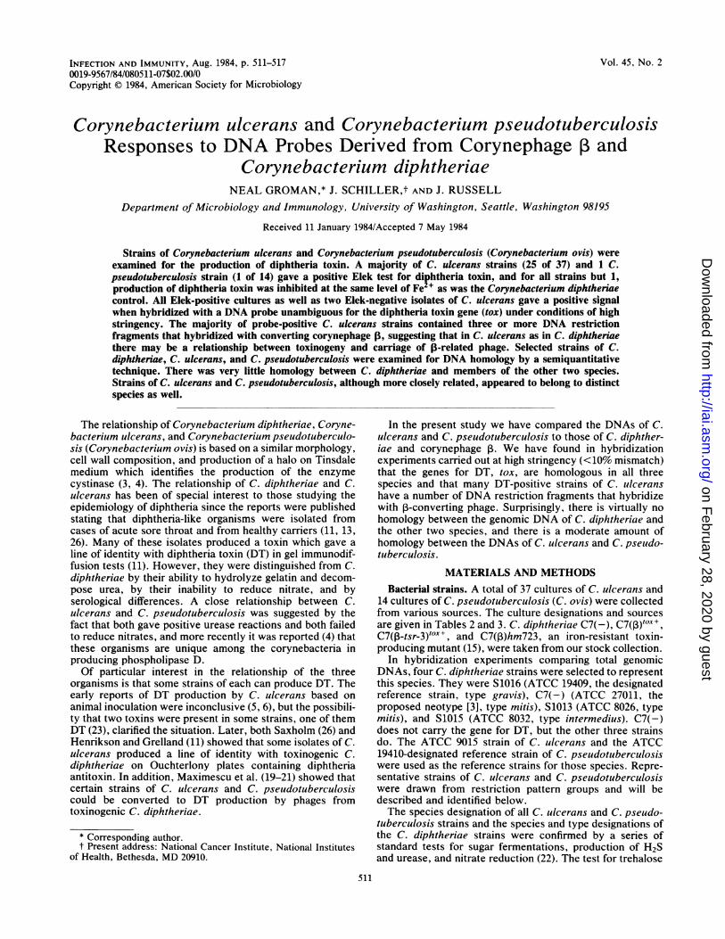
digests of representative strains of each of the species withlabeled genomic DNA of the others. The results with C.diphtheriae and C. ulcerans probes are given in Fig. 5 and 6.Results similar to those in Fig. 6 were obtained when C.pseudotuberculosis S1019 was used as a probe. At highstringency, there was very little homology between C.diphtheria and either C. ulcerans or C. pseudotuberculosis.Only isolated fragments of these strains appeared to behomologous. However, C. ulcerans and C. pseudotubercu-losis were more closely related. In a separate experiment(data not shown), chromosomal digests of C. ulcerans werehybridized with the C. pseudotuberculosis probe at interme-diate stringency, ca. 20% mismatch. The same fragments ofthe C. ulcerans strains simply gave a more intense reaction,and there was no change in the reactions of the C. diphther-iae strains. These results, although only semiquantitative,are consonant with a finding in preliminary experiments,employing the Si exonuclease technique, in which only 3 to10% homology was detected between C. diphtheriae andeither C. ulcerans or C. pseudotuberculosis, whereas ca.40% homology was detected between strains of C. ulceransand C. pseudotuberculosis.
DISCUSSIONWe have compared the genes for DT in the three Coryne-
bacterium species known to produce this toxin. DNA probesspecific for segments coding for the A and B fragments of DTin C. diphtheriae were used. The probes were isolated fromthe tox gene of the same ,-converting phage carried by theDT-positive C. diphtheriae strain used in the Elek tests. A
A B C D E F G H I J K L
FIG. 5. Hybridization of selected strains of C. diphtheriae, C.ulcerans, and C. pseudotuberculosis with labeled C. diphtheriaeprobe. Similar amounts of BamHl-digested genomic DNA wereplaced in each well, separated by agarose gel electrophoresis as inFig. 1, transferred to nitrocellulose, and hybridized to 32P-labeled C.diphtheriae S1016 (ATCC 19409) genomic DNA at 68'C for 48 h.The filter was then washed under stringent conditions (ca. 10 to 12%mismatch) and autoradiographed. Lanes A to D are C. ulceransstrains, and their restriction group designations are given withinparentheses. Lanes E to H are C. diphtheriae strains, and theircolonial types are noted within parentheses. Lanes I to L are C.pseudotuberculosis strains, and their restriction enzyme digestgroup pattern is given within parentheses. Lanes: A, 690 (I); B, 751(II; C, 716 (III); D, 691 (miscellaneous); E, S1016 (gravis); F, S1013(mitis); G, S1015 (intermedius); H, C7 (mitis); I, S1019 (I); J, 722 (I):K, 766 (II); L, 728 (II).
high degree of homology was demonstrated under stringentconditions by DNA fragments from the one strain of C.pseudotuberculosis and from all of the strains of C. ulceranswhich were DT positive in a carefully controlled Elek (gel-immunodiffusion) test. It has already been shown (7) thatthese probes hybridize with DNA fragments from a varietyof DT-positive C. diphtheriae. It also has been shown thatDT-positive C. lulcerans and C. pseudotuberculosis (28)produce proteins with the molecular weight of DT and theADP-ribosylating activity characteristic of that toxin.There was some variation among the C. ulcerans strains in
the position of their tox-bearing DNA fragments on agarosegels. Variation could be due to placement of the gene indifferent sites within the chromosome or to differencesamong strains in the position of the restriction sites whichflank the tox-bearing fragment. If a converting phage isassumed to be involved in toxinogeny, these differences
VOL. 45, 1984
on February 28, 2020 by guest
http://iai.asm.org/
Dow
nloaded from
516 GROMAN, SCHILLER, AND RUSSELL
A B C D E F G H I J K L
FIG. 6. Hybridization of selected strains of C. diphtheriae, C.
ulcerans, and C. pseudotuberculosis with labeled C. ulcerans probe.
The conditions and strains used are as given in Fig. 5, except that
the 32P-labeled C. ulcerans 690 (ATCC 9015) genomic DNA was
used as the probe. Lanes A to D are C. ulcerans strains, lanes E to H
are C. diphtheriae strains, and lanes I to L are C. pseudotuberculo-
sis strains.
could be in the phage genome itself or in the adjacent
bacterial genome, for it is known that the tox gene is located
at the extreme end of a prophage arm (17). It has been shown
for C. diphtheriae (24) that there are at least two bacterial
sites into which 13- or 13-related-converting phages can insert
as prophage, and our data suggest that there are two sites for
the fox gene in at least one strain of C. ulcerans and
therefore at least two sites for the integration of converting
phage in that strain.
Although it is well established in laboratory experiments
that DT-negative strains of C. ulcerans and C. pseudotuber-
culosis can, like C. diphtheriae, be converted by 13-like
phages (20), it is not known whether such conversions occur
naturally. We have observed that a large proportion of DT-
producing C. ulcerans strains carry a number of DNA
restriction fragments highly homologous to 13-converting
phage DNA. Although suggestive, it remains to be estab-
lished whether these 13-related phages actually carry the tox
gene. The fact that a number of DT-producing strains do not
have 13-related DNA suggests two additional possibilities.
Either the tox gene is being carried by a phage or phagesunrelated or distantly related to 1-converting phage, or thegene is present independently of phage. In principle, it isvirtually impossible to prove that the DT gene is not associ-ated with some intact or defective phage unrelated to 1.
It has been shown in this and other studies that there are anumber of similarities in the three Corynebacterium specieswith respect to DT production. They contain highly homolo-gous tox genes, toxin production is depressed at similarconcentrations of Fe2+, nontoxinogenic strains of all can beconverted to toxinogeny by 13 or 1-related phages in thelaboratory (20), and there is a strong correlation between DTproduction and carriage of these phages in natural isolates ofC. ulcerans as well as C. diphtheriae (9). In view of thesesimilarities, the limited homology between the genomicDNAs of representative strains of these three species issurprising but provides important support for their designa-tion as distinct species. It does appear from the hybridizationexperiments that, in addition to the DT gene, these organ-isms share a small number of other genes or gene sequences.Given the observed similarities, these could be a gene orgenes governing a phage receptor for certain corynephages,sites for the integration of P-related phages, and genesgoverning iron regulation of protein synthesis. Such com-monalities could reflect strong selection for a pathwaypermitting exchange of phage or host genetic material orboth. However, as of now, the only genes known to betransported across these species barriers are phage genesand the often associated tox gene (see above).There is some information about the relationship between
the epidemiological characteristics of our sample of C.ulcerans and C. pseudotuberculosis and groups of thesestrains with similar restriction enzyme digest patterns (Ta-bles 2 and 3). Thus, the group II strains of C. ulcerans whichwere sent from Norway and isolated there between 1949 and1952 probably represent a cohort of epidemiologically relat-ed strains. In recent work with C. diphtheriae (7), restrictionpatterns helped establish the similarity of strains isolatedfrom a common site over a period of 5 years. It also has beenshown with Vibrio cholerae (16) that similar restrictionpatterns were shared by geographically related strains isolat-ed over a period of 6 to 8 years. Epidemiological informationis also available for some of the C. ulcerans and C. pseudotu-berculosis strains obtained from Romania (20). The geo-graphical and chronological origins of the C. ulcerans strainswith the group II pattern are quite diverse, and those of C.pseudotuberculosis also have diverse geographical and ani-mal origins. The dichotomy between the restriction enzymedigest patterns of the nitrate-positive and -negative C. pseu-dotuberculosis strains is also of interest. Whether this ismore than just an accidental correlation remains to be seen.Arden and Barksdale (2) have shown that nitrate-negativemutants of C. diphtheriae can easily be produced in thelaboratory, although such a loss in natural isolates is rare.However, it is possible that a loss of this trait by naturalisolates of C. pseudotuberculosis is of some epidemiologicalor environmental significance. Finally, it should be notedthat 7 of the 13 phage typing groups defined by Saragea et al.(25) covering both C. ulcerans and C. pseudotuberculosisstrains are represented in the sample sent to us fromRomania. In no case was a given phage type present inorganisms with different restriction enzyme digest patterns.
ACKNOWLEDGMENTSWe acknowledge the assistance of Myron Rabin and Mark Strom
in this study.
INFECT. IMMUN.
on February 28, 2020 by guest
http://iai.asm.org/
Dow
nloaded from

RELATION OF TOXINOGENIC CORYNEBACTERIUM SPP. 517
This investigation was supported by Public Health Service grantAI-10492 from the National Institute of Allergy and InfectiousDiseases and by National Research Service award GM-07270 fromthe National Institutes of Health.
LITERATURE CITED
1. Abrehem, K., and I. Zamiri. 1980. Inhibition of Corynebacteri-um ulcerans toxin production by Tween 80. J. Med. Microbiol.13:581-585.
2. Arden, S. B., and L. Barksdale. 1976. Nitrate reductase activi-ties in lysogenic and nonlysogenic strains of Corynebacteriumdiphtheriae and related species. Int. J. Syst. Bacteriol. 26:66-73.
3. Barksdale, L. 1970. Corynebacterium diphtheriae and its rela-tives. Bacteriol. Rev. 34:378-422.
4. Barksdale, L., R. Linder, I. T. Sulea, and M. Pollice. 1981.Phospholipase D activity of Corynebacterium pseudotuberculo-sis (Corynebacterium ovis) and Corynebacterium ulcerans, adistinctive marker within the genus Corynebacterium. J. Clin.Microbiol. 13:335-343.
5. Barratt, M. M. 1933. A group of aberrant members of the genusCorynebacterium isolated from the human nasopharynx. J.Pathol. Bacteriol. 36:369-397.
6. Gilbert, R., and C. Stewart. 1927. Corynebacterium ulcerans, apathogenic microorganism resembling C. diphtheriae. J. Lab.Clin. Med. 12:756-761.
7. Groman, N., N. Cianciotto, M. Bjorn, and M. Rabin. 1983.Detection and expression ofDNA homologous to the tox gene innontoxinogenic isolates of Corynbacterium diphtheriae. Infect.Immun. 42:48-56.
8. Groman, N., and K. Judge. 1979. Effect of metal ions ondiphtheria toxin production. Infect. Immun. 26:1065-1070.
9. Groman, N. B., and R. Memmer. 1958. Lysogeny and conver-sion in mitis and mitis-like Corynebacterium diphtheriae. J.Gen. Microbiol. 19:634-644.
10. Henriksen, S. D. 1955. Some bacteriophages of Corynebacteri-um ulcerans and their lack of effect on toxigenicity. Acta Pathol.Microbiol. Scand. 37:65-70.
11. Henriksen, S. D., and R. Grelland. 1952. Toxigenicity, serologi-cal reactions, and relationships of the diphtheria-like corynebac-terium. J. Pathol. Bacteriol. 64:503-511.
12. Hermann, G. J., M. S. Moore, and E. I. Parsons. 1958. Asubstitute for serum in the diphtheria in vitro toxigenicity test.Am. J. Clin. Pathol. 29:181-183.
13. Jebb, W. H. H. 1948. Starch-fermenting, gelatin-liquefyingcorynebacteria isolated from the human nose and throat. J.Pathol. Bacteriol. 60:403-411.
14. Kaczorek, M., F. Delpeyroux, N. Chenciner, R. E. Streeck, J. R.Murphy, P. Boquet, and P. Tiollais. 1983. Nucleotide sequence
and expression of the diphtheria tox 228 gene in Escherichiacoli. Science 221:855-858.
15. Kanei, C., T. Uchida, and M. Yoneda. 1977. Isolation fromCorynebacterium diphtheriae C7(,) of bacterial mutants thatproduce toxin in medium with excess iron. Infect. Immun.18:203-209.
16. Kaper, J. B., H. B. Bradford, N. C. Roberts, and S. Falkow.1982. Molecular epidemiology of Vibrio cholerae in the U.S.Gulf Coast. J. Clin. Microbiol. 16:129-134.
17. Laird, W., and N. Groman. 1976. Prophage map of convertingcorynebacteriophage beta. J. Virol. 19:208-219.
18. Laird, W., and N. Groman. 1976. Isolation and characterizationof tox mutants of corynebacteriophage beta. J. Virol. 19:220-227.
19. Maximescu, P. 1%8. New host strains for the lysogenic Coryne-bacterium diphtheriae Park Williams No. 8 strains. J. Gen.Microbiol. 53:125-133.
20. Maximescu, P., A. Oprisan, A. Pop, and E. Potorac. 1974.Further studies on Corynebacterium species capable of produc-ing diphtheria toxin. (C. diphtheriae, C. ulcerans, C. ovis). J.Gen. Microbiol. 82:49-56.
21. Maximescu, P., A. Pop, A. Oprisan, and E. Potorac. 1974.Diphtheria tox+ gene expressed in Corynebacterium speciesother than C. diphtheriae. J. Hyg. Epidemiol. Microbiol. Im-munol. (Prague) 18:324-326.
22. McGonagle, L. 1978. Procedures for diagnostic microbiology,4th ed. University of Washington, Seattle.
23. Petrie, G. F., and D. McLean. 1934. Inter-relations of Coryne-bacterium ovis, Corynebacterium diphtheriae, and certain diph-theroid strains derived from the human nasopharynx. J. Pathol.Bacteriol. 39:635-663.
24. Rappuoli, R., J. L. Michel, and J. R. Murphy. 1983. Integrationof corynebacteriophages "OX+, wOX+, and yfX- into two attach-ment sites on the Corynebacterium diphtheriae chromosome. J.Bacteriol. 153:1202-1210.
25. Saragea, A., P. Maximescu, and E. Meitert. 1976. Corynebacte-rium diphtheriae: microbiological methods used in clinical andepidemiological investigations, p. 62-176. In T. Bergan and J. R.Norris (ed.), Methods in microbiology, vol. 13. Academic Press,Inc., New York.
26. Saxholm, R. 1951. Toxin-producing diphtheria-like organismsisolated from cases of sore throat. J. Pathol. Bacteriol. 63:303-311.
27. SchiDler, J., N. Groman, and N. Coyle. 1980. Plasmids inCorynebacterium diphtheriae and diphtheroids mediating eryth-romycin resistance. Antimicrob. Agents Chemother. 18:814-821.
28. Wong, T. P., and N. Groman. 1984. Production of diphtheriatoxin by selected isolates of Corynebacterium ulcerans andCorynebacterium pseudotuberculosis. Infect. Immun. 43:1114-1116.
VOL. 45, 1984
on February 28, 2020 by guest
http://iai.asm.org/
Dow
nloaded from