Embed Size (px)
Citation preview

HAL Id: hal-00903023https://hal.archives-ouvertes.fr/hal-00903023
Submitted on 1 Jan 2006
HAL is a multi-disciplinary open accessarchive for the deposit and dissemination of sci-entific research documents, whether they are pub-lished or not. The documents may come fromteaching and research institutions in France orabroad, or from public or private research centers.
L’archive ouverte pluridisciplinaire HAL, estdestinée au dépôt et à la diffusion de documentsscientifiques de niveau recherche, publiés ou non,émanant des établissements d’enseignement et derecherche français ou étrangers, des laboratoirespublics ou privés.
Corynebacterium pseudotuberculosis: microbiology,biochemical properties, pathogenesis and molecular
studies of virulenceFernanda Dorella, Luis Gustavo Carvalho Pacheco, Sergio Oliveira, Anderson
Miyoshi, Vasco Azevedo
To cite this version:Fernanda Dorella, Luis Gustavo Carvalho Pacheco, Sergio Oliveira, Anderson Miyoshi, Vasco Azevedo.Corynebacterium pseudotuberculosis: microbiology, biochemical properties, pathogenesis and molecu-lar studies of virulence. Veterinary Research, BioMed Central, 2006, 37 (2), pp.201-218. <10.1051/ve-tres:2005056>. <hal-00903023>
201Vet. Res. 37 (2006) 201–218© INRA, EDP Sciences, 2006DOI: 10.1051/vetres:2005056
Review article
Corynebacterium pseudotuberculosis: microbiology, biochemical properties, pathogenesis and molecular
studies of virulence
Fernanda Alves DORELLAa, Luis Gustavo Carvalho PACHECOa, Sergio Costa OLIVEIRAb, Anderson MIYOSHIa, Vasco AZEVEDOa*
a Laboratório de Genética Celular e Molecular, Departamento de Biologia Geral, Instituto de Ciências Biológicas, Universidade Federal de Minas Gerais, CP 486, CEP 31270-901, Belo Horizonte, MG, Brazil
b Laboratório de Imunologia de Doenças Infecciosas, Departamento de Bioquímica e Imunologia, Instituto de Ciências Biológicas, Universidade Federal de Minas Gerais, CP 486, CEP 31270-901,
Belo Horizonte, MG, Brazil
(Received 2 February 2005; accepted 4 November 2005)
Abstract – Corynebacterium pseudotuberculosis is the etiological agent of caseous lymphadenitis(CLA), a common disease in small ruminant populations throughout the world. Once established,this disease is difficult to eradicate because drug therapy is not effective and because the clinicaldetection of infected animals is of limited efficiency. We reviewed the microbiological, biochemicaland taxonomic features of C. pseudotuberculosis, general aspects of infection, the main virulencedeterminants and currently available commercial vaccines. We also examined the current molecularstrategies for the study of virulence in C. pseudotuberculosis, including the latest research on theidentification of novel virulence factors and genes, which will help us to better understand thebiology of this microorganism. This knowledge may also contribute to the development ofimproved CLA vaccines, including subunit and DNA-based types, as well as to improve thediagnosis, treatment and control of this disease.
Corynebacterium pseudotuberculosis / caseous lymphadenitis / pathogenesis / virulence /vaccine
Table of contents
1. Introduction ..................................................................................................................................... 2022. Microbiological, biochemical and taxonomic features of C. pseudotuberculosis ........................... 202
2.1. Microbiological aspects .......................................................................................................... 2022.2. Biochemical properties............................................................................................................ 2032.3. Antimicrobial susceptibility .................................................................................................... 2032.4. Taxonomy ............................................................................................................................... 206
3. General aspects of C. pseudotuberculosis infection ........................................................................ 2073.1. Transmission ........................................................................................................................... 2073.2. Human cases............................................................................................................................ 2073.3. Caseous lymphadenitis............................................................................................................ 207
* Corresponding author: [email protected]
Article published by EDP Sciences and available at http://www.edpsciences.org/vetres or http://dx.doi.org/10.1051/vetres:2005056

202 F.A. Dorella et al.
3.4. Epidemiology of CLA .............................................................................................................2083.5. Diagnosis and control of CLA .................................................................................................208
4. From proteins to DNA: Commercial and experimental vaccines ....................................................2084.1. Commercial vaccines ...............................................................................................................2084.2. Experimental vaccines .............................................................................................................210
5. Determinants of virulence ...............................................................................................................2105.1. Phospholipase D ......................................................................................................................2105.2. Toxic cell-wall lipids ...............................................................................................................2115.3. New candidates ........................................................................................................................211
6. Molecular strategies for the study of virulence in C. pseudotuberculosis .......................................2116.1. Identification of immunodominant peptides............................................................................2116.2. Generation of mutants..............................................................................................................212
7. Future directions ...............................................................................................................................212
1. INTRODUCTION
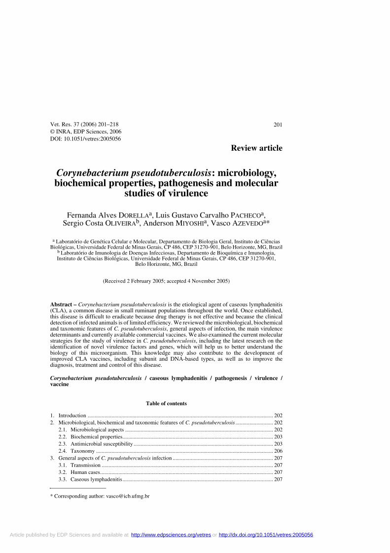
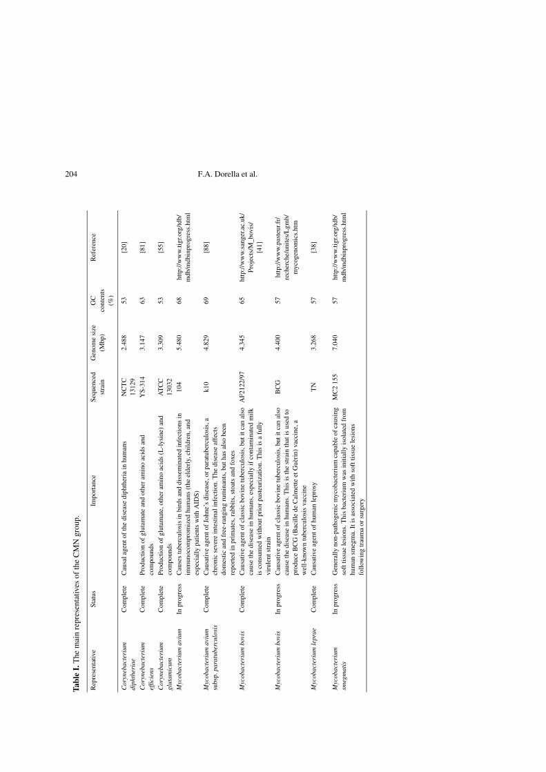
The genus Corynebacterium belongs toa suprageneric group of actinomycetes thatalso includes the genera Mycobacterium,Nocardia and Rhodococcus [46, 87, 100, 102].These gram-positive bacteria (Corynebac-terium, Mycobacterium, Nocardia andRhodococcus species), termed the CMNgroup, constitute a very heterogeneous group;however, most of the species share partic-ular characteristics, such as: (i) a specificcell wall organization, mainly character-ized by the presence of a huge polymercomplex composed of peptidoglycan, ara-binogalactan and mycolic acids [5, 26–28,39, 45, 48] and (ii) high G+C content (47–74%) [39, 40, 43, 80]. The genomes of sev-eral species of this group have already beencompletely sequenced; this fact reflects theconsiderable medical, veterinary and bio-technological importance of these organ-isms (Tab. I).
Corynebacterium pseudotuberculosis isan important animal pathogen. It is the eti-ological agent of a disease that is commonlycalled caseous lymphadenitis (CLA) orcheesy gland [114]. This disease is found inall the world’s major sheep and goat pro-duction areas, causing significant eco-nomic losses [85, 114].
In this review, we present the mainmicrobiological characteristics of C. pseu-dotuberculosis. Bacterial virulence deter-minants, including previously reported vir-
ulence factors and recently identifiedmolecules, are discussed, with emphasis onthe molecular strategies that have been usedto identify and study such determinants.The aspects regarding CLA are also cov-ered, focusing on the currently-availablecommercial and experimental vaccines.
2. MICROBIOLOGICAL, BIOCHEMICAL AND TAXONOMIC FEATURES OF C. PSEUDOTUBERCULOSIS
2.1. Microbiological aspects
C. pseudotuberculosis was isolated frombovine farcy in 1888 by Nocard. Preisz, in1894, was the first to completely describethis microorganism and to observe itsresemblance to the diphtheria bacillus.Synonyms for C. pseudotuberculosis wereBacillus pseudotuberculosis ovis, Bacilluspseudotuberculosis, Corynebacterium ovisand Preisz-Nocard bacillus [59, 72].
This microorganism is a facultativeintracellular pathogen that exhibits pleo-morphic forms, such as coccoids and fila-mentous rods, ranging in size from 0.5 μmto 0.6 μm by 1.0 μm to 3.0 μm [17, 28, 72,97]. It is a non-sporulating, non-capsulatedand non-motile bacterium; however, it hasfimbriae [17, 46, 72]. This bacterium is afacultative anaerobe and grows best at

The role of C. pseudotuberculosis in pathogenesis 203
37 °C, at a pH of 7.0 to 7.2 [17, 72, 97]. Itgrows sparse initially on the agar surfaceand then becomes organized in clumps or inpalisades, taking on a cream to orange col-oration; colonies are dry, opaque and con-centrically ringed. Growth in fluid mediumdevelops as a granular deposit with a sur-face pellicle [17, 72, 77]. Haemolysis onblood agar is variable, but large zonesdevelop in the presence of Rhodococcusequi [17]. C. pseudotuberculosis toxininhibits the action of staphylococcal β-lysin[59].
C. pseudotuberculosis stains Gram-positive and when stained by Albert’s orNeisser’s method, volutin granules can bevisualized. These metachromatic granulesare clearly observed in the bacillary form,but are absent from coccoid cells; they con-tain high-energy phosphate reserves [46,72].
2.2. Biochemical properties
Cell wall peptidoglycan is based onmeso-diaminopimelic acid (meso-DAP).Arabinose and galactose are major cell wallsugars. Short-chain mycolic acids (coryno-mycolic acids, 22–36 carbon atoms) arepresent [59, 94, 97]. Biochemical reactionsof C. pseudotuberculosis isolates vary con-siderably, mainly in their fermenting ability[72, 100, 105]. All strains produce acid, butnot gas, from many carbon sources, includ-ing glucose, fructose, maltose, mannose,and sucrose [17, 53, 59, 72]. This bacteriumis phospholipase D and catalase positive,oxidase negative, and it is beta-hemolytic[59, 77, 100]. Strains isolated from smallruminants generally do not reduce nitrate[17, 72, 100, 114].
A well-established biochemical test forcoryneform bacteria identification is theAPI Coryne system (API-bioMérieux, Inc.,La Balme les Grottes, France). This methodconsists of 21 biochemical tests; it can beperformed in 24–48 h. The test contains20 tubes containing substrates that allowfor 11 enzyme tests (pyrazinamidase,
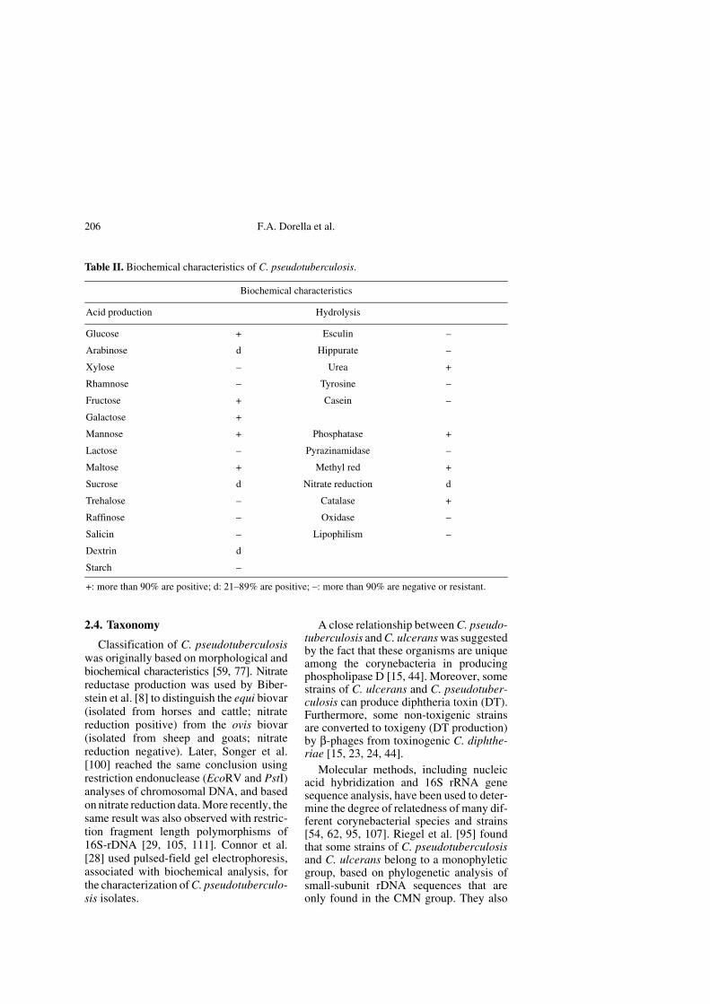
pyrrolidonyl arylamidase, β-galactosidase,alkaline phosphatase, α-glucosidase, N-acetylglucosaminidase, β-glucuronidase, andnitrate reduction and gelatin, urea and escu-lin hydrolysis) and eight carbohydrate fer-mentation tests (glucose, ribose, D-xylose,mannitol, maltose, lactose, sucrose and gly-cogen). This system is more reliable andrapid when it is compared with standardidentification methods (API-bioMérieux,Inc.). A summary of general biochemicalproperties of C. pseudotuberculosis is pre-sented in Table II.
2.3. Antimicrobial susceptibility
The susceptibility pattern of C. pseudo-tuberculosis to antimicrobial agents variesamong isolates obtained from varioussources [28, 37, 66]. Muckle and Gyles[77], in a study of 26 strains isolated fromlesions of caseous lymphadenitis in goats,reported that all strains were susceptible tothe antibiotics ampicillin, chlorampheni-col, lincomycin, gentamicin, tetracycline,penicillin G and sulfamethoxazole-trimeth-oprim. Only three isolates were susceptibleto neomycin, and all strains were resistantto streptomycin. Garg et al. [40] reportedstrains of C. pseudotuberculosis that werestrongly resistant to penicillin but suscepti-ble to neomycin. A strain highly resistant tostreptomycin (500 μg/mL) was observed ina study of 22 isolates of C. pseudotubercu-losis from sheep and goat abscesses [90].Minimal inhibitory concentration (MIC)values for all isolates were similar for thevarious antimicrobial agents. Later studiesalso indicated a similarity of MIC valuesamong strains [1, 29, 60]. However, Fern-ández et al. [35] found higher MIC valuesfor several antimicrobial agents, in an anal-ysis of corynebacteria isolated from ewemastitis.
Olson et al. [82] grew C. pseudotuber-culosis as a biofilm, in an attempt to repro-duce the environment of a natural infection.They observed that this bacterium washighly resistant to all the drugs that theytested under such growth conditions.
204 F.A. Dorella et al.
Tabl
e I.
The
mai
n re
pres
enta
tives
of
the
CM
N g
roup
.
Rep
rese
ntat
ive
Stat
usIm
port
ance
Seq
uenc
ed
stra
inG
enom
e si
ze
(Mbp
)G
C
cont
ents
(%
)
Ref
eren
ce
Cor
yneb
acte
rium
di
phth
eria
eC
ompl
ete
Cau
sal a
gent
of
the
dise
ase
diph
ther
ia in
hum
ans
NC
TC
13
129
2.48
853
[20]
Cor
yneb
acte
rium
ef
ficie
nsC
ompl
ete
Prod
ucti
on o
f gl
utam
ate
and
othe
r am
ino
acid
s an
d co
mpo
unds
YS-
314
3.14
763
[81]
Cor
yneb
acte
rium
gl
utam
icum
Com
plet
ePr
oduc
tion
of
glut
amat
e, o
ther
am
ino
acid
s (L
-lys
ine)
and
co
mpo
unds
AT
CC
13
032
3.30
953
[55]
Myc
obac
teri
um a
vium
In p
rogr
ess
Cau
ses
tube
rcul
osis
in b
irds
and
dis
sem
inat
ed in
fect
ions
in
imm
unoc
ompr
omiz
ed h
uman
s (t
he e
lder
ly, c
hild
ren,
and
es
peci
ally
pat
ient
s w
ith A
IDS)
104
5.48
068
http
://w
ww
.tigr
.org
/tdb/
mdb
/mdb
inpr
ogre
ss.h
tml
Myc
obac
teri
um a
vium
subs
p. p
arat
uber
culo
sis
Com
plet
eC
ausa
tive
agen
t of
John
e’s
dise
ase,
or
para
tube
rcul
osis
, a
chro
nic
seve
re in
test
inal
infe
ctio
n. T
he d
isea
se a
ffec
ts
dom
estic
and
fre
e-ra
ngin
g ru
min
ants
, but
has
als
o be
en
repo
rted
in p
rim
ates
, rab
bits
, sto
ats
and
foxe
s
k10
4.82
969
[88]
Myc
obac
teri
um b
ovis
Com
plet
eC
ausa
tive
agen
t of
clas
sic
bovi
ne tu
berc
ulos
is, b
ut it
can
als
o ca
use
the
dise
ase
in h
uman
s, e
spec
ially
if c
onta
min
ated
mil
k is
con
sum
ed w
ithou
t pri
or p
aste
uriz
atio
n. T
his
is a
ful
ly
viru
lent
str
ain
AF2
122/
974.
345
65ht
tp://
ww
w.s
ange
r.ac.
uk/
Pro
ject
s/M
_bov
is/
[41]
Myc
obac
teri
um b
ovis
In p
rogr
ess
Cau
sativ
e ag
ent o
f cl
assi
c bo
vine
tube
rcul
osis
, but
it c
an a
lso
caus
e th
e di
seas
e in
hum
ans.
Thi
s is
the
stra
in th
at is
use
d to
pr
oduc
e B
CG
(B
acill
e de
Cal
met
te e
t Guè
rin)
vac
cine
, a
wel
l-kn
own
tube
rcul
osis
vac
cine
BC
G4.
400
57ht
tp:/
/ww
w.p
aste
ur.f
r/re
cher
che/
unit
es/L
gmb/
myc
ogen
omic
s.ht
m
Myc
obac
teri
um le
prae
Com
plet
eC
ausa
tive
agen
t of
hum
an le
pros
yT
N3.
268
57[3
8]
Myc
obac
teri
um
smeg
mat
isIn
pro
gres
sG
ener
ally
non
-pat
hoge
nic
myc
obac
teri
um c
apab
le o
f cau
sing
so
ft ti
ssue
lesi
ons.
Thi
s ba
cter
ium
was
init
ially
isol
ated
fro
m
hum
an s
meg
ma.
It i
s as
soci
ated
wit
h so
ft ti
ssue
lesi
ons
follo
win
g tr
aum
a or
sur
gery
MC
2 15
57.
040
57ht
tp:/
/ww
w.ti
gr.o
rg/td
b/m
db/m
dbin
prog
ress
.htm
l
The role of C. pseudotuberculosis in pathogenesis 205
Tabl
e I.
Con
tinue
d.
Rep
rese
ntat
ive
Sta
tus
Impo
rtan
ceS
eque
nced
st
rain
Gen
ome
size
(M
bp)
GC
co
nten
ts
(%)
Ref
eren
ce
Myc
obac
teri
um
tube
rcul
osis
Com
plet
eC
ausa
tive
agen
t of
tube
rcul
osis
. It i
s hi
ghly
con
tagi
ous,
in
fect
ing
appr
oxim
atel
y 80
% o
f th
e pa
tien
t’s s
ocia
l con
tact
sC
DC
1551
4.40
365
[36]
Myc
obac
teri
um
tube
rcul
osis
Com
plet
eC
ausa
tive
agen
t of t
uber
culo
sis.
Unl
ike
som
e cl
inic
al is
olat
es,
it r
etai
ns f
ull v
irul
ence
in a
nim
al m
odel
s of
tube
rcul
osis
and
is
sus
cept
ible
to d
rugs
and
rec
eptiv
e to
gen
etic
man
ipul
atio
n
H37
Rv
4.41
165
[25]
Myc
obac
teri
um
tube
rcul
osis
In p
rogr
ess
Cau
sativ
e ag
ent o
f tu
berc
ulos
is. I
t was
sub
sequ
ently
fou
nd
that
this
str
ain
is o
ne o
f th
e m
ost w
ide-
spre
ad a
nd v
irul
ent
Myc
obac
teri
um tu
berc
ulos
is s
trai
ns
210
4.40
057
http
://w
ww
.tigr
.org
/tdb/
mdb
/mdb
inpr
ogre
ss.h
tml
Noc
ardi
a fa
rcin
ica
Com
plet
eT
he c
ausa
tive
agen
t of
noca
rdio
sis,
aff
ectin
g th
e lu
ng, c
entr
al
nerv
ous
syst
em, a
nd c
utan
eous
tiss
ues
of h
uman
s an
d an
imal
s T
his
spec
ies
exhi
bits
a g
reat
er d
egre
e of
vir
ulen
ce th
an th
e m
ore
com
mon
Noc
ardi
a as
tero
ides
IFM
101
526.
021
(Chr
omos
ome)
0.18
4(P
lasm
id p
NF1
)0.
087
(Pla
smid
pN
F2)
70 67 68
[56]
Rho
doco
ccus
sp.
In p
rogr
ess
Mic
robe
cap
able
of
degr
adin
g a
wid
e va
riet
y of
po
lych
lori
nate
d bi
phen
yls
RH
A1
9.70
0ht
tp:/
/w
ww
.rho
doco
ccus
.ca
206 F.A. Dorella et al.
2.4. Taxonomy
Classification of C. pseudotuberculosiswas originally based on morphological andbiochemical characteristics [59, 77]. Nitratereductase production was used by Biber-stein et al. [8] to distinguish the equi biovar(isolated from horses and cattle; nitratereduction positive) from the ovis biovar(isolated from sheep and goats; nitratereduction negative). Later, Songer et al.[100] reached the same conclusion usingrestriction endonuclease (EcoRV and PstI)analyses of chromosomal DNA, and basedon nitrate reduction data. More recently, thesame result was also observed with restric-tion fragment length polymorphisms of16S-rDNA [29, 105, 111]. Connor et al.[28] used pulsed-field gel electrophoresis,associated with biochemical analysis, forthe characterization of C. pseudotuberculo-sis isolates.
A close relationship between C. pseudo-tuberculosis and C. ulcerans was suggestedby the fact that these organisms are uniqueamong the corynebacteria in producingphospholipase D [15, 44]. Moreover, somestrains of C. ulcerans and C. pseudotuber-culosis can produce diphtheria toxin (DT).Furthermore, some non-toxigenic strainsare converted to toxigeny (DT production)by β-phages from toxinogenic C. diphthe-riae [15, 23, 24, 44].
Molecular methods, including nucleicacid hybridization and 16S rRNA genesequence analysis, have been used to deter-mine the degree of relatedness of many dif-ferent corynebacterial species and strains[54, 62, 95, 107]. Riegel et al. [95] foundthat some strains of C. pseudotuberculosisand C. ulcerans belong to a monophyleticgroup, based on phylogenetic analysis ofsmall-subunit rDNA sequences that areonly found in the CMN group. They also
Table II. Biochemical characteristics of C. pseudotuberculosis.
Biochemical characteristics
Acid production Hydrolysis
Glucose + Esculin –
Arabinose d Hippurate –
Xylose – Urea +
Rhamnose – Tyrosine –
Fructose + Casein –
Galactose +
Mannose + Phosphatase +
Lactose – Pyrazinamidase –
Maltose + Methyl red +
Sucrose d Nitrate reduction d
Trehalose – Catalase +
Raffinose – Oxidase –
Salicin – Lipophilism –
Dextrin d
Starch –
+: more than 90% are positive; d: 21–89% are positive; –: more than 90% are negative or resistant.

The role of C. pseudotuberculosis in pathogenesis 207
concluded that the equi and ovis biovars ofC. pseudotuberculosis should not be classifiedas subspecies, due to their high genomicsimilarity. In two other independent studies[54, 107], C. pseudotuberculosis was foundto be closely related to C. ulcerans.
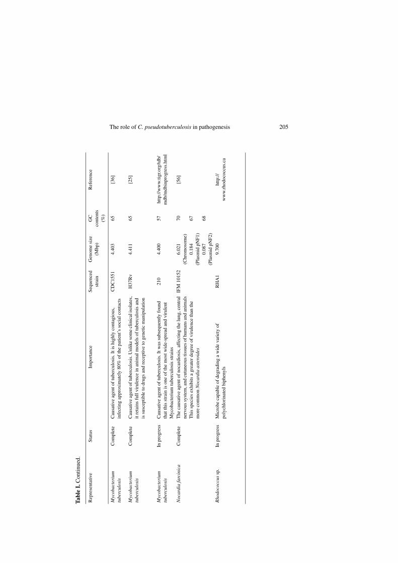
More recently, analysis of partial genesequences from the β-subunit of RNApolymerase (rpoB) has been shown to bemore accurate for the identification ofCorynebacterium species than analysesbased on 16S rDNA [61, 62]. This methodhas also been successfully used to identifymycobacterial species [63]. Although therpoB gene is a powerful identification tool,many authors propose that it may be usedto complement the 16S rRNA gene analysisin the phylogenetic studies of Corynebac-terium and Mycobacterium species [61–63,74]. We have constructed a phylogenetictree based on rpoB gene sequences of ref-erence strains from the CMN group (Fig. 1).Based on this phylogenetic tree, we canobserve a clear relationship between C. pseu-dotuberculosis and C. ulcerans. Moreover,analysis using the rpoB gene allowed theidentification of the group that these twospecies belong to, as previously observed[61, 62].
3. GENERAL ASPECTS OF C. PSEU-DOTUBERCULOSIS INFECTION
Though C. pseudotuberculosis was orig-inally identified as the causative microor-ganism of CLA in sheep and goats, this bac-terium has also been isolated from otherspecies, including horses, in which it causesulcerative lymphangitis and pigeon fever incattle, camels, swine, buffaloes, and humans[89, 97, 114, 117].
3.1. Transmission
The potential of C. pseudotuberculosisto survive for several weeks in the environ-ment likely contributes to its ability tospread within a herd or flock [4, 117].
Transmission among sheep or goats occursmainly through contamination of superfi-cial wounds, which can appear during com-mon procedures, such as shearing, castra-tion and ear tagging, or through injuries ofthe animal’s bodies generated by other trau-matic events. Not infrequently, contami-nated sheep cough bacteria onto skin cuts ofother sheep, constituting another means oftransmission [84, 114]. In cattle, as well asin buffaloes, there is evidence of mechani-cal transmission of this bacterium by house-flies and by other Diptera, though the naturalmechanisms of infection with C. pseudotu-berculosis are not well documented [97,116, 117].
3.2. Human cases
Human infection caused by C. pseudo-tuberculosis is a rare event, and most of thereported cases have been related to occupa-tional exposure; one case, diagnosed in1988, involved the ingestion of raw goatmeat and cow milk [89]. About 25 cases ofinfection of humans with this microorgan-ism have been reported in the literature [67,73, 89].
Peel et al. [89] reviewed 22 cases, inwhich infected humans were generally pre-sented with lymphadenitis, abscesses, andconstitutional symptoms. Mills et al. [73]described suppurative granulomatous lym-phadenitis in a boy, due to contact with con-taminated farm animals. Liu et al. [67]reported a C. pseudotuberculosis infectionin a patient’s eye, due to an ocular implant.
In most cases, the patients received anti-biotic therapy and the affected lymph nodeswere surgically removed [67, 73, 89].
3.3. Caseous lymphadenitis
Caseous lymphadenitis causes significanteconomic losses to sheep and goat produc-ers worldwide, mainly due to the reductionof wool, meat and milk yields, decreasedreproductive efficiencies of affected animalsand condemnation of carcasses and skins in

208 F.A. Dorella et al.
abattoirs [3, 83]. The manifestations ofCLA in small ruminants are characterizedmainly by bacteria-induced caseation necro-sis of the lymph glands. The most frequentform of the disease, external CLA, is char-acterized by abscess formation in superfi-cial lymph nodes and in subcutaneous tis-sues. These abscesses can also developinternally in organs, such as the lungs, kid-neys, liver and spleen, characterizing visceralCLA [72, 91]. In some cases, the infectionproduces few obvious clinical signs in theanimal, remaining unrecognized until apost-mortem examination has been carriedout, making it difficult to obtain definitivedata about the prevalence of this disease [3,17, 83].
3.4. Epidemiology of CLA
Recent epidemiological surveys haveexamined the prevalence of CLA in differ-ent countries [2, 3, 6, 11, 28, 85]. Amongflocks surveyed in Australia, the averageprevalence of CLA in adult sheep was 26%[85]. Forty-five percent of the farmers inter-viewed in a study in the United Kingdomhad seen abscesses in their sheep; however,this could be an overestimation of CLAprevalence since few farmers had investi-gated the causes of the abscesses [11].Twenty-one percent of 485 culled sheepexamined in Canadian slaughterhouses hadCLA [3]. This disease remains an importantsubject of veterinary concern throughoutthe world.
3.5. Diagnosis and control of CLA
Controlling CLA with antibiotics is notan easy task, since viable bacteria stay pro-tected inside abscesses due to the thick cap-sule that surrounds them [91, 103, 114]. Itis generally agreed that the best strategy tocontrol the disease is vaccination of healthyanimals, along with the identification/removalof infected animals [13, 71, 84, 114]. How-ever, the difficulties associated with theearly clinical identification of infected ani-mals can be a hindrance to such a strategy.
Several serodiagnostic tests have beendeveloped to overcome the problem of clin-ical identification of CLA, but most havebeen reported to lack either sensitivity orspecificity [14, 16, 70, 71, 104, 114, 118].Nevertheless, some enzyme-linked immu-nosorbent assay (ELISA)-based diagnostictests have been reported to be effective incontrol and eradication programs [32, 33,110]. Recently, ELISA tests to detectgamma interferon (IFN-γ), as a marker ofcell-mediated immunity against C. pseudo-tuberculosis, have been developed [71, 86,93]. The IFN-γ ELISA test appears to bemore sensitive than the normal antibodyELISA in detecting prior infection in goats,and it does not seem to be affected by vac-cination in sheep [71]. Another novel strat-egy that holds promise for the diagnosis ofCLA is the use of polymerase chain reaction(PCR) tests specific for C. pseudotubercu-losis to identify bacteria isolated fromabscesses [21].
4. FROM PROTEINS TO DNA: COMMERCIAL AND EXPERIMENTAL VACCINES
4.1. Commercial vaccines
Most of the currently-available commer-cial vaccines for caseous lymphadenitis arecombined with vaccines against other path-ogens. These include Clostridium tetani,Cl. perfringens, Cl. septicum, Cl. novyi andCl. chauvoei [85, 91, 103, 114]. These vac-cines are based on inactivated phospholi-pase D (PLD) and are called toxoid vaccines.
Paton et al. [84], in an analysis of theeffectiveness of a combined toxoid vaccineagainst CLA, reported a reduction in thenumber and size of CLA lung abscesses anda decrease in the spread of this diseasewithin the flock. However, in another study[85], it was reported that although 43% ofthe farmers applied commercial CLA vac-cines, only 12% used them correctly. It wasconcluded that adjustments in vaccination

The role of C. pseudotuberculosis in pathogenesis 209
Figure 1. Dendrogram representing thephylogenetic relationships of the CMNgroup (Corynebacterium, Mycobacterium,Nocardia and Rhodococcus species)obtained by the neighbor-joining method[96]. The tree was derived from the align-ments of rpoB gene sequences. The phyl-ogenetic distances were calculated by thesoftware MEGA 3 [64]. The support ofeach branch, as determined from 1 000bootstrap samples, is indicated by the valueat each node (in percent).

210 F.A. Dorella et al.
programs would dramatically diminish theprevalence of CLA.
Not all the vaccines licensed for use insheep can be used to vaccinate goats. More-over, while the recommended vaccinationprogram for sheep consists of two primingdoses in lambs and yearly boosters in adultsheep, revaccination is recommended atsix-month intervals in goats [85, 114].
A live attenuated vaccine strain of C.pseudotuberculosis, strain 1002, has beenlicensed for use in Brazil since 2000. It isalready being produced industrially and isavailable in a liquid form that must beadministrated yearly to the animals, subcu-taneously; a lyophilized version is alsobeing developed by the Empresa Baianade Desenvolvimento Agrícola (http://www.ebda.ba.gov.br). This live vaccinewas reported to confer around 83% protec-tion against CLA in goats in experimentalassays and in field trials.
4.2. Experimental vaccines
C. pseudotuberculosis Toxminus (pldmutant) has been used as a live bacterialvector to deliver heterologous antigenicproteins [75]. Five heterologous genes(the gene coding for Mycobacterium leprae18-kDa antigen, Taenia ovis 45W gene,Babesia bovis 11C5 antigen, the Dichelo-bacter nodosus gene encoding mature basicprotease (bprV) and Anaplasma marginaleApH antigen), plus a genetically inacti-vated analogue of PLD, were used to con-struct plasmids expressing foreign genes inthe Toxminus strain. Three proteins elicitedspecific antibody responses in experimen-tally vaccinated sheep. The expression byToxminus of mature basic protease (bprV)of D. nodosus fused to the carboxy-termi-nus of Mycobacterium leprae 18-kDa anti-gen against ovine footrot [76] was alsotested. Though the animals were not pro-tected from footrot, this live recombinantvaccine was capable of eliciting a humoralimmune response, and it may be capable ofsuccessfully delivering a foreign antigen.
Recently, the immune responses of sheepvaccinated with a DNA vaccine expressingthe extracellular domain of bovine CTLA-4,fused to HIg and a genetically detoxifiedphospholipase D (boCTLA-4-HIg-ΔPLD)from C. pseudotuberculosis have beeninvestigated [22]. CTLA-4 binds with highaffinity to the B7 membrane antigen onantigen-presenting cells (APC), enhancingthe humoral immune response to a vaccineantigen. Though the genetically attenuatedvaccine was found to be only partiallyeffective against experimental challengewith C. pseudotuberculosis, the targetedDNA vaccine provided sheep with a signif-icantly improved antibody response. Inorder to improve the efficacy of this DNAvaccine, De Rose et al. [31] tested differentroutes of immunization: (i) intramuscularDNA injection, (ii) subcutaneous DNAinjection and (iii) gene gun bombardment.Intramuscular vaccination gave a level ofprotection similar to that observed with pro-tein vaccination, while subcutaneous andgene gun vaccination did not protect sheepagainst bacterial challenge.
5. DETERMINANTS OF VIRULENCE
5.1. Phospholipase D
Phospholipase D (PLD) is a potent exo-toxin produced by C. pseudotuberculosisand it has been considered as the major vir-ulence factor for this bacterium [51, 65].
This exotoxin is a permeability factorthat promotes the hydrolysis of ester bondsin sphingomyelin in mammalian cell mem-branes, possibly contributing to the spreadof the bacteria from the initial site of infec-tion to secondary sites within the host [19,30, 65, 69, 89, 106, 108]. Moreover, it pro-vokes dermonecrotic lesions, and at higherdoses it is lethal to a number of differentspecies of laboratory and domestic animals[34, 102]. Damage and destruction ofcaprine macrophages have been observedduring infection with C. pseudotuberculosis.

The role of C. pseudotuberculosis in pathogenesis 211
This lethal effect is due to action of PLD[109].
Several of the biological activities of C.pseudotuberculosis PLD, as well as itsmolecular structure, have also been foundin sphingomyelinases in the venom of themedically important spider genus Loxosceles[7, 10, 30, 102, 108, 112].
The use of an antitoxin has prevented thespread of C. pseudotuberculosis within thehost; however, it is not able to prevent thedevelopment of abscesses [114]. Moreover,vaccination of goats with formalized exo-toxin, i.e. with inactive PLD, also preventedthe spread of bacteria, following experi-mental challenge [13].
5.2. Toxic cell-wall lipids
The surface lipids of C. pseudotubercu-losis have long been described as major fac-tors contributing to its pathogenesis [18, 47,48, 58]. The toxicity of the extracted lipidmaterial has been demonstrated by theinduction of hemorrhagic necrosis follow-ing intradermal injection in guinea pigs[58]. Mouse peritoneal macrophages werefound to be highly susceptible to the necro-tizing action of C. pseudotuberculosis sur-face lipids, but this cytotoxic effect is notobserved in rabbit cells [48]. However,infection with C. pseudotuberculosis in theguinea pig invariably progresses untildeath, while guinea pig macrophages arenot susceptible to the cytotoxic action of thebacterial lipids [48, 57]. Tashjian et al.[109] observed that C. pseudotuberculosiswas resistant to killing and digestion bycaprine macrophages due to its lipid coat.
A study carried out in mice with 25 iso-lates of C. pseudotuberculosis proposedthat there is a direct relationship of the per-centage of surface lipids with the inductionof chronic abscessation [78].
5.3. New candidates
Recently, it has been proposed that aputative C. pseudotuberculosis iron uptake
gene cluster has a role in its virulence [9].The four genes in this putative operon wereidentified downstream from the pld gene.They were designated as Fe acquisitiongenes (fag) A, B, C and D. Since C. pseudo-tuberculosis is an intracellular pathogen,this bacterium must be able to acquire ironfrom an environment in which this nutrientis scarce. Although there was no alterationin the utilization of iron by a fagB(C)mutant in vitro, this mutant had a decreasedability to survive and to cause abscesses inexperimentally-infected goats [9].
6. MOLECULAR STRATEGIES FOR THE STUDY OF VIRULENCE IN C. PSEUDOTUBERCULOSIS
6.1. Identification of immunodominant peptides
To date, the most widely studied C. pseu-dotuberculosis protein is PLD. It hasalready been purified, cloned and expressedin E. coli [34, 50, 69, 101].
A protective antigen, corynebacterialsecreted protease 40 (CP40) [115], has beenidentified in C. pseudotuberculosis byapplying a strategy that involves the localimmune response, analyzing the specificityof antibodies produced by B cells [113].Antibody secreting cells (ASC), obtainedfrom induced infections in sheep, produceantibodies with high specificity. These anti-bodies are used as probes to screen whole-cell antigens of C. pseudotuberculosis byimmunoblots. CP40 was one of the earliestantigens recognized in immunoblots ofsera. ELISA tests confirmed the resultsobtained with immunoblots, and field trialswith this semipurified antigen showed thatCP40 was highly protective against exper-imentally-induced CLA [113].
Some researchers have analyzed andcharacterized soluble and insoluble pro-teins that have immunodominant potential[12, 79]. Though many other immunogenicexcreted-secreted components have been

212 F.A. Dorella et al.
described, using immunoblot techniques[86, 87], these proteins have not been iden-tified. However, they reliably detectedCLA infection in goats, and they could beused as vaccine components.
6.2. Generation of mutants
Random chemical mutagenesis, withformic acid, was used by Haynes et al. [49]to produce enzymatically-inactive PLD.This analog protein, though inactive, stillhad immunological activity [49]. Hodgsonet al. [51] and McNamara et al. [68] usedsite-specific mutagenesis to produce pldmutants that had reduced ability to establishinfection and were unable to disseminate insheep and goats.
Site-specific amino acid substitution hasalso been used to generate genetic inactiva-tion of the pld gene in two independentexperiments. Tachedjian et al. [106] substi-tuted the His20 in the PLD active site withother amino acids, obtaining mutants thatwere able to produce a genetically-inacti-vated version of PLD. After analysis ofmutant gene expression, two mutants wereselected that retained features useful fortoxoid vaccine development. In another study,the inactivated protein, in which His20 wassubstituted by Ser, gave 44% protection insheep challenged with the bacterium [52].
A mutant of the C. pseudotuberculosisrecA gene was generated by site-specificinactivation [92]. The mutant had its homol-ogous recombination efficiency decreased 8–10 fold. Nevertheless, in vivo analysisrevealed that the mutated recA gene did notaffect the virulence of this bacterium inmice.
Reduction of virulence of C. pseudotu-berculosis mutants was obtained by Sim-mons et al. [98]. Allelic exchange was usedto generate aroQ-attenuated mutants thatwere unable to cause CLA in murine mod-els. It was suggested that highly attenuatedaroQ mutants of C. pseudotuberculosiscould be used as vaccine vectors [99].
The ability of the fag genes to be inducedby limited iron was studied by transcrip-tional fusions with the lacZ reporter gene,followed by an assay for β-galactosidaseactivity [9]. The resultant mutants weregrown in both iron-rich and iron-limitedmedia. The mutants expressed very lowlevels of β-galactosidase activity in iron-rich medium and almost three-fold more iniron-limited medium. Although not wellexpressed in vitro, this putative operonappears to be induced by limited iron.
Our research group has identified 34insertional mutants of genes coding for fim-brial and transport subunits, and also forhypothetical and unknown function pro-teins from C. pseudotuberculosis, usingrandom transposon mutagenesis with theTnFuZ transposition system [42], a tool thatgenerates transcriptional and translationalfusions with the phoZ gene (encoding alka-line phosphatase) of Enterococcus faecalis1.This discovery indicates promising targetgenes that could contribute to the develop-ment of attenuated vaccine strains.
7. FUTURE DIRECTIONS
Despite the various molecular strategiesthat have been employed, efficient tools forthe genetic study of C. pseudotuberculosisare still scarce. In fact, the main reason forthe lack of molecular investigation of thisorganism is that the genetics of the genushave been little studied with modern tech-niques, making it difficult to identify andcharacterize factors that could be involvedin virulence [20]. Nevertheless, other rep-resentatives of the CMN group are bettercharacterized, and the genetic tools thathave been developed could be directlyapplicable to C. pseudotuberculosis infuture studies.
1 Dorella F.A., Estevam E.M., Pacheco L.G.C.,Guimarães C.T., Lana U.G.P., Gomes E.A.,Miyoshi A., Azevedo V., unpublished results.
The role of C. pseudotuberculosis in pathogenesis 213
ACKNOWLEDGEMENTS
Miyoshi A. and Azevedo V. share the samecredit in the senior authorship of this work. Thiswork was supported by CNPq (Conselho Nacionalde Desenvolvimento Científico e Tecnológico,Brasil), CAPES (Coordenação de Aperfeiçoa-mento de Pessoal de Nível Superior, Brasil),FINEP (Financiadora de Estudos e Projetos-01.04.760.00) and FAPEMIG (Fundação deAmparo à Pesquisa do Estado de Minas Gerais,Brasil).
REFERENCES
[1] Adamson P.J., Wilson W.D., Hirsh D.C., Bag-got J.D., Martin L.D., Susceptibility of equinebacterial isolates to antimicrobial agents, Am.J. Vet. Res. 46 (1985) 447–450.
[2] Al-Rawashdeh O.F., al-Qudah K.M., Effectof shearing on the incidence of caseous lym-phadenitis in Awassi sheep in Jordan, J. Vet.Med. B Infect. Dis. Vet. Public Health 47(2000) 287–293.
[3] Arsenault J., Girard C., Dubreuil P., DaignaultD., Galarneau J.-R., Boisclair J., Simard C.,Bélanger D., Prevalence of and carcass con-demnation from maedi-visna, paratuberculo-sis and caseous lymphadenitis in culled sheepfrom Quebec, Canada, Prev. Vet. Med. 59(2003) 67–81.
[4] Augustine J.L., Renshaw H.W., Survival ofCorynebacterium pseudotuberculosis inaxenic purulent exudate on common barnyardfomites, Am. J. Vet. Res. 47 (1986) 713–715.
[5] Bayan N., Houssin C., Chami M., Leblon G.,Mycomembrane and S-layer: two importantstructures of Corynebacterium glutamicumcell envelope with promising biotechnologyapplications, J. Biotechnol. 104 (2003) 55–56.
[6] Ben Said M.S., Ben Maitigue H., Benzarti M.,Messadi L., Rejeb A., Amara A., Epidemio-logical and clinical studies of ovine caseouslymphadenitis, Arch. Inst. Pasteur Tunis 79(2002) 51–57.
[7] Bernheimer A.W., Campbell B.J., ForresterL.J., Comparative toxinology of Loxoscelesreclusa and Corynebacterium pseudotuber-culosis, Science 228 (1985) 590–591.
[8] Biberstein E.L., Knight H.D., Jang S., Twobiotypes of Corynebacterium pseudotubercu-losis, Vet. Rec. 89 (1971) 691–692.
[9] Billington S.J., Esmay P.A., Songer J.G., JostB.H., Identification and role in virulence ofputative iron acquisition genes from Coryne-
bacterium pseudotuberculosis, FEMS Micro-biol. Lett. 208 (2002) 41–45.
[10] Binford G.J., Cordes M.H.J., Wells M.A.,Sphingomyelinase D from venoms of Loxos-celes spiders: evolutionary insights fromcDNA sequences and gene structure, Toxicon45 (2005) 547–560.
[11] Binns S.H., Bairley M., Green L.E., Postalsurvey of ovine caseous lymphadenitis in theUnited Kingdom between 1990 and 1999,Vet. Rec. 150 (2002) 263–268.
[12] Braithwaite C.E., Smith E.E., Songer J.G.,Reine A.H., Characterization of detergent-soluble proteins of Corynebacterium pseudo-tuberculosis, Vet. Microbiol. 38 (1993) 59–70.
[13] Brown C.C., Olander H.J., Biberstein E.L.,Morse S.M., Use of a toxoid vaccine to protectgoats against intradermal challenge exposureto Corynebacterium pseudotuberculosis, Am.J. Vet. Res. 47 (1986) 1116–1119.
[14] Brown C.C., Olander H.J., Alves S.F., Syner-gistic hemolysis-inhibition titers associatedwith caseous lymphadenitis in a slaughter-house survey of goats and sheep in Northeast-ern Brazil, Can. J. Vet. Res. 51 (1987) 46–49.
[15] Buck G.A., Cross R.E., Wong T.P., Loera J.,Groman N., DNA relationships among sometox-bearing corynebacteriphages, Infect. Immun.49 (1985) 679–684.
[16] Burrell D.H., A simplified double immunod-iffusion technique for detection of Coryne-bacterium ovis antitoxin, Res. Vet. Sci. 28(1980) 234–237.
[17] Buxton A., Fraser G., Corynebacterium, in:Buxton A., Fraser G. (Eds.), Animal Microbi-ology, Blackwell Scientific Publications,Edinburgh, 1977, pp. 177–183.
[18] Carne H.R., Kater J.C., Wickham N., A toxiclipid from the surface of Corynebacteriumovis, Nature 178 (1956) 701–702.
[19] Carne H.R., Onon E.O., Action of Corynebac-terium ovis exotoxin on endothelial cells ofblood vessels, Nature 271 (1978) 246–248.
[20] Cerdeño-Tárraga A.M., Efstratiou A., DoverL.G., Holden M.T.G., Pallen M., BentleyS.D., et al., The complete genome sequenceand analysis of Corynebacterium diphthteriaeNCTC13129, Nucleic Acids Res. 31 (2003)6516–6523.
[21] Çetinkaya B., Karahan M., Atil E., Kalin R.,De Baere T., Vaneechoutte M., Identificationof Corynebacterium pseudotuberculosis iso-lates from sheep and goats by PCR, Vet.Microbiol. 2359 (2002) 1–9.
[22] Chaplin P.J., De Rose R., Boyle J.S., McWatersP., Kelly J., Tennent J.M., Lew A.M., ScheerlinckJ.-P.Y., Targeting improves the efficacy of a

214 F.A. Dorella et al.
DNA vaccine against Corynebacterium pseu-dotuberculosis in sheep, Infect. Immun. 67(1999) 6434–6438.
[23] Cianciotto N., Groman N., A beta-relatedcorynebacteriophage which lacks a tox allelebut can acquire it by recombination withphage, Infect. Immun. 49 (1985) 32–35.
[24] Cianciotto N., Rappuoli R., Groman N.,Detection of homology to the beta bacteri-ophage integration site in a wide variety ofCorynebacterium ssp., J. Bacteriol. 168(1986) 103–108.
[25] Cole S.T., Brosch R., Parkhill J., Garnier T.,Churcher C., Harris D., et al., Deciphering thebiology of Mycobacterium tuberculosis fromthe complete genome sequence, Nature 393(1998) 537–544.
[26] Collins M.D., Goodfellow M., Minnikin D.E.,Fatty acid composition of some mycolic acid-containing coryneform bacteria, J. Gen.Microbiol. 128 (1982) 2503–2509.
[27] Collins M.D., Falsen E., Akervall E., SjodenB., Alvarez A., Corynebacterium kroppenst-edtii sp. Nov., a novel corynebacterium thatdoes not contain mycolic acids, Int. J. Syst.Bacteriol. 48 (1998) 1449–1454.
[28] Connor K.M., Quirie M.M., Baird G.,Donachie W., Characterization of UnitedKingdom isolates of Corynebacterium pseu-dotuberculosis using pulsed-field gel electro-phoresis, J. Clin. Microbiol. 38 (2000) 2633–2637.
[29] Costa L.R.R., Spier S.J., Hirsh D.C., Compar-ative molecular characterization of Coryne-bacterium pseudotuberculosis of differentorigin, Vet. Microbiol. 62 (1998) 135–143.
[30] Coyle M.B., Lipsky B.A., Coryneform bacte-ria in infections diseases: clinical and labora-tory aspects, Clin. Microbiol. Rev. 3 (1990)227–246.
[31] De Rose R., Tennent J., McWaters P., ChaplinP.J., Wood P.R., Kimpton W., Cahill R.,Scheerlinck J.P., Efficacy of DNA vaccina-tion by different routes of immunisation insheep, Vet. Immunol. Immunopathol. 90(2002) 55–63.
[32] Dercksen D.P., ter Laak E.A., Schreuder B.E.,Eradication programme for caseous lymphad-enitis in goats in The Netherlands, Vet. Rec.138 (1996) 237.
[33] Dercksen D.P., Brinkhof J.M.A., Dekker-Nooren T., van Maanen K., Bode C.F., BairdG., Kamp E.M., A comparison of four sero-logical tests for the diagnosis of caseous lym-phadenitis in sheep and goats, Vet. Microbiol.75 (2000) 167–175.
[34] Egen N.B., Cuevas W., McNamara P.J.,Sammons D.W., Humphreys R., Songer J.G.,Purification of the phospholipase D ofCorynebacterium pseudotuberculosis byrecycling isoelectric focusing, Am. J. Vet.Res. 50 (1989) 1319–1322.
[35] Fernández E.P., Vela A.I., Las Heras A.,Domínguez L., Fernández-Garayzábal J.F.,Moreno M.A., Antimicrobial susceptibility ofcorynebacteria isolated from ewe’s mastitis,Int. J. Antimicrob. Agents 18 (2001) 571–574.
[36] Fleischmann R.D., Alland D., Eisen J.A.,Carpenter L., White O., Peterson J., et al.,Whole-genome comparison of Mycobacte-rium tuberculosis clinical and laboratorystrains, J. Bacteriol. 184 (2002) 5479–5490.
[37] Foley J.E., Spier S.J., Mihalyi J., DrazenovichN., Leutenegger C.M., Molecular epidemio-logic features of Corynebacterium pseudotu-berculosis isolated from horses, Am. J. Vet.Res. 65 (2004) 1734–1737.
[38] Fsihi H., Cole S.T., The Mycobacterium lep-rae genome: systematic sequence analysisidentifies key catabolic enzymes, ATP-depend-ent transport systems and a novel polA locusassociated with genomic variability, Mol.Microbiol. 16 (1995) 909–919.
[39] Funke G., Lawson P.A., Collins M.D., Heter-ogeneity within human-derived centers fordisease control and prevention (CDC) coryne-form group ANF-1-like bacteria and descrip-tion of Corynebacterium auris sp. nov., Int. J.Syst. Bacteriol. 45 (1995) 735–739.
[40] Garg D.N., Nain S.P.S., Chandiramani N.K.,Isolation and characterization of Corynebac-terium ovis from sheep and goats, Indian Vet.J. 62 (1985) 805–808.
[41] Garnier T., Eiglmeier K., Camus J.C., MedinaN., Mansoor H., Pryor M., et al., The completegenome sequence of Mycobacterium bovis,Proc. Natl. Acad. Sci. USA 100 (2003) 7877–7882.
[42] Gibson C.M., Caparon M.G., Alkaline phos-phatase reporter transposon for identificationof genes encoding secreted proteins in gram-positive microorganisms, Appl. Environ.Microbiol. 68 (2002) 928–932.
[43] Goodfellow M., Suprageneric classificationof actinomycetes, in: Williams S.T. (Ed.),Bergey’s manual of systematic bacteriology,Williams and Wilkins, Baltimore, 1989,pp. 2333–2343.
[44] Groman N., Schiller J., Russell J., Corynebac-terium ulcerans and Corynebacterium pseu-dotuberculosis responses to DNA probes derivedfrom corynephage β and Corynebacterium

The role of C. pseudotuberculosis in pathogenesis 215
diphtheriae, Infect. Immun. 45 (1984) 511–517.
[45] Hall V., Collins M.D., Hutson R.A., LawsonP.A., Falsen E., Duerden B.I., Corynebacte-rium atypicum sp. nov., from a human clinicalsource, does not contain corynomycolic acids,Int. J. Syst. Evol. Microbiol. 53 (2003) 1065–1068.
[46] Hard G.C., Electron microscopy examinationof Corynebacterium ovis, J. Bacteriol. 97(1969) 1480–1485.
[47] Hard G.C., Examination by electron micros-copy of the interaction between peritonealphagocytes and Corynebacterium ovis, J.Med. Microbiol. 5 (1972) 483–491.
[48] Hard G.C., Comparative toxic effect on thesurface lipid of Corynebacterium ovis on peri-toneal macrophages, Infect. Immun. 12(1975) 4139–1449.
[49] Haynes J.A., Tkalcevic J., Nisbet I.T., Produc-tion of an enzymatically inactive analog ofphospholipase D from Corynebacteriumpseudotuberculosis, Gene 119 (1992) 119–121.
[50] Hodgson A.L., Bird P., Nisbet I.T., Cloning,nucleotide sequence, and expression inEscherichia coli of the phospholipase D genefrom Corynebacterium pseudotuberculosis, J.Bacteriol. 172 (1990) 1256–1261.
[51] Hodgson A.L.M., Krywult J., Corner L.A.,Rothel J.S., Radford A.J., Rational attenua-tion of Corynebacterium pseudotuberculosis:potential cheesy gland vaccine and live deliv-ery vehicle, Infect. Immun. 60 (1992) 2900–2905.
[52] Hodgson A.L., Carter K., Tachedjian M.,Krywult J., Corner L.A., McColl M., CameronA., Efficacy of an ovine caseous lymphaden-itis vaccine formulated using a geneticallyinactive form of the Corynebacterium pseu-dotuberculosis phospholipase D, Vaccine 17(1999) 802–808.
[53] Holt J.G., Krieg N.R., Sneath P.H.A., StaleyJ.T., Williams S.T., Irregular, nonsporingGram-positive rods, in: Holt J.G., Krieg N.R.,Sneath P.H.A., Staley J.T., Williams S.T.(Eds.), Bergey’s manual of determinative bac-teriology, Williams and Wilkins, Baltimore,1994, p. 593.
[54] Hou X.-G., Kawamura Y., Sultana F., HiroseK., Miyake M., Otsuka Y., Misawa S., OguriT., Yamamoto H., Ezaki T., Genetic identifi-cation of members of the genus Corynebacte-rium at genus and species levels with 16SrDNA-targeted probes, Microbiol. Immunol.41 (1997) 453–460.
[55] Ikeda M., Nakagawa S., The Corynebacte-rium glutamicum genome: features and
impacts on biotechnological processes, Appl.Microbiol. Biotechnol. 62 (2003) 99–109.
[56] Ishikawa J., Yamashita A., Mikami Y.,Hoshino Y., Kurita H., Hotta K., Shiba T.,Hattori M., The complete genomic sequenceof Nocardia farcinica IFM 10152, Proc. Natl.Acad. Sci. USA 101 (2004) 14925–14930.
[57] Jolly R.D., The pathogenesis of experimentalCorynebacterium ovis infection in mice, N. Z.Vet. J. 13 (1965) 141–147.
[58] Jolly R.D., Some observations on surface lip-ids of virulent and attenuated strains ofCorynebacterium ovis, J. Appl. Bacteriol. 29(1966) 189–196.
[59] Jones D., Collins M.D., Irregular, nonsporingGram-positive rods, in: Smeath P.H.A., MairN.S., Sharpe M.E., Holt J.G. (Eds.), Bergey’smanual of systematic bacteriology, Williamsand Wilkins, Baltimore, 1986, pp. 1261–1282.
[60] Judson R., Songer J.G., Corynebacteriumpseudotuberculosis: in vitro susceptibility to39 antimicrobial agents, Vet. Microbiol. 27(1991) 145–150.
[61] Khamis A., Raoult D., La Scola B., rpoB genesequencing for identification of Corynebacte-rium species, J. Clin. Microbiol. 42 (2004)3925–3931.
[62] Khamis A., Raoult D., La Scola B., Compar-ison between rpoB and 16S rRNA genesequencing for molecular identification of168 clinical isolates of Corynebacterium, J.Clin. Microbiol. 43 (2005) 1934–1936.
[63] Kim B.J., Lee S.H., Lyu M.A., Kim S.J., BaiG.H., Kim S.J., Chae G.T., Kim E.C., ChaC.Y., Kook Y.H., Identification of Mycobac-terial species by comparative sequence anal-ysis of the RNA polymerase gene (rpoB), J.Clin. Microbiol. 37 (1999) 1714–1720.
[64] Kumar S., Tamura K., Nei M., MEGA3: Inte-grated software for Molecular EvolutionaryGenetics Analysis and sequence alignment,Brief. Bioinform. 5 (2004) 150–163.
[65] Lipsky B.A., Goldberger A.C., TompkinsL.S., Plorde J.J., Infections caused by non-diphtheria corynebacteria, Rev. Infect. Dis. 4(1982) 1220–1235.
[66] Literák I., Horváthová A., Jahnová M., RychlíkI., Skalka B., Phenotype and genotype of theSlovak and Czech Corynebacterium pseudo-tuberculosis strains isolated from sheep andgoats, Small Rumin. Res. 32 (1999) 107–111.
[67] Liu D.T., Chan W.M., Fan D.S., Lam D.S., Aninfected hydrogel buckle with Corynebacte-rium pseudotuberculosis, Br. J. Ophthalmol.89 (2005) 245–246.
[68] McNamara P.J., Bradley G.A., Songer J.G.,Targeted mutagenesis of the phospholipase D

216 F.A. Dorella et al.
results in decreased virulence of Corynebac-terium pseudotuberculosis, Mol. Microbiol.12 (1994) 921–930.
[69] McNamara P.J., Cuevas W.A., Songer J.G.,Toxic phospholipases D of Corynebacteriumpseudotuberculosis, C. ulcerans and Arcano-bacterium haemolyticum: cloning andsequence homology, Gene 156 (1995) 113–118.
[70] Menzies P.I., Muckle C.A., The use of amicroagglutination assay for the detection ofantibodies to Corynebacterium pseudotuber-culosis in naturally infected sheep and goatflocks, Can. J. Vet. Res. 53 (1989) 313–318.
[71] Menzies P.I., Hwang T.-I., Prescott J.F., Com-parison of an interferon-gamma to a phosphol-ipase D enzyme-linked immunosorbent assayfor diagnosis of Corynebacterium pseudotu-berculosis infection in experimentallyinfected goats, Vet. Microbiol. 100 (2004)129–137.
[72] Merchant I.A., Packer R.A., The GenusCorynebacterium, in: Merchant I.A., PackerR.A. (Eds.), Veterinary bacteriology andvirology, The Iowa State University Press,Iowa, 1967, pp. 425–440.
[73] Mills A.E., Mitchell R.D., Lim E.K., Coryne-bacterium pseudotuberculosis is a cause ofhuman necrotising granulomatous lymphad-enitis, Pathology 29 (1997) 231–233.
[74] Mollet C., Drancourt M., Raoult D., rpoBsequence analysis as a novel basis for bacterialidentification, Mol. Microbiol. 26 (1997)1005–1011.
[75] Moore R.J., Rothel L., Krywult J., RadfordA.J., Lund K., Hodgson A.L., Foreign geneexpression in C. pseudotuberculosis: devel-opment of a live vaccine vector, Vaccine 18(2000) 487–497.
[76] Moore R.J., Stewart D.J., Lund K., HodgsonA.L., Vaccination against ovine footrot usinga live bacterial vector to deliver basic proteaseantigen, FEMS Microbiol. Lett. 194 (2001)193–196.
[77] Muckle C.A., Gyles C.L., Characterization ofstrains of Corynebacterium pseudotuberculo-sis, Can. J. Comp. Med. 46 (1982) 206–208.
[78] Muckle C.A., Gyles C.L., Relation of lipidcontent and exotoxin production to virulenceof Corynebacterium pseudotuberculosis inmice, Am. J. Vet. Res. 44 (1983) 1149–1153.
[79] Muckle C.A., Menzies P.I., Li Y., HwangY.T., van Wesenbeeck M., Analysis of theimmunodominant antigens of Corynebacte-rium pseudotuberculosis, Vet. Microbiol. 30(1992) 47–58.
[80] Navas J., Genetic tools in pathogenic nocar-dioform actinomycetes, Microbiologia 12(1996) 297–304.
[81] Nishio Y., Nakamura Y., Kawarabayasi Y.,Usuda Y., Kimura E., Sugimoto S., Matsui K.,Yamagishi A., Kikuchi H., Ikeo K., GojoboriT., Comparative complete genome sequenceanalysis of the amino acid replacementsresponsible for the thermostability of Coryne-bacterium efficiens, Genome Res. 13 (2003)1572–1579.
[82] Olson M.E., Ceri H., Morck D.W., BuretA.G., Read R.R., Biofilm bacteria: formationand comparative susceptibility to antibiotics,Can. J. Vet. Res. 66 (2002) 86–92.
[83] Paton M.W., Rose I.R., Hart R.A., SutherlandS.S., Mercy A.R., Ellis T.M., Dhaliwal J.A.,New infection with Corynebacterium pseudo-tuberculosis reduces wool production, Aust.Vet. J. 71 (1994) 47–49.
[84] Paton M.W., Sutherland S.S., Rose I.R., HartR.A., Mercy A.R., Ellis T.M., The spread ofCorynebacterium pseudotuberculosis infec-tion to unvaccinated and vaccinated sheep,Aust. Vet. J. 72 (1995) 266–269.
[85] Paton M.W., Walker S.B., Rose I.R., WattG.F., Prevalence of caseous lymphadenitisand usage of caseous lymphadenitis vaccinesin sheep flocks, Aust. Vet. J. 81 (2003) 91–95.
[86] Paule B.J.A., Azevedo V., Regis L.F., CarminatiR., Bahia C.R., Vale V.L.C., Moura-CostaL.F., Freire S.M., Nascimento I., Schaer R.,Goes A.M., Meyer R., Experimental Coryne-bacterium pseudotuberculosis primary infec-tion in goats: kinetics of IgG and interferon-γproduction, IgG avidity and antigen recogni-tion by Western blotting, Vet. Immunol.Immunopathol. 96 (2003) 129–139.
[87] Paule B.J.A., Meyer R., Moura-Costa L.F.,Bahia C.R., Carminati R., Regis L.F., ValeV.L.C., Freire S.M., Nascimento I., Schaer R.,Azevedo V., Three-phase partitioning as anefficient method for extraction/concentrationof immunoreactive excreted-secreted proteinsof Corynebacterium pseudotuberculosis, Pro-tein Expr. Purif. 34 (2004) 311–166.
[88] Paustian M.L., Amonsin A., Kapur V.,Bannantine J.P., Characterization of novelcoding sequences specific to Mycobacteriumavium subsp. paratuberculosis: implicationsfor diagnosis of Johne’s Disease, J. Clin.Microbiol. 42 (2004) 2675–2681.
[89] Peel M.M., Palmer G.G., Stacpoole A.M.,Kerr T.G., Human lymphadenitis due toCorynebacterium pseudotuberculosis: reportof ten cases from Australia and review, Clin.Infect. Dis. 24 (1997) 185–191.

The role of C. pseudotuberculosis in pathogenesis 217
[90] Pepin M., Boisrame A., Marly J., Corynebac-terium pseudotuberculosis: biochemical prop-erties, production of toxin and virulence ofovine and caprine strains, Ann. Rech. Vet. 20(1989) 111–115.
[91] Piontkowski M.D., Shivvers D.W., Evalua-tion of a commercially available vaccineagainst Corynebacterium pseudotuberculo-sis for use in sheep, J. Am. Vet. Med. Assoc.212 (1998) 1765–1768.
[92] Pogson C.A., Simmons C.P., Strugnell R.A.,Hodgson A.L.M., Cloning and manipulationof the Corynebacterium pseudotuberculosisrecA gene for live vaccine vector develop-ment, FEMS Microbiol. Lett. 142 (1996)139–145.
[93] Prescott J.F., Menzies P.I., Hwang Y.T., Aninterferon-gamma assay for diagnosis ofCorynebacterium pseudotuberculosis infec-tion in adult sheep from a research flock, Vet.Microbiol. 88 (2002) 287–297.
[94] Puech V., Chami M., Lemassu A., LanéelleM.-A., Schiffler B., Gounon P., Bayan N.,Benz R., Daffé M., Structure of the cell enve-lope of corynebacteria: importance of the noncovalently bound lipids in the formation ofthe cell wall permeability barrier and fractureplane, Microbiology 147 (2001) 1365–1382.
[95] Riegel P., Ruimy R., de Briel D., Prévost G.,Jehl F., Christen R., Monteil H., Taxonomyof Corynebacterium diphthteriae and relatedtaxa, with recognition of Corynebacteriumulcerans sp. nov. nom. rev., FEMS Micro-biol. Lett. 126 (1995) 271–276.
[96] Saitou N., Nei M., The neighbor-joiningmethod: a new method for reconstructingphylogenetic trees, Mol. Biol. Evol. 4 (1987)406–425.
[97] Selim A.S., Oedematous skin disease of buf-falo in Egypt, J. Vet. Med. B Infect. Dis. Vet.Public Health 48 (2001) 241–258.
[98] Simmons C.P., Hodgson A.L.M., StrugnellR.A., Attenuation and vaccine potential ofaroQ mutants of Corynebacterium pseudotu-berculosis, Infect. Immun. 65 (1997) 3048–3056.
[99] Simmons C.P., Dunstan S.J., Tachedjian M.,Krywult J., Hodgson A.L., Strugnell R.A.,Vaccine potential of attenuated mutants ofCorynebacterium pseudotuberculosis, Infect.Immun. 66 (1998) 474–479.
[100] Songer J.G., Beckenbach K., Marshall M.M.,Olson G.B., Kelley L., Biochemical andgenetic characterization of Corynebacteriumpseudotuberculosis, Am. J. Vet. Res. 49(1988) 223–226.
[101] Songer J.G., Libby S.J., Iandolo J.J., CuevasW.A., Cloning and expression of the phos-
pholipase D gene from Corynebacteriumpseudotuberculosis in Escherichia coli,Infect. Immun. 58 (1990) 131–136.
[102] Songer J.G., Bacterial phospholipases andtheir role in virulence, Trends Microbiol. 5(1997) 156–160.
[103] Stanford K., Brogden K.A., McClellandL.A., Kozub G.C., Audibert F., The inci-dence of caseous lymphadenitis in Albertasheep and assessment of impact by vaccina-tion with commercial and experimental vac-cines, Can. J. Vet. Res. 62 (1998) 38–43.
[104] Sutherland S.S., Ellis T.M., Mercy A.R.,Paton M., Middleton H., Evaluation of anenzyme-linked immunosorbent assay for thedetection of Corynebacterium pseudotuber-culosis infection in sheep, Aust. Vet. J. 64(1987) 263–266.
[105] Sutherland S.S., Hart R.A., Buller N.B.,Genetic differences between nitrate-negativeand nitrate-positive C. pseudotuberculosisstrains using restriction fragment length pol-ymorphisms, Vet. Microbiol. 49 (1996) 1–9.
[106] Tachedjian M., Krywult J., Moore R.J.,Hodgson A.L., Caseous lymphadenitis vac-cine development: site-specific inactivationof the Corynebacterium pseudotuberculosisphospholipase D gene, Vaccine 13 (1995)1785–1792.
[107] Takahashi T., Mori Y., Kobayashi H., OchiM., Kikuchi N., Hiramune T., Phylogeneticpositions and assignments of swine and ovinecorynebacteria isolated based on the 16SrDNA sequence, Microbiol. Immunol. 41(1997) 649–655.
[108] Tambourgi D.V., Da Silva M.S., BillingtonS.J., Gonçalves De Andrade R.M., MagnoliF.C., Songer J.G., Van Den Berg C.W.,Mechanism of induction of complement sus-ceptibility of erythrocytes by spider and bac-terial sphingomyelinases, Immunology 107(2002) 93–101.
[109] Tashjian J.J., Campbell S.G., Interactionbetween caprine macrophages and Coryne-bacterium pseudotuberculosis: an electronmicroscopy study, Am. J. Vet. Res. 44 (1983)690–693.
[110] ter Laak E.A., Bosch J., Bijl G.C., SchreuderB.E., Double-antibody sandwich enzyme-linked immunosorbent assay and immunob-lot analysis used for control of caseous lym-phadenitis in goats and sheep, Am. J. Vet.Res. 53 (1992) 1125–1132.
[111] Vaneechoutte M., Riegel P., de Briel D.,Monteil H., Verschraegen G., De Rouck A.,Claeys G., Evaluation of applicability of ampli-fied rDNA-restriction analysis (ARDRA)to identification of species of the genus
218 F.A. Dorella et al.
Corynebacterium, Res. Microbiol. 146(1995) 633–641.
[112] van Meeteren L.A., Frederiks F., GiepmansB.N.G., Pedrosa M.F.F., Billington S.J., JostB.H., Tambourgi D.V., Moolenaar W.H.,Spider and bacterial sphingomyelinases Dtarget cellular lysophosphatidic acid recep-tors by hydrolyzing lysophosphatidylcho-line, J. Biol. Chem. 279 (2004) 10833–10836.
[113] Walker J., Jackson H.J., Eggleton D.G.,Meeusen E.N.T., Wilson M.J., BrandonM.R., Identification of a novel antigen fromCorynebacterium pseudotuberculosis thatprotects sheep against caseous lymphadeni-tis, Infect. Immun. 62 (1994) 2562–2567.
[114] Williamson L.H., Caseous lymphadenitis insmall ruminants, Vet. Clin. North Am. FoodAnim. Pract. 17 (2001) 359–371.
[115] Wilson M.J., Brandon M.R., Walker J.,Molecular and biochemical characterizationof a protective 40-kDa antigen from Coryne-bacterium pseudotuberculosis, Infect. Immun.63 (1995) 206–211.
[116] Yeruham I., Braverman Y., Shpigel N.Y.,Chizov-Ginzburg A., Saran A., Winkler M.,Mastitis in dairy cattle caused by Corynebac-terium pseudotuberculosis and the feasibilityof transmission by houseflies, Vet. Q. 18(1996) 87–89.
[117] Yeruham I., Friedman S., Perl S., Elad D.,Berkovich Y., Kalgard Y., A herd level anal-ysis of a Corynebacterium pseudotuberculo-sis outbreak in a dairy cattle herd, Vet. Der-matol. 15 (2004) 315–320.
[118] Zaki M.M., The application of a new tech-nique for diagnosing Corynebacterium ovisinfection, Res. Vet. Sci. 9 (1968) 489–493.
To access this journal online: www.edpsciences.org