Embed Size (px)
Citation preview
LETTERdoi:10.1038/nature10845
Corticostriatal plasticity is necessary for learningintentional neuroprosthetic skillsAaron C. Koralek1*, Xin Jin5*, John D. Long II1, Rui M. Costa5,6 & Jose M. Carmena1,2,3,4
The ability to learn new skills and perfect them with practiceapplies not only to physical skills but also to abstract skills1, likemotor planning or neuroprosthetic actions. Although plasticity incorticostriatal circuits has been implicated in learning physicalskills2–4, it remains unclear if similar circuits or processes arerequired for abstract skill learning. Here we use a novel behaviouraltask in rodents to investigate the role of corticostriatal plasticity inabstract skill learning. Rodents learned to control the pitch of anauditory cursor to reach one of two targets by modulating activity inprimary motor cortex irrespective of physical movement. Degrada-tion of the relation between action and outcome, as well as sensory-specific devaluation and omission tests, demonstrate that theselearned neuroprosthetic actions are intentional and goal-directed,rather than habitual. Striatal neurons change their activity withlearning, with more neurons modulating their activity in relationto target-reaching as learning progresses. Concomitantly, strongrelations between the activity of neurons in motor cortex and thestriatum emerge. Specific deletion of striatal NMDA receptorsimpairs the development of this corticostriatal plasticity, anddisrupts the ability to learn neuroprosthetic skills. These resultssuggest that corticostriatal plasticity is necessary for abstract skilllearning, and that neuroprosthetic movements capitalize on theneural circuitry involved in natural motor learning.
The ability to learn new actions and perfect them with practiceallows us to master skills like playing the piano or riding a bicycle.Learning these skills usually implies moving faster, more accuratelyand less variably5. However, mastering other types of skills, like playingboard games or controlling neuroprosthetic devices, often does notdirectly involve changes in physical movement1,6. Cortico-basal gangliacircuits have been implicated in the learning, selection and execution ofphysical skills2–4,7,8. In particular, plasticity in the motor cortices and thestriatum, the major input region of the basal ganglia, has been shown toaccompany the learning of physical skills2,9. The motor cortex andfrontal cortices have also been implicated in the learning of abstractskills10–13, and in learning to control neuroprosthetic devices irrespec-tive of physical movement14–17. Some studies suggest that not onlycortical areas, but also the striatum, are involved in learning abstractskills18–20. However, it is still unclear if the striatum is required forabstract skill learning, and if corticostriatal circuits undergo plasticityduring the learning of such skills as they do during the learning ofphysical skills. Here, we use a novel behavioural task in conjunctionwith electrophysiology and genetic manipulation in rodents to investi-gate the role of corticostriatal circuits and corticostriatal plasticity inthe learning of intentional neuroprosthetic actions: that is, actions per-formed with disembodied actuators based on the modulation of specificneural activity and irrespective of physical movement6.
We developed a novel operant brain–machine interface task inwhich rodents were required to modulate activity in M1, rather thanexecute a physical movement, to obtain reward (Fig. 1a). Modulation
of M1 ensemble activity resulted in changes in the pitch of an auditorycursor, which provided constant auditory feedback to rodents abouttask performance. Reward was delivered when rodents preciselymodulated M1 activity to move this auditory cursor to one of two
*These authors contributed equally to this work.
1Helen Wills Neuroscience Institute, University of California, Berkeley, California 94720, USA. 2Department of Electrical Engineering and Computer Sciences, University of California, Berkeley, California94720, USA. 3Program in Cognitive Science, University of California, Berkeley, California 94720, USA. 4UC Berkeley and UC San Francisco Joint Graduate Group in Bioengineering, University of California,Berkeley, California 94720, USA. 5Laboratory for Integrative Neuroscience, National Institute on Alcohol Abuse and Alcoholism, National Institutes of Health, 5625 Fishers Lane, Bethesda, Maryland 20892-9412, USA. 6Champalimaud Neuroscience Programme, Champalimaud Center for the Unknown, Avenida de Brası́lia, 1400-038 Lisbon, Portugal.
Ensem
ble
firing
rate
Cursor frequency
M1 spikes Transform
n1
n2
Σ − Σ
Auditory
feedback
Target 1
Reward when
target achieved
Target 2
a
bTarget 1
Target 2
Firin
g r
ate
(H
z)
1
12
2
10
Time (s)0−4 4
Ensemble 1Ensemble 2Indirect
Ensemble 1Ensemble 2Indirect
100 μV
0.5 ms
Am
plit
ud
e (
μV)
Time (ms)
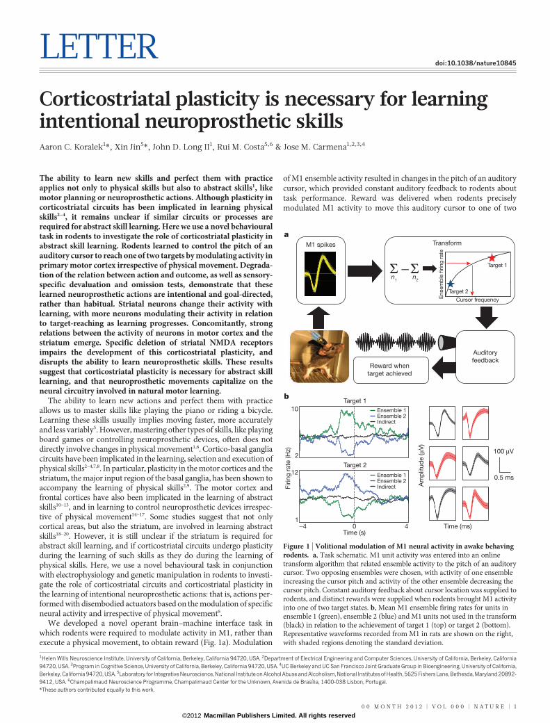
Figure 1 | Volitional modulation of M1 neural activity in awake behavingrodents. a, Task schematic. M1 unit activity was entered into an onlinetransform algorithm that related ensemble activity to the pitch of an auditorycursor. Two opposing ensembles were chosen, with activity of one ensembleincreasing the cursor pitch and activity of the other ensemble decreasing thecursor pitch. Constant auditory feedback about cursor location was supplied torodents, and distinct rewards were supplied when rodents brought M1 activityinto one of two target states. b, Mean M1 ensemble firing rates for units inensemble 1 (green), ensemble 2 (blue) and M1 units not used in the transform(black) in relation to the achievement of target 1 (top) or target 2 (bottom).Representative waveforms recorded from M1 in rats are shown on the right,with shaded regions denoting the standard deviation.
0 0 M O N T H 2 0 1 2 | V O L 0 0 0 | N A T U R E | 1
Macmillan Publishers Limited. All rights reserved©2012
target tones, and a trial was marked incorrect if no target had been hitwithin a set time limit (30 s). One of these targets was associated with areward of sucrose solution, whereas the other target was associatedwith a pellet reward (see Methods). Two neural ensembles consistingof two- to four-well-isolated units each were used to control theauditory cursor (Supplementary Figs 1 and 2). The action of thesetwo ensembles opposed each other, such that increased activity inone ensemble produced increases in cursor pitch, whereas increasedactivity in the other ensemble caused decreases in cursor pitch. Thus,to achieve a high-pitched target, rodents had to increase activity in thefirst ensemble and decrease activity in the second; the opposite wasrequired to hit a low-pitched target (Fig. 1b and Supplementary Fig. 3).These firing-rate modulations had to be maintained for several timebins (200 ms bin size) for a target to be hit (Supplementary Methods).Hence, in this operant task, rodents had to bring the two M1 ensemblesinto a desired state irrespective of motor output.
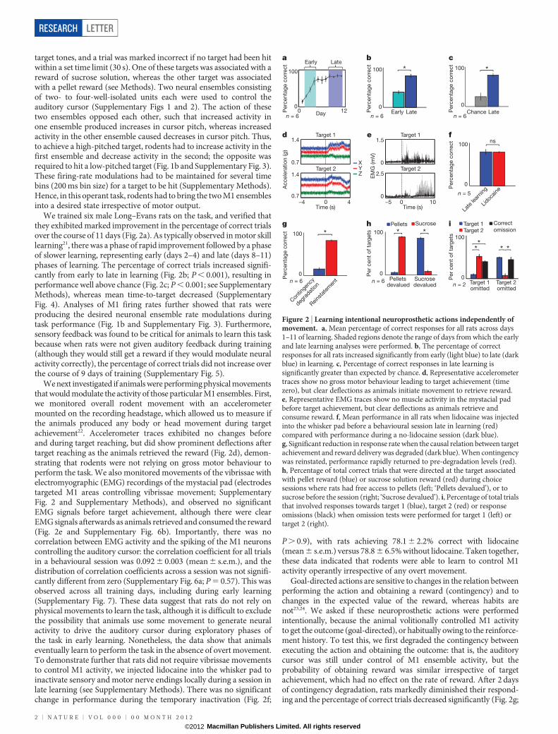
We trained six male Long–Evans rats on the task, and verified thatthey exhibited marked improvement in the percentage of correct trialsover the course of 11 days (Fig. 2a). As typically observed in motor skilllearning21, there was a phase of rapid improvement followed by a phaseof slower learning, representing early (days 2–4) and late (days 8–11)phases of learning. The percentage of correct trials increased signifi-cantly from early to late in learning (Fig. 2b; P , 0.001), resulting inperformance well above chance (Fig. 2c; P , 0.001; see SupplementaryMethods), whereas mean time-to-target decreased (SupplementaryFig. 4). Analyses of M1 firing rates further showed that rats wereproducing the desired neuronal ensemble rate modulations duringtask performance (Fig. 1b and Supplementary Fig. 3). Furthermore,sensory feedback was found to be critical for animals to learn this taskbecause when rats were not given auditory feedback during training(although they would still get a reward if they would modulate neuralactivity correctly), the percentage of correct trials did not increase overthe course of 9 days of training (Supplementary Fig. 5).
We next investigated if animals were performing physical movementsthat would modulate the activity of those particular M1 ensembles. First,we monitored overall rodent movement with an accelerometermounted on the recording headstage, which allowed us to measure ifthe animals produced any body or head movement during targetachievement22. Accelerometer traces exhibited no changes beforeand during target reaching, but did show prominent deflections aftertarget reaching as the animals retrieved the reward (Fig. 2d), demon-strating that rodents were not relying on gross motor behaviour toperform the task. We also monitored movements of the vibrissae withelectromyographic (EMG) recordings of the mystacial pad (electrodestargeted M1 areas controlling vibrissae movement; SupplementaryFig. 2 and Supplementary Methods), and observed no significantEMG signals before target achievement, although there were clearEMG signals afterwards as animals retrieved and consumed the reward(Fig. 2e and Supplementary Fig. 6b). Importantly, there was nocorrelation between EMG activity and the spiking of the M1 neuronscontrolling the auditory cursor: the correlation coefficient for all trialsin a behavioural session was 0.092 6 0.003 (mean 6 s.e.m.), and thedistribution of correlation coefficients across a session was not signifi-cantly different from zero (Supplementary Fig. 6a; P 5 0.57). This wasobserved across all training days, including during early learning(Supplementary Fig. 7). These data suggest that rats do not rely onphysical movements to learn the task, although it is difficult to excludethe possibility that animals use some movement to generate neuralactivity to drive the auditory cursor during exploratory phases ofthe task in early learning. Nonetheless, the data show that animalseventually learn to perform the task in the absence of overt movement.To demonstrate further that rats did not require vibrissae movementsto control M1 activity, we injected lidocaine into the whisker pad toinactivate sensory and motor nerve endings locally during a session inlate learning (see Supplementary Methods). There was no significantchange in performance during the temporary inactivation (Fig. 2f;
P . 0.9), with rats achieving 78.1 6 2.2% correct with lidocaine(mean 6 s.e.m.) versus 78.8 6 6.5% without lidocaine. Taken together,these data indicated that rodents were able to learn to control M1activity operantly irrespective of any overt movement.
Goal-directed actions are sensitive to changes in the relation betweenperforming the action and obtaining a reward (contingency) and tochanges in the expected value of the reward, whereas habits arenot23,24. We asked if these neuroprosthetic actions were performedintentionally, because the animal volitionally controlled M1 activityto get the outcome (goal-directed), or habitually owing to the reinforce-ment history. To test this, we first degraded the contingency betweenexecuting the action and obtaining the outcome: that is, the auditorycursor was still under control of M1 ensemble activity, but theprobability of obtaining reward was similar irrespective of targetachievement, which had no effect on the rate of reward. After 2 daysof contingency degradation, rats markedly diminished their respond-ing and the percentage of correct trials decreased significantly (Fig. 2g;
Perc
enta
ge c
orr
ect
Perc
enta
ge c
orr
ect
Perc
enta
ge c
orr
ect
Perc
enta
ge c
orr
ect
0
100
Day0 12
Early
Early Late Chance Late
Late
0
100 *
Perc
enta
ge c
orr
ect
0
100 *Pellets Sucrose
Per
cent
of
targ
ets
0
100
Pelletsdevalued
Sucrose devalued
* *
0
100ns
n = 6
n = 6 n = 6
n = 5
n = 6
Accele
ratio
n (g)
Target 1
Target 2
1.4
0.7
1.4
0.7
Time (s)−4 40
YZ
X
Target 1
Target 2
Time (s)−5 100
Per
cent
of
targ
ets
0
100
Target 1omitted
Target 2omitted
Target 1Target 2
Correct
omission
0
100 *
EM
G (m
V)
1.5
0
2.5
0
**
* *
n = 6
n = 2
Late
lear
ning
Lidoc
aine
Con
tinge
ncy
degra
datio
n
Reins
tate
men
t
a b
f
g
d
h
e
i
c
Figure 2 | Learning intentional neuroprosthetic actions independently ofmovement. a, Mean percentage of correct responses for all rats across days1–11 of learning. Shaded regions denote the range of days from which the earlyand late learning analyses were performed. b, The percentage of correctresponses for all rats increased significantly from early (light blue) to late (darkblue) in learning. c, Percentage of correct responses in late learning issignificantly greater than expected by chance. d, Representative accelerometertraces show no gross motor behaviour leading to target achievement (timezero), but clear deflections as animals initiate movement to retrieve reward.e, Representative EMG traces show no muscle activity in the mystacial padbefore target achievement, but clear deflections as animals retrieve andconsume reward. f, Mean performance in all rats when lidocaine was injectedinto the whisker pad before a behavioural session late in learning (red)compared with performance during a no-lidocaine session (dark blue).g, Significant reduction in response rate when the causal relation between targetachievement and reward delivery was degraded (dark blue). When contingencywas reinstated, performance rapidly returned to pre-degradation levels (red).h, Percentage of total correct trials that were directed at the target associatedwith pellet reward (blue) or sucrose solution reward (red) during choicesessions where rats had free access to pellets (left; ‘Pellets devalued’), or tosucrose before the session (right; ‘Sucrose devalued’). i, Percentage of total trialsthat involved responses towards target 1 (blue), target 2 (red) or responseomissions (black) when omission tests were performed for target 1 (left) ortarget 2 (right).
RESEARCH LETTER
2 | N A T U R E | V O L 0 0 0 | 0 0 M O N T H 2 0 1 2
Macmillan Publishers Limited. All rights reserved©2012
P , 0.001). When contingency was reinstated, rats resumed respond-ing and the percentage of correct trials returned to plateau levels seen inlate learning (Fig. 2g).
To investigate further the intentional nature of these neuro-prosthetic skills, we performed a test where each of the outcomeswas devalued using sensory-specific satiety. Rats were given free accesseither to sucrose solution or pellets for 1 h before the behaviouralsession, thereby reducing the expected value of that outcome25. Afterspecific devaluation of each outcome/reward, rats chose the targetleading to that reward much less than the target leading to the rewardthat was not devalued (Fig. 2h; P , 0.001), indicating that their actionswere sensitive to changes in outcome value. Importantly, there were nosignificant differences in reward preference during normal task per-formance when neither of the outcomes was devalued (Supplemen-tary Fig. 8; P . 0.25). Finally, we asked whether rats were able tointentionally inhibit the reaching of one of the two targets to obtainthe specific reward associated with that target. To examine this weperformed an omission test, where the reward previously associated withreaching a particular target was only delivered when rats successfullyinhibited reaching that target throughout the duration of the trial. If thetarget was reached during the 30 s of trial duration, no reward wasdelivered and a new trial was initiated. Importantly, reaching the othertarget continued to lead to reward as during training. Animals behavedin a goal-directed manner in the omission test for both targets, becausethey reduced the number of reaches for the target they had to omitversus the no-omission target, while increasing the number of correctlyomitted responses (Fig. 2i; P , 0.001 for both comparisons). Takentogether, these data show that the neuroprosthetic actions in our taskare sensitive to changes in the causal relation between performingthe action and obtaining the reward (contingency degradation andomission test), and to changes in the expected value of the outcome(sensory-specific devaluation), indicating that they are intentional andgoal-directed rather than habitual.
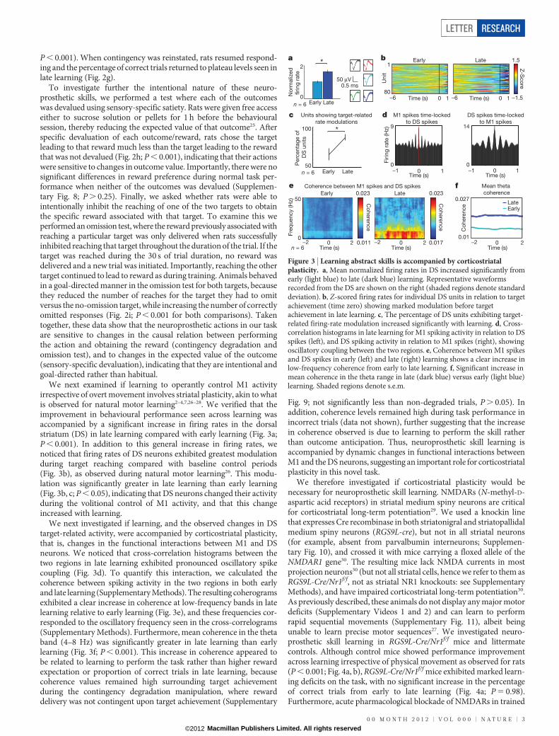
We next examined if learning to operantly control M1 activityirrespective of overt movement involves striatal plasticity, akin to whatis observed for natural motor learning2–4,7,26–28. We verified that theimprovement in behavioural performance seen across learning wasaccompanied by a significant increase in firing rates in the dorsalstriatum (DS) in late learning compared with early learning (Fig. 3a;P , 0.001). In addition to this general increase in firing rates, wenoticed that firing rates of DS neurons exhibited greatest modulationduring target reaching compared with baseline control periods(Fig. 3b), as observed during natural motor learning26. This modu-lation was significantly greater in late learning than early learning(Fig. 3b, c; P , 0.05), indicating that DS neurons changed their activityduring the volitional control of M1 activity, and that this changeincreased with learning.
We next investigated if learning, and the observed changes in DStarget-related activity, were accompanied by corticostriatal plasticity,that is, changes in the functional interactions between M1 and DSneurons. We noticed that cross-correlation histograms between thetwo regions in late learning exhibited pronounced oscillatory spikecoupling (Fig. 3d). To quantify this interaction, we calculated thecoherence between spiking activity in the two regions in both earlyand late learning (Supplementary Methods). The resulting coherogramsexhibited a clear increase in coherence at low-frequency bands in latelearning relative to early learning (Fig. 3e), and these frequencies cor-responded to the oscillatory frequency seen in the cross-correlograms(Supplementary Methods). Furthermore, mean coherence in the thetaband (4–8 Hz) was significantly greater in late learning than earlylearning (Fig. 3f; P , 0.001). This increase in coherence appeared tobe related to learning to perform the task rather than higher rewardexpectation or proportion of correct trials in late learning, becausecoherence values remained high surrounding target achievementduring the contingency degradation manipulation, where rewarddelivery was not contingent upon target achievement (Supplementary
Fig. 9; not significantly less than non-degraded trials, P . 0.05). Inaddition, coherence levels remained high during task performance inincorrect trials (data not shown), further suggesting that the increasein coherence observed is due to learning to perform the skill ratherthan outcome anticipation. Thus, neuroprosthetic skill learning isaccompanied by dynamic changes in functional interactions betweenM1 and the DS neurons, suggesting an important role for corticostriatalplasticity in this novel task.
We therefore investigated if corticostriatal plasticity would benecessary for neuroprosthetic skill learning. NMDARs (N-methyl-D-aspartic acid receptors) in striatal medium spiny neurons are criticalfor corticostriatal long-term potentiation29. We used a knockin linethat expresses Cre recombinase in both striatonigral and striatopallidalmedium spiny neurons (RGS9L-cre), but not in all striatal neurons(for example, absent from parvalbumin interneurons; Supplemen-tary Fig. 10), and crossed it with mice carrying a floxed allele of theNMDAR1 gene30. The resulting mice lack NMDA currents in mostprojection neurons30 (but not all striatal cells, hence we refer to them asRGS9L-Cre/Nr1f/f, not as striatal NR1 knockouts: see SupplementaryMethods), and have impaired corticostriatal long-term potentiation30.As previously described, these animals do not display any major motordeficits (Supplementary Videos 1 and 2) and can learn to performrapid sequential movements (Supplementary Fig. 11), albeit beingunable to learn precise motor sequences27. We investigated neuro-prosthetic skill learning in RGS9L-Cre/Nr1f/f mice and littermatecontrols. Although control mice showed performance improvementacross learning irrespective of physical movement as observed for rats(P , 0.001; Fig. 4a, b), RGS9L-Cre/Nr1f/f mice exhibited marked learn-ing deficits on the task, with no significant increase in the percentageof correct trials from early to late learning (Fig. 4a; P 5 0.98).Furthermore, acute pharmacological blockade of NMDARs in trained
0.5 ms
No
rmaliz
ed
firing
rate
0
2*
Time (s)Time (s)
Early Late
Z-S
co
re
0−6 10−6 1
Unit
−1.5
1.5
80
1
50
100
Perc
enta
ge o
f
DS
units
0
9
0
14
Firin
g r
ate
(H
z)
Time (s) Time (s)0−1 1 0−1 1
DS spikes time-locked
to M1 spikes
M1 spikes time-locked
to DS spikesC
ohere
nce
0.023
Fre
quency (H
z)
0
50
Time (s) Time (s)0−2 −2 −22 0.017
*
0 2
Early Late
Coherence between M1 spikes and DS spikes
Units showing target-related
rate modulations
50 μV
Time (s)0 2
Co
here
nce
0.01
0.027
Mean theta
coherence
EarlyLateC
ohere
nce
0.023
0.011
n = 6
n = 6
n = 6
Early Late
Early Late
a b
c d
e f
Figure 3 | Learning abstract skills is accompanied by corticostriatalplasticity. a, Mean normalized firing rates in DS increased significantly fromearly (light blue) to late (dark blue) learning. Representative waveformsrecorded from the DS are shown on the right (shaded regions denote standarddeviation). b, Z-scored firing rates for individual DS units in relation to targetachievement (time zero) showing marked modulation before targetachievement in late learning. c, The percentage of DS units exhibiting target-related firing-rate modulation increased significantly with learning. d, Cross-correlation histograms in late learning for M1 spiking activity in relation to DSspikes (left), and DS spiking activity in relation to M1 spikes (right), showingoscillatory coupling between the two regions. e, Coherence between M1 spikesand DS spikes in early (left) and late (right) learning shows a clear increase inlow-frequency coherence from early to late learning. f, Significant increase inmean coherence in the theta range in late (dark blue) versus early (light blue)learning. Shaded regions denote s.e.m.
LETTER RESEARCH
0 0 M O N T H 2 0 1 2 | V O L 0 0 0 | N A T U R E | 3
Macmillan Publishers Limited. All rights reserved©2012
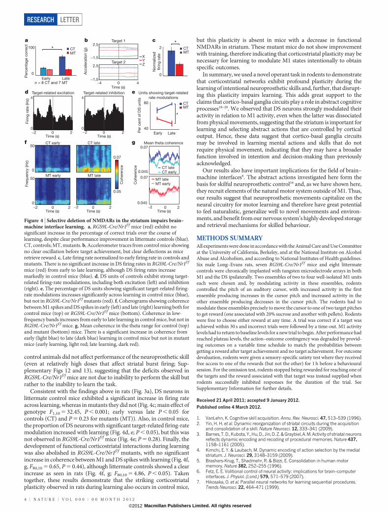
control animals did not affect performance of the neuroprosthetic skill(even at relatively high doses that affect striatal burst firing; Sup-plementary Figs 12 and 13), suggesting that the deficits observed inRGS9L-Cre/Nr1f/f mice are not due to inability to perform the skill butrather to the inability to learn the task.
Consistent with the findings above in rats (Fig. 3a), DS neurons inlittermate control mice exhibited a significant increase in firing rateacross learning, whereas in mutants they did not (Fig. 4c; main effect ofgenotype F1,10 5 32.45, P , 0.001; early versus late P , 0.05 forcontrols (CT) and P 5 0.23 for mutants (MT)). Also, in control mice,the proportion of DS neurons with significant target-related firing-ratemodulation increased with learning (Fig. 4d, e; P , 0.05), but this wasnot observed in RGS9L-Cre/Nr1f/f mice (Fig. 4e; P 5 0.28). Finally, thedevelopment of functional corticostriatal interactions during learningwas also abolished in RGS9L-Cre/Nr1f/f mutants, with no significantincrease in coherence between M1 and DS spikes with learning (Fig. 4f,g, F80,10 5 0.65, P 5 0.44), although littermate controls showed a clearincrease as seen in rats (Fig. 4f, g; F80,10 5 4.86, P , 0.05). Takentogether, these results demonstrate that the striking corticostriatalplasticity observed in rats during learning also occurs in control mice,
but this plasticity is absent in mice with a decrease in functionalNMDARs in striatum. These mutant mice do not show improvementwith training, therefore indicating that corticostriatal plasticity may benecessary for learning to modulate M1 states intentionally to obtainspecific outcomes.
In summary, we used a novel operant task in rodents to demonstratethat corticostriatal networks exhibit profound plasticity during thelearning of intentional neuroprosthetic skills and, further, that disrupt-ing this plasticity impairs learning. This adds great support to theclaims that cortico-basal ganglia circuits play a role in abstract cognitiveprocesses18–20. We observed that DS neurons strongly modulated theiractivity in relation to M1 activity, even when the latter was dissociatedfrom physical movements, suggesting that the striatum is important forlearning and selecting abstract actions that are controlled by corticaloutput. Hence, these data suggest that cortico-basal ganglia circuitsmay be involved in learning mental actions and skills that do notrequire physical movement, indicating that they may have a broaderfunction involved in intention and decision-making than previouslyacknowledged.
Our results also have important implications for the field of brain–machine interfaces6. The abstract actions investigated here form thebasis for skilful neuroprosthetic control16 and, as we have shown here,they recruit elements of the natural motor system outside of M1. Thus,our results suggest that neuroprosthetic movements capitalize on theneural circuitry for motor learning and therefore have great potentialto feel naturalistic, generalize well to novel movements and environ-ments, and benefit from our nervous system’s highly developed storageand retrieval mechanisms for skilled behaviour.
METHODS SUMMARYAll experiments were done in accordance with the Animal Care and Use Committeeat the University of California, Berkeley, and at the National Institute on AlcoholAbuse and Alcoholism, and according to National Institutes of Health guidelines.Six male Long–Evans rats, seven RGS9L-Cre/Nr1f/f mice and eight littermatecontrols were chronically implanted with tungsten microelectrode arrays in bothM1 and the DS ipsilaterally. Two ensembles of two to four well-isolated M1 unitseach were chosen and, by modulating activity in these ensembles, rodentscontrolled the pitch of an auditory cursor, with increased activity in the firstensemble producing increases in the cursor pitch and increased activity in theother ensemble producing decreases in the cursor pitch. The rodents had tomodulate these ensembles precisely to move the cursor to one of two target pitchesto get reward (one associated with 20% sucrose and another with pellets). Rodentswere free to choose either reward at any time. A trial was correct if a target wasachieved within 30 s and incorrect trials were followed by a time-out. M1 activitylevels had to return to baseline levels for a new trial to begin. After performance hadreached plateau levels, the action–outcome contingency was degraded by provid-ing outcomes on a variable time schedule to match the probabilities betweengetting a reward after target achievement and no target achievement. For outcomedevaluation, rodents were given a sensory-specific satiety test where they receivedfree access to one of the rewards (but not the other) for 1 h before a behaviouralsession. For the omission test, rodents stopped being rewarded for reaching one ofthe targets and the reward associated with that target was instead supplied whenrodents successfully inhibited responses for the duration of the trial. SeeSupplementary Information for further details.
Received 21 April 2011; accepted 9 January 2012.
Published online 4 March 2012.
1. VanLehn, K. Cognitive skill acquisition. Annu. Rev. Neurosci. 47, 513–539 (1996).2. Yin, H. H. et al. Dynamic reorganization of striatal circuits during the acquisition
and consolidation of a skill. Nature Neurosci. 12, 333–341 (2009).3. Barnes, T. D., Kubota, Y., Hu, D., Jin, D. Z. & Graybiel, A. M. Activity of striatal neurons
reflects dynamic encoding and recoding of procedural memories. Nature 437,1158–1161 (2005).
4. Kimchi, E. Y. & Laubach, M. Dynamic encoding of action selection by the medialstriatum. J. Neurosci. 29, 3148–3159 (2009).
5. Brashers-Krug, T., Shadmehr, R. & Bizzi, E. Consolidation in human motormemory. Nature 382, 252–255 (1996).
6. Fetz, E. E. Volitional control of neural activity: implications for brain–computerinterfaces. J. Physiol. (Lond.) 579, 571–579 (2007).
7. Hikosaka, O. et al. Parallel neural networks for learning sequential procedures.Trends Neurosci. 22, 464–471 (1999).
Perc
enta
ge c
orr
ect
0
100
Early Late
No
rmaliz
ed
firing
rate
0
2
Accele
ratio
n (g)
Target 1
Target 2
2.5
−1.5
2.5
−1.5
Time (s)−4 40
YZ
X
CTMT
CTMT
CTMT
4
0
Firin
g r
ate
(H
z)
Time (s)−2 0 2 −2 0 2 P
er
cent
of
DS
units
40
80
Early Late
Co
here
nce
0.07
0.05
Fre
quency (H
z)
0
50
0
50CT early CT late
MT early MT late
Mean theta coherence
0.045
0.07
0.045
0.07
Co
here
nce
Time (s)20
Units showing target-related
rate modulations
CT earlyCT late
MT earlyMT late
Target-related excitation Target-related inhibition
4
0
*
*
*
Time (s)
Time (s)−2 0 2 −2 −20 2
Time (s)
n = 8 CT and 7 MT
a b
d
f
c
g
e
Figure 4 | Selective deletion of NMDARs in the striatum impairs brain–machine interface learning. a, RGS9L-Cre/Nr1f/f mice (red) exhibit nosignificant increase in the percentage of correct trials over the course oflearning, despite clear performance improvement in littermate controls (blue).CT, controls; MT, mutants. b, Accelerometer traces from control mice showingno clear oscillation before target achievement, but clear deflections as miceretrieve reward. c, Late firing rate normalized to early firing rate in controls andmutants. There is no significant increase in DS firing rates in RGS9L-Cre/Nr1f/f
mice (red) from early to late learning, although DS firing rates increasemarkedly in control mice (blue). d, DS units of controls exhibit strong target-related firing-rate modulations, including both excitation (left) and inhibition(right). e, The percentage of DS units showing significant target-related firing-rate modulations increases significantly across learning in control mice (blue),but not in RGS9L-Cre/Nr1f/f mutants (red). f, Coherograms showing coherencebetween M1 spikes and DS spikes in early (left) and late (right) learning both forcontrol mice (top) or RGS9L-Cre/Nr1f/f mice (bottom). Coherence in low-frequency bands increases from early to late learning in control mice, but not inRGS9L-Cre/Nr1f/f mice. g, Mean coherence in the theta range for control (top)and mutant (bottom) mice. There is a significant increase in coherence fromearly (light blue) to late (dark blue) learning in control mice but not in mutantmice (early learning, light red; late learning, dark red).
RESEARCH LETTER
4 | N A T U R E | V O L 0 0 0 | 0 0 M O N T H 2 0 1 2
Macmillan Publishers Limited. All rights reserved©2012
8. Brasted, P. J. & Wise, S. P. Comparison of learning-related neuronal activity in thedorsal premotor cortex and striatum. Eur. J. Neurosci. 19, 721–740 (2004).
9. Rioult-Pedotti, M. S., Friedman, D. & Donghue, J. P. Learning-induced LTP inneocortex. Science 290, 533–536 (2000).
10. Georgopoulos, A. P., Taira, M. & Lukashin, A. Cognitive neurophysiology of themotor cortex. Science 260, 47–52 (1993).
11. Gandolfo, F., Li, C., Benda, B. J., Schioppa, C. P. & Bizzi, E. Cortical correlates oflearning in monkeys adapting to a new dynamical environment. Proc. Natl Acad.Sci. USA 97, 2259–2263 (2000).
12. Fincham, J. M. & Anderson, J. R. Distinct roles of the anterior cingulate andprefrontal cortex in the acquisition and performance of a cognitive skill. Proc. NatlAcad. Sci. USA 103, 12941–12946 (2006).
13. Badre, D., Kayser, A. S. & D’Esposito, M. Frontal cortex and the discovery of abstractaction rules. Neuron 66, 315–326 (2010).
14. Taylor, D. M., Tillery, S. I. & Schwartz, A. B. Direct cortical control of 3Dneuroprosthetic devices. Science 296, 1829–1832 (2002).
15. Carmena, J.M. et al. Learning to control a brain-machine interface for reachingandgrasping by primates. PLoS Biol. 1, 193–208 (2003).
16. Ganguly, K. & Carmena, J. M. Emergence of a stable cortical map forneuroprosthetic control. PLoS Biol. 7, e1000153 (2009).
17. Ganguly, K., Dimitrov, D. F., Wallis, J. D. & Carmena, J. M. Reversible large-scalemodification of cortical networks during neuroprosthetic control. Nature Neurosci.14, 662–667 (2011).
18. Beauchamp, M. H., Dagher, A., Aston, J. A. & Doyon, J. Dynamic functional changesassociated with cognitive skill learning of an adapted version of the Tower ofLondon task. Neuroimage 20, 1649–1660 (2003).
19. Poldrack, R. A., Prabhakaran, V., Seger, C. A. & Gabrieli, J. D. Striatal activationduring acquisition of a cognitive skill. Neuropsychology 13, 564–574 (1999).
20. Pasupathy, A. & Miller, E. K. Different time courses of learning-relatedactivity in theprefrontal cortex and striatum. Nature 433, 873–876 (2005).
21. Karni, A. et al. The acquisition of skilled motor performance: fast and slowexperience-driven changes in primary motor cortex. Proc. Natl Acad. Sci. USA 95,861–868 (1998).
22. Venkatraman, S., Jin, X., Costa, R. M. & Carmena, J. M. Investigating neuralcorrelates of behavior in freely behaving rodents using inertial sensors.J. Neurophysiol. 104, 569–575 (2010).
23. Balleine, B. W. & Dickinson, A. Goal-directed instrumental action: contingency andincentive learning and their cortical substrates. Neuropharmacology 37, 407–419(1998).
24. Yin, H. H., Knowlton, B. J. & Balleine, B. W. Inactivation of dorsolateral striatumenhances sensitivity to changes in the action-outcome contingency ininstrumental conditioning. Behav. Brain Res. 166, 189–196 (2006).
25. Hilario, M. R., Clouse, E., Yin, H. H. & Costa, R. M. Endocannabinoid signaling iscritical for habit formation. Front. Integr. Neurosci. 1, 1–12 (2007).
26. Costa, R. M., Cohen, D. & Nicolelis, M. A. Differential corticostriatal plasticity duringfast and slow motor skill learning in mice. Curr. Biol. 14, 1124–1134 (2004).
27. Jin, X. & Costa, R. M. Start/stop signals emerge in nigrostriatal circuits duringsequence learning. Nature 466, 457–462 (2010).
28. Miyachi, S., Hikosaka, O. & Lu, X. Differential activation of monkey striatal neuronsin the early and late stages of procedural learning. Exp. Brain Res. 146, 122–126(2002).
29. Calabresi, P., Pisani, A., Mercuri, N. B. & Bernardi, G. Long-term potentiation in thestriatum is unmasked by removing the voltage-dependent magnesium block ofNMDA receptor channels. Eur. J. Neurosci. 4, 929–935 (1992).
30. Dang, M. T. et al. Disrupted motor learning and long-term synaptic plasticity inmice lacking NMDAR1 in the striatum. Proc. Natl Acad. Sci. USA 103,15254–15259 (2006).
Supplementary Information is linked to the online version of the paper atwww.nature.com/nature.
Acknowledgements We thank S. Venkatraman for the three-axis accelerometer, Y. Lifor the RGS9L-Cre mice, K. Nakazawa for theNMDAR1-loxP mice,G. Luo for genotyping,M. Davis for advice on staining and G. Martins for performing immunohistochemistry.This work was supported by National Science Foundation CAREER Award 0954243,the Multiscale Systems Research Center and the Defense Advanced Research ProjectsAgency contract N66001-10-C-2008 to J.M.C., and the Division of Intramural Clinicaland Basic Research of the National Institute on Alcohol Abuse and Alcoholism, MarieCurie International Reintegration Grant 239527 and European Research Council STG243393 to R.M.C.
Author Contributions A.C.K., X.J., J.D.L., R.M.C. and J.M.C. designed experiments. A.C.K.,X.J. and J.D.L. conducted experiments. A.C.K., X.J., R.M.C. and J.M.C. analysed data andwrote the paper.
Author Information Reprints and permissions information is available atwww.nature.com/reprints. The authors declare no competing financial interests.Readers are welcome to comment on the online version of this article atwww.nature.com/nature. Correspondence and requests for materials should beaddressed to J.M.C. ([email protected]) or R.M.C.([email protected]).
LETTER RESEARCH
0 0 M O N T H 2 0 1 2 | V O L 0 0 0 | N A T U R E | 5
Macmillan Publishers Limited. All rights reserved©2012