Embed Size (px)
Citation preview

Cortical Porosity
– Its regulation and association with fracture
Daniel Sundh
Department of Internal Medicine and Clinical Nutrition, Institute of Medicine at Sahlgrenska Academy University of Gothenburg Gothenburg, Sweden, 2017

Cover illustration by Daniel Sundh Cortical Porosity – Its regulation and association with fracture © 2017 Daniel Sundh [email protected] ISBN 978-91-629-0127-1 (PRINT) ISBN 978-91-629-0128-8 (PDF) http://hdl.handle.net/2077/51732 Printed in Gothenburg, Sweden 2017 Ineko AB

The more I practice, the luckier I get - Gary Player Eller som min största idol skulle ha sagt: Heja Heja!

Abstract
Objective: Osteoporosis is a disease characterized by low bone mineral density and deteriorated bone microstructure. This thesis aimed to determine whether cortical porosity is associated with previous fracture and increase the knowledge regarding the regulation of this bone trait. Methods: The studies included in this thesis were based on two well-defined cohorts. The first was a sub-sample from the Swedish part of the Osteoporotic fractures in men (MrOS) study. This cohort comprised of 456 older men (mean age 80.2 years). The second cohort was the Sahlgrenska University hospital Prospective Evaluation of Risk of Bone fractures – SUPERB study, which is based on 3030 elderly women (75-80 years). Two sub-populations were used selected either on an X-ray verified hip fracture or available measurements of bone material properties. Bone mineral density was assessed with dual-energy X-ray absorptiometry. Bone geometry and microstructure were measured at the tibia with high-resolution peripheral quantitative computed tomography. Microindentation was performed with the hand-held Osteoprobe to assess bone material strength. Results: Cortical porosity was associated with prevalent fracture in older men and prevalent hip fracture in older women independently of areal bone mineral density and clinical risk factors. Serum levels of 25-hydroxyvitamin D were inversely associated with cortical porosity independently of parathyroid hormone, indicating that vitamin D might directly regulate this bone trait. A high amount of adipose tissue was associated with higher cortical porosity and lower bone material strength. Conclusions: Cortical porosity is higher in individuals with a prevalent fracture, low vitamin D levels, or large amount of adipose tissue. These results indicate that cortical porosity is important for bone strength and has a role in the etiology of bone fractures.
Keywords
Cortical porosity, fracture, osteoporosis, adipose tissue, vitamin D, high-resolution peripheral quantitative computed tomography

Sammanfattning på svenska
Bakgrund: Osteoporos eller benskörhet är en folksjukdom i Sverige. Sjukdomen leder till svagare skelett på grund av förlust av benmineral samt försämrad mikrostruktur vilket ökar risken för fraktur. Frågeställning: Målet med denna avhandling är att undersöka om det finns en skillnad i kortikal porositet (hålrum i kortikalt ben) mellan individer med en tidigare fraktur eller inte, samt öka kunskapen om hur dessa porer är reglerade. Metoder: Avhandlingen är baserad på två väldefinierade kohorter. Den första var en subpopulation av uppföljningsstudien för den svenska delen av den internationella studien MrOS. Kohorten bestod av 456 äldre män med en medelålder på 80.2 år. Den andra kohorten var Sahlgrenska Universitetssjukhus Prospektiva Estimering av Risk för Benfrakturer – SUPERB studien – som är baserad på 3030 äldre kvinnor i ålder 75-80 år. Från SUPERB-studien erhölls två subpopulationer. Den första bestod av kvinnor med uppmätt värde för benmaterialstyrka och den andra var selekterad med avseende på röntgenverifierad höftfraktur. Bentäthet mättes med dubbelfotonröntgenabsorbtiometri. Benmikrostruktur mättes på skenbenet med högupplöst datortomografi. Mikroindentering utfördes med s.k. Osteoprobe för att fastställa benmaterialstyrka. Resultat: Hög kortikal porositet var associerad med tidigare fraktur hos äldre män och tidigare höftfraktur hos äldre kvinnor. Identifierade associationer kvarstod även efter att man tagit hänsyn till bentäthet och kliniska riskfaktorer. Låga serumnivåer av vitamin D var associerade med höga nivåer av kortikal porositet. Denna association kvarstod även med hänsyn tagen till paratyreoideahormon, vilket innebär att vitamin D kan utöva en direkt effekt på kortikalt ben. Kortikalt ben och dess benmaterialstyrka var också inverst relaterade till höga nivåer fettvävnad. Slutsatser: Kortikal porositet är högre hos personer med tidigare fraktur, låga vitamin D nivåer och mycket fettvävnad. Dessa resultat tyder på att kortikal porositet har betydelse för benstyrka och kan bidra till uppkomsten av benfraktur.

i
List of papers
This thesis is based on the following studies, referred to in the text by their Roman numerals.
I . Sundh D*, Mellström D*, Nilsson M, Karlsson M, Ohlsson C, and Lorentzon M.
Increased Cortical Porosity in Older Men With Fracture
Journal of Bone and Mineral Research. 2015; 30(9): 1692–700.
I I . Sundh D, Nilsson AG, Nilsson M, Johansson L, Mellström D, and Lorentzon M.
Increased Cortical Porosity in Women With Hip Fracture
Journal of Internal Medicine. 2017; epub before print.
I I I . Sundh D, Rudäng R, Zoulakis M, Nilsson AG, Darelid A, and Lorentzon M.
A High Amount of Local Adipose Tissue Is Associated With High Cortical Porosity and Low Bone Material Strength in Older Women
Journal of Bone and Mineral Research. 2016; 31(4): 749–57.
IV. Sundh D*, Mellström D*, Ljunggren Ö, Karlsson MK, Ohlsson C, Nilsson M, Nilsson AG, and Lorentzon M.
Low Serum Vitamin D Is Associated with Higher Cortical Porosity in Older Men
Journal of Internal Medicine. 2016; 280(5): 496-508.
*Contributed equally
Article reprints were used with permission from the publishers.


iii
Content
Listofpapers................................................................................i
Content........................................................................................iii
Abbreviations..........................................................................vii1.Introduction............................................................................11.1Theskeleton.............................................................................11.2Bonecells...................................................................................3
1.2.1Osteoblasts....................................................................................31.2.2Osteocytes......................................................................................41.2.3Osteoclasts.....................................................................................5
1.3Boneremodeling.....................................................................51.4Peakbonemassanditsdeterminants.............................6
1.4.1Peakbonemass...........................................................................61.4.2Determinantsofpeakbonemass........................................7
1.5Hormonalregulationofboneremodeling.....................81.5.1VitaminD........................................................................................81.5.2Parathyroidhormone...............................................................9
1.6Bodycompositionandtheskeleton...............................101.7Osteoporosisandfractures...............................................121.8Bonegeometryandmicrostructure...............................14
1.8.1Corticalporosity.......................................................................161.8.2Thediversityofcorticalporosity......................................171.8.3Corticalporosity,bonestrength,andfracture............18
1.9Finiteelementanalysisandbonestrength..................181.10Bonematerialproperties................................................19
2.Aims........................................................................................213.SubjectsandMethods.......................................................233.1Subjects....................................................................................233.2Ethics.........................................................................................243.3Questionnaire........................................................................253.4Anthropometrics...................................................................263.5Fractures..................................................................................26
3.5.1Prevalentfractures.................................................................263.5.2Prevalenthipfractures..........................................................26
iv
3.6BoneMeasurements............................................................273.6.1Dual-energyX-rayabsorptiometry..................................273.6.2High-resolutionperipheralquantitativecomputed
tomography................................................................................283.6.2.1Corticalevaluation.....................................................................29
3.6.3Referencepointindentation................................................303.6.4Peripheralquantitativecomputedtomography.........31
3.7Bodycompositionmeasurements..................................313.8Serumanalyses......................................................................32
3.8.1VitaminD.....................................................................................323.8.2Parathyroidhormone.............................................................33
3.9Statistics...................................................................................334.Results...................................................................................354.1PaperI......................................................................................35
IncreasedCorticalPorosityinOlderMenWithFracture......354.1.1Mainresults................................................................................354.1.2Conclusion...................................................................................364.1.3Discussion....................................................................................36
4.2PaperII.....................................................................................38IncreasedCorticalPorosityinWomenWithHipFracture...384.2.1Mainresults................................................................................384.2.2Conclusion...................................................................................394.2.3Discussion....................................................................................39
4.3PaperIII...................................................................................40AHighAmountofLocalAdiposeTissueIsAssociatedWithHighCorticalPorosityandLowBoneMaterialStrengthinOlderWomen.........................................................................................404.3.1Mainresults................................................................................404.3.2Conclusion...................................................................................414.3.3Discussion....................................................................................41
4.4PaperIV...................................................................................43LowSerumVitaminDIsAssociatedWithHigherCorticalPorosityinElderlyMen......................................................................434.4.1Mainresults................................................................................434.4.2Conclusion...................................................................................434.4.3Discussion....................................................................................44
5.Generaldiscussion.............................................................455.1Additionalvalueofcorticalporosity.............................465.2Bodycompositionanditseffectsonbone....................475.3Challenges...............................................................................48

v
5.4Futurebenefits......................................................................496.Conclusion............................................................................51
7.FuturePerspective............................................................53
Relatedpublicationsnotincludedinthethesis...........55Acknowledgement..................................................................57
References................................................................................59

ABBREVIATIONS vii
Abbreviations
aBMD Areal bone mineral density BMC Bone mineral content BMD Bone mineral density BMI Body mass index BMSi Bone material strength index BMU Basic multicellular unit CI Confidence interval CV Coefficient of variation Ct.Bv Cortical bone volume Ct.Po Cortical porosity Ct.Po.V Cortical pore volume DXA Dual-energy X-ray absorptiometry FEA Finite element analyses FRAX Fracture risk assessment tool HR-pQCT High-resolution peripheral quantitative computed tomography IDI Indentation distance increase MrOS Osteoporotic Fractures in Men OPG Osteoprotegerin OC Osteocalcin PASE Physical Activity Scale for the Elderly PBM Peak bone mass PTH Parathyroid hormone PHPT Primary hyperparathyroidism RANKL Receptor activator of nuclear factor- ß ligand RPI Reference point indentation SD Standard deviation SUPERB Sahlgrenska University hospital Prospective Evaluation of Risk
of Bone fractures vBMD Volumetric bone mineral density 25-OH-D 25-hydroxyvitamin D


Daniel Sundh
1 . INTRODUCTION 1
1. Introduction
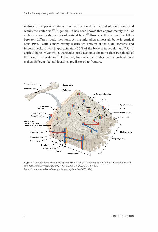
1.1 The skeleton The human skeleton is an organ with many functions and is commonly divided into two major categories, the axial and appendicular skeleton. Its main function is to provide support for the human body and to, by providing locations for the muscles to attach, enable us to move. The axial skeleton also works as a protection for vital organs such as the brain, heart, and lungs.(1,2) Moreover, our skeleton is the location for haematopoiesis and is a reservoir of important minerals, such as calcium and phosphorous, which are used for many important physiological actions.(1,2) Hence, the bone tissue is therefore vital for a wide variety of physiological functions. Bone strength is determined by the material composition and structure.(3) Bone must be stiff and resist deformation to be able to bear the loading. Also, bone must be flexible and absorb energy by deformation through the ability to shorten and widen due to compression as well as be able to lengthen and narrow in tension without cracking. Bone must on top of these features be light for good mobility.(4) Altered bone strength due to any of these features will result in the bone to fracture. The appendicular skeleton consists mainly of the long tubular bones in the extremities (tibia, femur, radius, and humerus) as well as the pectoral and pelvic girdle. The axial skeleton mainly consists of flat bones (ribs, skull, and sternum) and vertebras.(1) There are two main histological matured bone types: cortical or compact bone and trabecular or spongy bone (Figure 1). Cortical bone is mainly found in shafts of the long bones and at the surface of flat bones(5) and has a dense and ordered structure. The structure is composed of several columns (osteons), which are delineated with a cement line. The osteon is constructed from concentric layers of cortical bone (lamellae) around a central Haversian canal. In between the lamellae, osteocytes reside in certain cavities named lacunae. Inter-connected with the Haversian canals are the perforating Volkmann canals, providing the osteons with nutrients (Figure 1). Trabecular bone structure consists of interconnecting plates and bars defined as trabeculae, with marrow in-between, giving it a porous and open construction. Due to its ability to
Cortical Porosity – Its regulation and association with fracture
2 1 . INTRODUCTION
withstand compressive stress it is mainly found in the end of long bones and within the vertebras.(5) In general, it has been shown that approximately 80% of all bone in our body consists of cortical bone.(6) However, this proportion differs between different body locations. At the midradius almost all bone is cortical bone (95%) with a more evenly distributed amount at the distal forearm and femoral neck, in which approximately 25% of the bone is trabecular and 75% is cortical bone. Meanwhile, trabecular bone accounts for more than two thirds of the bone in a vertebra.(7) Therefore, loss of either trabecular or cortical bone makes different skeletal locations predisposed to fracture.
Figure 1 Cortical bone structure (By OpenStax College - Anatomy & Physiology, Connexions Web site. http://cnx.org/content/col11496/1.6/, Jun 19, 2013., CC BY 3.0, https://commons.wikimedia.org/w/index.php?curid=30131428)

Daniel Sundh
1 . INTRODUCTION 3
1.2 Bone cells
1.2.1 Osteoblasts
Osteoblasts are responsible for building bone and have their origin from mesenchymal stem cells. These stem cells are pluripotent and have the potential to differentiate into several different tissues: fat, muscle, cartilage, and bone.(8,9) Their differentiation is controlled by cytokines that regulates different transcription factors. Transcription factors involved in osteoblast differentiation are hedgehogs, bone morphogenetic proteins, transforming growth factor beta, parathyroid hormone (PTH), and wingless-type MMTV integration site family (WNT) proteins. One of the most essential factor in osteoblast differentiation is the Runt-related transcription factor 2 (Runx2).(5) Without this factor, as in mice with gene deletions of Runx2, mice completely lack osteoblasts, resulting in a cartilaginous skeleton, which has no mineralized components.(10) In humans, such depletion results in a disease called cleidocranial dysplasia, which is characterized by not fully developed cranial bones and partial or complete absence of collar bone.(11) Also, a second transcription factor, osterix, has been shown to be of great importance for osteoblast differentiation.(12) To evaluate the differentiation, different markers can be measured in serum and urine such as alkaline phosphatase (ALP) activity, levels of bone sialoprotein, osteopontin, and osteocalcin (OC), and products of synthesis or degradation of type I collagen. Some are expressed early (ALP) and others are expressed late (OC) in the process.(13) Osteoblasts secrete both receptor activator of nuclear factor- ß ligand (RANKL) and osteoprotegerin (OPG),(14) where RANKL induce osteoclast activation and OPG binds to RANKL and inhibit the activation of osteoclasts. Both these factors are affected by estradiol, which interferes with RANK signaling(15,16) and up-regulates the expression of OPG.(17,18) Estradiol deficiency therefore leads to increased bone resorption via increased RANKL-signaling.(19) Approximately 4-6% of the cells in the adult human skeleton are osteoblasts(20) and they build bone through secreting an unmineralized matrix (osteoid) consisting mainly of collagen type I but also of other bone proteins. The secreted osteoid acts as a scaffold for the mineralization of the calcium-phosphate-hydroxide salt (hydroxyapatite). Osteoblasts have a life span of approximately 3 months and, as they are aging, they have four possible ways to evolve: i) programmed cell death called apoptosis, ii) become imbedded as an osteocyte,

Cortical Porosity – Its regulation and association with fracture
4 1 . INTRODUCTION
iii) become a lineage cell, which covers the surface of the bone, or iv) transdifferentiate into cells that deposit chondroid or chondroid bone.(21)
1.2.2 Osteocytes
Osteocytes are said to be the conductors of bone remodeling. These spider-shaped cells are generated from the osteoblasts and are embedded into the bone matrix as the most abundant bone cell in an adult skeleton (90-95%).(22) Osteocytes are connected with each other and cells at the bone surface through dendritic processes, which are traveling in small canals called canaliculi, while the cell body is located in a lacuna.(23) With the canaliculi network, the osteocytes are able to detect changes in loading through fluid shear stress and thereby initiate bone remodeling if necessary. Osteocytes also regulate bone renewal by self-destruction via apoptosis.(4) As a result of fatigue, the bone tissue sustains microdamage, which has been shown to be highly associated with apoptotic osteocytes.(24) Osteocyte apoptosis leads to an osteoclastogenic response at the protein level. Increased number and differentiation of osteoclast precursors in the presence of apoptotic osteocytes are partly due to increased soluble RANKL.(25) Another way of controlling bone remodeling is through sclerostin, a protein synthesized by the SOST gene in the osteocytes. This protein is a negative regulator of the Wnt/ß-catenin pathway, which promotes bone formation.(2) Osteocytes have also shown to be systemically active by their release of fibroblast growth factor 23, which acts on the kidneys and increases the excretion of phosphate.(26) Osteocytes clearly have an important role in regulating bone remodeling indirectly by affecting osteoclasts and osteoblasts. However, it has for a long time been discussed whether osteocytes directly contribute to calcium and phosphate mobilization. By osteocytic remodeling of the perilacunar bone matrix, osteocytes are believed to resorb and replace bone matrix.(27) The osteocyte lacunocanalicular system has a 400-fold larger cell surface than the complete Haversian and Volkmann system and up to 133 times larger surface area than trabecular bone.(28) With such large surface areas also a minor resorption would have significant effects on ion circulation.
Daniel Sundh
1 . INTRODUCTION 5
1.2.3 Osteoclasts
Osteoclasts are multinuclear cells, generated from a fusion of several osteoclast precursor cells with a hematopoietic origin, and are the least abundant bone cell type (1-2%) within an adult skeleton. These cells are the only cells that can resorb bone, and are generated through activation by binding of RANKL, produced by osteoblasts, to the receptor activator of nuclear factor- ß (RANK) expressed at the osteoclast cell surface.(29) During osteoclast maturation, the cell is able to polarize and segregate important domains of the cell membrane. This polarization creates an outer circular domain enriched with adhesion structures creating the sealing zone.(30) The sealing zone comprises a structural important unit called the podosomes. These cell structures start as single units and as the maturation process progresses they begin to cluster and in the end forms a podosomal belt in the peripheral area of the cell. With these podosomes the osteoclasts are able to create a sealing zone, resulting in a resorption lacuna, in which bone resorption takes place. Within this pit, acidic molecules are secreted, which resolve the mineralized upper layer of the bone, exposing the organic matrix consisting of primary type I collagen. The exposed organic matrix is then further degraded by the lysosomal enzyme cathepsin K, also secreted by osteoclasts.(31) After the osteoclasts have travelled a certain distance within their predicted two weeks lifespan they die and are quickly removed by phagocytes.(32)
1.3 Bone remodeling Human bone, as all other constructions (e.g., roads, bridges, and buildings), needs to be repaired to stay in good condition. To assure that micro-cracks and other damages are repaired, osteoclasts remove bone, which osteoblasts later fill in with new bone. Also, by removing bone, the osteoclasts resolve calcium into the bloodstream, maintaining calcium homeostasis. However, bone removal and formation are not separated and independent processes. Both cell types are incorporated in a basic multicellular unit (BMU) where the osteoclasts act first by removing bone by acidification and proteolytic digestion followed by the osteoblasts filling in the voids created by secreting osteoid, which is eventually mineralized into new bone. In cortical bone, this process takes place via a cutting cone, where the osteoclasts drill holes into the hard bone, leaving tunnels, which appear as cavities in cross-sections. Subsequently, osteoblasts fill the cavity with new osteoid, which results in a new osteon. Trabecular bone remodeling takes place at open surfaces.(33) The BMU has a lifespan of approximately 6-9 months, which is a lot longer than each participating cell-type (osteoblast (3 months) and

Cortical Porosity – Its regulation and association with fracture
6 1 . INTRODUCTION
osteoclast (2 weeks)). The BMU therefore needs a large and continuous supply of new cells to perform its actions at the bone surface.(34) With increasing age or bone-affecting diseases, this process becomes unbalanced and more bone is being removed than formed, resulting in bone loss and subsequent increased risk of osteoporosis development.(35)
1.4 Peak bone mass and its determinants
1.4.1 Peak bone mass
Peak bone mass (PBM) is a measure of maximal acquired bone mass at the end of skeletal maturation.(36) When PBM is reached has long been debated but it is most probably obtained at different ages at different skeletal locations. PBM is generally thought to be reached in the late second or early third decade of life,(36) especially at clinically relevant locations such as the hip and spine.(37-40) For peripheral body parts, PBM is reached later. Low areal bone mineral density (aBMD) at older age might either be due to accelerated bone loss or failure to reach adequate bone mineral density (BMD) in childhood and adolescence. This means that maximizing bone accrual during development is of great importance to prevent future osteoporosis and consequent fractures. Computer simulations of bone loss over a lifetime have shown that an increase in PBM by 10% can postpone the onset of osteoporosis by 13 years(41) and may decrease the risk of fracture for postmenopausal women by 50%.(42) In contrast, a similar change in age at menopause or the rate of non-menopausal bone loss only postpones the disease by approximately 2 years.(41) Since 26% of all adult bone mineral is laid down at two years around peak skeletal growth, this period is of crucial importance for optimizing bone accrual.(43) The amount of bone laid down during these 2 years is the same as that which is lost between the ages 50 – 80.(44)
Daniel Sundh
1 . INTRODUCTION 7
1.4.2 Determinants of peak bone mass
The achieved PBM is dependent on several factors, where heredity is the strongest determining factor. Twin-studies have reported that heredity can explain 60-80% of the individual variance in PBM,(45) whereas studies performed on family members, where the genetics are less similar, reported that heredity explained approximately 50% of its variance.(46,47) Except for heredity, other environmental factors affect PBM,(48) such as physical activity,(49) nutrition, and smoking.(50) Evidence for physical activity affecting accrual of bone mass was presented by Kannus et al. They showed that female tennis and squash players that started playing before their menarche had more bone accrued than women who started after the start point of their menarche.(51) A large cross-sectional study performed on young men reported that present and previous duration of physical activity was associated with both trabecular volumetric bone mineral density (vBMD) and cortical bone structure. The same study also indicated that amount of load was positively associated with several bone parameters.(52) A large longitudinal study performed on men found an important contribution, in optimizing PBM, of physical activity for development of aBMD, trabecular vBMD, and cortical bone size.(53) The attained PBM is also affected by smoking. Small cross-sectional studies have shown that smoking is associated with lower PBM.(54,55) In a larger study, smoking was shown to be associated with lower aBMD and that reduction was mainly due to decreased cortical thickness by an increase in endosteal circumference.(56) A large proportion of these men later participated in a follow-up study five years later. With this first longitudinal study, start of smoking was associated with lower aBMD, vBMD, and cortical cross-sectional area.(50) Intake of certain nutrients and vitamins probably also affects bone development. Studies performed on vitamin D and its effect on or association with PBM reached conflicting results. Some cross-sectional studies have not been able to show an association between levels of vitamin D and lower PBM,(57-59) whereas some studies have.(60,61) A randomized controlled trial in Finland showed that adolescent girls, with adequate calcium intake, treated with vitamin D developed a higher femur aBMD than the placebo group.(62) But on the other hand, a meta-analysis based on several RCT studies indicates that only children and adolescents with low vitamin D levels benefit from vitamin D supplementation.(63)
Cortical Porosity – Its regulation and association with fracture
8 1 . INTRODUCTION
1.5 Hormonal regulation of bone remodeling
1.5.1 Vitamin D
Vitamin D stimulates the intestinal absorption of calcium, which has a large effect on the bone mineralization process.(64) Small amounts (20%) of our vitamin D requirement come through food such as fat fish, dairy products, and egg. Meanwhile, most of our vitamin D (80%) is produced, by ultraviolet irradiation in the skin, when exposed to sunlight.(65,66) Because of the low zenith angle of the sun in the winter months, photons cannot reach the northern part of the globe, above the ~35° latitude, which results in little or no vitamin D production.(67) At higher latitudes, or in the summer season at lower latitudes, energy from these photons results in a non-enzymatic reaction, which converts 7-dehydrocholesterol into previtamin D3,(67,68) which is later isomerized to Vitamin D3.(69) Vitamin D3 is transported via the bloodstream to the liver and is further hydroxylated into 25-hydroxyvitamin D (25-OH-D) by several enzymes of the cytochrome P-450 system, mainly by the CYP2R1.(70) This metabolite is measured in blood samples and used as a measurement of vitamin D status.(71) The second hydroxylation occurs in the kidneys, by CYP27B1,(70) promoted by PTH(65) and results in the most active metabolite 1,25-dihydroxyvitamin D (1,25-(OH)2-D). This metabolite plays a crucial role in bone physiology where it promotes increased intestinal uptake of calcium,(72) increased renal tubular calcium reabsorption,(71) and suppress PTH levels,(73) which enables a normal mineralization of our skeleton. It is still debated what levels that are sufficient for vitamin D. Ross et al. showed that 50 nmol L-1 was enough to cover 97.5% of the population.(74) Serum levels of 25-OH-D are positively associated with aBMD in some cohorts(75,76) but the association with bone microstructure has not been carefully investigated. The few existing studies on microstructure have not shown any conclusive results. 25-OH-D levels have been shown to be associated with trabecular but not cortical bone parameters in younger but not older men,(77) whereas another study, reported no association for similar trabecular bone parameters with levels of 25-OH-D regardless of age and sex.(78) In vitamin D treated subjects, the group with high levels had fewer but thicker trabeculae compared to the group with low levels.(79) Bone geometry measured at the hip was also associated with levels of 25-OH-D, where higher levels were associated with higher cortical volume.(80) On top of these limited data regarding 25-OH-D and bone mineral density and microstructure, vitamin D’s effect on fracture is even more uncertain. Fracture
Daniel Sundh
1 . INTRODUCTION 9
prevention was seen for vitamin D treated elderly women living in nursing homes(81) whereas vitamin D treated community-dwelling women showed an increased risk for fracture, although the association was no longer significant after adjustments for covariates.(82) The effect of vitamin D alone or together with calcium on fracture prevention was investigated in a large meta-analysis, which did not show any effect on fracture by vitamin D alone but a small effect when administered together with calcium.(83)
1.5.2 Parathyroid hormone
PTH is a polypeptide containing 84 amino acids and is secreted by chief cells located in the parathyroid glands. The production and secretion of PTH is regulated by the calcium-sensing receptor, located in the parathyroid cell membrane, where high calcium levels decrease the release of PTH and low levels increase it.(84) PTH is one of the key players in regulating calcium homeostasis.(84) This hormone is also closely connected to vitamin D levels. Decreased serum levels of vitamin D result in decreased calcium absorption from the intestines, which result in an increased serum level of PTH. PTH is also important in bone physiology and regulates both bone formation and bone resorption. The achieved net result on bone mass by PTH is dependent on mode of exposure. Continuous exposure to PTH, such as in primary hyperparathyroidism (PHPT), will result in bone loss whereas intermittent, low dose exposure of PTH will result in an anabolic response of bone formation, resulting in bone mass gain.(85) Such bone gain was reported as increased BMD in patients treated with PTH and a decreased risk for both vertebral and non-vertebral fractures.(86) The dominant cause of continuous high exposure to PTH is PHPT, which is characterized by high serum PTH and calcium levels. PTH produces a higher bone turnover state by increasing the frequency of BMU activation.(87) However, it seems that PTH has a greater effect on cortical than trabecular bone. Patients with PHPT have a larger loss of bone at the distal third of the forearm, largely enriched in cortical bone, compared to the lumbar spine, rich in trabecular bone. Bone loss at the hip is somewhere in between, where there is a mixture of both bone types.(88) These findings are strengthened by studies investigating bone structure for PHPT patients in whom trabecular bone is often preserved.(89-91) However, these findings are somewhat contradictory to the fracture pattern where this patient group have a higher risk for both vertebral and non-vertebral fractures.(92,93) Such fracture pattern could be explained by more recent studies,
Cortical Porosity – Its regulation and association with fracture
10 1 . INTRODUCTION
where patients with PHPT were found to have an impaired bone microstructure in both the trabecular and cortical bone compartment of the radius(87,94) and the tibia.(94) Also, parathyroid hormone as a treatment has been shown to increase cortical porosity(95-98) in most studies but not in all.(99)
1.6 Body composition and the skeleton Body composition is commonly described in proportions of body lean and fat mass. Fat mass can further be divided into gynoid fat, located around the hips and thighs, and android fat located in the torso area. The location of fat in relation to other body tissue is of interest. Fat located under the skin is subcutaneous fat and fat located within muscles and in association with organ tissue is called visceral fat.(100) Increased fat mass correlates with increased body weight. High body weight is believed to result in high BMD due to increased loading(101) and has been seen as a positive factor for the human skeleton. If higher BMD is due to only the weight load or if other mechanistic pathways also play a role is still being discussed. Some of the existing data indicates that adipose tissue, via several different mechanisms, has an independent effect on bone remodeling, which leads to increased bone mass. Such a mechanism could be the higher leptin levels in obese compared to non-obese subjects.(102) Higher serum levels of leptin are correlated with a higher BMD in most studies(103-105) but not in all.(106) The role of leptin in bone remodeling is therefore not clearly established, as some have reported an effect(104,107,108) whereas others have not.(103,105) In addition to leptin, hyperinsulinemia has been proposed to contribute to stronger bones in the obese, via higher levels of free sex hormones.(109) Furthermore, serum levels of free insulin-like growth factor I (IGF-I) are higher in the obese(110) and have shown to be, independently of age, associated with higher aBMD(111,112) as well as able to predict fracture independently of hip BMD.(112) For the same reason as fat might be good for bone, its endocrine function could also be devastating. For example, a fat-produced hormone, adiponectin, has been shown to be associated with bone development, bone biology, and fractures. Johansson et al. showed that higher levels of adiponectin were associated with higher risk for fracture in older men.(113) Low body mass index (BMI) is an established risk factor for fracture.(114) Whether adipose tissue is a positive factor for bone strength due to higher BMD and reduced impact of falls due to soft-tissue padding, has not been completely elucidated. Several studies indicate that the protective function of higher BMI is
Daniel Sundh
1 . INTRODUCTION 11
site-specific and decreases the risk of hip,(115-117) pelvis,(116,117) and wrist fracture.(117) Meanwhile, other fracture types such as ankle,(117) spine,(118,119) and humerus(115,116) are more common in subjects with overweight and obesity. The reason for this site-specific effect of body composition on fracture risk is not known. There are several potential factors where differences in fall pattern and shock absorbing tissue padding being the most common arguments. In addition, there could be an increase in BMD due to loading, although the increase may not be large enough to withstand the higher applied force in case of a fall. Another potential reason could be deteriorated bone traits, not measurable with DXA, due to higher fat levels. One potential bias with studies investigating the correlation between body weight (or BMI) and bone mass is that they may not necessarily represent a correlation between obesity per se and osteoporosis, because obesity is defined by excessive fat mass rather than total body weight. Even if many previous studies have used fat mass(120) to assess the correlation between obesity and bone mass, they often do not adjust for the mechanical loading effects on the skeleton due to body weight. It is of great importance to establish whether bone mineral density is different between obese and non-obese individuals. To fully understand the differences between the two groups an evaluation of geometry and microstructure is of importance. Evans et al. performed a study where they investigated obese and non-obese subjects matched for BMI and age. When these two groups were compared, almost all microstructural parameters were better in the obese group.(121) In another study, where the subjects were divided into obese and non-obese, subjects within the obese group were found to have greater femoral neck vBMD, bone size, and cortical thickness than non-obese. In the non-obese subjects, increased BMI was linearly associated with increased cross-sectional area, cortical and trabecular vBMD, whereas there was no positive association for increased BMI and bone density or structural parameters for individuals classified as obese, indicating that bone adaptation to loading reaches a plateau.(122) However, BMI is a somewhat rough measurement and do not separate fat from lean mass. High abdominal fat measured with DXA (highly correlated with CT)(123,124) was inversely correlated with bone microarchitecture measured at trans-iliac bone biopsies. Subjects in the highest tertile of abdominal fat had higher cortical porosity and lower bone volume fraction.(123) Recently, Liu et al. showed in a large cross-sectional study that higher levels of visceral adipose tissue were associated with higher trabecular density and trabecular number as well as significantly higher cortical porosity in the radius but not in the tibia. These associations were no longer apparent when adjusting for BMI, indicating that the effects on microstructure may be due to skeleton loading.(125)
Cortical Porosity – Its regulation and association with fracture
12 1 . INTRODUCTION
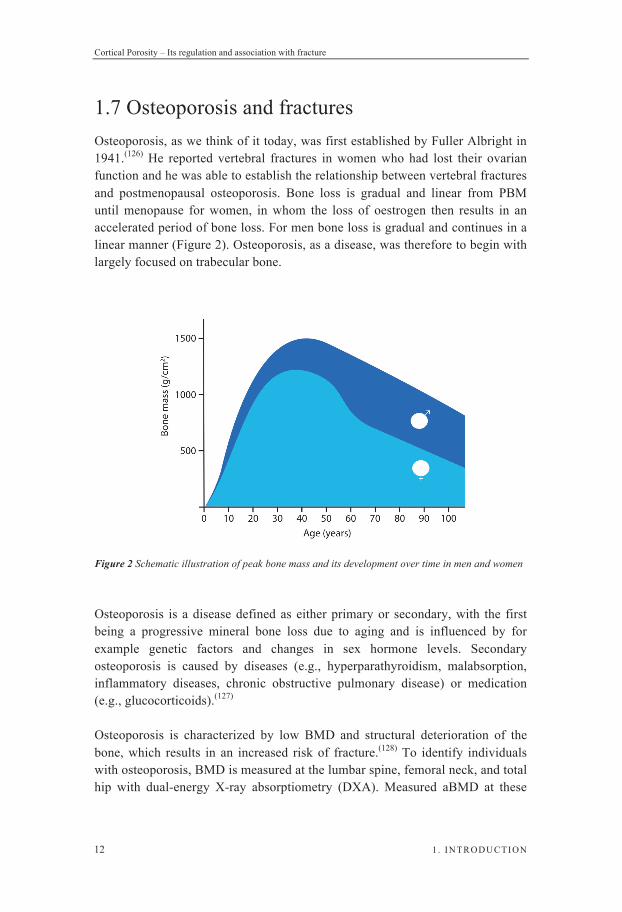
1.7 Osteoporosis and fractures Osteoporosis, as we think of it today, was first established by Fuller Albright in 1941.(126) He reported vertebral fractures in women who had lost their ovarian function and he was able to establish the relationship between vertebral fractures and postmenopausal osteoporosis. Bone loss is gradual and linear from PBM until menopause for women, in whom the loss of oestrogen then results in an accelerated period of bone loss. For men bone loss is gradual and continues in a linear manner (Figure 2). Osteoporosis, as a disease, was therefore to begin with largely focused on trabecular bone.
Figure 2 Schematic illustration of peak bone mass and its development over time in men and women
Osteoporosis is a disease defined as either primary or secondary, with the first being a progressive mineral bone loss due to aging and is influenced by for example genetic factors and changes in sex hormone levels. Secondary osteoporosis is caused by diseases (e.g., hyperparathyroidism, malabsorption, inflammatory diseases, chronic obstructive pulmonary disease) or medication (e.g., glucocorticoids).(127) Osteoporosis is characterized by low BMD and structural deterioration of the bone, which results in an increased risk of fracture.(128) To identify individuals with osteoporosis, BMD is measured at the lumbar spine, femoral neck, and total hip with dual-energy X-ray absorptiometry (DXA). Measured aBMD at these
Daniel Sundh
1 . INTRODUCTION 13
locations is used to calculate a T-score based on a healthy young population. The diagnosis osteoporosis was defined, by the World health organization (WHO) in 1994, as a bone density -2.5 standard deviations (SD) of the mean of a population of young white adult women.(129) Areal BMD has proven to be as good a predictor of hip fracture as blood pressure for stroke and even better than cholesterol levels predict heart disease.(42,129) Osteoporosis is asymptomatic and many of the patients first come to clinical attention after a fragility fracture, which is the primary manifestation of the disease.(130) Traditionally, osteoporotic fractures are those sustained at a specific location (i.e., hip, wrist, humerus, or vertebra)(131) after a fall from standing height.(132) The same definition is partly used today, but studies have shown that low aBMD increases the risk for most fractures,(133) indicating that also these can be of osteoporotic origin.(132) The incidence of osteoporotic fractures is high. In the US 39.7% of the women and 13.1% of the men will sustain a fracture after the age of 50.(134) Osteoporotic fractures are even more common in Scandinavia, where the risk of a fracture after the age of 50 is 46.4% for women and 22.4% for men.(135) In 2005 there were 2 million osteoporosis-related fractures reported in the US. This number may increase towards 3 million by the year 2025 resulting in an increase in annual fracture-related cost from 16.9 billion to 25.3 billion dollars.(136) Out of the osteoporotic fractures, hip fracture is the most costly and results in the largest patient suffering. The age-specific incidence rate of hip fracture has decreased somewhat in the first decade of the 21st century.(137,138) Despite this observed decrease, the absolute number of annual hip fractures might still increase due to the growing elderly population,(139) which will result in increased societal cost and patient suffering. Of all fractures, hip fracture has the largest effect on morbidity and mortality(140) and it often results in severe negative effects on quality of life.(141) The hip fracture incidence rate increases exponentially with age, and more than half of all hip fractures occur after the age of 80,(142) which is partly due to a decrease in aBMD at the proximal femur (a proxy for bone strength), as well as an increased frequency of falls.(143) Out of all hip fractures, women sustain 70%.(144) Due to complications from a hip fracture, one in five dies within the first year(128) and a higher risk for postoperative mortality is seen for individuals with three or more comorbidities independent of age and sex.(145) About half of the women who lived independently before the hip fracture, are afterwards in long-term care or in need of assistance by other people or devices for mobility.(146) A survey in older women found that as much as 80% would rather die than experience a hip fracture with complications that lead to loss of independence and quality of life followed by a subsequent admission to a nursing home.(147)

Cortical Porosity – Its regulation and association with fracture
14 1 . INTRODUCTION
Areal BMD is used as a biomarker for bone strength and has been shown to be a strong predictor of future fracture risk accounting for approximately 60-70% of the variance in bone strength.(148,149) Since aBMD has been used as a biomarker for a long time, worldwide and in several ethnicities, it is a well validated method to predict fracture. Even so, for women with hip fracture, only 46% had osteoporosis at the total hip as defined by DXA.(150) One reason for this low ability to predict which patient that will fracture might be that the method only measures BMD and has limited ability to differentiate between trabecular and cortical bone. Fracture prediction might be improved if bone microstructure is taken into account. The arrival of new imaging techniques, such as the high-resolution peripheral quantitative computed tomography (HR-pQCT), now provides the opportunity to investigate this issue not only at the macrostructural level of changes in bone mass, but also at the level of changes in bone microarchitecture.
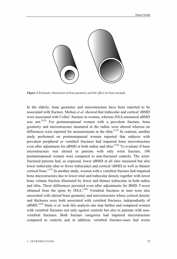
1.8 Bone geometry and microstructure The definition of osteoporosis includes structural deterioration of bone, which results in increased risk of fracture.(128) However, in clinics today fracture risk is mainly determined by aBMD, with limited abilities to measure such structural bone traits. Even if bone structure is a part of the definition for osteoporosis, methodological limitations have made it difficult to measure these bone traits in vivo in humans. This is now possible with a fairly new technique. The HR-pQCT, with a resolution of 82 m, measures bone microstructure at the radius and tibia.(151) Information regarding the spatial distribution of bone mass results in another level of possibility to estimate bone strength. Assume that the two schematic bones (Figure 3) have an equal mass and cortical area. The bone on the right side has its bone mass distributed further from the bending axis, which results in an increased cross-sectional moment of inertia and thereby a substantially increased resistance to bending.(152) This is one of the large determinants to why men, at any given value for aBMD, have bone that is more resilient to bending than women. Fragile bones generated by osteoporosis result in lower bone strength, which will fracture due to low-energy trauma.(132) An osteoporotic bone is characterized by disrupted trabecular bone microstructure with either thinning of the trabeculaes (men) or loss of trabeculae (women).(153) But also the cortical bone is affected, with thinning of the cortex and higher cortical porosity.(154) Therefore, measurement of bone geometry and microstructure could potentially improve the ability to predict an individual’s fracture risk.
Daniel Sundh
1 . INTRODUCTION 15
Figure 3 Schematic illustration of bone geometry and the effect on bone strength.
In the elderly, bone geometry and microstructure have been reported to be associated with fracture. Melton et al. showed that trabecular and cortical vBMD were associated with Colles’ fracture in women, whereas DXA-measured aBMD was not.(155) For postmenopausal women with a prevalent fracture, bone geometry and microstructure measured at the radius were altered whereas no differences were reported for measurements at the tibia.(151) In contrast, another study performed on postmenopausal women reported that subjects with prevalent peripheral or vertebral fractures had impaired bone microstructure even after adjustment for aBMD at both radius and tibia.(156) To evaluate if bone microstructure was altered in patients with only wrist fracture, 100 postmenopausal women were compared to non-fractured controls. The wrist-fractured patients had, as expected, lower aBMD at all sites measured but also lower trabecular (due to fewer trabeculae) and cortical vBMD as well as thinner cortical bone.(157) In another study, women with a vertebral fracture had impaired bone microstructure due to lower total and trabecular density together with lower bone volume fraction illustrated by fewer and thinner trabeculae in both radius and tibia. These differences persisted even after adjustments for BMD T-score obtained from the spine by DXA.(158) Vertebral fractures in men were also associated with altered bone geometry and microstructure where cortical density and thickness were both associated with vertebral fractures, independently of aBMD.(159) Stein et al. took this analysis one step further and compared women with vertebral fractures not only against controls but also to patients with non-vertebral fractures. Both fracture categories had impaired microstructure compared to controls and in addition, vertebral fracture-cases had worse
Cortical Porosity – Its regulation and association with fracture
16 1 . INTRODUCTION
microstructure than subjects with non-vertebral fractures with lower total and trabecular density due to lower trabecular number and larger trabecular separation.(160) Because of various results for association between bone microarchitecture and fracture prevalence, Boutroy and colleagues performed a large multicenter study. They found that women with prevalent fractures had lower total, trabecular, and cortical vBMD as well as fewer trabeculae and thinner cortical bone than women without fractures.(161)
1.8.1 Cortical porosity
Cortical bone, or compact bone as it is sometimes called, is not really compact (Figure 4).(6) Cortical porosity is a term used to define the bone voids, holes, or pores perforating the compact cortex. In fact, cortical bone consists of many different levels of porosity and the term might need some explanation. Cortical porosity can be divided into five size dependent groups here arranged from the largest to the smallest: 1) the marrow cavity or similar large cavities, 2) channels for nutrient arteries that traverse the cortex, 3) vascular porosity within the cortex, 4) lacuno-canalicular porosity, and 5) nanoporosity at the level of the collagen and hydroxyapatite crystals.(162) With modern high-resolution measuring techniques, such as nano and micro CT, these small structures are measurable in three-dimensions. However, only the first three categories are captured using HR-pQCT, in vivo in humans. The second category consists of Haversian (alongside the cortical bone) and Volkmann canals (traversing between Haversian canals), mainly providing the bone with nutrients. These canals are the main providers of bone surface for the BMUs to resorb bone within the cortex. This intracortical resorption increases with age resulting in a change in the surface-to-volume ratio of trabecular versus cortical bone resorption. More and more of the trabecular bone is removed without being replaced while the cortical pores grow and increase in number, resulting in a shift in the ratio (Figure 4). With increasing surface area within the pores the loss of cortical bone begins to accelerate.(6) Such increased cortical porosity is associated with lower bone strength through lower shear and tensile fracture toughness.(163)

Daniel Sundh
1 . INTRODUCTION 17
Figure 4 Illustration of cortical porosity and its development over time. A woman aged 75 (upper) compared to a postmenopausal woman aged 52 (lower)
1.8.2 The diversity of cortical porosity
During the time of peak height velocity, both young boys and girls sustain a large proportion of all forearm fractures.(164) This increase in fracture incidence, for both sexes, might be due to an increase in cortical porosity at this stage of puberty.(165) During the intense bone growth period, in which 90% of the radius lengthening occurs at the distal growth plate,(166) there is an extensive need for calcium. The body therefore redistributes minerals by decreasing cortical vBMD and increasing the cortical porosity leading to decreased bone strength before the epiphyseal fusion.(167) The levels of cortical porosity are higher in growing boys compared to growing girls.(168,169) This sex dependent difference in cortical porosity starts at the early stage of puberty(169) and could be one factor explaining why a higher proportion of young boys fracture the distal end of the radius than young girls.(164) One reason for this discrepancy could be the greater height velocity in boys compared to girls (4.9 versus 2.9 cm/year).(164) As puberty progresses, cortical porosity decrease in both sexes but the sex difference still remains. This difference in cortical porosity between sexes is apparent also in older ages when women have lower cortical porosity than men.(170-175) However, at old age an exponential increase(172,174) is seen in cortical porosity, and this increase is higher in women(172) and predominantly affects the
Cortical Porosity – Its regulation and association with fracture
18 1 . INTRODUCTION
midcortical compartment.(173) It has also been reported that the amount of cortical porosity differs when dividing the bone into four different quadrants and that the increase in porosity with age differs within these four regions.(171) The highest value for cortical porosity was found at the medial section at the radius and posterior-lateral section of the tibia. Except for age and sex, also ethnicity has a large impact on cortical porosity.(176,177) Individuals with heritage from Africa or Asia have thicker cortical bone and lower cortical porosity compared to the white population in the United States of America.(176,177)
1.8.3 Cortical porosity, bone strength, and fracture
Cortical porosity is highly important for bone strength. Increased cortical porosity is associated with decreased shear and tensile fracture toughness at both the femur and tibia.(163) In addition, patients with a femoral neck fracture were reported to have higher cortical porosity compared to non-fractured controls.(178) To evaluate if cortical porosity was associated with prevalent fractures, postmenopausal women with a prevalent wrist fracture were compared to non-fractured controls. There was, however, no difference in cortical porosity between these two groups.(157) In women diagnosed with osteoporosis by DXA, cortical porosity, measured using the STRAX method (StraxCorp PTY LTD, Melbourne, Australia), did not discriminate fracture cases from controls. In contrast, in osteopenic women, increased cortical porosity was associated with a substantially increased odds ratio for a distal forearm fracture.(179) Cortical porosity was also recently shown to be higher in a small study of patients (n=24) with a prevalent hip fracture compared to non-fractured controls (n=24).(180) However, after adjustments for covariates there was no significant association between cortical porosity and hip fracture.
1.9 Finite element analysis and bone strength The only way to establish the needed force to break a bone is to actually measure the force while breaking the bone. Many ex vivo studies have measured this force using several different methods. The clinically used method DXA is considered to be good at explaining the variance in bone failure load and stiffness. Even so, bone mineral content (BMC) measured at the distal radius can only explain 76% and aBMD 60% of the variance in bone failure load.(148) If BMC and aBMD were measured further proximally, at the 33% level, the degree of explanation is reduced to 48% and 31% respectively.(181) aBMD measured at
Daniel Sundh
1 . INTRODUCTION 19
the femoral neck can explain 57% of the variance in bone strength.(149) When taking trabecular and cortical bone into consideration, using the pQCT, a higher amount of the variance can be explained. Cortical content measured at 4% of the radius length could explain 85%,(148) whereas cortical thickness only could explain 53%.(181) To be able to increase the amount of variance explained even further, additional information is needed. With finite element analysis (FEA) applied to the HR-pQCT images, every voxel is converted into an equally sized brick element where material properties can be determined. With such method the variance explained of the bone failure load amounts to 66-94% at the tibia(181-183) and 87% at the hip.(184) For stiffness, FEA could explain 97% of the variance in the tibia.(185) Thus, the use of FEA approximations of the force needed to break a bone improves the degree of explanation considerably.
1.10 Bone material properties For a bone to withstand fractures it has to maintain bone mass, bone geometry, and microstructure. In addition, bone material properties also contribute to bone strength.(186,187) It has until recently not been possible to measure bone material properties in vivo in humans. With the new Osteoprobe device (Active Life Scientific, Santa Barbara, CA, USA) these bone properties have become measurable. Bone material properties, measured as an index, are lower in patients with a prevalent osteoporotic fracture.(188) Also, a recent study indicates that the higher frequency of hip fracture in Norway compared to Spain could partly be explained by material properties.(189) Norwegian women were found to have higher BMD in the total hip but lower bone material properties than Spanish women.(189) It is however not certain that bone material strength index (BMSi) measured with the Osteoprobe is associated with fracture. A larger population based cross-sectional study could not find any associations between BMSi and prevalent fractures in older women.(190) Reduced bone material strength has also been observed in diseases and treatments associated with increased fracture risk.(191) Case control studies showed that patients with type 2 diabetes have lower BMSi than non-diabetic controls.(192,193) Little is known about bone material properties and its association with bone geometry and microstructure. Studies performed on human cadavers have shown an association between BMSi and cortical porosity.(194,195)

Cortical Porosity – Its regulation and association with fracture
20 1 . INTRODUCTION
Daniel Sundh
2 . AIMS 21
2. Aims
The general aim of the thesis was to study the regulation of cortical bone microstructure, predominantly cortical porosity, and its association with fracture. The specific aims for each included paper were:
1) To evaluate if cortical porosity measured in elderly men was associated with prevalent fracture and if this association was independent of BMD, measured with DXA, and clinical risk factors.
2) To investigate if cortical porosity was associated with prevalent hip
fracture in older women and if this association was independent of femoral neck BMD and clinical risk factors.
3) To investigate if different adipose tissue depots were associated with BMSi and cortical bone microstructure.
4) To study if serum levels of 25-OH-D were associated with cortical
porosity in elderly men and if this association also was observed for a sub-group of patients eligible for vitamin D treatment.

Cortical Porosity – Its regulation and association with fracture
22 2 . AIMS
Daniel Sundh
3 . SUBJECTS AND METHODS 23
3. Subjects and Methods
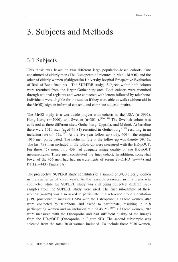
3.1 Subjects This thesis was based on two different large population-based cohorts. One constituted of elderly men (The Osteoporotic Fractures in Men - MrOS) and the other of elderly women (Sahlgrenska University hospital Prospective Evaluation of Risk of Bone fractures – The SUPERB study). Subjects within both cohorts were recruited from the larger Gothenburg area. Both cohorts were recruited through national registers and were contacted with letters followed by telephone. Individuals were eligible for the studies if they were able to walk (without aid in the MrOS), sign an informed consent, and complete a questionnaire.
The MrOS study is a worldwide project with cohorts in the USA (n=5995), Hong Kong (n=2000), and Sweden (n=3014).(196-198) The Swedish cohort was collected at three different sites, Gothenburg, Uppsala, and Malmö. At baseline there were 1010 men (aged 69-81) recruited in Gothenburg,(198) resulting in an inclusion rate of 45%.(199) At the five-year follow-up study, 600 of the original 1010 men participated. The inclusion rate at the follow-up was thereby 59.4%. The last 478 men included in the follow-up were measured with the HR-pQCT. For these 478 men, only 456 had adequate image quality on the HR-pQCT measurements. These men constituted the final cohort. In addition, somewhat fewer of the 456 men had had measurements of serum 25-OH-D (n=444) and PTH (n=443)(Figure 5A). The prospective SUPERB study constitutes of a sample of 3030 elderly women in the age range of 75-80 years. As the research presented in this thesis was conducted while the SUPERB study was still being collected, different sub-samples from the SUPERB study were used. The first sub-sample of these women (n=496) was also asked to participate in a reference probe indentation (RPI) procedure to measure BMSi with the Osteoprobe. Of these women, 482 were contacted by telephone and asked to participate, resulting in 218 participating women and an inclusion rate of 45.2%.(190) Of these women, 202 were measured with the Osteoprobe and had sufficient quality of the images from the HR-pQCT (Osteoprobe in Figure 5B). The second subsample was selected from the total 3030 women included. To include these 3030 women,
Cortical Porosity – Its regulation and association with fracture
24 3 . SUBJECTS AND METHODS
6833 were contacted. From these, 435 (6.4%) women did not meet the inclusion criteria and were excluded due to reasons such as bilateral hip replacements, not able to communicate in Swedish, or not able to walk with or without a walking aid. Of all contacted women meeting the inclusion criteria, 3368 (52.6%) declined to participate in the study. The inclusion rate for the SUPERB study was therefore 47.4%. From the whole cohort, 49 women were identified with a prevalent X-ray verified hip fracture. Of these women, 46 had sufficient quality of their HR-pQCT images (Hip Frx in Figure 5B) and could therefore be included as cases. The cases were then compared to all women without any self-reported fracture after the age of 50, extracted from the first 1093 consecutively included women with complete HR-pQCT data, and with adequate quality of the HR-pQCT images (n=361) (controls in Figure 5B).
Figure 5 Study inclusion for investigated cohorts: MrOS (A) and SUPERB (B). Quality defines the subjects with approvable quality of the HR-pQCT images
3.2 Ethics The ethical considerations for both studies concern mainly X-ray exposure, blood sampling, and confirmation of fractures in X-ray registers. The radiation exposure due to X-rays and bone densitometry was low and approved by the local radiation protection committee for both studies. Venous blood sampling is an invasive procedure but the risk of serious complications is extremely low. Some of the participants in the SUPERB study were also measured with the Osteoprobe device. The invasiveness of this procedure can be compared to a venous blood sample. Since this method is fairly new, the first 102 participants

Daniel Sundh
3 . SUBJECTS AND METHODS 25
were contacted after the procedure and no complications were reported. Epidemiologic studies are associated with issues regarding personal integrity for the included participants. To ensure that data security was not violated, all data was collected in a coded form and was only handled by authorized personnel. Furthermore, analyses and presentation of results were only performed on group level with no possibilities to identify unique individuals. The study participants could withdraw their consent at any time and thereby be excluded from the study. For all studies in this thesis, all participants signed an informed consent. All studies were approved by the ethical review board in Gothenburg.
3.3 Questionnaire For the elderly men, current smoking was established by asking if their smoking habits had changed since baseline measurements, and if so, in what way. For the elderly women, current smoking was assessed through asking if they currently smoked (yes/no) followed by if they smoke regularly or when they most recently smoked, if they had quitted.(200) Medical history regarding fall inducing and bone affecting diseases such as Parkinson’s, stroke, rheumatoid arthritis, diabetes, heart failure, angina pectoris, and colon or prostate cancer was assessed. Questions included known diagnoses (yes/no). Use of medications was assessed as current use and was defined as usage at least three times per week for the past 30 days. Information regarding previous fracture was obtained by asking if the participant had sustained a fracture (yes/no). If so, further questions were asked regarding location and time of the fracture event. The questionnaire physical activity scale for the elderly (PASE) was used to assess physical activity habits. It is a validated self-reported questionnaire designed to measure physical activity in individuals 65 years or older.(201) The questionnaire consists of twelve items about physical activity during a seven-day period prior to the assessment. A total PASE-score was calculated with these twelve items by multiplying the amount of time spent (hours/week) or participation (yes/no) in different activities by empirically derived weights and finally summing the products for all twelve items. Daily intake of calcium (mg/day) was assessed and calculated from questions regarding calcium supplements and calcium containing foods (e.g., dairy products and vegetables). For the elderly women, daily calcium intake was assessed with questions regarding amount of dairy products consumed.(202) The total amount of calcium was calculated by summarizing daily intake and supplements. For the women, alcohol consumption was estimated by questions regarding the amount and frequency of drinking.(203) Also, a physical component summary was established by the standardized SF-12 questionnaire.(204)
Cortical Porosity – Its regulation and association with fracture
26 3 . SUBJECTS AND METHODS
3.4 Anthropometrics Anthropometrics were obtained by the same methods for both cohorts. Height was measured with a standardized, wall-mounted stadiometer. Two consecutive measurements were performed, and if they differed 5 mm a third measurement was performed and the two most similar were used. An average was calculated and used in the analyses. Tibia length was measured with a ruler between the medial malleolus to the medial condyle of the tibia. Weight was measured to the nearest 0.1 kg with the same scale for both cohorts. Both height and weight had a coefficient of variation (CV) below 1%.
3.5 Fractures
3.5.1 Prevalent fractures
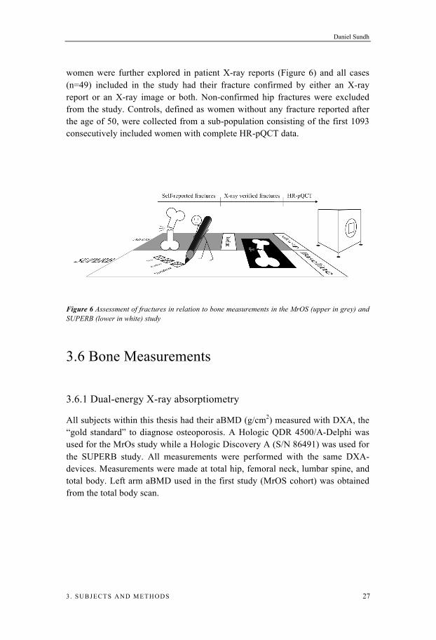
A standardized questionnaire was used at baseline in the MrOS to obtain information regarding self-reported previous fractures after the age of 50 years (Figure 6). X-ray verified fractures were collected between the date of baseline and that of HR-pQCT measurements at follow-up (Figure 6). Two fracture groups were analyzed: (i) All identified fractures (both self-reported and X-ray verified fractures) and (ii) only X-ray verified fractures. All fractures were defined and categorized according to fracture site. Groups investigated were peripheral fractures (upper and lower arm and leg), osteoporotic fractures (hip, wrist, humerus, and vertebra), multiple fractures ( 2 fractures), and all fractures (excluding hand, finger, foot, toe, and skull fractures). Study subjects in these fracture categories were compared to controls without any fracture. Severity of trauma was not considered. All X-ray verified fractures were obtained from patient records and were collected by a research nurse and further inspected by an orthopedic surgeon. Only clinical vertebral fractures were included, defined by a radiologist and identified by investigating patient records.
3.5.2 Prevalent hip fractures
Prevalent hip fractures were identified using a questionnaire completed by the women included in the SUPERB-study. Questionnaires filled out by all participants provided information about all self-reported prevalent fractures. There were 64 women who reported a hip fracture after the age of 50. These
Daniel Sundh
3 . SUBJECTS AND METHODS 27
women were further explored in patient X-ray reports (Figure 6) and all cases (n=49) included in the study had their fracture confirmed by either an X-ray report or an X-ray image or both. Non-confirmed hip fractures were excluded from the study. Controls, defined as women without any fracture reported after the age of 50, were collected from a sub-population consisting of the first 1093 consecutively included women with complete HR-pQCT data.
Figure 6 Assessment of fractures in relation to bone measurements in the MrOS (upper in grey) and SUPERB (lower in white) study
3.6 Bone Measurements
3.6.1 Dual-energy X-ray absorptiometry
All subjects within this thesis had their aBMD (g/cm2) measured with DXA, the “gold standard” to diagnose osteoporosis. A Hologic QDR 4500/A-Delphi was used for the MrOs study while a Hologic Discovery A (S/N 86491) was used for the SUPERB study. All measurements were performed with the same DXA-devices. Measurements were made at total hip, femoral neck, lumbar spine, and total body. Left arm aBMD used in the first study (MrOS cohort) was obtained from the total body scan.
Cortical Porosity – Its regulation and association with fracture
28 3 . SUBJECTS AND METHODS
3.6.2 High-resolution peripheral quantitative computed tomography
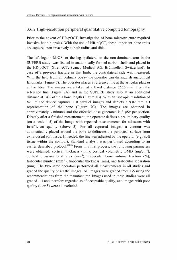
Prior to the advent of HR-pQCT, investigation of bone microstructure required invasive bone biopsies. With the use of HR-pQCT, these important bone traits are captured non-invasively at both radius and tibia. The left leg, in MrOS, or the leg ipsilateral to the non-dominant arm in the SUPERB study, was fixated in anatomically formed carbon shells and placed in the HR-pQCT (XtremeCT; Scanco Medical AG, Brüttisellen, Switzerland). In case of a previous fracture in that limb, the contralateral side was measured. With the help from an ordinary X-ray the operator can distinguish anatomical landmarks (Figure 7). The operator places a reference line at the articular plateau at the tibia. The images were taken at a fixed distance (22.5 mm) from the reference line (Figure 7A) and in the SUPERB study also at an additional distance at 14% of tibia bone length (Figure 7B). With an isotropic resolution of 82 μm the device captures 110 parallel images and depicts a 9.02 mm 3D representation of the bone (Figure 7C). The images are obtained in approximately 3 minutes and the effective dose generated is 3 μSv per section. Directly after a finished measurement, the operator defines a preliminary quality (on a scale 1-5) of the image with repeated measurements for all scans with insufficient quality (above 3). For all captured images, a contour was automatically placed around the bone to delineate the periosteal surface from extra-osseal soft tissue. If needed, the line was adjusted by the operator (e.g., soft tissue within the contour). Standard analysis was performed according to an earlier described protocol.(205) From this first process, the following parameters were obtained: cortical thickness (mm), cortical volumetric BMD (mg/cm3), cortical cross-sectional area (mm2), trabecular bone volume fraction (%), trabecular number (mm-1), trabecular thickness (mm), and trabecular separation (mm). The two same operators performed all measurements in all studies and graded the quality of all the images. All images were graded from 1-5 using the recommendations from the manufacturer. Images used in these studies were all graded 1-3 and therefore regarded as of acceptable quality, and images with poor quality (4 or 5) were all excluded.
Daniel Sundh
3 . SUBJECTS AND METHODS 29
Figure 7 Representative images for the manufacturer’s standard site (A) and the more proximal section (B) at 14% of tibia length. The 9.02 mm 3D construction of the complete bone (C), cortical shell (D), and cortical porosity (E)
3.6.2.1 Cortical evaluation
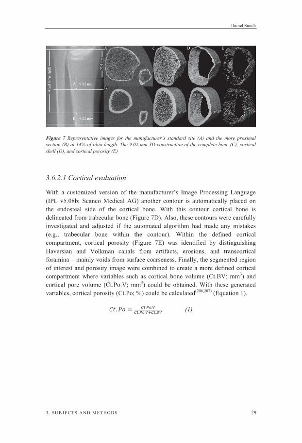
With a customized version of the manufacturer’s Image Processing Language (IPL v5.08b; Scanco Medical AG) another contour is automatically placed on the endosteal side of the cortical bone. With this contour cortical bone is delineated from trabecular bone (Figure 7D). Also, these contours were carefully investigated and adjusted if the automated algorithm had made any mistakes (e.g., trabecular bone within the contour). Within the defined cortical compartment, cortical porosity (Figure 7E) was identified by distinguishing Haversian and Volkman canals from artifacts, erosions, and transcortical foramina – mainly voids from surface coarseness. Finally, the segmented region of interest and porosity image were combined to create a more defined cortical compartment where variables such as cortical bone volume (Ct.BV; mm3) and cortical pore volume (Ct.Po.V; mm3) could be obtained. With these generated variables, cortical porosity (Ct.Po; %) could be calculated(206,207) (Equation 1).
(1)
Cortical Porosity – Its regulation and association with fracture
30 3 . SUBJECTS AND METHODS
3.6.3 Reference point indentation
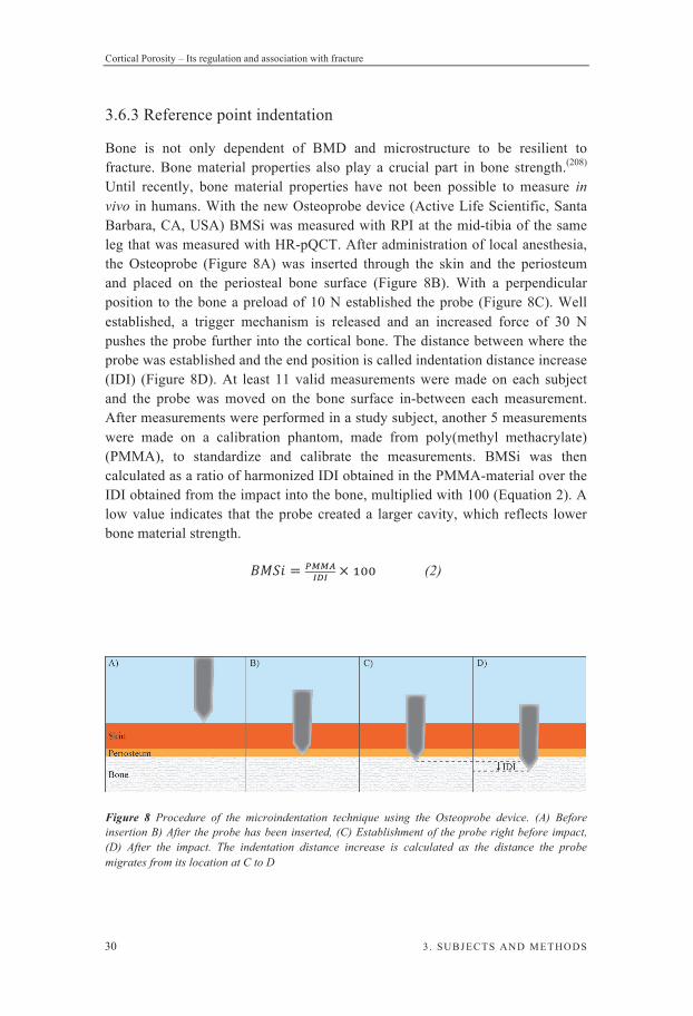
Bone is not only dependent of BMD and microstructure to be resilient to fracture. Bone material properties also play a crucial part in bone strength.(208) Until recently, bone material properties have not been possible to measure in vivo in humans. With the new Osteoprobe device (Active Life Scientific, Santa Barbara, CA, USA) BMSi was measured with RPI at the mid-tibia of the same leg that was measured with HR-pQCT. After administration of local anesthesia, the Osteoprobe (Figure 8A) was inserted through the skin and the periosteum and placed on the periosteal bone surface (Figure 8B). With a perpendicular position to the bone a preload of 10 N established the probe (Figure 8C). Well established, a trigger mechanism is released and an increased force of 30 N pushes the probe further into the cortical bone. The distance between where the probe was established and the end position is called indentation distance increase (IDI) (Figure 8D). At least 11 valid measurements were made on each subject and the probe was moved on the bone surface in-between each measurement. After measurements were performed in a study subject, another 5 measurements were made on a calibration phantom, made from poly(methyl methacrylate) (PMMA), to standardize and calibrate the measurements. BMSi was then calculated as a ratio of harmonized IDI obtained in the PMMA-material over the IDI obtained from the impact into the bone, multiplied with 100 (Equation 2). A low value indicates that the probe created a larger cavity, which reflects lower bone material strength.
(2)
Figure 8 Procedure of the microindentation technique using the Osteoprobe device. (A) Before insertion B) After the probe has been inserted, (C) Establishment of the probe right before impact, (D) After the impact. The indentation distance increase is calculated as the distance the probe migrates from its location at C to D
Daniel Sundh
3 . SUBJECTS AND METHODS 31
3.6.4 Peripheral quantitative computed tomography
In the SUPERB-study, 30 women’s distal tibia was measured at 14% of the tibial bone length with a peripheral quantitative computed tomography (XCT-2000; Stratec Medizintechnik, GmbH, Pforzheim, Germany). A two millimeter thick, single slice was captured with the resolution of 0.50 mm.
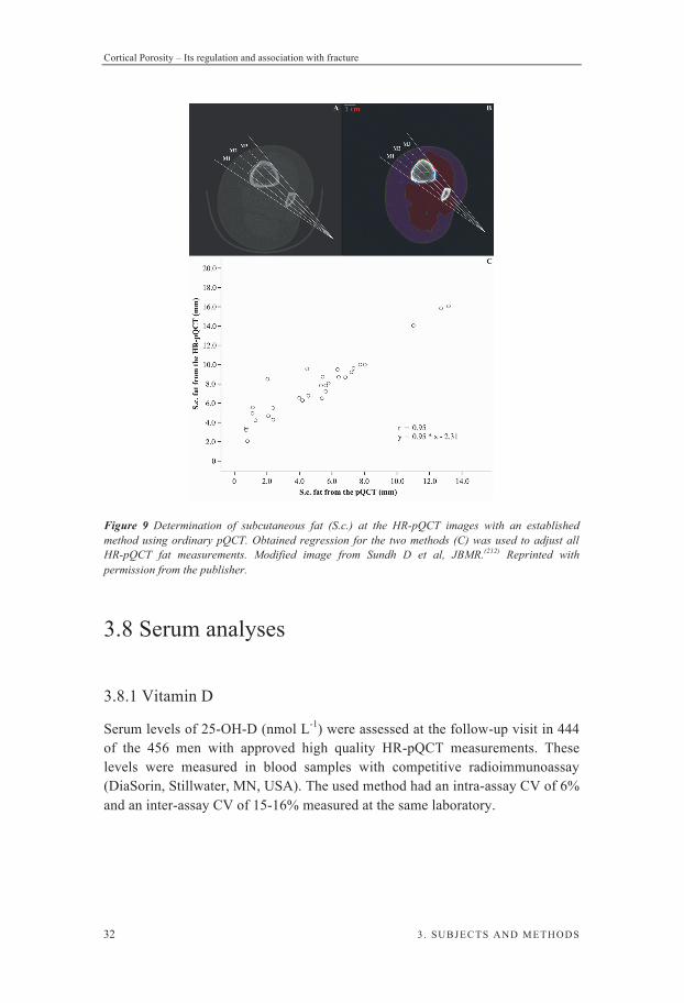
3.7 Body composition measurements Body composition such as total adipose and lean tissue was analyzed from the whole body measurement with DXA. This analysis was further subdivided into more precise areas (e.g., leg, arm, and trunk).(209) Subcutaneous fat was measured with the HR-pQCT device at the distal tibia section (14% of tibia length). Two lines were drawn in contact to both tibia and fibula and created an angle when crossed. This angle was divided by four, generating three measuring locations (M1, M2, and M3). The distance from the periosteal surface of the tibia bone to the surface of the skin layer was measured (Figure 9A). This procedure was performed on image 1, 55, and 110 for each individual and an average was calculated for the nine measurements. Interobserver (2.5%) and intraobserver (1.1%) CVs for this method were calculated for 30 of the participating women. To assure that subcutaneous fat was measured, a correlation study between the HR-pQCT and a pQCT was made. In 30 women aged 76.5 ± 0.98 years (mean ± SD), tibia was measured with both machines at 14% of bone length. The pQCT-images were further processed with BoneJ, a plug-in to the open source software ImageJ (NIH, Bethesda, MD, USA). This software enables segmentation of the soft tissue(210) and has been used earlier to assess subcutaneous fat.(211) The segmented subcutaneous fat was measured with the same procedure as for the HR-pQCT-images. Two lines were drawn in contact with both tibia and fibula generating an angle that was divided by four. The three generated measuring points were used to measure only the marked subcutaneous fat (purple area in Figure 9B). An average of the three measurements (since only a single slice is obtained) were then correlated with the average calculated from the HR-pQCT device (r=0.95; p<0.001). With a linear regression where fat measured with the pQCT was the dependent variable and fat from the HR-pQCT the independent variable a regression coefficient (0.95) and a constant (-2.31) were obtained (Figure 9C). The regression equation was used to adjust all subcutaneous fat measurements made with the HR-pQCT device.
Cortical Porosity – Its regulation and association with fracture
32 3 . SUBJECTS AND METHODS
Figure 9 Determination of subcutaneous fat (S.c.) at the HR-pQCT images with an established method using ordinary pQCT. Obtained regression for the two methods (C) was used to adjust all HR-pQCT fat measurements. Modified image from Sundh D et al, JBMR.(212) Reprinted with permission from the publisher.
3.8 Serum analyses
3.8.1 Vitamin D
Serum levels of 25-OH-D (nmol L-1) were assessed at the follow-up visit in 444 of the 456 men with approved high quality HR-pQCT measurements. These levels were measured in blood samples with competitive radioimmunoassay (DiaSorin, Stillwater, MN, USA). The used method had an intra-assay CV of 6% and an inter-assay CV of 15-16% measured at the same laboratory.
Daniel Sundh
3 . SUBJECTS AND METHODS 33
3.8.2 Parathyroid hormone
Serum levels of serum PTH (pmol L-1) were assessed at the follow-up in 443 men out of the 444 men with available 25-OH-D measurements. The analysis was performed with the Immulite 2000 intact PTH assay (Diagnostic Products, Los Angeles, CA, USA). The method reference range was between 1.1 to 6.9 pmol L-1 and had an intra-assay CV of 5% and inter-assay CV of 9%.
3.9 Statistics Group to group differences in anthropometrics, environmental factors, and bone parameters that were normally distributed were investigated using independent samples t-test. For categorical variables, 2 test or Fisher’s exact test for smaller samples were used. Parameters that were not normally distributed were investigated and groups compared based on their medians by a Mann-Whitney U test. When investigating the effect of 25-OH-D on bone microstructure, all subjects were divided into quartiles according to serum levels of PTH and 25-OH-D. The mean for bone variables and covariates was compared between quartiles with an analysis of variance (ANOVA) followed by a least significant difference post hoc test for group-to-group significance testing. Categorical variables were also here compared using a 2 test. Parameters that were not normally distributed were logarithmically transformed before used in any further statistical analysis. Bivariate correlations were used to evaluate body compositions association with bone microstructure and material properties. To assess if these correlations were still valid after adjustment for covariates, stepwise multivariable linear regression analyses were performed. Since measurements of BMSi differed between operators, adjustment for operator was carried out using dummy variables in the stepwise linear regression models. Quartiles of subcutaneous fat were defined to investigate, by ANOVA followed by a Bonferroni post hoc test, whether the associations with cortical porosity and BMSi were nonlinear. To assess the association between serum levels of 25-OH-D and bone microstructure, on top of other environmental factors and anthropometrics, hierarchical linear regression models were used. Furthermore, multivariable logistic regression models were performed to evaluate the association of bone microstructure and prevalent fracture in men or hip fracture in women independently of covariates and aBMD. Also, to evaluate if cortical porosity had a linear association with prevalent fracture in men or if there was a threshold value for cortical porosity the proportion of fractures was compared with a 2 test between quartiles of cortical porosity. Many statistical analyses were made in all four papers, where a p-value of 0.05 was considered significant.
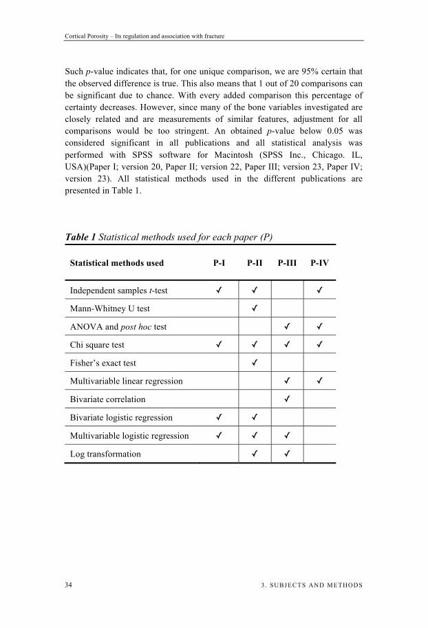
Cortical Porosity – Its regulation and association with fracture
34 3 . SUBJECTS AND METHODS
Such p-value indicates that, for one unique comparison, we are 95% certain that the observed difference is true. This also means that 1 out of 20 comparisons can be significant due to chance. With every added comparison this percentage of certainty decreases. However, since many of the bone variables investigated are closely related and are measurements of similar features, adjustment for all comparisons would be too stringent. An obtained p-value below 0.05 was considered significant in all publications and all statistical analysis was performed with SPSS software for Macintosh (SPSS Inc., Chicago. IL, USA)(Paper I; version 20, Paper II; version 22, Paper III; version 23, Paper IV; version 23). All statistical methods used in the different publications are presented in Table 1.
Table 1 Statistical methods used for each paper (P)
Statistical methods used P-I P-II P-III P-IV
Independent samples t-test
Mann-Whitney U test ANOVA and post hoc test
Chi square test
Fisher’s exact test Multivariable linear regression
Bivariate correlation
Bivariate logistic regression
Multivariable logistic regression
Log transformation

Daniel Sundh
4 . RESULTS 35
4. Results
4.1 Paper I
Increased Cortical Porosity in Older Men With Fracture
In the first study we investigated the association of bone geometry and microstructure parameters with prevalent fractures in older men. In the cohort of 456 men aged 80.2 ± 3.5 (mean ± SD) with HR-pQCT measurements, 87 had experienced a fracture from the age of 50 and onwards (all fracture group). Out of these 87 fracture cases, 52 (11.4%) were self-reported and 35 (7.7%) were X-ray verified.
4.1.1 Main results
Men in the all fracture group and the X-ray verified fractures had 15.8% and 21.6% higher cortical porosity respectively than non-fractured controls.
Fracture prevalence by quartiles of cortical porosity indicated a nonlinear relationship between this bone trait and fracture.
Men with any fracture had lower total hip, femoral neck, lumbar spine,
and left arm aBMD than men without a fracture.
Cortical porosity was, independently of aBMD and clinical risk factors, associated with all and peripheral X-ray verified fractures (Figure 10).
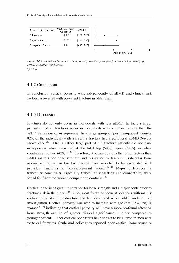
Cortical Porosity – Its regulation and association with fracture
36 4 . RESULTS
Figure 10 Associations between cortical porosity and X-ray verified fractures independently of aBMD and other risk factors. *p<0.05
4.1.2 Conclusion
In conclusion, cortical porosity was, independently of aBMD and clinical risk factors, associated with prevalent fracture in older men.
4.1.3 Discussion
Fractures do not only occur in individuals with low aBMD. In fact, a larger proportion of all fractures occur in individuals with a higher T-score than the WHO definition of osteoporosis. In a large group of postmenopausal women, 82% of the individuals with a fragility fracture had a peripheral aBMD T-score above -2.5.(213) Also, a rather large part of hip fracture patients did not have osteoporosis when measured at the total hip (54%), spine (54%), or when combining the two (42%).(150) Therefore, it seems obvious that other factors than BMD matters for bone strength and resistance to fracture. Trabecular bone microstructure has in the last decade been reported to be associated with prevalent fractures in postmenopausal women.(214) Major differences in trabecular bone traits, especially trabecular separation and connectivity were found for fractured women compared to controls.(157) Cortical bone is of great importance for bone strength and a major contributor to fracture risk in the elderly.(6) Since most fractures occur at locations with mainly cortical bone its microstructure can be considered a plausible candidate for investigation. Cortical porosity was seen to increase with age (r = 0.57-0.58) in women,(179) indicating that cortical porosity will have a more profound effect on bone strength and be of greater clinical significance in older compared to younger patients. Other cortical bone traits have shown to be altered in men with vertebral fractures. Szulc and colleagues reported poor cortical bone structure
Daniel Sundh
4 . RESULTS 37
through reduced cortical vBMD and thickness for those with vertebral fractures.(159) However, in comparison to our study, Szulc et al. did not find that any of the cortical measurements were significantly different between men with fracture and controls after adjusting for aBMD measured with DXA. This discrepancy could be due to the differences in age between the investigated cohorts; their cohort being relatively young (age mean ± SD; 73 ± 8 years for cases and 69.9 ± 9 years for controls). Cortical porosity measured in experiments with dehydration or deuterium oxide, where bone water concentration is measured, has been shown to be much higher(215) than with our method. One reason for this might be that our method only captures larger pores (approximately 130 m in diameter).(179) Even if the method we used to evaluate cortical porosity underestimates this bone trait, it could still reflect the interindividual differences between men with fracture compared to non-fractured controls. Also, with this study we could report a nonlinear association between cortical porosity and fracture, indicating that cortical porosity is only important for fracture risk when it is rather high. With this study being the first to report increased cortical porosity in older men with fracture, future studies are needed to evaluate if cortical porosity can predict future fracture.
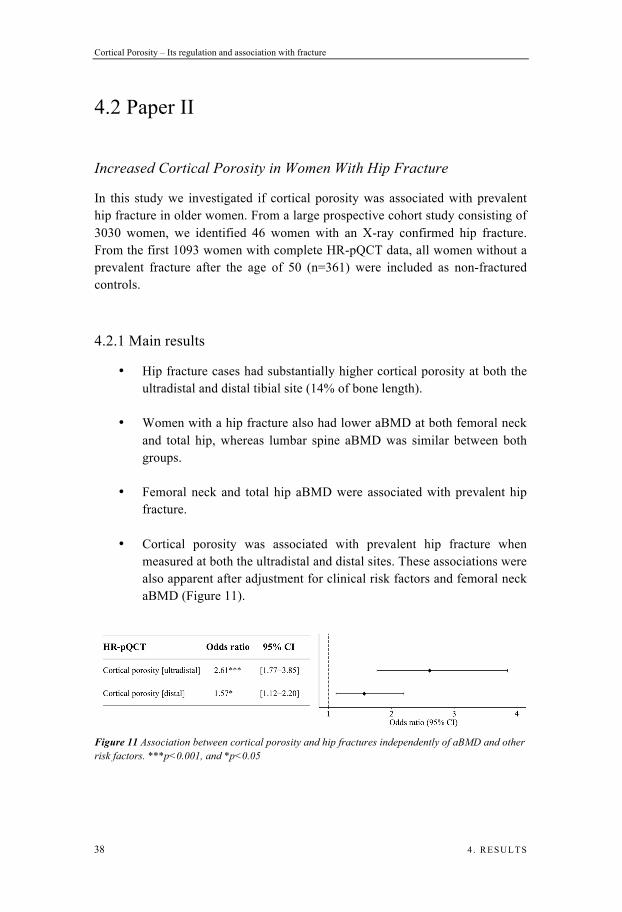
Cortical Porosity – Its regulation and association with fracture
38 4 . RESULTS
4.2 Paper II
Increased Cortical Porosity in Women With Hip Fracture
In this study we investigated if cortical porosity was associated with prevalent hip fracture in older women. From a large prospective cohort study consisting of 3030 women, we identified 46 women with an X-ray confirmed hip fracture. From the first 1093 women with complete HR-pQCT data, all women without a prevalent fracture after the age of 50 (n=361) were included as non-fractured controls.
4.2.1 Main results
Hip fracture cases had substantially higher cortical porosity at both the ultradistal and distal tibial site (14% of bone length).
Women with a hip fracture also had lower aBMD at both femoral neck and total hip, whereas lumbar spine aBMD was similar between both groups.
Femoral neck and total hip aBMD were associated with prevalent hip fracture.
Cortical porosity was associated with prevalent hip fracture when measured at both the ultradistal and distal sites. These associations were also apparent after adjustment for clinical risk factors and femoral neck aBMD (Figure 11).
Figure 11 Association between cortical porosity and hip fractures independently of aBMD and other risk factors. ***p<0.001, and *p<0.05

Daniel Sundh
4 . RESULTS 39
4.2.2 Conclusion
Women with a prevalent hip fracture in the age span of 75-80 have higher cortical porosity than non-fractured controls in the same age group. Cortical porosity was associated with prevalent hip fracture independently of femoral neck aBMD and clinical risk factors.
4.2.3 Discussion
Today, the commonly used and recommended clinical tool (i.e., DXA) to identify patients who will fracture, is not sufficiently accurate and fails to identify more than half of the patients who sustain a fracture.(213,216) By adding a more refined method, being able to measure bone microstructure, perhaps a larger proportion of fracture cases could be identified and treated before the fracture. In this study we found that women in the age of 75-80 years with a prevalent hip fracture had higher cortical porosity than non-fractured controls. Furthermore, this bone trait was associated with prevalent hip fracture after adjustment for other risk factors and femoral neck aBMD. This finding indicates that cortical porosity contributes with information regarding bone fragility in addition to aBMD measured at the femoral neck. Other studies have previously reported differences in bone geometry between hip fracture patients and non-fractured controls. Vico et al. showed that postmenopausal women with a hip fracture had a deteriorated bone microstructure in the trabecular bone compartment as well as altered bone geometry features in the cortical compartment with lower cortical bone area, thickness, and vBMD at the tibia.(217) However, this study did not investigate differences in cortical porosity. This bone trait was recently reported to be lower in 24 hip fracture cases compared to non-fractured controls. This somewhat smaller study could not show an association for cortical porosity at the tibia and prevalent hip fracture after adjustment for covariates.(180) This lack of association could also be due to earlier reported lower cortical porosity in Asian compared to white women(176) but could also be due to low statistical power in the above mentioned hip fracture study. In our larger cohort, cortical porosity together with cortical vBMD was associated with hip fracture even after adjustments for clinical risk factors including femoral neck aBMD.
Cortical Porosity – Its regulation and association with fracture
40 4 . RESULTS
4.3 Paper III
A High Amount of Local Adipose Tissue Is Associated With High Cortical Porosity and Low Bone Material Strength in Older Women
In this study we investigated how body composition, in forms of different depots of adipose tissues, was associated with BMSi and cortical bone microstructure in 202 women (78.2 ± 1.1 years [mean ± SD]).
4.3.1 Main results
High levels of subcutaneous fat at the tibia were correlated with increased cortical porosity and inversely correlated with cortical vBMD.
Women in the highest quartile of subcutaneous fat had lower BMSi (Figure 12A) and higher cortical porosity (Figure 12B) compared to women in the lowest quartile.
Increased BMSi was correlated to cortical vBMD and inversely
correlated to cortical porosity.
Local subcutaneous fat was associated with cortical porosity and inversely associated with BMSi, independently of covariates.
Cortical porosity was inversely associated with BMSi independently of
covariates.
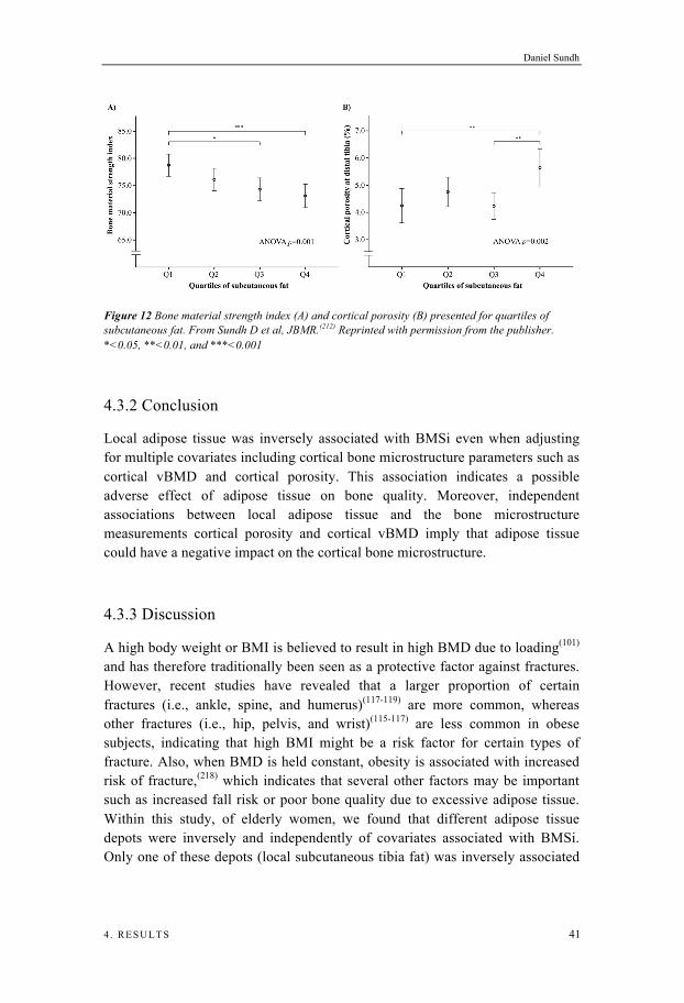
Daniel Sundh
4 . RESULTS 41
Figure 12 Bone material strength index (A) and cortical porosity (B) presented for quartiles of subcutaneous fat. From Sundh D et al, JBMR.(212) Reprinted with permission from the publisher. *<0.05, **<0.01, and ***<0.001
4.3.2 Conclusion
Local adipose tissue was inversely associated with BMSi even when adjusting for multiple covariates including cortical bone microstructure parameters such as cortical vBMD and cortical porosity. This association indicates a possible adverse effect of adipose tissue on bone quality. Moreover, independent associations between local adipose tissue and the bone microstructure measurements cortical porosity and cortical vBMD imply that adipose tissue could have a negative impact on the cortical bone microstructure.
4.3.3 Discussion
A high body weight or BMI is believed to result in high BMD due to loading(101) and has therefore traditionally been seen as a protective factor against fractures. However, recent studies have revealed that a larger proportion of certain fractures (i.e., ankle, spine, and humerus)(117-119) are more common, whereas other fractures (i.e., hip, pelvis, and wrist)(115-117) are less common in obese subjects, indicating that high BMI might be a risk factor for certain types of fracture. Also, when BMD is held constant, obesity is associated with increased risk of fracture,(218) which indicates that several other factors may be important such as increased fall risk or poor bone quality due to excessive adipose tissue. Within this study, of elderly women, we found that different adipose tissue depots were inversely and independently of covariates associated with BMSi. Only one of these depots (local subcutaneous tibia fat) was inversely associated
Cortical Porosity – Its regulation and association with fracture
42 4 . RESULTS
with cortical vBMD and positively associated with cortical porosity. These findings indicate that adipose tissue could exert a negative effect on cortical bone quality and microstructure and that this effect is local or paracrine rather than systemic. We therefore speculate that local adipose tissue may exert a direct negative effect on cortical bone microstructure (e.g., cortical porosity) and its material properties, which could be a contributing factor to why obese people are more prone to fractures in the extremities (e.g., ankle and upper arm) where cortical bone accounts for a large proportion of bone strength. Cohen et al. has earlier described associations between fat and bone microstructure and revealed an impaired bone microstructure in transiliac crest biopsies in individuals with higher trunk fat mass.(123) They reported that women with the highest levels of trunk fat had lower trabecular bone volume fraction, fewer and thinner trabecuale, and higher cortical porosity. In line with these results, we also found that women with higher subcutaneous fat at the tibia had thinner trabeculae, but since the number was increased, no association was seen for trabecular bone volume fraction. The reason for this interaction between fat and trabecular and cortical bone is still unclear but one explanation might be found at the cellular level. Since osteoblasts and adipocytes originate from the same mesenchymal stem cell and osteoporosis patients have been shown to have an increase in marrow adipocyte infiltration(219) one reason could be a shifted pattern in differentiation of these stem cells, which contribute to the pathogenesis of osteoporosis. BMSi measured with microindentation has previously been shown to be negatively associated with cortical porosity in cadaveric bones,(195) and with this study also proven to be valid in vivo in humans even after adjustment for several covariates. Although, we found that BMSi was associated with bone microstructure the variation of BMSi, explained by cortical porosity (3.3%) and cortical vBMD (5.1%), was low. This indicates that BMSi measures material properties distinct from structural parameters. Also, the association between local subcutaneous adipose tissue and BMSi was independent of cortical bone microstructure such as cortical porosity and cortical vBMD.
Daniel Sundh
4 . RESULTS 43
4.4 Paper IV
Low Serum Vitamin D Is Associated With Higher Cortical Porosity in Elderly Men
Serum levels of 25-OH-D and bone parameters using HR-pQCT were measured in 444 older men aged 80.2 ± 3.5 (mean ± SD) years. The included men were divided into quartiles in regards to their serum levels of 25-OH-D. Sub analyses were made between men with sufficient 25-OH-D levels ( 50 nmol L-1) and men with 25-OH-D deficiency (<25 nmol L-1) or insufficiency (25-49 nmol L-1) in combination with high PTH values (>6.8 pmol L-1).
4.4.1 Main results
Men in the lowest quartile of 25-OH-D had lower aBMD at the total hip and femoral neck.
Men in the lowest quartile had higher cortical porosity, measured at the tibia, than men in the highest quartile.
Low serum levels of 25-OH-D were associated with higher cortical porosity independently of covariates including PTH.
Men with 25-OH-D deficiency or insufficiency in combination with
high PTH values had lower aBMD at the total hip and femoral neck as well as markedly higher cortical porosity (17.2%).
Serum levels of PTH were not associated with cortical porosity
independently of covariates.
4.4.2 Conclusion
Low serum levels of 25-OH-D were associated with increased cortical porosity independently of confounding variables, including serum PTH levels. Moreover, cortical porosity was substantially higher in men with 25-OH-D deficiency or insufficiency in combination with elevated PTH levels.
Cortical Porosity – Its regulation and association with fracture
44 4 . RESULTS
4.4.3 Discussion
In this study we found that higher levels of 25-OH-D were associated with higher aBMD at the total hip and femoral neck, which confirms earlier findings.(75,76) Whether such differences also are valid for bone microstructure is unclear. An association has earlier been shown between low levels of 25-OH-D (<25 nmol L-1) and lower aBMD.(42,75) To fully elucidate the effect of vitamin D levels on calcium balance, consideration of PTH levels is often needed. We therefore identified a subgroup of individuals in accordance to their 25-OH-D and PTH levels. This group, eligible for vitamin D treatment, had lower aBMD measured at total hip and femoral neck, bone sites rich in cortical bone. Also, the geometrical and microstructure measurement showed similar results where the largest differences were seen for cortical measurements, with lower cortical vBMD, thickness, and area. They also had substantially higher cortical porosity. Levels of 25-OH-D, in a previous study, were associated with only trabecular bone microstructure in younger men (<65 years), whereas no associations were found for neither cortical nor trabecular bone in older men ( 65 years).(77) In contrast, we found associations for 25-OH-D with both trabecular and cortical bone microstructure at the tibia in older men. In another study investigating bone microstructure in men and women treated with high doses of vitamin D, individuals with higher 25-OH-D levels had fewer but thicker trabecuales.(79) These findings are difficult to compare to our results, since less than 10% of their cohort had a serum level of 25-OH-D below 75 nmol L-1, which is a level generally considered to be at least sufficient, while our results were obtained in a cohort with rather low percentage of men with vitamin D supplementation. As expected, the PTH levels were lower in men with higher levels of 25-OH-D. It is conceivable that increased PTH levels explained why individuals with low levels of 25-OH-D had higher cortical porosity. However, since the associations for 25-OH-D with cortical porosity were independent of PTH levels, we speculate that the levels of 25-OH-D could have a direct effect on cortical porosity. This would be possible since receptors for vitamin D have been found in osteoblasts, osteocytes, and osteoclasts and previous studies have shown both anabolic and catabolic effects of active vitamin D on bone.(220) Higher cortical porosity observed for men with lower 25-OH-D levels could be due to non-mineralized osteoid perceived as cortical pores. These bone voids were measured with a program that utilizes a BMD algorithm to define what is bone or pores. Both cortical vBMD and porosity was associated with 25-OH-D levels. However, 25-OH-D could explain a larger percentage of the variation in cortical vBMD (1.4%) than in cortical porosity (1.1%) indicating that 25-OH-D might primarily regulate cortical vBMD.
Daniel Sundh
5 . GENERAL DISCUSSION 45
5. General discussion
The inability of DXA to correctly identify the subjects who will sustain a fracture could potentially be due to its limited ability to measure bone geometry and microstructure. With measurement of these features, which is possible today, a larger proportion of those at high risk for fracture could potentially be identified. In addition to bone microstructure, bone material properties have a large impact on bone strength. Material properties have been difficult to measure until recently. With a newly developed method (Osteoprobe), bone material properties could be measured, but any association between BMSi and fracture incidence has not yet been investigated in prospective studies. Therefore, future studies are needed to determine if measuring bone material strength can improve fracture prediction, in addition to DXA derived BMD, bone microstructure and geometry. Fracture risk is not only dependent on bone strength, meaning that even if we are able to explain 100% of the variability in bone strength we probably will not be able to predict 100% of all fractures. To be able to predict a higher percentage of the patients who will sustain a fracture, more of the contributing risk factors must be taken into account. The fracture risk assessment tool (FRAX) was developed to enable us to better predict fractures based on several risk factors. With information regarding risk factors (i.e., smoking, parental history of hip fracture, previous fracture, use of per oral glucocorticoids, alcohol consumption, and rheumatoid arthritis), evaluated in worldwide prospective cohorts, the clinically used FRAX tool can calculate, with or without femoral neck aBMD, a ten-year probability of hip and major osteoporotic fracture.(131) Additional risk factor, such as frequent falls, is not included in FRAX but probably explains parts of the fracture risk. In persons aged 65 years or older, approximately 30% fall one or more times per year.(221) Few of these falls result in a fracture, but most hip fractures are the result from falling (approximately 90%),(222) which indicate that also the amount of trauma applied to the bone plays a large part in the risk of fracture.
Cortical Porosity – Its regulation and association with fracture
46 5 . GENERAL DISCUSSION
5.1 Additional value of cortical porosity In younger ages, bone loss occurs predominantly in the trabecular bone compartment. As we age, bone loss becomes more active in the cortical bone compartment.(223) This bone loss, in the form of cortical porosity, will therefore have a large impact on bone strength in the elderly and could potentially be a contributing factor to fracture.(6) The research behind this thesis, show that cortical porosity in both men and women could have an important role in bone strength. Cortical porosity was associated with fracture in both men and women independently of clinically used BMD. Therefore, additional measurement of bone microstructure could potentially identify a larger percentage of the individuals who will sustain a fracture. However, a major limitation in using the HR-pQCT in osteoporosis diagnostics is the fairly high cost of X-ray machines, resulting in expensive examinations. Health economic analyses are needed to estimate if the generated extra cost, measuring bone microstructure, is reasonable to find a larger proportion of the patients who will fracture. Such expensive medical care can be problematic, especially for developing countries. These countries need a method to establish the risk of fracture without expensive equipment. Partly due to this reason, the algorithm FRAX was developed. If measurements of cortical porosity, or any other bone microstructure or geometry trait, can independently predict fracture, it could be beneficial to include this bone variable into FRAX. Many individuals today are not classified to require osteoporosis medication based on DXA values (e.g., osteopenic patients) and clinical risk factors but still sustain a large proportion of the fractures.(161) Perhaps, by adding cortical porosity into FRAX, a truer estimation of bone strength could be obtained and result in a more precise selection of whom to treat. First off, the fracture predictive value of bone geometry or microstructure must be established. Until today, only one study with fairly few fractures has reported the ability of bone microstructure measurements to predict incident fractures in men, while no studies are available in women. Increased cortical porosity was associated with increased risk of fracture, although the association was not statistically significant, which could partly be explained by a limited numbers of fractures and rather weak associations.(224) In contrast, cortical bone area was found to predict incident fractures, independently of FRAX and aBMD in this cohort of older men. These findings do not rule out an important role of cortical porosity, but future well powered studies are needed to evaluate if cortical porosity can predict fractures and especially hip fracture in older men and women.
Daniel Sundh
5 . GENERAL DISCUSSION 47
5.2 Body composition and its effects on bone The prevalence of obesity has increased in the western world, which rightfully results in a large concern regarding an anticipated increase of cardiovascular disease(225) and diabetes.(226) These diseases cause enormous challenges in both societal costs and patient suffering. Although a normal to high BMI is traditionally believed to protect from fractures,(117) obesity has recently been associated with increased risk of certain fracture types in large cohorts and register studies.(115-119) With obesity being a risk factor for fracture an even larger increase in societal cost can be expected as both mean age of the population and prevalence of obesity increase. Until today, there is only consensus regarding unhealthy low BMI and risk of fracture.(114) Whether high BMI, and especially fat mass, exerts a negative effect on bone strength and thereby increases the risk of fracture is still to be proven. The perception of this matter has always been that larger individuals have better bone due to more loading, which results in higher BMD(101) and thereby stronger bones. If adipose tissue is associated with increased risk of fracture it might be due to several reasons. The increased risk of fracture might be due to lowering of bone strength through either altered bone microstructure or bone material properties or it could be due to a different fall pattern. If bone quality is affected, such effect might be caused by the endocrine or paracrine function (e.g., hormones and adipokines) of adipose tissue. Within this thesis, we were able to determine associations between body composition, especially fat depots, and bone material properties and bone microstructure. Individuals with more subcutaneous fat had higher cortical porosity and lower bone material strength. The reasons for these associations are uncertain but one can speculate in several theories. One potential reason could be the serum level of vitamin D. With vitamin D being a fat-soluble substrate, obese people accumulate higher amounts of vitamin D in their adipose tissue, which results in lower levels of circulating serum vitamin D.(227) These lower levels of vitamin D could be the reason for higher cortical porosity and lower cortical vBMD resulting in higher risk of fracture. Another reason could be due to the paracrine or endocrine functions of adipose tissue. This tissue consists largely of adipocytes producing the hormone adiponectin.(228) High serum levels of adiponectin have in cross-sectional studies shown to be associated with low BMD in both men and women.(113) But more interestingly, high serum of adiponectin has been reported in the MrOS Sweden to predict incident fractures independently of BMD in older men.(113) One could speculate that these effects are mediated partly through worsening of the bone microstructure by higher cortical porosity and lower cortical vBMD.
Cortical Porosity – Its regulation and association with fracture
48 5 . GENERAL DISCUSSION
5.3 Challenges The main problem with measuring cortical porosity is first of all the resolution of the imaging technology used. Since Haversian canals are small, approximately 50 m,(229,230) the resolution needs to be high to be able to capture individual pores. Such resolution is not available today and although previous studies have shown a high correlation (r = 0.80) for cortical porosity between in vitro measurements (19 m) and the HR-pQCT device,(154) indicating trustworthy measurements of porosity, it still has problems in measuring the smallest pores. With today’s resolution, more than half of all pores are being omitted due to the small size.(179) A newer version of the HR-pQCT (i.e., HR-pQCT II) has recently been released. This newer version measures bone microstructure with a higher resolution (61 m). The increased resolution however, results in higher radiation doses. Also, even if the second version may measure the bone microstructure more accurately, it still seems to be problematic to capture the smallest pores leaving us with the same problem as for the original version (used in this thesis). Cortical porosity, as measured today, is beneficial for bone strength as shown in this thesis. Whether bone strength is mostly dependent on enlargement of existing pores or the formation of new pores is not well established. If development of new pores is as important for bone strength as enlargement of existing pores is, then a better resolution is of great importance to be able to capture this change. The resolution issue also generates a known challenge called partial volume effect. This phenomenon appears when one and the same voxel includes two different materials (e.g., part of a pore and compact bone) with different attenuation values. In such a scenario the voxel will not be able to distinguish what it is measuring, which results in a weighted averaging or blurring of the attenuation, defined as partial volume averaging. This obtained value will not conform with either material attenuation, as generated with the calibrated hydroxyapatite density values, and therefore, it will not be possible to construct a correct microstructure for that voxel. The resolution is not the only problem when measuring cortical porosity. With bone being continuously remodeled it exists in many different mineralization stages. When measuring cortical bone, the pores are partly determined based on the reached degree of mineralization. It takes several days for newly placed osteoid to complete the mineralization process and eventually create compact bone. Therefore, such newly placed osteoid will be measured as a void instead of bone and thereby interpreted as a pore. These two issues can result in both over and under estimation of cortical porosity.
Daniel Sundh
5 . GENERAL DISCUSSION 49
5.4 Future benefits With a better understanding of the regulation of cortical bone, new potential targets for treatment could be found. The development of osteoporosis medication in use today was mainly achieved by randomized controlled trials in osteoporotic patients. Included subjects in these studies were selected based on a certain cut-off BMD-value at the spine or hip(231,232) and in some studies complemented with vertebral fracture.(86,233,234) By such patient inclusion criteria, many of the drugs have been proven efficacious in patients with reduced aBMD, which could be due to reduced trabecular or cortical volumetric BMD, reduced bone size or a combination of several of these features. Despite this fact, most used drugs have proven to have good efficacy primarily on vertebral and to some extent on other fractures.(232,234,235) However, more than half of all fractures occur in subjects without osteoporosis, indicating that there are other important factors besides BMD for bone strength. With greater knowledge of cortical bone regulation (e.g., cortical porosity) new potential treatment possibilities may arise. Since 80% of our skeleton consists of cortical bone, and at older ages most of the trabecular bone has vanished, most fractures (i.e., hip fracture) in the elderly seem to depend on cortical bone.(6) These fractures generate large costs for the society and great suffering for the patient. With increased understanding of how cortical porosity is regulated, potential drugs can be developed with better effect on cortical bone. With such compounds, a potential combination of today’s treatment and a more cortical bone specific treatment could probably lower the incidence of fracture even more.

Cortical Porosity – Its regulation and association with fracture
50 5 . GENERAL DISCUSSION
Daniel Sundh
6 . CONCLUSION 51
6. Conclusion
Osteoporosis is called a silent disease. However, fracture, which is the clinical outcome, is associated with risk of invalidity, morbidity, and mortality. Therefore, osteoporosis leads to high societal costs and patient suffering. The clinically used densitometry methods fail to detect more than half of all patients suffering from a fracture. New methods that are able to investigate bone in three dimensions and look at separate sections of the bone, could improve fracture prediction. Such method was evaluated in this thesis and revealed independent associations between cortical porosity and prevalent fractures in men and in women. We could also determine a clear association between cortical porosity and bone material strength, indicating that cortical porosity could affect over all bone strength. Furthermore, we found that cortical porosity is related to vitamin D status, suggesting a role of vitamin D in regulating cortical porosity. With a greater knowledge regarding the regulation and fracture predictive role of cortical porosity, improved diagnosis and prevention of fractures could be possible. Furthermore, increased knowledge regarding the regulation of cortical porosity could potentially result in new treatment targets, targeting mainly the cortical bone and the development of porosity, for osteoporosis.

Cortical Porosity – Its regulation and association with fracture
52 6 . CONCLUSION
Daniel Sundh
7 . FUTURE PERSPECTIVE 53
7. Future Perspective
There is still much remaining work within the field of osteoporosis and its diagnostics. The research presented in this thesis has shed some light over the regulation of cortical porosity and its importance in bone strength. However, it is still not known if cortical porosity as a bone trait can be used to predict fractures. Well-powered prospective studies with incident fractures as outcome are needed to establish if cortical porosity is a clinically useful bone trait that could be assessed to improve fracture prediction.
Since potential pharmacological treatment might be more effective in older ages it would be interesting to investigate if other non-pharmacological interventions could have an effect on cortical porosity. Therefore, a large intervention study investigating physical activity and its effect on cortical porosity would be of great interest. If an intervention study in men and women, over the age of 65, could show an effect on cortical bone microstructure and thereby decrease the number of fractures, exercise, in safe forms, could be recommended to high risk patients. With our cross-sectional study in older women we found that higher amount of adipose tissue was associated with higher cortical porosity and lower cortical volumetric bone mineral density. With this information, several studies would be interesting to perform. It is well known that bariatric surgery results in lower aBMD due to rapid decrease in loading of the skeleton.(236) Also, smaller studies have reported that gastric bypass surgery has a negative effect on cortical bone.(237,238) Frederiksen et al. reported an increased cortical porosity in the operated patients.(238) However, the reported difference between cases and controls was not statistically significant, possibly because of a power issue due to rather few individuals. Therefore, it would be highly interesting to perform a larger study, designed to investigate if cortical porosity is increased after bariatric surgery due to decreased loading, similar to what have been shown for unloading studies,(239) or if the decrease in fat mass could result in better cortical bone (lower cortical porosity and higher cortical vBMD) in line with the findings presented in this thesis. Another interesting study regarding weight and fat mass would be to study the other end of the spectra. Young girls with anorexia nervosa have a low BMI and an increased risk of fracture.(114) This patient group

Cortical Porosity – Its regulation and association with fracture
54 7 . FUTURE PERSPECTIVE
was also reported to have higher cortical porosity,(240) which could be a part of the explanation for the increased fracture risk. It would therefore be very interesting to investigate if weight gain by an intervention program could decrease the elevated cortical porosity and thereby potentially lower the fracture risk. The most compelling study to perform, is to investigate the ability of cortical porosity and other bone microstructure measures to predict incident fractures in the large and well powered SUPERB-study consisting of 3030 older women followed prospectively. In this study, an anticipated 400-500 fractures will have occurred at the time of the first registry follow-up (at 4-years since study start). This study will allow well powered Cox regression analyses to investigate the predictive role of various microstructure traits with aBMD and clinical risk factors as covariates.

Daniel Sundh
RELATED PUBLICATIONS NOT INCLUDED IN THE THESIS 55
Related publications not included in the thesis
1. Nilsson M, Ohlsson C, Sundh D, Mellström D, and Lorentzon M. Association of physical activity with trabecular microstructure and cortical bone at distal tibia and radius in young adult men. J Clin Endocrinol Metab. 2010 Jun; 95(6):2917-26.
2. Nilsson M, Sundh D, Ohlsson C, Karlsson M, Mellström D, and Lorentzon M. Exercise during growth and young adulthood is independently associated with cortical bone size and strength in old Swedish men. J Bone Miner Res. 2014 Aug; 29(8):1795-804.
3. Vandenput L, Lorentzon M, Sundh D, Nilsson ME, Karlsson MK, Mellström D, and Ohlsson C. Serum estradiol levels are inversely associated with cortical porosity in older men. J Clin Endocrinol Metab. 2014 Jul; 99(7):E1322-6.
4. Rudäng R, Zoulakis M, Sundh D, Brisby H, Diez-Perez A, Johansson L, Mellström D, Darelid A, and Lorentzon M. Bone material strength is associated with areal BMD but not with prevalent fractures in older women. Osteoporos Int. 2016 Apr; 27(4):1585-92.
5. Nilsson M, Sundh D, Mellström D, and Lorentzon M. Current physical activity is independently associated with cortical bone size and bone strength in elderly Swedish women. J Bone Miner Res. 2016 Sep 27.
6. *Ohlsson C, *Sundh D, Wallerek A, Nilsson M, Karlsson M, Johansson H, Mellström D, and Lorentzon M. Cortical bone area predicts incident fractures independently of areal bone mineral density in older men. J Clin Endocrinol Metab. 2016 Nov 22.
7. Nilsson AG, Sundh D, Johansson L, Nilsson M, Mellström D, Rudäng R, Zoulakis M, Wallander M, Darelid A, and Lorentzon M. Type 2 diabetes mellitus is associated with better bone microarchitecture but lower bone material strength and poorer physical function in elderly women – a population-based study. J Bone Miner Res. 2016 Dec 12.
*Contributed equally

Cortical Porosity – Its regulation and association with fracture
56 RELATED PUBLICATIONS NOT INCLUDED IN THE THESIS
Daniel Sundh
ACKNOWLEDGEMENT 57
Acknowledgement
Först och främst vill jag börja med att tacka min fantastiska handledare Mattias Lorentzon. Som person är Mattias extremt driven och har en fantastisk förmåga att plocka fram det bästa hos personer. Mattias som handledare har inte bara motiverat mig som doktorand och möjliggjort denna bok utan även varit en fantastisk människa att lära känna. Tack Dan som bihandledare för ditt stora stöd i alla mina projekt och inte minst för alla fantastiska historier som du har bjudit på under alla år. Stor del av denna avhandling tilldelar jag mina fantastiska kollegor. Först och främst ett stort tack till min på-prackade själsfrände Ulrika. Tongången har inte alltid varit vänlig men alltid med kärlek. Som ni alla vet skulle forskning aldrig fungera utan väl fungerande administration. Som tur är har vi gott om resurser på det området i fantastiska Maria. Enheten för geriatrik har varit en plats med mycket kunskap och många duktiga forskare att ta stor lärdom ifrån. Stort tack Martin för att du alltid varit närvarande för diskussioner om både forskning och andra intressanta frågor oavsett tid på dygnet. Anna N din stora kunskap och trygghet har bidragit mycket till den goda stämningen på enheten. Tack Cecilie för alla diskussioner om allt från personliga situationer till alla möjliga tänkbara ämnen. Du har varit en fantastiskt bra kontorsgranne och jag önskar dig all lycka till med din egen bok. Robert under min början av doktorandtiden var du i full gång att avsluta din. Jag skulle vilja passa på att tacka för ett extremt bra välkomnande in i gruppen, hade dock varit trevligt om kaffet hade smakat lite mer likt idag och inte som det den antika Melitta-bryggaren framställde. Tack Anna D för stor inspiration och för att varje gång du var en tid hos oss på enheten spred sådan värme och glädje. Kristian som på ett positivt sätt rör om lite i grytan med en stor och härlig personlighet (p.s, lovar att återkomma den dag jag har ett Excel-problem). Lisa för att alltid vara så nyfiken och genuint intresserad av vad jag faktiskt har gjort under mina år på enheten. Michail för din enastående förmåga att få allting att framstå som att det inte finns något problem i världen. Berit för dina in-puts i allt från forskning till tiden efter färdig doktorstitel.
Att genomföra SUPERB-studien har varit en stor del av mitt projekt. För att detta skulle vara möjligt var en personal av rang av högsta betydelse. Jag vill därför ägna ett oändligt stort tack till den fantastiska personal som har jobbat på

Cortical Porosity – Its regulation and association with fracture
58 ACKNOWLEDGEMENT
enheten. Maja du som först in bidrog mycket till att studien kom igång och med ett härligt skratt gav än mer liv till enheten. Robin för ditt tålamod med allt från logistik till oändliga samtal med alla damer. Om du förövrigt undrar hur man tillagar goda ägg så är det bara att du hör av dig. Linda med det fantastiska humöret och inte en antydan till någon form av dialekt. Annika som bidragit med stöd både på och utanför arbetsplatsen. Om jag lyckas lyfta blicken i skogen kanske vi eventuellt ses där. Karolin med ett fantastiskt humör och viljan att hjälpa allt och alla. Linn som med en helt otrolig effektivitet slutförde det hela trots inre motsättningar. Emelie som bidragit med en bra balans mellan jobb och det som faktiskt har betydelse, skratt och träning. Anja och Alexandra för att träget analysera. Under de år jag arbetat för Mattias har Osteoporosmottagningen alltid bidragit med ett stort stöd. Ni har inte bara varit intresserade av min forskning utan även utveckling på andra områden. Jag hoppas innerligt att jag har tagit ett litet steg ifrån flipp-flops och långt hår? Utveckling som doktorand är av högsta betydelse. CORS har under flera år kunnat erbjuda möten med de bästa i fältet och för det är jag väldigt tacksam. Som ny i fältet var Jonas en av mina första bekantskaper och under en lång tid höjde du CORS-mötena till en helt annan nivå. I denna kategori kvalar även Karin in som på alla möten bjudit på bra motstånd i både pistade backar samt löparspår. Jag vill också passa på att tacka alla medförfattare för bra åsikter och förslag på hur delarbetena kunde bli än bättre.
Jag skulle vilja tacka ”familjen” (Emelie och Karin) och Elisabeth för stort stöd och intresse för hur tesen har fortskridit. Jag skulle vilja tacka många för de sociala aktiviteter som fått mig att inte tänka på ben utan njuta av mycket skratt och trevligt sällskap: Blixt, Berlin Peeps, Daniel E, Daniel G, Fredrik M, George, Haak, Hannah, Johan L, John, Miche, Midgrens, Olle och Pierre H.
Ett extra tack vill jag ge till Bosse och Birgitta som med stor sponsring av diverse aktivitetsprojekt har varit en stor bidragande faktor till att detta gick att genomföra. Stort tack även till Anna, Manne, Simon, Felix och Elsy. Min familj har alltid betytt mycket för mig. Först och främst mina föräldrar Bengt och Christina som alltid har stöttat och motiverat mig till att utvecklas till vad jag vill. Min bror Johan som alltid finns där och som är den person som vet hur man kan ge en lillebror en rejäl ego-boost. Min syster Maria som inte bara varit ett stöd under doktorandtiden utan även en stor hjälp under alla aktiva studieår. Jag vill också ge ett tack till din underbara familj Jeppe och Emma.
Främst vill jag tacka min största idol och livskamrat Johanna Melin. Du har varit en klippa att luta sig mot i alla lägen och har med stor entusiasm motiverat mig i mina projekt både i och utanför arbetet. Med var sin doktorstitel kommer nu ”VonKeyboardFamily” ut starkare än någonsin!
Daniel Sundh
REFERENCES 59
References
1. Clark RK. Anatomy and Physiology: Understanding the Human Body: Jones and Bartlett Publishers; 2005.
2. Proff P, Romer P. The molecular mechanism behind bone remodelling: a review. Clin Oral Investig. Dec 2009; 13(4):355-62.
3. Currey JD. Bones: Structure and Mechanics: Princeton University Press; 2002.
4. Seeman E, Delmas PD. Bone quality--the material and structural basis of bone strength and fragility. N Engl J Med. May 25 2006; 354(21):2250-61.
5. Datta HK, Ng WF, Walker JA, Tuck SP, Varanasi SS. The cell biology of bone metabolism. J Clin Pathol. May 2008; 61(5):577-87.
6. Zebaze RM, Ghasem-Zadeh A, Bohte A, Iuliano-Burns S, Mirams M, Price RI, et al. Intracortical remodelling and porosity in the distal radius and post-mortem femurs of women: a cross-sectional study. Lancet. May 15 2010; 375(9727):1729-36.
7. Riggs BL, Wahner HW, Seeman E, Offord KP, Dunn WL, Mazess RB, et al. Changes in bone mineral density of the proximal femur and spine with aging. Differences between the postmenopausal and senile osteoporosis syndromes. J Clin Invest. Oct 1982; 70(4):716-23.
8. Caplan AI, Bruder SP. Mesenchymal stem cells: building blocks for molecular medicine in the 21st century. Trends Mol Med. Jun 2001; 7(6):259-64.
9. Jiang Y, Jahagirdar BN, Reinhardt RL, Schwartz RE, Keene CD, Ortiz-Gonzalez XR, et al. Pluripotency of mesenchymal stem cells derived from adult marrow. Nature. Jul 4 2002; 418(6893):41-9.
10. Otto F, Thornell AP, Crompton T, Denzel A, Gilmour KC, Rosewell IR, et al. Cbfa1, a candidate gene for cleidocranial dysplasia
Cortical Porosity – Its regulation and association with fracture
60 REFERENCES
syndrome, is essential for osteoblast differentiation and bone development. Cell. May 30 1997; 89(5):765-71.
11. Mundlos S, Otto F, Mundlos C, Mulliken JB, Aylsworth AS, Albright S, et al. Mutations involving the transcription factor CBFA1 cause cleidocranial dysplasia. Cell. May 30 1997; 89(5):773-9.
12. Nakashima K, Zhou X, Kunkel G, Zhang Z, Deng JM, Behringer RR, et al. The novel zinc finger-containing transcription factor osterix is required for osteoblast differentiation and bone formation. Cell. Jan 11 2002; 108(1):17-29.
13. Choi JY, Lee BH, Song KB, Park RW, Kim IS, Sohn KY, et al. Expression patterns of bone-related proteins during osteoblastic differentiation in MC3T3-E1 cells. J Cell Biochem. Jun 15 1996; 61(4):609-18.
14. Jabbar S, Drury J, Fordham JN, Datta HK, Francis RM, Tuck SP. Osteoprotegerin, RANKL and bone turnover in postmenopausal osteoporosis. J Clin Pathol. Apr 2011; 64(4):354-7.
15. Shevde NK, Bendixen AC, Dienger KM, Pike JW. Estrogens suppress RANK ligand-induced osteoclast differentiation via a stromal cell independent mechanism involving c-Jun repression. Proc Natl Acad Sci U S A. Jul 05 2000; 97(14):7829-34.
16. Srivastava S, Toraldo G, Weitzmann MN, Cenci S, Ross FP, Pacifici R. Estrogen decreases osteoclast formation by down-regulating receptor activator of NF-kappa B ligand (RANKL)-induced JNK activation. J Biol Chem. Mar 23 2001; 276(12):8836-40.
17. Hofbauer LC, Khosla S, Dunstan CR, Lacey DL, Spelsberg TC, Riggs BL. Estrogen stimulates gene expression and protein production of osteoprotegerin in human osteoblastic cells. Endocrinology. Sep 1999; 140(9):4367-70.
18. Saika M, Inoue D, Kido S, Matsumoto T. 17beta-estradiol stimulates expression of osteoprotegerin by a mouse stromal cell line, ST-2, via estrogen receptor-alpha. Endocrinology. Jun 2001; 142(6):2205-12.
19. Weitzmann MN, Pacifici R. The role of T lymphocytes in bone metabolism. Immunol Rev. Dec 2005; 208:154-68.
Daniel Sundh
REFERENCES 61
20. Capulli M, Paone R, Rucci N. Osteoblast and osteocyte: games without frontiers. Arch Biochem Biophys. Nov 01 2014; 561:3-12.
21. Franz-Odendaal TA, Hall BK, Witten PE. Buried alive: how osteoblasts become osteocytes. Dev Dyn. Jan 2006; 235(1):176-90.
22. Parfitt AM. The cellular basis of bone turnover and bone loss: a rebuttal of the osteocytic resorption--bone flow theory. Clin Orthop Relat Res. 1977; (127):236-47.
23. Knothe Tate ML, Adamson JR, Tami AE, Bauer TW. The osteocyte. Int J Biochem Cell Biol. Jan 2004; 36(1):1-8.
24. Verborgt O, Gibson GJ, Schaffler MB. Loss of osteocyte integrity in association with microdamage and bone remodeling after fatigue in vivo. J Bone Miner Res. Jan 2000; 15(1):60-7.
25. Al-Dujaili SA, Lau E, Al-Dujaili H, Tsang K, Guenther A, You L. Apoptotic osteocytes regulate osteoclast precursor recruitment and differentiation in vitro. J Cell Biochem. Sep 2011; 112(9):2412-23.
26. Riminucci M, Collins MT, Fedarko NS, Cherman N, Corsi A, White KE, et al. FGF-23 in fibrous dysplasia of bone and its relationship to renal phosphate wasting. J Clin Invest. Sep 2003; 112(5):683-92.
27. Bonewald LF. The amazing osteocyte. J Bone Miner Res. Feb 2011; 26(2):229-38.
28. Noble BS. The osteocyte lineage. Arch Biochem Biophys. May 15 2008; 473(2):106-11.
29. Yasuda H, Shima N, Nakagawa N, Yamaguchi K, Kinosaki M, Mochizuki S, et al. Osteoclast differentiation factor is a ligand for osteoprotegerin/osteoclastogenesis-inhibitory factor and is identical to TRANCE/RANKL. Proc Natl Acad Sci U S A. Mar 31 1998; 95(7):3597-602.
30. Vaananen HK, Zhao H, Mulari M, Halleen JM. The cell biology of osteoclast function. J Cell Sci. Feb 2000; 113 ( Pt 3):377-81.
31. Cappariello A, Maurizi A, Veeriah V, Teti A. Reprint of: The Great Beauty of the osteoclast. Arch Biochem Biophys. Nov 01 2014; 561:13-21.
Cortical Porosity – Its regulation and association with fracture
62 REFERENCES
32. Hughes DE, Dai A, Tiffee JC, Li HH, Mundy GR, Boyce BF. Estrogen promotes apoptosis of murine osteoclasts mediated by TGF-beta. Nat Med. Oct 1996; 2(10):1132-6.
33. Dempster DW, Luckey M, Cauley JA. Osteoporosis: Elsevier Science; 2013.
34. Manolagas SC. Birth and death of bone cells: basic regulatory mechanisms and implications for the pathogenesis and treatment of osteoporosis. Endocr Rev. Apr 2000; 21(2):115-37.
35. Lerner UH. Bone remodeling in post-menopausal osteoporosis. J Dent Res. Jul 2006; 85(7):584-95.
36. Bonjour JP, Theintz G, Law F, Slosman D, Rizzoli R. Peak bone mass. Osteoporos Int. 1994; 4 Suppl 1:7-13.
37. Bonjour JP, Theintz G, Buchs B, Slosman D, Rizzoli R. Critical years and stages of puberty for spinal and femoral bone mass accumulation during adolescence. J Clin Endocrinol Metab. Sep 1991; 73(3):555-63.
38. Lorentzon M, Mellstrom D, Ohlsson C. Age of attainment of peak bone mass is site specific in Swedish men--The GOOD study. J Bone Miner Res. Jul 2005; 20(7):1223-7.
39. Bachrach LK, Hastie T, Wang MC, Narasimhan B, Marcus R. Bone mineral acquisition in healthy Asian, Hispanic, black, and Caucasian youth: a longitudinal study. J Clin Endocrinol Metab. Dec 1999; 84(12):4702-12.
40. Ohlsson C, Darelid A, Nilsson M, Melin J, Mellstrom D, Lorentzon M. Cortical consolidation due to increased mineralization and endosteal contraction in young adult men: a five-year longitudinal study. J Clin Endocrinol Metab. Jul 2011; 96(7):2262-9.
41. Hernandez CJ, Beaupre GS, Carter DR. A theoretical analysis of the relative influences of peak BMD, age-related bone loss and menopause on the development of osteoporosis. Osteoporos Int. Oct 2003; 14(10):843-7.
42. Marshall D, Johnell O, Wedel H. Meta-analysis of how well measures of bone mineral density predict occurrence of osteoporotic fractures. BMJ. May 18 1996; 312(7041):1254-9.
Daniel Sundh
REFERENCES 63
43. Bailey DA, Martin AD, McKay HA, Whiting S, Mirwald R. Calcium accretion in girls and boys during puberty: a longitudinal analysis. J Bone Miner Res. Nov 2000; 15(11):2245-50.
44. Arlot ME, Sornay-Rendu E, Garnero P, Vey-Marty B, Delmas PD. Apparent pre- and postmenopausal bone loss evaluated by DXA at different skeletal sites in women: the OFELY cohort. J Bone Miner Res. Apr 1997; 12(4):683-90.
45. Pocock NA, Eisman JA, Hopper JL, Yeates MG, Sambrook PN, Eberl S. Genetic determinants of bone mass in adults. A twin study. J Clin Invest. Sep 1987; 80(3):706-10.
46. Krall EA, Dawson-Hughes B. Heritable and life-style determinants of bone mineral density. J Bone Miner Res. Jan 1993; 8(1):1-9.
47. Nordstrom P, Lorentzon R. Influence of heredity and environment on bone density in adolescent boys: a parent-offspring study. Osteoporos Int. 1999; 10(4):271-7.
48. Valimaki MJ, Karkkainen M, Lamberg-Allardt C, Laitinen K, Alhava E, Heikkinen J, et al. Exercise, smoking, and calcium intake during adolescence and early adulthood as determinants of peak bone mass. Cardiovascular Risk in Young Finns Study Group. BMJ. Jul 23 1994; 309(6949):230-5.
49. Lorentzon M, Mellstrom D, Ohlsson C. Association of amount of physical activity with cortical bone size and trabecular volumetric BMD in young adult men: the GOOD study. J Bone Miner Res. Nov 2005; 20(11):1936-43.
50. Rudang R, Darelid A, Nilsson M, Nilsson S, Mellstrom D, Ohlsson C, et al. Smoking is associated with impaired bone mass development in young adult men: a 5-year longitudinal study. J Bone Miner Res. Oct 2012; 27(10):2189-97.
51. Kannus P, Haapasalo H, Sankelo M, Sievanen H, Pasanen M, Heinonen A, et al. Effect of starting age of physical activity on bone mass in the dominant arm of tennis and squash players. Ann Intern Med. Jul 1 1995; 123(1):27-31.
52. Nilsson M, Ohlsson C, Sundh D, Mellstrom D, Lorentzon M. Association of physical activity with trabecular microstructure and

Cortical Porosity – Its regulation and association with fracture
64 REFERENCES
cortical bone at distal tibia and radius in young adult men. J Clin Endocrinol Metab. Jun 2010; 95(6):2917-26.
53. Nilsson M, Ohlsson C, Oden A, Mellstrom D, Lorentzon M. Increased physical activity is associated with enhanced development of peak bone mass in men: a five-year longitudinal study. J Bone Miner Res. May 2012; 27(5):1206-14.
54. Mazess RB, Barden HS. Bone density in premenopausal women: effects of age, dietary intake, physical activity, smoking, and birth-control pills. Am J Clin Nutr. Jan 1991; 53(1):132-42.
55. Neville CE, Robson PJ, Murray LJ, Strain JJ, Twisk J, Gallagher AM, et al. The effect of nutrient intake on bone mineral status in young adults: the Northern Ireland young hearts project. Calcif Tissue Int. Feb 2002; 70(2):89-98.
56. Lorentzon M, Mellstrom D, Haug E, Ohlsson C. Smoking is associated with lower bone mineral density and reduced cortical thickness in young men. J Clin Endocrinol Metab. Feb 2007; 92(2):497-503.
57. Kremer R, Campbell PP, Reinhardt T, Gilsanz V. Vitamin D status and its relationship to body fat, final height, and peak bone mass in young women. J Clin Endocrinol Metab. Jan 2009; 94(1):67-73.
58. Hatun S, Islam O, Cizmecioglu F, Kara B, Babaoglu K, Berk F, et al. Subclinical vitamin D deficiency is increased in adolescent girls who wear concealing clothing. J Nutr. Feb 2005; 135(2):218-22.
59. Kristinsson JO, Valdimarsson O, Sigurdsson G, Franzson L, Olafsson I, Steingrimsdottir L. Serum 25-hydroxyvitamin D levels and bone mineral density in 16-20 years-old girls: lack of association. J Intern Med. May 1998; 243(5):381-8.
60. Outila TA, Karkkainen MU, Lamberg-Allardt CJ. Vitamin D status affects serum parathyroid hormone concentrations during winter in female adolescents: associations with forearm bone mineral density. Am J Clin Nutr. Aug 2001; 74(2):206-10.
61. Lehtonen-Veromaa MK, Mottonen TT, Nuotio IO, Irjala KM, Leino AE, Viikari JS. Vitamin D and attainment of peak bone mass among peripubertal Finnish girls: a 3-y prospective study. Am J Clin Nutr. Dec 2002; 76(6):1446-53.
Daniel Sundh
REFERENCES 65
62. Viljakainen HT, Natri AM, Karkkainen M, Huttunen MM, Palssa A, Jakobsen J, et al. A positive dose-response effect of vitamin D supplementation on site-specific bone mineral augmentation in adolescent girls: a double-blinded randomized placebo-controlled 1-year intervention. J Bone Miner Res. Jun 2006; 21(6):836-44.
63. Winzenberg T, Powell S, Shaw KA, Jones G. Effects of vitamin D supplementation on bone density in healthy children: systematic review and meta-analysis. BMJ. Jan 25 2011; 342:c7254.
64. Holick MF. Sunlight and vitamin D for bone health and prevention of autoimmune diseases, cancers, and cardiovascular disease. Am J Clin Nutr. Dec 2004; 80(6 Suppl):1678S-88S.
65. Lips P, van Schoor NM, de Jongh RT. Diet, sun, and lifestyle as determinants of vitamin D status. Ann N Y Acad Sci. May 2014; 1317:92-8.
66. Fraser WD, Milan AM. Vitamin D assays: past and present debates, difficulties, and developments. Calcif Tissue Int. Feb 2013; 92(2):118-27.
67. Holick MF, Chen TC, Lu Z, Sauter E. Vitamin D and skin physiology: a D-lightful story. J Bone Miner Res. Dec 2007; 22 Suppl 2:V28-33.
68. Adams JS, Hewison M. Update in vitamin D. J Clin Endocrinol Metab. Feb 2010; 95(2):471-8.
69. Holick MF, Tian XQ, Allen M. Evolutionary importance for the membrane enhancement of the production of vitamin D3 in the skin of poikilothermic animals. Proc Natl Acad Sci U S A. Apr 11 1995; 92(8):3124-6.
70. DeLuca HF. Is there more to learn about functional vitamin D metabolism? J Steroid Biochem Mol Biol. Apr 2015; 148:3-6.
71. DeLuca HF. Overview of general physiologic features and functions of vitamin D. Am J Clin Nutr. Dec 2004; 80(6 Suppl):1689S-96S.
72. Busse B, Bale HA, Zimmermann EA, Panganiban B, Barth HD, Carriero A, et al. Vitamin D deficiency induces early signs of aging in human bone, increasing the risk of fracture. Sci Transl Med. Jul 10 2013; 5(193):193ra88.
Cortical Porosity – Its regulation and association with fracture
66 REFERENCES
73. Hernandez JL, Olmos JM, Pariente E, Nan D, Martinez J, Llorca J, et al. Influence of vitamin D status on vertebral fractures, bone mineral density, and bone turnover markers in normocalcemic postmenopausal women with high parathyroid hormone levels. J Clin Endocrinol Metab. Apr 2013; 98(4):1711-7.
74. Ross AC, Manson JE, Abrams SA, Aloia JF, Brannon PM, Clinton SK, et al. The 2011 Dietary Reference Intakes for Calcium and Vitamin D: what dietetics practitioners need to know. J Am Diet Assoc. Apr 2011; 111(4):524-7.
75. Bischoff-Ferrari HA, Dietrich T, Orav EJ, Dawson-Hughes B. Positive association between 25-hydroxy vitamin D levels and bone mineral density: a population-based study of younger and older adults. Am J Med. May 1 2004; 116(9):634-9.
76. Kull M, Kallikorm R, Lember M. Vitamin D as a possible independent predictor of bone mineral density in Estonian adults: a cross-sectional population-based study. Intern Med J. Jun 2012; 42(6):e89-94.
77. Chaitou A, Boutroy S, Vilayphiou N, Varennes A, Richard M, Blaizot S, et al. Association of bone microarchitecture with parathyroid hormone concentration and calcium intake in men: the STRAMBO study. Eur J Endocrinol. Jul 2011; 165(1):151-9.
78. Khosla S, Melton LJ, 3rd, Achenbach SJ, Oberg AL, Riggs BL. Hormonal and biochemical determinants of trabecular microstructure at the ultradistal radius in women and men. J Clin Endocrinol Metab. Mar 2006; 91(3):885-91.
79. Boyd SK, Burt LA, Sevick LK, Hanley DA. The relationship between serum 25(OH)D and bone density and microarchitecture as measured by HR-pQCT. Osteoporos Int. Sep 2015; 26(9):2375-80.
80. Martin EN, Haney EM, Shannon J, Cauley JA, Ensrud KE, Keaveny TM, et al. Femoral volumetric bone density, geometry, and strength in relation to 25-hydroxy vitamin D in older men. J Bone Miner Res. Mar 2015; 30(3):562-9.
81. Chapuy MC, Arlot ME, Duboeuf F, Brun J, Crouzet B, Arnaud S, et al. Vitamin D3 and calcium to prevent hip fractures in the elderly women. N Engl J Med. Dec 3 1992; 327(23):1637-42.

Daniel Sundh
REFERENCES 67
82. Sanders KM, Stuart AL, Williamson EJ, Simpson JA, Kotowicz MA, Young D, et al. Annual high-dose oral vitamin D and falls and fractures in older women: a randomized controlled trial. JAMA. May 12 2010; 303(18):1815-22.
83. Group D. Patient level pooled analysis of 68 500 patients from seven major vitamin D fracture trials in US and Europe. BMJ. Jan 12 2010; 340:b5463.
84. Rosen CJ, Bilezikian JP. Clinical review 123: Anabolic therapy for osteoporosis. J Clin Endocrinol Metab. Mar 2001; 86(3):957-64.
85. Goltzman D. Interactions of PTH and PTHrP with the PTH/PTHrP receptor and with downstream signaling pathways: exceptions that provide the rules. J Bone Miner Res. Feb 1999; 14(2):173-7.
86. Neer RM, Arnaud CD, Zanchetta JR, Prince R, Gaich GA, Reginster JY, et al. Effect of parathyroid hormone (1-34) on fractures and bone mineral density in postmenopausal women with osteoporosis. N Engl J Med. May 10 2001; 344(19):1434-41.
87. Hansen S, Beck Jensen JE, Rasmussen L, Hauge EM, Brixen K. Effects on bone geometry, density, and microarchitecture in the distal radius but not the tibia in women with primary hyperparathyroidism: A case-control study using HR-pQCT. J Bone Miner Res. Sep 2010; 25(9):1941-7.
88. Silverberg SJ, Shane E, de la Cruz L, Dempster DW, Feldman F, Seldin D, et al. Skeletal disease in primary hyperparathyroidism. J Bone Miner Res. Jun 1989; 4(3):283-91.
89. Dempster DW, Muller R, Zhou H, Kohler T, Shane E, Parisien M, et al. Preserved three-dimensional cancellous bone structure in mild primary hyperparathyroidism. Bone. Jul 2007; 41(1):19-24.
90. Parisien M, Cosman F, Mellish RW, Schnitzer M, Nieves J, Silverberg SJ, et al. Bone structure in postmenopausal hyperparathyroid, osteoporotic, and normal women. J Bone Miner Res. Sep 1995; 10(9):1393-9.
91. Parisien M, Silverberg SJ, Shane E, de la Cruz L, Lindsay R, Bilezikian JP, et al. The histomorphometry of bone in primary hyperparathyroidism: preservation of cancellous bone structure. J Clin Endocrinol Metab. Apr 1990; 70(4):930-8.
Cortical Porosity – Its regulation and association with fracture
68 REFERENCES
92. Khosla S, Melton LJ, 3rd, Wermers RA, Crowson CS, O'Fallon W, Riggs B. Primary hyperparathyroidism and the risk of fracture: a population-based study. J Bone Miner Res. Oct 1999; 14(10):1700-7.
93. Vignali E, Viccica G, Diacinti D, Cetani F, Cianferotti L, Ambrogini E, et al. Morphometric vertebral fractures in postmenopausal women with primary hyperparathyroidism. J Clin Endocrinol Metab. Jul 2009; 94(7):2306-12.
94. Stein EM, Silva BC, Boutroy S, Zhou B, Wang J, Udesky J, et al. Primary hyperparathyroidism is associated with abnormal cortical and trabecular microstructure and reduced bone stiffness in postmenopausal women. J Bone Miner Res. May 2013; 28(5):1029-40.
95. Hansen S, Hauge EM, Beck Jensen JE, Brixen K. Differing effects of PTH 1-34, PTH 1-84, and zoledronic acid on bone microarchitecture and estimated strength in postmenopausal women with osteoporosis: an 18-month open-labeled observational study using HR-pQCT. J Bone Miner Res. Apr 2013; 28(4):736-45.
96. Macdonald HM, Nishiyama KK, Hanley DA, Boyd SK. Changes in trabecular and cortical bone microarchitecture at peripheral sites associated with 18 months of teriparatide therapy in postmenopausal women with osteoporosis. Osteoporos Int. Jan 2011; 22(1):357-62.
97. Tsai JN, Uihlein AV, Burnett-Bowie SM, Neer RM, Derrico NP, Lee H, et al. Effects of Two Years of Teriparatide, Denosumab, or Both on Bone Microarchitecture and Strength (DATA-HRpQCT study). J Clin Endocrinol Metab. May 2016; 101(5):2023-30.
98. Gafni RI, Brahim JS, Andreopoulou P, Bhattacharyya N, Kelly MH, Brillante BA, et al. Daily parathyroid hormone 1-34 replacement therapy for hypoparathyroidism induces marked changes in bone turnover and structure. J Bone Miner Res. Aug 2012; 27(8):1811-20.
99. Dempster DW, Cosman F, Kurland ES, Zhou H, Nieves J, Woelfert L, et al. Effects of daily treatment with parathyroid hormone on bone microarchitecture and turnover in patients with osteoporosis: a paired biopsy study. J Bone Miner Res. Oct 2001; 16(10):1846-53.
100. School DotG, Vice Provost for Faculty Affairs University of Missouri Kansas City Denis M Medeiros PDRD, Medeiros DM, Wildman REC. Advanced Human Nutrition: Jones & Bartlett Learning, LLC; 2013.
Daniel Sundh
REFERENCES 69
101. Felson DT, Zhang Y, Hannan MT, Anderson JJ. Effects of weight and body mass index on bone mineral density in men and women: the Framingham study. J Bone Miner Res. May 1993; 8(5):567-73.
102. Ahima RS, Flier JS. Adipose tissue as an endocrine organ. Trends Endocrinol Metab. Oct 2000; 11(8):327-32.
103. Goulding A, Taylor RW. Plasma leptin values in relation to bone mass and density and to dynamic biochemical markers of bone resorption and formation in postmenopausal women. Calcif Tissue Int. Dec 1998; 63(6):456-8.
104. Blain H, Vuillemin A, Guillemin F, Durant R, Hanesse B, de Talance N, et al. Serum leptin level is a predictor of bone mineral density in postmenopausal women. J Clin Endocrinol Metab. Mar 2002; 87(3):1030-5.
105. Jurimae J, Jurimae T, Leppik A, Kums T. The influence of ghrelin, adiponectin, and leptin on bone mineral density in healthy postmenopausal women. J Bone Miner Metab. 2008; 26(6):618-23.
106. Kontogianni MD, Dafni UG, Routsias JG, Skopouli FN. Blood leptin and adiponectin as possible mediators of the relation between fat mass and BMD in perimenopausal women. J Bone Miner Res. Apr 2004; 19(4):546-51.
107. Roux C, Arabi A, Porcher R, Garnero P. Serum leptin as a determinant of bone resorption in healthy postmenopausal women. Bone. Nov 2003; 33(5):847-52.
108. Weiss LA, Barrett-Connor E, von Muhlen D, Clark P. Leptin predicts BMD and bone resorption in older women but not older men: the Rancho Bernardo study. J Bone Miner Res. May 2006; 21(5):758-64.
109. Reid IR. Relationships between fat and bone. Osteoporos Int. May 2008; 19(5):595-606.
110. Frystyk J, Vestbo E, Skjaerbaek C, Mogensen CE, Orskov H. Free insulin-like growth factors in human obesity. Metabolism. Oct 1995; 44(10 Suppl 4):37-44.
111. Sugimoto T, Nishiyama K, Kuribayashi F, Chihara K. Serum levels of insulin-like growth factor (IGF) I, IGF-binding protein (IGFBP)-2,
Cortical Porosity – Its regulation and association with fracture
70 REFERENCES
and IGFBP-3 in osteoporotic patients with and without spinal fractures. J Bone Miner Res. Aug 1997; 12(8):1272-9.
112. Garnero P, Sornay-Rendu E, Delmas PD. Low serum IGF-1 and occurrence of osteoporotic fractures in postmenopausal women. Lancet. Mar 11 2000; 355(9207):898-9.
113. Johansson H, Oden A, Lerner UH, Jutberger H, Lorentzon M, Barrett-Connor E, et al. High serum adiponectin predicts incident fractures in elderly men: Osteoporotic fractures in men (MrOS) Sweden. J Bone Miner Res. Jun 2012; 27(6):1390-6.
114. De Laet C, Kanis JA, Oden A, Johanson H, Johnell O, Delmas P, et al. Body mass index as a predictor of fracture risk: a meta-analysis. Osteoporos Int. Nov 2005; 16(11):1330-8.
115. Gnudi S, Sitta E, Lisi L. Relationship of body mass index with main limb fragility fractures in postmenopausal women. J Bone Miner Metab. 2009; 27(4):479-84.
116. Prieto-Alhambra D, Premaor MO, Fina Aviles F, Hermosilla E, Martinez-Laguna D, Carbonell-Abella C, et al. The association between fracture and obesity is site-dependent: a population-based study in postmenopausal women. J Bone Miner Res. Feb 2012; 27(2):294-300.
117. Compston JE, Watts NB, Chapurlat R, Cooper C, Boonen S, Greenspan S, et al. Obesity is not protective against fracture in postmenopausal women: GLOW. Am J Med. Nov 2011; 124(11):1043-50.
118. Pirro M, Fabbriciani G, Leli C, Callarelli L, Manfredelli MR, Fioroni C, et al. High weight or body mass index increase the risk of vertebral fractures in postmenopausal osteoporotic women. J Bone Miner Metab. 2010; 28(1):88-93.
119. Laslett LL, Just Nee Foley SJ, Quinn SJ, Winzenberg TM, Jones G. Excess body fat is associated with higher risk of vertebral deformities in older women but not in men: a cross-sectional study. Osteoporos Int. Jan 2012; 23(1):67-74.
120. Reid IR. Relationships among body mass, its components, and bone. Bone. Nov 2002; 31(5):547-55.
Daniel Sundh
REFERENCES 71
121. Evans AL, Paggiosi MA, Eastell R, Walsh JS. Bone density, microstructure and strength in obese and normal weight men and women in younger and older adulthood. J Bone Miner Res. May 2015; 30(5):920-8.
122. Shen J, Nielson CM, Marshall LM, Lee DC, Keaveny TM, Orwoll ES, et al. The Association Between BMI and QCT-Derived Proximal Hip Structure and Strength in Older Men: A Cross-Sectional Study. J Bone Miner Res. Jul 2015; 30(7):1301-8.
123. Cohen A, Dempster DW, Recker RR, Lappe JM, Zhou H, Zwahlen A, et al. Abdominal fat is associated with lower bone formation and inferior bone quality in healthy premenopausal women: a transiliac bone biopsy study. J Clin Endocrinol Metab. Jun 2013; 98(6):2562-72.
124. Wiklund P, Toss F, Weinehall L, Hallmans G, Franks PW, Nordstrom A, et al. Abdominal and gynoid fat mass are associated with cardiovascular risk factors in men and women. J Clin Endocrinol Metab. Nov 2008; 93(11):4360-6.
125. Liu CT, Broe KE, Zhou Y, Boyd SK, Cupples LA, Hannan MT, et al. Visceral Adipose Tissue Is Associated With Bone Microarchitecture in the Framingham Osteoporosis Study. J Bone Miner Res. Jan 2017; 32(1):143-50.
126. Albright F, Smith PH, Richardson AM. Postmenopausal osteoporosis: its clinical features. J Am Med Assoc. 1941; 116(22):2465-74.
127. Walker-Bone K. Recognizing and treating secondary osteoporosis. Nature reviews Rheumatology. Aug 2012; 8(8):480-92.
128. Consensus development conference: prophylaxis and treatment of osteoporosis. Am J Med. Jan 1991; 90(1):107-10.
129. Organisation WH. Assessment of fracture risk and its application to screening for postmenopausal osteoporosis. Report of a WHO Study Group. World Health Organ Tech Rep Ser. 1994; 843:1-129.
130. Glaser DL, Kaplan FS. Osteoporosis - Definition and clinical presentation. Spine. Dec 15 1997; 22(24):12S-6S.

Cortical Porosity – Its regulation and association with fracture
72 REFERENCES
131. Kanis JA, Oden A, Johansson H, Borgstrom F, Strom O, McCloskey E. FRAX and its applications to clinical practice. Bone. May 2009; 44(5):734-43.
132. Strom O, Borgstrom F, Kanis JA, Compston J, Cooper C, McCloskey EV, et al. Osteoporosis: burden, health care provision and opportunities in the EU: a report prepared in collaboration with the International Osteoporosis Foundation (IOF) and the European Federation of Pharmaceutical Industry Associations (EFPIA). Archives of osteoporosis. 2011; 6:59-155.
133. Stone KL, Seeley DG, Lui LY, Cauley JA, Ensrud K, Browner WS, et al. BMD at multiple sites and risk of fracture of multiple types: long-term results from the Study of Osteoporotic Fractures. J Bone Miner Res. Nov 2003; 18(11):1947-54.
134. Melton LJ, Chrischilles EA, Cooper C, Lane AW, Riggs BL. How many women have osteoporosis? (Reprinted from Journal Bone & Mineral Research, vol 7, pg 1005, 1992). J Bone Miner Res. May 2005; 20(5):886-92.
135. Kanis JA, Johnell O, Oden A, Sembo I, Redlund-Johnell I, Dawson A, et al. Long-term risk of osteoporotic fracture in Malmo. Osteoporos Int. 2000; 11(8):669-74.
136. Burge R, Dawson-Hughes B, Solomon DH, Wong JB, King A, Tosteson A. Incidence and economic burden of osteoporosis-related fractures in the United States, 2005-2025. J Bone Miner Res. Mar 2007; 22(3):465-75.
137. Rosengren BE, Karlsson M, Petersson I, Englund M. The 21st-century landscape of adult fractures: cohort study of a complete adult regional population. J Bone Miner Res. Mar 2015; 30(3):535-42.
138. Sogaard AJ, Holvik K, Meyer HE, Tell GS, Gjesdal CG, Emaus N, et al. Continued decline in hip fracture incidence in Norway: a NOREPOS study. Osteoporos Int. Jul 2016; 27(7):2217-22.
139. Omsland TK, Magnus JH. Forecasting the burden of future postmenopausal hip fractures. Osteoporos Int. Oct 2014; 25(10):2493-6.
140. Sernbo I, Johnell O. Consequences of a hip fracture: a prospective study over 1 year. Osteoporos Int. May 1993; 3(3):148-53.
Daniel Sundh
REFERENCES 73
141. Wiktorowicz ME, Goeree R, Papaioannou A, Adachi JD, Papadimitropoulos E. Economic implications of hip fracture: health service use, institutional care and cost in Canada. Osteoporos Int. 2001; 12(4):271-8.
142. U.S. Congress OoTA. Hip Fracture Outcomes in People Age 50 and Over-Background Paper, OTA-BP-H- 120 (Washington, DC: U.S. Government Printing Office, July 1994). 1994.
143. Winner SJ, Morgan CA, Evans JG. Perimenopausal risk of falling and incidence of distal forearm fracture. BMJ. Jun 3 1989; 298(6686):1486-8.
144. Johnell O, Kanis JA. An estimate of the worldwide prevalence, mortality and disability associated with hip fracture. Osteoporos Int. Nov 2004; 15(11):897-902.
145. Roche JJ, Wenn RT, Sahota O, Moran CG. Effect of comorbidities and postoperative complications on mortality after hip fracture in elderly people: prospective observational cohort study. BMJ. Dec 10 2005; 331(7529):1374.
146. Cummings SR, Kelsey JL, Nevitt MC, O'Dowd KJ. Epidemiology of osteoporosis and osteoporotic fractures. Epidemiol Rev. 1985; 7:178-208.
147. Salkeld G, Cameron ID, Cumming RG, Easter S, Seymour J, Kurrle SE, et al. Quality of life related to fear of falling and hip fracture in older women: a time trade off study. BMJ. Feb 5 2000; 320(7231):341-6.
148. Muller ME, Webber CE, Bouxsein ML. Predicting the failure load of the distal radius. Osteoporos Int. Jun 2003; 14(4):345-52.
149. Cody DD, Gross GJ, Hou FJ, Spencer HJ, Goldstein SA, Fyhrie DP. Femoral strength is better predicted by finite element models than QCT and DXA. J Biomech. Oct 1999; 32(10):1013-20.
150. Wainwright SA, Marshall LM, Ensrud KE, Cauley JA, Black DM, Hillier TA, et al. Hip fracture in women without osteoporosis. J Clin Endocrinol Metab. May 2005; 90(5):2787-93.
151. Boutroy S, Bouxsein ML, Munoz F, Delmas PD. In vivo assessment of trabecular bone microarchitecture by high-resolution peripheral
Cortical Porosity – Its regulation and association with fracture
74 REFERENCES
quantitative computed tomography. J Clin Endocrinol Metab. Dec 2005; 90(12):6508-15.
152. Snow-Harter C, Marcus R. Exercise, bone mineral density, and osteoporosis. Exerc Sport Sci Rev. 1991; 19:351-88.
153. Khosla S, Riggs BL, Atkinson EJ, Oberg AL, McDaniel LJ, Holets M, et al. Effects of sex and age on bone microstructure at the ultradistal radius: a population-based noninvasive in vivo assessment. J Bone Miner Res. Jan 2006; 21(1):124-31.
154. Nishiyama KK, Macdonald HM, Buie HR, Hanley DA, Boyd SK. Postmenopausal women with osteopenia have higher cortical porosity and thinner cortices at the distal radius and tibia than women with normal aBMD: an in vivo HR-pQCT study. J Bone Miner Res. Apr 2010; 25(4):882-90.
155. Melton LJ, 3rd, Riggs BL, van Lenthe GH, Achenbach SJ, Muller R, Bouxsein ML, et al. Contribution of in vivo structural measurements and load/strength ratios to the determination of forearm fracture risk in postmenopausal women. J Bone Miner Res. Sep 2007; 22(9):1442-8.
156. Sornay-Rendu E, Boutroy S, Munoz F, Delmas PD. Alterations of cortical and trabecular architecture are associated with fractures in postmenopausal women, partially independent of decreased BMD measured by DXA: The OFELY study. J Bone Miner Res. Mar 2007; 22(3):425-33.
157. Melton LJ, 3rd, Christen D, Riggs BL, Achenbach SJ, Muller R, van Lenthe GH, et al. Assessing forearm fracture risk in postmenopausal women. Osteoporos Int. Jul 2010; 21(7):1161-9.
158. Sornay-Rendu E, Cabrera-Bravo JL, Boutroy S, Munoz F, Delmas PD. Severity of vertebral fractures is associated with alterations of cortical architecture in postmenopausal women. J Bone Miner Res. Apr 2009; 24(4):737-43.
159. Szulc P, Boutroy S, Vilayphiou N, Chaitou A, Delmas PD, Chapurlat R. Cross-sectional analysis of the association between fragility fractures and bone microarchitecture in older men: the STRAMBO study. J Bone Miner Res. Jun 2011; 26(6):1358-67.
Daniel Sundh
REFERENCES 75
160. Stein EM, Liu XS, Nickolas TL, Cohen A, McMahon DJ, Zhou B, et al. Microarchitectural abnormalities are more severe in postmenopausal women with vertebral compared to nonvertebral fractures. J Clin Endocrinol Metab. Oct 2012; 97(10):E1918-26.
161. Boutroy S, Khosla S, Sornay-Rendu E, Zanchetta MB, McMahon DJ, Zhang CA, et al. Microarchitecture and Peripheral BMD are Impaired in Postmenopausal White Women With Fracture Independently of Total Hip T-Score: An International Multicenter Study. J Bone Miner Res. Jun 2016; 31(6):1158-66.
162. Currey JD, Shahar R. Cavities in the compact bone in tetrapods and fish and their effect on mechanical properties. J Struct Biol. Aug 2013; 183(2):107-22.
163. Yeni YN, Brown CU, Wang Z, Norman TL. The influence of bone morphology on fracture toughness of the human femur and tibia. Bone. Nov 1997; 21(5):453-9.
164. Bailey DA, Wedge JH, McCulloch RG, Martin AD, Bernhardson SC. Epidemiology of fractures of the distal end of the radius in children as associated with growth. J Bone Joint Surg Am. Sep 1989; 71(8):1225-31.
165. Kirmani S, Christen D, van Lenthe GH, Fischer PR, Bouxsein ML, McCready LK, et al. Bone structure at the distal radius during adolescent growth. J Bone Miner Res. Jun 2009; 24(6):1033-42.
166. Pritchett JW. Growth plate activity in the upper extremity. Clin Orthop Relat Res. Jul 1991; (268):235-42.
167. Parfitt AM. The two faces of growth: benefits and risks to bone integrity. Osteoporos Int. Nov 1994; 4(6):382-98.
168. Gabel L, Macdonald HM, McKay HA. Sex Differences and Growth-Related Adaptations in Bone Microarchitecture, Geometry, Density, and Strength From Childhood to Early Adulthood: A Mixed Longitudinal HR-pQCT Study. J Bone Miner Res. Feb 2017; 32(2):250-63.
169. Nishiyama KK, Macdonald HM, Moore SA, Fung T, Boyd SK, McKay HA. Cortical porosity is higher in boys compared with girls at the distal radius and distal tibia during pubertal growth: an HR-pQCT study. J Bone Miner Res. Feb 2012; 27(2):273-82.
Cortical Porosity – Its regulation and association with fracture
76 REFERENCES
170. Burghardt AJ, Kazakia GJ, Ramachandran S, Link TM, Majumdar S. Age- and gender-related differences in the geometric properties and biomechanical significance of intracortical porosity in the distal radius and tibia. J Bone Miner Res. May 2010; 25(5):983-93.
171. Kazakia GJ, Nirody JA, Bernstein G, Sode M, Burghardt AJ, Majumdar S. Age- and gender-related differences in cortical geometry and microstructure: Improved sensitivity by regional analysis. Bone. Feb 2013; 52(2):623-31.
172. Macdonald HM, Nishiyama KK, Kang J, Hanley DA, Boyd SK. Age-related patterns of trabecular and cortical bone loss differ between sexes and skeletal sites: a population-based HR-pQCT study. J Bone Miner Res. Jan 2011; 26(1):50-62.
173. Nirody JA, Cheng KP, Parrish RM, Burghardt AJ, Majumdar S, Link TM, et al. Spatial distribution of intracortical porosity varies across age and sex. Bone. Jun 2015; 75:88-95.
174. Vilayphiou N, Boutroy S, Sornay-Rendu E, Van Rietbergen B, Chapurlat R. Age-related changes in bone strength from HR-pQCT derived microarchitectural parameters with an emphasis on the role of cortical porosity. Bone. Feb 2016; 83:233-40.
175. Shanbhogue VV, Brixen K, Hansen S. Age- and Sex-Related Changes in Bone Microarchitecture and Estimated Strength: A Three-Year Prospective Study Using HRpQCT. J Bone Miner Res. Aug 2016; 31(8):1541-9.
176. Boutroy S, Walker MD, Liu XS, McMahon DJ, Liu G, Guo XE, et al. Lower cortical porosity and higher tissue mineral density in Chinese American versus white women. J Bone Miner Res. Mar 2014; 29(3):551-61.
177. Putman MS, Yu EW, Lee H, Neer RM, Schindler E, Taylor AP, et al. Differences in skeletal microarchitecture and strength in African-American and white women. J Bone Miner Res. Oct 2013; 28(10):2177-85.
178. Squillante RG, Williams JL. Videodensitometry of osteons in females with femoral neck fractures. Calcif Tissue Int. Apr 1993; 52(4):273-7.
179. Bala Y, Zebaze R, Ghasem-Zadeh A, Atkinson EJ, Iuliano S, Peterson JM, et al. Cortical porosity identifies women with osteopenia
Daniel Sundh
REFERENCES 77
at increased risk for forearm fractures. J Bone Miner Res. Jun 2014; 29(6):1356-62.
180. Zhu TY, Hung VW, Cheung WH, Cheng JC, Qin L, Leung KS. Value of Measuring Bone Microarchitecture in Fracture Discrimination in Older Women with Recent Hip Fracture: A Case-control Study with HR-pQCT. Sci Rep. Sep 27 2016; 6:34185.
181. Pistoia W, van Rietbergen B, Lochmuller EM, Lill CA, Eckstein F, Ruegsegger P. Estimation of distal radius failure load with micro-finite element analysis models based on three-dimensional peripheral quantitative computed tomography images. Bone. Jun 2002; 30(6):842-8.
182. Pistoia W, van Rietbergen B, Lochmuller EM, Lill CA, Eckstein F, Ruegsegger P. Image-based micro-finite-element modeling for improved distal radius strength diagnosis: moving from bench to bedside. J Clin Densitom. Summer 2004; 7(2):153-60.
183. Varga P, Dall'Ara E, Pahr DH, Pretterklieber M, Zysset PK. Validation of an HR-pQCT-based homogenized finite element approach using mechanical testing of ultra-distal radius sections. Biomechanics and modeling in mechanobiology. Jul 2011; 10(4):431-44.
184. Pottecher P, Engelke K, Duchemin L, Museyko O, Moser T, Mitton D, et al. Prediction of Hip Failure Load: In Vitro Study of 80 Femurs Using Three Imaging Methods and Finite Element Models-The European Fracture Study (EFFECT). Radiology. Sep 2016; 280(3):837-47.
185. Macneil JA, Boyd SK. Bone strength at the distal radius can be estimated from high-resolution peripheral quantitative computed tomography and the finite element method. Bone. Jun 2008; 42(6):1203-13.
186. Abraham AC, Agarwalla A, Yadavalli A, McAndrew C, Liu JY, Tang SY. Multiscale Predictors of Femoral Neck In Situ Strength in Aging Women: Contributions of BMD, Cortical Porosity, Reference Point Indentation, and Nonenzymatic Glycation. J Bone Miner Res. Dec 2015; 30(12):2207-14.
187. Granke M, Coulmier A, Uppuganti S, Gaddy JA, Does MD, Nyman JS. Insights into reference point indentation involving human cortical bone: sensitivity to tissue anisotropy and mechanical behavior. Journal
Cortical Porosity – Its regulation and association with fracture
78 REFERENCES
of the mechanical behavior of biomedical materials. Sep 2014; 37:174-85.
188. Diez-Perez A, Guerri R, Nogues X, Caceres E, Pena MJ, Mellibovsky L, et al. Microindentation for in vivo measurement of bone tissue mechanical properties in humans. J Bone Miner Res. Aug 2010; 25(8):1877-85.
189. Duarte Sosa D, Vilaplana L, Guerri R, Nogues X, Wang-Fagerland M, Diez-Perez A, et al. Are the High Hip Fracture Rates Among Norwegian Women Explained by Impaired Bone Material Properties? J Bone Miner Res. Oct 2015; 30(10):1784-9.
190. Rudang R, Zoulakis M, Sundh D, Brisby H, Diez-Perez A, Johansson L, et al. Bone material strength is associated with areal BMD but not with prevalent fractures in older women. Osteoporos Int. Apr 2016; 27(4):1585-92.
191. Mellibovsky L, Prieto-Alhambra D, Mellibovsky F, Guerri-Fernandez R, Nogues X, Randall C, et al. Bone Tissue Properties Measurement by Reference Point Indentation in Glucocorticoid-Induced Osteoporosis. J Bone Miner Res. Sep 2015; 30(9):1651-6.
192. Farr JN, Drake MT, Amin S, Melton LJ, 3rd, McCready LK, Khosla S. In vivo assessment of bone quality in postmenopausal women with type 2 diabetes. J Bone Miner Res. Apr 2014; 29(4):787-95.
193. Nilsson AG, Sundh D, Johansson L, Nilsson M, Mellstrom D, Rudang R, et al. Type 2 Diabetes Mellitus is Associated with Better Bone Microarchitecture But Lower Bone Material Strength and Poorer Physical Function in Elderly Women - a Population-Based Study. J Bone Miner Res. Dec 12 2016.
194. Abraham AC, Agarwalla A, Yadavalli A, Liu JY, Tang SY. Microstructural and compositional contributions towards the mechanical behavior of aging human bone measured by cyclic and impact reference point indentation. Bone. Jun 2016; 87:37-43.
195. Karim L, Van Vliet M, Bouxsein ML. Comparison of cyclic and impact-based reference point indentation measurements in human cadaveric tibia. Bone. Apr 7 2015.

Daniel Sundh
REFERENCES 79
196. Orwoll E, Blank JB, Barrett-Connor E, Cauley J, Cummings S, Ensrud K, et al. Design and baseline characteristics of the osteoporotic fractures in men (MrOS) study--a large observational study of the determinants of fracture in older men. Contemp Clin Trials. Oct 2005; 26(5):569-85.
197. Mellstrom D, Johnell O, Ljunggren O, Eriksson AL, Lorentzon M, Mallmin H, et al. Free testosterone is an independent predictor of BMD and prevalent fractures in elderly men: MrOS Sweden. J Bone Miner Res. Apr 2006; 21(4):529-35.
198. Ohlsson C, Mellstrom D, Carlzon D, Orwoll E, Ljunggren O, Karlsson MK, et al. Older Men With Low Serum IGF-1 Have an Increased Risk of Incident Fractures: The MrOS Sweden Study. J Bone Miner Res. Apr 2011; 26(4):865-72.
199. Mellstrom D, Vandenput L, Mallmin H, Holmberg AH, Lorentzon M, Oden A, et al. Older men with low serum estradiol and high serum SHBG have an increased risk of fractures. J Bone Miner Res. Oct 2008; 23(10):1552-60.
200. Vartiainen E, Seppala T, Lillsunde P, Puska P. Validation of self reported smoking by serum cotinine measurement in a community-based study. J Epidemiol Community Health. Mar 2002; 56(3):167-70.
201. Washburn RA, McAuley E, Katula J, Mihalko SL, Boileau RA. The physical activity scale for the elderly (PASE): evidence for validity. J Clin Epidemiol. Jul 1999; 52(7):643-51.
202. Angbratt M, Moller M. Questionnaire about calcium intake: can we trust the answers? Osteoporos Int. 1999; 9(3):220-5.
203. Bergman H, Kallmen H. Alcohol use among Swedes and a psychometric evaluation of the alcohol use disorders identification test. Alcohol Alcohol. May-Jun 2002; 37(3):245-51.
204. Ware J, Jr., Kosinski M, Keller SD. A 12-Item Short-Form Health Survey: construction of scales and preliminary tests of reliability and validity. Med Care. Mar 1996; 34(3):220-33.
205. Laib A, Hauselmann HJ, Ruegsegger P. In vivo high resolution 3D-QCT of the human forearm. Technol Health Care. Dec 1998; 6(5-6):329-37.
Cortical Porosity – Its regulation and association with fracture
80 REFERENCES
206. Burghardt AJ, Buie HR, Laib A, Majumdar S, Boyd SK. Reproducibility of direct quantitative measures of cortical bone microarchitecture of the distal radius and tibia by HR-pQCT. Bone. Sep 2010; 47(3):519-28.
207. Ostertag A, Peyrin F, Fernandez S, Laredo JD, de Vernejoul MC, Chappard C. Cortical measurements of the tibia from high resolution peripheral quantitative computed tomography images: a comparison with synchrotron radiation micro-computed tomography. Bone. Jun 2014; 63:7-14.
208. Chavassieux P, Seeman E, Delmas PD. Insights into material and structural basis of bone fragility from diseases associated with fractures: how determinants of the biomechanical properties of bone are compromised by disease. Endocr Rev. Apr 2007; 28(2):151-64.
209. Kelly TL, Wilson KE, Heymsfield SB. Dual energy X-Ray absorptiometry body composition reference values from NHANES. PLoS One. 2009; 4(9):e7038.
210. Rantalainen T, Nikander R, Heinonen A, Daly RM, Sievanen H. An open source approach for regional cortical bone mineral density analysis. J Musculoskelet Neuronal Interact. Sep 2011; 11(3):243-8.
211. Frank-Wilson AW, Johnston JD, Olszynski WP, Kontulainen SA. Measurement of muscle and fat in postmenopausal women: precision of previously reported pQCT imaging methods. Bone. Jun 2015; 75:49-54.
212. Sundh D, Rudang R, Zoulakis M, Nilsson AG, Darelid A, Lorentzon M. A High Amount of Local Adipose Tissue Is Associated With High Cortical Porosity and Low Bone Material Strength in Older Women. J Bone Miner Res. Apr 2016; 31(4):749-57.
213. Siris ES, Chen YT, Abbott TA, Barrett-Connor E, Miller PD, Wehren LE, et al. Bone mineral density thresholds for pharmacological intervention to prevent fractures. Arch Intern Med. May 24 2004; 164(10):1108-12.
214. Boutroy S, Van Rietbergen B, Sornay-Rendu E, Munoz F, Bouxsein ML, Delmas PD. Finite element analysis based on in vivo HR-pQCT images of the distal radius is associated with wrist fracture in postmenopausal women. J Bone Miner Res. Mar 2008; 23(3):392-9.
Daniel Sundh
REFERENCES 81
215. Techawiboonwong A, Song HK, Leonard MB, Wehrli FW. Cortical bone water: in vivo quantification with ultrashort echo-time MR imaging. Radiology. Sep 2008; 248(3):824-33.
216. Schuit SC, van der Klift M, Weel AE, de Laet CE, Burger H, Seeman E, et al. Fracture incidence and association with bone mineral density in elderly men and women: the Rotterdam Study. Bone. Jan 2004; 34(1):195-202.
217. Vico L, Zouch M, Amirouche A, Frere D, Laroche N, Koller B, et al. High-resolution pQCT analysis at the distal radius and tibia discriminates patients with recent wrist and femoral neck fractures. J Bone Miner Res. Nov 2008; 23(11):1741-50.
218. Nielson CM, Marshall LM, Adams AL, LeBlanc ES, Cawthon PM, Ensrud K, et al. BMI and fracture risk in older men: the osteoporotic fractures in men study (MrOS). J Bone Miner Res. Mar 2011; 26(3):496-502.
219. Griffith JF, Yeung DK, Antonio GE, Wong SY, Kwok TC, Woo J, et al. Vertebral marrow fat content and diffusion and perfusion indexes in women with varying bone density: MR evaluation. Radiology. Dec 2006; 241(3):831-8.
220. Ryan JW, Reinke D, Kogawa M, Turner AG, Atkins GJ, Anderson PH, et al. Novel targets of vitamin D activity in bone: action of the vitamin D receptor in osteoblasts, osteocytes and osteoclasts. Curr Drug Targets. Dec 2013; 14(14):1683-8.
221. Cumming RG, Kelsey JL, Nevitt MC. Methodologic issues in the study of frequent and recurrent health problems. Falls in the elderly. Ann Epidemiol. Oct 1990; 1(1):49-56.
222. Grisso JA, Kelsey JL, Strom BL, Chiu GY, Maislin G, O'Brien LA, et al. Risk factors for falls as a cause of hip fracture in women. The Northeast Hip Fracture Study Group. N Engl J Med. May 09 1991; 324(19):1326-31.
223. Riggs BL, Melton Iii LJ, 3rd, Robb RA, Camp JJ, Atkinson EJ, Peterson JM, et al. Population-based study of age and sex differences in bone volumetric density, size, geometry, and structure at different skeletal sites. J Bone Miner Res. Dec 2004; 19(12):1945-54.
Cortical Porosity – Its regulation and association with fracture
82 REFERENCES
224. Ohlsson C, Sundh D, Wallerek A, Nilsson M, Karlsson M, Johansson H, et al. Cortical bone area predicts incident fractures independently of areal bone mineral density in older men. J Clin Endocrinol Metab. Nov 22 2016:jc20163177.
225. Bastien M, Poirier P, Lemieux I, Despres JP. Overview of epidemiology and contribution of obesity to cardiovascular disease. Prog Cardiovasc Dis. Jan-Feb 2014; 56(4):369-81.
226. Shaw JE, Sicree RA, Zimmet PZ. Global estimates of the prevalence of diabetes for 2010 and 2030. Diabetes Res Clin Pract. Jan 2010; 87(1):4-14.
227. Holick MF, Binkley NC, Bischoff-Ferrari HA, Gordon CM, Hanley DA, Heaney RP, et al. Evaluation, treatment, and prevention of vitamin D deficiency: an Endocrine Society clinical practice guideline. J Clin Endocrinol Metab. Jul 2011; 96(7):1911-30.
228. Fantuzzi G. Adipose tissue, adipokines, and inflammation. J Allergy Clin Immunol. May 2005; 115(5):911-9; quiz 20.
229. Martiniakova M, Grosskopf B, Omelka R, Vondrakova M, Bauerova M. Differences among species in compact bone tissue microstructure of mammalian skeleton: use of a discriminant function analysis for species identification. J Forensic Sci. Nov 2006; 51(6):1235-9.
230. Singh IJ, Gunberg DL. Estimation of age at death in human males from quantitative histology of bone fragments. Am J Phys Anthropol. Nov 1970; 33(3):373-81.
231. Cummings SR, Black DM, Thompson DE, Applegate WB, Barrett-Connor E, Musliner TA, et al. Effect of alendronate on risk of fracture in women with low bone density but without vertebral fractures: results from the Fracture Intervention Trial. JAMA. Dec 23-30 1998; 280(24):2077-82.
232. Cummings SR, San Martin J, McClung MR, Siris ES, Eastell R, Reid IR, et al. Denosumab for prevention of fractures in postmenopausal women with osteoporosis. N Engl J Med. Aug 20 2009; 361(8):756-65.
233. Black DM, Cummings SR, Karpf DB, Cauley JA, Thompson DE, Nevitt MC, et al. Randomised trial of effect of alendronate on risk of
Daniel Sundh
REFERENCES 83
fracture in women with existing vertebral fractures. Fracture Intervention Trial Research Group. Lancet. Dec 07 1996; 348(9041):1535-41.
234. Black DM, Delmas PD, Eastell R, Reid IR, Boonen S, Cauley JA, et al. Once-yearly zoledronic acid for treatment of postmenopausal osteoporosis. N Engl J Med. May 03 2007; 356(18):1809-22.
235. Miller PD, Hattersley G, Riis BJ, Williams GC, Lau E, Russo LA, et al. Effect of Abaloparatide vs Placebo on New Vertebral Fractures in Postmenopausal Women With Osteoporosis: A Randomized Clinical Trial. JAMA. Aug 16 2016; 316(7):722-33.
236. Fleischer J, Stein EM, Bessler M, Della Badia M, Restuccia N, Olivero-Rivera L, et al. The decline in hip bone density after gastric bypass surgery is associated with extent of weight loss. J Clin Endocrinol Metab. Oct 2008; 93(10):3735-40.
237. Stein EM, Carrelli A, Young P, Bucovsky M, Zhang C, Schrope B, et al. Bariatric surgery results in cortical bone loss. J Clin Endocrinol Metab. Feb 2013; 98(2):541-9.
238. Frederiksen KD, Hanson S, Hansen S, Brixen K, Gram J, Jorgensen NR, et al. Bone Structural Changes and Estimated Strength After Gastric Bypass Surgery Evaluated by HR-pQCT. Calcif Tissue Int. Mar 2016; 98(3):253-62.
239. Kazakia GJ, Tjong W, Nirody JA, Burghardt AJ, Carballido-Gamio J, Patsch JM, et al. The influence of disuse on bone microstructure and mechanics assessed by HR-pQCT. Bone. Jun 2014; 63:132-40.
240. Faje AT, Karim L, Taylor A, Lee H, Miller KK, Mendes N, et al. Adolescent girls with anorexia nervosa have impaired cortical and trabecular microarchitecture and lower estimated bone strength at the distal radius. J Clin Endocrinol Metab. May 2013; 98(5):1923-9.