Embed Size (px)
Citation preview
Cortical dysplasia and skull defects in mice witha Foxc1 allele reveal the role of meningealdifferentiation in regulating cortical developmentKonstantinos Zarbalis, Julie A. Siegenthaler, Youngshik Choe, Scott R. May*, Andrew S. Peterson†,and Samuel J. Pleasure‡
Department of Neurology, University of California, 1550 Fourth Street, San Francisco, CA 94158
Edited by Gail R. Martin, University of California, San Francisco, CA, and approved July 23, 2007 (received for review March 20, 2007)
We report the identification of a hypomorphic mouse allele forFoxc1 (Foxc1hith) that survives into adulthood revealing previouslyunknown roles for Foxc1 in development of the skull and cerebralcortex. This line of mice was recovered in a forward genetic screenusing ENU mutagenesis to identify mutants with cortical defects. Inthe hith allele a missense mutation substitutes a Leu for a con-served Phe at amino acid 107, leading to destabilization of theprotein without substantially altering transcriptional activity. Em-bryonic and postnatal histological analyses indicate that dimin-ished Foxc1 protein expression in all three layers of meningeal cellsin Foxc1hith/hith mice contributes to the cortical and skull defects inmutant mice and that the prominent phenotypes appear as themeninges differentiate into pia, arachnoid, and dura. Carefulanalysis of the cortical phenotypes shows that Foxc1hith/hith micedisplay detachment of radial glial endfeet, marginal zone hetero-topias, and cortical dyslamination. These abnormalities have somefeatures resembling defects in type 2 (cobblestone) lissencephalyor congenital muscular dystrophies but appear later in corticogen-esis because of the delay in breakdown of the basement mem-brane. Our data reveal that the meninges regulate the develop-ment of the skull and cerebral cortex by controlling aspects of theformation of these neighboring structures. Furthermore, we pro-vide evidence that defects in meningeal differentiation can lead tosevere cortical dysplasia.
genetic screen � meninges � migration
The forkhead transcription factor Foxc1 is a crucial regulatorof a host of developmental processes including somitic,
cardiovascular, calvarial, renal, and ocular development (1–4).Previous studies show that, at least during the formation of thecalvarium, Foxc1 exerts its activity through FGF and BMPsignaling, both important regulators of cell differentiation andproliferation (5). Targeted deletion of Foxc1 in mice is perinatalor embryonic lethal and produces a wide variety of develop-mental defects in homozygotes (6). Analysis of compoundmutants of Foxc1 and Foxc2, factors with virtually identicalDNA-binding domains, demonstrate that in many systems theyhave redundant function. Interestingly, mutations in FOXC1were identified in numerous cases as causative for the dominanthuman disorder Axenfeld–Rieger syndrome (ARS) (7–11) in-dicating nonredundant functions for Foxc1 in at least someregions. ARS is characterized by dysgenesis of the anteriorsegment of the eye and is often associated with craniofacialabnormalities and abnormal dentition. ARS has also presentedas an autosomal recessive disorder with mental retardation,hydrocephalus, and meningeal calcification (12) or as part of achromosomal deletion syndrome (including Foxc1 and possiblyother genes) associated with mental retardation and hydroceph-alus (13–15).
During cephalic development, cranial neural crest cells mi-grate to cover the head, and these cells ultimately differentiateinto the bones of the face and the frontal bone of the skull,
cephalic mural cells (i.e., pericytes and blood vessel smoothmuscle cells), and the meninges (16, 17). The meninges consistof three distinct mesenchymal cell layers with different functionsthat form via differentiation of the immature meningeal fibro-blasts of neural crest origin. The inner meningeal layer, the pia,produces the basement membrane (BM) covering the cortex andserves as the origin of blood vessels that supply the superficialcerebral cortex (18–20). The middle meningeal layer, the arach-noid, includes specialized structures called arachnoid granula-tions, which resorb cerebrospinal f luid after circulation throughthe ventricular system (21). The third and outermost meningeallayer, the dura, is a thick, fibrous, collagenous structure that istightly associated with the calvarium. Interestingly, the dura haspreviously been shown to secrete signals that induce calvarialossification (17, 22). In this study we show that mice with ahypomorphic mutation in the Foxc1 gene have defects in all threemeningeal layers with severe consequences for the brain andskull. Our data demonstrate the central importance of Foxc1 inthe development of all meningeally based structures and revealinsights into the role of the meninges in controlling the devel-opment of adjacent structures: the skull and cerebral cortex. Inaddition, the mutant mice reveal that defects in meningealdifferentiation leading to breakdown of the pial BM at laterstages of development can cause a severe cortical dyspla-sia syndrome associated with marginal zone heterotopias anddyslamination.
ResultsIn a forward genetic ENU mutagenesis screen (23) we recovereda line of mice (#351) with homozygous mutants characterized bymicrophthalmia and incomplete skull closure at the most dorsalaspect of the frontal bones (Fig. 1 a and b); we called the mouseline hole-in-the-head (hith). Homozygous animals showed thesedefects in full penetrance with little variability and were viableand fertile; hith/� mice were phenotypically indistinguishablefrom wild-type mice. Examination of the eyes revealed a numberof defects similar to anomalies seen in ARS. In whole mount the
Author contributions: K.Z. and J.A.S. contributed equally to this work; K.Z., J.A.S., A.S.P.,and S.J.P. designed research; K.Z., J.A.S., Y.C., and S.R.M. performed research; K.Z., Y.C.,S.R.M., and A.S.P. contributed new reagents/analytic tools; K.Z., J.A.S., Y.C., S.R.M., A.S.P.,and S.J.P. analyzed data; and K.Z., J.A.S., Y.C., S.R.M., A.S.P., and S.J.P. wrote the paper.
The authors declare no conflict of interest.
This article is a PNAS Direct Submission.
Abbreviations: ARS, Axenfeld–Rieger syndrome; BM, basement membrane; En, embryonicday n; Pn, postnatal day n; CHX, cycloheximide.
*Present address: The Salk Institute for Biological Studies, P.O. Box 85800, San Diego, CA 92186.
†To whom correspondence may be sent at the present address: Genentech, Inc., 1 DNA Way,South San Francisco, CA 94080. E-mail: [email protected].
‡To whom correspondence may be addressed. E-mail: [email protected].
This article contains supporting information online at www.pnas.org/cgi/content/full/0702618104/DC1.
© 2007 by The National Academy of Sciences of the USA
14002–14007 � PNAS � August 28, 2007 � vol. 104 � no. 35 www.pnas.org�cgi�doi�10.1073�pnas.0702618104
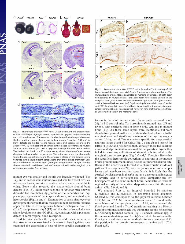
mutant eye was smaller and the iris was irregularly shaped (Fig.1a), and in sections the mutant eyes had smaller vitreal cavities,misshapen lenses, anterior chamber defects, and corneal thick-ening. Bone stains revealed the characteristic frontal bonedefects (Fig. 1b). Adult brain sections in hith/hith mice showeddramatic hydrocephalus, dysgenesis of the neocortex and hip-pocampus, agenesis of the corpus callosum, and marginal zoneheterotopias (Fig. 1 c and d). Examination of brain histology overdevelopment showed that the most prominent dysplastic featuresappeared late in corticogenesis between embryonic day 18.5(E18.5) and postnatal day 7 (P7) and that the hydrocephalus wasa late development after P7 (Fig. 1c), consistent with a postnataldefect in cerebrospinal f luid resorption.
To determine whether the dysgenesis of dorsomedial neocor-tical structures was associated with neuronal layering defects, weexamined the expression of several layer-specific transcription
factors in the adult mutant cortex (as recently reviewed in ref.24). In P10 control mice Tbr1 prominently stained layer 2/3 andlayer 6, with scattered cells in layer 5 (Fig. 2a), and in mutantbrain (Fig. 2b) these same layers were identifiable but wereclearly disorganized, with areas of stained cells displaced into themarginal zone and significant waviness of the layering organi-zation. Using two different markers specific for deep corticalneurons [layers 5 and 6 for Ctip2 (Fig. 2 c and d) and layer 5 forER81 (Fig. 2 e and f)] showed that, although these two markersalso revealed prominent waviness of the deep cortical layers, theyfailed to show any collections of stained cells included in themarginal zone heterotopias (Fig. 2 d and f). Thus, it is likely thatthe superficial heterotopic collections of neurons in the mutantbrains predominantly contained neurons of superficial layer fate.Because the neocortex is formed in a characteristic inside-outpattern of neurogenesis (24), with early-born neurons in deeperlayers and later-born neurons superficially, it is likely that thecortical dysplasia seen in the hith mutants develops and increasesin severity later in corticogenesis. Also, comparing the twohemispheres of the same mutant brain demonstrated variableregional severity of the cortical dysplasia even within the sameanimal (Fig. 2 b, d, and f).
We mapped hith to an interval bounded by markersD13Mit135 and D13Mit275. In Ensembl’s latest assembly,NCBIM36, this corresponds to the physical interval between21.93 Mb and 37.33 Mb on mouse chromosome 13. Based on theresemblance of the eye phenotype to ARS, we sequenced theFoxc1 gene and found a T-to-C transition resulting in a Phe-to-Leu substitution at amino acid 107 within the second helix of theDNA binding forkhead domain (Fig. 3 a and b). Interestingly, inthe mouse mutant dysgenetic lens (dyl), a T-to-C transition in theFoxe3 gene results in an amino acid substitution at amino acid 93,the position in the protein homologous to amino acid 107 inFoxc1 (25).
Fig. 1. Phenotype of Foxc1hith/hith mice. (a) Whole mount and cross sectionsof Foxc1hith/hith eyes highlight the micropthalmalia, dysgenic iris (white arrow),and thickened cornea. The anterior chamber is also lost (the space betweenthe lens and the cornea, black arrow) in the mutants. (Scale bars: 500 �m.) (b)Bony defects are limited to the frontal bone and sagittal suture in theFoxc1hith/hith. (c) Hemisections of cortex at three ages in control and mutantanimals shows that major cortical dysplasia develops between E18.5 and P7.The dashed red line in the P7 mutant cortex shows the area of most severedysplasia in dorsomedial cortical areas. The red arrows show the abnormallyformed hippocampal layers, and the asterisk is placed in the dilated lateralventricle in the adult mutant cortex. Note that there is not prominent ven-tricular dilatation at earlier ages. (d) Higher-magnification images showingthree examples from different brains of heterotopic cells in the marginal zoneof mutant neocortex (red arrows).
Fig. 2. Dyslamination in Foxc1hith/hith mice. (a and b) Tbr1 staining of P10brains shows labeling of layers 2/3, 5, and 6 in control and mutant brains. Themutant brains are montages generated by merging two images of both brainhemispheres. In mutant brains Tbr1� cells form heterotopic clusters in themarginal zone (blue arrows in Insets) and reveal significant disorganization ofcortical layers (black arrows). (c–f) Ctip2 staining labels cells in layers 5 and 6,and ER81 labels cells in layer 5, and both show significant laminar disorgani-zation in mutant brains (black arrows); however, note that there are no Ctip2-or ER81-stained cells in the marginal zone.
Zarbalis et al. PNAS � August 28, 2007 � vol. 104 � no. 35 � 14003
DEV
ELO
PMEN
TAL
BIO
LOG
Y

Transcriptional assays using a vector with four repeats of theFoxc1 binding motif upstream of a minimal promoter and aluciferase reporter gene showed that F107L-Foxc1 had essentiallynormal transcriptional activity compared with wild-type Foxc1whereas truncated Foxc1 without the transcriptional activationdomain showed no transcriptional activity (Fig. 3c). Because ofsuggestions that amino acid 107 is important in regulating thetertiary structure of the forkhead domain (26), we examined thepossibility that the mutant protein was unstable. Indeed, proteinstability experiments in transfected cells showed that F107L-Foxc1protein was less stable than Foxc1 (n � 6; P � 0.004) (Fig. 3d). Itis likely that the dramatic overexpression of Foxc1 in our transfec-tion assays is the reason for the apparently normal transactivationby mutant protein in our transcriptional assays. Stability of Foxc1has been linked to the C-terminal phosphorylation of the protein(27), and evaluation of the phosphorylation state of F107L-Foxc1protein revealed reduced phosphorylation compared with wild-type controls (Fig. 3e).
Because the analysis in transfected cells relies on heterologousexpression of nonphysiologic levels of Foxc1, we examined Foxc1expression in situ as well. Foxc1 mRNA expression was normal inthe mutants and, as in the controls and consistent with previousreports (6, 7, 28), was detected in the meninges surrounding thebrain and in the head mesenchyme (Fig. 4 a and b). Immunohis-tochemistry for Foxc1 showed nuclear expression in the meningesand head mensenchyme in unaffected embryos (n � 8) (Fig. 4 c ande). However, in similar areas in mutants, the Foxc1 protein level wasdramatically reduced (n � 7) (Fig. 4 d and f). This is consistent withthe idea that the F107L mutation in Foxc1 leads to destabilizationof the protein and that mice homozygous for this mutation haveintermediate levels of Foxc1 function between those of the previ-ously described Foxc1�/� and Foxc1�/� mice (6). Consistent withthis, crosses of Foxc1hith/� mice with Foxc1�/� mice yielded mutantembryos, further establishing that the Foxc1 mutation that we haveidentified is the pathogenic mutation (data not shown).
Fig. 3. Point mutation in the Foxc1 hith allele does not affect transcription butresults in destabilization of the protein in vitro. (a) Sequence histograms depict-ing amino acids 105–109 in the wild-type (WT) and hith mutant (MUT) Foxc1gene. The asterisk denotes the T-to-C point mutation in amino acid 107 thatresults in phenylalanine being replaced by leucine. (b) The forkhead domain ofFoxc1 is composed of three �-helices, two �-sheets, and two loops that form a‘‘wing-like’’ formation. The mutation in the hith allele is located in the second�-helix (asterisk). (c) Transcriptional activity of the mutant protein was tested viacotransfection of a control construct (CMV7-FLAG), wild-type Foxc1-containingvector (FLAG-FOXC1), mutant Foxc1 (FLAG-Foxc1F107L), or a transcriptionally in-active truncation mutant of Foxc1 (FLAG-FOXC1144) and a luciferase reporterconstructwith(4xFoxc1bp-luc)orwithout(G5-luc)FOXC1bindingsites.Wild-typeFoxc1 protein and mutated Foxc1 had similar transcriptional activity when co-transfected with the Foxc1 luciferase reporter, whereas the truncation mutant ofFoxc1showednospecifictranscriptionalactivity. (d)Threehoursafterapplicationof the protein translation inhibitor CHX (20 mg/ml) to 293 cells transfected withFLAG-tagged wild-type or mutant Foxc1, the mutant Foxc1 protein levels wereclearly reduced. This effect was reversed with application of a proteasome inhib-itor, MG132 (10 mM), treated 1 h before CHX. A time course of CHX treatmentshows a marked decrease in Foxc1 mutant protein by 3 h and further reductionby 6 h after addition of CHX. Results from six independent experiments wereplotted (error bars indicate SD). (e) Separation of FLAG-Foxc1 and FLAG-Foxc1F107L protein on an acrylamide gel revealed that both were present in arange of MW species, but mutant protein never attained the highest MW seen inthe wild-type protein. Treatment of lysates with the phosphatase �-PPase indi-cated that this isoform complexity was due to differences in phosphorylationstatus of the protein.
Fig. 4. Foxc1 expression in vivo at E14.5. (a and b) In both Foxc1�/ andFoxc1hith/hith heads, ventral head structures and the meninges (MN) surround-ing both the cortex and the midbrain (MB) express Foxc1 mRNA. The Foxc1signal intensity is similar in control and mutant mice. (c and d) Foxc1 proteinis expressed intensely by mesenchymal cells and meningeal cells ventral to theforebrain in Foxc1�/ tissue sections. Expression of Foxc1 is not seen in neuraltissue (except in pericytes associated with blood vessels; data not shown).Foxc1 protein expression in Foxc1hith/hith tissue is similarly distributed, but thestaining intensity is markedly reduced. (e and f ) Foxc1� cells are distributedthroughout the primitive meninges in control mice, but in Foxc1hith/hith me-ninges expression of Foxc1 in these cells is less. (Scale bars: 1 mm in a and b, 100�m in c and d, and 50 �m in e and f.)
14004 � www.pnas.org�cgi�doi�10.1073�pnas.0702618104 Zarbalis et al.

The marginal zone heterotopias and dyslamination seen in thehith mice indicate potential defects in the meningeal-derived pialBM and the interaction of the radial glial cells that serve as aguide for cortical migration and the pial surface where theynormally attach. To test this, we examined the continuity of theBM at E14.5, E18.5, and P7 by examining the distribution oflaminin and Foxc1. At E14.5 BM integrity was intact (n � 8)(Fig. 5 a and b), but by E18.5 the BM of Foxc1hith/hith brains wasless well organized and significant breaches in the BM wereevident at the pial surface (n � 3) (Fig. 5 c and d). Soon afterbirth, areas of BM disruption were associated with heterotopicneurons (Fig. 5 e and f ), indicating that disruption in the BMleads to the disorganization of cortical neurons in the marginalzone. When we examined the distribution of Foxc1� cells inrelationship to the BM in the medial cortex there were dramaticdifferences between mutant and wild-type animals. In theFoxc1�/ brain strongly labeled Foxc1� cells were distributedwidely and in intimate association with the laminin-expressingBM and with the mesenchymal tissue at the dorsal midline of thehead, whereas in brains of Foxc1hith/hith mice there were fewFoxc1� cells in this area at any age (Fig. 5 a–f ).
Beyond providing a physical barrier to migrating neuronsduring brain development, the pial BM serves as a criticalattachment point for radial glial endfeet whose fibers span thewidth of the growing cerebral wall, providing a migratoryscaffold for neuronal migration (29). At E14.5, before the loss ofBM integrity, radial glial endfeet could be seen properly attach-ing to the pial laminin� BM in both control and hith mice (Fig.5 g and h). However, by E18.5, whereas in the Foxc1�/ brainsradial glial endfeet still projected all of the way to the laminin�
BM (Fig. 5i), the mutants had disorganized radial glia withdetached endfeet in areas of BM disruption (Fig. 5h). It is likelythat these radial glial defects are associated with the dyslami-nation and neuronal heterotopias seen in the mutant brains.
Foxc1 is strongly expressed in the meninges from very early indevelopment, and Foxc1-null mice demonstrate correspondinglyearly defects in meningeal development (6). In contrast, as wehave shown, hith mice have intact early stages of pial BMdevelopment with later breakdown. Importantly, the pial me-ninges are one of the three meningeal layers that becomedistinguishable in the last few days of gestation (17). At E18.5 inFoxc1�/ mice, the three distinct meningeal layers were estab-lished in the space between ossified calvarium and cortical tissueand Foxc1� cells occupied all three layers [supporting informa-tion (SI) Fig. 6 a and b]. In contrast, the Foxc1hith/hith meningeshad defects most apparent at the edge of the holes in thecalvarium. In areas of bone formation of hith/hith mutants,weakly Foxc1� cells populated all layers of the meninges whereasonly scattered pial Foxc1� cells were found where bone hadfailed to form (SI Fig. 6c). To further clarify the defects in theseareas, we examined the expression of Foxc2, a close homolog ofFoxc1 also reportedly expressed in the meninges (28, 30).Interestingly, in our higher-resolution analysis we found that onlydural cells strongly expressed Foxc2 (SI Fig. 6e), indicating that,whereas Foxc1 is expressed within all layers of meninges, Foxc2is a dura-specific marker. In mutants, the Foxc2� dura was intactbeneath areas of bone growth but no dura formed within thebone holes (SI Fig. 6f). Because the dura contains signalsrequired to induce ossification, the lack of dura is likely to becritical in the failure to form a continuous skull. In addition, itis likely that the failure to produce differentiated three-layeredmeninges, including the arachnoid, is responsible for the post-natal development of hydrocephalus due to defects in cerebro-spinal f luid resorption.
DiscussionHere we describe the spectrum of phenotypes in mice homozy-gous for a Foxc1 mutation (hith) resulting in reduced Foxc1
protein stability. Mutants have reduced Foxc1 activity sufficientfor early developmental events to proceed normally, uncoveringpreviously unrecognized roles in the maintenance or late devel-opment of the meninges. Thus, our mutant mice have features
Fig. 5. Progressive breakdown of midline BM and development of corticaldysplasia in Foxc1hith/hith animals. (a and b) Laminin (red) and Foxc1 (green)expression at the cortical midline in Foxc1�/ tissue at E14.5 shows Foxc1� cellsclosely associated with the laminin BM (white arrows) and around laminin�
blood vessels (blue arrow). At this early age, the laminin BM is intact inFoxc1hith/hith brains and weakly Foxc1� cells are associated with both themeninges and blood vessels at the midline. (c and d) At E18.5, a continuous BMsurrounds the cortical tissue and Foxc1� cells are embedded within the pialBM (white arrows). In the Foxc1hith/hith brain, breeches in the laminin BM areapparent at E18.5 (blue arrows), and, although some weakly Foxc1� cells areobserved within in the interhemispheric region, these cells are largely absentin the areas in and around the laminin breeches. (e and f ) At P7, severe breaksin the laminin BM are accompanied by infiltrating MAP2� neural tissue anddisorganized laminin deposits within the heterotopias. (g–j) Organized radialglial endfeet (green) overlap with the laminin� BM at the pial surface inFoxc1�/ brain as an essentially unbroken layer of staining where the endfeetattach to the BM in control and mutants at E14.5 but only in controls at E18.5.In contrast, in the Foxc1hith/hith mice disorganized laminin is associated withdetached radial glial endfeet (white arrows) and gaps where no nestin-labeled endfeet are attached to the BM (asterisks) at E18.5. (Scale bars: 500 �min a and b, 100 �m in c, d, and g–j, and 200 �m in e and f.)
Zarbalis et al. PNAS � August 28, 2007 � vol. 104 � no. 35 � 14005
DEV
ELO
PMEN
TAL
BIO
LOG
Y
in common with the engineered Foxc1-null mice and the spon-taneously occurring congenital hydrocephalus mouse line [alsopredicted to be a null allele (6)], but with some importantdifferences. Unlike hith mice, the Foxc1�/� mice completely lackskull ossification and produce only a primitive meningeal mes-enchyme (6). Consequently, these mice have earlier and moresevere defects incompatible with postnatal life or with detailedanalysis of cortical development. In the viable Foxc1hith/hith micewe were able to characterize brain malformations first appearinglate in gestation and progressing postnatally. Importantly, ARSpatients with Foxc1 mutations are generally heterozygous forpredicted null mutations and thus are genetically equivalent toFoxc1�/� mice, and in fact a mild version of anterior chamberdysgenesis is seen in Foxc1�/� mice (31). Importantly, corticaldysplasia has not been reported in typical ARS patients and isnot reported in Foxc1�/� mice; however, ARS patients withautosomal recessive inheritance have mental retardation andmeningeal calcifications (12), and this is reminiscent of thephenotype of the Foxc1hith/hith mice. Thus, in mice the completeloss of Foxc1 leads to a severe early developmental, lethalphenotype, half dosage leads predominantly to anterior segmentdysgenesis, and an intermediate between these (the Foxc1hith/hith
mice) leads to a viable but severe malformation syndrome in allof the differentiated derivatives of the meningeal mesenchyme,and it is likely that similar human alleles will be found and willexpand the spectrum of ARS to include patients with corticaldysplasia.
The cortical dysgenesis and BM defects seen in Foxc1hith/hith
animals are quite distinct but have some similarities to defectsfound in the congenital muscular dystrophies. In the brains ofpatients with these syndromes (or in engineered mouse modelsof these diseases), deletion of structural elements of the BM(including laminin subtypes, perlecan, or their cellular receptorsdystroglycan, �1, or �6 integrin) or downstream signaling mol-ecules leads to cortical ectopias, detachment of radial glial fibers,and overall laminar disorganization (32–35), reminiscent of, butmore severe than, what we have observed here. Our data areconsistent with the idea that reduced expression of Foxc1 proteinin meningeal cells disrupts the ability of the meninges to produceand maintain the BM as the cortex expands during embryonicand early postnatal development, leading to a later degradationof BM function and causing a distinct later-appearing corticaldysplasia phenotype. This is a mechanism for the production ofthis cortical phenotype because it appears that meningeal de-velopment is sufficiently intact to proceed during early corticaldevelopment [unlike the situations where structural BM proteinsare mutated and the pial BM is presumably always of low qualityfrom earlier in development (32)] but later fails. It is possible thatFoxc1 plays an important role in the extensive remodeling thatmust occur to maintain a well ordered BM able to cover thegrowing cortex in late gestation and early postnatal life. Inaddition, there is currently no literature available addressingwhether there are subgroups of patients with cortical dysplasiapresenting at the stage of cortical expansion (analogous to earlypostnatal life in rodents) rather than at earlier stages when radialmigration is first ongoing. However, the large numbers ofepilepsy patients who present with more limited forms of corticaldysplasia sharing some features with cobblestone lissencephaly(as our mice do) make this quite likely (36).
Materials and MethodsPositional Mapping. The hith mutation is fully penetrant, and bothmale and female homozygous mutants can survive until adult-hood and are fertile. The polymorphic markers on chromosome13 used for fine-scale mapping of hith are from proximal to distalas follows: D13Mit218, D13Mit16, D13Mit17, D13Mit135,D13Mit275, D13Mit117, D13Mit177, D13Mit244, andD13Mit91. Meiotic mapping results support this order of mark-
ers that corresponds to the one displayed in Ensembl. The hithmutation recombined away from D13Mit135 at least 34 timesand recombined away from D13Mit275 at least 27 times. In allcases where one of these two markers segregated from hith theother associated with the mutation. We sequenced all exons(including the UTRs) of six candidate genes within the intervaland discovered no other polymorphisms besides the causativemutation in Foxc1. The sequenced candidate genes are asfollows: Foxc1 (EntrezGene ID code 17300), Mboat1 (Entrez-Gene ID code 218121), Dusp22 (EntrezGene ID code 105352),Exoc2 (EntrezGene ID code 66482), Gmds (EntrezGene IDcode 218138), and Cdkal (EntrezGene ID code 68916). Sequenc-ing of candidate genes was performed at the Ernest Gallo Clinicand Research Center core sequencing facility using AppliedBiosystems (Foster City, CA) Big Dye (3.1) reactions. Sequenc-ing reactions were electrophoresed and analyzed in an AppliedBiosystems Genetic Analyzer 3730XL.
Animal Husbandry and Genotyping. Mice are housed in specificpathogen-free facilities approved by the American Associationof Laboratory Animal Care. All animals were handled inaccordance with protocols approved by the University of Cali-fornia, San Francisco, Committee on Animal Research. Thecolony of animals carrying the Foxc1hith allele (induced onC57BL/6 background) is maintained by crossing male carrierswith FVB females. This mode of outcross is currently in thefourth generation without any changes in penetrance or vari-ability of the mutant phenotype. Heterozygote animals weredetermined by genotyping with markers D13Mit135 andD13Mit275. Once Foxc1 had been identified as the mutated geneprimers for a polymorphic marker closely linked to the mutation(�12.8 kb) were devised, and genotyping was based on using thismarker. Primer sequences are as follows: D10Egc3 left, ACCT-TGTCCTAGTGACTCCGATGG; D10Egc3 right, TTACAG-CAGTTCTCCTGACAACTTAATATCTAAG. All phenotyp-ically mutant animals were genotyped by using this marker andwere positive for the C57BL/6 allele consistent with the geneticbackground on which the mutation had been caused.
Transcriptional and Protein Stability Assays. 293 cells were seededonto polyD-lysine-coated 12-well culture plates 1 day beforetransfection. Cells were transfected with 1 �g of FLAG-FOXC1or CMV7-FLAG control construct, 1 �g of luciferase reporter,and 0.7 �g of Renilla luciferase reporter vector with 3 ml ofLipofectamine 2000 for dual luciferase assay using a detection kit(Promega, Madison, WI).
For the protein stability assays, cells grown on a 10-cm dishwere transfected with FOXC1 constructs. One day after trans-fection, cells were divided onto polyD-lysine-coated 12-wellculture plates. MG132 (10 mM) was added 1 h before cyclohex-imide (CHX) treatment. Cells were incubated with CHX (20mg/ml) for 0 (DMSO vehicle only), 0.5, 1, 3, and 6 h andsubjected to Western blotting. Signals from Western blot analysiswere analyzed with ImageQuant TL software (GE Healthcare,Fairfield, CT) and plotted using data from six independentexperiments.
Immunohistochemistry. Heads from E14.5 fetuses and P0 eyes werecollected and fixed overnight in 4% paraformaldehyde, cryopro-tected by using graduated sucrose concentrations (15% and 30%),and then frozen in Tissue-Tek OCT Compound (Sakura Fineteld,Torrance, CA). E18.5, P0, and P7 heads were fresh-frozen in OCT.All tissue was cryosectioned in 10-�m increments. Before antibodyapplication, sections were steamed in 0.01 M citric acid buffer (pH6.0) for 15 min then cooled in 0.1 M PBS for 15 min at roomtemperature. Sections were blocked in 1.0% BSA and 0.75% TritonX-100 for 45 min and then incubated in either goat anti-Foxc1(1:300; Novus Biologicals, Littleton, CO) or goat anti-Foxc2 (1:300;
14006 � www.pnas.org�cgi�doi�10.1073�pnas.0702618104 Zarbalis et al.
Novus Biologicals) for 1 h. After washing in PBS, horseradishperoxidase-conjugated anti-goat secondary antibody (1:100; VectorLaboratories, Burlingame, CA) was applied for 1 h followed by ashort incubation in Alexa Fluor 488-linked tyramide (1:100; In-vitrogen, Carlsbad, CA). For double or triple immunolabeling,Foxc1-labeled sections were incubated in rabbit anti-laminin (1:75;Sigma, St. Louis, MO), mouse anti-nestin (1:500; Chemicon, Te-mecula, CA), and mouse anti-MAP2 (1:300; Chemicon) followedby incubation in Alexa Fluor 594 anti-rabbit secondary and/orAlexa Fluor 633 anti-mouse secondary antibodies (Invitrogen).
Statistical Analysis. A one-way ANOVA was performed on den-sitometry data collected from the protein stability experimentsusing SigmaStat software. To identify statistically significantdifferences between wild-type and Foxc1 mutant values atspecific time points, an all pairwise multiple comparison proce-dure (Student–Newman–Keuls method) was performed.
Phosphorylation Experiment. Removal of phosphates of FLAG-FOXC1 with �-protein phosphatase (NEB, Ipswich, MA) was
done with 5 �g of cell extract obtained from 293 cells transfectedwith FOXC1 expression constructs. Cell extracts were incubatedwith 400 units of � protein phosphatase at 30°C for 30 min. Thereaction was stopped with SDS gel loading buffer.
In Situ Hybridization. Foxc1 in situ hybridization was performed aspreviously described (37) by using an 806-bp 35S-UTP-labeledriboprobe. The riboprobe corresponds to base pairs 2192–2997on the Foxc1 mRNA sequence (GenBank accession no.NM�008592).
We thank Margaret Robertson and Raymond Chui at the Ernest GalloClinic and Research Center core sequencing facility for technicalassistance in the sequencing of candidate genes. We also thank Drs.Viktor Kharazia and Holger Russig for contributions and assistance inthe histological analysis of mutant mice and Drs. Douglas Gould, JeremyReiter, and John Rubenstein for helpful discussions. This work wassupported by funding from the National Institute on Drug Abuse,Parents Against Childhood Epilepsy, the National Institute of MentalHealth, and Autism Speaks.
1. Kume T, Deng K, Hogan BL (2000) Development (Cambridge, UK) 127:1387–1395.2. Kume T, Jiang H, Topczewska JM, Hogan BL (2001) Genes Dev 15:2470–2482.3. Seo S, Fujita H, Nakano A, Kang M, Duarte A, Kume T (2006) Dev Biol
294:458–470.4. Wilm B, James RG, Schultheiss TM, Hogan BL (2004) Dev Biol 271:176–189.5. Rice R, Rice DP, Thesleff I (2005) Dev Dyn 233:847–852.6. Kume T, Deng KY, Winfrey V, Gould DB, Walter MA, Hogan BL (1998) Cell
93:985–996.7. Mears AJ, Jordan T, Mirzayans F, Dubois S, Kume T, Parlee M, Ritch R, Koop
B, Kuo WL, Collins C, et al. (1998) Am J Hum Genet 63:1316–1328.8. Nishimura DY, Swiderski RE, Alward WL, Searby CC, Patil SR, Bennet SR,
Kanis AB, Gastier JM, Stone EM, Sheffield VC (1998) Nat Genet 19:140–147.9. Nishimura DY, Searby CC, Alward WL, Walton D, Craig JE, Mackey DA,
Kawase K, Kanis AB, Patil SR, Stone EM, et al. (2001) Am J Hum Genet68:364–372.
10. Panicker SG, Sampath S, Mandal AK, Reddy AB, Ahmed N, Hasnain SE(2002) Invest Ophthalmol Visual Sci 43:3613–3616.
11. Honkanen RA, Nishimura DY, Swiderski RE, Bennett SR, Hong S, Kwon YH,Stone EM, Sheffield VC, Alward WL (2003) Am J Ophthalmol 135:368–375.
12. Moog U, Bleeker-Wagemakers EM, Crobach P, Vles JS, Schrander-StumpelCT (1998) Am J Med Genet 78:263–266.
13. Caluseriu O, Mirza G, Ragoussis J, Chow EW, MacCrimmon D, Bassett AS(2006) Am J Med Genet A 140:1208–1213.
14. Le Caignec C, De Mas P, Vincent MC, Boceno M, Bourrouillou G, Rival JM,David A (2005) Am J Med Genet A 132:175–180.
15. Maclean K, Smith J, St Heaps L, Chia N, Williams R, Peters GB, Onikul E,McCrossin T, Lehmann OJ, Ades LC (2005) Am J Med Genet A 132:381–385.
16. Etchevers HC, Vincent C, Le Douarin NM, Couly GF (2001) Development(Cambridge, UK) 128:1059–1068.
17. Jiang X, Iseki S, Maxson RE, Sucov HM, Morriss-Kay GM (2002) Dev Biol241:106–116.
18. McLone DG, Bondareff W (1975) Am J Anat 142:273–293.19. Bauer HC, Bauer H, Lametschwandtner A, Amberger A, Ruiz P, Steiner M
(1993) Brain Res 75:269–278.20. Sievers J, Pehlemann FW, Gude S, Berry M (1994) J Neurocytol 23:135–149.21. Davson H, Domer FR, Hollingsworth JR (1973) Brain 96:329–336.22. Ito Y, Yeo JY, Chytil A, Han J, Bringas P, Jr, Nakajima A, Shuler CF, Moses
HL, Chai Y (2003) Development (Cambridge, UK) 130:5269–5280.23. Zarbalis K, May SR, Shen Y, Ekker M, Rubenstein JL, Peterson AS (2004)
PLoS Biol 2:E219.24. Hevner RF, Daza RA, Rubenstein JL, Stunnenberg H, Olavarria JF, Englund
C (2003) Dev Neurosci 25:139–151.25. Blixt A, Mahlapuu M, Aitola M, Pelto-Huikko M, Enerback S, Carlsson P
(2000) Genes Dev 14:245–254.26. Saleem RA, Banerjee-Basu S, Berry FB, Baxevanis AD, Walter MA (2001)
Am J Hum Genet 68:627–641.27. Berry FB, Mirzayans F, Walter MA (2006) J Biol Chem 281:10098–10104.28. Sasaki H, Hogan BL (1993) Development (Cambridge, UK) 118:47–59.29. Marin O, Rubenstein JL (2003) Annu Rev Neurosci 26:441–483.30. Rice R, Rice DP, Olsen BR, Thesleff I (2003) Dev Biol 262:75–87.31. Smith RS, Zabaleta A, Kume T, Savinova OV, Kidson SH, Martin JE,
Nishimura DY, Alward WL, Hogan BL, John SW (2000) Hum Mol Genet9:1021–1032.
32. Hu H, Yang Y, Eade A, Xiong Y, Qi Y (2007) J Comp Neurol 501:168–183.33. Halfter W, Dong S, Yip YP, Willem M, Mayer U (2002) J Neurosci 22:6029–
6040.34. Olson EC, Walsh CA (2002) Curr Opin Genet Dev 12:320–327.35. Beggs HE, Schahin-Reed D, Zang K, Goebbels S, Nave KA, Gorski J, Jones
KR, Sretavan D, Reichardt LF (2003) Neuron 40:501–514.36. Rickert CH (2006) Childs Nerv Syst 22:821–826.37. Zarbalis K, Wurst W (2000) Mech Dev 93:165–168.
Zarbalis et al. PNAS � August 28, 2007 � vol. 104 � no. 35 � 14007
DEV
ELO
PMEN
TAL
BIO
LOG
Y