Embed Size (px)
Citation preview
Cortical and subcortical mechanisms
in persistent stuttering
Dissertation
for the award of the degree
„Doctor rerum naturalium“
Division of Mathematics and Natural Sciences
of the Georg-August-Universität Göttingen
submitted by
Nicole Neef
from Karl-Marx-Stadt
Göttingen 2010
Doctoral Thesis Committee:
PD Dr. med. Martin Sommer (Supervisor, Reviewer)
Abteilung Klinische Neurophysiologie
Universitätsmedizin Göttingen
Robert-Koch-Straße 40
37075 Göttingen
Prof. Dr. rer. nat. Julia Fischer (Reviewer)
Deutsches Primatenzentrum
Abteilung Kognitive Ethologie
Kellnerweg 4
37077 Göttingen
Prof. Dr. med.Walter Paulus
Abteilung Klinische Neurophysiologie
Universitätsmedizin Göttingen
Robert-Koch-Straße 40
37075 Göttingen
Prof. Dr. phil. Marcus Hasselhorn
Georg-August-Universität Göttingen
Abt. 4: Pädagogische Psychologie und Entwicklungspsychologie
37075 Göttingen
Date of thesis submission: 30th November 2010
Date of the oral examination: 10th January 2011

Statement of Originality I hereby declare that this thesis is my own work and has been written independently, with no
other sources and aids than quoted in the text, references and acknowledgements.
Göttingen, 30th November 2010
Nicole Neef

Content
Abbreviations .......................................................................................................................... 7
1 Introduction ............................................................................................................................ 9
1.1 Components of the process of fluent articulation ...................................................... 10
1.2 What is stuttering? ..................................................................................................... 12
1.3 Subtypes of stuttering ................................................................................................ 13
1.3.1 Acquired stuttering ....................................................................................................... 13
1.3.2 Psychogenic stuttering .................................................................................................. 13
1.3.3 Persistent stuttering ...................................................................................................... 13
1.4 Approaches to explain stuttering ............................................................................... 15
1.4.1 Psycholinguistic approach ............................................................................................ 15
1.4.2 Motor deficit perspective .............................................................................................. 16
1.5 Neurophysiological approaches to explain stuttering ................................................ 17
1.5.1 The cerebral dominance hypothesis ............................................................................. 18
1.5.2 The basal ganglia hypothesis ........................................................................................ 21
1.5.3 The disconnection hypothesis ....................................................................................... 24
1.6 Scope of the dissertation ............................................................................................ 27
2 Original Articles ................................................................................................................... 29
2.1 Right-shift for non-speech motor processing in adults who stutter ................................ 31
2.2 Reduced intracortical inhibition and facilitation in the primary motor tongue
representation of adults who stutter ...................................................................................... 61
2.3 Instable phoneme categorization in adults who stutter ................................................... 95
3 Summary ............................................................................................................................ 123
4 Conclusions and Future Prospects ...................................................................................... 125
4.1 The TMS approach ....................................................................................................... 125
4.2 The speech perception approach ................................................................................... 126
4.3 Future directions ........................................................................................................... 129
Appendix A – Levelt’s psycholinguistic model and the DIVA model .................................. 131
5

6
Appendix B - Stuttering and acquired brain lesions .............................................................. 137
Appendix C - Psycholinguistic approaches to explain stuttering ........................................... 143
Appendix D - Transcranial magnetic stimulation .................................................................. 145
Literatur .................................................................................................................................. 147
Acknowledgements ................................................................................................................ 161

Abbreviations
AADC Aromatic L-amino acid decarboxylase
AI ambivalence interval
AMT active motor threshold
ANOVA analysis of variance
AWNS adults who do not stutter
AWS adults who stutter
CS conditioning stimulus
CV consonant-vowel
CVS consonant-vowel-syllable-continua
DBS deep brain stimulation
DIVA directions into velocities of articulators model
DTI diffusion tensor imaging
EEG electroencephalography
EMG electromyogram
FA fractional anisotropy
FDI first dorsal interosseous
fMRI functional magnetic resonance imaging
FS fluent speaker
GABA γ-Aminobutyric acid
ICCunjust unjust intra-class correlation coefficient
ICD International Classification of Functioning, Disability and Health
ICF intracortical facilitation
IFG inferior frontal gyrus
ISI inter-stimulus interval
ITI inter-tap interval
M1 primary motor cortex
MEG magnetencephalography
MEP motor evoked potential
MMN mismatch negativity
MRI magnetic resonance imaging
MT motor threshold
NMDA N-methyl-D-aspartate
PB phoneme boundary
PET positron emissions tomography
7

8
PM premotor cortex
PMd dorsolateral premotor cortex
PMv ventral premotor cortex
rTMS repetitive transcranial magnetic stimulation
SD standard deviation
SEM standard error of mean
SICI short-term intracortical inhibition
SMA supplementary motor area
SSI-3 stuttering severity index 3th edition
STG superior temporal gyrus
TMS transcranial magnetic stimulation
TS test stimulus
VOT voice onset time
WHO world health organization
Chapter 1 Introduction
1 Introduction
Stuttering is a speech disorder that occurs without known origin between 3 and 8 years of age
and often remits before puberty. When it persists after puberty it becomes a chronic adult
speech disorder throughout the lifespan (Andrews et al., 1983). The advances in neuroimaging
promoted insights into the highly distributed system of speech production and its alterations
in adulthood stuttering. One important motivation in stuttering research is to separate
neurobiological correlates of the core symptoms of stuttering from neurobiological correlates
associated with compensation, attempts to avoid stuttering. Results so far indicate a variety of
complex dysfunctional systems and it appeared problematic to distinguish between
mechanisms responsible for speech dysfluencies and those connected to compensation in the
adult system (Ludlow, 2000). The first study of this dissertation will tie in with this problem.
Another important aspect is again motivated by neuroimaging studies that stress irregularities
in the activation of the primary motor cortex during different functional states associated with
speech production. The regulation of blood supply which is the basis of functional magnetic
resonance imaging (fMRI) is correlated with summed neuronal activity but conclusions about
states of cortical excitability and thus neurophysiological mechanisms are indirect. The
modulation of cortical excitability is an inherent principle in the encoding of output signal by
the primary motor cortex and the method of choice to non-invasively study this in humans is
transcranial magnetic stimulation (TMS). This method is well established in Professor Paulus’
Department of Clinical Neurophysiology, for instance to investigate neuromodulation in the
primary motor cortex representation of small hand muscles. One aim of the dissertation was to
establish recordings of TMS induced motor evoked potentials (MEPs) from facial muscles.
This enabled me to conduct the first study of the intracortical excitability of the primary
tongue motor representation in adults who stutter.
A seminal work motivated the third study included in this dissertation: diffusion tensor
imaging (DTI) identified reduced white matter integrity in fibres connecting frontal, temporal
and parietal speech related areas (Sommer et al., 2002a). Affected are possibly fibres
mediating the mapping of speech sounds to articulation. The interaction of speech production
and speech perception and the interference of stuttering with this interaction is the topic of the
third study reported in this dissertation.
9
During the course of my doctoral studies I furthermore contributed to a longitudinal DTI
study aiming at identifying a biological marker of stuttering persistency at stuttering onset. In
10
addition I conducted an fMRI study of the neuroanatomical correlates of continuous
performance in stuttering in cooperation with the Biomedical NMR Research GmbH at the
Max Planck Institute for Biophysical Chemistry. An internship at the speech Laboratory of
Purdue University allowed me to study the consolidation of speech motor learning in children
who stutter by using the Optotrak, a system that delivers a 3D tracking and measuring of
speech kinematics. These ongoing studies are not included here.
As the drafts of the studies themselves give only limited space to introduction of the topic, a
more detailed introduction is given in sections 1.1 to 1.5.
1.1 Components of the process of fluent articulation
Stuttering is a disorder with intermittent interruptions of fluent articulation. Fluent articulation
is one of human‘s most complex motor skills. It comprises the coordinated use of
approximately 100 muscles (Ackermann, 2008) and it is fascinating how effortless this skill is
managed by almost every human being. Rapid, complex movements are essential to articulate
the sounds of speech. Here I briefly summarize the structures involved in articulation, which
is the ultimate readout of language planning and speech motor control processes.
Subsequently, I refer to influential theories on language planning and speech motor control
because all these aspects are implied in different approaches to explain stuttering.
Articulation involves three anatomically distinct subsystems: the respiratory, laryngeal and
supralaryngeal system. The respiratory system regulates the outflow of air during speech and
thus provides the energy for the acoustic targets of speech. The core structures of the
laryngeal system are the vocal folds, controlling voicing and loudness of speech. During
voicing the oscillation of the vowel folds generates the fundamental frequency on which
resonation builds. The larynx provides the quasiperiodic and tone-like sound fundamental for
vowels and voiced consonants (e.g. [b], [z] and [m]). The supralaryngeal system contains the
pharyngeal, oral and nasal cavities whose architecture and configuration shape the timbre and
the sound of the generated acoustic signal. The supralaryngeal system also called the vocal
tract can be constricted at different places for example via lip closure, lip protrusion, tongue
tip or body elevation or retraction, and velum elevation. Characteristic sound features of
speech vowels are generated by overlapping vocal tract actions such as jaw lowering, tongue
body elevation, and lip protrusion. In contrast, the striking acoustic features of consonants are
generated by the magnitude of obstruction, resulting in bursts due to closure and friction-like
noise due to fine-tuned constriction.
Chapter 1 Introduction
The respiratory, laryngeal and supralaryngeal systems recruit distributed neural networks to
channel the muscle activation into organized spatio-temporal speech movement patterns. The
following neural structures are central for this function:
(1) Orofacial and laryngeal sensorimotor cortex and the corticobulbar and the corticospinal
tracts
(2) Premotor cortex, insula, supplementary motor area, and cingulate motor area
(3) Motoneurons in the brainstem (nucleus trigeminus, nucleus facialis, nucleus
glossopharyngeus, nucleus vagus, nucleus accessories, nucleus hypoglossus)
(4) Motoneurons and associated spinal interneurons which control the respiratory
musculature are found distributed across cervical segments (C1-C8) and thoracic
segments (T1-T12) of the spinal cord
(5) Peripheral nerve fibers of the mentioned motoneurons and their neuromuscular junctions
(6) Extrapyramidal tracts, basal ganglia-thalamocortical pathway
(7) Cerebellum with its efferent and afferent fibers, cerebello-thalamocortical pathway
Anatomy and physiology of speech production is comprehensively described by Steven M.
Barlow or Kenneth N. Stevens (Barlow et al., 1999; Stevens, 2000).
In normal conversation, a speaker produces 3 to 5 intelligible syllables per second (Smith,
1992); thus, the nervous system manages to simultaneously control and coordinate the
overlapping articulatory gestures to produce rapidly altering configurations of the multilevel
executing speech organs.
Preceding and simultaneously, a message that is intended to be transferred to a
communication partner has to be created and transformed into the verbal code. This cognitive
process is detailed by Levelt in his influential model of speech production (Levelt, 1989c). A
brief summary of this psycholinguistic model which shaped several theories on stuttering is
given in Appendix A.
Speech motor control is a further aspect that needs to be considered for the complex process
of speech production. A current model of speech motor control is the Directions into
Velocities of Articulators model (DIVA; Golfinopoulos et al., 2010; Guenther, 1994, 1995).
In this model speech motor control is based on a feed forward and a feedback control
subsystem. The feedforward process is supposed to control the execution of speech
movements. Additionally, the feedforward subsystem activates predictive internal models
(efference copies) in the feedback subsystem. These internal models represent the expectation
of the incoming somatosensory and auditory feedback resulting from current speech
11
12
movements which enable a fast detection and correction of articulation. Psycholinguistic
aspects of language generation are not considered in the model. Rather it details the
production process and the link of production perception interaction. By providing a
neuroanatomical framework to understand fluent as well as stuttered (Civier et al., 2010; Max
et al., 2004) speech production it is helpful to consider this model for studies on stuttering.
More details on the DIVA model are given in Appendix A.
1.2 What is stuttering?
Stuttering is an impairment of “Speech that is characterized by frequent repetition or
prolongation of sounds or syllables or words, or by frequent hesitations or pauses that disrupt
the rhythmic flow of speech. It should be classified as a disorder only if its severity is such as
to markedly disturb the fluency”, (“International Classification of Functioning, Disability and
Health” ICD-10 F98.5 A; WHO, 2007a). As a consequence of stuttering, the affected
individual is disabled in performing daily tasks that rely on spoken communication. This
handicaps the individual to maintain a desired occupation or to fulfill economic needs
(Yaruss, 2010).
The core symptoms of stuttering are dysfluencies. These are features in speech production
that can be observed to a different extent in everybody’s speech. The discrimination between
typical dysfluencies and stuttering-like dysfluencies requires their qualitative description.
Typical or so called other dysfluencies include interjections (“mhm”, “yes”), phrase
repetitions (“this is a this is a phrase repetition”), multisyllabic repetitions (“multi
multisyllabic”), revisions (“revi repetition”) that are not perceived as stuttering. As to
stuttering-like dysfluencies consensus exists regarding part word repetition (p-p-p-partword)
and dysrythmic phonation such as unintended audible prolongations of sounds and unintended
momentary cessation of phonation/articulation (block) (Yairi and Ambrose, 1992; Yairi and
Ambrose, 2005). There is, however, an ongoing debate on whether undue tension or struggle
is a criterion to rate a single syllable word repetition (“I-I-I see”) as a stuttering-like
dysfluency or not (Bloodstein and Ratner, 2008; Ward, 2006; Yairi and Ambrose, 2005) and
whether a cut-off value (e.g. 3 % of stuttered syllables) is necessary to label stuttering
(Sandrieser and Schneider, 2008; Ward, 2006). This debate reflects the two opposing views of
stuttering as either a quantitative variation along the continuum of normal speech dysfluency
(continuity hypothesis; Bloodstein, 1970; van Lieshout et al., 2007) or a qualitatively separate
disorder with a distinction between stuttering and normal dysfluency (Johnson, 1959; Yairi
and Ambrose, 2005). In the current studies I determined stuttering presence and severity
Chapter 1 Introduction
according to the German version of the stuttering severity index (Sandrieser and Schneider,
2008) as described in the methods sections of the included studies.
1.3 Subtypes of stuttering
Scientific approaches to explain stuttering are diverse and consequently many different
attempts to classify the disorder exist. These attempts are clearly influenced by the Zeitgeist in
which they emerged. Ehud Yairi wrote an excellent review on these attempts of subtyping
stuttering (Yairi, 2007). A reliable and standardized categorization would obviously be of
great advantage for scientific studies. A current PubMed search clearly indicates that a
separation between acquired [neurogenic] stuttering, psychogenic stuttering, and persistent
[developmental, idiopathic] stuttering is commonly used these days (Lundgren et al., 2010).
Therefore this etiology-based classification is briefly introduced here.
1.3.1 Acquired stuttering
Acquired stuttering occurs in adulthood and is related to aberrant neurogenic conditions
including for example cerebrovascular lesions, traumatic brain injuries, seizure disorders and
Parkinson’s disease (Lundgren et al., 2010). Various cortical and subcortical lesion sites are
related to acquired stuttering (see Appendix B, Table B-1). There is a lot to gain from studies
of acquired stuttering, where the causal disruption is more easily identified and the short
period between onset and examination helps to assure that observed abnormalities are not
secondary but indeed causal. Therefore, a detailed overview on locations of brain injuries that
induce speech dysfluencies, criteria for the differential diagnosis, cases of chased stuttering
due to brain lesions and current knowledge from deep brain simulation and stuttering is given
in Appendix B.
1.3.2 Psychogenic stuttering
Psychogenic stuttering occurs in adulthood as a result of psychological trauma (Baumgartner
and Duffy, 1997). A reliable differential diagnosis of acquired from psychogenic stuttering,
based on perceptual features of speech characteristics, is problematic. It appears that the rapid,
favorable response to the treatment serves best to differentiate the psychogenic cases from the
neurologic cases (Lundgren et al., 2010).
1.3.3 Persistent stuttering
All studies introduced in this dissertation aim at elucidating pathomechanisms in persistent
stuttering because it is a frequent disorder with unclear etiology. For that reason I give more
details on this disorder. Persistent stuttering occurs in childhood without obvious reason. The
13
14
aforementioned description of the symptoms of stuttering including sound and part word
repetitions, sound prolongations and blocks, are accompanied by further characteristics in
persistent stuttering. There are physical concomitants as for example facial grimacing, fist
clenching, and eye blinking. Additionally many persons with persistent stuttering develop
negative emotions like fear and embarrassment and avoidance behavior including for example
the avoidance of certain words or speech sounds that are expected to provoke stuttering, or do
avoid situations such as telephoning or ordering food in a restaurant (Büchel and Sommer,
2004; Wingate, 1964).
Age of onset
Persistent stuttering most often occurs in childhood between age 2 and 5 (Andrews and
Harris, 1964; Dworzynski et al., 2007; Mansson, 2000; Yairi and Ambrose, 2005) without
obvious reason. An extensive study on childhood stuttering yielded an onset of stuttering prior
to the age of 3 in 85% of 103 examined children who stutter (Yairi and Ambrose, 2005). The
sole epidemiological study that continued until the children were aged 15 reported 25 % of
stuttering onset after the age of 8 (Andrews and Harris, 1964).
Incidence
The risk of developing stuttering ranged between 5% and 7% depending on the age range and
study duration (Andrews and Harris, 1964; Dworzynski et al., 2007; Mansson, 2000). A
recent community-ascertained cohort study of 1619 Australian children recruited at 8 months
of age reported a cumulative incidence of stuttering onset of 8.5% at age 3 (Reilly et al.,
2009).
Recovery rate and prevalence
2 to 6 years after stuttering onset recovery rates range between 65% and 85% (Mansson,
2000; Yairi and Ambrose, 2005). For a considerable number of affected individuals, however,
stuttering continues unmitigated, resulting in a prevalence of about 1% among adults
(Andrews and Harris, 1964; Yairi and Ambrose, 1999).
The sex ratio
For stuttering the sex ratio appears to be roughly equal at the onset of the disorder
(3 girls : 4 boys), and studies indicate that among those children who continue to stutter in
adulthood, 75% to 80% are males (Bloodstein, 1970; Howell, 2007).
Chapter 1 Introduction
Genetic susceptibility
Stuttering has been long recognized to have a genetic component (Suresh et al., 2006). Family
clustering is frequently reported, several twin studies document a high degree of heritability
and male relatives of female stutterers are at greater risk to develop stuttering; an excellent
overview is given by Yairi and Ambrose (2005). The role of genetic contributions in the
aetiology of stuttering is complex, multifactorial, and heterogeneous (Fisher, 2010). Genetic-
linkage studies yielded suggestive evidence of linkage at multiple chromosomal sites with
little overlap among independent data sets (Kang et al., 2010). One example of suggestive
linkage has its locus on chromosome 12q and was found in a study which included
consanguineous families in Pakistan (Riaz et al., 2005). A continuative analysis of
chromosome 12q23.3 genomic region in consanguineous Pakistani families revealed genetic
abnormalities in the lysosomal enzyme–targeting pathway (Kang et al., 2010).
1.4 Approaches to explain stuttering
The phenomenon of stuttering gave rise to manifold theories, each shaped by the perspective
of a certain field such as for example analytic psychology (Damste et al., 1968), speech and
language pathology e.g. (Bloodstein and Ratner, 2008; Van Riper, 1971; Yairi and Ambrose,
2005), psychology e.g. (Smith and Kelly, 1997; Starkweather and Gottwald, 1990), linguistics
e.g. (Coulter et al., 2009; Howell, 2004; Postma and Kolk, 1993), biomechanics e.g. (Civier et
al., 2010; Namasivayam et al., 2009; Van Lieshout, 2004) and neuroscience e.g. (Alm, 2004;
Brown et al., 2005; Büchel and Sommer, 2004; Kell et al., 2009; Ludlow, 2000). This
multiplicity of approaches is plausible due to the fact that a broad assortment of linguistic,
cognitive, and sensorimotor processes is involved in speech production.
We focus on stuttering as a motor disorder. Before I detail this speech motor control
perspective I briefly mention the psycholinguistic perspective, not only because it has strongly
influenced stuttering research but also because this perspective was considered in the third
study (phoneme identification) included in this dissertation. An awareness of the diverse
approaches to problems in stuttering is important, because a certain experimental result may
be given disparate interpretations by different investigators.
1.4.1 Psycholinguistic approach
It is still a matter of debate, whether stuttering is a language disorder or a motor control
disorder (Kent, 2000). The challenge in understanding stuttering is the distinction between
impairments of the language system and impairments of motor control per se (Kent, 2000).
Several attempts to explain stuttering favor the fluency failure resulting from weakness in
15
16
encoding lexical, grammatical, phonological or suprasegmental (e.g. word stress) targets in
speech production (Bloodstein and Ratner, 2008). Phonological encoding is the linguistic
process that is most often considered to be disturbed in stuttering (Smith et al., 2010).
Prominent theories are the neuropsycholinguistic theory (Perkins et al., 1991), the Covert
Repair Hypothesis (Postma and Kolk, 1993) and the EXPLAN theory (Howell, 2004). These
theoretical accounts posit that motor breakdowns result from slowed or faulty phonological
encoding, a linguistic processing stage that precedes motor planning and execution as detailed
in Appendix A. These accounts suggest primary a deficit in language competence and
language performance. An elaborated review on psycholinguistic accounts is given in
(Bloodstein and Ratner, 2008) and a brief summary, focusing on accounts of a phonological
encoding deficit in stuttering, is given in Appendix C.
1.4.2 Motor deficit perspective
Persons who stutter exhibit difficulties in initiating and controlling speech movements (Peters
et al., 2000). Mechanisms governing a precise adjustment of the respiratory, laryngeal and
articulatory system are operating less efficiently or are disrupted in timing or coordination and
thus interfere with the smooth course of articulatory movements (Adams, 1974; Kent, 1984;
Van Riper, 1971; Zimmermann, 1980b).
Difficulties in initiating speech movements have been extensively examined by means of
acoustic reaction time studies. Compared to control subjects, persons who stutter were slower
in initiating speech movements unequivocally for the initiation of complex utterances (Peters
et al., 1989). Because reaction time is a cumulative measure of linguistic and motor processes
a general conclusion regarding the initiation of speech movements in stuttering is pending
(Smits-Bandstra, 2010).
The control of timing is one important aspect of speech motor control and in several attempts
it has been hypothesized that stuttering is a disorder of timing (Kent, 1984; Ludlow and
Loucks, 2003; Olander et al., 2010). Several studies of perceptually fluent speech of persons
who stutter reveal deviations in variability, speed and relative timing of speech movements
(Kleinow and Smith, 2000; Max et al., 2003; Zimmermann, 1980a).
Comparisons of non-speech oral movements and finger movements between persons who
stutter and control subjects suggest a general neuromotor deficit in stuttering (Cooper and
Allen, 1977; Max et al., 2003; Zelaznik et al., 1997). Examinations of unimanual and
bimanual rhythmic finger tapping or finger sequencing studies reveal unequivocal results and
differences between persons who stutter and control subjects manifest mainly in complex
conditions (Olander et al., 2010). To conclude, the complex spatial-temporal coordination
Chapter 1 Introduction
independent of the executing organs (orofacial /limb) is constrained in the system of persons
who stutter.
Aberrant production-perception-interaction
Current theories of speech production integrate perceptive processes and production-
perception-interactions. Several researchers consider an aberrant sensory feedback system as
potential cause of stuttering. Civier and Guenther (2010) distinguish three views:
(1) persons who stutter differ from control subjects by relying too heavily on sensory
feedback (Tourville et al., 2008; van Lieshout et al., 1993);
(2) persons who stutter benefit from reliance on sensory feedback (Max et al., 2004;
Namasivayam et al., 2008; van Lieshout et al., 1996);
(3) due to an impaired feedforward control system, persons who stutter rely more heavily
on a feedback-based motor control strategy (Civier et al., 2010; De Nil et al., 2001;
Kalveram and Jancke, 1989; Zimmermann, 1980b). This suggests that an over-reliance
towards an auditory feedback control strategy increases the systems’ vulnerability to
produce errors. Those errors might cause the motor system to “reset” and repeat the
current syllable (Civier et al., 2010). Repetitions would then result from the attempts
to repair large sensorimotor errors as simulated in the computer model and proven in
one person who stutters.
1.5 Neurophysiological approaches to explain stuttering
Since the formulation of the cerebral dominance theory (Orton, 1928) researchers have
speculated about potential involvement of aberrant neural processes in the onset and
development of stuttering (De Nil, 2004). Early research into the nature of these deviations
was mainly based on behavioural observations and electromyographic measurements. With
advances in neuroimaging techniques such as positron emissions tomography (PET) and
magnetic resonance imaging (MRI) manifold findings about the neural differences between
persons who stutter and control subjects has been aggregated, motivating the emergence of
different hypothesis of brain function in stuttering. The following section targets to introduce
three of these hypotheses leading to the motivation of the studies presented in this
dissertation.
17
18
1.5.1 The cerebral dominance hypothesis
Already in 1931, Travis introduced the idea of the cerebral dominance theory of stuttering:
“Stuttering is caused by aberrant interhemispheric relationships. These aberrancies could
include the creation of a mistiming of nerve impulses to the bilateral speech musculatures.”
(Travis, 1978)
The author took note of the fact that the midline speech structures such as jaw, lips, tongue,
velum and glottis were innervated by separate sources of the two hemispheres of the brain.
High spatio-temporal coordination of these structures during speaking depends on the
precisely timed, synchronized streams of nerve impulses. To avoid competing timing signals,
Travis hypothesized, one cerebral hemisphere dominates the other. Speech “breakdowns”
were proposed to arise from insufficient dominance.
With the progress in neuroimaging techniques, it became possible to scrutinize the cerebral
dominance hypothesis, and indeed several fMRI and PET studies revealed that the lateralized
activation pattern during speech tasks differs between persons who stutter and control
subjects. They found increased right-hemispheric activations and decreased left-hemispheric
activations in stuttering. Specifically, hyperactivations were localized in right motor and
premotor cerebral and left cerebellar areas, while absent or decreased activations were
described in left auditory and sensory cortical areas (Brown et al., 2005). As interesting as
these findings are, the interpretation with respect to functional alterations is unclear. PET and
fMRI detect changes in the cerebral blood flow, the haemodynamic response, upon a
particular behavior (e.g. speaking). While the haemodynamic response is thought to report
quantitative changes in the average local neuronal activity, it reveals neither the quality, the
functional consequence of these changes nor the neurophysiological mechanisms that underlie
the aberrant activity patterns in persons who stutter. Using transcranial magnetic stimulation
(TMS, see Appendix D), a neurostimulation technique that allows direct interference with
local brain activity, I addressed those points in two studies.
Asking for the functional consequence of this right hemispheric hyperactivity in stuttering,
neuroscientists suggest a compensatory role, because the level of activation for example in the
right frontal operculum correlated negatively with stuttering severity (Preibisch et al., 2003).
Fluency-inducing maneuvers, like choral reading or metronome speaking, which relieve the
need for compensation, also reduce the right-hemispheric motor-system overactivations and
left temporal auditory-system deactivations, this is, they approximate the activation pattern of
persons who stutter to that of control subjects (Fox et al., 1996; Fox et al., 2000; Ingham et
Chapter 1 Introduction
al., 2004). A direct test of the role of right-hemispheric motor areas to proposed specific
behaviors is so far missing in stuttering research.
In the first study of the current dissertation TMS was used to induce a virtual lesion in the
dorsolateral premotor cortex (PMd) to test its role in movement timing in persons who stutter.
In healthy subjects it has been reported that the left PMd is crucially involved in the control of
paced finger movements (Pollok et al., 2008). It is unclear whether this cortical lateralization
of timing control holds true in persons who stutter. Supporting evidence for an imbalanced
functional lateralization of the control of finger tapping in stuttering is given by a recent fMRI
study (Morgan et al., 2008). While in healthy subjects finger tapping with the right hand
activated the contralateral motor and premotor cortex, in persons who stutter the precentral
gyrus of either hemisphere was activated. In the study presented here we tested whether the
right premotor cortex is indeed functionally involved in a paced finger tapping task in persons
who stutter.
The second study included in this dissertation took aim at the neurophysiological mechanisms
in the primary motor tongue representation. From a neurophysiological point of view, the
right hemispheric hyperactivity of the primary motor cortex during symptom production
(Braun et al., 1997; Fox et al., 1996; Fox et al., 2000) has been interpreted as increased
cortical excitability (Ludlow and Loucks, 2003). By applying TMS it is possible to determine
cortical excitability (see Appendix D). Although TMS is well established and a widely used
technique there are only two reports on cortical excitability in stuttering research preceding
this dissertation (Sommer et al., 2009a; Sommer et al., 2003). The objective of the most recent
study (Sommer et al., 2009a) is to elucidate transcallosal interactions between the motor
cortices in adults who stutter. The interplay between hemispheres which is operationalized
with measures of transcallosal inhibition and ipsilateral silent period was normal in the
cortical hand representation in stuttering, not indicating that this interplay between motor
cortices is likely to play a decisive role in stuttering. The earlier study (Sommer et al., 2003)
ascertains the intracortical excitability of the cortical representation of a right hand. The
critical parameters are intracortical inhibition which is likely mediated by inhibitory motor
cortical interneurons (Hallett, 2000), and intracortical facilitation which is hypothesized to be
a net facilitation consisting of prevailing facilitation and weaker inhibition mediated, among
other mechanisms, by glutamatergic N-methyl-D-aspartate (NMDA) receptors and γ-
Aminobutyric acid (GABA)ergic receptors (Hanajima and Ugawa, 2008; Paulus et al., 2008).
Again, intracortical inhibition and intracortical facilitation were found to be normal in the
19
20
motor hand area in adults who stutter. Thus, so far there is no evidence for an abnormal
excitability of the primary motor representation in persons who stutter.
It is plausible to examine neurophysiological mechanisms in the primary motor hand
representation in persons who stutter because various studies on finger movements indicate a
compromised function on a subclinical level in stuttering (see section 1.4.2 and study 1) and a
fMRI study indicates an altered activation pattern (Morgan et al., 2008). Nonetheless, two
aspects should be considered: the physiological state and the executing system (limb system
versus orofacial system).
On the one hand previous TMS measurements in stuttering reflect the neurophysiological
state during rest and not during a mode in which the neural populations contribute to a certain
function such as finger tapping or speaking. The context-dependent influence of remote brain
areas interconnected with the primary motor cortex changes with the current functional state.
Thus, the primary motor cortex provides not a fixed, context-invariant neurophysiological
picture. The context dependence is clearly illustrated by neuroimaging studies on stuttering,
reporting a right hemispheric overactivity of the primary motor cortex during symptom
production (Braun et al., 1997; Fox et al., 1996; Fox et al., 2000), a bilateral overactivity
during perceptively fluent speech production and a bilateral decreased activity during speech
perception and speech planning (Chang et al., 2009). As a consequence, state-dependent
measures are necessary to exclude an altered motor cortical excitability in stuttering.
On the other hand, one should be careful when generalizing mechanisms in the motor hand
representation to that of speech relevant structures, as the underlying network architecture
differs, providing bilateral innervation of midline speech structures. However, the recording
of TMS induced motor evoked potentials (MEPs) in orofacial structures is challenging. The
reasons for that are (1) the direct peripheral stimulation of the innervating nerve, (2) the short
latency of the MEP which might be masked by the TMS artefact, (3) the persistent tonic
activity of orofacial muscles, and (4) the relevant cortical representation lies beneath thicker
skull or deeper inside (Devlin and Watkins, 2008). Although methodologically challenging,
we were able to establish a set up to measure TMS induced potentials from the primary motor
tongue representation. Thus the second study included in this dissertation is the first report of
the neurophysiological properties of oral muscles in persons who stutter, measured by means
of TMS at rest and under voluntary contraction. These properties were obtained from the
primary motor cortices of both hemispheres to consider potential imbalances of cortical
excitability measures between hemispheres as it is suggested by the cerebral dominance
hypothesis.
Chapter 1 Introduction
The cerebral dominance hypothesis is not the only concept explaining stuttering as a motor-
deficit. The basal ganglia hypothesis extends the view to subcortical structures and their
involvement in motor control while the disconnection hypothesis broadens the picture to
include the left-perisylvian deficit of white matter integrity (Sommer et al., 2002a). Both
hypotheses and their relation to the studies in this dissertation are introduced in the next
sections.
1.5.2 The basal ganglia hypothesis
The second hypothesis postulates an altered basal ganglia function in stuttering. Although the
basal ganglia lie beyond the range of direct interference by TMS, the cortico-striato-thalamo-
cortical loop is an important connection shaping the output of the primary motor cortex as
well as the PMd, i.e. the stimulation sites targeted in the TMS studies included here. The basal
ganglia comprise subcortical gray matter in the forebrain, diencephalon and midbrain.
Macroscopically one can separate two primary input structures (striatum and subthalamic
nucleus), two intrinsic nuclei (globus pallidus external segment, substantia nigra pars
compacta), and two primary output structures (substantia nigra pars reticularis, globus
pallidus internal segment, see Figure 1-1). Multiple loops between the cerebral cortex, the
basal ganglia, thalamus and cerebellum contribute to the motor function such as planning,
selecting, initiating and regulating voluntary movements. Excellent insights into the
functional organization of the basal ganglia are given by Roberta M. Kelly and Peter L. Strick
as well as by Jonathan W. Mink (Kelly and Strick, 2004; Mink, 1996).
Early findings supporting a basal ganglia involvement in stuttering came from
pharmacological studies. Clinical trials with dopamine antagonists such as haloperidol,
risperidone and olanzapine resulted in a fluency enhancement while dopamine agonists,
including L-dopa, aggravate stuttering (Brady, 1991; Maguire et al., 2004). Moreover, long
time medication with levodopa in Parkinson’s disease is reported to be accompanied with
acquired stuttering (Louis et al., 2001). That stuttering is likely to be related to abnormal
elevations of cerebral dopamine activity was reinforced by an early study with PET. Wu and
colleagues examined three persons who stutter and six control subjects. They labeled
presynaptic dopamine production and reported an increased uptake of the administered ligand
[6FDOPA, ligand for Aromatic L-amino acid decarboxylase (AADC) enzyme which
generates dopamine] in medial prefrontal cortex, deep orbital cortex, insular cortex, extended
amygdala, auditory cortex and caudate tails (Wu et al., 1997).
21
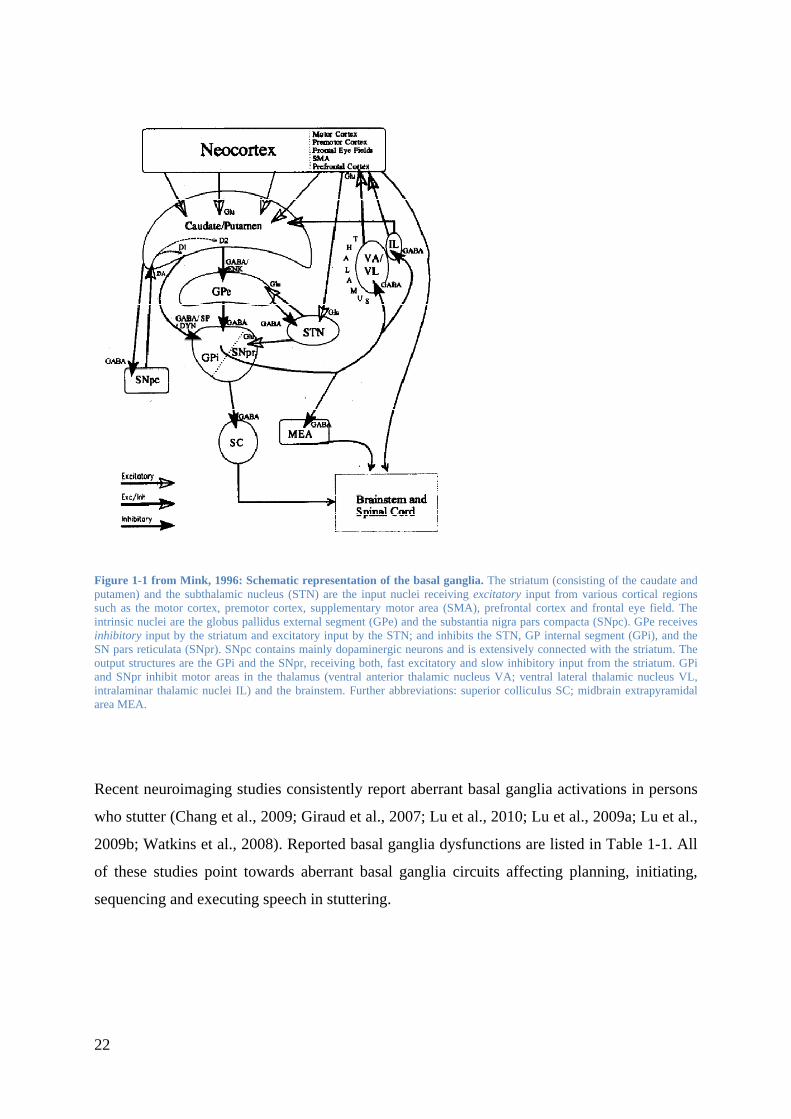
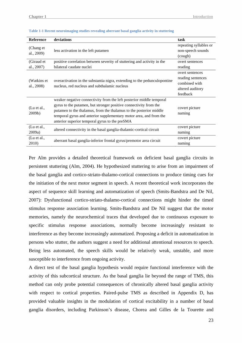
Figure 1-1 from Mink, 1996: Schematic representation of the basal ganglia. The striatum (consisting of the caudate and putamen) and the subthalamic nucleus (STN) are the input nuclei receiving excitatory input from various cortical regions such as the motor cortex, premotor cortex, supplementary motor area (SMA), prefrontal cortex and frontal eye field. The intrinsic nuclei are the globus pallidus external segment (GPe) and the substantia nigra pars compacta (SNpc). GPe receives inhibitory input by the striatum and excitatory input by the STN; and inhibits the STN, GP internal segment (GPi), and the SN pars reticulata (SNpr). SNpc contains mainly dopaminergic neurons and is extensively connected with the striatum. The output structures are the GPi and the SNpr, receiving both, fast excitatory and slow inhibitory input from the striatum. GPi and SNpr inhibit motor areas in the thalamus (ventral anterior thalamic nucleus VA; ventral lateral thalamic nucleus VL, intralaminar thalamic nuclei IL) and the brainstem. Further abbreviations: superior collicuIus SC; midbrain extrapyramidal area MEA.
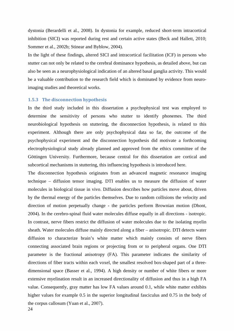
Recent neuroimaging studies consistently report aberrant basal ganglia activations in persons
who stutter (Chang et al., 2009; Giraud et al., 2007; Lu et al., 2010; Lu et al., 2009a; Lu et al.,
2009b; Watkins et al., 2008). Reported basal ganglia dysfunctions are listed in Table 1-1. All
of these studies point towards aberrant basal ganglia circuits affecting planning, initiating,
sequencing and executing speech in stuttering.
22
Chapter 1 Introduction
Table 1-1 Recent neuroimaging studies revealing aberrant basal ganglia activity in stuttering
Reference deviations task
(Chang et al., 2009)
less activation in the left putamen repeating syllables or non-speech sounds (cough)
(Giraud et al., 2007)
positive correlation between severity of stuttering and activity in the bilateral caudate nuclei
overt sentences reading
(Watkins et al., 2008)
overactivation in the substantia nigra, extending to the pedunculopontine nucleus, red nucleus and subthalamic nucleus
overt sentences reading sentences combined with altered auditory feedback
(Lu et al., 2009b)
weaker negative connectivity from the left posterior middle temporal gyrus to the putamen, but stronger positive connectivity from the putamen to the thalamus, from the thalamus to the posterior middle temporal gyrus and anterior supplementary motor area, and from the anterior superior temporal gyrus to the preSMA
covert picture naming
(Lu et al., 2009a)
altered connectivity in the basal ganglia-thalamic-cortical circuit covert picture naming
(Lu et al., 2010)
aberrant basal ganglia-inferior frontal gyrus/premotor area circuit covert picture naming
Per Alm provides a detailed theoretical framework on deficient basal ganglia circuits in
persistent stuttering (Alm, 2004). He hypothesized stuttering to arise from an impairment of
the basal ganglia and cortico-striato-thalamo-cortical connections to produce timing cues for
the initiation of the next motor segment in speech. A recent theoretical work incorporates the
aspect of sequence skill learning and automatization of speech (Smits-Bandstra and De Nil,
2007): Dysfunctional cortico-striato-thalamo-cortical connections might hinder the timed
stimulus response association learning. Smits-Bandstra and De Nil suggest that the motor
memories, namely the neurochemical traces that developed due to continuous exposure to
specific stimulus response associations, normally become increasingly resistant to
interference as they become increasingly automatized. Proposing a deficit in automatization in
persons who stutter, the authors suggest a need for additional attentional resources to speech.
Being less automated, the speech skills would be relatively weak, unstable, and more
susceptible to interference from ongoing activity.
A direct test of the basal ganglia hypothesis would require functional interference with the
activity of this subcortical structure. As the basal ganglia lie beyond the range of TMS, this
method can only probe potential consequences of chronically altered basal ganglia activity
with respect to cortical properties. Paired-pulse TMS as described in Appendix D, has
provided valuable insights in the modulation of cortical excitability in a number of basal
ganglia disorders, including Parkinson’s disease, Chorea and Gilles de la Tourette and
23
24
dystonia (Berardelli et al., 2008). In dystonia for example, reduced short-term intracortical
inhibition (SICI) was reported during rest and certain active states (Beck and Hallett, 2010;
Sommer et al., 2002b; Stinear and Byblow, 2004).
In the light of these findings, altered SICI and intracortical facilitation (ICF) in persons who
stutter can not only be related to the cerebral dominance hypothesis, as detailed above, but can
also be seen as a neurophysiological indication of an altered basal ganglia activity. This would
be a valuable contribution to the research field which is dominated by evidence from neuro-
imaging studies and theoretical works.
1.5.3 The disconnection hypothesis
In the third study included in this dissertation a psychophysical test was employed to
determine the sensitivity of persons who stutter to identify phonemes. The third
neurobiological hypothesis on stuttering, the disconnection hypothesis, is related to this
experiment. Although there are only psychophysical data so far, the outcome of the
psychophysical experiment and the disconnection hypothesis did motivate a forthcoming
electrophysiological study already planned and approved from the ethics committee of the
Göttingen University. Furthermore, because central for this dissertation are cortical and
subcortical mechanisms in stuttering, this influencing hypothesis is introduced here.
The disconnection hypothesis originates from an advanced magnetic resonance imaging
technique – diffusion tensor imaging. DTI enables us to measure the diffusion of water
molecules in biological tissue in vivo. Diffusion describes how particles move about, driven
by the thermal energy of the particles themselves. Due to random collisions the velocity and
direction of motion perpetually change - the particles perform Brownian motion (Dhont,
2004). In the cerebro-spinal fluid water molecules diffuse equally in all directions - isotropic.
In contrast, nerve fibers restrict the diffusion of water molecules due to the isolating myelin
sheath. Water molecules diffuse mainly directed along a fiber – anisotropic. DTI detects water
diffusion to characterize brain’s white matter which mainly consists of nerve fibers
connecting associated brain regions or projecting from or to peripheral organs. One DTI
parameter is the fractional anisotropy (FA). This parameter indicates the similarity of
directions of fiber tracts within each voxel, the smallest resolved box-shaped part of a three-
dimensional space (Basser et al., 1994). A high density or number of white fibers or more
extensive myelination result in an increased directionality of diffusion and thus in a high FA
value. Consequently, gray matter has low FA values around 0.1, while white matter exhibits
higher values for example 0.5 in the superior longitudinal fasciculus and 0.75 in the body of
the corpus callosum (Yuan et al., 2007).
Chapter 1 Introduction
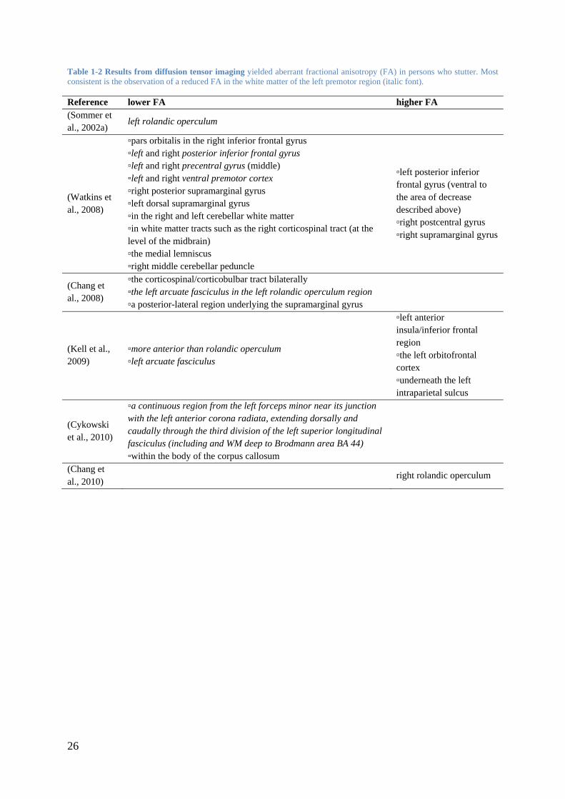
The first assessment of FA in brains of adults who stutter yielded one main result: The FA in
the white matter underlying the left rolandic operculum was decreased compared to control
subjects (Sommer et al., 2002a). Subsequent studies examining adults and adolescents who
stutter, independently confirmed the finding of compromised white matter integrity in left
frontal regions (Chang et al., 2008; Cykowski et al., 2010; Watkins et al., 2008).
These described brain alterations are evident in adults who stutter and adolescents. It is not
clear yet, whether lifelong stuttering caused these deviations similar to reported training
effects on white matter (Scholz et al., 2009; Takeuchi et al., 2010). Nonetheless, it is tempting
to speculate that inborn, genetic aberrations are the cause of the observed white matter
abnormalities (Büchel and Watkins, 2010). The latest indication in that direction came with
the aforementioned discovery of stuttering related mutations of proteins controlling the
lysosomal enzyme–targeting pathway (Kang et al., 2010). Other, apparently more severe,
mutations in the same pathway lead to mucolipidosis type II and III, and affected subjects
show severe white-matter abnormality (Folkerth, 1999).
The consequence of decreased fiber integrity in the frontal motor and premotor regions might
be a vulnerability of speech relevant cortico-cortical (Salmelin et al., 2000) or cortical-
subcortical connections in stuttering (Lu et al., 2009a). One important link for speech
production is the neural link between the motor production of speech sounds and the
representation of speech sounds in cortex (Hickok and Poeppel, 2007; Scott and Johnsrude,
2003), because the production of speech sounds is substantially modified by real-time
auditory feedback. Altered auditory feedback of speech produces automatic adjustment by the
speaker to compensate for the alteration such that feedback remains predictable (Houde and
Jordan, 1998; Tourville et al., 2008). Moreover, experience with speech sounds shapes their
perception (Nasir and Ostry, 2009; Shiller et al., 2009) suggesting that laryngeal disorders that
affect speaking, such as spasmodic dysphonia, alaryngeal speech or stuttering, may have
consequences on the perception of speech sounds in humans (Heiser and Cheung, 2008).
These considerations motivated the experiment conducted in the third study presented in this
dissertation.
25
26
Table 1-2 Results from diffusion tensor imaging yielded aberrant fractional anisotropy (FA) in persons who stutter. Most consistent is the observation of a reduced FA in the white matter of the left premotor region (italic font).
Reference lower FA higher FA (Sommer et al., 2002a)
left rolandic operculum
(Watkins et al., 2008)
▫pars orbitalis in the right inferior frontal gyrus ▫left and right posterior inferior frontal gyrus ▫left and right precentral gyrus (middle) ▫left and right ventral premotor cortex ▫right posterior supramarginal gyrus ▫left dorsal supramarginal gyrus ▫in the right and left cerebellar white matter ▫in white matter tracts such as the right corticospinal tract (at the level of the midbrain) ▫the medial lemniscus ▫right middle cerebellar peduncle
▫left posterior inferior frontal gyrus (ventral to the area of decrease described above) ▫right postcentral gyrus ▫right supramarginal gyrus
(Chang et al., 2008)
▫the corticospinal/corticobulbar tract bilaterally ▫the left arcuate fasciculus in the left rolandic operculum region ▫a posterior-lateral region underlying the supramarginal gyrus
(Kell et al., 2009)
▫more anterior than rolandic operculum ▫left arcuate fasciculus
▫left anterior insula/inferior frontal region ▫the left orbitofrontal cortex ▫underneath the left intraparietal sulcus
(Cykowski et al., 2010)
▫a continuous region from the left forceps minor near its junction with the left anterior corona radiata, extending dorsally and caudally through the third division of the left superior longitudinal fasciculus (including and WM deep to Brodmann area BA 44) ▫within the body of the corpus callosum
(Chang et al., 2010)
right rolandic operculum
Chapter 1 Scope of the dissertation
1.6 Scope of the dissertation
The objective of this dissertation is to explore cortical and subcortical mechanisms in
stuttering.
In the first study, repetitive transcranial magnetic stimulation (rTMS) helped discovering a
dysfunction of the left dorsolateral premotor cortex in control of paced finger movements and
a compensatory role of its right hemispheric homologue in stuttering. While previous
neuroimaging studies elucidated altered activation patterns, we were able to directly show for
the first time that the right hemisphere might indeed play a compensatory rather than
maladaptive role for non-speech functions in persistent developmental stuttering.
In the second study, we aimed at detecting neurophysiological changes in the primary motor
tongue representation of the left and right hemisphere in adults with persistent stuttering.
Overcoming methodological challenges of transcranial magnetic stimulation at orofacial
structures, this is the first study demonstrating an abnormality in intracortical excitability in
persistent stuttering.
The third study operationalized a behavioral approach to elucidate a possible disconnection
between parieto-temporal regions involved in the phonological bottom up processing of
speech stimuli and frontal regions mainly involved in the planning, programming and
execution of speech movements. Behavioral deviations on a subclinical level might indicate a
disturbed functional connectivity of these main networks of speech processing.
The following studies aim particularly:
(1) to test the lateralization of cortical control of paced finger movement timing in stuttering
(2) to detect neurophysiological changes in the intracortical excitability in the primary motor
tongue representation of the left and right hemisphere by employing single-pulse and
paired-pulse TMS in adults who stutter and matched control subjects
(3) to test the stability of phoneme percepts in stuttering by analyzing participants’ sensitivity
to identify voiced and voiceless stop-consonants near the phoneme boundary.
27

Chapter 2 Original articles
29
2 Original Articles
The following published and submitted articles are presented in this chapter:
I. Neef N .E., Jung, K., Rothkegel H., Pollok B., Wolff von Gudenberg A., Paulus W.,
Sommer M. “Right-shift for non-speech motor processing in adults who stutter” (2010
Jun 30. [Epub ahead of print]). The study was designed by Martin Sommer, Holger
Rothkegel, Bettina Pollok and Nicole Neef. The program to present the stimuli was provided
by Bettina Pollok. Nicole Neef wrote the ethic proposal, recruited the subjects, examined the
subjects and analyzed the data. Reanalysis of the speech sample to test inter-rater reliability
was performed by Kristina Jung. Statistics were performed by Martin Sommer and Nicole
Neef. The manuscript was written by Martin Sommer and Nicole Neef with contributions of
all authors.
II. Neef N. E., Paulus W., Neef A., Wolff von Gudenberg A., Sommer M. “Reduced
intracortical inhibition and facilitation in the primary motor tongue representation of
adults who stutter” (resubmitted after revision to Clinical Neurophysiology; version of
November 26th 2010). The study was designed by Martin Sommer and Nicole Neef. Nicole
Neef wrote the ethic proposal, recruited the subjects, examined the subjects and analyzed the
data. A data browser was programmed by Andreas Neef. The quantification of the
Electromyography (EMG) activity at baseline and area under the MEP amplitude were
performed by Andreas Neef. Statistics were performed by Martin Sommer and Nicole Neef.
The manuscript was written by Nicole Neef with contributions of all authors.
III. Neef N. E., Sommer M., Paulus W., Wolff von Gudenberg A., Wüstenberg T.
“Instable phoneme categorization in adults who stutter” (under revision to resubmit to
Journal of Speech, Language, and Hearing Research; version of November 30th 2010). This
study was designed by Torsten Wüstenberg, Martin Sommer, Veronika Gutmann and Nicole
Neef. Torsten Wüstenberg prepared the auditory stimuli and programmed the psychophysical
test. Nicole Neef wrote the ethic proposal, recruited the subjects, examined the subjects and
analyzed the data. Veronika Gutmann collected some of the pilot data. Statistics were
performed by Torsten Wüstenberg, Martin Sommer and Nicole Neef. Graph 1 and 2 was
prepared by Torsten Wüstenberg all other graphs were designed by Torsten Wüstenberg and
Nicole Neef. The manuscript was written by Torsten Wüstenberg (methods sections: stimuli,
data analysis, Appendix A and B) and Nicole Neef (introduction; methods sections:
participants, fluency assessment, experimental procedure, statistics; results; discussion) with
contributions of all authors.

Chapter 2 Study 1 Non-speech motor processing in stuttering
31
2.1 Right-shift for non-speech motor processing in adults who stutter
Nicole E. Neef1,2, Kristina Jung3, Holger Rothkegel1,2, Bettina Pollok4,
Alexander Wolff von Gudenberg3, Walter Paulus1,2, and Martin Sommer1,2
1Department of Clinical Neurophysiology, Georg-August-University of Goettingen, Germany, 2Center for Systems Neuroscience, Goettingen, Germany 3Institut der Kasseler Stottertherapie, Bad Emstal, Germany 4Institute of Clinical Neuroscience and Medical Psychology, Heinrich-Heine-University
Duesseldorf, Germany
Running title: non-speech motor processing in stuttering
Address for correspondence:
Martin Sommer, M.D.
Department of Clinical Neurophysiology, University of Goettingen,
Robert-Koch-Str. 40, D-37075 Goettingen, Germany
Tel. +49-551-39 66 50, Fax. +49-551-39 81 26; Email: [email protected]

Chapter 2 Study 1 Non-speech motor processing in stuttering
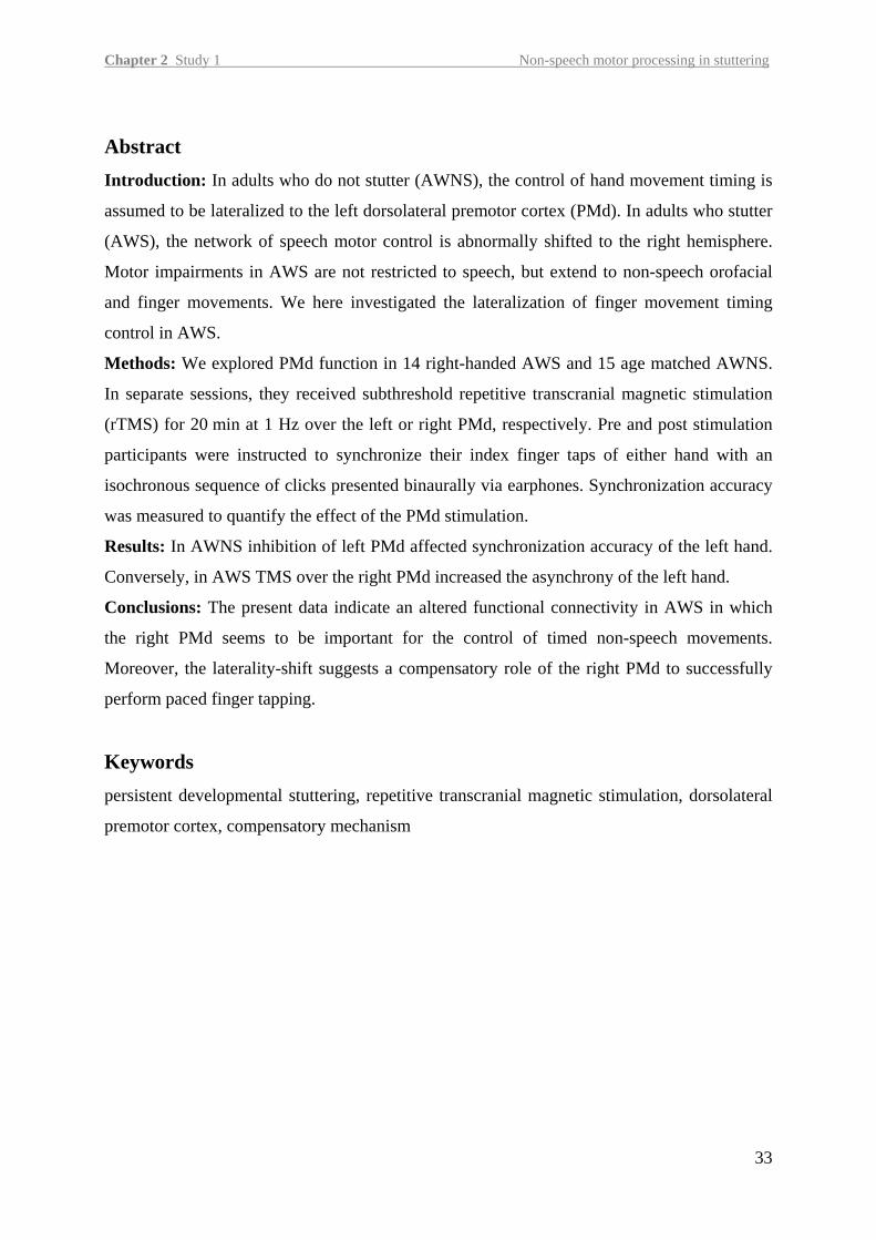
Abstract Introduction: In adults who do not stutter (AWNS), the control of hand movement timing is
assumed to be lateralized to the left dorsolateral premotor cortex (PMd). In adults who stutter
(AWS), the network of speech motor control is abnormally shifted to the right hemisphere.
Motor impairments in AWS are not restricted to speech, but extend to non-speech orofacial
and finger movements. We here investigated the lateralization of finger movement timing
control in AWS.
Methods: We explored PMd function in 14 right-handed AWS and 15 age matched AWNS.
In separate sessions, they received subthreshold repetitive transcranial magnetic stimulation
(rTMS) for 20 min at 1 Hz over the left or right PMd, respectively. Pre and post stimulation
participants were instructed to synchronize their index finger taps of either hand with an
isochronous sequence of clicks presented binaurally via earphones. Synchronization accuracy
was measured to quantify the effect of the PMd stimulation.
Results: In AWNS inhibition of left PMd affected synchronization accuracy of the left hand.
Conversely, in AWS TMS over the right PMd increased the asynchrony of the left hand.
Conclusions: The present data indicate an altered functional connectivity in AWS in which
the right PMd seems to be important for the control of timed non-speech movements.
Moreover, the laterality-shift suggests a compensatory role of the right PMd to successfully
perform paced finger tapping.
Keywords persistent developmental stuttering, repetitive transcranial magnetic stimulation, dorsolateral
premotor cortex, compensatory mechanism
33
34
Introduction Fluent speech requires the well timed selection, initiation, execution and monitoring of motor
sequences. The relevant cortical and subcortical neural systems appear to be malfunctioning
in developmental stuttering (Brown et al., 2005; Fox et al., 1996; Ludlow and Loucks, 2003).
Stuttering is characterized by an impairment of speech rhythm or fluency (Bloodstein and
Ratner, 2008). Speech disruptions typically include blocks, repetitions, or prolongations of
speech segments ((WHO), 2007a), and may be accompanied by movements of face and limb
muscles and by negative emotions such as fear or embarrassment. About 5% of the population
stutters at some point during childhood (Mansson, 2000). Although spontaneous recovery rate
is high, stuttering without obvious neurological origin persists after puberty in about 1% of
adults (Andrews and Harris, 1964; Bloodstein and Ratner, 2008; Craig et al., 2002). Exploring
the underlying neural mechanisms of this disorder provides insights into mechanisms of
dysfluent speech production and into models of speech planning and production in general.
These insights into the physiology of stuttering may ultimately serve to improve treatments
enhancing speech fluency.
Temporal patterns in speech occur on multiple timescales (i.e. subsegmental, segmental and
suprasegmental, (Levelt, 1989c). In adults who stutter (AWS), acoustic-temporal and spatio-
temporal characteristics are affected in stuttered and fluent speech on all these time scales
(Jancke, 1994; Kleinow and Smith, 2000; Max and Gracco, 2005; Prins and Hubbard, 1992).
Most consistent are the observations of increased variability of duration and relative timing of
acoustic and kinematic features. Additionally, stuttering has been associated with altered
auditory feedback control mechanisms (Max et al., 2004; Tourville et al., 2008). Altogether,
these facts underline a deficit of speech motor timing and the impact of the timing of auditory
information during speaking in AWS.
Alterations of timing abilities in AWS exceed the domain of speech and affect the motor
control of non-speech movements as well. For example, AWS performed poorly in
reproducing varying rhythmic patterns (Hunsley, 1937) or unpredictable digit sequences
(Webster, 1986). Additionally, AWS exhibit prolonged initiation and execution times in
finger movement sequencing tasks (Smits-Bandstra et al., 2006; Webster, 1997) and increased
manual reaction times (Bishop et al., 1991; Webster and Ryan, 1991). Phase variability is
greater during bimanual coordination of auditory paced movements (Zelaznik et al., 1997)
and movement variability is increased during simultaneous synchronization of speech and
hand movements (Hulstijn et al., 1992). However, studies on auditory paced isochronous
finger movements did not find differences of timing accuracy and timing variability between
Chapter 2 Study 1 Non-speech motor processing in stuttering
AWS and controls (Hulstijn et al., 1992; Max and Yudman, 2003; Melvine et al., 1995;
Zelaznik et al., 1994).
Two separate processes have been related to timing accuracy: a neural clock mechanism (Ivry
and Spencer, 2004; Rao et al., 1997), and an emergent property of the kinematics of
movements itself (Ivry and Spencer, 2004; Mauk and Buonomano, 2004). This dissociation
between event timing and emergent timing has been corroborated by previous findings
(Spencer et al., 2003; Zelaznik et al., 2005; Zelaznik et al., 2002). Timing in the sub- and
supra-second range involves dissociable neural networks (Gibbon et al., 1997; Lewis and
Miall, 2003; Wiener et al., 2010). Sub-second timing engages cerebello-thalamo-cortical
network (Pollok et al., 2005), whereas supra-second timing tasks were more prone to activate
cortical structures such as supplementary motor area (SMA) and prefrontal cortex (Wiener et
al., 2010). For an event timing task like self-paced finger tapping, Wing and Kristofferson
(Wing and Kristofferson, 1973) indicate a dichotomy between central clock and motor
execution by suggesting that a central timekeeper supplies intervals of the adequate length and
drives motor commands at the end of each interval. The original Wing-Kristofferson model
was concerned with the special case of self-paced finger tapping and therefore neglected the
process of integrating external cues. This contrasts with finger tapping in synchrony with an
acoustically presented pacer, a timed motion task that additionally involves the integration of
the external event and the monitoring of the synchrony of the pacer and the tapping.
Finger tapping accuracy can be disturbed by transcranial magnetic stimulation (TMS)
(Doumas et al., 2005; Levit-Binnun et al., 2007; Malcolm et al., 2008; Pollok et al., 2008), a
neurophysiological technique inducing a brief electric current in the brain using a magnetic
field to pass the scalp and the skull safely and painlessly. Repetitive TMS (rTMS) is capable
of inducing excitability changes of neural networks outlasting the stimulation period (Hallett,
2000; Miniussi et al., 2008; Siebner et al., 2009; Siebner and Rothwell, 2003), thereby
temporarily disrupting activity in local or remote cortical areas (Wagner et al., 2009; Walsh
and Rushworth, 1999). Thus, rTMS disrupts brain functions for a finite time with relatively
high spatial resolution.
In the present study rTMS was employed to induce a transient virtual lesion of the
dorsolateral premotor cortex (PMd). Traditionally the premotor cortices (PM) were assumed
to be key structures in the motor domain and thereby associated with the preparation and the
organization of movements and actions (Wise, 1985). Imaging studies suggest a specific
significance of the PMd for cognitive functions (Abe and Hanakawa, 2009), sensorimotor
integration (Pollok et al., 2009; Schubotz et al., 2003) and rhythm perception (Bengtsson et
35
36
al., 2009), as well. Recent studies provide evidence for a specific role of the left PMd for
movement timing of both hands (Pollok et al., 2009; Pollok et al., 2008). Interestingly,
externally paced finger movements as well as syllable repetition seem to recruit the same
cerebral network involving the left PMd (Riecker et al., 2006). However, the PMd seems to
play a role during fluency enhancing mechanisms in AWS. Fluency is reliably enhanced when
speech is timed to a pacer: either an external pacer such as a rhythmic beat (Wingate, 2002;
Wohl, 1968), the unison speaking with another person (Adams and Ramig, 1980; Ingham and
Carroll, 1977; Saltuklaroglu et al., 2009), or an internal pacer such as rhythmic arm swinging
or a finger tapping (Bloodstein and Ratner, 2008). Alternative fluency enhancing techniques
are delayed or frequency shifted auditory feedback (Antipova et al., 2008; Van Riper, 1970).
Such fluency enhancing mechanisms involve right premotor regions as well as the cerebellum
(Braun et al., 1997; Fox et al., 1996; Tourville et al., 2008; Watkins et al., 2008). Hence, the
PMs seem to play an important role for motor timing control as well as the implementation of
fluency enhancing techniques.
Theoretical frameworks on stuttering suggest an aberrant timing of neural activity in different
brain regions that are relevant for speech processing (Alm, 2004; Howell, 2004; Ludlow and
Loucks, 2003). Specifically, the basal ganglia-cortical route might be impaired in providing
internal cues for the exact timing of movements, while the PMd in concert with the
cerebellum successfully utilizes external time cues resulting in enhanced fluency for example
during metronome speaking (Alm, 2004). Interestingly, in AWS even a non-speech motor
task like externally paced finger tapping mirrored an irregular right-shifted activation
(Morgan et al., 2008). This increased right pre-central activation suggests that the cortical
contribution to the process of timed movements is less left lateralized. The present study aims
at further investigating the assumption of a hemispheric shift of motor functions in AWS by
means of an induced virtual lesion of the left and right PMd in AWS and adults who do not
stutter (AWNS).
Chapter 2 Study 1 Non-speech motor processing in stuttering
Methods
Participants Fourteen right-handed AWS [mean age 30.3 ± 11.4 (SD); one female] and fifteen AWNS
[mean age 28.1 ± 5.0 (SD); one female] participated in this study. Table 1 contains details of
the participants. Stuttering participants were recruited from the Stuttering self-help group of
Goettingen and the Institute for the Kassel Stuttering Therapy. Three AWS had already taken
part in an earlier TMS study (Sommer et al., 2009b). The groups were matched and statistics
did not yield any group differences for age (T = .65, p = .5), handedness (Oldfield, 1971);
Z = -.73, p = .46) and level of education (Z = -1.28, p = .2), amount of musical training and
gender. AWS produced significantly more stuttered syllables than AWNS [meanAWS 9.0 ± 8.0
(SD), meanAWNS .6 ± .4 (SD); Z = -4.6; p < .001; for details on statistics see data analysis
section]. Stuttering severity was very mild in five, mild in three, moderate in two, severe in
two and very severe in two AWS according to the Stuttering Severity Index (SSI-3). Inter-
rater reliability analysis yielded an unjust intra-class correlation coefficient (ICCunjust) of .94
(95% CI .82 -.98) and intra-rater reliability analysis yielded an ICCunjust of .97 (95% CI .81 -
.98).
None of the participants had a self-reported history of speech, language or hearing problems,
with the exception of stuttering in AWS. According to the definition ((WHO), 2007b)
cluttering was recognized by rapid, erratic, and dysrhythmic speech dysfluency with distinct
speech timing abnormalities. On this ground we excluded one fifteenth putative participant
who exhibited both stuttering and cluttering. None of the participants showed neurological or
medical abnormalities on routine examination. None of the participants were taking drugs
affecting the central nervous system at the time of the study. The local Ethics Committee
approved the study and all participants gave written informed consent according to the
declaration of Helsinki.
please insert Tab. 1 about here
Fluency assessment The fluency assessments were performed and independently analyzed by a qualified speech-
language pathologist (N.N.) and a qualified clinical linguist (K.J.). In compliance with the
37
38
German version of the SSI-3 (Sandrieser and Schneider, 2008; Riley, 1994), speech samples
of all participants containing a conversation about job or school and a reading task were
videotaped (Sony Handycam DCR-TRV16E Mini DV digital Camcorder) and audio recorded
(Edirol R-09; sample rate: 16 bit/44.1 kHz; format: WAV). SSI-3 norms were adapted from
Riley (Riley, 1994). Software for offline analysis was DivX player (DivX software, San
Diego) and WavePad (NCH software, Canberra). The offline analysis of dysfluencies
included 500 syllables for the conversation and not less than 340 syllables for the reading
task. Sound prolongations, blocks (silent prolongation of an articulatory posture), sound and
syllable repetitions were counted as stuttered syllables. Monosyllabic words that were
repeated with apparent undue stress or tension were counted too (Sandrieser and Schneider,
2008). Furthermore, the estimated duration of the three longest blocks and observation of
physic tants were included for the estimate of stuttering severity in AWS. al concomiProcedure The experiment consisted of two sessions, one for stimulating the left and the other for
stimulating the right PMd. During each session participants performed one run of left index
and one run of right index finger tapping before rTMS. Both runs were repeated immediately
(about 30 sec) after rTMS. The order of stimulation site and hand was counterbalanced across
participants. To avoid carry-over effects of the magnetic stimulation the second rTMS session
was performed not less than 48 hours after the first one.
Participants sat in a silent room in front of a computer keyboard connected to the computer
via a PS/2 cable. The keyboard was shielded to the participant’s visual field. Participants were
requested to synchronize their unimanual index finger taps with a metronome. The
acoustically presented metronome signals contained clicks of 10 msec duration with an inter
click interval of 800 msec. Each experimental run comprised a continuous series of 56 clicks.
The clicks were presented binaurally via dynamic, closed-ear headphones (Sennheiser HD
280; up to 32 dB attenuation of outside noise). Click intensity was individually adjusted to a
level perceived as loud by the participants. The pacing signal was triggered and the onsets of
space bar presses were recorded by using Eprime (http://www.pstnet.com). We quantified
performance by calculating (1) the asynchrony, the averaged temporal distance between the
onset of the pacing signal and finger taps, and (2) the inter-tap interval (ITI)-variability, the
variation of the time between two consecutive taps.
Chapter 2 Study 1 Non-speech motor processing in stuttering
Stimulation technique TMS was applied while participants sat comfortably in a reclining chair. A figure-8-shaped
stimulation coil connected to a Magstim rapid2 stimulator (Magstim Company, Dyfed, Wales,
UK) was positioned tangentially to the scalp with the handle pointing backwards and rotated
away from the midline by 45 degrees. The junction of the two wings of the figure-8-coil was
held flat on the skull. The pulse configuration was biphasic with an initial posterior-anterior
current flow in the brain. The motor hot spot was localized at the optimal point for eliciting
motor evoked potentials (MEPs) in the contralateral first dorsal interosseous (FDI) muscle
over the primary motor cortex (M1). Active motor threshold (AMT) was determined as the
minimum intensity needed to evoke MEPs in the tonically contracted contralateral FDI
muscle of about 200 µV in five of ten consecutive trials. For the rTMS of the PMd the
intersection of the coil was placed 2.5 cm anterior to the M1 representational hot spot of FDI.
This procedure is in accordance with previous studies (Doumas et al., 2005; Mochizuki et al.,
2004; Pollok et al., 2008; Schluter et al., 1998) and fits with functional imaging data
displaying the PMd to be positioned about 1.8 - 2.5 cm (Picard and Strick, 2001) and 2.0 cm
(Fink et al., 1997) anterior to the M1 hand area. The coil was held with the handle pointing
backward and rotated away from the midline by 45° to induce a final anterior-posterior
directed current in the stimulated cortex. Surface electromyogram (EMG) was recorded from
the FDI through a pair of silver–silver chloride surface electrodes in a belly-tendon montage.
Raw signals were amplified, band-pass filtered (2 - 2500 Hz), digitized with a micro 1401 AD
converter (Cambridge Electronic Design, Cambridge, United Kingdom) and controlled by
Signal Software (Cambridge Electronic Design, version 2.13). Complete muscle relaxation
was controlled through visual feedback of EMG activity. Subthreshold rTMS was applied at
90% of ipsilateral AMT intensity for 20 minutes at 1 Hz over the left PMd in one session and
the right PMd in another. This rTMS protocol has been shown to decrease cortico-spinal
excitability for several minutes (Gerschlager et al., 2001; Walsh and Rushworth, 1999) and
compl recommendations (Rossi et al., 2009; Wassermann, 1998). ies with safetyData analysis The mean values of the two dependent variables, asynchrony and ITI-variability, were
calculated separately for each group (AWNS/AWS), each hand (left hand/ right hand) and
each site of stimulation (left rTMS/ right rTMS); thus, yielding 16 values of asynchrony and
ITI-variability, respectively.
39
40
To control for group differences in the finger tapping performance before rTMS we compared
the individual mean baseline asynchrony values using a two-way mixed design analysis of
variance (ANOVA) with the between-subjects factor group (AWS/AWNS) and the within-
subjects factor hand (left/right). A similar ANOVA was calculated with the baseline ITI-
variability.
At baseline finger taps preceded the acoustic signal in most participants resulting in negative
asynchrony values. However, there were two AWS and seven AWNS that showed a positive
asynchrony in most runs. To test the impact of rTMS we therefore normalized the asynchrony
after stimulation for each participant and each session by subtracting the asynchrony before
stimulation.
We entered the normalized values in a three-way mixed design ANOVA with the between-
subjects factor group (AWS/AWNS) and the within-subjects factors stimulation site (rTMS
over the left PMd/rTMS over the right PMd) and hand (left/right). In addition, the expected
rTMS-induced increases of asynchrony values (Pollok et al., 2008) were tested with one-
tailed t-tests. We tested the impact of rTMS on ITI-variability similarly by entering the
normalized to baseline values.
To exclude differences of age between groups we used a two-tailed t-test for independent
samples, for education, handedness and percentage of stuttered syllables, we used Mann-
Whitney U-tests. Nonparametric testing was chosen since education is an ordinally scaled
variable and handedness as well as percentage of stuttered syllables did not show normal
distribution in AWNS. The AMT comparison was calculated with a repeated measures
ANOVA with hand as a within-subjects factor and group as a between-subjects factor.
Statistics were performed by SPSS Statistics 17.0 (http://www.spss.com/de/software).
Results At baseline the two-way mixed design ANOVA with asynchrony values before rTMS as
dependent variable revealed no significant difference between AWS and AWNS (factor group
F1,27 = 1.4, p = .3). However, the ANOVA revealed a more pronounced negative asynchrony
in the right hand than in the left hand [factor hand F1,27 = 7.73, p = .01; left hand -28 ± 52
msec (mean ± SD) vs right hand -39 ± 60 msec]. Analysis yielded no further effect. ITI-
variability before rTMS revealed no main effect or interaction for group and hand.
After rTMS, the analysis of normalized asynchrony values revealed no main effects of hand
(F1,27 = 1.5, p = .2), stimulation site (F1,27 = .4, p = .5) and group (F1,27 = .6, p = .4) but a
significant interaction between hand, stimulation site and group (F1,27 = 5.82, p = .023).
Chapter 2 Study 1 Non-speech motor processing in stuttering
Post hoc one-tailed t-tests, not corrected for multiple comparisons (Perneger, 1998), revealed
that rTMS over the left PMd significantly increased left hand asynchrony in AWNS
(T = 1.9, p = .036) as previously shown (Pollok et al., 2008). By contrast, in AWS rTMS over
the right PMd resulted in a significant increase of left hand asynchrony (T = 2.34, p = .015)
(Fig. 1).
Please insert Fig. 1 about here
After rTMS the analysis of normalized ITI-variability revealed no main effects of group,
stimulation site or hand and no interactions of any of these factors. Interaction between group,
hand and stimulation site was marginally significant (F1,27 = 5.82, p = .06). Post-hoc one-
tailed t-tests, not corrected for multiple comparisons, yielded in an increased normalized ITI-
variability after rTMS over the left PMd in the left hand of AWNS (T = -2.01, p = .032). This
is in concordance with previous findings (Pollok et al., 2008). All other statistics yielded no
significant differences (Fig. 1).
Discussion We studied the cortical control of auditory paced finger movements in AWS and AWNS. In
AWNS, rTMS over the left PMd increased left hand asynchrony and increased ITI-variability,
whereas rTMS over the right PMd was ineffective. By contrast, in AWS rTMS over the left
PMd w synchrony. as ineffective, whereas rTMS over the right PMd prolonged left hand aLeft-hemispheric dominance on movement timing control in AWNS In AWNS rTMS over the left PMd increased asynchrony and ITI variability of the left hand.
This finding agrees well with previous studies confirming a particular role of the left PMd in
auditory paced rhythmic finger tapping (Pollok et al., 2009; Pollok et al., 2008). Although it is
not entirely clear via which connections the left PMd exerts dominance over the right
hemisphere, a specific significance of direct left PMd – right M1 connections (Pollok et al.,
2008); (Boroojerdi et al., 1996; Ferbert et al., 1992) as well as subcortical circuits (Chouinard
et al., 2003) has been evidenced. It is well established that the cerebellum is closely connected
to the cerebral cortex via a cerebello-thalamo-cortical loop (Horne and Butler, 1995) and that
auditory paced isochronous tapping engages the cerebellum (Ivry et al., 2002; Spencer et al.,
2005). Even perception of an isochronous rhythm involves the left PMd in concert with the
right cerebellum in healthy subjects suggesting the engagement of prediction mechanisms that
41
42
are used for motor preparation (Bengtsson et al., 2009). Therefore, the left PMd might serve
as an interface between sensory prediction and temporally precise motor initiation (Kurata et
al., 2000; Ramnani and Passingham, 2001). Consequently, an rTMS induced dysfunction of
the left PMd might alter the functional connectivity of the cerebello-thalamo-cortical loop
which results in less precise timed motor behavior.
This finding is consistent with a hemispheric dominance of the left PMd in AWNS reported
by Koch et al. (Koch et al., 2006) during a response selection task and by Pollok et al. (2008)
for movement timing during auditory paced finger tapping. Nevertheless, this hypotheses is
not unchallenged since in a response selection experiment, O`Shea et al. (O'Shea et al., 2007)
did not find evidence for such a hemispheric dominance. Rather, they demonstrated that
chang king PMd and contralateral M1. es in functional connectivity occur in the pathway linRight-shifted control of movement timing in AWS In contrast to AWNS, right rTMS prolonged left hand asynchrony in AWS, whereas left
rTMS was ineffective. Previous behavioral (Curry and Gregory, 1969; Sommers et al., 1975),
physiological (Biermann-Ruben et al., 2005; Moore and Lang, 1977) and neuroimaging
studies (Braun et al., 1997; De Nil and Brutten, 1991; Ingham et al., 2004; Preibisch et al.,
2003) provide evidence for a cerebral imbalance in AWS with an increased involvement of
the right hemisphere during speech production. Our results are in line with neural imaging
studies suggesting an aberrant role of the left PMd (Lu et al., 2010) and an additional
involvement of the right PMd during speech (Braun et al., 1997; Fox et al., 1996; Ingham,
2001) and even non-speech tasks in AWS (Chang et al., 2009; Morgan et al., 2008).
Accordingly, using functional magnetic resonance imaging right hand finger tapping has been
shown to be associated with bilateral pre- and post-central activation with increased activation
of the right hemisphere in AWS as compared to AWNS (Morgan et al., 2008). Thus, less
activation of the left premotor area and stronger activation of the right premotor area are not
specif he present findings. ic for speech in AWS, an interpretation corroborated by tNo effects on right hand performance in both groups Previous studies documented contradictory data resulting from rTMS over the left PMd on
right hand movement timing in non-stuttering adults (Del Olmo et al., 2007; Doumas et al.,
2005; Pollok et al., 2008). The present study showed an effect of left PMd rTMS on the
subdominant left hand only. Within our sample of fluently speaking participants right
handedness was less strongly developed (group average 76; median 70). Thus, one might
speculate that the rTMS effect occurs in strongly developed right handedness only (i.e.,
Chapter 2 Study 1 Non-speech motor processing in stuttering
Edinburgh Inventory score of 90 to 100). To insure that the degree of handedness did not
interfere with our main result we recalculated our statistics with three-way mixed analyses of
covariance (ANCOVAs) with handedness scores as additional covariate. The ANCOVAs
confirmed the three-way interaction between hand, site and group for asynchrony
(F1,26 = 6.28, p = .019) and the marginal interaction of the same factors for ITI variability
(F1,26 = 3.58, p = .07). ANCOVAs yielded no further effects.
Hence, the lack of modulation of right hand asynchrony cannot be explained by less
pronounced right handedness within the present sample. Our data suggest that networks
controlling the performance of the non-dominant hand may be more susceptible to rTMS
effects than those controlling the dominant hand (Meyer-Lindenberg et al., 2002). This idea is
also supported by a former diffusion tensor imaging study showing decreased fractional
anisotropy underneath the precentral gyrus of the non-dominant hand related to the dominant
hand (Buchel et al., 2004). Thus, morphologically the non-dominant hand relies on white
matter with less integrity contrasted to the dominant hand. Furthermore, rTMS studies
demonstrate an improvement of non-dominant left hand performance after inhibition of the
ipsilateral left M1 (Kobayashi et al., 2004), but no improvement of dominant right hand
performance after inhibition of the ipsilateral right M1 (Weiler et al., 2008). Additionally, in
AWNS, interhemispheric inhibition from the dominant to the non-dominant M1 is stronger
than vice versa (Netz et al., 1995; Samii et al., 1997). These results are compatible with our
hypothesis that the network subserving motor control of the dominant hand might be more
stable and thus, less prone to disturbance. Why did right PMd stimulation affect the contralateral hand in AWS, while left PMd stimulation did affect the ipsilateral hand in AWNS? In both groups rTMS affected the subdominant hand but, in AWNS this effect occurred after
left PMd stimulation, whereas in AWS right PMd stimulation yielded reduced timing
accuracy. Although speculative, this result supports the hypothesis that in AWS motor
functions are shifted to the right hemisphere. Thus, rTMS of the dominant hemisphere might
affect temporal accuracy of the subdominant hand.
Interestingly, the present data did not indicate differences between AWS and AWNS prior to
rTMS. Nevertheless, even a task which is not impaired in AWS, like unimanual auditory
paced finger tapping (Hulstijn et al., 1992; Max and Yudman, 2003; Melvine et al., 1995;
Zelaznik et al., 1994), is associated with altered brain functions. Since in our study, inhibition
of the right PMd elicited an aggravation of asynchrony but inhibition of the left PMd did not
43
44
elicit an effect, we assume that the right PMd involvement reflects a compensatory
mechanism rather than malfunction (Braun et al., 1997; Fox et al., 2000; Ludlow, 2000;
Preibisch et al., 2003).
This compensatory mechanism might be needed because in AWS a basal neural deficit has
been described in a left frontal brain region near the stimulation site. White matter integrity is
reduced in the left Rolandic Operculum in adults (Sommer et al., 2002a; Watkins et al., 2008)
and adolescents who stutter (Chang et al., 2008). This results in a disconnection within the
cerebral network processing speech motor behavior. Evidence in favor of such a weakened
connection has been given by an abnormal activating time course of left premotor and
primary motor regions (Salmelin et al., 2000) and altered left frontal-right cerebellar
interactions (Lu et al., 2010) in AWS.
The integration of motor areas of an undamaged hemisphere to adaptively compensate for
damaged or disconnected regions has been recently identified in recovered stroke patients
(Johansen-Berg et al., 2002; Riecker et al., 2010). Interestingly, a functional connectivity
analysis pinpointed the SMAs to provide a driver-like input to the contralesional premotor and
sensorimotor cortices in stroke patients (Riecker et al., 2010).
Anterior parts of the SMA are mainly connected with M1, PM and the putamen, posterior
parts are mainly connected with the inferior frontal gyrus, medial parietal, superior frontal
cortex and the caudatum (Johansen-Berg et al., 2004; Kim et al., 2010; Lehericy et al., 2004).
In AWS, SMA shows increased activation during speech production (Chang et al., 2009) and
even a more pronounced activation during stuttered as compared to fluent speech production
(Ingham et al., 2000), which is also mirrored in a correlation between stutter-rate and SMA
activation (Fox et al., 2000). Additionally, the involvement of the putamen, which interacts
with the SMA as well as with M1 and PM, is also altered in AWS (Braun et al., 1997; Lu et
al., 2009b; Ludlow and Loucks, 2003; Watkins et al., 2008). This over-activation may be
related to the fact that the SMA supports the involvement of different neural populations like
the rig lly recruited for functional reorganization. ht PMd that are additionaLimitations of the study Although we used a standard procedure for determining rTMS location, we did not verify the
exact PMd localization by structural or functional imaging. We therefore cannot rule out an
aberrant structural or functional organization of the left PMd in AWS. Cerebellar regions play
an important role for event timing (Spencer et al., 2003) and altered auditory feedback
(Howell and Sackin, 2002; Tourville et al., 2008), and behavioral evidence indicated
Chapter 2 Study 1 Non-speech motor processing in stuttering
cerebellar deficits in children who stutter (Howell et al., 1997; Howell et al., 2008). However,
we did not stimulate the cerebellum, because this procedure is quite uncomfortable and may
induce changes of the cortico-spinal excitability. This effect is related to the peripheral
stimulation of the neck muscles rather than the stimulation of the cerebellum itself
(Gerschlager et al., 2002).
Conclusion The present findings indicate a right-shifted neuronal organization for movement timing in
AWS supporting the hypothesis of a generally altered neurophysiological organization of the
motor control system in AWS. Since synchronization accuracy prior to rTMS did not differ
between AWS and AWNS we suggest that the increased involvement of the right PMd in
non-speech and possibly also in speech tasks represents a compensatory rather than a
maladaptive process.
Funding This work was supported by the Deutsche Forschungsgemeinschaft [SO 429/2-2 to M.S.];
Scholarship of the Stifterverband für die Deutsche Wissenschaft, Walter und Ilse Rose
Stiftung [to W.P.]; the Bernstein Center for Computational Neuroscience (BCCN)
[01GQ0432 to W.P.] and the Forschungskommission of the Medical Faculty of the Heinrich-
Heine University. Duesseldorf [9772328 to B.P.]. Nicole Neef, Kristina Jung, Holger
Rothkegel, Dr. Bettina Pollok, Dr. Alexander Wolff von Gudenberg, Prof. Dr. Walter Paulus
and Dr. Martin Sommer reported no biomedical financial interests or potential conflicts of
interest.
Acknowledgements We are thankful to Manuel Hewitt for the technical assistance, to Michael Nitsche for the
support in statistical questions, to Per Alm, Veronika Gutmann, Anne Smith, Vincent Walsh
and Christine Weber-Fox for fruitful discussions of the results, to Hayley Arnold for
thoughtful comments and confirming correct English usage and syntax, and to the reviewers
for their helpful feedback.
45
46
References (WHO) WHO. ICD-10 F98.5 Stuttering [Stammering]. Retrieved from
http://apps.who.int/classifications/apps/icd/icd10online/, 2007a.
(WHO) WHO. ICD-10 F98.6 Cluttering Retrieved from
www.who.int/classifications/apps/icd/icd10online, 2007b.
Abe M and Hanakawa T. Functional coupling underlying motor and cognitive functions of the
dorsal premotor cortex. Behavioural Brain Research, 198 (1): 13-23, 2009.
Adams MR and Ramig P. Vocal characteristics of normal speakers and stutterers during
choral reading. Journal of Speech and Hearing Research, 23 (2): 457-69, 1980.
Alm PA. Stuttering and the basal ganglia circuits: a critical review of possible relations.
Journal of Communication Disorders, 37 (4): 325-69, 2004.
Andrews G and Harris M. The Syndrome of Stuttering. London: Heinemann, 1964.
Antipova EA, Purdy SC, Blakeley M, and Williams S. Effects of altered auditory feedback
(AAF) on stuttering frequency during monologue speech production. Journal of
Fluency Disorders, 33 (4): 274-290, 2008.
Bengtsson SL, Ullen F, Ehrsson HH, Hashimoto T, Kito T, Naito E, Forssberg H, and Sadato
N. Listening to rhythms activates motor and premotor cortices. Cortex, 45 (1): 62-71,
2009.
Biermann-Ruben K, Salmelin R, and Schnitzler A. Right rolandic activation during speech
perception in stutterers: a MEG study. Neuroimage, 25 (3): 793-801, 2005.
Bishop JH, Williams HG, and Cooper WA. Age and task complexity variables in motor-
performance of stuttering and nonstuttering children. Journal of Fluency Disorders, 16
(4): 207-217, 1991.
Bloodstein O and Ratner BN. A Handbook on Stuttering. Canada: Delmar Thomson Learning,
2008.
Boroojerdi B, Diefenbach K, and Ferbert A. Transcallosal inhibition in cortical and
subcortical cerebral vascular lesions. Journal of the Neurological Sciences, 144 (1-2):
160-70, 1996.
Braun AR, Varga M, Stager S, Schulz G, Selbie S, Maisog JM, Carson RE, and Ludlow CL.
Altered patterns of cerebral activity during speech and language production in
developmental stuttering. An H2(15)O positron emission tomography study. Brain, 120
( Pt 5): 761-84, 1997.
Chapter 2 Study 1 Non-speech motor processing in stuttering
Brown S, Ingham RJ, Ingham JC, Laird AR, and Fox PT. Stuttered and fluent speech
production: an ALE meta-analysis of functional neuroimaging studies. Human Brain
Mapping, 25 (1): 105-17, 2005.
Buchel C, Raedler T, Sommer M, Sach M, Weiller C, and Koch MA. White matter
asymmetry in the human brain: a diffusion tensor MRI study. Cerebral Cortex, 14 (9):
945-51, 2004.
Chang SE, Erickson KI, Ambrose NG, Hasegawa-Johnson MA, and Ludlow CL. Brain
anatomy differences in childhood stuttering. Neuroimage, 39 (3): 1333-44, 2008.
Chang SE, Kenney MK, Loucks TMJ, and Ludlow CL. Brain activation abnormalities during
speech and non-speech in stuttering speakers. Neuroimage, 46 (1): 201-212, 2009.
Chouinard PA, Van Der Werf YD, Leonard G, and Paus T. Modulating neural networks with
transcranial magnetic stimulation applied over the dorsal premotor and primary motor
cortices. Journal of Neurophysiology, 90 (2): 1071-83, 2003.
Craig A, Hancock K, Tran Y, Craig M, and Peters K. Epidemiology of stuttering in the
community across the entire life span. Journal of Speech, Language, and Hearing
Research, 45 (6): 1097-105, 2002.
Curry FK and Gregory HH. The performance of stutterers on dichotic listening tasks thought
to reflect cerebral dominance. Journal of Speech and Hearing Research, 12 (1): 73-82,
1969.
De Nil LF and Brutten GJ. Speech-associated attitudes of stuttering and nonstuttering
children. Journal of Speech and Hearing Research, 34 (1): 60-6, 1991.
Del Olmo MF, Cheeran B, Koch G, and Rothwell JC. Role of the cerebellum in externally
paced rhythmic finger movements. Journal of Neurophysiology, 98 (1): 145-52, 2007.
Doumas M, Praamstra P, and Wing AM. Low frequency rTMS effects on sensorimotor
synchronization. Experimental Brain Research, 167 (2): 238-45, 2005.
Ferbert A, Priori A, Rothwell JC, Day BL, Colebatch JG, and Marsden CD. Interhemispheric
inhibition of the human motor cortex. Journal of Physiology, 453: 525-46, 1992.
Fink GR, Frackowiak RS, Pietrzyk U, and Passingham RE. Multiple nonprimary motor areas
in the human cortex. Journal of Neurophysiology, 77 (4): 2164-74, 1997.
Fox PT, Ingham RJ, Ingham JC, Hirsch TB, Downs JH, Martin C, Jerabek P, Glass T, and
Lancaster JL. A PET study of the neural systems of stuttering. Nature, 382 (6587): 158-
61, 1996.
47
48
Fox PT, Ingham RJ, Ingham JC, Zamarripa F, Xiong JH, and Lancaster JL. Brain correlates of
stuttering and syllable production. A PET performance-correlation analysis. Brain, 123
( Pt 10): 1985-2004, 2000.
Gerschlager W, Christensen LO, Bestmann S, and Rothwell JC. rTMS over the cerebellum
can increase corticospinal excitability through a spinal mechanism involving activation
of peripheral nerve fibres. Clinical Neurophysiology, 113 (9): 1435-40, 2002.
Gerschlager W, Siebner HR, and Rothwell JC. Decreased corticospinal excitability after
subthreshold 1 Hz rTMS over lateral premotor cortex. Neurology, 57 (3): 449-55, 2001.
Gibbon J, Malapani C, Dale CL, and Gallistel C. Toward a neurobiology of temporal
cognition: advances and challenges. Current Opinion in Neurobiology, 7 (2): 170-84,
1997.
Hallett M. Transcranial magnetic stimulation and the human brain. Nature, 406 (6792): 147-
50, 2000.
Horne MK and Butler EG. The role of the cerebello-thalamo-cortical pathway in skilled
movement. Progress in Neurobiology, 46 (2-3): 199-213, 1995.
Howell P. Assessment of Some Contemporary Theories of Stuttering That Apply to
Spontaneous Speech. Contemp Issues Commun Sci Disord, 31: 122-139, 2004.
Howell P, Au-Yeung J, and Rustin L. Clock and motor variance in lip tracking: A comparison
between children who stutter and those who do not. In W. Hulstijn, H. F. M. Peters, and
Lieshout PHHMv (Eds.), In Speech Production: Motor Control, Brain Research and
Fluency Disorders. Amsterdam: Elsevier, 1997, pp. 573-578.
Howell P, Davis S, and Williams R. Late childhood stuttering. Journal of Speech, Language,
and Hearing Research, 51 (3): 669-87, 2008.
Howell P and Sackin S. Timing interference to speech in altered listening conditions. Journal
of the Acoustical Society of America, 111 (6): 2842-2852, 2002.
Hulstijn W, Summers JJ, van Lieshout PH, and Peters HF. Timing in finger tapping and
speech: A comparison between stutters and fluent speakers Human Movement Science,
11: 113-124, 1992.
Hunsley YL. Dysintegration in the speech musculature of stutterers during the production of a
non-vocal temporal pattern. Psychological Monographs, 49 (1): 32-49, 1937.
Ingham RJ. Brain imaging studies of developmental stuttering. Journal of Communication
Disorders, 34 (6): 493-516, 2001.
Chapter 2 Study 1 Non-speech motor processing in stuttering
Ingham RJ and Carroll PJ. Listener judgement of differences in stutterers' nonstuttered speech
during chorus- and nonchorus-reading conditions. Journal of Speech and Hearing
Research, 20 (2): 293-302, 1977.
Ingham RJ, Fox PT, Costello Ingham J, and Zamarripa F. Is overt stuttered speech a
prerequisite for the neural activations associated with chronic developmental stuttering?
Brain and Language, 75 (2): 163-94, 2000.
Ingham RJ, Fox PT, Ingham JC, Xiong J, Zamarripa F, Hardies LJ, and Lancaster JL. Brain
correlates of stuttering and syllable production: gender comparison and replication.
Journal of Speech, Language, and Hearing Research, 47 (2): 321-41, 2004.
Ivry RB and Spencer RM. The neural representation of time. Current Opinion in
Neurobiology, 14 (2): 225-32, 2004.
Ivry RB, Spencer RM, Zelaznik HN, and Diedrichsen J. The cerebellum and event timing.
Annals of the New York Academy of Sciences, 978: 302-17, 2002.
Jancke L. Variability and duration of voice onset time and phonation in stuttering and
nonstuttering adults. Journal of Fluency Disorders, 19 (1): 21-37, 1994.
Johansen-Berg H, Behrens TE, Robson MD, Drobnjak I, Rushworth MF, Brady JM, Smith
SM, Higham DJ, and Matthews PM. Changes in connectivity profiles define
functionally distinct regions in human medial frontal cortex. Proceedings of the
National Academy of Sciences of the United States of America, 101 (36): 13335-40,
2004.
Johansen-Berg H, Rushworth MF, Bogdanovic MD, Kischka U, Wimalaratna S, and
Matthews PM. The role of ipsilateral premotor cortex in hand movement after stroke.
Proceedings of the National Academy of Sciences of the United States of America, 99
(22): 14518-23, 2002.
Kim JH, Lee JM, Jo HJ, Kim SH, Lee JH, Kim ST, Seo SW, Cox RW, Na DL, Kim SI, and
Saad ZS. Defining functional SMA and pre-SMA subregions in human MFC using
resting state fMRI: functional connectivity-based parcellation method. Neuroimage, 49
(3): 2375-86, 2010.
Kleinow J and Smith A. Influences of length and syntactic complexity on the speech motor
stability of the fluent speech of adults who stutter. Journal of Speech Language and
Hearing Research, 43 (2): 548-559, 2000.
Kobayashi M, Hutchinson S, Theoret H, Schlaug G, and Pascual-Leone A. Repetitive TMS of
the motor cortex improves ipsilateral sequential simple finger movements. Neurology,
62 (1): 91-8, 2004.
49
50
Koch G, Franca M, Del Olmo MF, Cheeran B, Milton R, Alvarez Sauco M, and Rothwell JC.
Time course of functional connectivity between dorsal premotor and contralateral motor
cortex during movement selection. Journal of Neuroscience, 26 (28): 7452-9, 2006.
Kurata K, Tsuji T, Naraki S, Seino M, and Abe Y. Activation of the dorsal premotor cortex
and pre-supplementary motor area of humans during an auditory conditional motor task.
Journal of Neurophysiology, 84 (3): 1667-72, 2000.
Lehericy S, Ducros M, Krainik A, Francois C, Van de Moortele PF, Ugurbil K, and Kim DS.
3-D diffusion tensor axonal tracking shows distinct SMA and pre-SMA projections to
the human striatum. Cerebral Cortex, 14 (12): 1302-9, 2004.
Levelt WJM. Speaking: from intention to articulation. Cambridge, MA: MIT Press, 1989.
Levit-Binnun N, Handzy NZ, Peled A, Modai I, and Moses E. Transcranial magnetic
stimulation in a finger-tapping task separates motor from timing mechanisms and
induces frequency doubling. Journal of Cognitive Neuroscience, 19 (5): 721-33, 2007.
Lewis PA and Miall RC. Distinct systems for automatic and cognitively controlled time
measurement: evidence from neuroimaging. Current Opinion in Neurobiology, 13 (2):
250-255, 2003.
Lu C, Chen C, Ning N, Ding G, Guo T, Peng D, Yang Y, Li K, and Lin C. The neural
substrates for atypical planning and execution of word production in stuttering.
Experimental Neurology, 221 (1): 146-56, 2010.
Lu C, Peng D, Chen C, Ning N, Ding G, Li K, Yang Y, and Lin C. Altered effective
connectivity and anomalous anatomy in the basal ganglia-thalamocortical circuit of
stuttering speakers. Cortex, 2009.
Ludlow CL. Stuttering: dysfunction in a complex and dynamic system. Brain, 123 ( Pt 10):
1983-4, 2000.
Ludlow CL and Loucks T. Stuttering: a dynamic motor control disorder. Journal of Fluency
Disorders, 28 (4): 273-95, 2003.
Malcolm MP, Lavine A, Kenyon G, Massie C, and Thaut M. Repetitive transcranial magnetic
stimulation interrupts phase synchronization during rhythmic motor entrainment.
Neuroscience Letters, 435 (3): 240-5, 2008.
Mansson H. Childhood stuttering: Incidence and development,. Journal of Fluency Disorders,
25: 47-57, 2000.
Mauk MD and Buonomano DV. The neural basis of temporal processing. Annual Review of
Neuroscience, 27: 307-40, 2004.
Chapter 2 Study 1 Non-speech motor processing in stuttering
Max L and Gracco VL. Coordination of oral and laryngeal movements in the perceptually
fluent speech of adults who stutter. Journal of Speech, Language, and Hearing
Research, 48 (3): 524-42, 2005.
Max L, Guenther FH, Gracco VL, Ghosh SS, and Wallace ME. Unstable or insufficiently
activated internal models and feedback-biased motor control as sources of dysfluency:
A theoretical model of stuttering. Contemporary Issues in Communication Science and
Disorders: CICSD, 31: 105-122, 2004.
Max L and Yudman EA. Accuracy and variability of isochronous rhythmic timing across
motor systems in stuttering versus nonstuttering individuals. Journal of Speech,
Language, and Hearing Research, 46 (1): 146-63, 2003.
Melvine C, Williams H, Bishop J, McClenaghan B, Cooper W, and McDade H. Vocal and
manual timing control of adult stutterers and nonstutterers. In Starkweather CW and
Peters UFM (Eds.), Stuttering: Proceedings of the first World Congress on Fluency
Disorders. Nijmegen, The Netherlands: University of Nijmegen, 1995, pp. 35-38.
Meyer-Lindenberg A, Ziemann U, Hajak G, Cohen L, and Berman KF. Transitions between
dynamical states of differing stability in the human brain. Proceedings of the National
Academy of Sciences of the United States of America, 99 (17): 10948-53, 2002.
Miniussi C, Cappa SF, Cohen LG, Floel A, Fregni F, Nitsche MA, Oliveri M, Pascual-Leone
A, Paulus W, Priori A, and Walsh V. Efficacy of repetitive transcranial magnetic
stimulation/transcranial direct current stimulation in cognitive neurorehabilitation. Brain
Stimulation, 1 (4): 326-336, 2008.
Mochizuki H, Huang YZ, and Rothwell JC. Interhemispheric interaction between human
dorsal premotor and contralateral primary motor cortex. Journal of Physiology, 561 (Pt
1): 331-8, 2004.
Moore WH, Jr. and Lang MK. Alpha asymmetry over the right and left hemispheres of
stutterers and control subjects preceding massed oral readings: a preliminary
investigation. Perceptual and Motor Skills, 44 (1): 223-30, 1977.
Morgan A, Reilly S, Anderson A, Reutens D, and Wood A. Functional brain activation
differences for motor versus language regions in adults with and without stuttering: An
fMRI study. Secondary Titl. Oxford, 2008.
Netz J, Ziemann U, and Homberg V. Hemispheric asymmetry of transcallosal inhibition in
man. Experimental Brain Research, 104 (3): 527-33, 1995.
51
52
O'Shea J, Sebastian C, Boorman ED, Johansen-Berg H, and Rushworth MF. Functional
specificity of human premotor-motor cortical interactions during action selection.
European Journal of Neuroscience, 26 (7): 2085-95, 2007.
Oldfield RC. The assessment and analysis of handedness: the Edinburgh inventory.
Neuropsychologia, 9 (1): 97-113, 1971.
Perneger TV. What's wrong with Bonferroni adjustments. BMJ, 316 (7139): 1236-8, 1998.
Picard N and Strick PL. Imaging the premotor areas. Current Opinion in Neurobiology, 11
(6): 663-72, 2001.
Pollok B, Gross J, Muller K, Aschersleben G, and Schnitzler A. The cerebral oscillatory
network associated with auditorily paced finger movements. Neuroimage, 24 (3): 646-
55, 2005.
Pollok B, Krause V, Butz M, and Schnitzler A. Modality specific functional interaction in
sensorimotor synchronization. Human Brain Mapping, 30 (6): 1783-90, 2009.
Pollok B, Rothkegel H, Schnitzler A, Paulus W, and Lang N. The effect of rTMS over left
and right dorsolateral premotor cortex on movement timing of either hand. European
Journal of Neuroscience, 27 (3): 757-64, 2008.
Preibisch C, Neumann K, Raab P, Euler HA, von Gudenberg AW, Lanfermann H, and Giraud
AL. Evidence for compensation for stuttering by the right frontal operculum.
Neuroimage, 20 (2): 1356-64, 2003.
Prins D and Hubbard CP. Constancy of interstress intervalls in the fluent speech of people
who stutter during adaptation trials. Journal of Speech and Hearing Research, 35 (4):
799-804, 1992.
Ramnani N and Passingham RE. Changes in the human brain during rhythm learning. Journal
of Cognitive Neuroscience, 13 (7): 952-66, 2001.
Rao SM, Harrington DL, Haaland KY, Bobholz JA, Cox RW, and Binder JR. Distributed
neural systems underlying the timing of movements. Journal of Neuroscience, 17 (14):
5528-35, 1997.
Riecker A, Groschel K, Ackermann H, Schnaudigel S, Kassubek JR, and Kastrup A. The role
of the unaffected hemisphere in motor recovery after stroke. Human Brain Mapping,
2010.
Riecker A, Kassubek J, Groschel K, Grodd W, and Ackermann H. The cerebral control of
speech tempo: opposite relationship between speaking rate and BOLD signal changes at
striatal and cerebellar structures. Neuroimage, 29 (1): 46-53, 2006.
Chapter 2 Study 1 Non-speech motor processing in stuttering
Riley GD. The Stuttering Severity Instrument for Children and Adults- Third Edition (SSI-3).
Austin, TX: Pro-Ed., 1994.
Rossi S, Hallett M, Rossini PM, and Leone AP. Safety, Ethical Considerations, and
Application Guidelines for the Use of Transcranial Magnetic Stimulation in Clinical
Practice and Research. Secondary Titl. Siena, 2009.
Salmelin R, Schnitzler A, Schmitz F, and Freund HJ. Single word reading in developmental
stutterers and fluent speakers. Brain, 123 ( Pt 6): 1184-202, 2000.
Saltuklaroglu T, Teulings HL, and Robbins M. Differential levels of speech and manual
dysfluency in adults who stutter during simultaneous drawing and speaking tasks.
Human Movement Science, 28 (5): 643-54, 2009.
Samii A, Canos M, Ikoma K, Wassermann EM, and Hallett M. Absence of facilitation or
depression of motor evoked potentials after contralateral homologous muscle activation.
Electroencephalography and Clinical Neurophysiology, 105 (3): 241-5, 1997.
Sandrieser P and Schneider P. Stottern im Kindesalter. Stuttgart: Thieme, 2008.
Schluter ND, Rushworth MF, Passingham RE, and Mills KR. Temporary interference in
human lateral premotor cortex suggests dominance for the selection of movements. A
study using transcranial magnetic stimulation. Brain, 121 ( Pt 5): 785-99, 1998.
Schubotz RI, von Cramon DY, and Lohmann G. Auditory what, where, and when: a sensory
somatotopy in lateral premotor cortex. Neuroimage, 20 (1): 173-85, 2003.
Siebner HR, Hartwigsen G, Kassuba T, and Rothwell JC. How does transcranial magnetic
stimulation modify neuronal activity in the brain? Implications for studies of cognition.
Cortex, 45 (9): 1035-42, 2009.
Siebner HR and Rothwell J. Transcranial magnetic stimulation: new insights into
representational cortical plasticity. Experimental Brain Research, 148 (1): 1-16, 2003.
Smits-Bandstra S, De Nil LF, and Saint-Cyr JA. Speech and nonspeech sequence skill
learning in adults who stutter. Journal of Fluency Disorders, 31 (2): 116-36, 2006.
Sommer M, Knappmeyer K, Hunter EJ, von Gudenberg AW, Neef N, and Paulus W. Normal
interhemispheric inhibition in persistent developmental stuttering. Movement Disorders,
2009.
Sommer M, Koch MA, Paulus W, Weiller C, and Büchel C. Disconnection of speech-relevant
brain areas in persistent developmental stuttering. Lancet, 360 (9330): 380-3, 2002.
Sommers RK, Brady WA, and Moore WH, Jr. Dichotic ear preferences of stuttering children
and adults. Perceptual and Motor Skills, 41 (3): 931-8, 1975.
53
54
Spencer RM, Ivry RB, and Zelaznik HN. Role of the cerebellum in movements: control of
timing or movement transitions? Experimental Brain Research, 161 (3): 383-96, 2005.
Spencer RM, Zelaznik HN, Diedrichsen J, and Ivry RB. Disrupted timing of discontinuous
but not continuous movements by cerebellar lesions. Science, 300 (5624): 1437-9, 2003.
Tourville JA, Reilly KJ, and Guenther FH. Neural mechanisms underlying auditory feedback
control of speech. Neuroimage, 39 (3): 1429-43, 2008.
Van Riper C. The use of DAF in stuttering therapy. British Journal of Disorders of
Communication, 5 (1): 40-5, 1970.
Wagner T, Rushmore J, Eden U, and Valero-Cabre A. Biophysical foundations underlying
TMS: setting the stage for an effective use of neurostimulation in the cognitive
neurosciences. Cortex, 45 (9): 1025-34, 2009.
Walsh V and Rushworth M. A primer of magnetic stimulation as a tool for neuropsychology.
Neuropsychologia, 37 (2): 125-35, 1999.
Wassermann EM. Risk and safety of repetitive transcranial magnetic stimulation: report and
suggested guidelines from the International Workshop on the Safety of Repetitive
Transcranial Magnetic Stimulation, June 5-7, 1996. Electroencephalography and
Clinical Neurophysiology, 108 (1): 1-16, 1998.
Watkins KE, Smith SM, Davis S, and Howell P. Structural and functional abnormalities of the
motor system in developmental stuttering. Brain, 131 (Pt 1): 50-9, 2008.
Webster WG. Response sequence organization and reproduction by stutterers.
Neuropsychologia, 24 (6): 813-821, 1986.
Webster WG. Principles of human brain organization related to lateralization of language and
speech motor functions in normal speakers and stutterers. In Hulstijn W, Peters HFM,
and Lieshout PHHMv (Eds.), Speech Production: Motor Control, Brain Research and
Fluency Disorders: Proceedings of the Third International Conference on Speech
Motor Production and Fluency Disorders. Amsterdam: Elsevier, 1997, pp. 119-139.
Webster WG and Ryan CR. Task complexity and manual reaction times in people who stutter.
Journal of Speech and Hearing Research, 34 (4): 708-14, 1991.
Weiler F, Brandao P, Barros-Filho J, Uribe CE, Pessoa VF, and Brasil-Neto JP. Low
frequency (0.5Hz) rTMS over the right (non-dominant) motor cortex does not affect
ipsilateral hand performance in healthy humans. Arquivos de Neuro-Psiquiatria, 66
(3B): 636-40, 2008.
Wiener M, Turkeltaub P, and Coslett HB. The image of time: A voxel-wise meta-analysis.
Neuroimage, 49 (2): 1728-1740, 2010.
Chapter 2 Study 1 Non-speech motor processing in stuttering
Wing AM and Kristofferson AB. Response delays and the timing of discrete motor responses.
Perception and Psychophysics (14): 5-12, 1973.
Wingate ME. Foundations of stuttering. San Diego: Academic Press, 2002.
Wise SP. The primate premotor cortex: past, present, and preparatory. Annual Review of
Neuroscience, 8: 1-19, 1985.
Wohl MT. The electric metronome--an evaluative study. British Journal of Disorders of
Communication, 3 (1): 89-98, 1968.
Zelaznik HN, Smith A, and Franz EA. Motor Performance of Stutterers and Nonstutterers on
Timing and Force Control Tasks. Journal of Motor Behavior, 26 (4): 340-347, 1994.
Zelaznik HN, Smith A, Franz EA, and Ho M. Differences in bimanual coordination associated
with stuttering. Acta Psychologica, 96 (3): 229-43, 1997.
Zelaznik HN, Spencer RM, Ivry RB, Baria A, Bloom M, Dolansky L, Justice S, Patterson K,
and Whetter E. Timing variability in circle drawing and tapping: probing the
relationship between event and emergent timing. Journal of Motor Behavior, 37 (5):
395-403, 2005.
Zelaznik HN, Spencer RMC, and Ivry RB. Dissociation of explicit and implicit timing in
repetitive tapping and drawing movements. Journal of Experimental Psychology-
Human Perception and Performance, 28 (3): 575-588, 2002.
55
56
Tab. 1 Characteristics of participants AWS = adults who stutter; m = male, f = female; sd =
standard deviation; AMT = active motor threshold; FDI = first dorsal interosseous; G =
German, K = Kannada, T = Turkish, H = Hungarian, I = Italian; * = median; level of
education was estimated as follows: 1 = school, 2 = high school, 3 = less than 2 years college,
4 = 2 years college, 5 = 4 years college, 6 = postgraduate; handedness was quantified with the
10-item scale of the Edinburgh Handedness Inventory23; stuttered syllables were mean
percentage out of not less than 340 read and 500 spoken syllables.
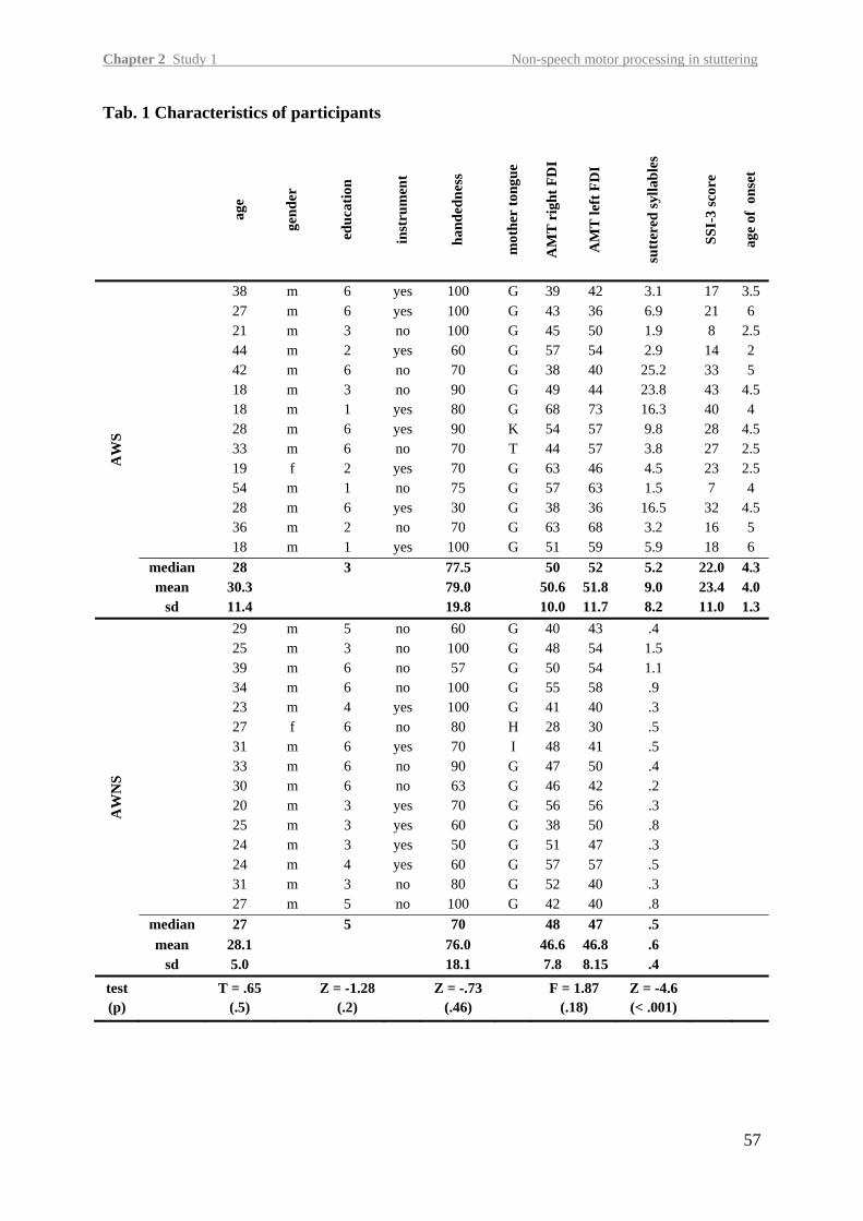
Chapter 2 Study 1 Non-speech motor processing in stuttering
Tab. 1 Characteristics of participants
age
gend
er
educ
atio
n
inst
rum
ent
hand
edne
ss
mot
her
tong
ue
AM
T r
ight
FD
I
AM
T le
ft F
DI
sutt
ered
sylla
bles
SSI-
3 sc
ore
age
of o
nset
AW
S
38 m 6 yes 100 G 39 42 3.1 17 3.5 27 m 6 yes 100 G 43 36 6.9 21 6 21 m 3 no 100 G 45 50 1.9 8 2.5 44 m 2 yes 60 G 57 54 2.9 14 2 42 m 6 no 70 G 38 40 25.2 33 5 18 m 3 no 90 G 49 44 23.8 43 4.5 18 m 1 yes 80 G 68 73 16.3 40 4 28 m 6 yes 90 K 54 57 9.8 28 4.5 33 m 6 no 70 T 44 57 3.8 27 2.5 19 f 2 yes 70 G 63 46 4.5 23 2.5 54 m 1 no 75 G 57 63 1.5 7 4 28 m 6 yes 30 G 38 36 16.5 32 4.5 36 m 2 no 70 G 63 68 3.2 16 5 18 m 1 yes 100 G 51 59 5.9 18 6
median 28 3 77.5 50 52 5.2 22.0 4.3 mean 30.3 79.0 50.6 51.8 9.0 23.4 4.0
sd 11.4 19.8 10.0 11.7 8.2 11.0 1.3
AW
NS
29 m 5 no 60 G 40 43 .4 25 m 3 no 100 G 48 54 1.5 39 m 6 no 57 G 50 54 1.1 34 m 6 no 100 G 55 58 .9 23 m 4 yes 100 G 41 40 .3 27 f 6 no 80 H 28 30 .5 31 m 6 yes 70 I 48 41 .5 33 m 6 no 90 G 47 50 .4 30 m 6 no 63 G 46 42 .2 20 m 3 yes 70 G 56 56 .3 25 m 3 yes 60 G 38 50 .8 24 m 3 yes 50 G 51 47 .3 24 m 4 yes 60 G 57 57 .5 31 m 3 no 80 G 52 40 .3 27 m 5 no 100 G 42 40 .8
median 27 5 70 48 47 .5 mean 28.1 76.0 46.6 46.8 .6
sd 5.0 18.1 7.8 8.15 .4
test (p)
T = .65
(.5) Z = -1.28
(.2) Z = -.73
(.46) F = 1.87
(.18) Z = -4.6 (< .001)
57
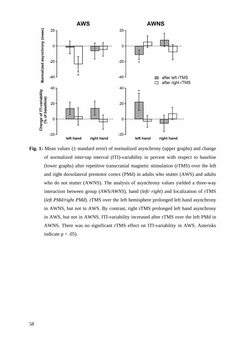
Fig. 1: Mean values (± standard error) of normalized asynchrony (upper graphs) and change
of normalized inter-tap interval (ITI)-variability in percent with respect to baseline
(lower graphs) after repetitive transcranial magnetic stimulation (rTMS) over the left
and right dorsolateral premotor cortex (PMd) in adults who stutter (AWS) and adults
who do not stutter (AWNS). The analysis of asynchrony values yielded a three-way
interaction between group (AWS/AWNS), hand (left/ right) and localization of rTMS
(left PMd/right PMd). rTMS over the left hemisphere prolonged left hand asynchrony
in AWNS, but not in AWS. By contrast, right rTMS prolonged left hand asynchrony
in AWS, but not in AWNS. ITI-variability increased after rTMS over the left PMd in
AWNS. There was no significant rTMS effect on ITI-variability in AWS. Asterisks
indicate p < .05).
58

Chapter 2 Study 1 Non-speech motor processing in stuttering
59

Chapter 2 Study 2 Excitability of the primary motor tongue representation in stuttering
61
2.2 Reduced intracortical inhibition and facilitation in the primary motor
tongue representation of adults who stutter
Authors: N. E. Neef, W. Paulus, A. Neef, A. Wolff von Gudenberg, M. Sommer
The work was done at: Department of Clinical Neurophysiology, Georg-August-University,
Göttingen
Corresponding author: Martin Sommer, Georg-August-University, Department of
Clinical Neurophysiology, Robert-Koch-Str. 40, 37075 Göttingen, Germany; phone: +49 551
39 8463; fax: +49 551 39 8126, email: [email protected]
Keywords stuttering, primary motor tongue representation, trancranial magnetic stimulation,
intrahemispheric inhibition, intrahemispheric facilitation, MEP input-output curve
Acknowledgements: We thank Dr. M. Röbel from the University Medicine Göttingen,
Department of Otorhinolaryngology and Prof. W. Engelke from the University Medicine
Göttingen, Department of Maxillofacial Surgery for technical assistance. This work was
supported by the Stifterverband für die Deutsche Wissenschaft, Walter und Ilse Rose Stiftung
[NN], the Deutsche Forschungsgemeinschaft [SO 429/2-2] and by the German Ministry of
Health [BMBF 01GQ0811 (AN); 01GQ0810 (WP)].

Chapter 2 Study 2 Excitability of the primary motor tongue representation in stuttering
Abstract
Objective: This study aimed at detecting neurophysiological changes, in the primary motor
tongue representation of the left and right hemisphere in adults with persistent stuttering.
Methods: Excitability was examined in 12 patients and in 14 control subjects. Using
transcranial magnetic stimulation (TMS) we examined motor threshold, motor-evoked
potential (MEP) input-output curves, short-term intracortical inhibition (SICI) and
intracortical facilitation (ICF).
Results: In control subjects a significant inhibition of the MEP-amplitude at short inter-
stimulus interval (ISI) as well as a significant facilitation of the MEP-amplitude at long ISIs
was evident. Patients with persistent stuttering showed an inhibition at ISI 3ms and lower
inhibition at 2 ms interval, not reaching statistical significance; this delay of inhibitory
activity was especially prominent in the right hemisphere. Facilitation was reduced at ISI 10
and 15 ms in patients. Furthermore, MEP input-output curve was steeper in patients with
persistent stuttering. Motor thresholds did not differ between groups.
Conclusions: In persistent stuttering intracortical excitability of the primary motor tongue
representation is altered with a deviant time course for inhibitory activity in the right
hemisphere and reduced paired-pulse facilitation.
Significance: These results specify changes in intracortical networks possibly mediated by
altered GABAergic regulations in persistent stuttering. Thus, a better understanding of disease
mechanisms and a potential role in understanding pharmacological treatment response
emerges by using TMS in persistent stuttering.
63
64
1. Introduction
Speech-relevant cortical and subcortical neural systems appear to be malfunctioning in
persistent stuttering (Brown et al., 2005; Fox et al., 1996; Ludlow and Loucks, 2003). This
speech disorder affects approximately 1% of the adult population, severely compromising
their quality of life (Bloodstein and Ratner, 2008; Craig et al., 2002). Stuttering is
characterized by involuntary, intermittent interruptions of fluent speech. Speech sound and
syllable repetitions, sound prolongations and speech blocks are prominent signs. It is still
unclear which changes in motor cortical function contribute to these disruptions to a smooth
execution of complex spatiotemporal commands of articulatory gestures.
Current knowledge about cortical mechanisms in stuttering relies mainly on imaging studies
reporting for instance a right-hemispheric hyperactivity in the primary motor cortex during
dysfluent speech (Braun et al., 1997; Fox et al., 1996; Fox et al., 2000). This hyperactivity has
been speculated to be related to an increased cortical excitability (Ludlow and Loucks, 2003).
In physiological terms motor cortical excitability can be explored using transcranial magnetic
stimulation (TMS). TMS-studies in stuttering are scarce, although this technique is an
established tool to assess noninvasively the functional integrity of descending corticospinal
and corticonuclear pathways of the human motor cortex. Activating descending pathways
originating from layer 5 pyramidal cells in the primary motor cortex requires suprathreshold
TMS stimuli and results in brief muscle response, the motor-evoked potential (MEP).
Subthreshold TMS stimuli can activate intracortical circuits in the motor cortex (Paulus et al.,
2008). Paired-pulse stimulation with a subthreshold conditioning stimulus (CS) followed by a
suprathreshold test stimulus (TS) induces either a reduced motor cortex excitability, short-
term intracortical inhibition (SICI); or an increased motor cortex excitability, the intracortical
facilitation (ICF). SICI and ICF depend on the length of the inter-stimulus interval (ISI)
between CS and TS (Kujirai et al., 1993; Ziemann et al., 1998). Figure 1 depicts modulated
intracortical excitability in a typical healthy subject. Hence, TMS is a tool for transynaptically
activating cortical neurons, providing a method to assess the strength of intracortical synaptic
connections.
Neurons of the primary motor cortex (M1) rececive influential input from frontal cortex
regions and from the basal ganglia; their descending output innervates coordinated, voluntary
movements. Intracortical inhibitory and facilitatory networks within M1 may promote the
selection and initiation of target movements and suppress synergisms. Speech requires a
highly coordinated interplay between different subsystems innervated bilaterally by six
different cranial nerves (V, VII, IX, X, XI, XII). Respiratory activity has to be synchronized
Chapter 2 Study 2 Excitability of the primary motor tongue representation in stuttering
as well, again involving paired spinal nerves. The excitability of the cortical neurons that
project through corticobulbar pathways to the target cranial nerves and further to the target
muscles is shaped by subcortical and intercortical as well as intracortical circuity. Intracortical
excitability modulations are induced by interactions of inhibitory and excitatory circuits.
A disturbed intracortical excitability of neurons in the primary motor cortex (M1) that project
to speech relevant muscles is a potential reason for the dysfluent speech (Ludlow and Loucks,
2003), but the intracortical excitability of speech relevant motor cortex regions had not been
investigated in adults who stutter (AWS). An adjacent region in M1, the hand representation,
was studied in our group (Sommer et al., 2003) with the finding of an unaltered intracortical
excitability in AWS.
Altered intracortical inhibition in stuttering might be a reasonable expectation because
imbalanced cortical excitability in motor regions has been implicated by functional
neuroimaging during symptom production (Brown et al., 2005). Besides; stuttering shares
clinical features of other movement disorders: tic-like involuntary movements (Mulligan et
al., 2003), focal dystonia -like excessive activation of task-related and task-unrelated muscles
(Sommer et al., 2003), and Parkinsonism-like freezing of articulatory gestures (Alm, 2008).
All these movement disorders are characterize by a reduced SICI (Berardelli et al., 2008).
In the present study, we used TMS elicited motor evoked potentials (MEPs) to test the
hypothesis that intracortical excitability in the M1 tongue representation is altered in adults
who stutter (AWS). Specifically, we hypothesized that the excitability of inhibitory circuits
within M1tongue representation is reduced in AWS. A decreased intracortical inhibition may
lead to a reduced inhibition for the prevention of movements (Stinear et al., 2009) thereby
contributing to the intermittent dysfluencies in stuttering.
2. Methods
2.1 Subjects
Twelve AWS (three female; mean 29.9 years, SD 8.2) and fourteen fluent speakers (FS, 3
females, mean 29.5 years, SD 7.6) participated in this study. Adults who stutter were recruited
from the local stuttering support group and the Institute for the Kassel Stuttering Therapy
(Euler et al., 2009). Fluent speakers were recruited by advertisement. The groups were
matched for age, handedness (Oldfield, 1971) and education. Seven AWS reported a family
history of stuttering. None of the FS reported having a family history of speech or language
disorders. Expect from stuttering in the AWS group, participants reported no medical history,
neurological impairment or drug use that would potentially affect their neurological function.
65
66
Before experimental measures were obtained with TMS, all subjects were screened for
exclusion criteria using a standard TMS safety screen (Keel et al., 2001). All subjects
provided written informed consent prior to inclusion into the study. This study received
ethical approval from the Goettingen Ethics Committee.
2.2 Fluency assessment
To judge stuttering severity, fluency assessments were performed regarding the German
version of the SSI-3 (Sandrieser and Schneider, 2008). Speech samples of all participants
containing a conversation about job or school and a reading task were videotaped and
analyzed by a qualified speech-language pathologist. SSI-3 norms were adapted from Riley
(Riley, 1994) . The offline analysis of dysfluencies included 500 syllables for the conversation
and not less than 340 syllables for the reading task. Sound prolongations, blocks (silent
prolongation of an articulatory posture) as well as sound and syllable repetitions were counted
as stuttered syllables. Monosyllabic words that were repeated with apparent undue stress or
tension were counted. Furthermore, the estimated duration of the three longest blocks and
observation of physical concomitants were included for the estimate of stuttering severity in
AWS.
2.3 Experimental procedures
Subjects were seated in a comfortable chair. Bilateral simultaneous surface recordings of the
lingual muscle were taken with two pairs of disposable pre-gelled silver/silver chloride ring
electrodes (5 mm x 100 mm, Viasys Neurocare, Hoechberg, Germany). Electrodes were
mounted at a customized spoon-shaped silicon mouthpiece. Contact area at the tongue was
5 mm x 10 mm at longitudinal and lateral inter-electrode distances of 25 and 20 mm,
respectively. The mouthpiece was placed on the upper surface of the tongue, and the subjects
were asked to close their lips and teeth leisurely without additional pressure and to hold the
end of the mouthpiece with the hand ipsilateral to TMS stimulation site, with the elbow
comfortably supported. While recording participants were asked to push the tongue tightly
against the electrodes and their lower teeth. This procedure was adapted from (Rödel et al.,
2003).
Surface EMG signals were recorded using a CED power 1401 interface with a sampling
frequency of 5 KHz, amplified × 1000 and Butterworth bandpass filtered between 20 Hz and
2000 Hz. Recordings were controlled by Signal Software (Cambridge Electronic Design,
version 2.13).
Chapter 2 Study 2 Excitability of the primary motor tongue representation in stuttering
2.4 Transcranial Magnetic Stimulation
Experiment 1
TMS was applied while participants sat comfortably in a reclining chair. Subjects were
instructed to stay relaxed throughout the assessment. The voluntary background contraction of
the tongue was maintained at approximately 10% of maximum activity (Muellbacher et al.,
2001). Muscle activation was controlled through visual feedback of EMG activity. TMS was
achieved using a monophasic stimulus applied through a figure-of-eight coil with an outer
wing diameter of 70 mm. The coil was positioned tangentially to the skull with the handle
pointing backwards and laterally at an angle of 45° to the sagittal plane. In this position, the
induced current flow in the brain was in the posterior–anterior direction. The scalp surface
was explored systematically and the position for consistently inducing maximal MEPs in the
contralateral tongue site at the lowest stimulus strength was identified as the “hot spot” and
marked with a pen to ensure accurate coil placement throughout the experiment (Muellbacher
et al., 2001). We found the motor tongue representation to be slightly more anterior and more
lateral than what we usually observe for the hand representation which is consistent with the
literature (Svensson et al., 2003). Motor threshold for SICI and ICF was assessed with the coil
connected to a Bistim module (Magstim Company Ltd., Whitland, Wales), which connected
two identical Magstim200 stimulators. Single TMS pulses were applied to determine the
minimal stimulus intensity to the nearest 1% of the maximum stimulator output required to
produce MEPs of greater than 100 µV in at least 3 of 6 consecutive stimuli. This intensity
defines the motor threshold (MT). Intracortical excitability was assessed according to a paired
conditioning test stimulus paradigm (Kujirai et al., 1993; Muellbacher et al., 2001), with a
subthreshold conditioning stimulus followed by a suprathreshold test stimulus at different
inter-stimulus intervals (ISIs). Four ISIs, 2, 3, 10, and 15 ms were tested and randomly
intermixed with the test stimulus given alone. Each of the four ISIs was applied 8 times while
the test stimulus alone was applied 18 times. The conditioning stimulus was applied at 90%
MT and the test stimulus was set at 130% MT. These ISIs and stimulation intensities were
chosen based on findings of a previous study to obtain sizeable SICI and ICF in lingual
muscles (Muellbacher et al., 2001).
This protocol was conducted in 14 FS and in 12 AWS in both hemispheres in a pseudo-
randomized order in two separate sessions.
67
68
Experiment 2
Motor threshold was reassessed and MEP input-output curves was determined with the same
coil, positioned as described before, but this time connected to a single pulse Magstim200
stimulator (Magstim Company Ltd., Whitland, Wales). The MEP input–output curve was
recorded using six intensity levels between 90% and 140% of MT (10% increments). Five
stimuli were applied at each intensity level in a consecutive order every 4 s. Afterwards the
MEP input-output curve was recorded while participants were asked to contract their tongue
with about 60 % maximum activity with a short (one minute) break between intensity levels.
This protocol was conducted in both hemispheres in a pseudo-randomized order in two
separate sessions, in 12 FS and 8 AWS who also participated in experiment 1.
2.6 Data analysis
Altogether participant recruitment comprised 17 AWS and 17 FS. We did not conduct the
whole procedure in 4 AWS and in 3 FS because M1 tongue representation could not be
determined properly in one or both hemispheres of these subjects. One other AWS was
excluded because fluency assessment yielded an additional cluttering component in this
patient. According to the definition ((WHO), 2007b) cluttering was recognized by rapid,
erratic, and dysrhythmic speech dysfluency with distinct speech timing abnormalities.
To determine pre-TMS tongue activity we analyzed the EMG signal of all valid recordings of
every single subject. Recordings with TMS artifacts outlasting the motor evoked response
were excluded. For the paired-pulse protocol 60 ms of the EMG signal immediately before the
TMS artifact and for the input-output curve data 79 ms of the EMG signal were considered.
These signals were corrected for offset, rectified and averaged using Matlab.
Motor evoked potential peak-to-peak amplitudes were analyzed with Signal 4.04 (Cambridge
Electronic Design). Mean peak-to-peak amplitudes were calculated for each condition,
including unconditioned MEP amplitudes of the MEP input-output curve procedure and of the
paired-pulse procedure, and conditioned MEP amplitudes of the paired-pulse procedure for
ISI 2, 3, 10 and 15 ms for either projection and either hemisphere. The conditioned MEP
amplitudes were normalized and are given as ratios of the unconditioned MEP amplitude
recorded in the paired-pulse protocol.
In addition to the peak-to-peak amplitude, MEP magnitude was also estimated by the area
under the baseline corrected and rectified EMG signal, where baseline was defined as average
pre-TMS amplitude of the EMG. The time interval of significant MEP response was defined
manually for each trial and each recording site. The interval selection was guided by the
Chapter 2 Study 2 Excitability of the primary motor tongue representation in stuttering
overall shape of the MEP amplitude envelope and by the rate of change of MEP amplitude.
Selection of intervals and computation of the area under the curve was performed in a custom
written EMG-Browser in Igor Pro (Wavemetrics).
2.7 Statistical analysis
To test for group differences for the variables age and percentage of stuttered syllables we
used two-tailed t-tests for independent samples; for the t of percentage of stuttered syllables
heterogeneity of variance was stated; education and handedness were analyzed with Mann-
Whitney U-tests. Nonparametric testing was chosen since education is an ordinally scaled
variable (1 = school, 2 = high school, 3 = less than 2 years college, 4 = 2 years college, 5 = 4
years college, 6 = postgraduate) and handedness did not show normal distribution in either
group.
Experiment 1
Motor threshold comparison was calculated with repeated-measures ANOVA with
hemisphere (left, right) as a within-subjects factor and group (AWS, FS) as a between-
subjects factor.
Unconditioned MEP amplitudes were tested by 2x2x2 repeated measures ANOVA with the
between-subjects factor group (AWS, FS) and the within-subjects factors hemisphere (left,
right) and projection (contralateral, ipsilateral).
For SICI and ICF analyses, conditioned MEP amplitudes were expressed as a percentage of
unconditioned MEP amplitudes (test stimulus only condition), employing a 2x2x2x2x4
omnibus ANOVA with the between-subjects factor group (AWS, FS) and the within-subjects
factors hemisphere (left, right), projection (contralateral, ipsilateral) and ISI (2 ms, 3ms, 10
ms, 15 ms).
Separate 2 x 2 x 2 x 2 repeated-measures ANOVAs were performed on SICI and ICF data
with the within-subjects factors hemisphere (left, right), projection (contralateral, ipsilateral)
and ISI (ICI: 2 ms, 3ms; ICF: 10 ms, 15 ms) and the between-subjects factor group (AWS,
FS). Post-hoc unpaired, two-tailed t-tests were conducted to determine group differences
considering single conditions.
Pre-TMS tongue activity and unconditioned MEP amplitude were assessed separately using
2 x 2 x 2 repeated measures ANOVAs with within-subjects factor hemisphere (left, right) and
tongue site (left, right) and between-subjects factor group (AWS, FS).
69
70
Experiment 2
Motor threshold comparison was calculated with repeated-measures ANOVA with
hemisphere (left, right) as a within-subjects factor and group (AWS, FS) as a between-
subjects factor.
MEP input-output curves were explored by analyzing MEP amplitudes and MEP areas
separately. A 2 x 2 x 2 x 6 x 2 repeated measures ANOVA with modus (10%contraction,
60%contraction), hemisphere (left, right), projection (contralateral, ipsilateral) and intensity
(90, 100, 110, 120, 130, 140% MT) as within-subjects factors and group (AWS, FS) as a
between-subjects factor served for these analyses.
Pre-TMS tongue activity of the input-output curve data were analyzed with a 2 x 2 x 2 x 6 x 2
repeated measures ANOVA with modus (10%contraction, 60%contraction), hemisphere (left,
right), tongue site (left, right) and intensity (90, 100, 110, 120, 130, 140% MT) as within-
subjects factors and group (AWS, FS) as a between-subjects factor.
Values were considered statistically significant if p < .05. Statistics were performed by SPSS
Statistics 17.0 (http://www.spss.com/de/software).
3. Results
3.1 Experiment 1
Participants
The groups matched for age, t(24) = -0.13, p = .9 (unpaired two tailed t-test), handedness,
p =.980; U-test) and education, p =.206 (U-test). Adults who stutter produced more stuttered
syllables than fluent speakers, t(11.054) = -4.96; p < .001 (unpaired two tailed t-test,
heterogeneity of variance). Stuttering severity was very mild in five, mild in two, moderate in
two, severe in two and very severe in one AWS. Averaged stuttering onset was at age 4.5±2.8
(see Table 1 for demographics and fluency scores).
Motor threshold
The values of MT are given in Table 1. Analysis of variance considering the factors group and
hemisphere yielded no effects or interaction. Previous TMS studies of the primary motor hand
area in AWS resulted in contradictory observations reporting increased motor thresholds in
AWS relative to FS (Sommer et al., 2003), as well as no differences (Neef et al., 2010;
Sommer et al., 2009a).
Pre-TMS tongue activity
Chapter 2 Study 2 Excitability of the primary motor tongue representation in stuttering
Pre-TMS tongue activity in the paired-pulse protocol was similar in both groups (Figure 2A).
ANOVA yielded no effect for hemisphere, projection and group as well as no interactions.
Please insert Figure 1 around here
Unconditioned MEP amplitude
A single unconditioned TMS pulse with 130% MT results in similar MEP amplitudes for FS
and AWS, ANOVA detected no effect of group. MEP amplitudes at contralateral projections
were significantly larger than at ipsilateral projections (effect of projection F(1,24) = 15.44,
p = .001). Post-hoc group-wise comparisons of contralateral and ipsilateral MEP amplitudes
via two-tailed, paired t-tests yielded significant differences for all conditions, expect the right
hemisphere projections in FS (see Table 2). This finding is in accordance with previous
studies and likely reflects a predominance of the contralateral projections present even in the
tongue (Meyer et al., 1997; Rödel et al., 2003). Analysis of variance yielded no other effects.
Please insert Table 2 around here
Intracortical Excitability
In FS ISIs of 2 and 3ms lead to inhibition of the MEP amplitude, whereas ISIs of 10 and
15 ms lead to facilitated the MEP amplitude (Figure 2B). Post hoc paired t-tests revealed
significant changes in intracortical exitability for all ISIs in FS with a significance level
consistently smaller than .0001. These results agree in time course and magnitude with a
previous report (Muellbacher et al., 2001). In AWS excitability was significantly changed for
ISI 3ms, t(11) = -4.9, p < .0001; 10 ms t(11) = 3.75, p = .003; and 15 ms t(11) = 2.4,
p = .035); but inhibition at ISI 2 ms did not reach significance, t(11) = -1.5, p = .15. Figure 2B
depicts the main effect of ISI with F(1,24) = 87.72, p < .0001 Thus, short ISIs at 2 and 3 ms
significantly inhibited the MEP amplitude in both groups expect for ISI 2 ms in AWS, while
longer ISIs at 10 and 15 ms significantly augmented motor evoked responses. The interaction
between ISI and group with F(3,22) = 7.69, p < .0001 indicates that the magnitude of the
effect differs between the two groups.
SICI
Short-term intracortical inhibition was reduced for the right hemispheric projections at an ISI
2 ms in AWS compared to FS (Figure 2D and F). ANOVA yielded an effect of projection
F(1,24) = 4.36, p = .048; an effect of ISI F(1,24) = 9.34, p = .005; an interaction of ISI and
71
72
group F(1,24) = 4.59, p = .043; and an interaction of hemisphere, ISI and group
F(1,24) = 5.41, p = .027.
Post-hoc t-tests revealed a significantly reduced SICI of the ipsilateral projections of the right
hemisphere at an ISI of 2 ms in AWS t(24) = -2.1; p = .046; for the same hemisphere and ISI,
SICI trends to decreased inhibition for the contralateral projection t(24) = -2.0; p = .056).
Analysis of variance yielded no other effects. Additionally we calculated separate
2x2 ANOVAs for each hemisphere and its contralateral projection and show the results in
Table 4.
Please insert Table 3 around here
Please insert Table 4 around here
ICF
Intracortical facilitation was consistently reduced in AWS (Figure 2C-F). ANOVA yielded an
effect of group F(1,24) = 10.34, p = .004; and an interaction of hemisphere and projection
F(1,24) = 6.06, p = .021.
Post-hoc unpaired, two-tailed t-tests revealed significantly reduced ICFs for right hemispheric
contralateral projection at either ISI (10 ms, p = .005; 15 ms, p = .011) and for the ipsilateral
projection at ISI 15 ms (p = .024). Left hemispheric contralateral projections exhibited a
significantly reduced ICF at ISI 10 ms (p = .034). Table 3 contains p values for all conditions
documenting a reduced ICF for all conditions at a level of marginally significance (p < .1) in
AWS related to FS. Analysis of variance yielded no other effects. Additionally we calculated
separate 2x2 ANOVAs for each hemisphere and its contralateral projection and show the
results in Table 4.
Please insert Figure 2 around here
3.3 Experiment 2
Participants
The groups matched for age, t(20) = -0.24, p = .811 ( unpaired two tailed t-test), handedness,
p =.680 (U-test) and education, p =.205(U-test). Adults who stutter produced more stuttered
syllables than fluent speakers, t(7.02) = -3.91; p = .006 (unpaired two tailed t-test,
heterogeneity of variance). Stuttering severity was very mild in three, mild in two, moderate
Chapter 2 Study 2 Excitability of the primary motor tongue representation in stuttering
in one, severe in one and very severe in one AWS. Averaged stuttering onset was at age
5.1±3.3 (see Table 1 for demographics and fluency scores).
Motor threshold
In FS MT was for the left and right hemisphere 42.6±6.7% and 44.3±8.8% maximum
stimulator output, respectively. AWS had a MT for the left and right hemisphere of 42.8±8.4
and 40.8±5.9% maximum stimulator output. ANOVA yielded no significant effects or
interaction (see Table 1 and Figure 3B).
Pre-TMS tongue activity
Pre-TMS tongue activity in the MEP input-output curve data did not differ between groups
(Figure 3A). ANOVA yielded a significant effect of mode F(1,18) = 9.33; p = .007, with an
increased tongue activation at a contraction of approximately 60 % of maximum contraction
0.048±0.05 mV versus tongue activation at a contraction of approximately 10 % of maximum
contraction 0.014±0.01mV (see Figure 3A).
MEP input-output curve
The ANOVA considering MEP peak-to-peak amplitude revealed an effect of mode,
F(1,18) = 11.45, p = .003 which reflects that MEP amplitudes are larger while the tongue is
more strongly contracted (mean MEP 1.01± 0.67 mV) compared to less contraction (mean
MEP 0.55± 0.28 mV). Furthermore, analysis yielded an effect of projection, F(1,18) = 31.29,
p < .001 with larger MEP amplitudes in the contralateral projection (0.91±0.48 mV) than in
the ipsilateral projection (0.65±0.38 mV). This again reflects slightly stronger contralateral
projections and is in accordance with previous reports from other labs (Muellbacher et al.,
2001).The effect of stimulus intensity, F(5,14) = 16.49, p < 0.0001, is reflected in the steady
increase of MEP amplitudes with increasing stimulus intensity (90% 0.31±0.19 mV,
100% 0.44±0.27, 110% 0.65±0.35, 120% 0.91±0.55, 130% 1.1±0.68, 140% 1.28±0.62; see
Figure 3C). Analysis yielded an interaction between stimulus intensity and group,
F(5,14) = 2.74, p = .023, because MEP recruitment was steeper in AWS related to FS (Figure
3B). Post-hoc unpaired t-tests of MEP amplitudes yielded no significant differences between
groups for separate conditions. Additionally, we found an interaction between mode and
stimulus intensity, F(1,18) = 3.86, p = .003, because the slope of the recruitment curve under
10% contraction was steeper at lower intensities and flatter at higher intensities compared to
the recruitment curve under 60% contraction which showed the reversed pattern (Figure 3E).
Finally, there was an effect of mode, hemisphere and projection, F(5,14) = 6.23 p = .02,
because under 60% contraction motor responses were enlarged in the contralateral projection
73
74
of the left hemisphere (1.29± 1.06 mV) compared to the contralateral projection of the right
hemisphere (1.05±0.66mV).
The ANOVA considering MEP area yielded one additional interaction between projection and
stimulus intensity with F(5,15) = 20.76; p < .0001. Areas of the MEP of the contralateral
projection had a steeper slope compared to the MEP areas of the ipsilateral projection (Figure
3F).
Furthermore, the ANOVA with MEP areas yielded almost the same effects compared to the
ANOVA with MEP peak-to-peak amplitudes: effect of mode F(1,18) = 10.95; p = .004; effect
of projection F(1,18) = 34.12, p < .0001, effect of stimulus intensity F(5,14) = 30.9;
p < .0001; an interaction of mode, hemisphere and projection F(5,14) = 4.92, p = .04; and an
interaction between mode, stimulus intensity and group F(5,14) = 2.38; p = .045. Post hoc
repeated measures ANOVAS separated for mode yielded in an significant interaction between
group and intensity at 60% of maximum contraction, F(5,14) = 2.55; p = .033. This
interaction was missing in the 10% of maximum contraction mode. Post hoc unpaired t-tests
yielded no differences between groups concerning the MEP area at 60% maximum
contraction at 140% MT or at 130% MT although the slope is steeper in AWS in this
condition (Figure 3D).
Please insert Figure 3 around here
4. Discussion
Here we present the first assessment of intracortical excitability in the M1 representations of
the lingual muscle in stuttering. The reduction of short-term intracortical inhibition at ISI 2ms
in our sample of stuttering subjects partly confirms our hypothesis of a reduced ICI.
Unexpected were the observations of a generally reduced intracortical facilitation, and of a
steeper MEP recruitment in adults who stutter related to fluent speakers.
4.1 Reduction of short-term intracortical inhibition in stuttering
The reduction of SICI indicates an altered excitability modulation of intracortical neural
networks in stuttering. Due to the local action of TMS and the limited conduction velocities
and synaptic delays it is assumed that SICI is mediated by the local neuronal circuits in the
motor cortex (Di Lazzaro et al., 1998; Ziemann and Rothwell, 2000). At intervals larger than
1 ms SICI is caused by the activation of GABAergic interneurons and the subsequent
inhibition of excitatory neurons (Fisher et al., 2002; Hanajima et al., 2003). We speculate that
Chapter 2 Study 2 Excitability of the primary motor tongue representation in stuttering
in AWS’s M1 tongue representation the interneuronal inhibitory network is less active at early
times. In the next paragraph we shortly introduce the cellular physiology framework to
discuss the effect.
A TMS pulse stimulates nerve fibers most likely at their terminations (Maccabee et al., 1993;
Rotem and Moses, 2008) thereby activating synaptic terminals in all cortical layers. A
suprathreshold TMS pulse activates enough excitatory synapses to elicit action potentials in
layer 5 excitatory cells, the cortical output responsible for muscle activation. The layer 2/3
excitatory cells are activated as well, and in turn stimulate layer 5 cells, thereby prolonging
and increasing the motor output. If the TMS pulse is weaker, excitatory cells are not
sufficiently activated to fire action potentials, no motor response is elicited; the TMS stimulus
is called subthreshold. Even in this case, however, the inhibitory interneurons are depolarized
enough to fire action potentials. Those action potentials reach inhibitory (GABAergic)
synapses onto the layer 2/3 and layer 5 excitatory neurons after a distance dependent
conduction delay of 0-1 ms (Esser et al., 2005). Over the next 2 ms the inhibitory postsynaptic
currents in the excitatory neurons increase. If a second, stronger TMS pulse is applied during
this period, it might still suffice to activate the excitatory layer 5 neurons, that cause the motor
response but due to the built up inhibition, the excitation of layer 5 and especially layer 2/3
neurons is weaker, leading to a reduction of action potential number, when compared to a
single, unconditioned TMS stimulus. This view is supported by large scale modeling (Esser et
al., 2005).
Our findings indicate that at an ISI of 2ms for FS there is significant inhibition but not in
AWS. In the right hemisphere in AWS the inhibition takes longer to develop. This delay of
the peak inhibitory activity implies that inhibitory inputs on the excitatory cells develop with
a slower time course in AWS. The reasons can be diverse, including altered kinetics of
synaptic signaling (altered subunit composition of GABA receptor complexes), longer
conduction delays (a larger fraction of long range, i.e. 1 mm, inhibitory connections; (Kang et
al., 1994) or short term synaptic plasticity e.g synaptic depression due to depletion of release-
competent synaptic vesicles at the excitatory synapses innervating the layer 5 excitatory
neurons.
GABAeric interneurons seem to play a key role in mediating the effect of intracortical
inhibition. Therefore it is interesting to note that stuttering can be induced by theophylline
(Movsessian, 2005) an adenosin receptor antagonist which has been previously described to
reduce the binding of GABA to GABAA receptors via a decoupling of the benzodiazepine
binding site that is present on the receptor (Roca et al., 1990). Additionally, theophylline
75
76
plasma concentration correlates positively with a reduction of SICI in healthy subjects
(Nardone et al., 2004). SICI is mediated by either the α2- or α3-subunit of the GABAA
receptor (Di Lazzaro et al., 2006). Thus, the susceptibility of the speech motor system in
stuttering might be mediated by GABAA neurons.
4.2 Reduced intracortical facilitation in stuttering
One possible explanation for a generally reduced ICF in stuttering might be a different level
of pre TMS tongue activation. It is known that orofacial muscles are rarely at rest (Devlin and
Watkins, 2008) and that muscle activation reduces ICF suggesting that voluntary drive
reduces the excitability of intracortical circuits (Ridding et al., 1995). Therefore one might
conclude that the pre TMS tongue activity was higher in AWS and hence ICF was reduced.
However, the comparison of pre TMS tongue activity revealed no group differences.
Consistent with this, previous EMG studies in stuttering do not provide evidence for elevated
tonic activity or a co-activation in the laryngeal or orofacial muscles, neither during fluent
speech nor during dysfluent speech (Smith et al., 1996; Smith et al., 1993). Even at rest lower
lip activity did not differ between AWS and FS, while upper lip activity has been reported to
be lower in AWS (de Felicio et al., 2007).
Another possible explanation for a reduced ICF might be that the MEP amplitude was already
saturated in AWS at a test pulse intensity of 130% MT. This point also does not hold true
because the MEP recruitment until 140% MT resulted in a steeper slope in AWS and the
comparison of MEP areas between groups yielded no differences. Thus, our findings
implicate a diminished excitability of excitatory neuronal circuits in motor cortex in the
sample of AWS examined in the current study.
While ICF is normal in the M1 hand representation in AWS (Sommer et al., 2003) the current
data imply a reduced intracortical facilitation for the M1 tongue representation. In contrast to
ICI which is reduced in several movement disorders, reduced ICF has been described only in
few movement disorders; unequivocally in cerebellar ataxia and equivocally in Huntington’s
disease (Berardelli et al., 2008). The fact, that these movement disorders as well as movement
disorders that are characterized by reduced ICI are related to a dysfunction of the basal
ganglia or the cerebellum, suggests that the dysfunction in these structures might influence
intracortical circuits that modulate motor cortex excitability. In stuttering the contribution of
altered cerebello-cortical loops (Lu et al., 2010) as well as deviant basal ganglia function are
proposed (Alm, 2004). Clinical trials with dopamine antagonists resulted in a positive effect
on speech fluency (Burns et al., 1978; Maguire et al., 2010) while dopamine agonists
Chapter 2 Study 2 Excitability of the primary motor tongue representation in stuttering
enhanced dysfluency. An early study with Positron Emission Tomography reports an
increased uptake of presynaptic dopamine in stuttering which indicates that stuttering is
related to a hyper-dopaminergic status (Wu et al., 1997). Although, physiological mechanisms
of ICF are not well understood we here speculate that the reduced ICF might be mediated by
disturbed interaction between cortical and subcortical networks modulating inhibitory and
facilitatory intracortical circuits.
Although, TMS paired-pulse techniques employed to study SICI and ICF in different
movement disorders provide important information about the pathophysiologies in the
primary motor cortex (Hanajima and Ugawa, 2008), the exact mechanisms of these
modulations of cortical excitability are still a matter of debate (Reis et al., 2008).
Furthermore, literature on intracortical excitability in speech relevant muscles is scarce.
Because the tongue muscles are rarely at rest and the innervations occurs bilaterally a direct
comparison with previous literature on SICI and ICF is limited and restricts general
conclusions.
4.3 MEP recruitment
The recording of the MEP input-output curve was necessary to address the question, whether
MEP amplitude was already statured in AWS in the facilitatory paired-pulse condition. To
take care for a potential ceiling effect we obtained MEP input-output curves under different
conditions of tongue activity. While recruitment under 10% of maximum tongue contraction
was characterized by a steep slope at high intensities, MEP recruitment seems to reach
staturation at high intensities under 60% contraction (Figure 3E). Figure 3C and 3D detail that
this saturation is more clearly displayed in FS compared to AWS were recruitment between
130 and 140% of maximum stimulator output shows a further trend towards a larger MEP
amplitude and MEP area. It was not possible to win all AWS that participated in the ICI/ICF
session to participate in the MEP input-output curve session. Thus, the data result from a
subsample of AWS of the first experiment which limits their interpretation.
4.5 Implications
We specified a reduced and possibly delayed SICI in the right hemisphere and a reduced ICF
in either hemisphere which implies altered intracortical modulation of inhibitory as well as
facilitatory circuits in stuttering. As the final cortical processing stage for voluntary
movement, the primary motor cortex is a critical site for the integration of movement
selection, initiation and prevention processes. Upcoming studies might further elucidate state-
77
78
dependent modulation of intracortical inhibition to conceive pathomechanisms of dysfluent
speech production in stuttering. Furthermore, single-pulse and paired-pulse TMS can be
employed to study underlying physiological mechanisms of a pharmacologically induced
enhancement of speech fluency and to compliment current pharmacological approaches.
Disclosure
The authors report no conflicts of interest.
Chapter 2 Study 2 Excitability of the primary motor tongue representation in stuttering
References
WHO. ICD-10 F98.6 Cluttering 2007:Retrieved from
www.who.int/classifications/apps/icd/icd10online.
Alm PA. Stuttering and the basal ganglia circuits: a critical review of possible relations. J
Commun Disord. 2004 Jul-Aug;37(4):325-69.
Alm PA. Fluency disorders: a discussion of possible causes and mechanisms, from a
neuroscience perspective. 8th Oxford Dysfluency Conference; 2008; Oxford, UK. 2008.
Berardelli A, Abbruzzese G, Chen R, Orth M, Ridding MC, Stinear C, et al. Consensus paper
on short-interval intracortical inhibition and other transcranial magnetic stimulation
intracortical paradigms in movement disorders. Brain Stimul. 2008 Jul;1(3):183-91.
Bloodstein O, Ratner BN. A Handbook on Stuttering. 6 ed. Canada: Delmar Thomson
Learning; 2008.
Braun AR, Varga M, Stager S, Schulz G, Selbie S, Maisog JM, et al. Altered patterns of
cerebral activity during speech and language production in developmental stuttering. An
H2(15)O positron emission tomography study. Brain. 1997 May;120 ( Pt 5):761-84.
Brown S, Ingham RJ, Ingham JC, Laird AR, Fox PT. Stuttered and fluent speech production:
an ALE meta-analysis of functional neuroimaging studies. Hum Brain Mapp. 2005
May;25(1):105-17.
Burns D, Brady JP, Kuruvilla K. The acute effect of haloperidol and apomorphine on the
severity of stuttering. Biol Psychiatry. 1978 Apr;13(2):255-64.
Craig A, Hancock K, Tran Y, Craig M, Peters K. Epidemiology of stuttering in the
community across the entire life span. J Speech Lang Hear Res. 2002 Dec;45(6):1097-
105.
de Felicio CM, Freitas RL, Vitti M, Regalo SC. Comparison of upper and lower lip muscle
activity between stutterers and fluent speakers. Int J Pediatr Otorhinolaryngol. 2007
Aug;71(8):1187-92.
Devlin JT, Watkins KE. Investigating language organization with TMS. In: Wassermann EM,
Epstein CM, Ziemann U, Walsh V, Paus T, Lisanby SH, editors. The Oxford Handbokk
of Transcranial Magnetic Stimulation. New York: Oxford University Press; 2008. p.
479-99.
Di Lazzaro V, Pilato F, Dileone M, Ranieri F, Ricci V, Profice P, et al. GABAA receptor
subtype specific enhancement of inhibition in human motor cortex. J Physiol. 2006 Sep
15;575(Pt 3):721-6.
79
80
Di Lazzaro V, Restuccia D, Oliviero A, Profice P, Ferrara L, Insola A, et al. Magnetic
transcranial stimulation at intensities below active motor threshold activates
intracortical inhibitory circuits. Exp Brain Res. 1998 Mar;119(2):265-8.
Esser SK, Hill SL, Tononi G. Modeling the effects of transcranial magnetic stimulation on
cortical circuits. J Neurophysiol. 2005 Jul;94(1):622-39.
Euler HA, Gudenberg AWv, Jung K, Neumann K. Computergestützte Therapie bei
Redeflussstörungen: Die langfristige Wirksamkeit der Kasseler Stottertherapie. Sprache,
Stimme, Gehör. 2009;33(4):193-97.
Fisher RJ, Nakamura Y, Bestmann S, Rothwell JC, Bostock H. Two phases of intracortical
inhibition revealed by transcranial magnetic threshold tracking. Exp Brain Res. 2002
Mar;143(2):240-8.
Fox PT, Ingham RJ, Ingham JC, Hirsch TB, Downs JH, Martin C, et al. A PET study of the
neural systems of stuttering. Nature. 1996 Jul 11;382(6587):158-61.
Fox PT, Ingham RJ, Ingham JC, Zamarripa F, Xiong JH, Lancaster JL. Brain correlates of
stuttering and syllable production. A PET performance-correlation analysis. Brain. 2000
Oct;123 ( Pt 10):1985-2004.
Hanajima R, Furubayashi T, Iwata NK, Shiio Y, Okabe S, Kanazawa I, et al. Further evidence
to support different mechanisms underlying intracortical inhibition of the motor cortex.
Exp Brain Res. 2003 Aug;151(4):427-34.
Hanajima R, Ugawa Y. Paired-pulse measures. In: Wassermann EM, Epstein CM, Ziemann
U, Paus T, Lisanby SH, editors. The Oxford handbook of transcranial magnetic
stimulation. New York: Oxford Univerity Press; 2008.
Kang Y, Kaneko T, Ohishi H, Endo K, Araki T. Spatiotemporally differential inhibition of
pyramidal cells in the cat motor cortex. J Neurophysiol. 1994 Jan;71(1):280-93.
Keel JC, Smith MJ, Wassermann EM. A safety screening questionnaire for transcranial
magnetic stimulation. Clin Neurophysiol. 2001;112(4):720.
Kujirai T, Caramia MD, Rothwell JC, Day BL, Thompson PD, Ferbert A, et al.
Corticocortical inhibition in human motor cortex. J Physiol. 1993 Nov;471:501-19.
Lu C, Chen C, Ning N, Ding G, Guo T, Peng D, et al. The neural substrates for atypical
planning and execution of word production in stuttering. Exp Neurol. [; Research
Support, Non-U.S. Gov't]. 2010 2010 Jan (Epub;221(1):146-56.
Ludlow CL, Loucks T. Stuttering: a dynamic motor control disorder. J Fluency Disord. 2003
Winter;28(4):273-95.

Chapter 2 Study 2 Excitability of the primary motor tongue representation in stuttering
Maccabee PJ, Amassian VE, Eberle LP, Cracco RQ. Magnetic coil stimulation of straight and
bent amphibian and mammalian peripheral nerve in vitro: locus of excitation. J Physiol.
1993 Jan;460:201-19.
Maguire G, Franklin D, Vatakis NG, Morgenshtern E, Denko T, Yaruss JS, et al. Exploratory
randomized clinical study of pagoclone in persistent developmental stuttering: the
Examining Pagoclone for peRsistent dEvelopmental Stuttering Study. J Clin
Psychopharmacol. 2010 Feb;30(1):48-56.
Meyer BU, Liebsch R, Roricht S. Tongue motor responses following transcranial magnetic
stimulation of the motor cortex and proximal hypoglossal nerve in man.
Electroencephalogr Clin Neurophysiol. 1997 Feb;105(1):15-23.
Movsessian P. Neuropharmacology of theophylline induced stuttering: the role of dopamine,
adenosine and GABA. Med Hypotheses. 2005;64(2):290-7.
Muellbacher W, Boroojerdi B, Ziemann U, Hallett M. Analogous corticocortical inhibition
and facilitation in ipsilateral and contralateral human motor cortex representations of the
tongue. J Clin Neurophysiol. 2001 Nov;18(6):550-8.
Mulligan HF, Anderson TJ, Jones RD, Williams MJ, Donaldson IM. Tics and developmental
stuttering. Parkinsonism Relat Disord. 2003 Jun;9(5):281-9.
Nardone R, Buffone E, Covi M, Lochner PG, Tezzon F. Changes in motor cortical excitability
in humans following orally administered theophylline. Neurosci Lett. 2004 Jan
23;355(1-2):65-8.
Neef N, Jung K, Rothkegel H, Pollok B, Wolff von Gudenberg A, Paulus W, et al. Right-shift
for non-speech motor processing in adults who stutter. Cortex. 2010.
Oldfield RC. The assessment and analysis of handedness: the Edinburgh inventory.
Neuropsychologia. 1971 Mar;9(1):97-113.
Paulus W, Classen J, Cohen LG, Large CH, Di Lazzaro V, Nitsche M, et al. State of the art:
Pharmacologic effects on cortical excitability measures tested by transcranial magnetic
stimulation. Brain Stimul. 2008 Jul;1(3):151-63.
Reis J, Swayne OB, Vandermeeren Y, Camus M, Dimyan MA, Harris-Love M, et al.
Contribution of transcranial magnetic stimulation to the understanding of cortical
mechanisms involved in motor control. J Physiol. 2008 Jan 15;586(2):325-51.
Ridding MC, Taylor JL, Rothwell JC. The effect of voluntary contraction on cortico-cortical
inhibition in human motor cortex. J Physiol. 1995 Sep 1;487 ( Pt 2):541-8.
Riley GD. The Stuttering Severity Instrument for Children and Adults- Third Edition (SSI-3).
Austin, TX: Pro-Ed.; 1994.
81
82
Roca DJ, Schiller GD, Friedman L, Rozenberg I, Gibbs TT, Farb DH. gamma-Aminobutyric
acidA receptor regulation in culture: altered allosteric interactions following prolonged
exposure to benzodiazepines, barbiturates, and methylxanthines. Mol Pharmacol. 1990
May;37(5):710-9.
Rödel RM, Laskawi R, Markus H. Tongue representation in the lateral cortical motor region
of the human brain as assessed by transcranial magnetic stimulation. Ann Otol Rhinol
Laryngol. 2003 Jan;112(1):71-6.
Rotem A, Moses E. Magnetic stimulation of one-dimensional neuronal cultures. Biophys J.
2008 Jun;94(12):5065-78.
Sandrieser P, Schneider P. Stottern im Kindesalter. 3 ed. Stuttgart: Thieme; 2008.
Smith A, Denny M, Shaffer LA, Kelly EM, Hirano M. Activity of intrinsic laryngeal muscles
in fluent and disfluent speech. J Speech Hear Res. 1996 Apr;39(2):329-48.
Smith A, Luschei E, Denny M, Wood J, Hirano M, Badylak S. Spectral analyses of activity of
laryngeal and orofacial muscles in stutterers. J Neurol Neurosurg Psychiatry. 1993
Dec;56(12):1303-11.
Sommer M, Knappmeyer K, Hunter EJ, Gudenberg AW, Neef N, Paulus W. Normal
interhemispheric inhibition in persistent developmental stuttering. Mov Disord. 2009
Apr 15;24(5):769-73.
Sommer M, Wischer S, Tergau F, Paulus W. Normal intracortical excitability in
developmental stuttering. Mov Disord. 2003 Jul;18(7):826-30.
Stinear CM, Coxon JP, Byblow WD. Primary motor cortex and movement prevention: where
Stop meets Go. Neurosci Biobehav Rev. 2009 May;33(5):662-73.
Svensson P, Romaniello A, Arendt-Nielsen L, Sessle BJ. Plasticity in corticomotor control of
the human tongue musculature induced by tongue-task training. Exp Brain Res. 2003
Sep;152(1):42-51.
Wu JC, Maguire G, Riley G, Lee A, Keator D, Tang C, et al. Increased dopamine activity
associated with stuttering. Neuroreport. 1997 Feb 10;8(3):767-70.
Ziemann U, Rothwell JC. I-waves in motor cortex. J Clin Neurophysiol. 2000 Jul;17(4):397-
405.
Ziemann U, Tergau F, Wassermann EM, Wischer S, Hildebrandt J, Paulus W. Demonstration
of facilitatory I wave interaction in the human motor cortex by paired transcranial
magnetic stimulation. J Physiol. 1998 Aug 15;511 ( Pt 1):181-90.

Chapter 2 Study 2 Excitability of the primary motor tongue representation in stuttering
83
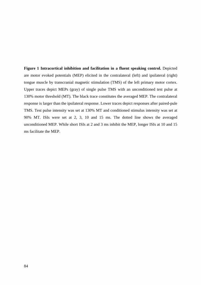
84
Figure 1 Intracortical inhibition and facilitation in a fluent speaking control. Depicted
are motor evoked potentials (MEP) elicited in the contralateral (left) and ipsilateral (right)
tongue muscle by transcranial magnetic stimulation (TMS) of the left primary motor cortex.
Upper traces depict MEPs (gray) of single pulse TMS with an unconditioned test pulse at
130% motor threshold (MT). The black trace constitutes the averaged MEP. The contralateral
response is larger than the ipsilateral response. Lower traces depict responses after paired-pule
TMS. Test pulse intensity was set at 130% MT and conditioned stimulus intensity was set at
90% MT. ISIs were set at 2, 3, 10 and 15 ms. The dotted line shows the averaged
unconditioned MEP. While short ISIs at 2 and 3 ms inhibit the MEP, longer ISIs at 10 and 15
ms facilitate the MEP.
Chapter 2 Study 2 Excitability of the primary motor tongue representation in stuttering
85
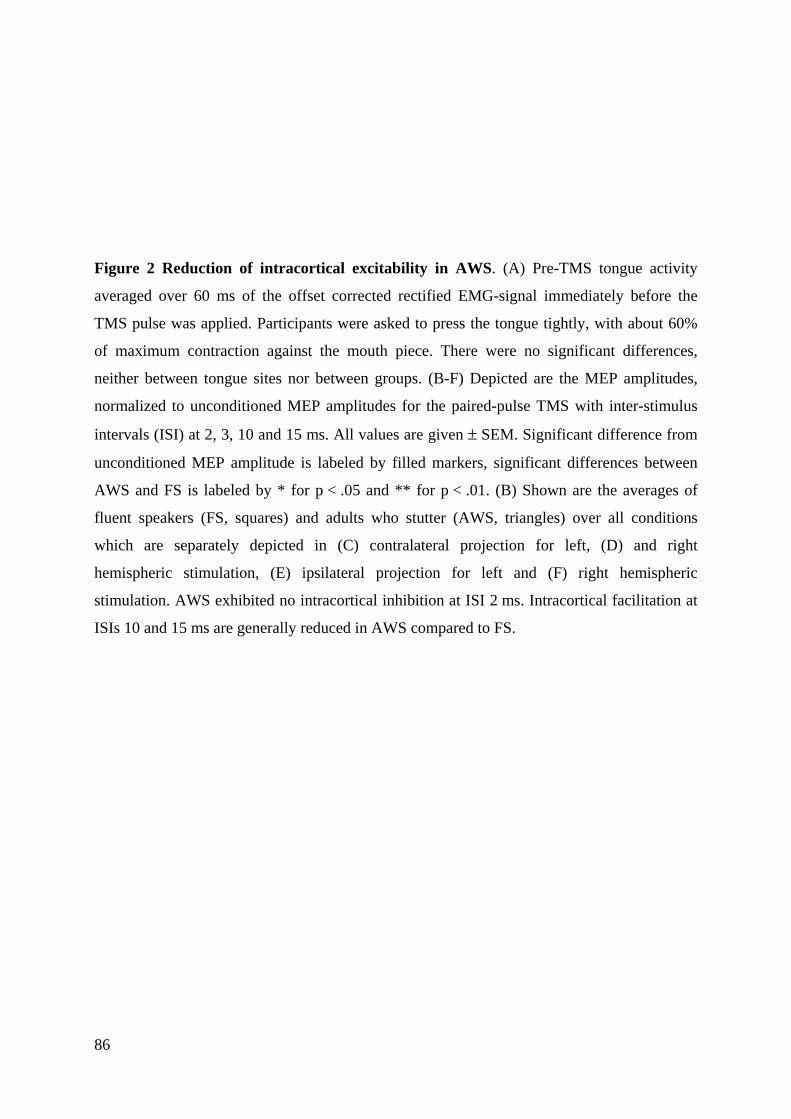
86
Figure 2 Reduction of intracortical excitability in AWS. (A) Pre-TMS tongue activity
averaged over 60 ms of the offset corrected rectified EMG-signal immediately before the
TMS pulse was applied. Participants were asked to press the tongue tightly, with about 60%
of maximum contraction against the mouth piece. There were no significant differences,
neither between tongue sites nor between groups. (B-F) Depicted are the MEP amplitudes,
normalized to unconditioned MEP amplitudes for the paired-pulse TMS with inter-stimulus
intervals (ISI) at 2, 3, 10 and 15 ms. All values are given ± SEM. Significant difference from
unconditioned MEP amplitude is labeled by filled markers, significant differences between
AWS and FS is labeled by * for p < .05 and ** for p < .01. (B) Shown are the averages of
fluent speakers (FS, squares) and adults who stutter (AWS, triangles) over all conditions
which are separately depicted in (C) contralateral projection for left, (D) and right
hemispheric stimulation, (E) ipsilateral projection for left and (F) right hemispheric
stimulation. AWS exhibited no intracortical inhibition at ISI 2 ms. Intracortical facilitation at
ISIs 10 and 15 ms are generally reduced in AWS compared to FS.
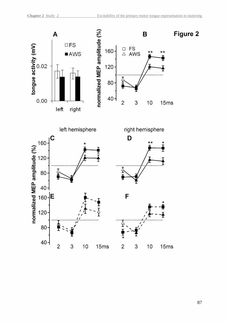
Chapter 2 Study 2 Excitability of the primary motor tongue representation in stuttering
87
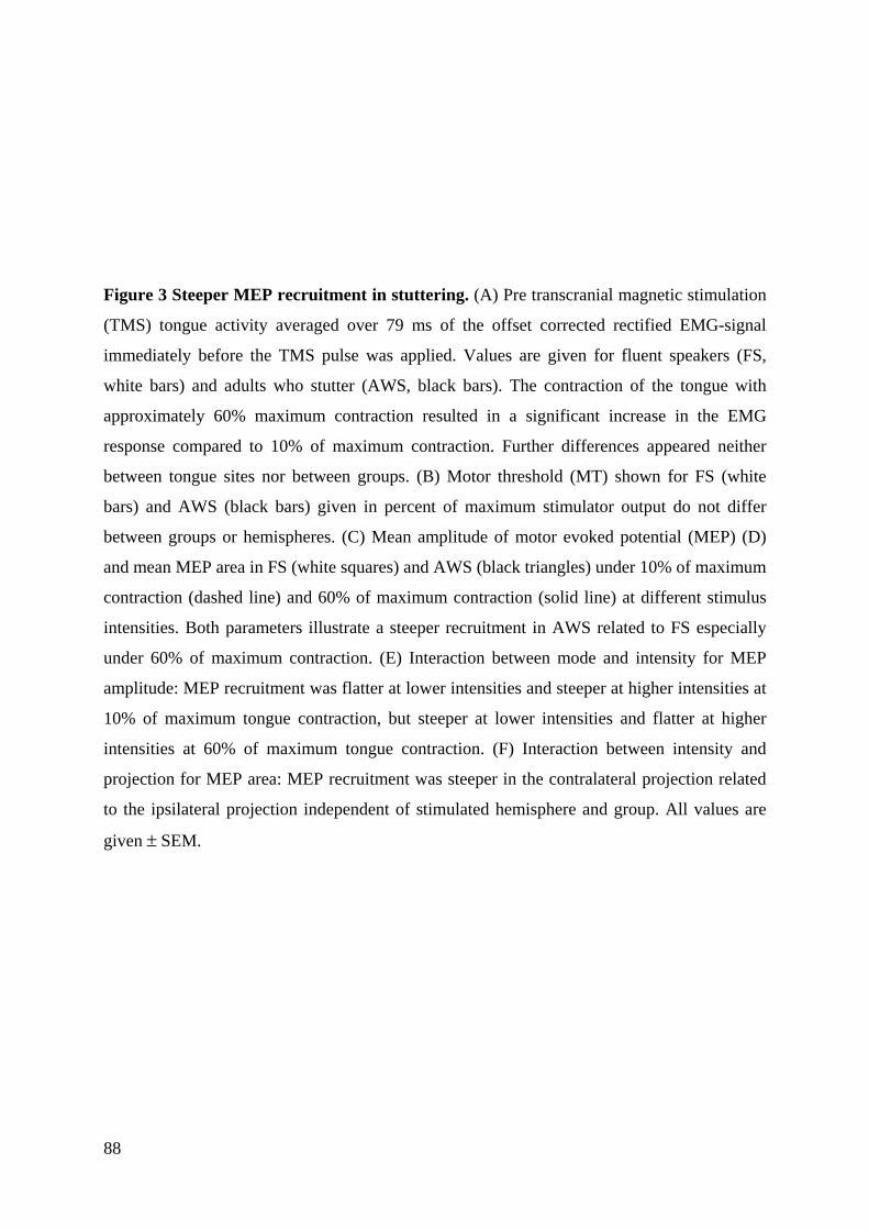
88
Figure 3 Steeper MEP recruitment in stuttering. (A) Pre transcranial magnetic stimulation
(TMS) tongue activity averaged over 79 ms of the offset corrected rectified EMG-signal
immediately before the TMS pulse was applied. Values are given for fluent speakers (FS,
white bars) and adults who stutter (AWS, black bars). The contraction of the tongue with
approximately 60% maximum contraction resulted in a significant increase in the EMG
response compared to 10% of maximum contraction. Further differences appeared neither
between tongue sites nor between groups. (B) Motor threshold (MT) shown for FS (white
bars) and AWS (black bars) given in percent of maximum stimulator output do not differ
between groups or hemispheres. (C) Mean amplitude of motor evoked potential (MEP) (D)
and mean MEP area in FS (white squares) and AWS (black triangles) under 10% of maximum
contraction (dashed line) and 60% of maximum contraction (solid line) at different stimulus
intensities. Both parameters illustrate a steeper recruitment in AWS related to FS especially
under 60% of maximum contraction. (E) Interaction between mode and intensity for MEP
amplitude: MEP recruitment was flatter at lower intensities and steeper at higher intensities at
10% of maximum tongue contraction, but steeper at lower intensities and flatter at higher
intensities at 60% of maximum tongue contraction. (F) Interaction between intensity and
projection for MEP area: MEP recruitment was steeper in the contralateral projection related
to the ipsilateral projection independent of stimulated hemisphere and group. All values are
given ± SEM.
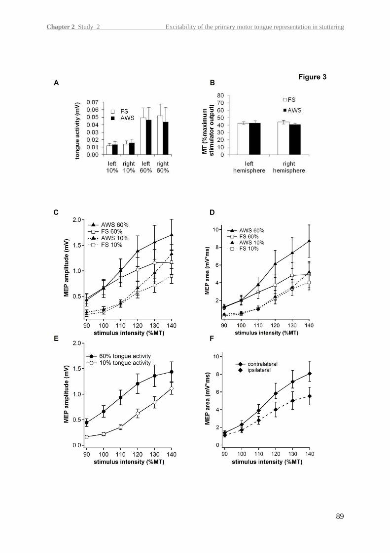
Chapter 2 Study 2 Excitability of the primary motor tongue representation in stuttering
89
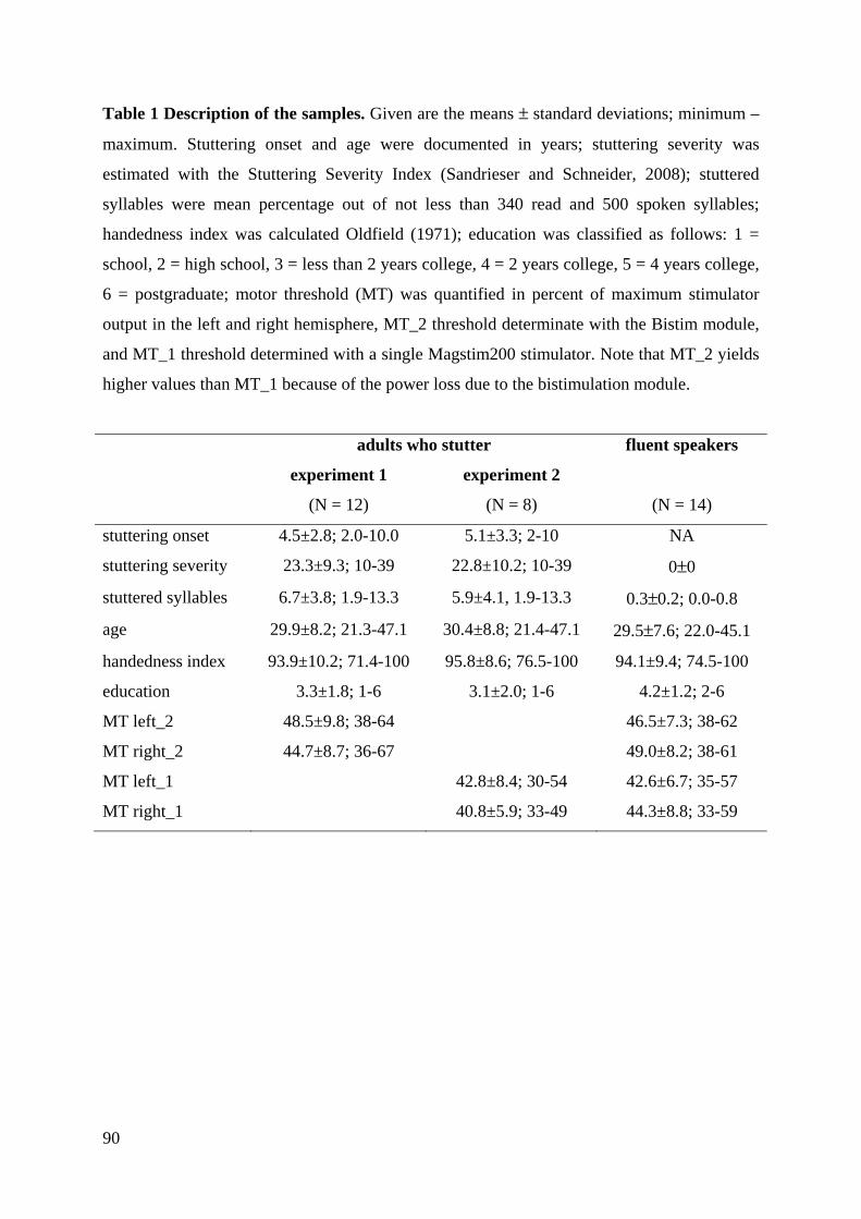
90
Table 1 Description of the samples. Given are the means ± standard deviations; minimum –
maximum. Stuttering onset and age were documented in years; stuttering severity was
estimated with the Stuttering Severity Index (Sandrieser and Schneider, 2008); stuttered
syllables were mean percentage out of not less than 340 read and 500 spoken syllables;
handedness index was calculated Oldfield (1971); education was classified as follows: 1 =
school, 2 = high school, 3 = less than 2 years college, 4 = 2 years college, 5 = 4 years college,
6 = postgraduate; motor threshold (MT) was quantified in percent of maximum stimulator
output in the left and right hemisphere, MT_2 threshold determinate with the Bistim module,
and MT_1 threshold determined with a single Magstim200 stimulator. Note that MT_2 yields
higher values than MT_1 because of the power loss due to the bistimulation module.
adults who stutter fluent speakers
experiment 1
(N = 12)
experiment 2
(N = 8)
(N = 14)
stuttering onset 4.5±2.8; 2.0-10.0 5.1±3.3; 2-10 NA
stuttering severity 23.3±9.3; 10-39 22.8±10.2; 10-39 0±0
stuttered syllables 6.7±3.8; 1.9-13.3 5.9±4.1, 1.9-13.3 0.3±0.2; 0.0-0.8
age 29.9±8.2; 21.3-47.1 30.4±8.8; 21.4-47.1 29.5±7.6; 22.0-45.1
handedness index 93.9±10.2; 71.4-100 95.8±8.6; 76.5-100 94.1±9.4; 74.5-100
education 3.3±1.8; 1-6 3.1±2.0; 1-6 4.2±1.2; 2-6
MT left_2 48.5±9.8; 38-64 46.5±7.3; 38-62
MT right_2 44.7±8.7; 36-67 49.0±8.2; 38-61
MT left_1 42.8±8.4; 30-54 42.6±6.7; 35-57
MT right_1 40.8±5.9; 33-49 44.3±8.8; 33-59
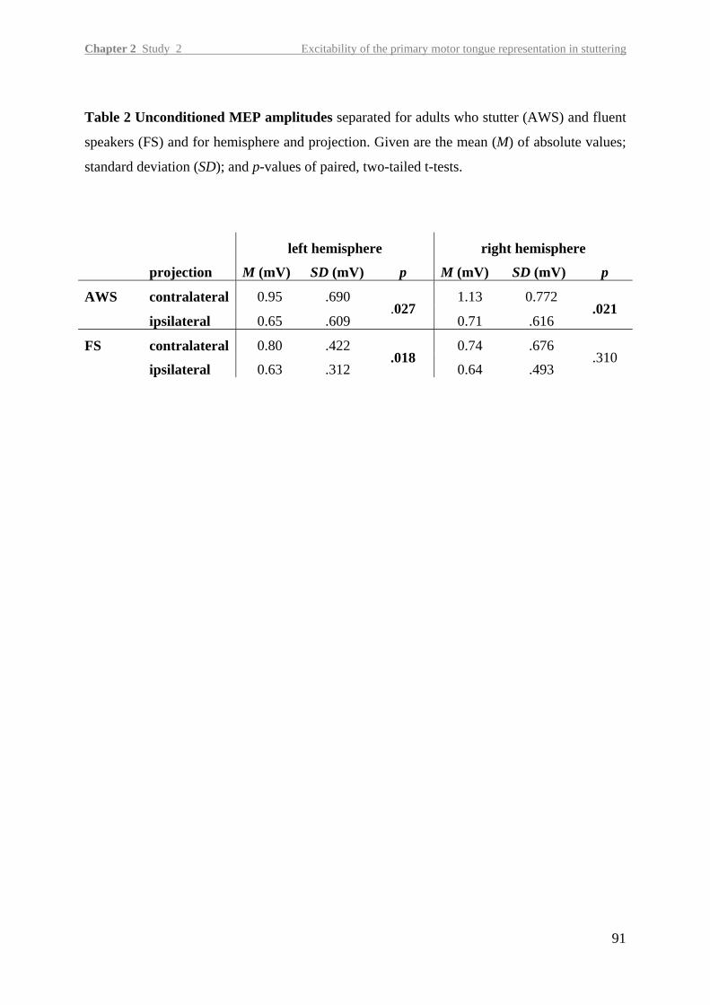
Chapter 2 Study 2 Excitability of the primary motor tongue representation in stuttering
Table 2 Unconditioned MEP amplitudes separated for adults who stutter (AWS) and fluent
speakers (FS) and for hemisphere and projection. Given are the mean (M) of absolute values;
standard deviation (SD); and p-values of paired, two-tailed t-tests.
projection
left hemisphere right hemisphere
M (mV) SD (mV) p M (mV) SD (mV) p
AWS contralateral 0.95 .690 .027
1.13 0.772 .021
ipsilateral 0.65 .609 0.71 .616
FS contralateral 0.80 .422 .018
0.74 .676 .310
ipsilateral 0.63 .312 0.64 .493
91
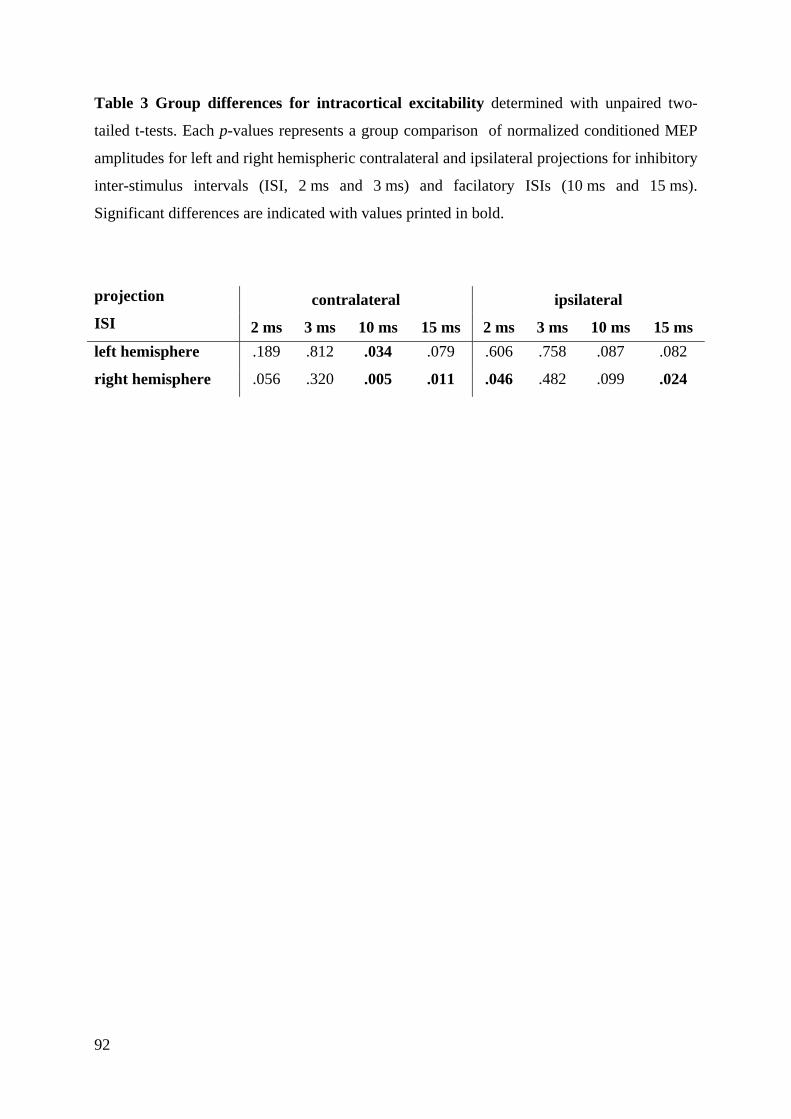
92
Table 3 Group differences for intracortical excitability determined with unpaired two-
tailed t-tests. Each p-values represents a group comparison of normalized conditioned MEP
amplitudes for left and right hemispheric contralateral and ipsilateral projections for inhibitory
inter-stimulus intervals (ISI, 2 ms and 3 ms) and facilatory ISIs (10 ms and 15 ms).
Significant differences are indicated with values printed in bold.
projection
ISI contralateral ipsilateral
2 ms 3 ms 10 ms 15 ms 2 ms 3 ms 10 ms 15 ms left hemisphere .189 .812 .034 .079 .606 .758 .087 .082
right hemisphere .056 .320 .005 .011 .046 .482 .099 .024
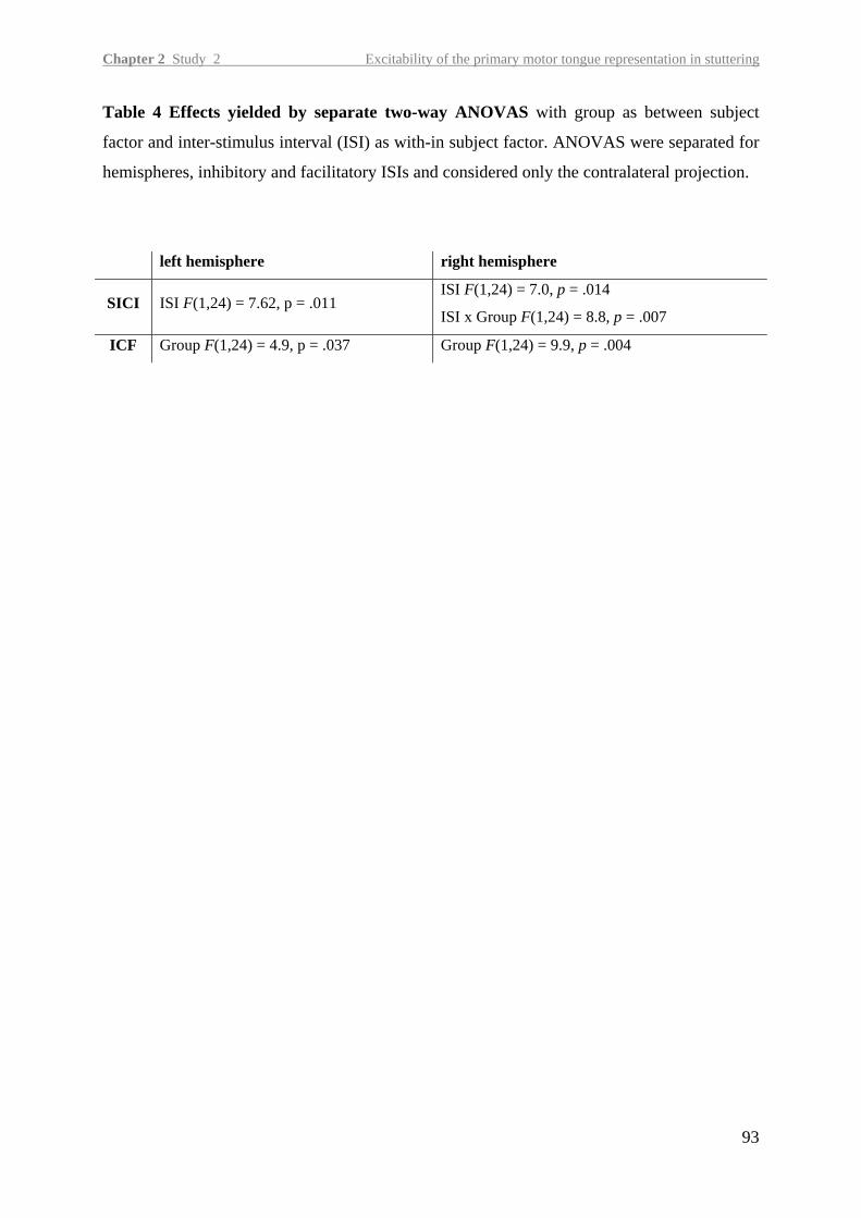
Chapter 2 Study 2 Excitability of the primary motor tongue representation in stuttering
93
Table 4 Effects yielded by separate two-way ANOVAS with group as between subject
factor and inter-stimulus interval (ISI) as with-in subject factor. ANOVAS were separated for
hemispheres, inhibitory and facilitatory ISIs and considered only the contralateral projection.
left hemisphere right hemisphere
SICI ISI F(1,24) = 7.62, p = .011 ISI F(1,24) = 7.0, p = .014
ISI x Group F(1,24) = 8.8, p = .007
ICF Group F(1,24) = 4.9, p = .037 Group F(1,24) = 9.9, p = .004

Chapter 2 Study 3 Phoneme Categorization in stuttering
95
2.3 Instable phoneme categorization in adults who stutter
Authors:
Nicole E. Neef
Martin Sommer
Walter Paulus
Department of Clinical Neurophysiology, Georg-August-University of Goettingen
Alexander Wolff von Gudenberg
Institut der Kasseler Stottertherapie, Bad Emstal, Germany
Torsten Wüstenberg
Department of Psychiatry and Psychotherapy, Charité Universitaetsmedizin Berlin
Running Head: Phoneme Categorization in Stuttering
Corresponding Author:
Martin Sommer, M.D
Department of Clinical Neurophysiology
Robert-Koch-Str. 40
D-37075 Goettingen, Germany
Phone: +49-551-39 66 50
Email: [email protected]

Chapter 2 Study 3 Phoneme Categorization in stuttering
Abstract
Purpose: It is currently unknown if persistent stuttering is accompanied by an irregular
sublexical speech perception. We tested the stability of phoneme percepts by analyzing
participants’ ability to discriminate between voiced and voiceless stop-consonants.
Method: In two synthetically generated syllable continua (/b/-/p/, /d/-/t/), voice onset
time was systematically modified. We determined the phoneme discrimination ability of
twenty patients and twenty matched control subjects by computing the phoneme boundaries,
and by quantifying the ambivalence interval - the interval of voice onset times where
phonemes were perceived ambiguously.
Results: Patients showed larger and less stable ambivalence intervals compared to healthy
subjects, while no difference between patients and controls was revealed concerning phoneme
boundaries.
Conclusion: Persistent developmental stuttering is associated with unreliable phonological
percepts, supporting current theories regarding the sensory-motor interaction in human
speech. Particularly, our findings might implicate an affected auditory feedback control
subsystem in AWS.
97
98
Introduction Stuttering is characterized by sudden interruptions of fluent speech due to an intermittent loss
of motor control (Ludlow and Loucks, 2003). The dynamic, multifactorial view of stuttering
postulates nonlinear interaction between a vulnerable speech motor system and factors like
genetic predisposition, emotional and autonomic arousal, linguistic and other cognitive
processing demands (Smith and Kelly, 1997; Smith et al., 2010). Although stuttering
manifests in the articulatory domain, speech perception is the focus of several studies
designed to elucidate its role in stuttering. Because proper articulation results in
distinguishable auditory targets, audition is a modality that serves mainly to control success of
articulation. Speech acquisition comprises subtle refinement of articulatory configurations
resulting in fine-tuned phonematic features relevant to distinguish meanings. Fine-tuning
requires the representation of a produced sound in the processing system and a mapping
between auditory target and produced item. This process is still active in adulthood,
demonstrated by the relation between a speaker’s production of a phoneme contrast and his or
her perception of this contrast (Newman, 2003; Perkell et al., 2004) and the influence of
speech motor learning on adult speaker's auditory maps (Nasir & Ostry, 2009). Because of the
tight link between production and perception it has been suggested that pathological speech
patterns are connected to altered speech perception (Heiser and Cheung, 2008).
The temporal processing demands of auditory and proprioceptive information during speaking
are presumed to challenge the speech motor system of adults who stutter (AWS) (Kent, 2000).
The auditory and proprioceptive information that is to be expected during speech production
are proposed to be held in internal models of speech sounds. The speech motor control deficit
is possibly linked to instability of these internal sensory models or to insufficient access on
them, restricting the feed forward control mechanisms in speech production (Max et al.,
2004).
The concept of instable speech sound maps in stuttering together with the demonstrated link
between speech production and perception lead us to probe a speech perception ability of
AWS – the phoneme categorization. Only few behavioral studies investigated speech sound
perception on the sublexical level in stuttering (Blood, 1996; Kramer et al., 1987). Sublexical
speech stimuli are eligible to test phoneme perception without contextually driven top down
information which facilitates recognition. We are not aware of a behavioral study
investigating phoneme categorization in AWS.
In contrast, sublexical speech perception abilities have been studied in a few
Electroencephalography (EEG) and Magnetencephalography (MEG) studies in AWS. Corbera
Chapter 2 Study 3 Phoneme Categorization in stuttering
and colleagues (2005) recorded the mismatch negativity (MMN) event-related brain potential
elicited to simple tone contrasts and to vowel contrasts. The MMN results from the detection
of a deviant stimulus. For example, in Corbera’s vowel contrast condition participants listened
to the repeated presentation of /o/. The unexpected presentation of /e/ resulted in an enlarged
electrophysiological response which indicated a detected mismatch between the previous
presented /o/ and the now presented /e/. This increase of the MMNs was significantly larger in
the left supratemporal region in AWS compared to control subjects. A larger MMN indicates
a higher sensitivity of the neural population to a certain deviation. Thus, the authors suggest
an abnormal speech sound representation within the auditory region of the left hemisphere in
AWS.
The MEG study demonstrates altered cortical activation patterns upon perception of an
unexpected phoneme. It remained unclear whether the performance in the underlying
categorization is also different in AWS on a subclinical level. This information is provided by
the psychophysical phoneme identification study presented here. One phonetically relevant
property of speech is the voice onset time (VOT). Differences in VOT are perceptually
essential to discriminate voiced from voiceless stop consonants. In word-initial position,
German voiced stops like /b/ and /d/ are typically produced with short VOTs or, in some
cases, with prevoicing; and German voiceless stops like /p/ and /t/ are produced with longer
VOTs (Keating, 1984). The stimuli of the present study were created by systematic variation
of the VOT, creating a /b/-/p/ and a /d/-/t/ continuum. Each VOT continuum can be
separated in three different sections according to the listener’s performance in the
psychophysical test. Two sections contain those stimuli that are reliably identified as the
voiced respectively voiceless phoneme. Between those sections lies a range of VOT in which
no reliable identification is possible and the listener reports different percepts upon repeated
presentation. The width of this section is termed here ambivalence interval (AI). It is directly
related to the slope of the psychometric function describing the discriminatory performance
(see Appendix B). The position of AI is conveniently defined by the VOT where
discrimination is at chance level. This VOT is here termed phoneme boundary PB. These two
parameters were extracted for each listener by fitting the results of the identification task with
a logistic curve (Fig 2 and Appendix B). The AI is a possible quantifier of ambiguity. A
narrow AI, corresponding to a steep slope, indicates a small range where stimuli are perceived
as ambiguous. Accordingly a wide AI (i.e. shallow slope) indicates that the range where
stimuli are not reliably identified is large (Fig 2). This concept has been employed in a recent
99
100
publication by Möttönen and Watkins (2009), who studied the influence of lip primary motor
cortex inhibition on phoneme categorization and discrimination. A logistic psychometric
function was used to fit the listener’s performance in a phoneme identification task (e.g. in the
acoustic /ba/-/da/ continuum). The slope of the function served as quantification of the
listener’s discrimination ability. It was reduced following inhibition of the primary motor lip
area.
Our study is designed to determine the ambivalence intervals and the phoneme boundaries of
two consonant-vowel (CV) continua (/b/-/p/ and /d/-/t/). We choose two different CV
continua to determine a general effect. We test the following specific hypotheses: First,
compared to fluently speaking participants, AWS will show broader ambivalence intervals,
indicating less stable phoneme representations on a subclinical level. Second, AWS and
fluently speaking participants will not differ in phoneme boundaries, a feature that depends
mainly on the native language and the idiom of the subjects (Braun, 1996; Lisker and
Abramson, 1964).
Methods Participants
Twenty AWS (aged 32.2±12.6 years, five women) and twenty control subjects (aged
31.9±11.0, four women) participated in the study. AWS were recruited from the local
stuttering support group and the Institute for the Kassel Stuttering Therapy. Control subjects
were recruited by advertisement. The groups were matched for age (t(39) = -0.09, p = .16;
unpaired two tailed t Test), level of education (p =.64; U test) and handedness (p = .43; U
test). Three AWS and one control subject were left handed according to the Edinburgh
inventory (Oldfield, 1971). AWS produced more stuttered syllables than control subjects
(German version of the SSI-3, mean±SD 11.83±9.45, vs. 0.27±0.23; p < .001; U test).
Stuttering severity was very mild in six, mild in two, moderate in three, severe in four and
very severe in five AWS (Sandrieser and Schneider, 2008). Averaged stuttering onset was at
age 5.0±2.6. Detailed description of participants is reported in Table 1.
All participants were native speakers of German, none of them was bilingual. Fifteen AWS
reported a family history of stuttering. None of the control subjects reported having a family
history of speech or language disorders. None of the participants reported a speech, language
or hearing deficit, except of stuttering in the stuttering group; or showed neurological
abnormalities on routine examination. No participant was taking drugs affecting the central
Chapter 2 Study 3 Phoneme Categorization in stuttering
nervous system at the time of the study. The Göttingen Ethics Committee approved the study,
and all participants gave written informed consent. All participants were paid for their
participation.
Fluency Assessment
To judge stuttering severity, fluency assessments were performed regarding the German
version of the SSI-3 (Sandrieser and Schneider, 2008). Speech samples of all participants
containing a conversation about job or school and a reading task were videotaped and
analyzed by a qualified speech-language pathologist. SSI-3 norms were adapted from Riley
(Riley, 1994). The offline analysis of dysfluencies included 500 syllables for the conversation
and not less than 340 syllables for the reading task. Sound prolongations, blocks (silent
prolongation of an articulatory posture) as well as sound and syllable repetitions were counted
as stuttered syllables. Monosyllabic words that were repeated with apparent undue stress or
tension were counted. Furthermore, the estimated duration of the three longest blocks and
observation of physical concomitants were included for the estimate of stuttering severity in
AWS.
Stimuli
Synthetically generated sets of /b/-/p/ and /d/-/t/consonant-vowel CV continua with a
varying VOT from 7 to 61 ms served as stimuli. These sets were created within a five step
process: (i) Voiceless syllables were generated using the AT&T Bell-Research Lab speech
synthesizer (http://www2.research.att.com/~ttsweb/tts/; sample rate = 16 kHz, number of bits
per sample = 16). (ii) Spectrograms of these syllables were computed with the software
package Praat (http://www.fon.hum.uva.nl/praat/). (iii) The first three formats of the vowel
were extracted from these spectrograms. (iv) Stimuli were segmented into consonant and
vowel regarding the formant with the earliest onset. (v) To form the CV continua, the
resulting segments were superimposed with a step width of 1 millisecond using an in house
made algorithm written in the MatLab programming language (MatLab 7.4, Release 2007a,
Mathworks, Inc., Natick, MA).
Experimental Procedure
Participants sat in front of a computer in a quiet room. Stimuli were presented binaurally via
dynamic, closed-ear headphones (Sennheiser HD 280; up to 32dB attenuation of outside
noise) at a comfortable hearing level they regulated by themselves. The two sets of CV
101
continua were presented in separate blocks in a balanced order. Each block comprised five
trial series. Each series started off with a staircase search for the approximate position of the
phoneme boundary. This was followed by a random sequence of stimuli with VOTs covering
20 ms around the tentative phoneme boundary. Participants were asked to listen to the stimuli
and to press the left mouse button when they perceive a /be/ and the right mouse button when
they perceive a /pe/. As a reminder this information was depicted on the screen during the
entire trial series. By pressing the space bar participants were able to repeat the last stimulus
until indicating their final decision by mouse button press in a two alternative forced choice.
The two blocks were separated by a 15-minute break. Speech samples for the SSI-3 fluency
assessment were recorded during this break.
The adaptive computation of stimulus VOTs was based on an accelerated stochastic
approximation (see Appendix – A) as described by Kesten (1958; Treutwein, 1995). The
staircase procedure started by a randomly chosen VOT and was aborted after ten reversals.
For the /b/-/p/continuum control subjects required a mean of 35.95 (±2.87) and AWS a
mean of 36.77 (±5.24) trials. For the /d/-/t/continuum the average amount of trials for one
repetition was 36.93 (±4.5) for control subjects and 38.06 (±5.96) for AWS. The adaptive
estimation of the phoneme boundary took about three minutes each.
It is an intrinsic property of the applied adaptive algorithm that in the last third of each
repetition, consecutive stimuli near the phoneme boundary, and thus within the ambivalence
interval, are presented. For control subjects 28.4% of /b/-/p/ and 29.1% of /d/-/t/
stimuli were presented in ambivalence interval. For AWS this ratio was slightly higher: 30.6%
for the /b/-/p/ and 35.2% for the /d/-/t/continuum (see Figure 1). This supports an
optimal fit of the later described psychometric model to the data.
The experiment was run on presentation software version 0.71 (Neurobehavioral Systems,
http://www.neurobs.com/).
------------------------------------------------
Please insert Figure 1 about here.
-----------------------------------------------
Data Analysis
To estimate the phoneme boundary and the ambivalence interval, we fitted a logistic
psychometric function for the voiceless percept to the data, including the trials of
the staircase search and the following catch trials, (see Appendix – B) using a maximum
likelihood algorithm (psygnifit,
)VOT(Ψ
http://www.bootstrap-software.org/psignifit/; Wichmann and
102
Chapter 2 Study 3 Phoneme Categorization in stuttering
Hill, 2001b, a). After modeling the data, phoneme boundary was estimated by the null of the
second derivative dVOT²
d²Ψ of the psychometric function. The ambivalence interval was
estimated by computing the absolute difference between the two nulls of the third derivative
³dVOT³d Ψ of the psychometric function (see Figure 2).
------------------------------------------------
Please insert Figure 2 about here.
-----------------------------------------------
Statistics
To test the differences between groups, we entered the values of the ambivalence interval in a
three-factorial mixed design omnibus ANOVA for repeated measures with the between-
subjects factor Group (AWS; control subjects) and the within-subjects factors CV-continuum
(/b/-/p/, /d/-/t/) and Repetition (1 to 5). A similar analysis was computed for the
estimates of the phoneme boundary. Main effects and interactions were tested via post hoc t
tests.
To test potential correlations between stuttering severity and ambivalence interval as well as
phoneme boundary, we calculated Spearman’s correlation coefficient between the stutterer’s
SSI-3 overall-scores and the ambivalence interval or phoneme boundary values respectively.
Statistics were performed with the Software Package for the Social Sciences (SPSS 17.0,
http://www.spss.com/de/software).
Results ANOVA regarding the ambivalence interval affirmed the following significant main effects
and interactions: (i) a main effect for the Group, F(1,38) = 4.46, p = .041, with a significant
greater ambivalence interval for AWS, t(39) = -2.12, p = .026; (ii) a main effect for CVS-
continuum, F(1,38) = 7.45, p = .01, with a greater ambivalence interval for the /d/-/t/
continuum, t(39) = -2.91, p = .006; (iii) a main effect of Repetition, F(4,35) = 5.43, p = .002,
with a greater ambivalence interval for the first repetition (r1) compared with the second, third
and fourth repetition, t(39) (r1>r2) = 2.99, p = .048; t(39) (r1>r3) = 3.41, p = .015 and t(39)
(r1>r4) = 3.48, p = .013 (all p-values were Bonferroni corrected for multiple comparisons);
(iv) an interaction between repetition and group, F(4,35) = 3.59, p = .015. Looking for the
effect of repetition for either group as indicated by a main interaction of these two factors, in
control subjects a significant decrease of the ambivalence interval already was found for
103
104
repetition 2, t(19) = 3.713, p = .001 (Bonferroni corrected for multiple comparisons) while in
AWS this decrease is only significant for repetition 3, t(19) = 2.84, p = .01 (uncorrected).
Moreover, a significant increase of the ambivalence interval which is evident between the
third and fifth repetition, t(19) = -2.57, p = .019 (uncorrected), occurred in AWS (Figure 3 C,
D).
The ANOVA regarding the phoneme boundary revealed no significant main effect for Group,
F(1,38) = 2.4, p = .127. As well known from the literature (e.g. (Phillips et al., 2000)
phoneme boundary of the /b/-/p/continuum differed significantly from that of /d/-
/t/continuum, F(1,38) = 274.95, p < .001.
Correlation analyses yielded no correlation between stuttering severity and ambivalence
interval, -.36 < rs < .25; p > .12 or phoneme boundary, -.24 < rs < .22; p > .30, respectively.
------------------------------------------------
Please insert Figure 3 about here.
-----------------------------------------------
Discussion The aim of the present study was to substantiate a putative dysfunction of phoneme
categorization in stuttering. While previous studies indicated neuroanatomical and
neurofunctional alterations in brain regions relevant for speech perception, behavioural
evidence for associated speech perception impairment is so far missing. Our results indicate a
diminished phoneme discriminatory power in AWS. We conclude that an instable speech
perception adds to the prominent motor disturbances in AWS. The broader ambivalence
intervals and less reliable phoneme categorization suggest less stable phoneme percepts in
AWS.
The repetition effect
Noteworthy is the delayed repetition effect in AWS relative to fluent speakers. This
observation is in line with previous reports from speech production experiments where a
delayed improvement of performance occurs in AWS (Smits-Bandstra and De Nil, 2009).
That an improvement is not maintained in AWS has also been described (Smits-Bandstra et
al., 2006). We assume that this variability in performance reflects the speech system’s
instability in AWS which is also prone to other than speech related disturbances like
fluctuations in attention or vigilance (Bosshardt, 2006), and therefore unable to maintain the
Chapter 2 Study 3 Phoneme Categorization in stuttering
acquired improvement of performance. On a broader level, this is reminiscent of a tendency
of relapse in AWS after successful fluency-shaping therapy (Euler, von Gudenberg, Jung &
Neumann, 2009).
Processes involved in phoneme categorization
State-of-the-art accounts of sublexical speech perception refer to four different processes:
acoustic processing, phonetic processing, phonological processing and categorical phoneme
perception (Turkeltaub and Coslett, 2010). (i) Acoustic processing concerns the
spectrotemporal analysis of speech and non-speech auditory signals independent of language
experience; (ii) phonetic processing is shaped by language experience and is speech-specific;
(iii) phonological processing selects from discrete abstract symbolic mental representations of
speech sounds of the language-specific phonemic system; and (iv) categorical phoneme
perception concerns assigning phoneme labels to speech sounds.
While each of these processes could contribute to our findings, literature on the initial three in
AWS is remarkably scarce. The current study therefore aimed at the fourth process:
discrimination of speech sounds within phoneme boundaries; a meta-linguistic process which
does not lead to lexical access or further semantic processing. Rather it includes acoustic and
phonetic processing which operate on continuous, analog auditory signals, possibly mediated
through a comparison between phonological features and an articulatory plan (Rauschecker
and Scott, 2009; Turkeltaub and Coslett, 2010).
(i) Acoustic processing
Spectrotemporal analysis of non-speech or speech auditory signals is assumed to be computed
in bilateral dorsal superior temporal lobe areas including Herschel’s gyrus and planum
temporale (e.g. (Overath et al., 2008), and speech auditory signals additionally in the bilateral
dorsal superior temporal gyrus (e.g. (Hickok and Poeppel, 2007). In AWS, EEG and MEG
studies indicated differences in the timing and amplitude of neurophysiologic responses of the
auditory cortex to auditory presented speech stimuli (Beal et al., 2010; Hampton and Weber-
Fox, 2008) and may be based on structural alterations in Herschel’s gyrus and the planum
temporale (Beal et al., 2007; Foundas et al., 2001; Foundas et al., 2004).
(ii) Phonetic processing
Phoneme perception involves the superior temporal gyrus, specifically the planum temporale
(Jäncke et al., 2002; Zatorre et al., 1996). Confirming our hypothesis, our sample of AWS
105
106
exhibited broader ambivalence intervals. This finding indicates an overlapping of the
acceptable ranges of the encoded acoustic reference frame of a certain phonemic feature such
as VOT in AWS relative to control subjects. Maybe this blurring is related to a different and
asynchronous neural timing in auditory cortices in AWS. A recent magnetencephalography
study documented the M 100 latencies (a response to the onset properties of an auditory
stimulus) to the auditory presented vowel /i/ to be delayed in AWS’s right hemisphere. This
delay was related to the M 100 in AWS’s left hemisphere as well as to the bilateral M 100 in
fluent speakers (Beal et al., 2010). It is therefore conceivable that deviant phonetic processing
contributed to the behavioral findings in the current study.
A broadened ambivalence interval is a behavioral deviant on a subclinical level in AWS. As
expected, all AWS were able to solve the task properly by identifying phonemes at VOTs,
which were distant from phoneme boundaries. We did not expect any group difference
concerning the phoneme boundaries itself, because this feature depends primarily on idiom
and mother tongue (Braun, 1996; Lisker and Abramson, 1964), and we did not control for
idiom in the present study. Unexpectedly, our data show a trend towards increased phoneme
boundaries in AWS, indicating that AWS demand longer VOT to perceive a stop consonant as
voiceless. This observation supports previous reports: EEG-mismatch negativities to auditory
presented phonetic contrasts embedded in meaningless syllables was significantly enhanced in
left supratemporal regions in AWS indicating a deviant phoneme perception (Corbera et al.,
2005). Thus, AWS exhibit abnormal behavioral and cortical processing in phoneme
perception tasks.
(iii) Phonological processing
The current study asked participants to decide whether a presented stop consonant embedded
in a consonant-vowel syllable was voiced or voiceless. This decision required access to the
discrete phonological feature: voicing.
Several theories on stuttering postulate an imprecise phonological encoding in speech
production (Sasisekaran et al., 2006). These theories comprise the Covert Repair Hypothesis
(Postma and Kolk, 1993), the EXPLAN theory (Howell, 2007), the neurolinguistic model
(Perkins et al., 1991) and the Fault line Hypothesis (Wingate, 1988). However, little is known
regarding phonological processing during sublexical speech perception in AWS. Evidence for
deviant phonological processing has been suggested by a recent functional magnetic
resonance imaging (fMRI) study; auditory presented meaningless speech syllables resulted in
a decreased activity in the left superior temporal gyrus in AWS (Chang et al., 2009).
Chapter 2 Study 3 Phoneme Categorization in stuttering
With regard to our results, we cannot exclude alterations of sublexical speech perception
abilities in AWS. It might be possible that these alterations are caused by an instable speech
production, or vice versa. Thus, studying the interaction of speech perception and speech
production is likely to expand our understanding about stuttering. The interface for the
mapping between perceived and produced speech is probably located in the left supramarginal
and angular gyrus (Rauschecker and Scott, 2009; Turkeltaub and Coslett, 2010). It is
assumed, that categorical perception involves these areas.
(iv) Phoneme categorization
Deciding whether the presented phoneme is either voiced or voiceless most likely requires the
activation of the articulatory model of the specific phoneme (Meister et al., 2007; Turkeltaub
and Coslett, 2010), the so called speech sound map in the ventral premotor cortex and
posterior inferior frontal gyrus (Golfinopoulos et al., 2010). In our experiment, due to the
presentation of a third of the stimuli within the ambivalence interval, the task demand was
quite high. With increasing ambiguity of the presented syllables, task demand increases as
indicated in a previous study by the significant increase of response times around the
phoneme boundary (Wüstenberg and Mattler, 2008). Repetitive matching requires a high level
of functional connectivity within the described network. An intense neural communication
requires an intact anatomical architecture and an undisturbed neural synchronization. If
connections were malfunctioning, discriminatory power is likely to be impaired and
ambivalence interval is broadened.
Supporting a disturbance of this internal matching process, left perisylvian fibers exhibit
reduced integrity in AWS (Cykowski et al., in press; Sommer et al., 2002; Watkins et al.,
2008). The dual-stream model of functional anatomy of language postulates the dorsal stream
to be mainly involved in this mapping (Hickok and Poeppel, 2007). Fibers linking the
superior temporal lobe and the premotor cortices via the dorsal stream are the arcuate and
superior longitudinal fascicle (Saur et al., 2008). A recent analysis of resting-state-functional-
connectivity confirms this cortico-cortical connectivity between Brodmann area (BA) 6 and
rostral supramarginal gyrus, and between BAs 44 and 45 and caudal supramarginal gyrus and
angular gyrus (Kelly et al.). Thus, less coherent connections in AWS might slow down the
transfer of information or diminish the quality of the transported signals, which challenges
discriminatory power under difficult conditions such as near the phoneme boundary.
107
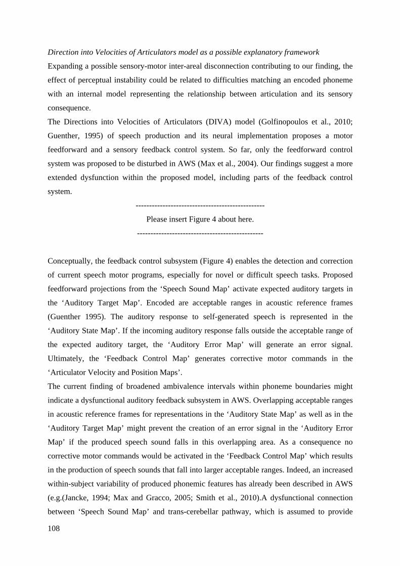
108
Direction into Velocities of Articulators model as a possible explanatory framework
Expanding a possible sensory-motor inter-areal disconnection contributing to our finding, the
effect of perceptual instability could be related to difficulties matching an encoded phoneme
with an internal model representing the relationship between articulation and its sensory
consequence.
The Directions into Velocities of Articulators (DIVA) model (Golfinopoulos et al., 2010;
Guenther, 1995) of speech production and its neural implementation proposes a motor
feedforward and a sensory feedback control system. So far, only the feedforward control
system was proposed to be disturbed in AWS (Max et al., 2004). Our findings suggest a more
extended dysfunction within the proposed model, including parts of the feedback control
system.
------------------------------------------------
Please insert Figure 4 about here.
-----------------------------------------------
Conceptually, the feedback control subsystem (Figure 4) enables the detection and correction
of current speech motor programs, especially for novel or difficult speech tasks. Proposed
feedforward projections from the ‘Speech Sound Map’ activate expected auditory targets in
the ‘Auditory Target Map’. Encoded are acceptable ranges in acoustic reference frames
(Guenther 1995). The auditory response to self-generated speech is represented in the
‘Auditory State Map’. If the incoming auditory response falls outside the acceptable range of
the expected auditory target, the ‘Auditory Error Map’ will generate an error signal.
Ultimately, the ‘Feedback Control Map’ generates corrective motor commands in the
‘Articulator Velocity and Position Maps’.
The current finding of broadened ambivalence intervals within phoneme boundaries might
indicate a dysfunctional auditory feedback subsystem in AWS. Overlapping acceptable ranges
in acoustic reference frames for representations in the ‘Auditory State Map’ as well as in the
‘Auditory Target Map’ might prevent the creation of an error signal in the ‘Auditory Error
Map’ if the produced speech sound falls in this overlapping area. As a consequence no
corrective motor commands would be activated in the ‘Feedback Control Map’ which results
in the production of speech sounds that fall into larger acceptable ranges. Indeed, an increased
within-subject variability of produced phonemic features has already been described in AWS
(e.g.(Jancke, 1994; Max and Gracco, 2005; Smith et al., 2010).A dysfunctional connection
between ‘Speech Sound Map’ and trans-cerebellar pathway, which is assumed to provide

Chapter 2 Study 3 Phoneme Categorization in stuttering
precise temporal information in the processing of time intervals during speech perceptual
tasks (Ackermann, 2008), might also contribute to the broadened ambivalence intervals. In
AWS a dysfunctional right cerebellar/ left frontal loop was recently described (Lu et al.,
2010).
Conclusion
In conclusion, our results demonstrate behavioral evidence for an altered speech perception in
AWS, underpinning neuroimaging studies that demonstrate irregularities in neuroanatomic
correlates of speech perception. In addition, our data help expand a theoretical framework on
stuttering assuming less stable speech sound representations or an insufficient access to them,
thereby highlighting the role of auditory perception in persistent developmental stuttering.
Future studies should evaluate the link between sublexical speech perception and production
to further elucidate the nature of stuttering. Such studies would provide data to complete
theoretical models of stuttering as well as of speech processing and its neural implementation
in general.
109

110
ACKNOWLEDGEMENT
This work was supported by the Stifterverband für die Deutsche Wissenschaft, Walter und
Ilse Rose Stiftung and the Deutsche Forschungsgemeinschaft [SO 429/2-2]. We thank
Veronika Gutman for her help in subject recruitment and testing.
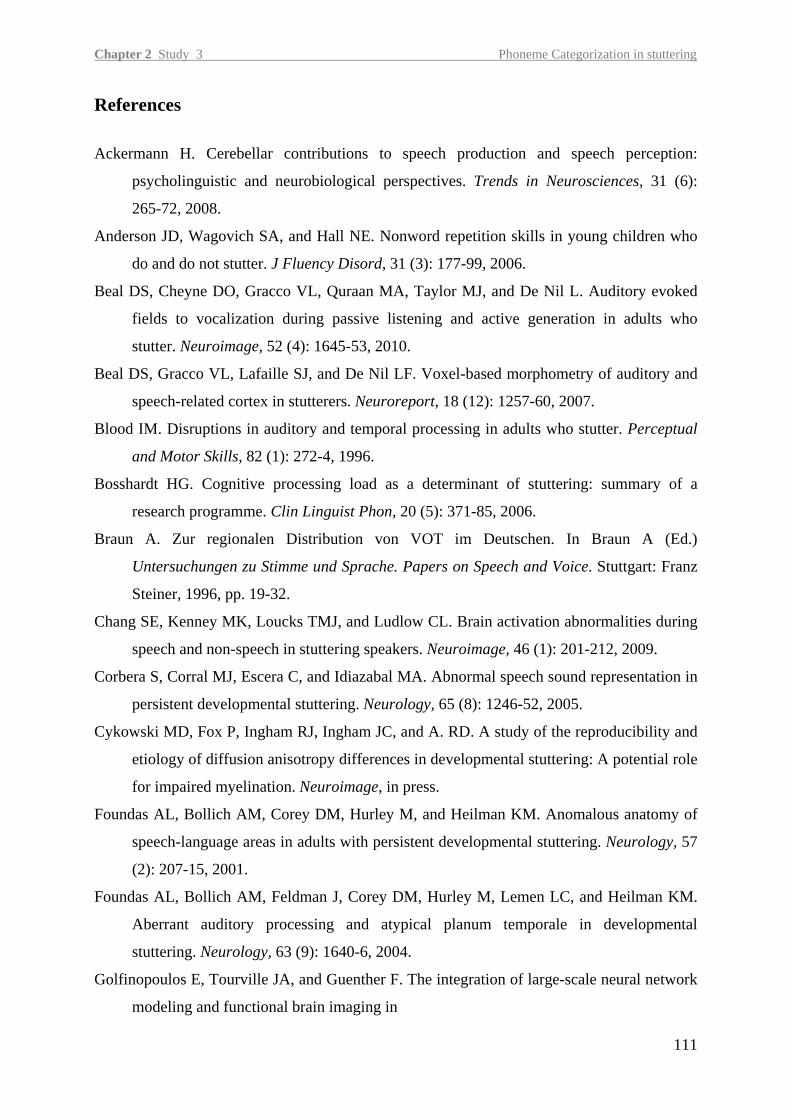
Chapter 2 Study 3 Phoneme Categorization in stuttering
References Ackermann H. Cerebellar contributions to speech production and speech perception:
psycholinguistic and neurobiological perspectives. Trends in Neurosciences, 31 (6):
265-72, 2008.
Anderson JD, Wagovich SA, and Hall NE. Nonword repetition skills in young children who
do and do not stutter. J Fluency Disord, 31 (3): 177-99, 2006.
Beal DS, Cheyne DO, Gracco VL, Quraan MA, Taylor MJ, and De Nil L. Auditory evoked
fields to vocalization during passive listening and active generation in adults who
stutter. Neuroimage, 52 (4): 1645-53, 2010.
Beal DS, Gracco VL, Lafaille SJ, and De Nil LF. Voxel-based morphometry of auditory and
speech-related cortex in stutterers. Neuroreport, 18 (12): 1257-60, 2007.
Blood IM. Disruptions in auditory and temporal processing in adults who stutter. Perceptual
and Motor Skills, 82 (1): 272-4, 1996.
Bosshardt HG. Cognitive processing load as a determinant of stuttering: summary of a
research programme. Clin Linguist Phon, 20 (5): 371-85, 2006.
Braun A. Zur regionalen Distribution von VOT im Deutschen. In Braun A (Ed.)
Untersuchungen zu Stimme und Sprache. Papers on Speech and Voice. Stuttgart: Franz
Steiner, 1996, pp. 19-32.
Chang SE, Kenney MK, Loucks TMJ, and Ludlow CL. Brain activation abnormalities during
speech and non-speech in stuttering speakers. Neuroimage, 46 (1): 201-212, 2009.
Corbera S, Corral MJ, Escera C, and Idiazabal MA. Abnormal speech sound representation in
persistent developmental stuttering. Neurology, 65 (8): 1246-52, 2005.
Cykowski MD, Fox P, Ingham RJ, Ingham JC, and A. RD. A study of the reproducibility and
etiology of diffusion anisotropy differences in developmental stuttering: A potential role
for impaired myelination. Neuroimage, in press.
Foundas AL, Bollich AM, Corey DM, Hurley M, and Heilman KM. Anomalous anatomy of
speech-language areas in adults with persistent developmental stuttering. Neurology, 57
(2): 207-15, 2001.
Foundas AL, Bollich AM, Feldman J, Corey DM, Hurley M, Lemen LC, and Heilman KM.
Aberrant auditory processing and atypical planum temporale in developmental
stuttering. Neurology, 63 (9): 1640-6, 2004.
Golfinopoulos E, Tourville JA, and Guenther F. The integration of large-scale neural network
modeling and functional brain imaging in
111
112
speech motor contro. Neuroimage: doi:10.1016/j.neuroimage.2009.10.023, 2010.
Guenther FH. Speech sound acquisition, coarticulation, and rate effects in a neural network
model of speech production. Psychological Review, 102 (3): 594-621, 1995.
Hampton A and Weber-Fox C. Non-linguistic auditory processing in stuttering: evidence
from behavior and event-related brain potentials. J Fluency Disord, 33 (4): 253-73,
2008.
Heiser MA and Cheung SW. Cortical representations of communication sounds. Curr Opin
Otolaryngol Head Neck Surg, 16 (5): 478-84, 2008.
Hickok G and Poeppel D. The cortical organization of speech processing. Nat Rev Neurosci, 8
(5): 393-402, 2007.
Howell P. The effects of gated speech on the fluency of speakers who stutter. Folia
Phoniatrica et Logopedica, 59 (5): 250-5, 2007.
Jancke L. Variability and duration of voice onset time and phonation in stuttering and
nonstuttering adults. Journal of Fluency Disorders, 19 (1): 21-37, 1994.
Jäncke L, Wüstenberg T, Scheich H, and Heinze H. Phonetic perception and the temporal
cortex. Neuroimage, 15 (4): 733-46, 2002.
Keating PA. Phonetic and phonological representation of stop consonant voicing. Language,
60, (2): 286-319, 1984.
Kelly C, Uddin LQ, Shehzad Z, Margulies DS, Castellanos FX, Milham MP, and Petrides M.
Broca's region: linking human brain functional connectivity data and non-human
primate tracing anatomy studies. European Journal of Neuroscience.
Kent RD. Research on speech motor control and its disorders: a review and prospective.
Journal of Communication Disorders, 33 (5): 391-427; quiz 428, 2000.
Kesten H. Accelerated stochastic approximation. Annals of Mathematical Statistics, 29: 41-
59, 1958.
Kramer MB, Green D, and Guitar B. A comparison of stutterers and nonstutterers on masking
level differences and synthetic sentence identification tasks. Journal of Communication
Disorders, 20 (5): 379-90, 1987.
Lisker L and Abramson AS. A cross-language study of voicing in initial stops: acoustical
measurements. Word, 20: 384-422, 1964.
Lu C, Chen C, Ning N, Ding G, Guo T, Peng D, Yang Y, Li K, and Lin C. The neural
substrates for atypical planning and execution of word production in stuttering.
Experimental Neurology, 221 (1): 146-56, 2010.
Chapter 2 Study 3 Phoneme Categorization in stuttering
Ludlow CL and Loucks T. Stuttering: a dynamic motor control disorder. Journal of Fluency
Disorders, 28 (4): 273-95, 2003.
Max L and Gracco VL. Coordination of oral and laryngeal movements in the perceptually
fluent speech of adults who stutter. Journal of Speech, Language, and Hearing
Research, 48 (3): 524-42, 2005.
Max L, Guenther FH, Gracco VL, Ghosh SS, and Wallace ME. Unstable or insufficiently
activated internal models and feedback-biased motor control as sources of dysfluency:
A theoretical model of stuttering. Contemporary Issues in Communication Science and
Disorders: CICSD, 31: 105-122, 2004.
Meister IG, Wilson SM, Deblieck C, Wu AD, and Iacoboni M. The essential role of premotor
cortex in speech perception. Current Biology, 17 (19): 1692-6, 2007.
Möttönen R and Watkins KE. Motor representations of articulators contribute to categorical
perception of speech sounds. Journal of Neuroscience, 29 (31): 9819-25, 2009.
Oldfield RC. The assessment and analysis of handedness: the Edinburgh inventory.
Neuropsychologia, 9 (1): 97-113, 1971.
Overath T, Kumar S, von Kriegstein K, and Griffiths TD. Encoding of spectral correlation
over time in auditory cortex. Journal of Neuroscience, 28 (49): 13268-73, 2008.
Perkins WH, Kent RD, and Curlee RF. A theory of neuropsycholinguistic function in
stuttering. Journal of Speech and Hearing Research, 34 (4): 734-52, 1991.
Phillips C, Pellathy T, Marantz A, Yellin E, Wexler K, Poeppel D, McGinnis M, and Roberts
T. Auditory cortex accesses phonological categories: an MEG mismatch study. Journal
of Cognitive Neuroscience, 12 (6): 1038-55, 2000.
Postma A and Kolk H. The covert repair hypothesis: prearticulatory repair processes in
normal and stuttered disfluencies. Journal of Speech and Hearing Research, 36 (3):
472-87, 1993.
Rauschecker JP and Scott SK. Maps and streams in the auditory cortex: nonhuman primates
illuminate human speech processing. Nature Neuroscience, 12 (6): 718-24, 2009.
Riley GD. The Stuttering Severity Instrument for Children and Adults- Third Edition (SSI-3).
Austin, TX: Pro-Ed., 1994.
Sandrieser P and Schneider P. Stottern im Kindesalter. Stuttgart: Thieme, 2008.
Sasisekaran J, De Nil LF, Smyth R, and Johnson C. Phonological encoding in the silent
speech of persons who stutter. J Fluency Disord, 31 (1): 1-21; quiz 19, 2006.
Saur D, Kreher BW, Schnell S, Kummerer D, Kellmeyer P, Vry MS, Umarova R, Musso M,
Glauche V, Abel S, Huber W, Rijntjes M, Hennig J, and Weiller C. Ventral and dorsal
113
114
pathways for language. Proceedings of the National Academy of Sciences of the United
States of America, 105 (46): 18035-40, 2008.
Smith A and Kelly E. Stuttering: A dynamic multifactorial model. In Curlee RF and Seigel
GM (Eds.), Nature and treatment of stuttering: New directions. New York: Allyn &
Bacon, 1997, pp. 204-217.
Smith A, Sadagopan N, Walsh B, and Weber-Fox C. Increasing phonological complexity
reveals heightened instability in inter-articulatory coordination in adults who stutter. J
Fluency Disord, 35 (1): 1-18, 2010.
Smits-Bandstra S and De Nil L. Speech skill learning of persons who stutter and fluent
speakers under single and dual task conditions. Clinical Linguistics & Phonetics, 23 (1):
38-57, 2009.
Smits-Bandstra S, De Nil LF, and Saint-Cyr JA. Speech and nonspeech sequence skill
learning in adults who stutter. Journal of Fluency Disorders, 31 (2): 116-36, 2006.
Sommer M, Koch MA, Paulus W, Weiller C, and Büchel C. Disconnection of speech-relevant
brain areas in persistent developmental stuttering. Lancet, 360 (9330): 380-3, 2002.
Treutwein B. Adaptive Psychophysical Procedures. Vision Research, 35 (17): 2503-2522,
1995.
Turkeltaub PE and Coslett HB. Localization of sublexical speech perception components.
Secondary Titl. 114: 1-15, 2010.
Watkins KE, Smith SM, Davis S, and Howell P. Structural and functional abnormalities of the
motor system in developmental stuttering. Brain, 131 (Pt 1): 50-9, 2008.
Wichmann FA and Hill NJ. The psychometric function: I. Fitting, sampling, and goodness of
fit. Perception and Psychophysics, 63 (8): 1293-313, 2001a.
Wichmann FA and Hill NJ. The psychometric function: II. Bootstrap-based confidence
intervals and sampling. Perception and Psychophysics, 63 (8): 1314-29, 2001b.
Wingate ME. The structure of stuttering. New York: Springer, 1988.
Wüstenberg T and Mattler U. Functional lateralization of feature specific processing in a
phonetic classification task. International Journal of Psychophysiology, 69 (3): 215,
2008.
Zatorre RJ, Meyer E, Gjedde A, and Evans AC. PET studies of phonetic processing of speech:
review, replication, and reanalysis. Cerebral Cortex, 6 (1): 21-30, 1996.
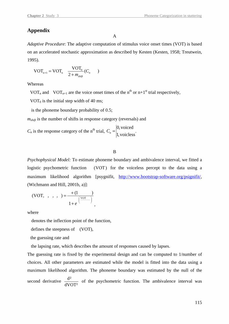
Chapter 2 Study 3 Phoneme Categorization in stuttering
Appendix A
Adaptive Procedure: The adaptive computation of stimulus voice onset times (VOT) is based
on an accelerated stochastic approximation as described by Kesten (Kesten, 1958; Treutwein,
1995).
)(2
VOTVOTVOT 0n1n φ−
+Δ−=+ n
shift
Cm
Whereas
ΔVOTn and ΔVOTn+1 are the voice onset times of the nth or n+1st trial respectively,
ΔVOT0 is the initial step width of 40 ms;
φ is the phoneme boundary probability of 0.5;
mshift is the number of shifts in response category (reversals) and
Cn is the response category of the nth trial, voicless,1voiced,0
=nC .
B
Psychophysical Model: To estimate phoneme boundary and ambivalence interval, we fitted a
logistic psychometric function for the voiceless percept to the data using a
maximum likelihood algorithm [psygnifit,
)VOT(Ψ
http://www.bootstrap-software.org/psignifit/,
(Wichmann and Hill, 2001b, a)]:
⎟⎟⎠
⎞⎜⎜⎝
⎛ −−+
−−+=Ψβ
α
λγγλγβαVOT
1
)1(),,,,VOT(
e ,
where
α denotes the inflection point of the function,
β defines the steepness of Ψ(VOT),
γ the guessing rate and
λ the lapsing rate, which describes the amount of responses caused by lapses.
The guessing rate is fixed by the experimental design and can be computed to 1/number of
choices. All other parameters are estimated while the model is fitted into the data using a
maximum likelihood algorithm. The phoneme boundary was estimated by the null of the
second derivative dVOT²
²d Ψ of the psychometric function. The ambivalence interval was
115

estimated by computing the absolute difference between the two nulls of the third derivative
dVOT³³d Ψ of the psychometric function (see Figure 2).
116
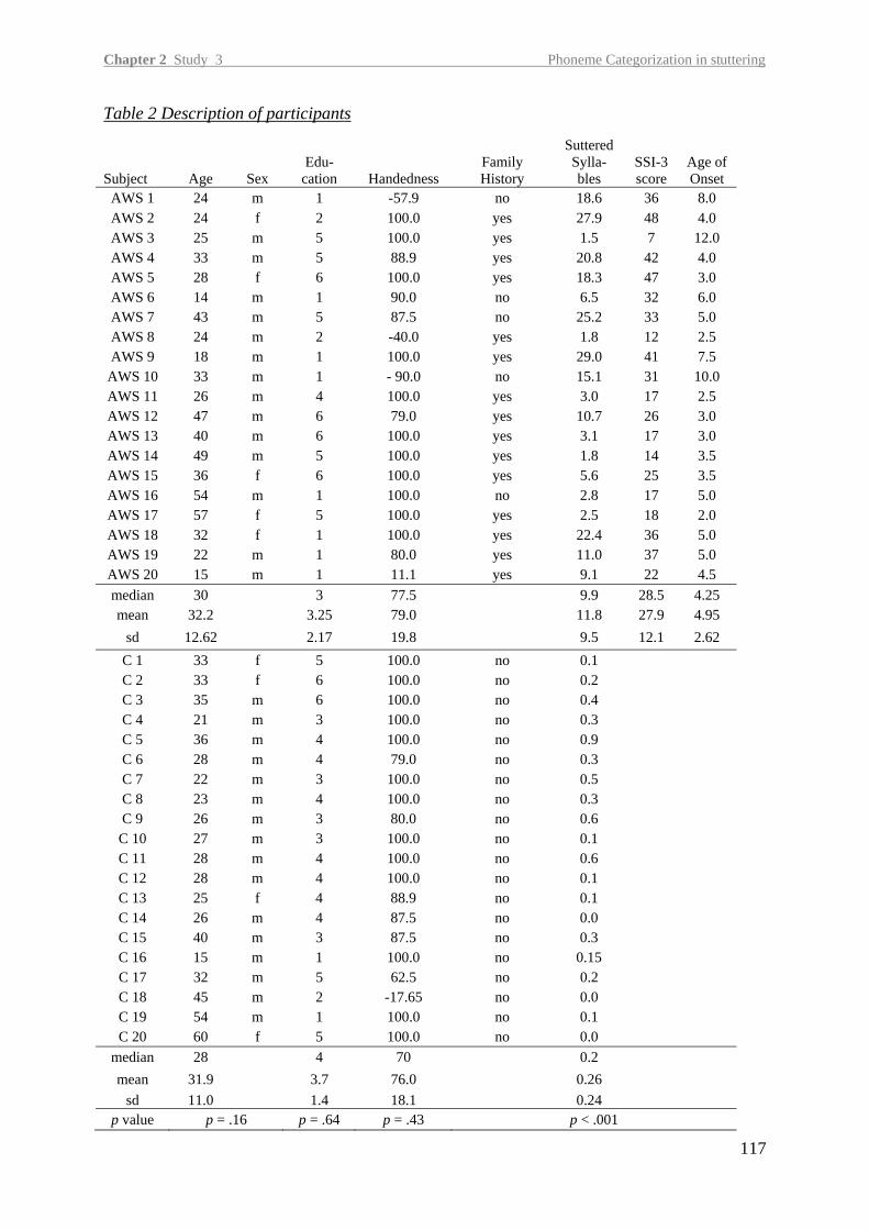
Chapter 2 Study 3 Phoneme Categorization in stuttering
Table 2 Description of participants
Subject Age Sex Edu-
cation Handedness Family History
Suttered Sylla-bles
SSI-3 score
Age of Onset
AWS 1 24 m 1 -57.9 no 18.6 36 8.0 AWS 2 24 f 2 100.0 yes 27.9 48 4.0 AWS 3 25 m 5 100.0 yes 1.5 7 12.0 AWS 4 33 m 5 88.9 yes 20.8 42 4.0 AWS 5 28 f 6 100.0 yes 18.3 47 3.0 AWS 6 14 m 1 90.0 no 6.5 32 6.0 AWS 7 43 m 5 87.5 no 25.2 33 5.0 AWS 8 24 m 2 -40.0 yes 1.8 12 2.5 AWS 9 18 m 1 100.0 yes 29.0 41 7.5
AWS 10 33 m 1 - 90.0 no 15.1 31 10.0 AWS 11 26 m 4 100.0 yes 3.0 17 2.5 AWS 12 47 m 6 79.0 yes 10.7 26 3.0 AWS 13 40 m 6 100.0 yes 3.1 17 3.0 AWS 14 49 m 5 100.0 yes 1.8 14 3.5 AWS 15 36 f 6 100.0 yes 5.6 25 3.5 AWS 16 54 m 1 100.0 no 2.8 17 5.0 AWS 17 57 f 5 100.0 yes 2.5 18 2.0 AWS 18 32 f 1 100.0 yes 22.4 36 5.0 AWS 19 22 m 1 80.0 yes 11.0 37 5.0 AWS 20 15 m 1 11.1 yes 9.1 22 4.5 median 30 3 77.5 9.9 28.5 4.25 mean 32.2 3.25 79.0 11.8 27.9 4.95
sd 12.62 2.17 19.8 9.5 12.1 2.62 C 1 33 f 5 100.0 no 0.1 C 2 33 f 6 100.0 no 0.2 C 3 35 m 6 100.0 no 0.4 C 4 21 m 3 100.0 no 0.3 C 5 36 m 4 100.0 no 0.9 C 6 28 m 4 79.0 no 0.3 C 7 22 m 3 100.0 no 0.5 C 8 23 m 4 100.0 no 0.3 C 9 26 m 3 80.0 no 0.6 C 10 27 m 3 100.0 no 0.1 C 11 28 m 4 100.0 no 0.6 C 12 28 m 4 100.0 no 0.1 C 13 25 f 4 88.9 no 0.1 C 14 26 m 4 87.5 no 0.0 C 15 40 m 3 87.5 no 0.3 C 16 15 m 1 100.0 no 0.15 C 17 32 m 5 62.5 no 0.2 C 18 45 m 2 -17.65 no 0.0 C 19 54 m 1 100.0 no 0.1 C 20 60 f 5 100.0 no 0.0
median 28 4 70 0.2 mean 31.9 3.7 76.0 0.26
sd 11.0 1.4 18.1 0.24 p value p = .16 p = .64 p = .43 p < .001
117
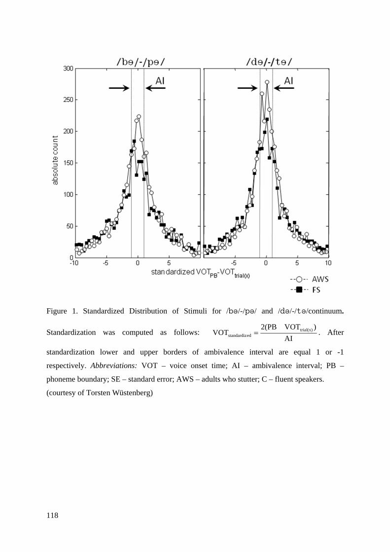
Figure 1. Standardized Distribution of Stimuli for /b/-/p/ and /d/-/t/continuum.
Standardization was computed as follows: AI
)VOT2(PBVOT trial(x)
edstandardiz
−=Δ . After
standardization lower and upper borders of ambivalence interval are equal 1 or -1
respectively. Abbreviations: VOT – voice onset time; AI – ambivalence interval; PB –
phoneme boundary; SE – standard error; AWS – adults who stutter; C – fluent speakers.
(courtesy of Torsten Wüstenberg)
118
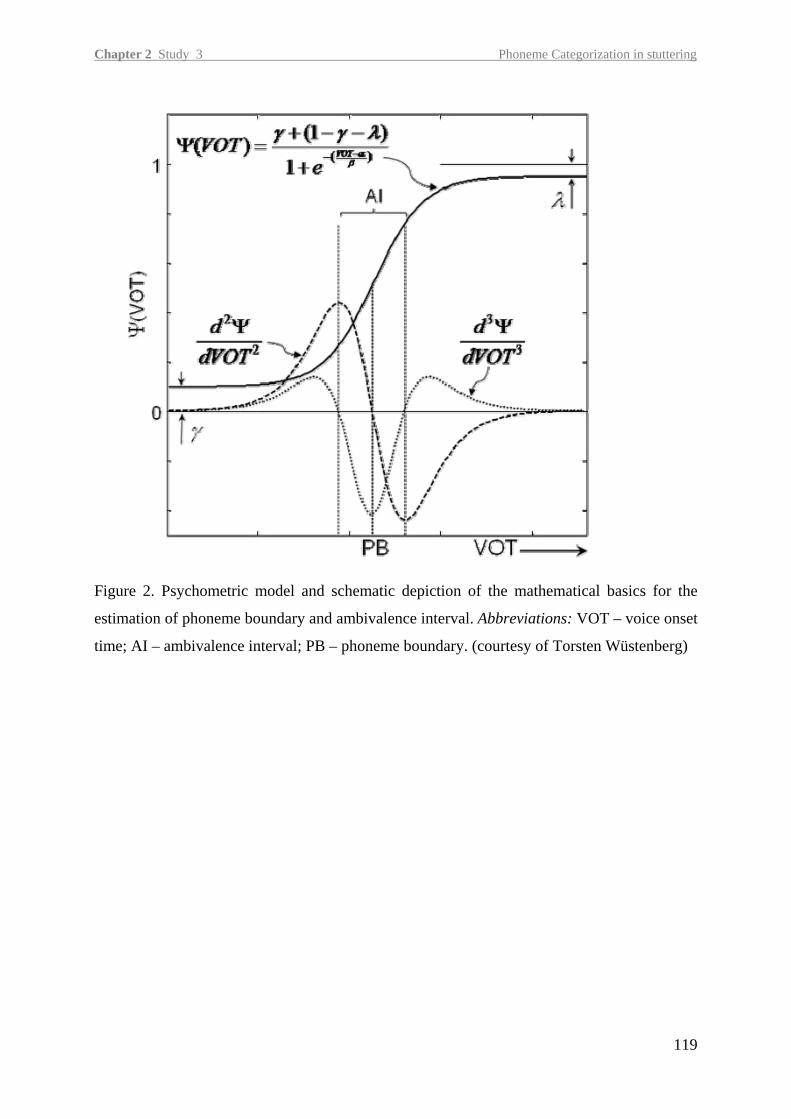
Chapter 2 Study 3 Phoneme Categorization in stuttering
Figure 2. Psychometric model and schematic depiction of the mathematical basics for the
estimation of phoneme boundary and ambivalence interval. Abbreviations: VOT – voice onset
time; AI – ambivalence interval; PB – phoneme boundary. (courtesy of Torsten Wüstenberg)
119
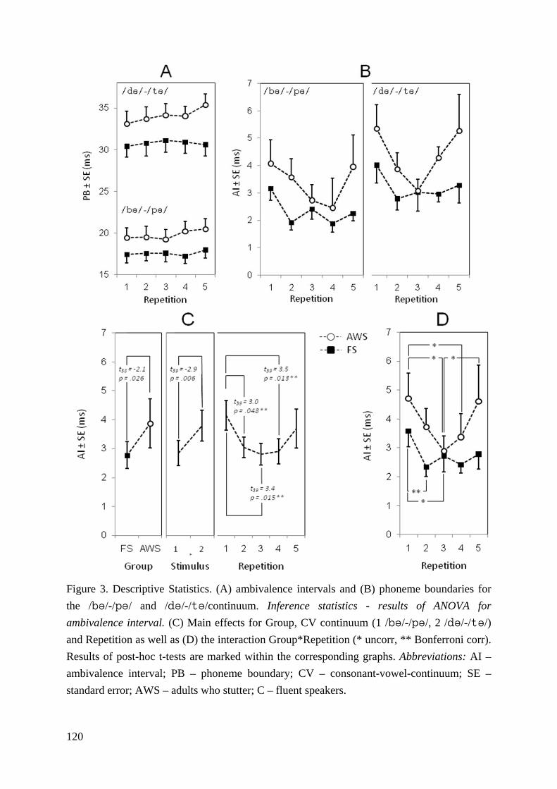
Figure 3. Descriptive Statistics. (A) ambivalence intervals and (B) phoneme boundaries for the /b/-/p/ and /d/-/t/continuum. Inference statistics - results of ANOVA for ambivalence interval. (C) Main effects for Group, CV continuum (1 /b/-/p/, 2 /d/-/t/) and Repetition as well as (D) the interaction Group*Repetition (* uncorr, ** Bonferroni corr). Results of post-hoc t-tests are marked within the corresponding graphs. Abbreviations: AI – ambivalence interval; PB – phoneme boundary; CV – consonant-vowel-continuum; SE – standard error; AWS – adults who stutter; C – fluent speakers.
120
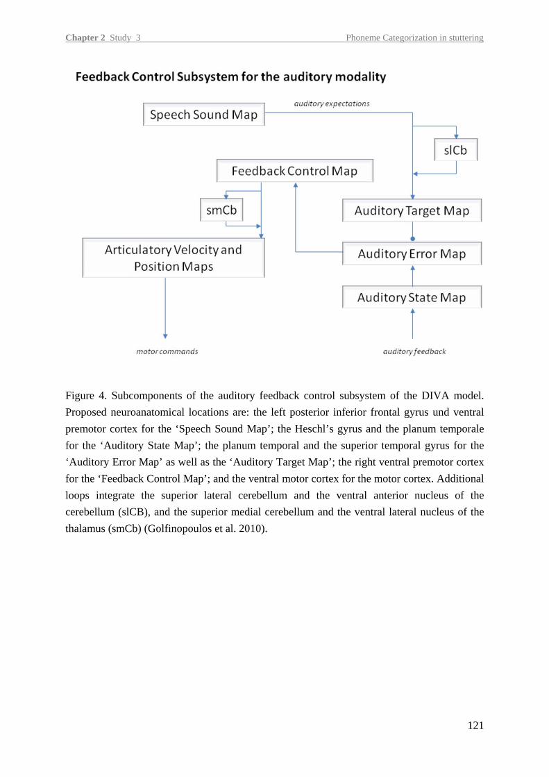
Chapter 2 Study 3 Phoneme Categorization in stuttering
121
Figure 4. Subcomponents of the auditory feedback control subsystem of the DIVA model. Proposed neuroanatomical locations are: the left posterior inferior frontal gyrus und ventral premotor cortex for the ‘Speech Sound Map’; the Heschl’s gyrus and the planum temporale for the ‘Auditory State Map’; the planum temporal and the superior temporal gyrus for the ‘Auditory Error Map’ as well as the ‘Auditory Target Map’; the right ventral premotor cortex for the ‘Feedback Control Map’; and the ventral motor cortex for the motor cortex. Additional loops integrate the superior lateral cerebellum and the ventral anterior nucleus of the cerebellum (slCB), and the superior medial cerebellum and the ventral lateral nucleus of the thalamus (smCb) (Golfinopoulos et al. 2010).

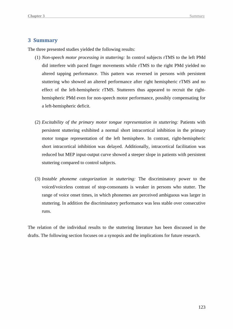
Chapter 3 Summary
123
3 Summary
The three presented studies yielded the following results:
(1) Non-speech motor processing in stuttering: In control subjects rTMS to the left PMd
did interfere with paced finger movements while rTMS to the right PMd yielded no
altered tapping performance. This pattern was reversed in persons with persistent
stuttering who showed an altered performance after right hemispheric rTMS and no
effect of the left-hemispheric rTMS. Stutterers thus appeared to recruit the right-
hemispheric PMd even for non-speech motor performance, possibly compensating for
a left-hemispheric deficit.
(2) Excitability of the primary motor tongue representation in stuttering: Patients with
persistent stuttering exhibited a normal short intracortical inhibition in the primary
motor tongue representation of the left hemisphere. In contrast, right-hemispheric
short intracortical inhibition was delayed. Additionally, intracortical facilitation was
reduced but MEP input-output curve showed a steeper slope in patients with persistent
stuttering compared to control subjects.
(3) Instable phoneme categorization in stuttering: The discriminatory power to the
voiced/voiceless contrast of stop-consonants is weaker in persons who stutter. The
range of voice onset times, in which phonemes are perceived ambiguous was larger in
stuttering. In addition the discriminatory performance was less stable over consecutive
runs.
The relation of the individual results to the stuttering literature has been discussed in the
drafts. The following section focuses on a synopsis and the implications for future research.

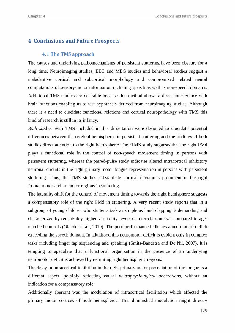
Chapter 4 Conclusions and future prospects
4 Conclusions and Future Prospects
4.1 The TMS approach
The causes and underlying pathomechanisms of persistent stuttering have been obscure for a
long time. Neuroimaging studies, EEG and MEG studies and behavioral studies suggest a
maladaptive cortical and subcortical morphology and compromised related neural
computations of sensory-motor information including speech as well as non-speech domains.
Additional TMS studies are desirable because this method allows a direct interference with
brain functions enabling us to test hypothesis derived from neuroimaging studies. Although
there is a need to elucidate functional relations and cortical neuropathology with TMS this
kind of research is still in its infancy.
Both studies with TMS included in this dissertation were designed to elucidate potential
differences between the cerebral hemispheres in persistent stuttering and the findings of both
studies direct attention to the right hemisphere: The rTMS study suggests that the right PMd
plays a functional role in the control of non-speech movement timing in persons with
persistent stuttering, whereas the paired-pulse study indicates altered intracortical inhibitory
neuronal circuits in the right primary motor tongue representation in persons with persistent
stuttering. Thus, the TMS studies substantiate cortical deviations prominent in the right
frontal motor and premotor regions in stuttering.
The laterality-shift for the control of movement timing towards the right hemisphere suggests
a compensatory role of the right PMd in stuttering. A very recent study reports that in a
subgroup of young children who stutter a task as simple as hand clapping is demanding and
characterized by remarkably higher variability levels of inter-clap interval compared to age-
matched controls (Olander et al., 2010). The poor performance indicates a neuromotor deficit
exceeding the speech domain. In adulthood this neuromotor deficit is evident only in complex
tasks including finger tap sequencing and speaking (Smits-Bandstra and De Nil, 2007). It is
tempting to speculate that a functional organization in the presence of an underlying
neuromotor deficit is achieved by recruiting right hemispheric regions.
The delay in intracortical inhibition in the right primary motor presentation of the tongue is a
different aspect, possibly reflecting causal neurophysiological aberrations, without an
indication for a compensatory role.
Additionally aberrant was the modulation of intracortical facilitation which affected the
primary motor cortices of both hemispheres. This diminished modulation might directly
125
126
contribute to the intermittent involuntary loss of speech-control, interrupting fluent speech in
stuttering. Interneuronal modulation of primary motor cortex’ excitability is an important
neurobiological principle enabling this neural structure to encode signals contributing to the
selection, initiation and inhibition of complex spatial-temporal speech movements (Stinear et
al., 2009). The study included here, is the first step towards a systematic neurophysiological
evaluation of the primary motor tongue representation in stuttering. As it receives input from
frontal cortical regions and the basal ganglia and drives the corticobulbar projection, changes
in its excitability are likely to reflect an altered modulation of corticobulbar neurons by basal
and frontal regions. Neuroimaging studies suggest that this modulation is state dependent
(Chang et al. 2009). So far, we examined the intracortical excitability and its modulation at
rest and under voluntary contraction. Having established MEP recordings from the tongue, we
can now advance towards function-related questions.
Current studies
In direct extension of the tongue-MEP-project a current study of our laboratory integrates
speech production. Here we study the modulation of the excitability of the primary motor
tongue representation during the preparation of a subsequent articulatory gesture (Hoang et
al., in preparation). Preliminary results show that excitability is indeed state dependent,
increasing before initiation of the new gesture. While at rest the left and right primary motor
representation show a reduced intracortical facilitation, during a speech related mode only the
left primary motor cortex exhibits a reduced facilitation in persons who stutter; the right
primary motor cortex shows the tendency to more facilitation. This state-dependent
dissociation between the two hemispheres evokes the question for underlying mechanisms. Is
the interplay between the speech motor cortices via the corpus callosum impaired? Or are
those intercortical connections altered, which are involved in the regulation of excitation to
initiate efferent volleys of motor commands and to prevent unwanted movements? It seems
that the cerebral dominance hypothesis, although well advanced in years, comes to the fore
again.
4.2 The speech perception approach
Mapping of sounds to articulation
In adults who stutter the phoneme categorization study suggested a diminished sensitivity to
identify voiced and voiceless plosives near the phoneme boundary. One possible
interpretation attributes this vulnerability to the decreased integrity of fiber tracts of the
Chapter 4 Conclusions and future prospects
fasciculus longitudionalis superior which has been consistently reported by four independent
research groups (Chang et al., 2008; Cykowski et al., 2010; Sommer et al., 2002a; Watkins et
al., 2008). It is not exactly clear yet, which fiber tracts are affected: fiber tracts connecting
Broca’s area (inferior frontal gurus) with the ventral premotor and primary motor cortex,
related to the encoding of the phonetic plan (Lu et al., 2009a; Salmelin et al., 2000), or
connections of the dorsal route between premotor areas and superior temporal lobe related to
the sensory-motor mapping of sound to articulation (Chang et al., 2008; Cykowski et al.,
2010; Neef et al., 2009). Therefore we already planned the following future studies:
(1) A study with transcranial magnetic stimulation to elucidate whether the lesioning of
critical cortical sites influences the identification of the contrast of voicing. Planned
stimulation sites are the left superior temporal gyrus, the left ventral premotor cortex
and the left primary motor cortex. An effect of stimulation will be operationalized by
quantifying and comparing ambivalence intervals before and after stimulation. (i) We
expect a broadening of the ambivalence intervals due to a lesioning of the STG
because this cortical region is mainly involved in speech perception. (ii) A broadening
of the ambivalence interval due to an inhibition of the ventral premotor cortex might
indicate an increased vulnerability of the dorsal route (connection between PMv and
STG). (iii) An effect of lesioning the primary motor cortex might indicate that the
motor programs themselves may constitute phonological primitives, which as a
consequence would demand a rethinking of the targeted reference frame in speech
production.
(2) A study with electroencephalography (EEG) will elucidate the temporal coordination
of neural activity and thus will answer the question whether the neural populations in
frontal and temporal regions are simultaneously engaged in the mentioned phoneme
identification task as it is proposed for a sensory-motor mapping of sound to
articulation. By using distributed source models we will estimate the functional
connectivity of the dorsal route for the processing of perceptually ambiguous and
unambiguous stimuli, respectively. In control subjects the phoneme identification task
is expected to be mirrored in a quantifiable functional connectivity during the
perception of unambiguous stimuli. This functional connectivity is expected to be
diminished during the perception of ambiguous stimuli. In persons who stutter a
deficient dorsal route caused by diminished fiber integrity is expected to be mirrored
in an altered time pattern.
127
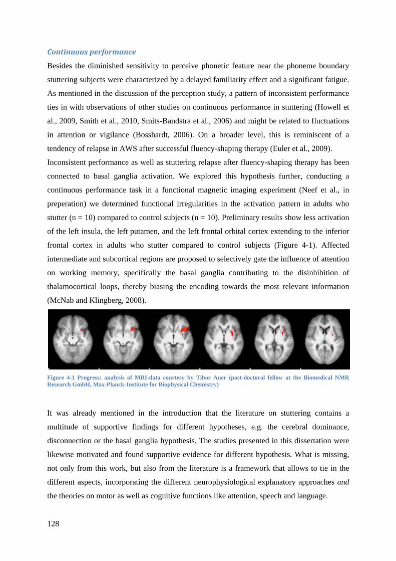
Continuous performance
Besides the diminished sensitivity to perceive phonetic feature near the phoneme boundary
stuttering subjects were characterized by a delayed familiarity effect and a significant fatigue.
As mentioned in the discussion of the perception study, a pattern of inconsistent performance
ties in with observations of other studies on continuous performance in stuttering (Howell et
al., 2009, Smith et al., 2010, Smits-Bandstra et al., 2006) and might be related to fluctuations
in attention or vigilance (Bosshardt, 2006). On a broader level, this is reminiscent of a
tendency of relapse in AWS after successful fluency-shaping therapy (Euler et al., 2009).
Inconsistent performance as well as stuttering relapse after fluency-shaping therapy has been
connected to basal ganglia activation. We explored this hypothesis further, conducting a
continuous performance task in a functional magnetic imaging experiment (Neef et al., in
preperation) we determined functional irregularities in the activation pattern in adults who
stutter (n = 10) compared to control subjects (n = 10). Preliminary results show less activation
of the left insula, the left putamen, and the left frontal orbital cortex extending to the inferior
frontal cortex in adults who stutter compared to control subjects (Figure 4-1). Affected
intermediate and subcortical regions are proposed to selectively gate the influence of attention
on working memory, specifically the basal ganglia contributing to the disinhibition of
thalamocortical loops, thereby biasing the encoding towards the most relevant information
(McNab and Klingberg, 2008).
Figure 4-1 Progress: analysis of MRI-data courtesy by Tibor Auer (post-doctoral fellow at the Biomedical NMR Research GmbH, Max-Planck-Institute for Biophysical Chemistry)
It was already mentioned in the introduction that the literature on stuttering contains a
multitude of supportive findings for different hypotheses, e.g. the cerebral dominance,
disconnection or the basal ganglia hypothesis. The studies presented in this dissertation were
likewise motivated and found supportive evidence for different hypothesis. What is missing,
not only from this work, but also from the literature is a framework that allows to tie in the
different aspects, incorporating the different neurophysiological explanatory approaches and
the theories on motor as well as cognitive functions like attention, speech and language.
128
Chapter 4 Conclusions and future prospects
129
4.3 Future directions
Future efforts to unravel the causes of stuttering might profit from a change in perspective.
New inputs could come from the research on the cortical and subcortical reorganizations
underlying skill acquisition and automation. It might account for the connection between
aberration in basal ganglia function and a pathological interplay between hemispheres in the
emergence of dysfluent speech movements. Interestingly, the extent of the basal ganglia
involvement in a skill is related to the skill’s degree of automaticity. During maturation
speaking becomes an automatized skill. The automation of a skill involves a restructuring of
implementation and a reorganization of functional anatomy including a decreased activation
of cortical areas and an increased activation of the intermediate cortical structures and the
basal ganglia (Saling and Phillips, 2007).
Fluency-enhancing techniques such as speaking with a gentle voice onset and no voice offset
or speaking under altered auditory feedback invokes additional monitoring to control for the
target speech pattern. This shifts the speaking away from automatized toward a monitored,
controlled process involving additional cortical resources. Both referred methods are very
efficient at the beginning of an intervention but prone to become less beneficial with time
exercised (Euler et al., 2009). This suggests that an increasing automaticity which is related to
an increasing involvement of the basal ganglia leads to reoccurrence of the dysfluent
symptoms.
The modulation of cortical excitability also plays a role in skill acquisition: when new motor
patterns are acquired, initially some degrees of freedom which are redundant, not crucial for
the task, are “frozen”. This reduces the capacity needed for monitoring and thus speeds up
motor learning. When automation sets in, however, the degrees of freedom are freed again.
Freezing and freeing involves the modulation of cortical inhibition (Salling and Phillips,
2007), which brings in other aspect of this dissertation, the excitability of primary motor
cortex and the contribution of other cortical areas like premotor cortex.
Whether the field of automaticity-related restructuring of cortical and subcortical processing
can help to create an integrative framework in which different aspects and hypothesis on the
cause of stuttering can be tied in, is not clear. Thus, a promising direction in stuttering
research might lie in studies that correlate shifts in activation from cortical towards
subcortical structures with the behavioural changes associated with automation. For me, this
option is an attractive perspective in future works on stuttering.

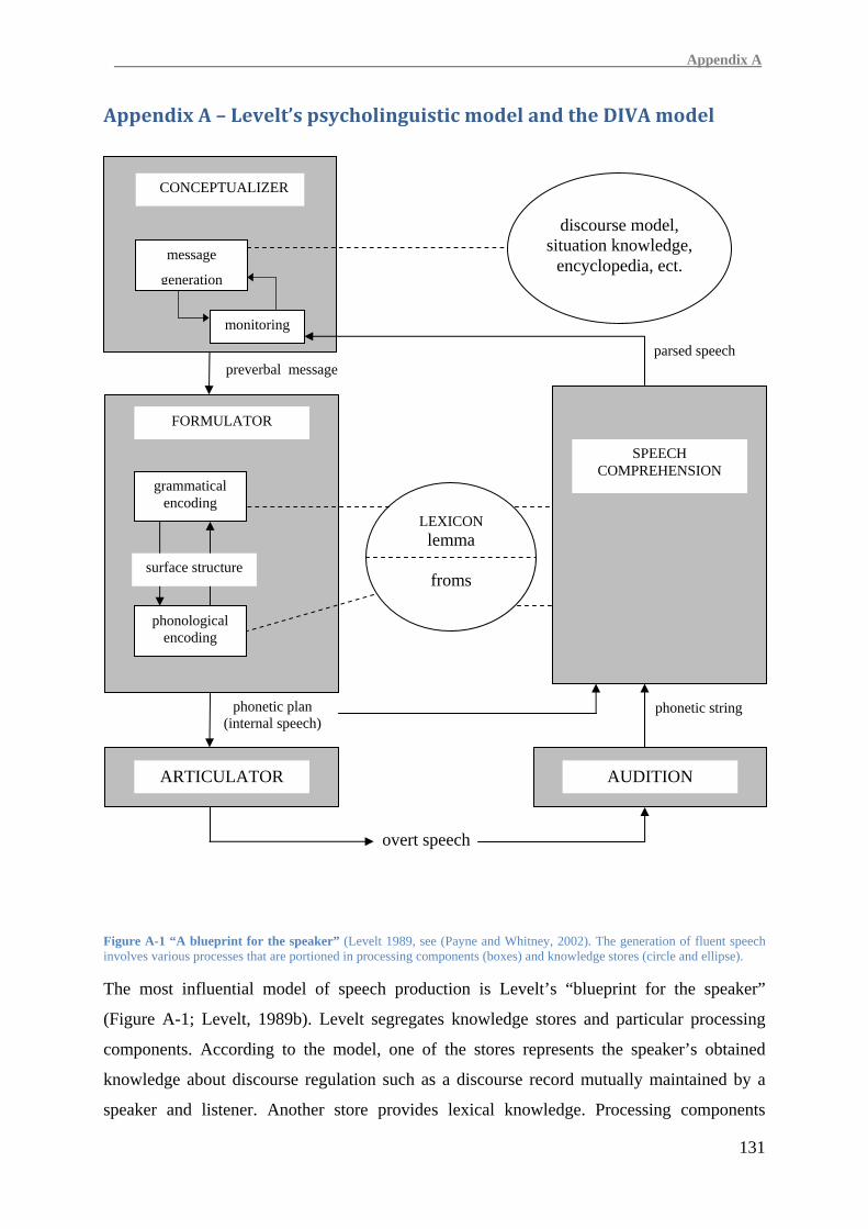
A Appendix A
Appendix A – Levelt’s psycholinguistic model and the DIVA model
CONCEPTUALIZER
discourse model, situation knowledge,
encyclopedia, ect. message
131
Figure A-1 “A blueprint for the speaker” (Levelt 1989, see (Payne and Whitney, 2002). The generation of fluent speech involves various processes that are portioned in processing components (boxes) and knowledge stores (circle and ellipse).
The most influential model of speech production is Levelt’s “blueprint for the speaker”
(Figure A-1; Levelt, 1989b). Levelt segregates knowledge stores and particular processing
components. According to the model, one of the stores represents the speaker’s obtained
knowledge about discourse regulation such as a discourse record mutually maintained by a
speaker and listener. Another store provides lexical knowledge. Processing components
generation
monitoring
FORMULATOR
grammatical encoding
phonological encoding
LEXICON lemma
froms
SPEECH COMPREHENSION
ARTICULATOR AUDITION
phonetic plan (internal speech)
preverbal message parsed speech
phonetic string
overt speech
surface structure
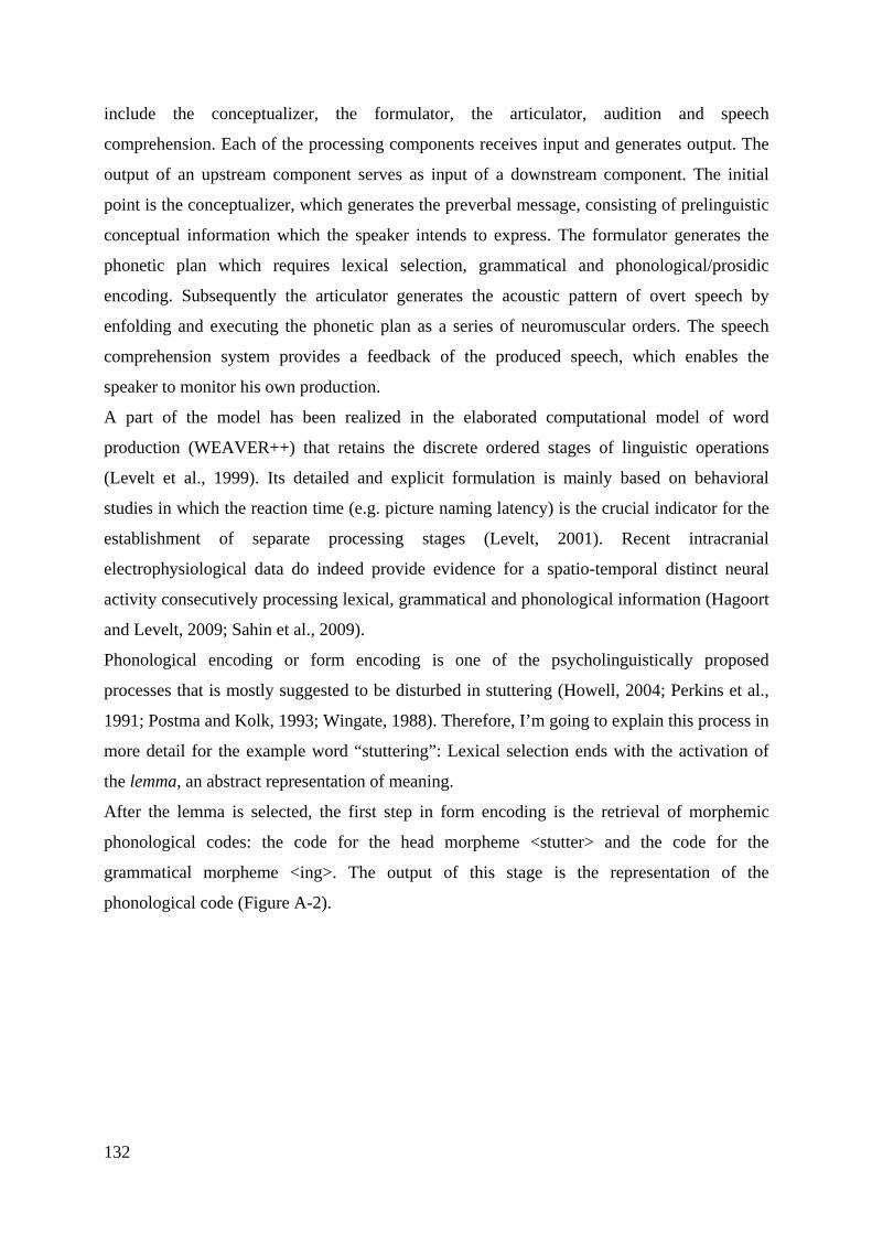
132
include the conceptualizer, the formulator, the articulator, audition and speech
comprehension. Each of the processing components receives input and generates output. The
output of an upstream component serves as input of a downstream component. The initial
point is the conceptualizer, which generates the preverbal message, consisting of prelinguistic
conceptual information which the speaker intends to express. The formulator generates the
phonetic plan which requires lexical selection, grammatical and phonological/prosidic
encoding. Subsequently the articulator generates the acoustic pattern of overt speech by
enfolding and executing the phonetic plan as a series of neuromuscular orders. The speech
comprehension system provides a feedback of the produced speech, which enables the
speaker to monitor his own production.
A part of the model has been realized in the elaborated computational model of word
production (WEAVER++) that retains the discrete ordered stages of linguistic operations
(Levelt et al., 1999). Its detailed and explicit formulation is mainly based on behavioral
studies in which the reaction time (e.g. picture naming latency) is the crucial indicator for the
establishment of separate processing stages (Levelt, 2001). Recent intracranial
electrophysiological data do indeed provide evidence for a spatio-temporal distinct neural
activity consecutively processing lexical, grammatical and phonological information (Hagoort
and Levelt, 2009; Sahin et al., 2009).
Phonological encoding or form encoding is one of the psycholinguistically proposed
processes that is mostly suggested to be disturbed in stuttering (Howell, 2004; Perkins et al.,
1991; Postma and Kolk, 1993; Wingate, 1988). Therefore, I’m going to explain this process in
more detail for the example word “stuttering”: Lexical selection ends with the activation of
the lemma, an abstract representation of meaning.
After the lemma is selected, the first step in form encoding is the retrieval of morphemic
phonological codes: the code for the head morpheme <stutter> and the code for the
grammatical morpheme <ing>. The output of this stage is the representation of the
phonological code (Figure A-2).
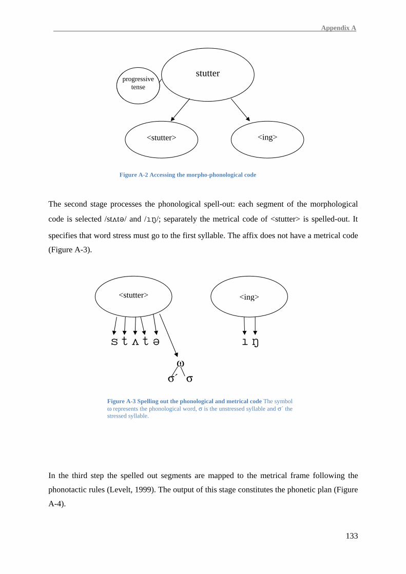
A Appendix A
stutter progressive
tense
133
<stutter> <ing>
s t t
ω σ´ σ
<ing> <stutter>
The second stage processes the phonological spell-out: each segment of the morphological
code is selected /stt/ and //; separately the metrical code of <stutter> is spelled-out. It
specifies that word stress must go to the first syllable. The affix does not have a metrical code
(Figure A-3).
Figure A-2 Accessing the morpho-phonological code
Figure A-3 Spelling out the phonological and metrical code The symbol ω represents the phonological word, σ is the unstressed syllable and σ´ the stressed syllable.
In the third step the spelled out segments are mapped to the metrical frame following the
phonotactic rules (Levelt, 1999). The output of this stage constitutes the phonetic plan (Figure
A-4).
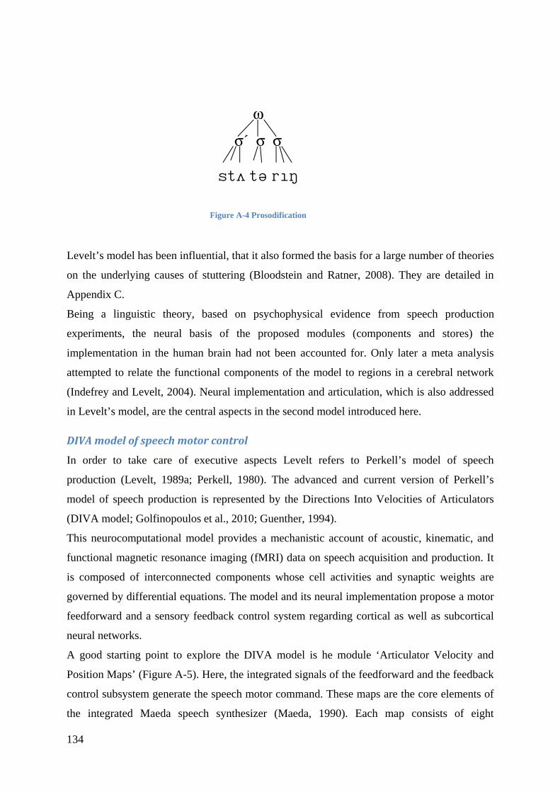
ω
σ´ σ σ
st t r
Figure A-4 Prosodification
Levelt’s model has been influential, that it also formed the basis for a large number of theories
on the underlying causes of stuttering (Bloodstein and Ratner, 2008). They are detailed in
Appendix C.
Being a linguistic theory, based on psychophysical evidence from speech production
experiments, the neural basis of the proposed modules (components and stores) the
implementation in the human brain had not been accounted for. Only later a meta analysis
attempted to relate the functional components of the model to regions in a cerebral network
(Indefrey and Levelt, 2004). Neural implementation and articulation, which is also addressed
in Levelt’s model, are the central aspects in the second model introduced here.
DIVA model of speech motor control
In order to take care of executive aspects Levelt refers to Perkell’s model of speech
production (Levelt, 1989a; Perkell, 1980). The advanced and current version of Perkell’s
model of speech production is represented by the Directions Into Velocities of Articulators
(DIVA model; Golfinopoulos et al., 2010; Guenther, 1994).
This neurocomputational model provides a mechanistic account of acoustic, kinematic, and
functional magnetic resonance imaging (fMRI) data on speech acquisition and production. It
is composed of interconnected components whose cell activities and synaptic weights are
governed by differential equations. The model and its neural implementation propose a motor
feedforward and a sensory feedback control system regarding cortical as well as subcortical
neural networks.
A good starting point to explore the DIVA model is he module ‘Articulator Velocity and
Position Maps’ (Figure A-5). Here, the integrated signals of the feedforward and the feedback
control subsystem generate the speech motor command. These maps are the core elements of
the integrated Maeda speech synthesizer (Maeda, 1990). Each map consists of eight
134
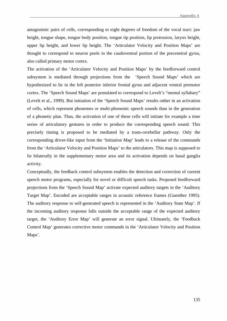
A Appendix A
antagonistic pairs of cells, corresponding to eight degrees of freedom of the vocal tract: jaw
height, tongue shape, tongue body position, tongue tip position, lip protrusion, larynx height,
upper lip height, and lower lip height. The ‘Articulator Velocity and Position Maps’ are
thought to correspond to neuron pools in the caudoventral portion of the precenetral gyrus,
also called primary motor cortex.
The activation of the ‘Articulator Velocity and Position Maps’ by the feedforward control
subsystem is mediated through projections from the ‘Speech Sound Maps’ which are
hypothesized to lie in the left posterior inferior frontal gyrus and adjacent ventral premotor
cortex. The ‘Speech Sound Maps’ are postulated to correspond to Levelt’s “mental syllabary”
(Levelt et al., 1999). But initiation of the ‘Speech Sound Maps’ results rather in an activation
of cells, which represent phonemes or multi-phonemic speech sounds than in the generation
of a phonetic plan. Thus, the activation of one of these cells will initiate for example a time
series of articulatory gestures in order to produce the corresponding speech sound. This
precisely timing is proposed to be mediated by a trans-cerebellar pathway. Only the
corresponding driver-like input from the ‘Initiation Map’ leads to a release of the commands
from the ‘Articulator Velocity and Position Maps’ to the articulators. This map is supposed to
lie bilaterally in the supplementary motor area and its activation depends on basal ganglia
activity.
Conceptually, the feedback control subsystem enables the detection and correction of current
speech motor programs, especially for novel or difficult speech tasks. Proposed feedforward
projections from the ‘Speech Sound Map’ activate expected auditory targets in the ‘Auditory
Target Map’. Encoded are acceptable ranges in acoustic reference frames (Guenther 1995).
The auditory response to self-generated speech is represented in the ‘Auditory State Map’. If
the incoming auditory response falls outside the acceptable range of the expected auditory
target, the ‘Auditory Error Map’ will generate an error signal. Ultimately, the ‘Feedback
Control Map’ generates corrective motor commands in the ‘Articulator Velocity and Position
Maps’.
135
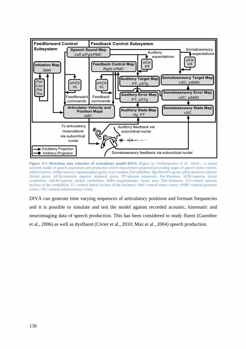
Figure A-5 Directions into velocities of articulators model DIVA (Figure by Golfinopoulos et al., 2010) – a neural network model of speech acquisition and production which characterizes proposed processing stages of speech motor control. Abbreviations: aSMg=anterior supramarginal gyrus; Cau=caudate; Pal=pallidum; Hg=Heschl's gyrus; pIFg=posterior inferior frontal gyrus; pSTg=posterior superior temporal gyrus; PT=planum temporale; Put=Putamen; slCB=superior lateral cerebellum; smCB=superior medial cerebellum; SMA=supplementary motor area; Tha=thalamus; VA=ventral anterior nucleus of the cerebellum; VL=ventral lateral nucleus of the thalamus; vMC=ventral motor cortex; vPMC=ventral premotor cortex; vSC=ventral somatosensory cortex.
DIVA can generate time varying sequences of articulatory positions and formant frequencies
and it is possible to simulate and test the model against recorded acoustic, kinematic and
neuroimaging data of speech production. This has been considered to study fluent (Guenther
et al., 2006) as well as dysfluent (Civier et al., 2010; Max et al., 2004) speech production.
136
A Appendix B
Appendix B - Stuttering and acquired brain lesions
This dissertation focuses on cortical and subcortical mechanisms in persistent developmental
stuttering. Due to the long course and the often very long delay between onset and
examination in a study, the causal origins of developmental stuttering are notoriously hard to
address and consequently they are still largely unclear. There is, however, a lot to gain from
studies of the related acquired and induced stuttering, where the causal disruption is more
easily identified and the short period between onset and examination helps to assure that
observed abnormalities are not secondary but indeed causal. Similarly to aphasiology where
lesion studies elucidated and facilitated the understanding of language processing in the brain
I am going to give a short review on locations of brain injuries that induces speech
dysfluencies to further understand the emergence of stuttering and general processes of
speech production.
Acquired stuttering
In 1835, Franz Joseph Gall and Johann Gaspar Spurzheim might have delivered the first
report on acquired [neurogenic] stuttering (Andy and Bhatnagar, 1992). They mentioned a
patient with a sword wound across the left nasal fossa and a penetrated internal posterior part
of the anterior left lobe of the brain which was followed by speech and voice problems,
hemiplegia and loss of vision. Later on only a slight stuttering remained. 150 years later,
Nancy Helm and colleagues provided the first comprehensive description of the syndrome
(Helm et al., 1978) but guidelines in its assessment were critically reviewed e.g. (Lundgren et
al., 2010; Ringo and Dietrich, 1995) because the perceptual distinction between
developmental and acquired stuttering remains indefinite (Van Borsel and Taillieu, 2001). A
diagnosic certainty is possible if a documented neurologic condition and the following
behaviors are associated: (1) dysfluencies occurre at a similar rate on open class words (e.g.
nouns, verbs, adjectives) as well as on closed class words (pronouns, determiners,
conjunctions, prepositions, particles); (2) repetitions, prolongations, and blocks occur in all
positions in words; (3) dysfluencies occur consistently across speech tasks (e.g. free speech
production, reading); (4) patients appear not overtly anxious about the stuttering behavior; (5)
accompanying physical concomitants (facial grimacing, fist clenching, and eye blinking)
occur rarely and; (6) no adaptation effect is evident (repeated reading of a passage enhances
fluency) (Jokel et al., 2007; Lundgren et al., 2010). Challenging aspects among the
differential diagnosis of acquired stuttering are the distinction of dysfluency from those
137
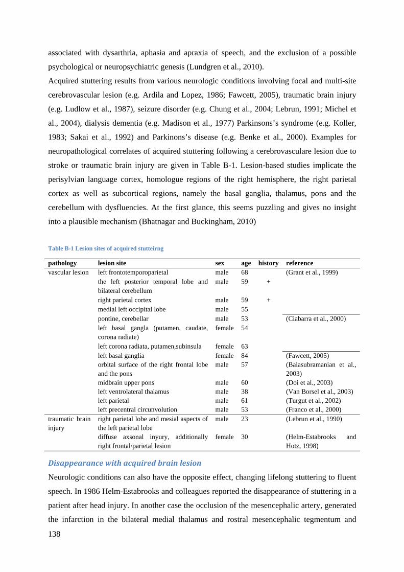
138
associated with dysarthria, aphasia and apraxia of speech, and the exclusion of a possible
psychological or neuropsychiatric genesis (Lundgren et al., 2010).
Acquired stuttering results from various neurologic conditions involving focal and multi-site
cerebrovascular lesion (e.g. Ardila and Lopez, 1986; Fawcett, 2005), traumatic brain injury
(e.g. Ludlow et al., 1987), seizure disorder (e.g. Chung et al., 2004; Lebrun, 1991; Michel et
al., 2004), dialysis dementia (e.g. Madison et al., 1977) Parkinsons’s syndrome (e.g. Koller,
1983; Sakai et al., 1992) and Parkinons’s disease (e.g. Benke et al., 2000). Examples for
neuropathological correlates of acquired stuttering following a cerebrovasculare lesion due to
stroke or traumatic brain injury are given in Table B-1. Lesion-based studies implicate the
perisylvian language cortex, homologue regions of the right hemisphere, the right parietal
cortex as well as subcortical regions, namely the basal ganglia, thalamus, pons and the
cerebellum with dysfluencies. At the first glance, this seems puzzling and gives no insight
into a plausible mechanism (Bhatnagar and Buckingham, 2010)
Table B-1 Lesion sites of acquired stutteirng
pathology lesion site sex age history reference vascular lesion left frontotemporoparietal male 68 (Grant et al., 1999)
the left posterior temporal lobe and bilateral cerebellum
male 59 +
right parietal cortex male 59 + medial left occipital lobe male 55 pontine, cerebellar male 53 (Ciabarra et al., 2000) left basal gangla (putamen, caudate, corona radiate)
female 54
left corona radiata, putamen,subinsula female 63 left basal ganglia female 84 (Fawcett, 2005) orbital surface of the right frontal lobe and the pons
male 57 (Balasubramanian et al., 2003)
midbrain upper pons male 60 (Doi et al., 2003) left ventrolateral thalamus male 38 (Van Borsel et al., 2003) left parietal male 61 (Turgut et al., 2002) left precentral circunvolution male 53 (Franco et al., 2000)
traumatic brain injury
right parietal lobe and mesial aspects of the left parietal lobe
male 23 (Lebrun et al., 1990)
diffuse axsonal inyury, additionally right frontal/parietal lesion
female 30 (Helm-Estabrooks and Hotz, 1998)
Disappearance with acquired brain lesion
Neurologic conditions can also have the opposite effect, changing lifelong stuttering to fluent
speech. In 1986 Helm-Estabrooks and colleagues reported the disappearance of stuttering in a
patient after head injury. In another case the occlusion of the mesencephalic artery, generated
the infarction in the bilateral medial thalamus and rostral mesencephalic tegmentum and
A Appendix B
139
ceased stuttering (Muroi et al., 1999). In two cases the progress of multiple sclerosis ceased
stuttering (Miller, 1985). An elaborated study of four cases documents the disappearance of
stuttering after neurosurgery (Jones, 1966). In all four cases neurosurgery was required on one
cerebral hemisphere because of a neurologic disease (aneurysm clipping following a
haemorrhage, tumour resection). Whereas pre-operative intracarotid amytal tests yielded a
bilateral cerebral representation of language, post-operatively language function was shifted
to the non-operated hemisphere in all patients. This co-occurrence of emerging language
dominance with cessation of stuttering fits nicely with the cerebral dominance theory, a
hypothesis that postulated that stuttering might be caused by an aberrant cerebral language
lateralization (Travis, 1978).
Brain stimulation
Further cues towards a neurobiological understanding of the phenomenon stuttering has been
delivered by a contemporary surgical treatment: deep brain stimulation (DBS). An implanted
multicontact electrode stimulates the brain tissue with high-frequency electric current pulses.
First implantations provided efficient relief from symptoms of advanced Parkinson’s disease:
for example an improvement from rigidity when delivered to the subthalamic nucleus
(Lanotte et al., 2002), a decrease of tremor when delivered to the thalamic nucleus ventralis
intermedius (Benabid et al., 1991; Benabid et al., 1987); or relieve from dyskinesia when
delivered to the internal pallidum (Limousin-Dowsey et al., 1999). DBS is now employed to
treat a broad range of chronic brain disorders in patients resistant to pharmacological
therapies, including dystonia, epilepsy, pain, obsessive compulsive disorders, Gilles de la
Tourette syndrome, depression and to improve the condition of brain-injured patients in a
vegetative or minimally conscious state (Deniau et al., 2010; Schiff et al., 2007). Although
DBS can be an effective symptomatic therapy, it is accompanied by adverse effects e.g. (Seijo
et al., 2007; Voges and Krauss, 2010).
In October 2010, a PubMed recherché with the keywords “deep brain stimulation” and
“stuttering” yielded five articles, included in Table B-2. Most frequent are case reports on
cessation of acquired stuttering due to DBS on the left thalamus centromedian nucleus
(Bhatnagar and Buckingham, 2010; Bhatnagar and Andy, 1989). In three cases, DBS in
patients with dystonia induced stuttering as an adverse effect. While stuttering ceased under
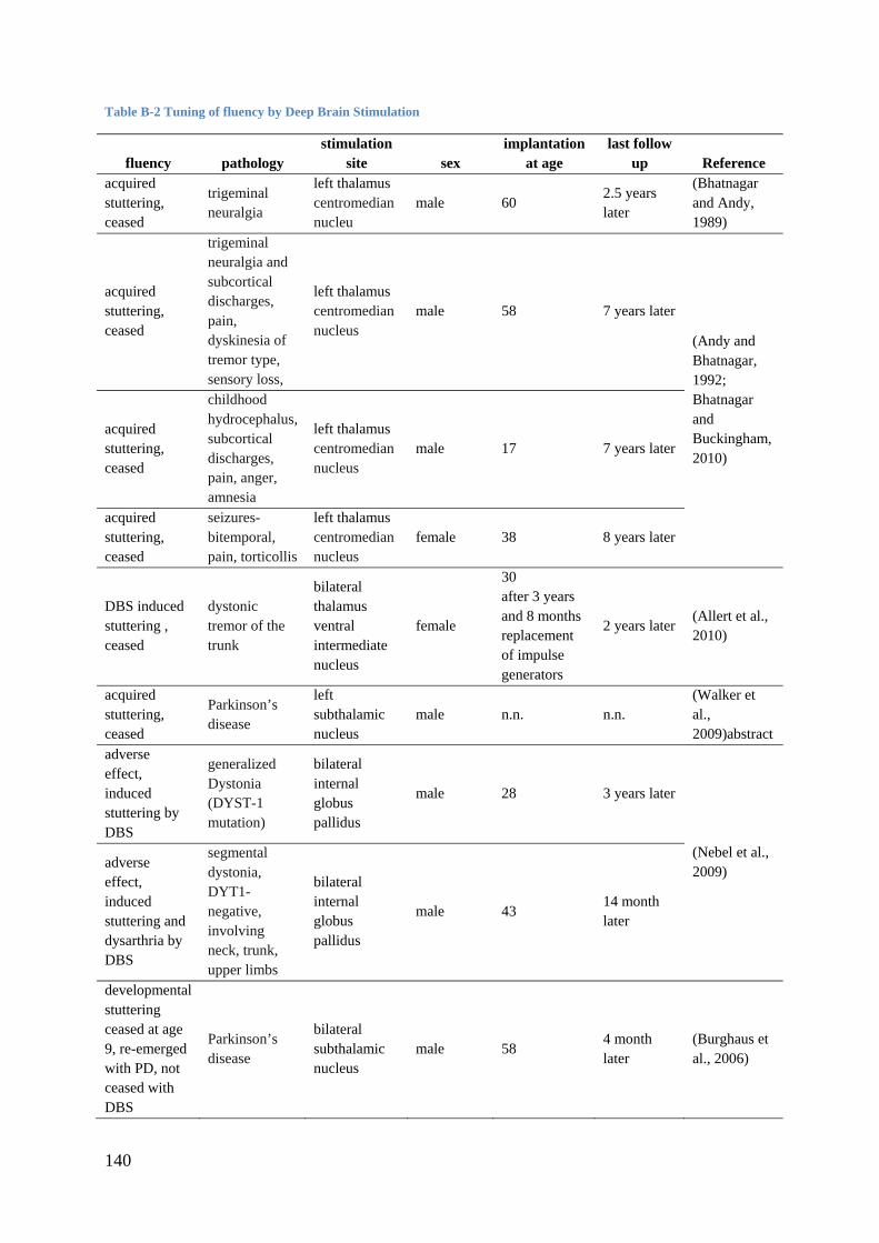
140
Table B-2 Tuning of fluency by Deep Brain Stimulation
fluency pathology stimulation
site sex implantation
at age last follow
up Reference acquired stuttering, ceased
trigeminal neuralgia
left thalamus centromedian nucleu
male 60 2.5 years later
(Bhatnagar and Andy, 1989)
acquired stuttering, ceased
trigeminal neuralgia and subcortical discharges, pain, dyskinesia of tremor type, sensory loss,
left thalamus centromedian nucleus
male 58 7 years later
(Andy and Bhatnagar, 1992; Bhatnagar and Buckingham, 2010)
acquired stuttering, ceased
childhood hydrocephalus, subcortical discharges, pain, anger, amnesia
left thalamus centromedian nucleus
male 17 7 years later
acquired stuttering, ceased
seizures-bitemporal, pain, torticollis
left thalamus centromedian nucleus
female 38 8 years later
DBS induced stuttering , ceased
dystonic tremor of the trunk
bilateral thalamus ventral intermediate nucleus
female
30 after 3 years and 8 months replacement of impulse generators
2 years later (Allert et al., 2010)
acquired stuttering, ceased
Parkinson’s disease
left subthalamic nucleus
male n.n. n.n. (Walker et al., 2009)abstract
adverse effect, induced stuttering by DBS
generalized Dystonia (DYST-1 mutation)
bilateral internal globus pallidus
male 28 3 years later
(Nebel et al., 2009) adverse
effect, induced stuttering and dysarthria by DBS
segmental dystonia, DYT1-negative, involving neck, trunk, upper limbs
bilateral internal globus pallidus
male 43 14 month later
developmental stuttering ceased at age 9, re-emerged with PD, not ceased with DBS
Parkinson’s disease
bilateral subthalamic nucleus
male 58 4 month later
(Burghaus et al., 2006)

A Appendix B
141
adjusted stimulation of bilateral thalamic nucleus ventralis intermedius (Allert et al., 2010), it
persisted under bilateral simulation of the internal globus pallidus (Nebel et al., 2009).

A Appendix C
143
Appendix C - Psycholinguistic approaches to explain stuttering
The neuropsycholinguistic theory suggests that fluency breakdowns result from a
desynchronized arrival of segmental (phonological) and suprasegemental (stress pattern)
information which are processed by different neural systems that converge on a common
output system. Time pressure forces the speaker to begin, continue, or accelerate an utterance
which results in a loss of motor control – stuttered dysfluencies (Perkins et al., 1991).
The Covert Repair Hypothesis is extensively pursued in recent years. The hypothesis targets
the pre-articulatory internal monitor established in Levelts psycholinguistic model. The
authors propose internal error sources such as phonemic encoding error detected before word
execution has started (resulting in a block), phonemic encoding error detected after word
execution has been produced (resulting in a prolongation or a repetition), phonemic encoding
error detected after the first sound has been produced (resulting in a prolongation of the initial
sound(s) or in a mid-syllable block) (Postma and Kolk, 1993).
The Execution and Planning model (EXPLAN) distinguishes between linguistic planning and
motor execution of utterance components. The theory proposes an incremental processing in
spontaneous speech production, which means, the planning of an upcoming utterance
component is simultaneously processed to the execution of the current utterance component.
In stuttering the motor execution plan is not available due to planning difficulties caused by
potential phonetic or lexical complexity, resulting in speech dysfluencies (Howell, 2004).

A Appendix D
Appendix D - Transcranial magnetic stimulation
Transcranial magnetic stimulation (TMS) is a noninvasive technique used to stimulate a
subset of the neurons in a small part, approximately 1 cm³, of the cerebral cortex. The
technique uses the principle of electromagnetic induction; a very brief magnetic pulse induces
an electrical field and thereby an electrical current in conductive brain tissue.
The mechanisms by which the brief electrical current stimulates neurons are not entirely clear.
Strong stimuli (suprathreshold) applied over the motor cortex directly elicit motor evoked
potentials (MEP) in the target muscles of the stimulated area. It is thought that the electric
stimulus suffices to depolarize the axons of the layer 5 pyramidal cells above action potential
threshold. Weaker stimuli alter the excitatory state even when no muscle evoked potentials
are detectable (see below: paired-pulse TMS). It is thought that these subthreshold stimuli
only depolarize presynaptic terminals of interneurons increasing inhibitory input to the
primary neurons (Esser et al., 2005).
Different TMS protocols specifically manipulate stimulated neural populations. For example,
the repetitive stimulation (rTMS) of the primary motor cortex with subthreshold stimulation
intensity and a low frequent pulse rate of 1 Hz over 10 minutes results in a temporary
reduction of the excitability of the stimulated neuron pool (Romero et al., 2002). This reduced
excitability is for example reflected in diminished amplitudes of the motor potentials evoked
by single suprathreshold TMS pulses applied over the same stimulation site. The same
stimulation protocol applied over Wernicke’s area resulted in slowed picture word verification
(Drager et al., 2004). The temporary reduction in excitability that is introduced by this TMS
protocol is termed “virtual lesion”.
Intracortical excitability can be obtained by positioning one TMS coile at a target site of the
primary motor cortex. A subthreshold conditioning stimulus precedes a suprathreshold test
stimulus which elicites a motor evoked potential (MEP) with an amplitude that depends on
the time interval between the pulses. In healthy subjects, short inter-stimulus intervals
(1 - 6 ms) inhibit the motor responses and long inter-stimulus intervals (10-15 ms) facilitate
them (Kujirai et al., 1993). The intracortical inhibition is likely mediated by inhibitory motor
cortical interneurons (Hallett, 2000) whereas intracortical facilitation is hypothesized to be a
net facilitation consisting of prevailing facilitation and weaker inhibition mediated by
glutamatergic N-methyl-D-aspartate receptors (NMDA receptors; Hanajima and Ugawa,
2008). Paired-pulse paradigms, where conditioning and test pulse are applied to separate sites,
145

146
are useful tools to investigate intracortical and intercortical connectivity (e.g.Mochizuki et al.,
2004).
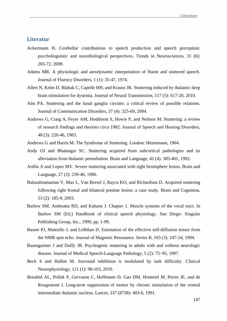
L LiteratureD
Literatur
Ackermann H. Cerebellar contributions to speech production and speech perception:
psycholinguistic and neurobiological perspectives. Trends in Neurosciences, 31 (6):
265-72, 2008.
Adams MR. A physiologic and aerodynamic interpretation of fluent and stuttered speech.
Journal of Fluency Disorders, 1 (1): 35-47, 1974.
Allert N, Kelm D, Blahak C, Capelle HH, and Krauss JK. Stuttering induced by thalamic deep
brain stimulation for dystonia. Journal of Neural Transmission, 117 (5): 617-20, 2010.
Alm PA. Stuttering and the basal ganglia circuits: a critical review of possible relations.
Journal of Communication Disorders, 37 (4): 325-69, 2004.
Andrews G, Craig A, Feyer AM, Hoddinott S, Howie P, and Neilson M. Stuttering: a review
of research findings and theories circa 1982. Journal of Speech and Hearing Disorders,
48 (3): 226-46, 1983.
Andrews G and Harris M. The Syndrome of Stuttering. London: Heinemann, 1964.
Andy OJ and Bhatnagar SC. Stuttering acquired from subcortical pathologies and its
alleviation from thalamic perturbation. Brain and Language, 42 (4): 385-401, 1992.
Ardila A and Lopez MV. Severe stuttering associated with right hemisphere lesion. Brain and
Language, 27 (2): 239-46, 1986.
Balasubramanian V, Max L, Van Borsel J, Rayca KO, and Richardson D. Acquired stuttering
following right frontal and bilateral pontine lesion: a case study. Brain and Cognition,
53 (2): 185-9, 2003.
Barlow SM, Andreatta RD, and Kahane J. Chapter 1. Muscle systems of the vocal tract. In
Barlow SM (Ed.) Handbook of clinical speech physiology. San Diego: Singular
Publishing Group, Inc., 1999, pp. 1-99.
Basser PJ, Mattiello J, and LeBihan D. Estimation of the effective self-diffusion tensor from
the NMR spin echo. Journal of Magnetic Resonance. Series B, 103 (3): 247-54, 1994.
Baumgartner J and Duffy JR. Psychogenic stuttering in adults with and without neurologic
disease. Journal of Medical Speech-Language Pathology, 5 (2): 75–95, 1997.
Beck S and Hallett M. Surround inhibition is modulated by task difficulty. Clinical
Neurophysiology, 121 (1): 98-103, 2010.
Benabid AL, Pollak P, Gervason C, Hoffmann D, Gao DM, Hommel M, Perret JE, and de
Rougemont J. Long-term suppression of tremor by chronic stimulation of the ventral
intermediate thalamic nucleus. Lancet, 337 (8738): 403-6, 1991. 147
148
Benabid AL, Pollak P, Louveau A, Henry S, and de Rougemont J. Combined (thalamotomy
and stimulation) stereotactic surgery of the VIM thalamic nucleus for bilateral
Parkinson disease. Applied Neurophysiology, 50 (1-6): 344-6, 1987.
Benke T, Hohenstein C, Poewe W, and Butterworth B. Repetitive speech phenomena in
Parkinson's disease. Journal of Neurology, Neurosurgery and Psychiatry, 69 (3): 319-
24, 2000.
Berardelli A, Abbruzzese G, Chen R, Orth M, Ridding MC, Stinear C, Suppa A, Trompetto
C, and Thompson PD. Consensus paper on short-interval intracortical inhibition and
other transcranial magnetic stimulation intracortical paradigms in movement disorders.
Brain Stimul, 1 (3): 183-91, 2008.
Bhatnagar S and Buckingham H. Neurogenic stuttering: its reticular modulation. Curr Neurol
Neurosci Rep, 10 (6): 491-8, 2010.
Bhatnagar SC and Andy OJ. Alleviation of acquired stuttering with human centremedian
thalamic stimulation. Journal of Neurology, Neurosurgery and Psychiatry, 52 (10):
1182-4, 1989.
Bloodstein O. Stuttering and normal nonfluency a continuity hypothesis. British Journal of
Disorders of Communication, 5 (1): 30-9, 1970.
Bloodstein O and Ratner BN. A Handbook on Stuttering. Canada: Delmar Thomson Learning,
2008.
Bosshardt HG. Cognitive processing load as a determinant of stuttering: summary of a
research programme. Clin Linguist Phon, 20 (5): 371-85, 2006.
Brady JP. The pharmacology of stuttering: a critical review. American Journal of Psychiatry,
148 (10): 1309-16, 1991.
Braun AR, Varga M, Stager S, Schulz G, Selbie S, Maisog JM, Carson RE, and Ludlow CL.
Altered patterns of cerebral activity during speech and language production in
developmental stuttering. An H2(15)O positron emission tomography study. Brain, 120
( Pt 5): 761-84, 1997.
Brown S, Ingham RJ, Ingham JC, Laird AR, and Fox PT. Stuttered and fluent speech
production: an ALE meta-analysis of functional neuroimaging studies. Human Brain
Mapping, 25 (1): 105-17, 2005.
Büchel C and Sommer M. What causes stuttering? PLoS Biol, 2 (2): E46, 2004.
Büchel C and Watkins KE. Genetic susceptibility to persistent stuttering. New England
Journal of Medicine, 362 (23): 2226; author reply 2227, 2010.
L Literature
Burghaus L, Hilker R, Thiel A, Galldiks N, Lehnhardt FG, Zaro-Weber O, Sturm V, and
Heiss WD. Deep brain stimulation of the subthalamic nucleus reversibly deteriorates
stuttering in advanced Parkinson's disease. Journal of Neural Transmission, 113 (5):
625-31, 2006.
Chang SE, Erickson KI, Ambrose NG, Hasegawa-Johnson MA, and Ludlow CL. Brain
anatomy differences in childhood stuttering. Neuroimage, 39 (3): 1333-44, 2008.
Chang SE, Kenney MK, Loucks TMJ, and Ludlow CL. Brain activation abnormalities during
speech and non-speech in stuttering speakers. Neuroimage, 46 (1): 201-212, 2009.
Chang SE, Synnestvedt A, Ostuni J, and Ludlow CL. Similarities in speech and white matter
characteristics in idiopathic developmental stuttering and adult-onset stuttering. J
Neurolinguistics, 23 (5): 455-469, 2010.
Chung SJ, Im JH, Lee JH, and Lee MC. Stuttering and gait disturbance after supplementary
motor area seizure. Movement Disorders, 19 (9): 1106-9, 2004.
Ciabarra AM, Elkind MS, Roberts JK, and Marshall RS. Subcortical infarction resulting in
acquired stuttering. Journal of Neurology, Neurosurgery and Psychiatry, 69 (4): 546-9,
2000.
Civier O, Tasko SM, and Guenther FH. Overreliance on auditory feedback may lead to
sound/syllable repetitions: simulations of stuttering and fluency-inducing conditions
with a neural model of speech production. J Fluency Disord, 35 (3): 246-79, 2010.
Cooper MH and Allen GD. Timing control accuracy in normal speakers and stutterers.
Journal of Speech and Hearing Research, 20 (1): 55-71, 1977.
Coulter CE, Anderson JD, and Conture EG. Childhood stuttering and dissociations across
linguistic domains: a replication and extension. J Fluency Disord, 34 (4): 257-78, 2009.
Cykowski MD, Fox PT, Ingham RJ, Ingham JC, and Robin DA. A study of the
reproducibility and etiology of diffusion anisotropy differences in developmental
stuttering: a potential role for impaired myelination. Neuroimage, 52 (4): 1495-504,
2010.
Damste PH, Zwaan EJ, and Schoenaker TJ. Learning principles applied to the stuttering
problem. Folia Phoniatrica, 20 (5): 327-41, 1968.
De Nil L. Recent developments in brain imaging research in stuttering. In Maasan B, Kent
RD, Peters HFM, Van Lieshout PHMM, and Hulstijn W (Eds.), Speech motor control in
normal and disorderd speech. New York: Oxford University Press, 2004, pp. 113-137.
149
150
De Nil LF, Kroll RM, and Houle S. Functional neuroimaging of cerebellar activation during
single word reading and verb generation in stuttering and nonstuttering adults.
Neuroscience Letters, 302 (2-3): 77-80, 2001.
Deniau J-M, Degos B, Bosch C, and Maurice N. Deep brain stimulation mechanisms: beyond
the concept of local functional inhibition. European Journal of Neuroscience, 32 (7):
1080-1091, 2010.
Devlin JT and Watkins KE. Investigating language organization with TMS. In Wassermann
EM, Epstein CM, Ziemann Uet al (Eds.), The Oxford Handbokk of Transcranial
Magnetic Stimulation. New York: Oxford University Press, 2008, pp. 479-499.
Dhont JKG. Translational Brownian motion. In Gerhard Gompper UBK, Jan K.G. Dhont,
Dieter Richter, Roland G. Winkler (Ed.) Physics Meets biology: From Soft
Matter to Cell Biology, 35th Spring School of the Institut of Solid State Research. Jülich:
Forschungszentrum Jülich, 2004.
Doi M, Nakayasu H, Soda T, Shimoda K, Ito A, and Nakashima K. Brainstem infarction
presenting with neurogenic stuttering. Internal Medicine, 42 (9): 884-7, 2003.
Drager B, Breitenstein C, Helmke U, Kamping S, and Knecht S. Specific and nonspecific
effects of transcranial magnetic stimulation on picture-word verification. European
Journal of Neuroscience, 20 (6): 1681-7, 2004.
Dworzynski K, Remington A, Rijsdijk F, Howell P, and Plomin R. Genetic etiology in cases
of recovered and persistent stuttering in an unselected, longitudinal sample of young
twins. Am J Speech Lang Pathol, 16 (2): 169-78, 2007.
Esser SK, Hill SL, and Tononi G. Modeling the effects of transcranial magnetic stimulation
on cortical circuits. Journal of Neurophysiology, 94 (1): 622-39, 2005.
Euler HA, Gudenberg AWv, Jung K, and Neumann K. Computergestützte Therapie bei
Redeflussstörungen: Die langfristige Wirksamkeit der Kasseler Stottertherapie. Sprache,
Stimme, Gehör, 33 (4): 193-97, 2009.
Fawcett RG. Stroke-associated acquired stuttering. CNS Spectr, 10 (2): 94-5, 2005.
Fisher SE. Genetic susceptibility to stuttering. New England Journal of Medicine, 362 (8):
750-2, 2010.
Folkerth RD. Abnormalities of developing white matter in lysosomal storage diseases. Journal
of Neuropathology and Experimental Neurology, 58 (9): 887-902, 1999.
Fox PT, Ingham RJ, Ingham JC, Hirsch TB, Downs JH, Martin C, Jerabek P, Glass T, and
Lancaster JL. A PET study of the neural systems of stuttering. Nature, 382 (6587): 158-
61, 1996.
L Literature
Fox PT, Ingham RJ, Ingham JC, Zamarripa F, Xiong JH, and Lancaster JL. Brain correlates of
stuttering and syllable production. A PET performance-correlation analysis. Brain, 123 (
Pt 10): 1985-2004, 2000.
Franco E, Casado JL, Lopez Dominguez JM, Diaz Espejo C, Blanco A, and Robledo A.
[Stuttering as the only manifestation of a cerebral infarct]. Neurologia, 15 (9): 414-6,
2000.
Giraud AL, Neumann K, Bachoud-Levi AC, von Gudenberg AW, Euler HA, Lanfermann H,
and Preibisch C. Severity of dysfluency correlates with basal ganglia activity in
persistent developmental stuttering. Brain and Language, 2007.
Golfinopoulos E, Tourville JA, and Guenther F. The integration of large-scale neural network
modeling and functional brain imaging in speech motor contro. Neuroimage:
doi:10.1016/j.neuroimage.2009.10.023, 2010.
Grant AC, Biousse V, Cook AA, and Newman NJ. Stroke-associated stuttering. Archives of
Neurology, 56 (5): 624-7, 1999.
Guenther FH. A neural network model of speech acquisition and motor equivalent speech
production. Biological Cybernetics, 72 (1): 43-53, 1994.
Guenther FH. Speech sound acquisition, coarticulation, and rate effects in a neural network
model of speech production. Psychological Review, 102 (3): 594-621, 1995.
Guenther FH, Ghosh SS, and Tourville JA. Neural modeling and imaging of the cortical
interactions underlying syllable production. Brain and Language, 96 (3): 280-301, 2006.
Hagoort P and Levelt WJ. Neuroscience. The speaking brain. Science, 326 (5951): 372-3,
2009.
Hallett M. Transcranial magnetic stimulation and the human brain. Nature, 406 (6792): 147-
50, 2000.
Hanajima R and Ugawa Y. Paired-pulse measures. In Wassermann EM, Epstein CM,
Ziemann U, Paus T, and Lisanby SH (Eds.), The Oxford handbook of transcranial
magnetic stimulation. New York: Oxford Univerity Press, 2008.
Heiser MA and Cheung SW. Cortical representations of communication sounds. Curr Opin
Otolaryngol Head Neck Surg, 16 (5): 478-84, 2008.
Helm-Estabrooks N and Hotz G. Sudden onset of "stuttering" in an adult: neurogenic or
psychogenic? Seminars in Speech and Language, 19 (1): 23-9, 1998.
Helm NA, Butler RB, and Benson DF. Acquired stuttering. Neurology, 28 (11): 1159-65,
1978.
151
152
Hickok G and Poeppel D. The cortical organization of speech processing. Nat Rev Neurosci,
8 (5): 393-402, 2007.
Hoang LN, Neef NE, Neef A, Ambrus G, Paulus W, Wolff von Gudenberg A, and Sommer
M. Inter-gestural motor cortex excitability of the tongue representation in adults who
stutter. in preparation.
Houde JF and Jordan MI. Sensorimotor adaptation in speech production. Science, 279 (5354):
1213-6, 1998.
Howell P. Assessment of Some Contemporary Theories of Stuttering That Apply to
Spontaneous Speech. Contemp Issues Commun Sci Disord, 31: 122-139, 2004.
Howell P, Anderson AJ, Bartrip J, and Bailey E. Comparison of acoustic and kinematic
approaches to measuring utterance-level speech variability. Journal of Speech,
Language, and Hearing Research, 52 (4): 1088-96, 2009.
Howell P. Signs of developmental stuttering up to age eight and at 12 plus. Clinical
Psychology Review, 27 (3): 287-306, 2007.
Indefrey P and Levelt WJ. The spatial and temporal signatures of word production
components. Cognition, 92 (1-2): 101-44, 2004.
Ingham RJ, Fox PT, Ingham JC, Xiong J, Zamarripa F, Hardies LJ, and Lancaster JL. Brain
correlates of stuttering and syllable production: gender comparison and replication.
Journal of Speech, Language, and Hearing Research, 47 (2): 321-41, 2004.
Johnson W. The onset of stuttering: Research findings and implications. Minneapolis:
University of Minnesota, 1959.
Jokel R, De Nil LF, and Sharpe AK. Speech disfluencies in adults with neurogenic stuttering
associated with stroke and traumatic brain injury. Journal of Medical Speech-Language
Pathology, 14: 243-261, Retrieved at http://goliath.ecnext.com/coms2/gi_0199-
7265968/Speech-disfluencies-in-adults-with.html, 2007.
Jones RK. Observations on stammering after localized cerebral injury. Journal of Neurology,
Neurosurgery and Psychiatry, 29 (3): 192-5, 1966.
Kalveram KT and Jancke L. Vowel duration and voice onset time for stressed and nonstressed
syllables in stutterers under delayed auditory feedback condition. Folia Phoniatrica, 41
(1): 30-42, 1989.
Kang C, Riazuddin S, Mundorff J, Krasnewich D, Friedman P, Mullikin JC, and Drayna D.
Mutations in the lysosomal enzyme-targeting pathway and persistent stuttering. New
England Journal of Medicine, 362 (8): 677-85, 2010.
L Literature
Kell CA, Neumann K, von Kriegstein K, Posenenske C, von Gudenberg AW, Euler H, and
Giraud AL. How the brain repairs stuttering. Brain, 132 (Pt 10): 2747-60, 2009.
Kelly RM and Strick PL. Macro-architecture of basal ganglia loops with the cerebral cortex:
use of rabies virus to reveal multisynaptic circuits. Progress in Brain Research, 143:
449-59, 2004.
Kent RD. Stuttering as a temporal programming disorder. In Curlee RF and Perkins WH
(Eds.), Nature and treatment of stuttering: new directions. San Diego: College-Hill
Press, 1984.
Kent RD. Research on speech motor control and its disorders: a review and prospective.
Journal of Communication Disorders, 33 (5): 391-427; quiz 428, 2000.
Kleinow J and Smith A. Influences of length and syntactic complexity on the speech motor
stability of the fluent speech of adults who stutter. Journal of Speech Language and
Hearing Research, 43 (2): 548-559, 2000.
Koller WC. Dysfluency (stuttering) in extrapyramidal disease. Archives of Neurology, 40 (3):
175-7, 1983.
Kujirai T, Caramia MD, Rothwell JC, Day BL, Thompson PD, Ferbert A, Wroe S, Asselman
P, and Marsden CD. Corticocortical inhibition in human motor cortex. Journal of
Physiology, 471: 501-19, 1993.
Lanotte MM, Rizzone M, Bergamasco B, Faccani G, Melcarne A, and Lopiano L. Deep brain
stimulation of the subthalamic nucleus: anatomical, neurophysiological, and outcome
correlations with the effects of stimulation. Journal of Neurology, Neurosurgery and
Psychiatry, 72 (1): 53-8, 2002.
Lebrun Y. Stuttering and epilepsy. Journal of Neurolinguistics, 6 (4): 433–444, 1991.
Lebrun Y, Bijleveld H, and Rousseau JJ. A case of persistent neurogenic stuttering following
a missile wound. Journal of Fluency Disorders, 15 (5-6): 251-258, 1990.
Levelt WJ. Chapter 11. Articulating. In Joshi A (Ed.) Speaking. From intention to articulation.
Cambridge, MA: MIT Press, 1989a, pp. 413-457.
Levelt WJ. Models of word production. Trends in Cognitive Sciences, 3 (6): 223-232, 1999.
Levelt WJ. Spoken word production: a theory of lexical access. Proceedings of the National
Academy of Sciences of the United States of America, 98 (23): 13464-71, 2001.
Levelt WJ, Roelofs A, and Meyer AS. A theory of lexical access in speech production.
Behavioral and Brain Sciences, 22 (1): 1-38; discussion 38-75, 1999.
Levelt WJM. Chapter 1. The speaker as information processor. In Joshi A (Ed.) Speaking.
From Intention to articulation. Cambridge, Ma: MIT Press, 1989b.
153
154
Levelt WJM. Speaking: from intention to articulation. Cambridge, MA: MIT Press, 1989c.
Limousin-Dowsey P, Pollak P, Van Blercom N, Krack P, Benazzouz A, and Benabid A.
Thalamic, subthalamic nucleus and internal pallidum stimulation in Parkinson's disease.
Journal of Neurology, 246 Suppl 2: II42-5, 1999.
Louis ED, Winfield L, Fahn S, and Ford B. Speech dysfluency exacerbated by levodopa in
Parkinson's disease. Movement Disorders, 16 (3): 562-5, 2001.
Lu C, Chen C, Ning N, Ding G, Guo T, Peng D, Yang Y, Li K, and Lin C. The neural
substrates for atypical planning and execution of word production in stuttering.
Experimental Neurology, 221 (1): 146-56, 2010.
Lu C, Ning N, Peng D, Ding G, Li K, Yang Y, and Lin C. The role of large-scale interactions
for developmental stuttering. Neuroscience, 161 (4): 1008-1026, 2009a.
Lu C, Peng D, Chen C, Ning N, Ding G, Li K, Yang Y, and Lin C. Altered effective
connectivity and anomalous anatomy in the basal ganglia-thalamocortical circuit of
stuttering speakers. Cortex, 2009b.
Ludlow CL. Stuttering: dysfunction in a complex and dynamic system. Brain, 123 ( Pt 10):
1983-4, 2000.
Ludlow CL and Loucks T. Stuttering: a dynamic motor control disorder. Journal of Fluency
Disorders, 28 (4): 273-95, 2003.
Ludlow CL, Rosenberg J, Salazar A, Grafman J, and Smutok M. Site of penetrating brain
lesions causing chronic acquired stuttering. Annals of Neurology, 22 (1): 60-6, 1987.
Lundgren K, Helm-Estabrooks N, and Klein R. Stuttering Following Acquired Brain Damage:
A Review of the Literature. J Neurolinguistics, 23 (5): 447-454, 2010.
Madison DP, Baehr ET, Bazell M, Hartman RW, Mahurkar SD, and Dunea G.
Communicative and cognitive deterioration in dialysis dementia: two case studies.
Journal of Speech and Hearing Disorders, 42 (2): 238-46, 1977.
Maeda S. Compensatory articulation during speech: evidence from the analysis and synthesis
of vocal tract shapes using an articulatory model. In Hardcastle WJ and Marchal A
(Eds.), Speech Production and Speech Modeling. Boston: Kluwer Academic Publishers,
1990, pp. 131–149.
Maguire GA, Yu BP, Franklin DL, and Riley GD. Alleviating stuttering with pharmacological
interventions. Expert Opin Pharmacother, 5 (7): 1565-71, 2004.
Mansson H. Childhood stuttering: Incidence and development,. Journal of Fluency Disorders,
25: 47-57, 2000.
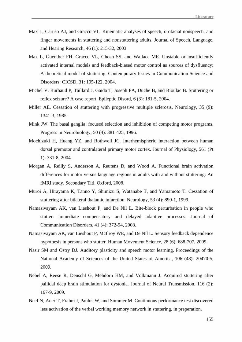
L Literature
Max L, Caruso AJ, and Gracco VL. Kinematic analyses of speech, orofacial nonspeech, and
finger movements in stuttering and nonstuttering adults. Journal of Speech, Language,
and Hearing Research, 46 (1): 215-32, 2003.
Max L, Guenther FH, Gracco VL, Ghosh SS, and Wallace ME. Unstable or insufficiently
activated internal models and feedback-biased motor control as sources of dysfluency:
A theoretical model of stuttering. Contemporary Issues in Communication Science and
Disorders: CICSD, 31: 105-122, 2004.
Michel V, Burbaud P, Taillard J, Gaida T, Joseph PA, Duche B, and Bioulac B. Stuttering or
reflex seizure? A case report. Epileptic Disord, 6 (3): 181-5, 2004.
Miller AE. Cessation of stuttering with progressive multiple sclerosis. Neurology, 35 (9):
1341-3, 1985.
Mink JW. The basal ganglia: focused selection and inhibition of competing motor programs.
Progress in Neurobiology, 50 (4): 381-425, 1996.
Mochizuki H, Huang YZ, and Rothwell JC. Interhemispheric interaction between human
dorsal premotor and contralateral primary motor cortex. Journal of Physiology, 561 (Pt
1): 331-8, 2004.
Morgan A, Reilly S, Anderson A, Reutens D, and Wood A. Functional brain activation
differences for motor versus language regions in adults with and without stuttering: An
fMRI study. Secondary Titl. Oxford, 2008.
Muroi A, Hirayama K, Tanno Y, Shimizu S, Watanabe T, and Yamamoto T. Cessation of
stuttering after bilateral thalamic infarction. Neurology, 53 (4): 890-1, 1999.
Namasivayam AK, van Lieshout P, and De Nil L. Bite-block perturbation in people who
stutter: immediate compensatory and delayed adaptive processes. Journal of
Communication Disorders, 41 (4): 372-94, 2008.
Namasivayam AK, van Lieshout P, McIlroy WE, and De Nil L. Sensory feedback dependence
hypothesis in persons who stutter. Human Movement Science, 28 (6): 688-707, 2009.
Nasir SM and Ostry DJ. Auditory plasticity and speech motor learning. Proceedings of the
National Academy of Sciences of the United States of America, 106 (48): 20470-5,
2009.
Nebel A, Reese R, Deuschl G, Mehdorn HM, and Volkmann J. Acquired stuttering after
pallidal deep brain stimulation for dystonia. Journal of Neural Transmission, 116 (2):
167-9, 2009.
Neef N, Auer T, Frahm J, Paulus W, and Sommer M. Continuous performance test discovered
less activation of the verbal working memory network in stuttering. in preperation.
155
156
Neef N, Paulus W, and Sommer M. Dem Stottern auf der Spur. L.O.G.O.S. Interdiziplinär, 17
(2): 129-135, 2009.
Olander L, Smith A, and Zelaznik H. Evidence That a Motor Timing Deficit Is a Factor in the
Development of Stuttering. Journal of Speech, Language, and Hearing Research, 2010.
Orton ST. A physiological theory of reading disability and stuttering in children. New
England Journal of Medicine, 199 (21): 1045-1052, 1928.
Paulus W, Classen J, Cohen LG, Large CH, Di Lazzaro V, Nitsche M, Pascual-Leone A,
Rosenow F, Rothwell JC, and Ziemann U. State of the art: Pharmacologic effects on
cortical excitability measures tested by transcranial magnetic stimulation. Brain Stimul,
1 (3): 151-63, 2008.
Payne JS and Whitney PJ. Developing L2 Oral Proficiency through Synchronous CMC:
Output, Working Memory, and Interlanguage Development. CALICO, 20: 7-32, 2002.
Perkell JS. Phonetic features and the physiology of speech production. In Butterworth B (Ed.)
Language Production: Vol. 1. Speech and talk. London: Academic Press, 1980, pp. 337-
372.
Perkins WH, Kent RD, and Curlee RF. A theory of neuropsycholinguistic function in
stuttering. Journal of Speech and Hearing Research, 34 (4): 734-52, 1991.
Peters HF, Hulstijn W, and Starkweather CW. Acoustic and physiological reaction times of
stutterers and nonstutterers. Journal of Speech and Hearing Research, 32 (3): 668-80,
1989.
Peters HF, Hulstijn W, and Van Lieshout PH. Recent developments in speech motor research
into stuttering. Folia Phoniatrica et Logopedica, 52 (1-3): 103-19, 2000.
Pollok B, Rothkegel H, Schnitzler A, Paulus W, and Lang N. The effect of rTMS over left
and right dorsolateral premotor cortex on movement timing of either hand. European
Journal of Neuroscience, 27 (3): 757-64, 2008.
Postma A and Kolk H. The covert repair hypothesis: prearticulatory repair processes in
normal and stuttered disfluencies. Journal of Speech and Hearing Research, 36 (3): 472-
87, 1993.
Preibisch C, Neumann K, Raab P, Euler HA, von Gudenberg AW, Lanfermann H, and Giraud
AL. Evidence for compensation for stuttering by the right frontal operculum.
Neuroimage, 20 (2): 1356-64, 2003.
Reilly S, Onslow M, Packman A, Wake M, Bavin EL, Prior M, Eadie P, Cini E, Bolzonello
C, and Ukoumunne OC. Predicting stuttering onset by the age of 3 years: a prospective,
community cohort study. Pediatrics, 123 (1): 270-7, 2009.
L Literature
Riaz N, Steinberg S, Ahmad J, Pluzhnikov A, Riazuddin S, Cox NJ, and Drayna D.
Genomewide significant linkage to stuttering on chromosome 12. American Journal of
Human Genetics, 76 (4): 647-51, 2005.
Ringo CC and Dietrich S. Neurogenic stuttering: An analysis and critique. Journal of Medical
Speech-Language Pathology, 3 (2): 111-122, 1995.
Romero JR, Anschel D, Sparing R, Gangitano M, and Pascual-Leone A. Subthreshold low
frequency repetitive transcranial magnetic stimulation selectively decreases facilitation
in the motor cortex. Clinical Neurophysiology, 113 (1): 101-7, 2002.
Sahin NT, Pinker S, Cash SS, Schomer D, and Halgren E. Sequential processing of lexical,
grammatical, and phonological information within Broca's area. Science, 326 (5951):
445-9, 2009.
Sakai T, Miyamura M, and Kuzuhara S. [Palilalia and acquired stuttering in a case of
Parkinson's disease]. Rinsho Shinkeigaku. Clinical Neurology, 32 (8): 859-63, 1992.
Saling LL and Phillips JG. Automatic behaviour: efficient not mindless. Brain Research
Bulletin, 73 (1-3): 1-20, 2007.
Salmelin R, Schnitzler A, Schmitz F, and Freund HJ. Single word reading in developmental
stutterers and fluent speakers. Brain, 123 ( Pt 6): 1184-202, 2000.
Sandrieser P and Schneider P. Stottern im Kindesalter. Stuttgart: Thieme, 2008.
Schiff ND, Giacino JT, Kalmar K, Victor JD, Baker K, Gerber M, Fritz B, Eisenberg B,
Biondi T, O'Connor J, Kobylarz EJ, Farris S, Machado A, McCagg C, Plum F, Fins JJ,
and Rezai AR. Behavioural improvements with thalamic stimulation after severe
traumatic brain injury. Nature, 448 (7153): 600-3, 2007.
Scholz J, Klein MC, Behrens TE, and Johansen-Berg H. Training induces changes in white-
matter architecture. Nature Neuroscience, 12 (11): 1370-1, 2009.
Scott SK and Johnsrude IS. The neuroanatomical and functional organization of speech
perception. Trends in Neurosciences, 26 (2): 100-7, 2003.
Seijo FJ, Alvarez-Vega MA, Gutierrez JC, Fdez-Glez F, and Lozano B. Complications in
subthalamic nucleus stimulation surgery for treatment of Parkinson's disease. Review of
272 procedures. Acta Neurochirurgica, 149 (9): 867-75; discussion 876, 2007.
Shiller DM, Sato M, Gracco VL, and Baum SR. Perceptual recalibration of speech sounds
following speech motor learning. Journal of the Acoustical Society of America, 125 (2):
1103-13, 2009.
Smith A. The control of orofacial movements in speech. Critical Reviews in Oral Biology and
Medicine, 3 (3): 233-67, 1992.
157
158
Smith A and Kelly E. Stuttering: A dynamic multifactorial model. In Curlee RF and Seigel
GM (Eds.), Nature and treatment of stuttering: New directions. New York: Allyn &
Bacon, 1997, pp. 204-217.
Smith A, Sadagopan N, Walsh B, and Weber-Fox C. Increasing phonological complexity
reveals heightened instability in inter-articulatory coordination in adults who stutter. J
Fluency Disord, 35 (1): 1-18, 2010.
Smits-Bandstra S. Methodological considerations in the measurement of reaction time in
persons who stutter. J Fluency Disord, 35 (1): 19-32, 2010.
Smits-Bandstra S and De Nil LF. Sequence skill learning in persons who stutter: implications
for cortico-striato-thalamo-cortical dysfunction. J Fluency Disord, 32 (4): 251-78, 2007.
Smits-Bandstra S, De Nil L, and Rochon E. The transition to increased automaticity during
finger sequence learning in adult males who stutter. Journal of Fluency Disorders, 31
(1): 22-42; quiz 39-40, 2006.
Sommer M, Knappmeyer K, Hunter EJ, Gudenberg AW, Neef N, and Paulus W. Normal
interhemispheric inhibition in persistent developmental stuttering. Movement Disorders,
24 (5): 769-73, 2009.
Sommer M, Koch MA, Paulus W, Weiller C, and Büchel C. Disconnection of speech-relevant
brain areas in persistent developmental stuttering. Lancet, 360 (9330): 380-3, 2002a.
Sommer M, Ruge D, Tergau F, Beuche W, Altenmuller E, and Paulus W. Intracortical
excitability in the hand motor representation in hand dystonia and blepharospasm.
Movement Disorders, 17 (5): 1017-25, 2002b.
Sommer M, Wischer S, Tergau F, and Paulus W. Normal intracortical excitability in
developmental stuttering. Movement Disorders, 18 (7): 826-30, 2003.
Starkweather CW and Gottwald S. The demands and capacities model. II: Clinical
applications. Journal of Fluency Disorders, 15: 143-157, 1990.
Stevens KN. Chapter 1. Anatomy and physiology of speech production In Stevens KN (Ed.)
Acoustic phonetics. Cambridge, MA: MIT Press, 2000, pp. 1-54.
Stinear CM and Byblow WD. Elevated threshold for intracortical inhibition in focal hand
dystonia. Movement Disorders, 19 (11): 1312-7, 2004.
Stinear CM, Coxon JP, and Byblow WD. Primary motor cortex and movement prevention:
where Stop meets Go. Neuroscience and Biobehavioral Reviews, 33 (5): 662-73, 2009.
Suresh R, Ambrose N, Roe C, Pluzhnikov A, Wittke-Thompson JK, Ng MC, Wu X, Cook
EH, Lundstrom C, Garsten M, Ezrati R, Yairi E, and Cox NJ. New complexities in the
L Literature
genetics of stuttering: significant sex-specific linkage signals. American Journal of
Human Genetics, 78 (4): 554-63, 2006.
Takeuchi H, Sekiguchi A, Taki Y, Yokoyama S, Yomogida Y, Komuro N, Yamanouchi T,
Suzuki S, and Kawashima R. Training of working memory impacts structural
connectivity. Journal of Neuroscience, 30 (9): 3297-303, 2010.
Tourville JA, Reilly KJ, and Guenther FH. Neural mechanisms underlying auditory feedback
control of speech. Neuroimage, 39 (3): 1429-43, 2008.
Travis LE. The cerebral dominance theory of stuttering: 1931--1978. Journal of Speech and
Hearing Disorders, 43 (3): 278-81, 1978.
Turgut N, Utku U, and Balci K. A case of acquired stuttering resulting from left parietal
infarction. Acta Neurologica Scandinavica, 105 (5): 408-10, 2002.
Van Borsel J and Taillieu C. Neurogenic stuttering versus developmental stuttering: an
observer judgement study. Journal of Communication Disorders, 34 (5): 385-95, 2001.
Van Borsel J, van der Made S, and Santens P. Thalamic stuttering: a distinct clinical entity?
Brain and Language, 85 (2): 185-9, 2003.
Van Lieshout P. Dynamical Systems Theory and its application in speech. In Maasan B, Kent
R, Peters H, Van Lieshout P, and Hulstijn W (Eds.), Speech motor control in normal
and disordered speech. New York: Oxford University Press, 2004, pp. 51-81.
Van Lieshout P, Hulstijn W, and Peters H. Searching for the weak link in the speech
production chain of people who stutter: a motor skill approach. In Maassen B, Kent R,
Peters H, Lieshout Pv, and Hulstijn W (Eds.), Speech Motor Control in Normal and
Disordered Speech. New York: Oxford University Press, 2007, pp. 313-356.
Van Lieshout PH, Hulstijn W, and Peters HF. From planning to articulation in speech
production: what differentiates a person who stutters from a person who does not
stutter? Journal of Speech and Hearing Research, 39 (3): 546-64, 1996.
Van Lieshout PH, Peters HF, Starkweather CW, and Hulstijn W. Physiological differences
between stutterers and nonstutterers in perceptually fluent speech: EMG amplitude and
duration. Journal of Speech and Hearing Research, 36 (1): 55-63, 1993.
Van Riper C. The nature of stuttering. Prentice-Hall: Englewood Cliffs, NJ, 1971.
Voges J and Krauss JK. [Neurological and technical aspects of deep brain stimulation].
Nervenarzt, 81 (6): 702-10, 2010.
Walker HC, Phillips DE, Boswell DB, Guthrie BL, Guthrie SL, Nicholas AP, Montgomery
EB, and Watts RL. Relief of acquired stuttering associated with Parkinson's disease by
159
160
unilateral left subthalamic brain stimulation. Journal of Speech, Language, and Hearing
Research, 52 (6): 1652-7, 2009.
Ward D. Stuttering & Cluttering: Frameworks for Understanding and Treatment. Hove, East
Sussex: Psychology Press, 2006.
Watkins KE, Smith SM, Davis S, and Howell P. Structural and functional abnormalities of the
motor system in developmental stuttering. Brain, 131 (Pt 1): 50-9, 2008.
WHO. ICD-10 F98.6 Cluttering Retrieved from
www.who.int/classifications/apps/icd/icd10online, 2007.
Wingate ME. A Standard Definition of Stuttering. Journal of Speech and Hearing Disorders,
29: 484-9, 1964.
Wingate ME. The structure of stuttering. New York: Springer, 1988.
Wu JC, Maguire G, Riley G, Lee A, Keator D, Tang C, Fallon J, and Najafi A. Increased
dopamine activity associated with stuttering. Neuroreport, 8 (3): 767-70, 1997.
Yairi E. Subtyping stuttering I: a review. J Fluency Disord, 32 (3): 165-96, 2007.
Yairi E and Ambrose N. A longitudinal study of stuttering in children: a preliminary report.
Journal of Speech and Hearing Research, 35 (4): 755-60, 1992.
Yairi E and Ambrose NG. Early childhood stuttering I: persistency and recovery rates. Journal
of Speech, Language, and Hearing Research, 42 (5): 1097-112, 1999.
Yairi E and Ambrose NG. Early childhood stuttering. Austin, TX: Pro-Ed, 2005.
Yaruss JS. Assessing quality of life in stuttering treatment outcomes research. J Fluency
Disord, 35 (3): 190-202, 2010.
Yuan W, Holland SK, Schmithorst VJ, Walz NC, Cecil KM, Jones BV, Karunanayaka P,
Michaud L, and Wade SL. Diffusion tensor MR imaging reveals persistent white matter
alteration after traumatic brain injury experienced during early childhood. AJNR.
American Journal of Neuroradiology, 28 (10): 1919-25, 2007.
Zelaznik HN, Smith A, Franz EA, and Ho M. Differences in bimanual coordination associated
with stuttering. Acta Psychologica, 96 (3): 229-43, 1997.
Zimmermann G. Articulatory dynamics of fluent utterances of stutterers and nonstutterers.
Journal of Speech and Hearing Research, 23 (1): 95-107, 1980a.
Zimmermann G. Stuttering: a disorder of movement. Journal of Speech and Hearing
Research, 23 (1): 122-36, 1980b.

A Acknowledgements D
161
Acknowledgements
The work included in this thesis would never have been possible without abundant expert
support. First and foremost, I would like to express my deepest gratitude to Dr. Martin
Sommer for his constant and terrific support, academic supervision, innovative ideas and
patience provided the basis for the work included in my thesis. I am very grateful for his
guidance, the numerous opportunities he offered me during the course of the years and for the
independence with which I could conduct this research.
I like to express my sincere gratitude to Professor Walter Paulus. Without him, giving me the
chance to work in his department and constantly supporting the ambitioned DTI study with
children who stutter the progress during the course would never have been possible.
Furthermore, he introduced me the fascinating field of brain stimulation.
I am very grateful to the members of my thesis committee Professor Julia Fischer and
Professor Marcus Hasselhorn for their important advices and constructive criticism,
advancing my progress over the years.
I wish to thank all members of the Institute of the Kasseler stuttering therapy. My special
thanks goes to Dr. Alexander Wolff von Gudenberg for giving us the opportunity to recruit
persons who stutter at his Institute and to Kristina Jung, without her help the measurement of
the inter-rater reliability of the stuttering severity of the subjects in the fingertapping study
would not have been possible.
I am very grateful to Torsten Wüstenberg for his tremendous support in the study on phoneme
categorization in stuttering.
I would also like to thank Dr. Bettina Pollok for introducing me to the exciting topic of
movement timing.
In addition I would like to thank all members of the Department of Clinical Neurophysiology
who have supported me. I especially would like to acknowledge Dr. Andrea Antal for the
inspiring journal clubs and Holger Rothkegel, Manuel Hewitt, Nivethida
Tirugnanasambandam, Géza Ambrus, Klara Boros and Linh Hoang supporting me in various
technical and practical TMS issues. A very special note of gratitude also goes to Marion
Kurze helping me in a variety of organizational situations.
Furthermore, I deeply acknowledge the help of the members of the Göttingen stuttering
support group and all the persons participating in the current studies promoting the research in
stuttering.
162
I thank Tibor Auer for his constant and patient help in questions concerned with magnetic
resonance imaging and I thank Professor Jens Frahm for giving me the opportunity to visit a
methods course at the Biomedical NMR Research GmbH and to conduct an fMRI study with
adults who stutter there.
I thank Dr. M. Röbel from the Department of Otorhinolaryngology and Prof. W. Engelke
from the Department of Maxillofacial Surgery for their technical assistance concerning the
tongue MEP recordings.
I acknowledge the supported by the „Stifterverband für die Deutsche Wissenschaft“, „Walter
und Ilse Rose Stiftung“.
I would also like to acknowledge the valuable help of Prof. J. Gärtner, director of the
Department of Pediatrics and Pediatric Neurology; Dr. Ch. Eich, consultant at the Department
of Anaesthesiology, Emergency and Intensive Care Medicine; Dr. M. Röbl, consultant at the
Pädiatrie 2 of the University Medicin Göttingen; Dr. Helms, physicist at the MR-Research in
Neurology and Psychiatry; and D. Wettschereck, referee at the ethics committee of the
University Medicin Göttingen for supporting the important DTI project to investigate a
potential biomarker in stuttering at its onset.
Finally, I would like to express my deepest gratitude to my husband Andreas Neef, who has
been a source of inspiration and analytical solutions, and to the members of my family who
have always supported me, gave me the time to visit conferences and a speech science
laboratory abroad.


![Subcortical Modulating Systems 3 11 04.ppt [Read-Only]zlab.rutgers.edu/classes/behaviorCogNeuro/Subcortical Modulating... · EEG with brainstem transections A: Cortical LVFA typical](https://img.pdfslide.us/doc/110x75/5b2d0d4a7f8b9ab66e8bad5e/subcortical-modulating-systems-3-11-04ppt-read-onlyzlab-modulating-eeg.jpg)