Embed Size (px)
Citation preview
Oskar anchoring restricts pole plasm to the posterior of the Drosophila oocyteVanzo, N. F. and Ephrussi, A. Development129, 3705-3714.
The reproduction of Fig. 6 in this article was innaccurate. The correct version of the figure is given below. We apologise to theauthors and readers for the mistake.
ERRATUM5551
A zebrafish sox9 gene required for cartilage morphogenesisYan, Y.-L., Miller, C. T., Nissen, R., Singer, A., Liu, D., Kirn, A., Draper, B., Willoughby, J., Morcos, P. A., Amsterdam, A.,Chung B.-c., Westerfield, M., Haffter, P., Hopkins, N., Kimmel, C. and Postlethwait, J. H. Development129, 5065-5079.
The name of the third author was published incorrectly in the printed version. The correct name is Robert M. Nissen.
CORRIGENDUM

INTRODUCTION
Cartilage cushions joints and provides a template for thedevelopment of cartilage-replacement bones. Aberrantcartilage development results in craniofacial anomalies andcartilage damage results in diseases such as osteoarthritis(Hamerman, 1989). The genetic pathway leading tochondrocyte differentiation is under active investigation(Cancedda et al., 1995; de Crombrugghe et al., 2001), but lessis known about the cellular mechanisms that control theassemblage of chondrocytes into higher order cartilage organs.
As chondrocytes begin to differentiate, they surroundthemselves with matrix, including collagen encoded byCOL2A1. In many cartilages, cells organize themselves intorows called stacks as they begin to form their mature spatialpatterns (Kimmel et al., 2001b). Chondrocytes continuedirectional proliferation, and then hypertrophy. Theextracellular matrix mineralizes before the hypertrophicchondrocytes undergo apoptosis and are replaced by theirbone-forming cell replacements.
Most cartilage replacement bones fail to develop normallyin individuals with campomelic dysplasia (CD), causing
5065Development 129, 5065-5079 (2002)Printed in Great Britain © The Company of Biologists Limited 2002DEV1848
The molecular genetic mechanisms of cartilageconstruction are incompletely understood. Zebrafishembryos homozygous for jellyfish (jef) mutations showcraniofacial defects and lack cartilage elements of theneurocranium, pharyngeal arches, and pectoral girdlesimilar to humans with campomelic dysplasia. We showthat two alleles of jef contain mutations in sox9a,one of twozebrafish orthologs of the human transcription factorSOX9. A mutation induced by ethyl nitrosourea changed aconserved nucleotide at a splice junction and severelyreduced splicing of sox9atranscript. A retrovirus insertioninto sox9adisrupted its DNA-binding domain. Inhibitingsplicing of the sox9atranscript in wild-type embryos withsplice site-directed morpholino antisense oligonucleotidesproduced a phenotype like jef mutant larvae, and causedsox9a transcript to accumulate in the nucleus; thisaccumulation can serve as an assay for the efficacy of amorpholino independent of phenotype. RNase-protectionassays showed that in morpholino-injected animals, thepercent of splicing inhibition decreased from 80% at 28hours post fertilization to 45% by 4 days. Homozygousmutant embryos had greatly reduced quantities of col2a1
message, the major collagen of cartilage. Analysis of dlx2expression showed that neural crest specification andmigration was normal in jef (sox9a) embryos. Confocalimages of living embryos stained with BODIPY-ceramiderevealed at single-cell resolution the formation ofprecartilage condensations in mutant embryos. Besides thelack of overt cartilage differentiation, pharyngeal archcondensations in jef (sox9a) mutants lacked three specificmorphogenetic behaviors: the stacking of chondrocytesinto orderly arrays, the individuation of pharyngealcartilage organs and the proper shaping of individualcartilages. Despite the severe reduction of cartilages,analysis of titin expression showed normal musclepatterning in jef (sox9a) mutants. Likewise, calcein labelingrevealed that early bone formation was largely unaffectedin jef (sox9a) mutants. These studies show that jef (sox9a)is essential for both morphogenesis of condensations andovert cartilage differentiation.
Key words: sox9a,col2a1, titin, Zebrafish, Chondrogenesis,Pharyngeal arches, Campomelic dysplasia, Cartilage
SUMMARY
DEVELOPMENT AND DISEASE
A zebrafish sox9 gene required for cartilage morphogenesis
Yi-Lin Yan 1, Craig T. Miller 1, Robert M. Nissen 2, Amy Singer 1, Dong Liu 1, Anette Kirn 3, Bruce Draper 1,4,John Willoughby 1, Paul A. Morcos 5, Adam Amsterdam 2, Bon-chu Chung 6, Monte Westerfield 1,Pascal Haffter 3, Nancy Hopkins 2, Charles Kimmel 1 and John H. Postlethwait 1,* 1Institute of Neuroscience, University of Oregon, Eugene, OR 97403, USA2Center for Cancer Research, Department of Biology, Massachusetts Institute of Technology, 40 Ames Street, E17-341,Cambridge, MA 02139, USA3MPI für Entwicklungsbiologie, Spemannstr. 35/III, D-72076 Tübingen, Germany4Division of Basic Science, Fred Hutchinson Cancer Research Center, B2-152, 1100 Fairview Ave. N., Seattle, WA 98109-1024, USA5Gene Tools, LLC, One Summerton Way, Philomath, OR 97370, USA6Institute of Molecular Biology, Academia Sinica, Nankang, Taipei, Taiwan ROC *Author for correspondence (e-mail: [email protected])
Accepted 12 August 2002

5066
macrocephaly, small jaw, cleft palate, lowset ears andsometimes lack of olfactory bulbs. Individuals with CD oftenhave underdeveloped sclerotome derivatives, including non-mineralized thoracic pedicles and 11, rather than 12, pairs ofribs; and they have poorly developed limbs, including bowedlimb bones, hypoplastic scapula and an insufficiently ossifiedpelvis (Houston et al., 1983; McKusick, 1990; Mansour et al.,1995). In addition, most XY individuals with CD display avariable female phenotype. The cartilage and sex-reversalphenotypes of CD are both caused by mutations in thetranscription factor gene SOX9(Foster et al., 1994; Wagner etal., 1994; Hageman et al., 1998; Cameron et al., 1996; Huanget al., 1999; Vidal et al., 2001). Individuals with CD areheterozygous for new mutations in SOX9,showing that CDis due to a dominant lethal mutation, either fromhaploinsufficiency or a dominant-negative effect. Mutations inthe coding region of SOX9 or in presumed regulatory elementscan cause CD or a similar phenotype in mouse (Wagner et al.,1994; Foster et al., 1994; Kwok et al., 1995; Cameron et al.,1996; Wunderle et al., 1998). Thus, phenotypic analysis of CDshows that SOX9is a regulator of chondrogenesis, but becauseno homozygous tetrapod mutant animals have been observed,and the affected skeletal elements in lethal heterozygotes aremerely hypoplastic, we do not know the extent of the functionof the gene.
Sox9 belongs to a family of DNA-binding proteins thatcontain a 79 amino acid long HMG (high mobility group)domain with at least 50% similarity to that of SRY, the sex-determining factor on the Y chromosome (Wright et al., 1993;Wegner, 1999). Sox proteins bind to a seven base pair sequencein the minor groove of DNA (Lefebvre et al., 1997; Ng et al.,1997) and bend DNA (Conner et al., 1994; Werner et al., 1995).Sox9 may also participate in transcript splicing (Ohe et al.,2002). The SOX9 protein has a C-terminal transcriptionactivation domain (Südbeck et al., 1996; Ng et al., 1997),suggesting that it acts by regulating expression of other genes.
Consistent with its role in chondrogenesis, Sox9is expressedin the pharyngeal arches and neurocranium, the sclerotomesand the lateral plate mesoderm (Lefebvre et al., 1997; Chianget al., 2001). In these domains, the expression of Sox9slightlyprecedes and directly regulates the expression of Col2a1,which encodes the major collagen of cartilage (Wright et al.,1995; Bell et al., 1997; Lefebvre et al., 1997; Ng et al., 1997;Zhao et al., 1997; Chiang et al., 2001).
Despite this knowledge of Sox9 activity, we have insufficientunderstanding of the morphogenetic roles Sox9 plays inchondrogenesis or the pathogenesis of CD. Heterozygous Sox9mutant mice show phenotypes similar to individuals with CDand die at birth, so permanent lines have not been established(Bi et al., 2001). Delayed or defective pre-cartilaginouscondensations are present in heterozygous Sox9mutant mouseembryos, but the precise morphogenetic steps that require Sox9function remain obscure. Some bones showed prematuremineralization in the heterozygous mouse embryos, suggestingthat Sox9plays a role in regulating the transition to hypertrophicchondrocytes in the growth plates. Sox9is thought to regulatethis transition by mediating the effects of parathyroid hormonerelated peptide (PTHrP) (Huang et al., 2001).
We have shown that the zebrafish genome contains twoduplicate orthologs of the human SOX9gene, called sox9aandsox9b, and that these map on zebrafish chromosomes that are
duplicates of much of human chromosome 17, the location ofSOX9 (Chiang et al., 2001). The two zebrafish sox9 genesapparently arose in a whole genome duplication eventhypothesized to have taken place near the base of the teleostradiation (Postlethwait et al., 1998; Amores et al., 1998). Thesox9aand sox9bgenes are expressed in partially overlappingpatterns that together approximate the expression pattern ofSox9in mouse (Wright et al., 1995; Chiang et al., 2001), aspredicted by the duplication, degeneration, complementationhypothesis (Force et al., 1999). Interestingly, however, inzebrafish the testis expresses sox9abut the ovary expressessox9b(Chiang et al., 2001), whereas in mammals, only thetestis expresses Sox9(Morais da Silva et al., 1996). Wereasoned that a mutation in one of the two zebrafish genesmight not be a dominant lethal mutation as in mammals, andso we investigated recessive lethal zebrafish mutations withphenotypes similar to individuals with CD. We show thattwo alleles of jellyfish(jef), one resulting from chemicalmutagenesis (jeftw37) (Piotrowski et al., 1996; van Eeden et al.,1996) and the other from the insertion of a retrovirus(Amsterdam et al., 1999), disrupt sox9a. Confocal microscopydemonstrated that jef (sox9a) is required not for precartilagecondensation formation, but for overt differentiation ofcartilage and for three morphogenetic processes: stacking,shaping and individuation. The results suggest that sox9aor itsdownstream targets, perhaps including extracellular matrixproteins, play morphogenetic roles in chondrogenesis.
MATERIALS AND METHODS
Animals, histology and gene expressionThe jeftw37 mutation was identified in an ENU screen for abnormaljaw and fin morphology (Piotrowski et al., 1996; van Eeden et al.,1996). The hi1134mutation was isolated in a retroviral insertionscreen (Amsterdam et al., 1999). Cartilages were Alcian stained,dissected and flat mounted (Kimmel et al., 1998). Larval bones werevisualized with calcein (Molecular Probes, catalog number C-381)(Du et al., 2001) (C. K., unpublished). In situ hybridization wasperformed as described (Jowett and Yan, 1996) using probes asdescribed (Akimenko et al., 1994; Yan et al., 1995; Chiang et al.,2001). BODIPY-ceramide labeling was performed essentially asdescribed (Cooper et al., 1999). Late epiboly stage embryos wereimmersed in BODIPY7 FL C5-ceramide (Molecular Probes, catalognumber D-3521) dissolved to 10 mM in DMSO, then diluted to 10µM in Embryo Medium (EM) (Westerfield, 2001) with 10 mMHepes. Embryos were placed in 150-200 µL of dye solution in 1.2%agarose dishes and stored in the dark. On day 2, embryos wereanesthetized, mounted on bridged coverslips, and one side of the headz-sectioned at 3 µm intervals with a Zeiss 310 upright confocalmicroscope. Live developing animals were stored in the dark and atsubsequent time points, the same side of each animal was re-examined.
MorpholinosMorpholino antisense oligonucleotides (MO) were obtained fromGene Tools (Philomath, OR) with the sequences: intron 1 splice donorjunction (i1d), AATGAATTACTCACCTCCAAAGTTT; and intron-2splice donor junction (i2d), CGAGTCAAGTTTAGTGTCCCAC-CTG. Morpholinos were injected as described (Draper et al., 2001).In the i1d MO, the 14th base from the 5′ end, a C, pairs with the Gimmediately following the splice junction (the one mutated in jeftw37).In MO i2d, the 4th base from the 3′ end, a C, pairs with the conservedG just after the splice junction.
Y.-L. Yan and others

5067Zebrafish sox9 and cartilage development
MappingTo map jeftw37, we identified a single strand conformationpolymorphism (SSCP) (see Postlethwait et al., 1998) in sox9a[(mapping primers were sox9a.+9 (CTTTCGCAGACACCAGCAGA)and sox9a –190 (CAGGTAGGGGTCGAGGAGATTCAT)]. Femalesheterozygous for jeftw37 were mated to WIK wild-type males, and theF1 were crossed to make an F2, which were scored for recombinationwith microsatellite markers (Knapik et al., 1998; Shimoda et al., 1999)near sox9aand sox9b (Chiang et al., 2001). The 95% confidenceinterval around the map distance between jeftw37 and an SSCP in the5′ untranslated region of sox9awas calculated according to Crow(Crow, 1950). To map the insertion allele jefhi1134,we used sox9a-RT2(CTCCTCCACGAAGGGACGCTTTTCCA), t2a (GGCACTGAGA-GTTTTCTGCATCTG) and 5′LTR (AGACCCCACCTGTAGGTTT-GGC) (see Fig. 3).
CloningGenomic clones of sox9awere isolated by amplifying genomic DNAisolated from Oregon AB wild type, Tübingen AB (TÜ, the geneticbackground of jeftw37) or homozygous jeftw37 embryos. The forwardcloning primer binds in the 5′ untranslated region (UTR, sox9a.+11:TTCGCAGACACCAGCAGACAACAAA) and the reverse primerbinds near the end of the 3′ UTR (sox9a.-1784: GTCTTTCC-CATCATGCACTGAACG). These primers amplified a 3.6 kbfragment including most of exon 1, intron 1, exon 2 and intron 2, andnearly all of exon 3. To minimize PCR errors, Platinum Taq DNApolymerase high fidelity (CAT#11304-029 from Gibco BRL) wasused in a touch-down PCR protocol. A BAC-containing sox9a(clone174 (I13)) was identified by screening the BAC zebrafish library-8549from Incyte Genomics with the primers sox9a.+441 (CCATG-CCGGTGAGGGTGAAC) and sox9a.-691 (CTTATAGTCGGGG-TGATCTTTCTTGTG). We cloned and sequenced DNA flanking thepro-viral insert linked to the hi1134mutant phenotype using inversePCR as previously described (Amsterdam et al., 1999).
RNA protection assaysRibonuclease protection assays used the RPA III kit (Ambion, #1414)according to manufacturer’s instructions. For each sample, RNA wasextracted from about 50 embryos, and 10 µg of totalRNA was loaded per lane. The protection probewas a 402 bp long PCR fragment from nucleotide494 to 896 of the cDNA, including 110 bp of exon1, all 252 bp of exon 2 and 40 bp of exon-3. Theantisense RNA probe for sox9awas generated byamplifying a fragment of the sox9a cDNA using theprimers sox9a.f2 (CCGATGAACGCGTTTATG-GTGT) and sox9a.r2 (TTTTCGGGGTGGTGG-
GAGGAG). The PCR product was cloned into the pCR4-TOP0 vector(Invitrogen; catalog number K4575-J10). The probe was made usingMAXIscript Invitro Transcription T3/T7 Kit (catalog number 1326).The amount of protected fragment was quantified using a Storm 860storage phosphor system (Johnson et al., 1990) with ImageQuant 4.2software (Molecular Dynamics, Sunnyvale, CA). Normalization usedthe ubiquitously expressed housekeeping gene ornithinedecarboxylase (odc),expressed sequence tag clone fc54f04; M. Clarkand S. Johnson, WUZGR; http//zfish.wustl.edu) as an internal control)(see Draper et al., 2001).
RT-PCR experiments to amplify the sox9amessage from jefhi1134
mutants used the primers sox9a.F (CCATGCCGGTGAGGGTGAAC)and sox9a.R (CGTTCGGCGGGAGGTATTGG).
RESULTS
Chondrogenesis requires jellyfish activityZebrafish homozygous for the recessive lethal ENU-inducedjellyfish allele jeftw37 have severely reduced cartilaginouselements (Piotrowski et al., 1996; van Eeden et al., 1996). Inwild-type larvae, jaw elements extend anterior and ventral tothe eye (Fig. 1A,B), but larvae homozygous for jeftw37 lackthese tissues (Fig. 1E,F) (Piotrowski et al., 1996). Likewise,cartilage supports the fin buds of normal larvae (Fig. 1D), butjeftw37 larvae have mis-shaped pectoral fins and lack thescapulocoracoid cartilage (Fig. 1F,H) (van Eeden et al., 1996).These phenotypes mimic CD (Houston et al., 1983; McKusick,1990; Mansour et al., 1995).
A screen for lethal mutations induced by retroviral insertion(Amsterdam et al., 1999; Burgess and Hopkins, 2000)identified a mutation (hi1134) giving a phenotype similar tothat of jeftw37 (Fig. 1I-L). To determine whether the hi1134mutation is allelic to jeftw37, we mated a male heterozygous forhi1134 to a female heterozygous for jeftw37 and observed anapprox. Mendelian ratio of phenotypically mutant offspring
Fig. 1.Activity of jef is essential for developmentof many cartilages. (A,E,I,M, lateral views,anterior towards the left; B,F,J,N, ventral views,anterior towards the left; C,G,K,O, dissectedpharyngeal cartilages stained with Alcian, anteriortowards the left; D,H,L,P, Alcian stained rightpectoral and fin skeletons, anterior towards thetop. (A-D) Wild type; (E-H) homozygous jeftw37;(I-L) homozygous jefhi1134; (M-P) heterozygousjeftw37/jefhi1134. All animals are 5 dpf. bh, basihyal;bsr, branchiostegal rays; cb, ceratobranchials; ch,ceratohyal; ch?, presumed ceratohyal; cl,cleithrum; ed, endoskeletal disk; co,scapulocoracoid; hs, hyosymplectic; m, Meckel’scartilage; m/de? putative Meckel’s cartilage anddentary bone; op, opercule; pq, palatoquadrate.Scale bars: 100 µm in O for C,G,K,O; 100 µm inP for D,H,L,P.

5068
(37 wild-type individuals and nine phenotypically jellyfishindividuals; Fig. 1M-P). Because these mutations fail tocomplement, we call the insertion allele jefhi1134.
Alcian staining demonstrated that all neurocranial cartilageand most cartilage elements of the pharyngeal arches weremissing from animals homozygous for jeftw37 or jefhi1134, oranimals heterozygous for the two alleles (Fig. 1C,G,K,O).Small regions of Alcian-positive material remained in bothjeftw37and jefhi1134homozygotes in approximately the positionexpected for Meckel’s cartilage and the ventral region of theceratohyal cartilage. In addition, cells in the pharyngealendoderm, possibly mucus secreting cells, were Alcian positivein both wild-type and mutant embryos. In the pectoral girdle,mutant animals lacked the scapulocoracoid cartilage, but thecleithrum bone and endoskeletal disk cartilage appearednormal (Fig. 1D,H,L,P). We conclude that many cartilageelements require jefactivity.
Molecular genetic nature of jef tw37
The jeftw37 mutationBecause the phenotype of jef mutations is similar to thephenotype of people with CD, we tried to rule out that sox9aor sox9b is disrupted in jeftw37. Bulked segregant analysis(Postlethwait et al., 1994) of an F2 mapping cross revealedlinkage to microsatellite marker z1176on the upper arm ofLG12 near sox9a,thus ruling out sox9bon LG3 as a candidate.We mapped jeftw37 at higher resolution relative to a
polymorphism in sox9a, and uncovered no recombinantsbetween jeftw37and sox9aamong 491 F2 diploid embryos. Thisrepresents 982 meioses, and a distance of 0.1±0.2 cM(centiMorgan) with 95% confidence. Thus, if there are onaverage 600 kb per cM (Postlethwait et al., 1994), this wouldbe a distance of 60±120 kb. We conclude that jeftw37maps veryclose to or within the sox9agene.
To determine whether jeftw37 lesions the sox9agene, wecloned and sequenced sox9a genomic DNA from homozygousjeftw37 embryos (Accession Number, AY090036) andcompared it with genomic DNA cloned from the wild-typestocks AB (Accession Number, AY090034) and TÜ(Accession Number, AY090035). We found differencesbetween the three strains at 49 locations in the 3560 bpconsensus sequence. The two wild-type strains differed at 48positions, and at the remaining position, the two wild-typestrains were the same, but the jeftw37 strain was different.All but one of the 48 differences between the two wildstrains were in non-translated parts of the gene, the exceptionbeing a silent change in codon Ala 406. Eleven of the 49differences were indels between 1 and 10 bp long; the otherswere single nucleotide differences. The TÜ and jeftw37 strainsdiffered at only three locations, all of them in introns. In twoof those positions, jeftw37 had the same sequence as the ABwild-type strain, but in the other, the two wild-type strainshad a G, but jeftw37 had a T (Fig. 2A). The unique change wasin the first base after the splice donor site of intron 1 in codon
Arg146, which alters an HphI restrictionendonuclease site, allowing identificationof jeftw37 heterozygotes by PCR.
Because nearly all introns have a Gimmediately after the splice junction(Mount, 1982; Zhang, 1998), this raised the
Y.-L. Yan and others
Fig. 2. jeftw37 alters a conserved splice sitesequence in sox9a and inhibits message splicing.(A) The sequence of sox9ain zebrafish wild-typestrains AB and TÜ, splice junction (arrow). Anin frame stop codon directly followed the G to Tchange in jeftw37. (B) The primer pair f1/r1 inexon 1 and intron 1 amplified a band of theexpected size (544 bp) from wild-type (WT)genomic DNA and from jeftw37 cDNA, showingthat intronic sequences of sox9a were present inmutant cDNA. With normal splicing in wild-typecDNA, there was no strong product of this size.(C) RNase protection assays (RPA, upper panel)on RNA extracted from wild-type andhomozygous jeftw37 mutants 4 days old. tRNAserved as a negative control. odc, the internalcontrol. Probe for RPA is made from a 402 bp ofsox9acDNA amplified using primer pair f2/r2.Quantifying band intensity (lower panel) shows adrastic decrease of message in jeftw37 embryos.(D,E) The jeftw37 allele behaves as a null whenheterozygous for the deletion allele b380. bh,basihyal; bsr, branchiostegal rays; cb,ceratobranchials; ch, ceratohyal; ch?, presumedceratohyal; cl, cleithrum; ed, endoskeletal disk;co, scapulocoracoid; hs, hyosymplectic; m,Meckel’s cartilage; m/de? putative Meckel’scartilage and dentary bone; op, opercule; pq,palatoquadrate. Scale bar: 100 µm.

5069Zebrafish sox9 and cartilage development
possibility that the lesion blocks transcript splicing. Becausethe lesion immediately follows the second nucleotide incodon 146 in the middle of the HMG domain, and an in-framestop codon follows two codons downstream, a non-splicedtranscript should be translated into a truncated proteincontaining only half of the HMG domain. Such a lesion islikely to lead to an ineffective protein.
The jeftw37 mutation inhibits splicingIf the G→T transversion in jeftw37 causes the jefphenotype,then it should disrupt the splicing of sox9a transcripts. To testthis prediction, we made cDNA from homozygous jeftw37
embryos and their homozygous wild-type siblings, andamplified various regions of the sox9a gene (Fig. 2A). Aforward primer, f1 in exon 1, and a reverse primer, r1 in intron1, should fail to amplify a product from mature wild-type sox9amRNA because the mature message lacks intron 1. Unsplicedtranscripts should give a band of 544 bp. The results showedthat genomic DNA from wild-type animals (a positive control)gave a band of the size predicted for a fragment that includesthe parts of exon 1 and intron 1, but cDNA from 4-day-oldwild-type animals had only a faint band at this location,consistent with normal splicing. By contrast, cDNA extractedfrom 4-day-old homozygous jeftw37animals behaved like wild-type genomic DNA (Fig. 2B), as expected if homozygousjeftw37 embryos accumulated unspliced transcript.
To learn the extent to which sox9amessage is reduced injeftw37 homozygotes, we prepared cDNA from 4-day-oldmutant and wild-type animals, and then conducted RNaseprotection assays using as probe a region of sox9a amplifiedby primer pair f2 and r2 that includes 110 bp of exon 1, all ofexon 2 (251 bp) and 40 bp in exon 3 (Fig. 2A). Theubiquitously expressed housekeeping gene, ornithinedecarboxylase(odc) provided an internal standard (Draper etal., 2001). The results revealed that jeftw37
embryos possessed only 28% of sox9atranscript compared with wild-type animals.We conclude that the mutation drasticallydecreases the efficiency of sox9atranscriptsplicing.
jeftw37 behaves as an amorphic mutationThe molecular genetic analysis of jeftw37 didnot rule out the possibility that somemessage may be spliced normally inhomozygous mutants. Coupled with theremnant bilateral patches of cartilage (Fig.1G), the protection assays made usconcerned that jeftw37might be a hypomorphrather than a null allele. The classical test fora null allele is the Müller test: for a nullallele, the phenotype of a homozygoteequals that of a heterozygote for onemutant allele and one deletion allele(Müller, 1932). To perform this test, wecrossed females heterozygous for jeftw37 toa male heterozygous for the deletionDf(LG12)dlx3b380 (Fritz et al., 1996), whichremoves a region of LG12 containing sox9a(data not shown). Fifteen of 56 offspringexamined showed a jellyfishphenotype, and
these were confirmed by PCR to be jeftw37/Df(LG12)dlx3 b380.Fig. 2D,E show that homozygous jeftw37/jeftw37 animals andheterozygous jeftw37/Df(LG12)dlx3 b380had the same severityof skeletal phenotype. We conclude that jeftw37 behaves as anull allele in the Müller test.
Molecular genetic nature of the jefhi1134 mutationThe jefhi1134 mutationAn allele with a molecular lesion that deletes protein functionwould strengthen interpretation of the mutant phenotype. Weidentified jefhi1134 in an insertional mutagenesis screen(Amsterdam et al., 1999; Burgess and Hopkins, 2000). Thevirus inserted into codon Leu147, 2 bp from the 5′ end of exon2 inside the HMG domain (Fig. 3), which would form atruncated Sox9a protein and probably destroy protein function.
To confirm that the mutant phenotype of jefhi1134
homozygotes is due to the viral insertion in sox9a,we mappedthe insertion site with respect to the mutant phenotype. Wemated a female heterozygous for jefhi1134 to the TAB14 wild-type strain and crossed the resulting F1 progeny to obtain anF2 mapping population. We scored 66 F2 individuals for theirgenotypes by PCR. The mutant and wild-type alleles weredistinguished by the forward primer 5′LTR which binds in theinsertion, and the forward primer t2a, which binds in intron-1(Fig. 3). When these are paired with a common reverse primerRT2, which binds in exon-2, the 5′LTR/RT2 primer pairamplified a band of 628 bp with mutant genomic DNA but noband with homozygous wild-type DNA. The t2a/RT2 pair gavea 237 bp fragment with wild-type DNA, and no band withmutant DNA (Fig. 3C). The results showed that of 45phenotypically mutant F2 individuals tested, all showed onlythe 628 bp band expected for homozygous insertions. Of 22phenotypically wild-type segregants tested, 14 showed bothbands expected for heterozygotes and eight showed only the
Fig. 3.The jefhi1134mutation results from a retroviral insertion into sox9aand inhibits theformation of mature message. (A) Primers relative to genomic structure of sox9a. (B) Avirus inserted into exon 2 in codon Leu147, two base pairs downstream of theintron/exon border in Arg146. 5′LTR indicates the position of a primer. (C) Amplificationproducts of mapping primers (see part A). (D) Primers F and R amplified a band of 627bp from wild-type (WT) embryos, but a 376 bp band from homozygousjefhi1134
embryos, showing that exon 2 (251 bp) was skipped in the splicing in jefhi1134mutants.
5070
237 bp band expected for wild-type genomic DNA. Thus,among 53 informative individuals, there were no recombinants.This places the insertion within 1.9±11 cM (95% confidence)(Crow, 1950) of the lesion causing the mutant phenotype.Taken with the failure of jefhi1134 to complement jef tw37, weconclude that the viral insertion in sox9ais responsible for thejellyfishphenotype in jefhi1134 homozygotes.
The jefhi1134 mutation inhibits production of maturemessageIf the insertion in jefhi1134 causes the mutant phenotype, itcould block the formation of mature sox9amRNA by one ofat least two mechanisms. First, because it is so close to thesplice acceptor site, it might cause the splicing machinery toskip exon 2 entirely. Alternatively, splicing might occurnormally, but the message with the insert would be unstable.If the insertion in jefhi1134causes skipping of the entire 251 bpexon 2, a primer pair in exon 1 and exon 3 (F/R, Fig. 3) wouldyield a transcript 251 bp shorter than wild-type. To test thesepossibilities, we prepared cDNA from mutant and wild-typeembryos and amplified the cDNAs. The results showed that inwild type, the predominant band is the 627 bp wild-typeproduct. In mutant animals, however, the predominant band isthe 376 bp product produced by neatly skipping exon 2. Directsequencing of wild-type and mutant PCR products from cDNAconfirmed that in jefhi1134, exon 1 splices directly to exon 3.
Translation of the resulting transcript should add 15 out-of-frame amino acids derived from exon-3 after Leu147. As a finaltest of sox9aexpression in mutant embryos, we performed insitu hybridization experiments on wild-type and homozygousjefhi1134 embryos. The experiments showed reduced signal inthe mutant embryos (see Figs 6, 7). Because the viral insertionalters sox9amRNA in a way that would produce a truncatedprotein with a disrupted HMG box, jefhi1134 is highly likely tobe a null mutation.
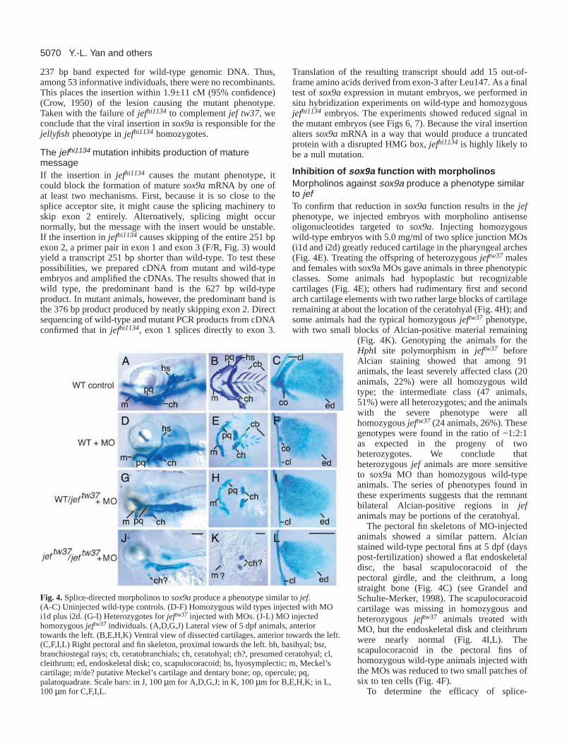
Inhibition of sox9a function with morpholinosMorpholinos against sox9a produce a phenotype similarto jefTo confirm that reduction in sox9a function results in the jefphenotype, we injected embryos with morpholino antisenseoligonucleotides targeted to sox9a. Injecting homozygouswild-type embryos with 5.0 mg/ml of two splice junction MOs(i1d and i2d) greatly reduced cartilage in the pharyngeal arches(Fig. 4E). Treating the offspring of heterozygous jeftw37 malesand females with sox9a MOs gave animals in three phenotypicclasses. Some animals had hypoplastic but recognizablecartilages (Fig. 4E); others had rudimentary first and secondarch cartilage elements with two rather large blocks of cartilageremaining at about the location of the ceratohyal (Fig. 4H); andsome animals had the typical homozygous jeftw37 phenotype,with two small blocks of Alcian-positive material remaining
(Fig. 4K). Genotyping the animals for theHphI site polymorphism in jeftw37 beforeAlcian staining showed that among 91animals, the least severely affected class (20animals, 22%) were all homozygous wildtype; the intermediate class (47 animals,51%) were all heterozygotes; and the animalswith the severe phenotype were allhomozygous jeftw37(24 animals, 26%). Thesegenotypes were found in the ratio of ~1:2:1as expected in the progeny of twoheterozygotes. We conclude thatheterozygous jef animals are more sensitiveto sox9a MO than homozygous wild-typeanimals. The series of phenotypes found inthese experiments suggests that the remnantbilateral Alcian-positive regions in jefanimals may be portions of the ceratohyal.
The pectoral fin skeletons of MO-injectedanimals showed a similar pattern. Alcianstained wild-type pectoral fins at 5 dpf (dayspost-fertilization) showed a flat endoskeletaldisc, the basal scapulocoracoid of thepectoral girdle, and the cleithrum, a longstraight bone (Fig. 4C) (see Grandel andSchulte-Merker, 1998). The scapulocoracoidcartilage was missing in homozygous andheterozygous jeftw37 animals treated withMO, but the endoskeletal disk and cleithrumwere nearly normal (Fig. 4I,L). Thescapulocoracoid in the pectoral fins ofhomozygous wild-type animals injected withthe MOs was reduced to two small patches ofsix to ten cells (Fig. 4F).
To determine the efficacy of splice-
Y.-L. Yan and others
Fig. 4.Splice-directed morpholinos to sox9aproduce a phenotype similar to jef.(A-C) Uninjected wild-type controls. (D-F) Homozygous wild types injected with MOi1d plus i2d. (G-I) Heterozygotes for jeftw37 injected with MOs. (J-L) MO injectedhomozygous jeftw37 individuals. (A,D,G,J) Lateral view of 5 dpf animals, anteriortowards the left. (B,E,H,K) Ventral view of dissected cartilages, anterior towards the left.(C,F,I,L) Right pectoral and fin skeleton, proximal towards the left. bh, basihyal; bsr,branchiostegal rays; cb, ceratobranchials; ch, ceratohyal; ch?, presumed ceratohyal; cl,cleithrum; ed, endoskeletal disk; co, scapulocoracoid; hs, hyosymplectic; m, Meckel’scartilage; m/de? putative Meckel’s cartilage and dentary bone; op, opercule; pq,palatoquadrate. Scale bars: in J, 100 µm for A,D,G,J; in K, 100 µm for B,E,H,K; in L,100 µm for C,F,I,L.
5071Zebrafish sox9 and cartilage development
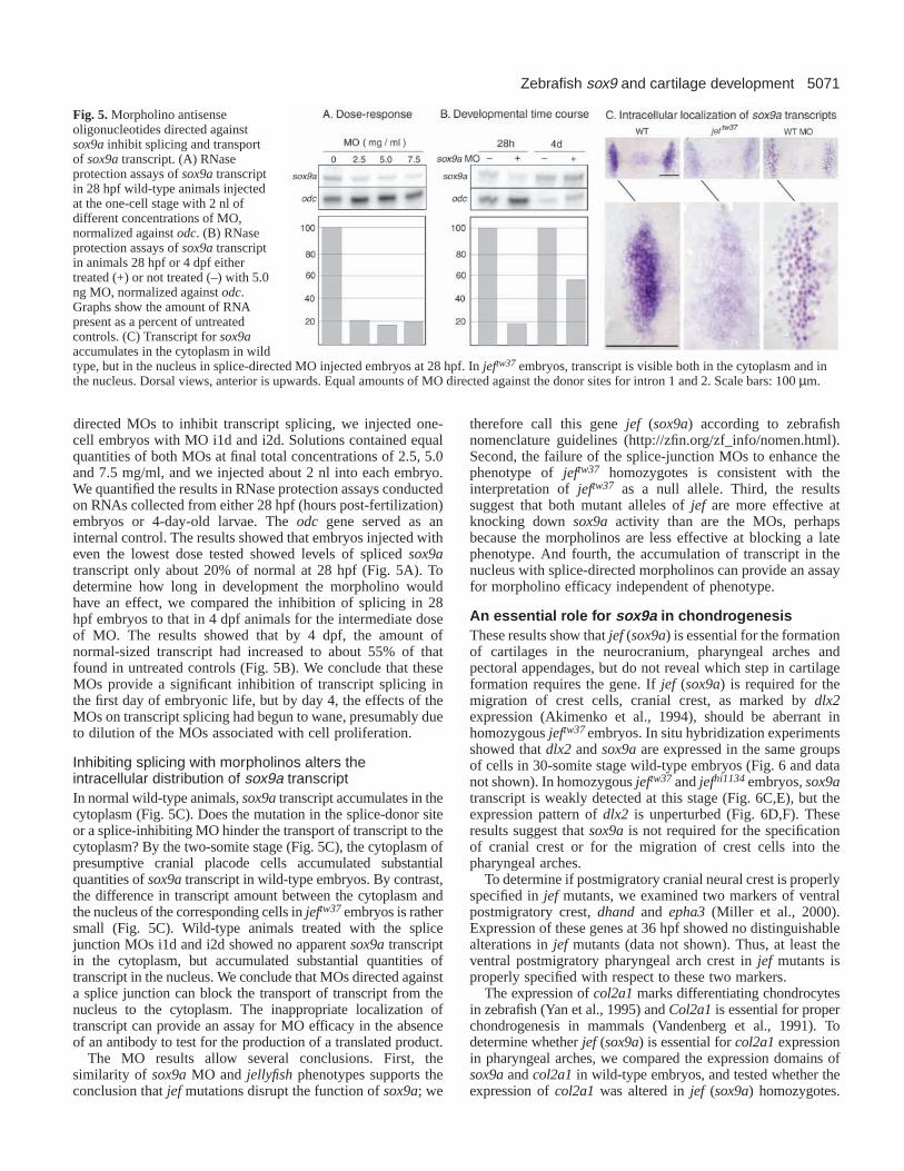
directed MOs to inhibit transcript splicing, we injected one-cell embryos with MO i1d and i2d. Solutions contained equalquantities of both MOs at final total concentrations of 2.5, 5.0and 7.5 mg/ml, and we injected about 2 nl into each embryo.We quantified the results in RNase protection assays conductedon RNAs collected from either 28 hpf (hours post-fertilization)embryos or 4-day-old larvae. The odc gene served as aninternal control. The results showed that embryos injected witheven the lowest dose tested showed levels of spliced sox9atranscript only about 20% of normal at 28 hpf (Fig. 5A). Todetermine how long in development the morpholino wouldhave an effect, we compared the inhibition of splicing in 28hpf embryos to that in 4 dpf animals for the intermediate doseof MO. The results showed that by 4 dpf, the amount ofnormal-sized transcript had increased to about 55% of thatfound in untreated controls (Fig. 5B). We conclude that theseMOs provide a significant inhibition of transcript splicing inthe first day of embryonic life, but by day 4, the effects of theMOs on transcript splicing had begun to wane, presumably dueto dilution of the MOs associated with cell proliferation.
Inhibiting splicing with morpholinos alters theintracellular distribution of sox9a transcriptIn normal wild-type animals, sox9atranscript accumulates in thecytoplasm (Fig. 5C). Does the mutation in the splice-donor siteor a splice-inhibiting MO hinder the transport of transcript to thecytoplasm? By the two-somite stage (Fig. 5C), the cytoplasm ofpresumptive cranial placode cells accumulated substantialquantities of sox9atranscript in wild-type embryos. By contrast,the difference in transcript amount between the cytoplasm andthe nucleus of the corresponding cells in jeftw37embryos is rathersmall (Fig. 5C). Wild-type animals treated with the splicejunction MOs i1d and i2d showed no apparent sox9atranscriptin the cytoplasm, but accumulated substantial quantities oftranscript in the nucleus. We conclude that MOs directed againsta splice junction can block the transport of transcript from thenucleus to the cytoplasm. The inappropriate localization oftranscript can provide an assay for MO efficacy in the absenceof an antibody to test for the production of a translated product.
The MO results allow several conclusions. First, thesimilarity of sox9aMO and jellyfishphenotypes supports theconclusion that jefmutations disrupt the function of sox9a; we
therefore call this gene jef (sox9a) according to zebrafishnomenclature guidelines (http://zfin.org/zf_info/nomen.html).Second, the failure of the splice-junction MOs to enhance thephenotype of jeftw37 homozygotes is consistent with theinterpretation of jeftw37 as a null allele. Third, the resultssuggest that both mutant alleles of jef are more effective atknocking down sox9aactivity than are the MOs, perhapsbecause the morpholinos are less effective at blocking a latephenotype. And fourth, the accumulation of transcript in thenucleus with splice-directed morpholinos can provide an assayfor morpholino efficacy independent of phenotype.
An essential role for sox9a in chondrogenesisThese results show that jef (sox9a) is essential for the formationof cartilages in the neurocranium, pharyngeal arches andpectoral appendages, but do not reveal which step in cartilageformation requires the gene. If jef (sox9a) is required for themigration of crest cells, cranial crest, as marked by dlx2expression (Akimenko et al., 1994), should be aberrant inhomozygous jeftw37 embryos. In situ hybridization experimentsshowed that dlx2and sox9aare expressed in the same groupsof cells in 30-somite stage wild-type embryos (Fig. 6 and datanot shown). In homozygous jeftw37and jefhi1134embryos, sox9atranscript is weakly detected at this stage (Fig. 6C,E), but theexpression pattern of dlx2is unperturbed (Fig. 6D,F). Theseresults suggest that sox9ais not required for the specificationof cranial crest or for the migration of crest cells into thepharyngeal arches.
To determine if postmigratory cranial neural crest is properlyspecified in jefmutants, we examined two markers of ventralpostmigratory crest, dhandand epha3(Miller et al., 2000).Expression of these genes at 36 hpf showed no distinguishablealterations in jef mutants (data not shown). Thus, at least theventral postmigratory pharyngeal arch crest in jef mutants isproperly specified with respect to these two markers.
The expression of col2a1marks differentiating chondrocytesin zebrafish (Yan et al., 1995) and Col2a1is essential for properchondrogenesis in mammals (Vandenberg et al., 1991). Todetermine whether jef (sox9a) is essential for col2a1expressionin pharyngeal arches, we compared the expression domains ofsox9aand col2a1in wild-type embryos, and tested whether theexpression of col2a1 was altered in jef (sox9a) homozygotes.
Fig. 5.Morpholino antisenseoligonucleotides directed againstsox9ainhibit splicing and transportof sox9atranscript. (A) RNaseprotection assays of sox9atranscriptin 28 hpf wild-type animals injectedat the one-cell stage with 2 nl ofdifferent concentrations of MO,normalized against odc. (B) RNaseprotection assays of sox9atranscriptin animals 28 hpf or 4 dpf eithertreated (+) or not treated (–) with 5.0ng MO, normalized against odc.Graphs show the amount of RNApresent as a percent of untreatedcontrols. (C) Transcript for sox9aaccumulates in the cytoplasm in wildtype, but in the nucleus in splice-directed MO injected embryos at 28 hpf. In jeftw37 embryos, transcript is visible both in the cytoplasm and inthe nucleus. Dorsal views, anterior is upwards. Equal amounts of MO directed against the donor sites for intron 1 and 2. Scale bars: 100 µm.

5072
The results show that the neurocranium, pharyngeal arches andpectoral fins co-express col2a1and sox9ain wild-type embryos(Fig. 7A,B,G,H), although col2a1shows additional expressionin the presumptive precursors of the cartilage capsule of the earand eye. In mutant animals, sox9aexpression is reduced (Fig.7C-F), and col2a1expression appears in only small regions ofthe pharyngeal arches (Fig. 7I-L). Ventral groups of cells in thefirst and second arches retain col2a1expression. The expressionin the second arch may correspond to the remaining bilateralAlcian-positive patches found later in mutant animals (see Fig.1C,G,K,O). These results show that in much of the pharyngealarch skeleton, the expression of col2a1 depends on sox9aactivity.
Prechondrogenic condensations form in jef (sox9a )mutants, but cartilage differentiation andcondensation morphogenesis fail to occurBecause migratory and postmigratory cranial neural crest are
present in jef(sox9a) mutant embryos (Fig. 6), the severereduction of differentiated (Alcian-positive) cranial cartilageseen later in jef(sox9a) mutant larvae is due either to the failureof prechondrogenic condensation formation or to the failureof condensation progression into differentiated cartilage. Todistinguish between these possibilities, we mated heterozygousjef (sox9a) males and females, and labeled the resultingembryos with the vital fluorescent dye BODIPY-ceramide.This dye fills extracellular spaces, thus labeling cell outlines,and has the powerful advantage of allowing histologicalidentification of nearly every cell type in live preparations(Cooper and Kimmel, 1998). We examined live developingBODIPY-ceramide-stained larvae at multiple time points from48 hours, when no pharyngeal cartilage differentiation hadoccurred in wild types (Schilling and Kimmel, 1997), until 76hours, when the primary scaffold of the larval pharyngealskeleton had chondrified. At 48 hours, wild type and mutantsfor both mutant alleles of jef(sox9a) had precartilagecondensations in the first two pharyngeal arches (Fig. 8A-B′and data not shown). Although at this stage no differentiated(Alcian-positive) pharyngeal cartilage was present (Schillingand Kimmel, 1997), the primordia of the major cartilages inthe first two arches were readily identifiable in wild type andmutant (Fig. 8A-B′). For example, the hyomandibular foramenwas present, as was the rudiment of the symplectic (Fig. 8A-B′). By 54 hours, differentiation had begun in wild type(Schilling and Kimmel, 1997), but had failed to occur injef (sox9a) mutants (data not shown). Concomitant withdifferentiation, in wild-type embryos chondrocytes organizedinto orderly stacks (data not shown) (Kimmel et al., 1998).
By 76 hours, cartilages in the first and second arches of wild-type embryos were well-formed, whereas jef(sox9a) mutantshad failed to undergo three major morphogenetic processes.First, jef (sox9a) mutants failed to form stacks of chondrocytes(Fig. 8C-D′ and data not shown), but cells in wild-typeprecartilage condensations oriented themselves with their longaxes parallel to each other (Fig. 8C,C′) (Kimmel et al., 1998).The only pharyngeal cartilages to differentiate in jef (sox9a)mutants, the small bilateral nodules of disorderly cartilage thatform in the ventral second arch (see Figs 1, 4) lacked orderlystacks of chondrocytes. Second, the precartilage condensationsin jef (sox9a) mutants failed to separate into individualizedregions. For example, the prominent dorsal/ventral joint that
Y.-L. Yan and others
Fig. 6.Activity of sox9ais not required for the specification ormigration of cranial neural crest. (A,B) Wild type (WT) embryos.(C,D) Homozygous jeftw37 embryos. (E,F) Homozygous jefhi1134
embryos. (A,C,E) sox9aexpression. (B,D,F) dlx2expression. Allanimals were 24 hpf. Dorsal views, anterior towards the left.
Fig. 7.Activity of sox9ais necessary for theexpression of col2a1in most developing cartilageof the neurocranial, pharyngeal and pectoralskeleton. (A-F) Expression of sox9a.(G-L) Expression of col2a1. (A,B,G,H) Wild-type animals. (C,D,I,J) Homozygous mutantjeftw37 embryos. (E,F,K,L) Homozygous mutantjefhi1134embryos. Photographs are a montagewith focus on the pharyngeal cartilages, and thepectoral girdle. All animals are 68 hpf. 1 and 2,first and second pharyngeal arches; co,scapulocoracoid; ed, endoskeletal disc; ov, oticvesicle. (A,C,E,G,I,K) Lateral views, anteriortowards the left. (B,D,F,H,J,L) Ventral views,anterior towards the left. Scale bars: in K, 100µm for A,C,E,G,I,K; in L, 100 µm forB,D,F,H,J,L.

5073Zebrafish sox9 and cartilage development
separates the upper and lower jaw (palatoquadrate andMeckel’s, Fig. 8C-D′) in wild-type embryos was undetectablein jef (sox9a) mutants. Third, jef(sox9a) mutant precartilagecondensations failed to transform into the specific shapes oftheir wild-type counterparts. For example, the symplecticregion of wild-type embryos formed a long, orderly rod ofcartilage, whereas this region in jef(sox9a) mutants wasdeformed into a jumbled region of mesenchyme (Fig. 8C-D′).For both mutant jef(sox9a) alleles, the phenotypically wild-type siblings (which should have included both heterozygotesand wild-type homozygotes) were indistinguishable fromone another. Thus, at this single-cell level of analysis, noevidence for heterozygous phenotype in either allele wasseen, consistent with the lack of a detectable heterozygousphenotype by Alcian staining.
These data show that jef (sox9a) function is not required forformation of pharyngeal precartilage condensations, butrather for subsequent differentiation of cells within thecondensations. Furthermore, jef (sox9a) function is requiredfor three morphogenetic processes: formation of orderly stacks,the individualization of cartilages and the shaping of specificskeletal elements.
Chondrogenesis and muscle patterningSignals from the cranial neural crest are required to pattern themesodermally derived muscles of the pharynx (Noden, 1983;Schilling et al., 1996). Cranial neural crest gives rise to muscleconnective tissue (Kontges and Lumsden, 1996), which couldbe one source of this signal. Alternatively, cartilage precursorsor the cartilages themselves might signal muscle patterning.Given the widespread expression of sox9ain cranial crest andthe severe defect in cartilage differentiation in jef (sox9a)mutants, jef mutants might have defects in muscle patterningfrom one of these sources. To test this possibility, we clonedand mapped a fragment of the muscle gene titin (GenBankAccession Number AY081167) (Xu et al., 2002) to use as amarker for muscle cells. A single molecule of Titin (the largestprotein known) spans half the length of a sarcomere (Labeitand Kolmerer, 1995). We report here the mapping of thezebrafish ttn gene to LG9 at 109.8 cM on the heat shock panel(Woods et al., 2000), a region of conserved synteny with thelong arm of human chromosome 2, the site of the human TTNgene. Expression of ttn showed a full complement ofpharyngeal muscles in jef animals homozygous for either allele(Fig. 9). Although the positions and shapes of muscles wereslightly distorted, presumably because of the absence ofcartilage-derived skeletal elements for muscle insertions, thepattern-forming process was normal in jef mutants. Weconclude that jef (sox9a) activity is not required to pattern theseanterior pharyngeal muscles.
Early bone formation is largely unaffected injef(sox9a) mutantsMice heterozygous for a Sox9 mutation exhibit expanded
Fig. 8.Prechondrogenic condensations form in jef (sox9a) mutants,but differentiation and morphogenesis fail to occur. Confocalmicrographs (lateral views shown as negative images, anteriortowards the left) of live wild-type (A,A′,C,C′) and jeftw37 mutants(B,B′,D,D′), stained with BODIPY-ceramide. Cells appear white andinterstitial space appears black. (The pharyngeal cavity does notretain the dye, so it also appears white in C-D′). At 48 hours, in wildtypes (A,A′) and jef mutants for both alleles (B,B′, and data notshown), prechondrogenic condensations have formed. The originalimages (A′,B′) are pseudo-colored (A,B) to highlight the first (blue)and second (yellow) pharyngeal arch precartilage condensations.Red, first aortic arch; green, adductor mandibulae muscle; pink,constrictor dorsalis premyogenic condensation. At this stage, acontiguous condensation prefigures the dorsal and ventral pharyngealcartilages, as seen best for the second arch in this focal plane (A-B′).In the second arch precartilage condensation, the symplecticrudiment (white asterisk) and hyomandibular foramen (blackasterisk) are apparent in wild types and jef mutants (A-B′). By 76hours, individuated and precisely shaped dorsal and ventral first andsecond pharyngeal arch cartilages with highly ordered stacks ofchondrocytes have formed in wild types (C,C′). Original images(C′,D′) are pseudocolored (C,D as in A,B) with overtly differentiatedcartilage in wild types colored purple in the first arch and orange inthe second arch cartilages. Stacking, individuation, and propershaping do not occur in jef mutants (D,D′). The precise boundary ofthe first and second arch is not visible in jef mutants and isapproximated in the pseudocolored panel (D). For jeftw37, fivemutants and 15 phenotypically wild-type siblings were examined; forjefhi1134, nine mutants and 21 phenotypically wild-type siblings wereexamined. ch, ceratohyal; e, eye; hs, hyosymplectic; M, Meckel’s;pq, palatoquadrate (see Fig. 1).

5074
ossification centers that prematurely ossify (Bi et al., 2001). Tolearn whether jef(sox9a) mutants show these phenotypes, weexamined larval bone development in jef(sox9a) mutant larvaeusing the fluorescent dye calcein (Du et al., 2001) (C. K.,unpublished). Despite the severe cartilage defects in jef (sox9a)mutants, most major cranial and pectoral fin bones appearedon schedule and were only slightly reduced in size in jef(sox9a) mutants (Fig. 9E, and data not shown). The secondarch dermal opercles and branchiostegal rays appeared onschedule in jef(sox9a) mutants and were mildly reduced. Theblade of the jef(sox9a) mutant opercular bone was reducedventrally and displaced anteriorly towards the eye. Thecleithrum in the fin girdle was present (Fig. 9, see also Fig.1D,H,L,P), as were pharyngeal teeth (Fig. 9). By contrast, thefifth ceratobranchial bone, which is a cartilage replacementbone (Cubbage and Mabee, 1996), was strikingly absent in jef(sox9a) mutants. Tiny remnants of bone were present adjacentto the teeth of jef(sox9a) mutants, perhaps the bone ofattachment of the teeth or the severely reduced remnant of thefifth ceratobranchial bone. Small remnants of bone were alsopresent in jef(sox9a) mutants in the position of the wild-typedentary, which normally forms in the lower jaw. Examining jef(sox9a) mutants at earlier time points (days 3 and 4) revealedno evidence for precocious bone development or enlargedossification centers (data not shown). The early larval lethalityof jef mutants thwarts analysis of ribs and other later-formingskeletal structures, as well as frustrating the analysis of gonadmorphogenesis.
DISCUSSION
These experiments identify jefas a mutation in sox9a,one ofthe two zebrafish orthologs of the human SOX9gene. Evidencefor this conclusion comes from the sequencing of sox9afromanimals homozygous for either of two jef alleles, one inducedby ENU and the other by retroviral insertion. The ENU-induced mutation changed a conserved G immediatelyfollowing the splice junction (Mount, 1982; Kreivi andLamond, 1996). Mutations of this nucleotide at thecorresponding site in the second intron of the human SOX9
gene can cause CD (Wagner et al., 1994), and similar mutationsdisrupt many other genes in humans (Freddi et al., 2000; Sironiet al., 2001; Targovnik et al., 2001) and in zebrafish (Lun andBrand, 1998; Childs et al., 2000). A quantitative assay forsox9a transcript and in situ experiments confirmed that themutation in jeftw37 severely inhibits splicing of the sox9atranscript. The insertion allele results in the skipping of exon2, and the predicted subsequent translation of a short proteintruncated within exon 3.
Two jef (sox9a ) alleles behave as null mutationsTo infer the role of a gene from its mutant phenotype, it isessential to know whether the alleles investigated lack allfunction of the gene. This is relevant here because, first, themammalian mutants retain one normal SOX9allele and,second. because the zebrafish mutants retain a small patch ofAlcian-positive material in the location of the second arch. Thezebrafish phenotype could result either from residual activityof sox9afrom the mutant allele, or from activity of a differentgene. We conclude that the two jef (sox9a) alleles are likelynull alleles because: (1) the viral insertion in jefhi1134 causesthe skipping of exon 2, which should result in a truncatedprotein; (2) animals heterozygous for jeftw37 over a deletion donot have a more severe phenotype than jeftw37 homozygotes;(3) morpholinos that affect wild-type and heterozygousembryos do not make the jeftw37 phenotype more severe; and(4) the phenotype of jeftw37/jefhi1134 heterozygotes have thesame phenotype as either homozygote.
Splice-directed morpholinos provide an independentassay for efficacyAnimals injected with splice-directed morpholinos displayeda weaker phenotype at day 4 than did homozygous jef(sox9a)mutations, presumably because of to the rebound of transcriptsplicing as evidenced by the RNase protection assays. MO-treated animals accumulated sox9atranscript in the nuclei ofsox9a-expressing cells, apparently because of a defect intranscript transport. We have also observed this phenomenonfor splice-directed morpholinos against sox9b (Y.-L. Y.,unpublished). Although it is well known that MOs can inhibitsplicing (Schmajuk et al., 1999; Draper et al., 2001), to our
Y.-L. Yan and others
Fig. 9.Muscle and bone in jef mutants. Expression of titinin wild-type (A) and homozygous jeftw37 (B) and jefhi1134
(C) animals at 3 dpf reveals normal patterning of cranialmuscles. Compiled stacks of confocal micrographs of wild-type (D) and a jeftw37 mutant (E) stained with calcein. Mostdermal bones (op, bsr, cl) are relatively unaffected in jefmutants, whereas the dermal dentary (de) and cartilagereplacement fifth ceratobranchial bone (cb5) are severelyreduced. Teeth (te) are present, as are tiny remnants of boneat the base of the teeth. Bone development occurs onschedule without enlarged ossification centers in jefmutants. am, adductor mandibulae; bsr, branchiostegal ray;cb5, fifth ceratobranchial; cl, cleithrum; de, dentary; hh,hyohyoideus; ih, interhyoideus; ima, intermandibularisanterior; imp, intermandibularis posterior; op, opercle; sh,sternohyoideus; te, teeth; tv1, transversus ventralis. Scalebar: in A, 100 µm for A-C.

5075Zebrafish sox9 and cartilage development
knowledge this inhibition had not previously been shown toretard the transport of transcript to the cytoplasm. Our novelfinding provides an assay for MO efficacy independent of anyphenotypic change. This assay or the RNase protection assay,is generally more convenient than an assay for the efficacy ofa translation-inhibiting MO because probes to measurenucleic acid quantity are much more readily available thanprobes to measure the quantity of a specific protein, whichoften requires a specific antibody. Furthermore, as pointedout by Draper et al. (Draper et al., 2001), splice-directedmorpholinos may allow one to distinguish between the effectsof different splice variants, and to distinguish between thefunctions of maternal and zygotic transcript.
Evaluating jef (sox9a ) mutants as a model forcampomelic dysplasiaPeople affected with CD are heterozygous for a mutation inSOX9, and display a syndrome of clinical features thatinclude bowing of the tibia and femur, hypoplastic scapula,absence of a pair of ribs, cleft palate and a small jaw (Houstonet al., 1983; McKusick, 1990; Foster et al., 1994; Wagner etal., 1994; Kwok et al., 1995; Mansour et al., 1995; Cameronet al., 1996; Hageman et al., 1998). Zebrafish homozygousfor jef (sox9a) mutations mimic at least two of thesephenotypes, but show them in more severe form. In humansand mice (Bi et al., 1999; Bi et al., 2001) heterozygous forSOX9mutations, the scapulas and jaws form, but they aresmall and thin. By contrast, the corresponding elements inzebrafish jef (sox9a) mutants, the scapulocoracoid cartilageand the first and second arch derivatives, are almostcompletely gone.
Why is the zebrafish sox9amutant phenotype more severein these aspects than the mammalian SOX9 mutantphenotypes? This question is significant because we need toknow whether SOX9is essential for development of theseelements or whether it merely facilitates completion of thesecartilages. Many SOX9 mutations in mammals are likely tobe null activity alleles rather than dominant negativemutations, judging from their predicted effect on the proteins(Foster et al., 1994; Wagner et al., 1994; Kwok et al., 1995;Mansour et al., 1995; Cameron et al., 1996; Hageman et al.,1998; Bi et al., 1999; Bi et al., 2001). Thus, the heterozygotesprobably have about half the normal amount of SOX9activityin tissues in which the gene is expressed. Homozygous jef(sox9a) animals should completely lack sox9activity in allcells in which sox9ais expressed in the absence of sox9b.Before hatching, the time during which the jef (sox9a)phenotype becomes apparent, sox9ais expressed strongly inthe first and second arches and in the scapulocoracoid, butsox9bis not expressed in these cells (Chiang et al., 2001). Wetherefore conclude that SOX9activity is essential forchondrogenesis of the arches, neurocranium andscapulocoracoid, and that mammals show a weak phenotypebecause SOX9activity is reduced, but not completely lost, inthe mammalian heterozygous genotypes. The evolution ofduplicated zebrafish genes has thus allowed analysis of null-activity embryos not yet available for the ortholog inmammals.
Many individuals with CD have XY sex reversal (Houstonet al., 1983; McKusick, 1990; Foster et al., 1994; Wagner etal., 1994; Kwok et al., 1995; Mansour et al., 1995; Cameron
et al., 1996; Hageman et al., 1998). Although sox9a isexpressed in the zebrafish testis, and sox9bis expressed in thezebrafish ovary (Chiang et al., 2001), consistent with a rolein sex determination, the late determination of sex inzebrafish has so far precluded the investigation of sexdetermination in jef(sox9a) animals. Because both male andfemale animals heterozygous for jef (sox9a) become sexuallymature adults of both sexes, jef(sox9a) appears not to havea fully penetrant dominant effect on sex determination inzebrafish.
Prechondrogenic condensations form in jef (sox9a )mutantsThe formation of prechondrogenic condensations in jef (sox9a)mutant zebrafish suggests that sox9ais not required forcondensation formation in zebrafish. The opposite conclusionwas drawn for mouse. Because cells homozygous for a Sox9mutation failed to contribute to condensations in geneticmosaics, Bi et al. (Bi et al., 1999) concluded that Sox9wasrequired for formation of condensations. These differencesmight reflect: (1) species differences in SOX9function; (2) thepresence of duplicated sox9genes in zebrafish; and/or (3) thedifference in experimental paradigms used. Although jef(sox9a) mutant cells form condensations in the context of atotally mutant environment, they might not contribute tocondensations when transplanted into a wild-type host, as wasfound with mouse. Mosaic analyses in zebrafish could test thispossibility.
Support for the idea that differences in experimentalparadigms could explain the contrasting conclusions on therequirement of SOX9for condensation formation comes fromanalysis of the zebrafish valentino mutant. Embryos thatlack val (mafb) activity make hindbrain tissue betweenrhombomeres 4 and 7, yet in genetic mosaics, val (mafb)mutant cells are excluded from this territory in a wild-type host(Moens et al., 1996). Thus, by analogy, homozygous Sox9mutant mice, like sox9a mutant zebrafish, might formprecartilage condensations even though mutant cells areexcluded from these domains in a mosaic. A mammalianphenotype analogous to that seen in zebrafish jef (sox9a)mutants might be the L-Sox5; Sox6double mutant inmouse, where condensations form, but no overt cartilagedifferentiation occurs (Smits et al., 2001). A conditional alleleof mouse Sox9has been made (Kist et al., 2002), which shouldfacilitate phenotypic analysis of skeletal development inhomozygous Sox9mutant mice.
The simultaneous failure of chondrocyte differentiation andmorphogenesis of condensations in jef (sox9a) mutantssuggests that jef (sox9a) regulates both morphogenesis anddifferentiation, and provides another example of specificationand morphogenesis going hand-in-hand (see Kimmel et al.,2001a; Kimmel et al., 2001b). These morphogenetic processesare separable from differentiation: the zebrafish pipetail [ppt(wnt5a)] mutation (Piotrowski et al., 1996; Rauch et al., 1997;Hammerschmidt et al., 1996) disrupts chondrocyte stacking butnot differentiation. Thus, differentiation does not requirestacking. Likewise, individuation of cartilage elements occursin ppt (wnt5a) mutants (Piotrowski et al., 1996), suggestingthat the jef (sox9a)-dependent morphogenetic processes aredistinct aspects of morphogenesis under regulation of separateloci.

5076
The failure of differentiation and morphogenesis in jef(sox9a) mutants correlates in time and space with the col2a1expression defect, and raises the possibility that col2a1 mightbe involved in one or both of these processes. Because COL2Ais a major component of differentiated cartilage matrix(Vandenberg et al., 1991), perhaps zebrafish col2a1 is requiredfor cartilage differentiation, and the failure to activate col2a1expression underlies the near complete absence of cartilage injef (sox9a) mutants. The lack of Col2a might also underlie themorphogenetic defects in jef (sox9a) mutants – it is possiblethat cells require a normal extracellular matrix in order toexhibit stacking cell behaviors. It would be interesting tosee if exogenously supplied Col2a could rescue eithermorphogenesis (e.g. stacking) or differentiation in jef (sox9a)mutants.
SOX9 regulates not only COL2A1,but other downstreamtargets as well. SOX9positively regulates expression of CDH2(Panda et al., 2001), which may mediate the jef (sox9a)-dependent stacking, individuation or shaping of pharyngealmesenchymal cells. Comparing expression of cadherin genes,col2a1and orthologs of other Sox9downstream targets in pptand jef mutants might suggest which genetic pathways underliestacking behavior and which cartilage differentiation. SOX9also regulates cell cycle genes (Panda et al., 2001), providinganother potential mechanism for the differentiation andmorphogenetic defects in jef (sox9a) mutants. Mitotic activityis enriched at the second arch dorsal/ventral joint, and wasproposed to partially drive the extension of the symplecticcartilage (Kimmel et al., 1998). This region of the second archcondensation never extends in jef (sox9a) mutants, possiblybecause mitotic behavior of cells within the precartilagecondensation is defective.
Pharyngeal muscle and bone differentiates in theabsence of differentiated cartilageCranial neural crest (CNC) patterns the pharyngealmusculature according to Noden’s experiments transplantingpresumptive first arch CNC into the position of presumptivesecond arch CNC (Noden, 1983). Although recentlyreinterpreted to be due to the organizing effects of atransplanted isthmus (Trainor et al., 2002), the conclusionremains that the transplant non-autonomously induced hostsecond arch muscles to adopt patterns appropriate to the firstarch muscles. Further evidence for an instructive role of CNCcomes from the zebrafish mutant chinless (chw), which lacksboth differentiated pharyngeal cartilage and muscles (Schillinget al., 1996). Wild-type CNC cells, when transplanted intohomozygous chw mutant hosts, induced local differentiation ofpharyngeal muscles (Schilling et al., 1996). A third studyrevealed severe ventral muscle defects in pharyngeal arches ofsuc (edn1) mutants, although mutant cells contributed tonormal ventral muscles when transplanted into a wild-type host(Miller et al., 2000). These results support the idea thatsignaling from the CNC patterns the pharyngeal archmesoderm. The populations of CNC which participate in thissignaling, however, remain unidentified. The CNC-derivedmuscle connective tissue (Kontges and Lumsden, 1996) is acandidate for this activity. The widespread expression of sox9araises the possibility that jef(sox9a) might function in a CNC-derived connective tissue lineage. The skeletogenic CNCderivatives, some of which are perturbed in jef(sox9a) mutants,
could also signal to the pharyngeal musculature. The presenceof a normal pattern of differentiated pharyngeal muscles in jef(sox9a) mutants shows that signals from CNC to thesurrounding mesoderm occur independently of jef (sox9a)function. Muscles in jef (sox9a) mutants are shorter and thickerthat their wild-type counterparts, suggesting that elongation ofthe muscles might require stiff, differentiated cartilage.
The precocious ossification and expanded ossificationcenters in heterozygous Sox9 mutant mice motivated thecharacterization of bone formation in jef (sox9a) mutants. Ourcalcein labeling confirms observations of bone (Piotrowski etal., 1996), and further demonstrates that cranial and pectoralfin bones form relatively normally in jef (sox9a) mutants, withthe exceptions of the dentary and fifth ceratobranchial bone, acartilage replacement bone (see Cubbage and Mabee, 1996)that appears to be surrounded by perichondral bone in wild-type 5-day-old zebrafish larvae. The absence of the fifthceratobranchial bone in jef(sox9a) mutants could be explainedif perichondral ossification requires a cartilage template. Thedentary bone, a dermal bone (see Cubbage and Mabee, 1996),is intimately associated with Meckel’s cartilage in wild type.Perhaps the reduction of the dentary in jef (sox9a) mutants isalso secondary to the cartilage defect. Weaker alleles ofzebrafish jef (sox9a) might allow analysis of bone developmentpast the early larval stage of currently available alleles.
Because dermal bones appear without a cartilage template,it might not seem surprising that most dermal bones formnormally in jef(sox9a) mutants. The widespread expression ofsox9a in postmigratory CNC, however, suggests that CNCosteocyte precursors might express sox9a. Expression ofsox9a, like that of dlx2in the zebrafish pharyngeal arches,appears to include the entire CNC population within each arch.The dlx2-expressing postmigratory CNC forms a cylindersurrounding the central cores of paraxial mesoderm (Miller etal., 2000; Kimmel et al., 2001a). This population presumablyincludes precursors of both the cartilage and bone skeleton,based on existing fate maps (Couly and Le Douarin, 1988;Schilling and Kimmel, 1997; Kontges and Lumsden, 1996).Thus, the absence of a major bone defect suggests that jef(sox9a) function in the pharyngeal skeleton might actually berequired only in a subset of sox9a-expressing postmigratoryCNC cells, the chondrocyte lineage. Fate-mapping experimentswill determine whether chondrocytes and osteocytes share alineage in the CNC, and whether the chondrocyte lineage isuniquely perturbed in jef (sox9a) mutants.
Sox9 and gene duplicationXenopus embryos treated with a morpholino antisenseoligonucleotide that inhibits the production of Sox9 proteinlack neural crest progenitors, suggesting that Sox9 is essentialfor the specification of neural crest in frogs (Spokony et al.,2002). By contrast,neural crest specification is apparentlynormal in individuals with CD, because they make cranial crestderivatives, but just have hypoplastic skeletons, and in theabsence of sox9afunction in zebrafish, because the expressionof dlx2 is normal, the cranial crest migrates on schedule, andprecartilage condensations form. How can we understand thesedifferences? We hypothesize that SOX9may play multipleessential roles in cranial crest development, and that these maybe revealed by further analysis of the two SOX9duplicates inzebrafish. As demonstrated by the work on Xenopus(Spokony
Y.-L. Yan and others

5077Zebrafish sox9 and cartilage development
et al., 2002), Sox9 protein plays an early role in crestspecification, and as revealed by our experiments on zebrafishand work with mouse, sox9plays a later role in chondrocytedifferentiation that includes the regulation of col2a1and themorphogenesis that accomplishes stacking. Because theantisense methodology blocked the early step of crestspecification in Xenopus,it was not possible to determinewhether Sox9 is required for the later morphogenetic roles.Zebrafish has two orthologs of SOX9 that have diverged insequence and expression pattern. The sox9bgene is expressedearly in the neural crest of the head and body axis, like theXenopus Sox9gene (Chiang et al., 2001; Li, 2002). We predictthat sox9bmay be necessary for the neural crest specificationfunction revealed in Xenopus(Spokony et al., 2002), and thatsox9a is necessary for the later step, as revealed by the jef(sox9a) mutations studied here. In the haploinsufficientmammalian mutants, the level of SOX9activity is likely to behalf the normal level, and this may be sufficient for the earlyrole in neural crest specification, and for much, but not all ofthe later role in cartilage morphogenesis.
The genome duplication that occurred in the ancestry ofzebrafish (Amores et al., 1998; Postlethwait et al., 1998) wasfollowed by nonfunctionalization so that zebrafish retainsduplicate orthologs of about 30% of tetrapod genes(Postlethwait et al., 2000). Because subfunctionalization maypreserve duplicate genes (Force et al., 1999; Stoltzfus, 1999),ancestral functions may assort to different duplicate copies.The expression patterns of sox9aand sox9b(Chiang et al.,2001) show overlapping subsets of the tetrapod SOX9expression pattern (Spokony et al., 2002; Wright et al., 1995),consistent with the hypothesis of subfunctionalization. Suchsubfunctionalization can reveal gene functions that are hiddenby analysis of morpholino-injected animals or knock-outmutations in tetrapods because the absence of an early functionin a cell lineage may preclude the detection of later functions.As has been the case with Nodal genes, where analysis ofzebrafish co-orthologs of Nodalrevealed an early function inthe induction of mesoderm, and a previously obscured laterfunction in neural plate patterning (Feldman et al., 1998;Sampath et al., 1998; Rebagliati et al., 1998; Blader andStrahle, 1998; Nomura and Li, 1998). Such may be the casewith sox9a and sox9b as well. In particular, the earlyexpression of sox9bin cranial crest may provide protein thatmight persist and partially compensate for the loss of sox9afunction, even though sox9bis not expressed in post-migratorycrest. Further analysis of sox9bcan test these possibilities.
Supported by NIH grants R01RR10715 (J. H. P.), R01 DC04186(M. W.), R01DE13834 (C. K.), RO1 RR12589 (N. H.), andP01HD22486 (C. K., J. H. P., M. W.); NSF grant IBN-9728587 (J. H.P), and a grant from Amgen (N. H.). We thank the NIH (1-G20-RR11724), NSF (STI-9602828), M. J. Murdock Charitable Trust(96127:JVZ:02/27/97) and W. M. Keck Foundation (961582) forsupporting renovation of the University of Oregon Zebrafish Facility.
REFERENCES
Akimenko, M.-A., Ekker, M., Wegner, J., Lin, W. and Westerfield, M.(1994). Combinatorial expression of three zebrafish genes related to Distal-less: part of a homeobox gene code for the head. J. Neurosci. 14, 3475-3486.
Amores, A., Force, A., Yan, Y.-L., Joly, L., Amemiya, C., Fritz, A., Ho, R.K., Langeland, J., Prince, V., Wang, Y.-L. et al. (1998). Zebrafish hoxclusters and vertebrate genome evolution. Science282, 1711-1714.
Amsterdam, A., Burgess, S., Golling, G., Chen, W. B., Sun, Z. X.,Townsend, K., Farrington, S., Haldi, M. and Hopkins, N.(1999). Alarge-scale insertional mutagenesis screen in zebrafish. Genes Dev. 13,2713-2724.
Bell, D. M., Leung, K. K., Wheatley, S. C., Ng, L.-J., Zhou, S., Ling, K.W., Sham, M. H., Koopman, P., Tam, P. P. L. and Cheah, K. S. E.(1997).SOX9 directly regulates the type-II collagen gene. Nat. Genet. 16, 174-178.
Bi, W., Deng, J. M., Zhang, Z., Behringer, R. R. and de Crombrugghe, B.(1999). Sox9is required for cartilage formation. Nat. Genet. 22, 85-89.
Bi, W., Huang, W., Whitworth, D. J., Deng, J. M., Zhang, Z., Behringer,R. R. and de Crombrugghe, B.(2001). Haploinsufficiency of Sox9resultsin defective cartilage primordia and premature skeletal mineralization. Proc.Natl. Acad. Sci. USA98, 6698-6703.
Blader, P. and Strähle, U.(1998). Developmental biology: casting an eye overcyclopia. Nature395, 112-113.
Burgess, S. and Hopkins, N.(2000). Use of pseudotyped retroviruses inzebrafish as genetic tags. Methods Enzymol. 327, 145-161.
Cameron, F. J., Hageman, R. M., Cooke-Yarborough, C., Kwok, C.,Goodwin, L. L., Sillence, D. O. and Sinclair, A. H.(1996). A novel germline mutation in SOX9 causes familial campomelic dysplasia and sexreversal. Hum. Mol. Genet. 5, 1625-1630.
Cancedda, R., Descalzi Cancedda, F. and Castagnola, P.(1995).Chondrocyte differentiation. Int. Rev. Cytol. 159, 265-358.
Chiang, E., Pai, C.-I., Wyatt, M., Yan, Y.-L., Postlethwait, J. and Chung,B.-C. (2001). Two sox9genes on duplicated zebrafish chromosomes:expression of similar transcription activators in distinct sites. Dev. Biol. 229,149-163.
Childs, S., Weinstein, B. M., Mohideen, M. A. P. K., Donohue, S.,Bonkovsky, H. and Fishman, M. C.(2000). Zebrafish draculaencodesferrochelatase and its mutation provides a model for erythropoieticprotoporphyria. Curr. Biol. 10, 1001-1004.
Connor, F., Cary, P. D., Read, C. M., Preston, N. S., Driscoll, P. C., Denny,P., Crane-Robinson, C. and Ashworth, A.(1994). DNA binding andbending properties of the post-meiotically expressed Sry-related proteinSox-5. Nucleic Acids Res. 22, 3339-3346.
Cooper, M. S., D’Amico, L. A. and Henry, C. A. (1999). Analyzingmorphogenetic cell behaviors in vitally stained zebrafish embryos. MethodsMol. Biol. 122, 185-204.
Cooper, M. S. and Kimmel, C. B. (1998). Morphogenetic cell behaviorsduring early teleost development. In Motion Analysis in Living Cells(ed. D.R. Soll), pp. 198-219. New York: Wiley-Liss.
Couly, G. and le Douarin, N. M.(1988). The fate map of the cephalic neuralprimordium at the presomitic to the 3-Somite stage in the avian embryo.Development103, 101-113.
Crow, J. F. (1950). Genetic Notes. Minneapolis: Burgess Publishing.Cubbage, C. C. and Mabee, P. M.(1996). Development of the cranium and
paired fins in the zebrafish, Danio rerio (Ostariophysi, cyprinidae). J.Morphol. 229, 121-160.
de Crombrugghe, B., Lefebvre, V. and Nakashima, K.(2001). Regulatorymechanisms in the pathways of cartilage and bone formation. Curr. Opin.Cell Biol. 13, 721-727.
Draper, B. W., Morcos, P. A. and Kimmel, C. B.(2001). Inhibition ofzebrafish fgf8 pre-mRNA splicing with morpholino oligos: A quantifiablemethod for gene knockdown. Genesis30, 154-156.
Du, S.J., Frenkel, V., Kindschi, G. and Zohar, Y.(2001). Visualizing normaland defective bone development in zebrafish embryos using the fluorescentchromophore calcein. Dev. Biol. 238, 239-246.
Feldman, B., Gates, M., Egan, E. S., Dougan, S. T., Rennebeck, G.,Sirotkin, H. I., Schier, A. F. and Talbot, W. S.(1998). Zebrafish organizerdevelopment and germ-layer formation require nodal-related signals. Nature395, 181-185.
Force, A., Lynch, M., Pickett, F. B., Amores, A., Yan, Y.-L. andPostlethwait, J.(1999). Preservation of duplicate genes by complementary,degenerative mutations. Genetics151, 1531-1545.
Foster, J. W., Dominguez-Steglich, M. A., Guioli, S., Kwock, C., Weller,P. A., Stevanovic, M., Weissenbach, J., Mansour, S., Young, I. D.,Goodfellow, P. N. et al. (1994). Campomelic dysplasia and autosomalsex reversal caused by mutations in an SRY-related gene. Nature372,525-530.
Freddi, S., Savarirayan, R. and Bateman, J. F.(2000). Molecular diagnosisof Stickler syndrome: a COL2A1 stop codon mutation screening strategy

5078
that is not compromised by mutant mRNA instability. Am. J. Med. Genet.90, 398-406.
Fritz, A., Rozowski, M., Walker, C. and Westerfield, M. (1996).Identification of selected gamma-ray induced deficiencies in zebrafish usingmultiplex polymerase chain reaction. Genetics144, 1735-1745.
Grandel, H. and Schulte-Merker, S.(1998). The development of the pairedfins in the zebrafish (Danio rerio). Mech. Dev. 79, 99-120.
Hageman, R. M., Cameron, F. J. and Sinclair, A. H.(1998). Mutationanalysis of the SOX9 gene in a patient with campomelic dysplasia. Hum.Mutat. 1, S112-S113.
Hamerman, D. (1989). The biology of osteoarthritis. New Engl. J. Med. 320,1322-1330.
Hammerschmidt, M., Pelegri, F., Mullins, M. C., Kane, D. A., Brand, M.,van Eeden, F. J., Furutani-Seiki, M., Granato, M., Haffter, P.,Heisenberg, C.-P. et al. (1996). Mutations affecting morphogenesis duringgastrulation and tail formation in the zebrafish, Danio rerio. Development123, 143-151.
Houston, C. S., Opitz, J. M., Spranger, J. W., Macpherson, R. I., Reed, M.H., Gilbert, E. F., Herrmann, J. and Schinzel, A.(1983). The campomelicsyndrome: review, report of 17 cases, and follow-up on the currently17-year-old boy first reported by Maroteaux et al., in 1971. Am. J. Med.Genet. 15, 3-28.
Huang, B., Wang, S., Ning, Y., Lamb, A. N. and Bartley, J.(1999).Autosomal XX sex reversal caused by duplication of SOX9. Am. J. Med.Genet. 87, 349-353.
Huang, W., Chung, U.-I., Kronenberg, H. M. and de Crombrugghe, B.(2001). The chondrogenic transcription factor Sox9is a target of signalingby the parathyroid hormone-related peptide in the growth plate ofendochondral bones. Proc. Natl. Acad. Sci. USA98, 160-165.
Johnson, R. F., Pickett, S. C. and Barker, D. L.(1990). Autoradiographyusing storage phosphor technology. Electrophoresis11, 355-360.
Jowett, T. and Yan, Y. L. (1996). Double fluorescent in situ hybridization tozebrafish embryos. Trends Genet. 12, 387-389.
Kimmel, C. B., Miller, C. T., Kruze, G., Ullmann, B., BreMiller, R. A.,Larison, K. D. and Snyder, H. C. (1998). The shaping of pharyngealcartilages during early development of the zebrafish. Dev. Biol. 203, 246-263.
Kimmel, C. B., Miller, C. T. and Keynes, R. J. (2001a). Neural crestpatterning and the evolution of the jaw. J. Anat. 199, 105-120.
Kimmel, C. B., Miller, C. T. and Moens, C. B.(2001b). Specification andmorphogenesis of the zebrafish larval head skeleton. Dev. Biol. 233,239-257.
Kist, R., Schrewe, H., Balling, R. and Scherer, G.(2002). Conditionalinactivation of Sox9: A mouse model for campomelic dysplasia. Genesis32,121-123.
Knapik, E., Goodman, A., Ekker, M., Chevrette, M., Delgado, J.,Neuhauss, S., Shimoda, N., Driever, W., Fishman, M. and Jacob, H.(1998). A microsatellite genetic linkage map for zebrafish (Danio rerio).Nat. Genet. 18, 338-343.
Kontges, G. and Lumsden, A. (1996). Rhombencephalic neural crestsegmentation is preserved throughout craniofacial ontogeny. Development122, 3229-3242.
Kreivi, J.-P. and Lamond, A. I. (1996). RNA splicing: unexpectedspliceosome diversity. Curr. Biol. 6, 802-805.
Kwok, C., Weller, P. A., Guioli, S., Foster, J. W., Mansour, S., Zuffardi,O., Punnett, H. H., Domiguez-Steglich, M. A., Brook, J. D., Young, I. D.et al. (1995). Mutations in SOX9, the gene responsible for Campomelicdysplasia and autosomal sex reversal. Am. J. Hum. Genet. 57, 1028-1036.
Labeit, S. and Kolmerer, B.(1995). Titins: giant proteins in charge of muscleultrastructure and elasticity. Science270, 293-296.
Lefebvre, V., Huang, W., Harley, V. R., Goodfellow, P. N. and deCrombrugghe, B. (1997). SOX9 is a potent activator of thechondrocyte-specific enhancer of the pro alpha1(II) collagen gene. Mol.Cell. Biol. 17, 2336-2346.
Li, M., Zhao, C., Wang, Y., Zhao, Z. and Meng, A. (2002) Zebrafish sox9bis an early neural crest marker. Dev. Genes Evol. 212, 203-206.
Lun, K. and Brand, M. (1998). A series of no isthmus(noi) alleles of thezebrafish pax2.1gene reveals multiple signaling events in development ofthe midbrain-hindbrain boundary. Development125, 3049-3062.
Mansour, S., Hall, C. M., Pembrey, M. E. and Young, I. D.(1995). A clinicaland genetic study of campomelic dysplasia. J. Med. Genet. 32, 415-420.
McKusick, V. A. (1990). Campomelic dwarfism. MIM no. 211970. InMendelian Inheritance in Man. Baltimore, MD: Johns Hopkins UniversityPress.
Miller, C. T., Schilling, T. F., Lee, K., Parker, J. and Kimmel, C. B.(2000).suckerencodes a zebrafish Endothelin-1 required for ventral pharyngeal archdevelopment. Development 127, 3815-3828.
Moens, C. B., Yan, Y.-L., Appel, B., Force, A. G. and Kimmel, C. B.(1996).valentino: a zebrafish gene required for normal hindbrain segmentation.Development122, 3981-3990.
Morais da Silva, S., Hacker, A., Harley, V., Goodfellow, P. N., Swain, A.and Lovell-Badge, R.(1996). Sox9expression during gonadal developmentimplies a conserved role for the gene in testis differentiation in mammalsand birds. Nat. Genet. 14, 62-68.
Mount, S. (1982). A catalogue of splice junction sequences. Nucleic AcidsRes. 10, 459-472.
Müller, H. J. (1932). Further studies on the nature and causes of genemutations. Int. Congr. Genet. 6, 213-255.
Ng, L.-J., Wheatley, S., Muscat, G. E. O., Conway-Campbell, J., Bowles,J., Wright, E., Bell, D. M., Tam, P. P. L., Cheah, K. S. E. and Koopman,P. (1997). SOX9 binds DNA, activates transcription, and coexpresses withtype II collagen during chondrogenesis in the mouse. Dev. Biol. 183,108-121.
Noden, D. M.(1983). The role of the neural crest in patterning of avian cranialskeletal, connective, and muscle tissues. Dev. Biol. 96, 144-165.
Nomura, M. and Li, E. (1998). Smad2role in mesoderm formation, left-rightpatterning and craniofacial development. Nature393, 786-790.
Ohe, K., Lalli, E. and Sassone-Corsi, P.(2002). A direct role of SRY andSOX proteins in pre-mRNA splicing. Proc. Natl. Acad. Sci. USA99, 1146-1151.
Panda, D. K., Miao, D., Lefebvre, V., Hendy, G. N. and Goltzman, D.(2001). The transcription factor SOX9 regulates cell cycle anddifferentiation genes in chondrocytic CFK2 cells. J. Biol. Chem. 276, 41229-41236.
Piotrowski, T., Schilling, T. F., Brand, M., Jiang, Y. J., Heisenberg, C.-P.,Beuchle, D., Grandel, H., van Eeden, F. J., Furutani-Seiki, M., Granato,M. et al. (1996). Jaw and branchial arch mutants in zebrafish II: anteriorarches and cartilage differentiation. Development123, 345-356.
Postlethwait, J., Johnson, S., Midson, C. N., Talbot, W. S., Gates, M.,Ballenger, E. W., Africa, D., Andrews, R., Carl, T., Eisen, J. S. et al.(1994). A genetic linkage map for the zebrafish. Science264, 699-703.
Postlethwait, J. H., Yan, Y.-L., Gates, M., Horne, S., Amores, A., Brownlie,A., Donovan, A., Egan, E., Force, A., Gong, Z. et al. (1998). Vertebrategenome evolution and the zebrafish gene map. Nat. Genet. 18, 345-349.
Postlethwait, J. H., Woods, I. G., Ngo-Hazelett, P., Yan, Y.-L., Kelly, P. D.,Chu, F., Huang, H., Hill-Force, A. and Talbot, W. S.(2000). Zebrafishcomparative genomics and the origins of vertebrate chromosomes. GenomeRes. 10, 1890-1902.
Rauch, G.-J., Hammerschmidt, M., Blader, P., Schauerte, H. E., Strähle,U., Ingham, P. W., McMahon, A. P. and Haffter, P.(1997). WNT5 isrequired for tail formation in the zebrafish embryo. Cold Spring Harb. Symp.Quant. Biol. 62, 227-233.
Rebagliati, M. R., Toyama, R., Haffter, P. and Dawid, I. B.(1998). cyclopsencodes a nodal-related factor involved in midline signaling. Proc. Natl.Acad. Sci. USA95, 9932-9937.
Sampath, K., Rubinstein, A. L., Cheng, A. M., Liang, J. O., Fekany, K.,Solnica-Krezel, L., Korzh, V., Halpern, M. E. and Wright, C. V. (1998).Induction of the zebrafish ventral brain and floorplate requires cyclops/nodalsignalling. Nature395, 185-189.
Schilling, T. F. and Kimmel, C. B.(1997). Musculoskeletal patterning in thepharyngeal segments of the zebrafish embryo. Development124, 2945-2960.
Schilling, T. F., Walker, C. and Kimmel, C. B.(1996). The chinlessmutationand neural crest interactions in zebrafish jaw development. Development122, 1417-1426.
Shimoda, N., Knapik, E. W., Ziniti, J., Sim, C., Yamada, E., Kaplan, S.,Jackson, D., de Sauvage, F., Jacob, H. and Fishman, M. C.(1999).Zebrafish genetic map with 2000 microsatellite markers. Genomics58, 219-232.
Schmajuk, G., Sierakowska, H. and Kole, R. (1999). Antisenseoligonucleotides with different backbones: modification of the splicingpathway and efficacy of uptake. J. Biol. Chem. 274, 21783-21789.
Smits, P., Li, P., Mandel, J., Zhang, Z., Deng, J. M., Behringer, R. R., deCrombrugghe, B. and Lefebvre, V.(2001). The transcription factors L-Sox5 and Sox6 are essential for cartilage formation. Dev. Cell1, 277-290.
Sironi, M., Corti, S., Locatelli, F., Cagliani, R. and Comi, G. P.(2001). Anovel splice site mutation (3157+1G>T) in the dystrophin gene causing totalexon skipping and DMD phenotype. Hum. Mutat. 17, 239.
Y.-L. Yan and others

5079Zebrafish sox9 and cartilage development
Spokony, R., Aoki, Y., Saint-Germain, N., Magner-Fink, E. and Saint-Jeannet, J. (2002). The transcription factor Sox9is required for cranialneural crest development in Xenopus. Development129, 421-432.
Stoltzfus, A. (1999). On the possibility of constructive neutral evolution. J.Mol. Evol. 49, 169-181.
Südbeck, P., Schmitz, M. L., Baeuerle, P. A. and Scherer, G.(1996). Sexreversal by loss of the C-terminal transactivation domain of human SOX9.Nat. Genet. 13, 230-232.
Targovnik, H. M., Rivolta, C. M., Mendive, F. M., Moya, C. M., Vono, J.and Medeiros-Neto, G.(2001). Congenital goiter with hypothyroidismcaused by a 5′ splice site mutation in the thyroglobulin gene. Thyroid11,685-690.
Trainor, P. A., Ariza-McNaughton, L. and Krumlauf, R. (2002). Role ofthe isthmusand FGFs in resolving the paradox of neural crest plasticity andprepatterning. Science295, 1288-1291.
Vandenberg, P., Khillan, J. S., Prockop, D. J., Helminen, H., Kontusaari,S. and Ala-Kokko, L. (1991). Expression of a partially deleted gene ofhuman type II procollagen (COL2A1) in transgenic mice produces achondrodysplasia. Proc. Natl. Acad. Sci. USA88, 7640-7644.
van Eeden, F. J. M., Granato, M., Schach, U., Brand, M., Furutani-Seiki,M., Haffter, P., Hammerschmidt, M., Heisenberg, C. P., Jiang, Y. J.,Kane, D. A. et al. (1996). Genetic analysis of fin formation in the zebrafish,Danio rerio. Development123, 255-262.
Vidal, V. P. I., Chaboissier, M. C., de Rooij, D. G. and Schedl, A.(2001).Sox9induces testis development in XX transgenic mice. Nat. Genet. 28,216-217.
Wagner, T., Wirth, J., Meyer, J., Zabel, B., Held, M., Zimmer, J., Pasantes,J., Bricarelli, F. D., Keutel, J. and Hustert, E. (1994). Autosomal sexreversal and campomelic dysplasia are caused by mutations in and aroundthe SRY-related gene SOX9. Cell79, 111-1120.
Wegner, M. (1999). From head to toes: the multiple facets of Sox proteins.Nucleic Acids Res. 27, 1409-1420.
Werner, M. H., Huth, J. R., Gronenborn, A. M. and Clore, G. M. (1995).Molecular basis of human 46X,Y sex reversal revealed from thethree-dimensional solution structure of the human SRY-DNA complex. Cell81, 705-714.
Westerfield, M. (2001). The Zebrafish Book: A Guide for the Laboratory Useof Zebrafish (Danio rerio). Eugene: University of Oregon Press.
Woods, I. G., Kelly, P. D., Chu, F., Ngo-Hazelett, P., Yan, Y.-L., Huang, H.,Postlethwait, J. H. and Talbot, W. S.(2000). A comparative map of thezebrafish genome. Genome Res. 10, 1903-1914.
Wright, E., Hargrave, M. R., Christiansen, J., Cooper, L., Kun, J., Evans,T., Gangadharan, U., Greenfield, A. and Koopman, P.(1995). TheSRY-related gene Sox9is expressed during chondrogenesis in mouseembryos. Nat. Genet. 9, 15-20.
Wright, E. M., Snopek, B. and Koopman, P.(1993). Seven new membersof the Soxgene family expressed during mouse development. Nucleic AcidsRes. 21, 744.
Wunderle, V. M., Critcher, R., Hastie, N., Goodfellow, P. N. and Schedl,A. (1998). Deletion of long-range regulatory elements upstream ofSOX9 causes campomelic dysplasia. Proc. Natl. Acad. Sci. USA95,10649-10654.
Xu, X., Meiler, S. E., Zhong, T. P., Mohideen, M. A., Crossley, D. A.,Burggren, W. W. and Fishman, M. C. (2002). Cardiomyopathy inzebrafish due to mutation in an alternatively spliced exon of titin. Nat. Genet.30, 205-209.
Yan, Y.-L., Hatta, K., Riggleman, B. and Postlethwait, J. H.(1995).Expression of a type II collagen gene in the zebrafish embryonic axis. Dev.Dyn. 203, 363-376.
Zhang, M. Q. (1998). Statistical features of human exons and their flankingregions. Hum. Mol. Genet. 7, 919-932.
Zhao, Q., Eberspaecher, H., Lefebvre, V. and de Crombrugghe, B.(1997).Parallel expression of Sox9 and Col2a1in cells undergoing chondrogenesis.Dev. Dyn. 209, 377-386.