Embed Size (px)
Citation preview
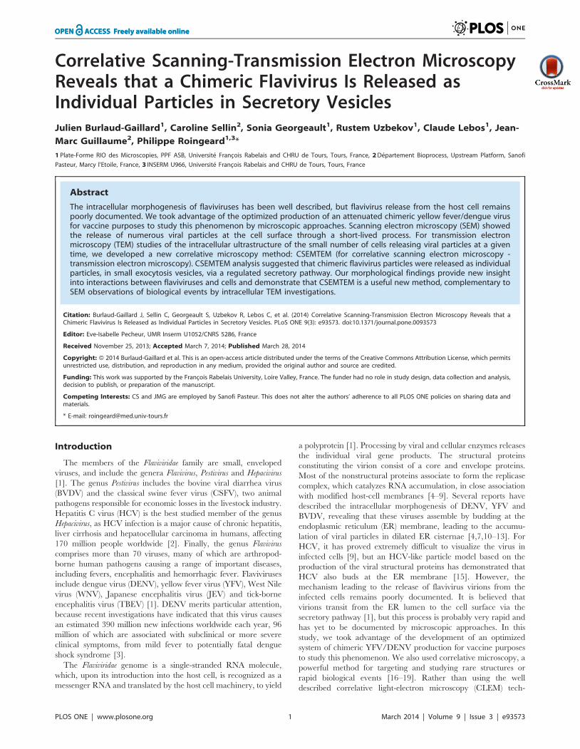
Correlative Scanning-Transmission Electron MicroscopyReveals that a Chimeric Flavivirus Is Released asIndividual Particles in Secretory VesiclesJulien Burlaud-Gaillard1, Caroline Sellin2, Sonia Georgeault1, Rustem Uzbekov1, Claude Lebos1, Jean-
Marc Guillaume2, Philippe Roingeard1,3*
1 Plate-Forme RIO des Microscopies, PPF ASB, Universite Francois Rabelais and CHRU de Tours, Tours, France, 2 Departement Bioprocess, Upstream Platform, Sanofi
Pasteur, Marcy l’Etoile, France, 3 INSERM U966, Universite Francois Rabelais and CHRU de Tours, Tours, France
Abstract
The intracellular morphogenesis of flaviviruses has been well described, but flavivirus release from the host cell remainspoorly documented. We took advantage of the optimized production of an attenuated chimeric yellow fever/dengue virusfor vaccine purposes to study this phenomenon by microscopic approaches. Scanning electron microscopy (SEM) showedthe release of numerous viral particles at the cell surface through a short-lived process. For transmission electronmicroscopy (TEM) studies of the intracellular ultrastructure of the small number of cells releasing viral particles at a giventime, we developed a new correlative microscopy method: CSEMTEM (for correlative scanning electron microscopy -transmission electron microscopy). CSEMTEM analysis suggested that chimeric flavivirus particles were released as individualparticles, in small exocytosis vesicles, via a regulated secretory pathway. Our morphological findings provide new insightinto interactions between flaviviruses and cells and demonstrate that CSEMTEM is a useful new method, complementary toSEM observations of biological events by intracellular TEM investigations.
Citation: Burlaud-Gaillard J, Sellin C, Georgeault S, Uzbekov R, Lebos C, et al. (2014) Correlative Scanning-Transmission Electron Microscopy Reveals that aChimeric Flavivirus Is Released as Individual Particles in Secretory Vesicles. PLoS ONE 9(3): e93573. doi:10.1371/journal.pone.0093573
Editor: Eve-Isabelle Pecheur, UMR Inserm U1052/CNRS 5286, France
Received November 25, 2013; Accepted March 7, 2014; Published March 28, 2014
Copyright: � 2014 Burlaud-Gaillard et al. This is an open-access article distributed under the terms of the Creative Commons Attribution License, which permitsunrestricted use, distribution, and reproduction in any medium, provided the original author and source are credited.
Funding: This work was supported by the Francois Rabelais University, Loire Valley, France. The funder had no role in study design, data collection and analysis,decision to publish, or preparation of the manuscript.
Competing Interests: CS and JMG are employed by Sanofi Pasteur. This does not alter the authors’ adherence to all PLOS ONE policies on sharing data andmaterials.
* E-mail: [email protected]
Introduction
The members of the Flaviviridae family are small, enveloped
viruses, and include the genera Flavivirus, Pestivirus and Hepacivirus
[1]. The genus Pestivirus includes the bovine viral diarrhea virus
(BVDV) and the classical swine fever virus (CSFV), two animal
pathogens responsible for economic losses in the livestock industry.
Hepatitis C virus (HCV) is the best studied member of the genus
Hepacivirus, as HCV infection is a major cause of chronic hepatitis,
liver cirrhosis and hepatocellular carcinoma in humans, affecting
170 million people worldwide [2]. Finally, the genus Flavivirus
comprises more than 70 viruses, many of which are arthropod-
borne human pathogens causing a range of important diseases,
including fevers, encephalitis and hemorrhagic fever. Flaviviruses
include dengue virus (DENV), yellow fever virus (YFV), West Nile
virus (WNV), Japanese encephalitis virus (JEV) and tick-borne
encephalitis virus (TBEV) [1]. DENV merits particular attention,
because recent investigations have indicated that this virus causes
an estimated 390 million new infections worldwide each year, 96
million of which are associated with subclinical or more severe
clinical symptoms, from mild fever to potentially fatal dengue
shock syndrome [3].
The Flaviviridae genome is a single-stranded RNA molecule,
which, upon its introduction into the host cell, is recognized as a
messenger RNA and translated by the host cell machinery, to yield
a polyprotein [1]. Processing by viral and cellular enzymes releases
the individual viral gene products. The structural proteins
constituting the virion consist of a core and envelope proteins.
Most of the nonstructural proteins associate to form the replicase
complex, which catalyzes RNA accumulation, in close association
with modified host-cell membranes [4–9]. Several reports have
described the intracellular morphogenesis of DENV, YFV and
BVDV, revealing that these viruses assemble by budding at the
endoplasmic reticulum (ER) membrane, leading to the accumu-
lation of viral particles in dilated ER cisternae [4,7,10–13]. For
HCV, it has proved extremely difficult to visualize the virus in
infected cells [9], but an HCV-like particle model based on the
production of the viral structural proteins has demonstrated that
HCV also buds at the ER membrane [15]. However, the
mechanism leading to the release of flavivirus virions from the
infected cells remains poorly documented. It is believed that
virions transit from the ER lumen to the cell surface via the
secretory pathway [1], but this process is probably very rapid and
has yet to be documented by microscopic approaches. In this
study, we took advantage of the development of an optimized
system of chimeric YFV/DENV production for vaccine purposes
to study this phenomenon. We also used correlative microscopy, a
powerful method for targeting and studying rare structures or
rapid biological events [16–19]. Rather than using the well
described correlative light-electron microscopy (CLEM) tech-
PLOS ONE | www.plosone.org 1 March 2014 | Volume 9 | Issue 3 | e93573
nique, we established a new method for this study: correlative
scanning electron microscopy-transmission electron microscopy
(CSEMTEM). This new type of correlative microscopy, based on
the detection of cells of interest by scanning electron microscopy
(SEM), for further investigation by transmission electron micros-
copy (TEM), made it possible to visualize the release of a flavivirus
at the cell surface. Our morphological data suggest that individual
viral particles are secreted from infected cells in small secretory
vesicles and that this new correlative microscopy method would be
useful for deciphering other biological processes.
Materials and Methods
Cell culture and virus infectionVero cells (African green monkey cell line) from the Sanofi
Pasteur cell bank were amplified in multitrays in a completely
animal-derived component-free process (serum-free media, re-
combinant trypsin and soybean inhibitor). For the production step,
cells were used to seed a 12-liter bioreactor containing serum-free
medium and 2.5 g/l Cytodex I microcarriers (GE). Cells were
amplified by incubation at 37uC, in the presence of 25% PO2, at
pH 7.2, with shaking at 30 rpm, for three days. The cell
amplification medium was then removed and replaced with
prewarmed viral medium. Cells were then infected with a chimeric
YFV/DENV based on the YFV 17D live attenuated vaccine
backbone containing the prM and envelope genes from DENV
[20]. The viral inoculum was introduced into the bioreactor at a
MOI of 0.001, two hours after the medium was changed. Two
days after infection, the medium was replaced. Bioreactor
sampling was undertaken on a daily basis, to monitor cells and
virus production.
Analysis of the secreted particles by negative stainingelectron microscopy and immunogold labeling
The clarified cell-culture supernatant was ultrafiltrated
(300 kDa) and the viral particles were purified by polyethylene
glycol precipitation followed by an utracentrifugation on sucrose
gradient. Fractions of interest were then pooled and concentrated
with an Amicon – 30 kDa (Millipore) device, before to be fixed (v/
v) with paraformaldehyde 2% (Sigma, St-Louis, MO), 0.1 M
phosphate buffer pH 7.2 overnight. Formvar/carbon-coated
nickel grids were deposited on a drop of fixed sample during five
minutes and rinsed three times with phosphate-buffered saline
(PBS). After a single wash with distilled water, the negative staining
was then performed with three consecutive contrasting steps using
2% uranyl acetate (Agar Scientific, Stansted, UK), before analysis
under the transmission electron microscope (JEOL 1011, Tokyo,
Japan).
For immunogold labeling, grids coated with the sample were
washed and further incubated for 45 minutes on a drop of PBS
containing 1:10 mouse monoclonal antibody against DENV E
glycoprotein (DE1, Abcam, Cambridge, UK). After six washes
with PBS, grids were further incubated for 45 minutes on a drop
of PBS containing 1:30 gold-conjugated (10 nm) goat-anti-mouse
IgG (Aurion, Wageningen, Netherlands). Grids were then washed
with 6 drops of PBS, post-fixed in 1% glutaraldehyde, rinsed with
two drops of distilled water, before to be negatively stained and
observed under the microscope as described above.
Ultrastructural analysis of the infected cells by scanningelectron microscopy
Chimeric YFV/DENV-infected Vero cells grown on micro-
carriers and were studied before infection (day 0) and every day
post-infection, from day 1 to day 4. On each day, 10 ml of
microcarrier suspension was fixed by incubation for 24 h in 4%
paraformaldehyde, 1% glutaraldehyde in 0.1 M phosphate buffer
(pH 7.2). Samples were then washed in phosphate-buffered saline
(PBS) and post-fixed by incubation with 2% osmium tetroxide for
1 h. Samples were then fully dehydrated in a graded series of
ethanol solutions and dried in hexamethyldisilazane (HMDS,
Sigma, St-Louis, MO). Finally, the dry sample was sprinkled onto
carbon disks and coated with 40 A platinum, with a GATAN
PECS 682 apparatus (Pleasanton, CA), before observation under a
Zeiss Ultra plus FEG-SEM scanning electron microscope
(Oberkochen, Germany).
Ultrastructural analysis of the infected cells bytransmission electron microscopy
Microcarriers were placed in a mixture of (1:1) propylene
oxide/Epon resin (Sigma) and then left overnight in pure resin for
impregnation of the samples. Microcarriers were then embedded
in Epon resin (Sigma), which was allowed to polymerize for
48 hours at 60uC. Ultra-thin sections (90 nm) of these blocks were
obtained with a Leica EM UC7 ultramicrotome (Wetzlar,
Germany), as previously described [21]. Sections were deposited
on formvar/carbon-coated nickel grids and stained with 5%
uranyl acetate, 5% lead citrate. Observations were made with a
JEOL 1011 transmission electron microscope.
Correlative scanning electron microscopy-transmissionelectron microscopy
Using SEM, we identified cells with chimeric viruses on their
surface at medium or high magnification (from 630,000 to
660,000). Microcarriers bearing such chimeric virus-coated cells
were monitored by SEM at low magnification (from630 to6300).
This made it possible to determine precisely the position of the
microcarriers of interest over the entire disk. We mapped the
entire disk, with the ZEISS multiscan module, to obtain larger
images at high resolution. Images were then compared with
observations of the disks under a Zeiss Stemi 2000c stereo
microscope. Regions of interest were selected by cropping the
carbon disk with a surgical scalpel. These selected regions were
then included in resin by performing a flat embedding, before to
be analyzed by TEM as described above. The location of the
beads of interest was initially checked by toluidine blue staining on
sequential 500 nm semithin sections. Photographs of these
semithin sections were taken with a Nikon Eclipse 80i (Tokyo,
Japan) equipped with a DS-Vi1 camera. Ultrathin sections
(90 nm) were then cut once the center of the microcarrier of
interest had been reached, allowing to specifically investigate the
cells present at the upper side of the microcarrier and previously
visualized by the SEM analysis. These selected sections of the
microcarriers bearing chimeric virus-coated cells were deposited
on formvar/carbon-coated nickel grids and stained with 5%
uranyl acetate, 5% lead citrate. They were then observed in a
JEOL 1011 transmission electron microscope.
Results
Analysis of the secreted particles by negative stainingelectron microscopy and immunogold labeling
Initial analysis by regular negative staining electron microscopy
of the purified viral particles led to the observation of numerous
spherical particles, 50 to 60 nm in diameter, that had the
morphological characterisitics of a typical flavivirus [14]
(Fig. 1A). The specificity of these structures was confirmed by
immunogold labeling with the anti-DENV E glycoprotein
CSEMTEM Analysis of Flavivirus Release
PLOS ONE | www.plosone.org 2 March 2014 | Volume 9 | Issue 3 | e93573

antibody, showing unambiguously the presence of the DENV
envelope glycoprotein at the surface of these viral particles
(Fig. 1B).
Ultrastructural analysis of the infected cells by scanningelectron microscopy
Low-magnification SEM was used to observe the Vero cell
monolayer on the microcarrier surface (Fig. 2A). These observa-
tions showed that the cells adhered efficiently to the surface of the
beads, which were found to be largely covered by the cells
(Fig. 2A). At high magnification and before infection, the surface of
the Vero cells was smooth, with small protrusions and no
detectable virus-like structures (Fig. 2B). Similar results were
obtained for observations made on the day after infection with the
chimeric YFV/DENV (not shown). Two and three days post-
infection, some cells had large numbers of chimeric viral particles,
50 to 60 nm in diameter, at their surface (Fig. 2C and 2D). In cells
displaying such particles, a large surface of the cell including
protrusions was covered by the chimeric viral particles, without no
evidence of any particular polarization for viral particle release or
morphological modification. However, cells bearing chimeric
YFV/DENV particles at their surface were very scarce. A full
screening of 100 microcarriers, accounting for the observation of
2300 cells, showed that only 6 cells (0.26%), detected on 6 different
microcarriers, exhibited this pattern of abundant viral release.
These structures were highly specific, as none were observed in
uninfected, control cells. Analyses of the Vero cells four days post-
infection showed an absence of chimeric viral particles at the cell
surface, despite extensive searches (not shown).
Ultrastructural analysis of the infected cells bytransmission electron microscopy
In analyses of ultrathin sections of Vero cells before infection or
one day post-infection with the chimeric YFV/DENV, no viral
particles were visualized (not shown). From days 2 to day 4 post-
infection, specific ultrastructural changes associated with the
presence of chimeric YFV/DENV and described elsewhere for
DENV [7,13,14] were easily observed (Figure 2). These changes
included dilated ER-derived cisternae containing viral particles,
often arranged in regular arrays (Fig. 3A and 3B) and small
clusters of smooth virus-induced vesicles (Fig. 3A). The interior of
these virus-induced vesicles was recently shown to be connected to
the cytosol and to constitute the probable site of viral RNA
replication [7]. Extensive observations of ultrathin sections of these
cells between days 2 and day 4 post-infection did not result in the
visualization of chimeric YFV/DENV particle release.
Correlative scanning electron microscopy-transmissionelectron microscopy
Samples of cells taken two days post-infection and initially
analyzed by SEM were used for this CSEMTEM experiment. The
observation of these samples at high magnification made it possible
to localize several microcarriers bearing cells with numerous
chimeric viral particles at their surface. Low magnification
analyses of these beads of interest were used to map their
neighborhood pattern (Fig. 4A), and the Multiscan module was
then used to record and reconstitute the whole SEM sample
Figure 1. Analysis by negative staining electron microscopyand immunogold labeling of the chimeric yellow fever virus/dengue virus (YFV/DENV) particles secreted in the cellsupernatant. (A): Numerous 50 to 60 nm spherical particles that hadmorphological characteristics of a typical flavivirus were observed byregular negative staining electron microscopy. (B): The specificity ofthese particles was confirmed by immunogold labeling with an anti-DENV E glycoprotein. Scale bars : 0.5 mm in A ; 100 nm in B.doi:10.1371/journal.pone.0093573.g001
Figure 2. Analysis, by scanning electron microscopy (SEM), ofVero cells grown on microcarrier beads and infected with thechimeric YFV/DENV. (A): At low magnification and before infection(day 0), Vero cells completely covered the microcarrier surface. (B): Onday 0, analysis of these cells at high magnification showed them tohave a smooth surface with protrusions. (C and D): On day 2 post-infection, several cells presented numerous chimeric viral particles attheir surface (arrows). Scale bars : 100 mm in A ; 1 mm in B and C; 0.2 mmin D.doi:10.1371/journal.pone.0093573.g002
CSEMTEM Analysis of Flavivirus Release
PLOS ONE | www.plosone.org 3 March 2014 | Volume 9 | Issue 3 | e93573

(Fig. 4B). Selected acquisition parameters applied to this 669
digital microphotograph map provided sufficient detail, with rapid
acquisition, and resulting in a final file of reasonable size (i.e. not
too heavy to handle). This large image was then compared with
the carbon disk observed directly under the stereo microscope.
The global pattern of the beads facilitated recognition of the zone
of interest and, finally, the microcarrier of interest. A surgical
scalpel was then used to cut and remove the carbon disk visualized
under the stereo microscope, resulting in the visualization of an
area of less than 1 mm2 on the SEM sample support (Fig. 4C).
Rapid control at the maximum magnification of the stereo
microscope confirmed the localization of the microcarrier of
interest in this selected portion of the SEM sample (Fig. 4D). This
resized sample was embedded in Epon resin and sectioned with an
ultramicrotome, to generate semi-thin sections that were stained
with toludine blue and observed under a light microscope, to
follow the bead of interest (Fig. 4E). Finally, several ultrathin
sections of this resized sample were examined by TEM, to
visualize the bead of interest at low (Fig. 4F) and high (insert in
Fig. 4F) magnifications. Dextran-based microcarriers were not
embedded in resin, resulting in these microcarriers appearing as
holes (corresponding to the space left by the beads) surrounded by
a monolayer of cells on semithin and ultrathin sections (Fig. 4E
and 4F). For this reason, ultrathin sections were deposited on
formvar membrane, to prevent the movement and tearing of the
sections under the electron beam. The observation, by TEM, of
these ultra-thin sections containing the selected cells, led to the
visualization of numerous chimeric YFV/DENV particles at the
cell surface (Fig. 5). The appearance of the chimeric viruses on the
cell surface differed from that on regular TEM, due to the
platinum coating step used in SEM analysis. Indeed, chimeric viral
particles appeared to be 60 to 70 nm in diameter and very dense
(Fig. 5). Clusters of these dense viral particles were frequently
found at the cell surface, but some were clearly associated with an
exocytosis-like pattern (Fig. 5B, 5C, 5D), suggesting that the
chimeric viral particles were released individually by the exocytosis
of a small secretory vesicle. Only few intracellular viral particles
were observed in these virus-covered cells as compared to the
surrounding cells, suggesting the occurrence of a massive release of
the chimeric viruses in these particular cells. To quantify the
presence of intracellular viral particles in the surrounding cells, we
examined carefully 200 cells on ultrathin sections obtained with
the CSEMTEM method and determined that 24 (12%) contained
viral particles, often arranged in regular arrays.
Discussion
The mechanism underlying flavivirus release from the host cell
remains unclear. As these viruses accumulate in dilated ER-
derived cisternae, it has been suggested that they may be released
by the exocytosis of these large virion-containing vacuoles and/or
as individual viral particles in secretory vesicles [12]. However, it
has been also suggested that WNV could be released by a budding
at the apical surface of the plasma membrane [22]. We report here
the first visualization, by SEM, of a chimeric flavivirus at the
surface of an infected cell. Our SEM photographs of chimeric
YFV/DENV-infected cells demonstrate that the viral particles are
not released in clusters in a polarized area of the cell. Instead, they
are released individually and evenly over a large surface of the cell.
The scarcity of chimeric virus-covered cells suggests that this
phenomenon is short-lived, probably accounting for the difficulties
encountered in attempts to observe the release of viral particles in
ultrathin sections analyzed by TEM. This led us to develop an
original method — CSEMTEM, for correlative scanning electron
microscopy-transmission electron microscopy — for identifying
virus-covered cells and selecting them precisely by SEM. These
cells were then embedded in resin and sectioned, for further
analysis by TEM. The ultrastructure of the cells that had
previously been prepared for SEM analysis was found to be very
well preserved when these cells were analyzed by TEM. No major
difference could be found between these cells and those prepared
by regular TEM protocols (i.e. without HMDS treatment and
platinum coating). The major difference concerned the chimeric
viral particles at the cell surface, which appeared as extremely
dense particles with a diameter of 60 to 70 nm. This particular
feature of the virions on CSEMTEM observation was due to the
platinum coating used for the initial SEM sample preparation,
resulting in the incorporation of large amounts of metal into these
small objects. We tested several alternative strategies, to try to
prevent this effect, including a carbon coating, but none gave
satisfactory SEM observations of the cell surface and final
visualization of the chimeric viral particles (not shown).
Nevertheless, our CSEMTEM method provided the first
observation of a chimeric flavivirus being released as an individual
particle in small exocytosis vesicles. These results are consistent
with recent gene silencing experiments showing that host cell
exocytosis factors, such as Sec3p and EXO70, are essential for
DENV egression or secretion [23]. This is also consistent with the
maturation of flavivirus particles in the Golgi compartment.
Hepacivirus and pestivirus virions are infectious immediately, or at
least very shortly after their envelopment [24,25], but flavivirus
particles remain immature until the acid-induced rearrangement
of their envelope E protein and the furin-mediated cleavage of
Figure 3. Analysis, by transmission electron microcopy (TEM),of Vero cells grown on microcarrier beads and infected withthe chimeric YFV/DENV. (A and B): Ultrathin sections of the cellsattached to the microcarriers showed the presence of numerouschimeric viral particles in cisterns (black arrows) linked to the roughendoplasmic reticulum. Some virus-induced smooth vesicles wereobserved close to rough ER cisternae containing chimeric YFV/DENVparticles (white arrows). Scale bars: 0.2 mm in A and B.doi:10.1371/journal.pone.0093573.g003
CSEMTEM Analysis of Flavivirus Release
PLOS ONE | www.plosone.org 4 March 2014 | Volume 9 | Issue 3 | e93573
their prM protein have occurred in the late Golgi compartment
[26,27].
The scarcity of virus-coated cells at any given time suggests that
this exocytosis is probably a very short-lived process. Moreover,
the presence of large numbers of chimeric flavivirus particles
evenly distributed over a large surface of these rare cells suggests
that this mechanism is driven by many exocytosis vesicles being
generated at the same time in a given cell. This suggests that the
release of the chimeric virions occurs via a regulated exocytosis
that may also account for the absence of morphological changes or
obvious cytotoxicity in the virus-coated cells studied by SEM.
Our data obtained with microscopic approaches may provide
new insight into basic flavivirus/cell interactions and may facilitate
the definition of targets for the development of preventive and
therapeutic strategies for combating infections due to these viruses.
However, it will be necessary to confirm these observations with
wild-type flavivirus strains. It will be also necessary to further
investigate this phenomenon by confirming our morphological
data with biological experiments. Nevertheless, our findings
suggest that CSEMTEM is a potentially useful new correlative
microscopy method for analyzing the intracellular ultrastructure of
cells presenting particular surface modifications, which could be
applied to the study of other important biological processes.
Although limited by technical constraints, CSEMTEM will be
particularly useful as compared to CLEM to study virus/cell
interactions, as fluorescence methods do not reveal detailed
information about the structure of viruses. Viruses can be
visualized as small spots on fluorescence microscopy, but the
resolution of this technique is too low to determine whether these
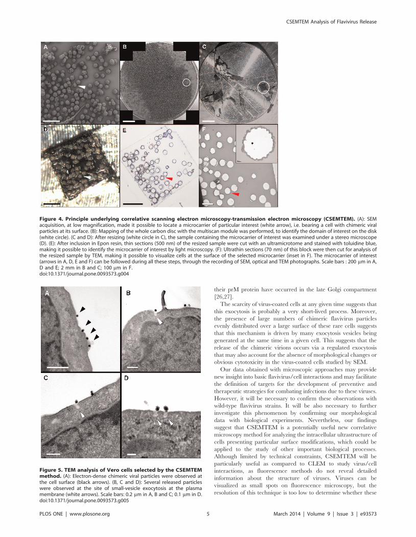
Figure 4. Principle underlying correlative scanning electron microscopy-transmission electron microscopy (CSEMTEM). (A): SEMacquisition, at low magnification, made it possible to locate a microcarrier of particular interest (white arrow), i.e. bearing a cell with chimeric viralparticles at its surface. (B): Mapping of the whole carbon disc with the multiscan module was performed, to identify the domain of interest on the disk(white circle). (C and D): After resizing (white circle in C), the sample containing the microcarrier of interest was examined under a stereo microscope(D). (E): After inclusion in Epon resin, thin sections (500 nm) of the resized sample were cut with an ultramicrotome and stained with toluidine blue,making it possible to identify the microcarrier of interest by light microscopy. (F): Ultrathin sections (70 nm) of this block were then cut for analysis ofthe resized sample by TEM, making it possible to visualize cells at the surface of the selected microcarrier (inset in F). The microcarrier of interest(arrows in A, D, E and F) can be followed during all these steps, through the recording of SEM, optical and TEM photographs. Scale bars : 200 mm in A,D and E; 2 mm in B and C; 100 mm in F.doi:10.1371/journal.pone.0093573.g004
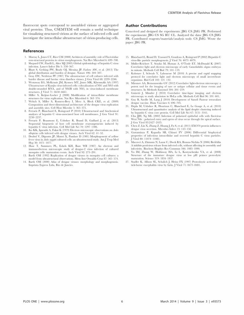
Figure 5. TEM analysis of Vero cells selected by the CSEMTEMmethod. (A): Electron-dense chimeric viral particles were observed atthe cell surface (black arrows). (B, C and D): Several released particleswere observed at the site of small-vesicle exocytosis at the plasmamembrane (white arrows). Scale bars: 0.2 mm in A, B and C; 0.1 mm in D.doi:10.1371/journal.pone.0093573.g005
CSEMTEM Analysis of Flavivirus Release
PLOS ONE | www.plosone.org 5 March 2014 | Volume 9 | Issue 3 | e93573
fluorescent spots correspond to assembled virions or aggregated
viral proteins. Thus, CSEMTEM will remain a useful technique
for visualizing structured virions at the surface of infected cells and
investigate the intracellular ultrastructure of virion-producing cells.
Author Contributions
Conceived and designed the experiments: JBG CS JMG PR. Performed
the experiments: JBG CS SG RU CL. Analyzed the data: JBG CS JMG
PR. Contributed reagents/materials/analysis tools: CS JMG. Wrote the
paper: JBG PR.
References
1. Murray L, Jones CT, Rice CM (2008) Architects of assembly: role of Flaviviridae
non-structural proteins in virion morphogenesis. Nat Rev Microbiol 6: 699–708.2. Shepard CW, Finelli L, Alter MJ (2005) Global epidemiology of hepatitis C virus
infection. Lancet Infect Dis 5: 558–567.
3. Bhatt S, Gething PW, Brady OJ, Messina JP, Farlow AW, et al. (2013) Theglobal distribution and burden of dengue. Nature 496: 504–507.
4. Gray EW, Nettleton PF (1987) The ultrastructure of cell cultures infected withborder disease and bovine virus diarrhea viruses. J Gen Virol 68: 2239–2246.
5. Westaway EG, McKenzie JM, Kenney MT, Jones MK, Khromykh AA (1997)Ultrastructure of Kunjin virus-infected cells: colocalization of NS1 and NS3 with
double-stranded RNA, and of NS2B with NS3, in virus-induced membrane
structures. J Virol 71: 6650–6661.6. Miller S, Krijnse-Locker J (2008) Modification of intracellular membrane
structures for virus replication. Nat Rev Microbiol 6: 363–374.7. Welsch S, Miller S, Romero-Brey I, Merz A, Bleck CKE, et al. (2009)
Composition and three-dimensional architecture of the dengue virus replication
and assembly sites. Cell Host Microbe 5: 365–375.8. Ferraris P, Blanchard E, Roingeard P (2010) Ultrastructural and biochemical
analyses of hepatitis C virus-associated host cell membranes. J Gen Virol 91:2230–2237.
9. Ferraris P, Beaumont E, Uzbekov R, Brand D, Gaillard J, et al. (2013)
Sequential biogenesis of host cell membrane rearrangements induced byhepatitis C virus infection. Cell Mol Life Sci 70: 1297–1306.
10. Ko KK, Igarashi A, Fukai K (1979) Electron microscopic observations on Aedes
albopictus cells infected with dengue viruses. Arch Virol 62: 41–52.
11. Deubel V, Digoutte JP, Mattei X, Pandare D (1981) Morphogenesis of yellowfever virus in Aedes aegypti cultured cells: an ultrastructural study. Am J Trop Med
Hyg 30: 1071–1077.
12. Hase T, Summers PL, Eckels KH, Baze WB (1987) An electron andimmunoelectron microscopic study of dengue-2 virus infection of cultured
mosquito cells: maturation events. Arch Virol 92: 273–291.13. Barth OM (1992) Replication of dengue viruses in mosquito cell cultures: a
model from ultrastructural observations. Mem Inst Oswaldo Cruz 87: 565–574.
14. Barth OM (2000) Atlas of dengue viruses: morphology and morphogenesis.Imprinta Express Ltda, Rio de Janeiro.
15. Blanchard E, Brand D, Trassard S, Goudeau A, Roingeard P (2002) Hepatitis C
virus-like particle morphogenesis. J Virol 76: 4073–4079.
16. Muller-Reichert T, Srayko M, Hyman A, O’Toole ET, McDonald K (2007)
Correlative light and electron microscopy of early Caenorhabditis elegans embryos
in mitosis. Methods Cell Biol 79: 101–119.
17. Kolotuev I, Schwab Y, Labouesse M (2010) A precise and rapid mapping
protocol for correlative light and electron microscopy of small invertebrate
organisms. Biol Cell 102: 121–132.
18. Mironov AA, Beznoussenko GV (2012) Correlative light-electron microscopy: a
potent tool for the imaging of rare or unique cellular and tissue events and
structures. In Methods Enzymol 504: 201–219.
19. Guizetti J, Mantler J (2010) Correlative time-lapse imaging and electron
microscopy to study abscission in HeLa cells. Methods Cell Biol 96: 591–601.
20. Guy B, Saville M, Lang J (2010) Development of Sanofi Pasteur tetravalant
dengue vaccine. Hum Vaccines 6: 696–705.
21. Depla M, Uzbekov R, Hourioux C, Blanchard E, Le Gouge A, et al. (2010)
Ultrastructural and quantitative analysis of the lipid droplet clustering induced
by hepatitis C virus core protein. Cell Mol Life Sci 67: 3151–3161.
22. Chu JJH, Ng ML (2002) Infection of polarized epithelial cells with flavivirus
West Nile : polarized entry and egress of virus occur through the apical surface.
J Gen Virol 83:2427–2435.
23. Chen Z, Lin X, Zhang Z, Huang J, Fu S, et al. (2011) EXO70 protein influences
dengue virus secretion. Microbes Infect 13: 143–150.
24. Gastaminza P, Kapadia SB, Chisari FV (2006) Differential biophysical
properties of infectious intracellular and secreted hepatitis C virus particles.
J Virol 80: 11074–11081.
25. Macovei A, Zitmann N, Lazar C, Dwek RA, Branza-Nichita N (2006) Brefeldin
A inhibits pestivirus release from infected cells, without affecting its assembly and
infectivity. Biochem Biophys Res Commun 346: 1083–1090.
26. Yu IM, Zhang W, Holdaway HA, Li L, Kostyuchenko VA, et al. (2008)
Structure of the immature dengue virus at low pH primes proteolytic
maturation. Science 319: 1834–1837.
27. Stadler K, Allison SL, Schalich J, Heinz FX (1997) Proteoloytic activation of
tick-borne encephalitis virus by furin. J Virol 71: 8475–8481.
CSEMTEM Analysis of Flavivirus Release
PLOS ONE | www.plosone.org 6 March 2014 | Volume 9 | Issue 3 | e93573