Embed Size (px)
Citation preview
Correlation of Change in Intraocular Pressure with Anterior Chamber Depth
& Angle after Cataract Surgery as measured by Anterior Segment OCT
Shilpi Pradhan, MD1,3; Martin Wilkes, MD1; Christopher T. Leffler, MD, MPH1,2; Dennis Pratt, MD1,2; Muneera Mahmood, MD1,2
1 Medical College of Virginia, Virginia Commonwealth University, Richmond, Virginia2 Hunter Holmes McGuire Veterans Affairs Hospital, Richmond, Virginia3 Current Cornea Fellow at University of Pittsburgh Medical Center, Pittsburgh, Pennsylvania
Authors have no financial interests.
Background
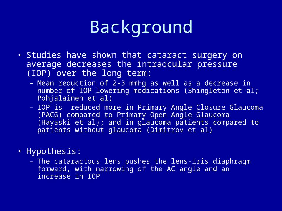
• Studies have shown that cataract surgery on average decreases the intraocular pressure (IOP) over the long term:– Mean reduction of 2-3 mmHg as well as a decrease in number of
IOP lowering medications (Shingleton et al; Pohjalainen et al)– IOP is reduced more in Primary Angle Closure Glaucoma
(PACG) compared to Primary Open Angle Glaucoma (Hayaski et al); and in glaucoma patients compared to patients without glaucoma (Dimitrov et al)
• Hypothesis:– The cataractous lens pushes the lens-iris diaphragm forward,
with narrowing of the AC angle and an increase in IOP
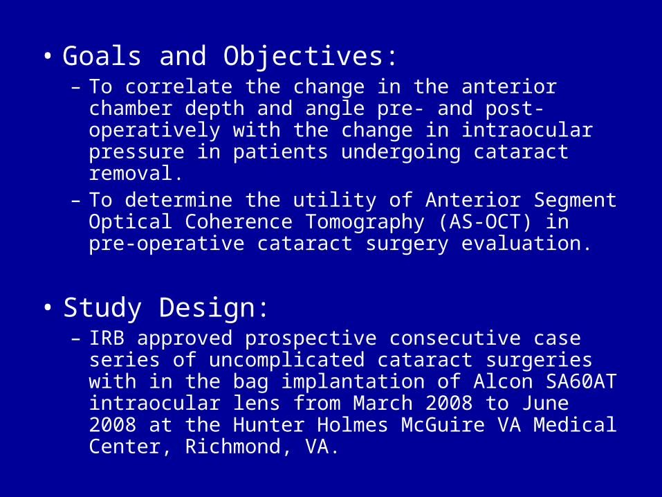
• Goals and Objectives:– To correlate the change in the anterior chamber depth
and angle pre- and post-operatively with the change in intraocular pressure in patients undergoing cataract removal.
– To determine the utility of Anterior Segment Optical Coherence Tomography (AS-OCT) in pre-operative cataract surgery evaluation.
• Study Design: – IRB approved prospective consecutive case series of
uncomplicated cataract surgeries with in the bag implantation of Alcon SA60AT intraocular lens from March 2008 to June 2008 at the Hunter Holmes McGuire VA Medical Center, Richmond, VA.

Data Collected– Demographics including age, race, gender– Approach to surgery, IOL location, and complications– Pre-op: IOL Master or immersion A-scan IOL
calculations– Pre- and Post-op (4-6 wks): vision, refraction,
horizontal single line scan un-dilated AS-OCT, gonioscopy, ultrasound pachymetry, IOP measurement by applanation tonometry
– Measurements taken from AS-OCT: Anterior Chamber (AC) diameter, AC depth, nasal and temporal horizontal AC angle measurements, pachymetry and pupil size

Visante AS-OCT• Visante Anterior Segment OCT scans the
anterior segment of the eye using 1310 nm wavelength of light. It takes 4000 axial scans or 8 frames per second with a lateral resolution of 60 μm and axial resolution of 18 μm (Dada et al).
• May be a more sensitive way to detect narrow or closed angles than gonioscopy (Sakata et al)
• Angle width was found to be significantly smaller with the presence of peripheral anterior synechiae in PACG and allowed quantification of the angle in an Asian population (Su et al)
• Horizontal (nasal and temporal) angles were found to be most accurate and reproducible (Radkrishnan et al)
• Comparable to Ultrasound biomicroscopic images and measurements (Dada et al)
• AS-OCT pachymetry correlates with ultrasound (US) pachymetry but has slightly lower measurements (Li et al)
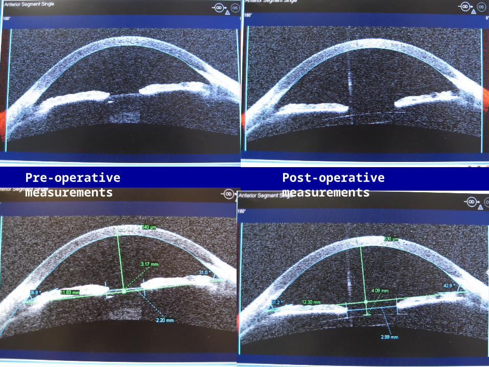
Pre-operative measurements Post-operative measurements
RESULTS
• 72 eyes in 72 patients were studied.
• Mean Age = 69.9 years old (SD 10.1)
• 70 out of 72 patients were male• 61 patients were white and 11
patients were black

Table 1. Preoperative and postoperative clinical parameters.
Pre-op Post-op Mean (SD) Mean (SD) p value
• OCT findings:• Anterior chamber depth (mm) 2.74 (0.44) 4.14 (0.32) <0.001• Anterior chamber angle (degrees) 27.0 (7.0) 38.0 (5.2) <0.001• Anterior chamber diameter (mm) 12.1 (0.5) 12.2 (0.4) 0.007• Corneal thickness (um) 547 (38) 538 (56) 0.30• Other findings:• Intraocular pressure (mean, mmHg) 14.8 (2.7) 11.8 (2.8) <0.001• …postoperative day 1 -- 18.2 (6.6) <0.001• …postoperative week 1 -- 12.8 (3.6) <0.001• …postoperative month 1 -- 10.8 (3.1) <0.001• Refraction (SE, D) 0.23 (2.5) -0.17 (0.7) 0.15• Gonioscopy (Schaffer scale) 2.8 (0.5) 3.2 (0.5) <0.001• Visual acuity (LogMar) 0.49 (0.29) 0.10 (0.22) <0.001
N=72 except as noted: anterior chamber OCT findings (n=65 postop.), gonioscopy (n=70 preop, n=61 postop), day 1 postoperative IOP (n=70), week 1 postoperative IOP (n=69), month 1 postop IOP (n=71). Mean postoperative IOP excludes day 1 postoperative.
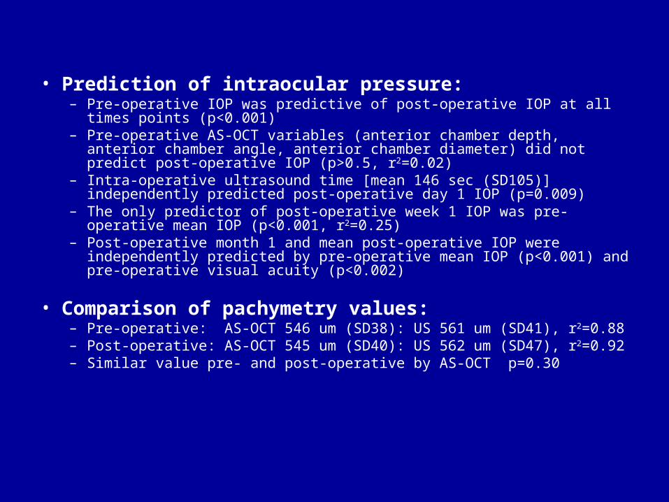
• Prediction of intraocular pressure:– Pre-operative IOP was predictive of post-operative IOP at all times points
(p<0.001) – Pre-operative AS-OCT variables (anterior chamber depth, anterior
chamber angle, anterior chamber diameter) did not predict post-operative IOP (p>0.5, r2=0.02)
– Intra-operative ultrasound time [mean 146 sec (SD105)] independently predicted post-operative day 1 IOP (p=0.009)
– The only predictor of post-operative week 1 IOP was pre-operative mean IOP (p<0.001, r2=0.25)
– Post-operative month 1 and mean post-operative IOP were independently predicted by pre-operative mean IOP (p<0.001) and pre-operative visual acuity (p<0.002)
• Comparison of pachymetry values:– Pre-operative: AS-OCT 546 um (SD38): US 561 um (SD41), r2=0.88– Post-operative: AS-OCT 545 um (SD40): US 562 um (SD47), r2=0.92– Similar value pre- and post-operative by AS-OCT p=0.30

Prediction of Post-operative Anterior Segment Anatomy
– Preoperative AC depth and angle by AS-OCT, lens power implanted, pre-operative gonioscopy, axial length and keratometry all predicted post-operative AC depth in univariate analyses.
– When pre-operative AC depth was controlled for, only pre-operative gonioscopy was independently predictive (p=0.04)
– Post-operative AC angle, AC diameter and pachymetry by AS-OCT were all independently predicted by their pre-operative values (p<0.001)
– Post-operative gonioscopy was poorly predicted by pre-operative gonioscopy (p=0.15)
Conclusions
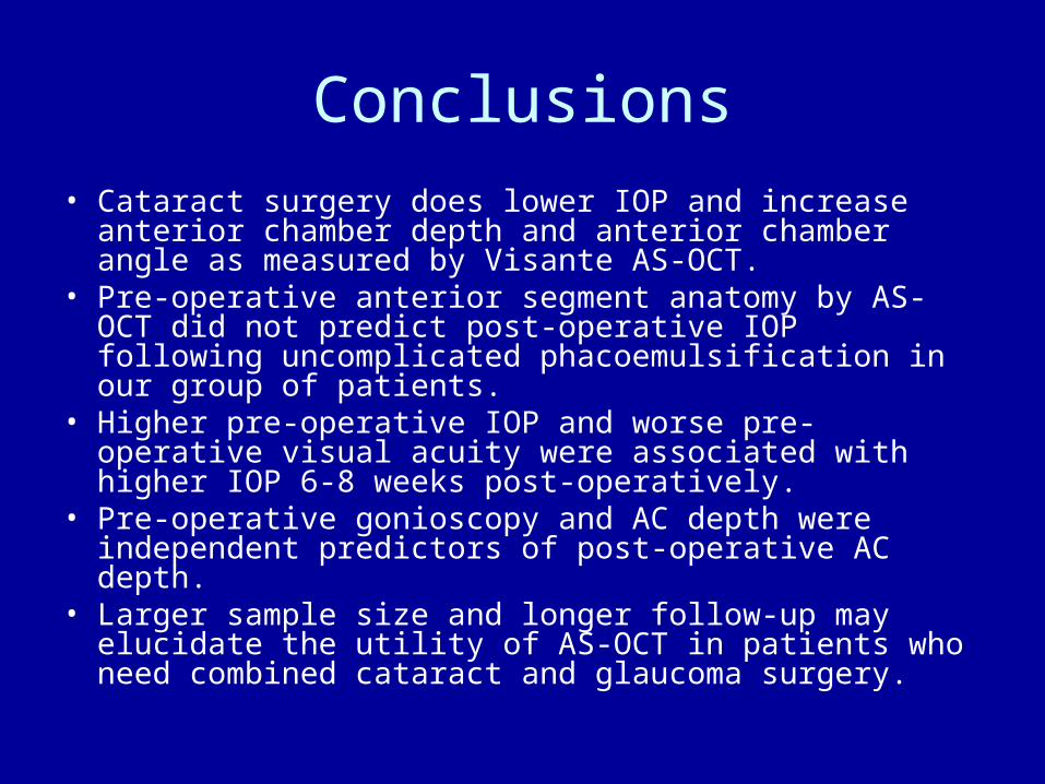
• Cataract surgery does lower IOP and increase anterior chamber depth and anterior chamber angle as measured by Visante AS-OCT.
• Pre-operative anterior segment anatomy by AS-OCT did not predict post-operative IOP following uncomplicated phacoemulsification in our group of patients.
• Higher pre-operative IOP and worse pre-operative visual acuity were associated with higher IOP 6-8 weeks post-operatively.
• Pre-operative gonioscopy and AC depth were independent predictors of post-operative AC depth.
• Larger sample size and longer follow-up may elucidate the utility of AS-OCT in patients who need combined cataract and glaucoma surgery.
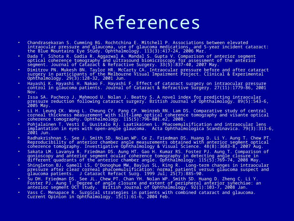
References• Chandrasekaran S. Cumming RG. Rochtchina E. Mitchell P. Associations between elevated intraocular pressure
and glaucoma, use of glaucoma medications, and 5-year incident cataract: the Blue Mountains Eye Study. Ophthalmology. 113(3):417-24, 2006 Mar.
• Dada T. Sihota R. Gadia R. Aggarwal A. Mandal S. Gupta V. Comparison of anterior segment optical coherence tomography and ultrasound biomicroscopy for assessment of the anterior segment. Journal of Cataract & Refractive Surgery. 33(5):837-40, 2007 May.
• Dimitrov PN. Mukesh BN. Taylor HR. McCarty CA. Intraocular pressure before and after cataract surgery in participants of the Melbourne Visual Impairment Project. Clinical & Experimental Ophthalmology. 29(3):128-32, 2001 Jun.
• Hayashi K. Hayashi H. Nakao F. Hayashi F. Effect of cataract surgery on intraocular pressure control in glaucoma patients. Journal of Cataract & Refractive Surgery. 27(11):1779-86, 2001 Nov.
• Issa SA. Pacheco J. Mahmood U. Nolan J. Beatty S. A novel index for predicting intraocular pressure reduction following cataract surgery. British Journal of Ophthalmology. 89(5):543-6, 2005 May.
• Li H. Leung CK. Wong L. Cheung CY. Pang CP. Weinreb RN. Lam DS. Comparative study of central corneal thickness measurement with slit-lamp optical coherence tomography and visante optical coherence tomography. Ophthalmology. 115(5):796-801.e2, 2008.
• Pohjalainen T. Vesti E. Uusitalo RJ. Laatikainen L. Phacoemulsification and intraocular lens implantation in eyes with open-angle glaucoma. Acta Ophthalmologica Scandinavica. 79(3):313-6, 2001 Jun.
• Radhakrishnan S. See J. Smith SD. Nolan WP. Ce Z. Friedman DS. Huang D. Li Y. Aung T. Chew PT. Reproducibility of anterior chamber angle measurements obtained with anterior segment optical coherence tomography. Investigative Ophthalmology & Visual Science. 48(8):3683-8, 2007 Aug.
• Sakata LM. Lavanya R. Friedman DS. Aung HT. Gao H. Kumar RS. Foster PJ. Aung T. Comparison of gonioscopy and anterior segment ocular coherence tomography in detecting angle closure in different quadrants of the anterior chamber angle. Ophthalmology. 115(5):769-74, 2008 May.
• Shingleton BJ, Gamell LS, O’Donoghue MW, Baylus SL, King R. Long-term changes in intraocular pressure after clear corneal phacoemulsification: normal patients versus glaucoma suspect and glaucoma patients. J Cataract Refract Surg. 1999 Jul; 25(7):885-90.
• Su DH. Friedman DS. See JL. Chew PT. Chan YH. Nolan WP. Smith SD. Huang D. Zheng C. Li Y. Foster PJ. Aung T. Degree of angle closure and extent of peripheral anterior synechiae: an anterior segment OCT study. British Journal of Ophthalmology. 92(1):103-7, 2008 Jan.
• Vass C. Menapace R. Surgical strategies in patients with combined cataract and glaucoma. Current Opinion in Ophthalmology. 15(1):61-6, 2004 Feb.