Embed Size (px)
Citation preview

MODULAR DESIGN OF STENT POLYMERS REGULATES HUMAN
CORONARY ARTERY CELL TYPE-SPECIFIC OXIDATIVE RESPONSE AND
PHENOTYPE
By
Spencer William Crowder
Thesis
Submitted to the Faculty of the
Graduate School of Vanderbilt University
in partial fulfillment of the requirements
for the degree of
MASTER OF SCIENCE
in
Biomedical Engineering
May, 2011
Nashville, Tennessee
Approved:
Dr. Hak-Joon Sung
Dr. Craig L. Duvall

ii
ACKNOWLEDGEMENTS
First and foremost, I would like to thank Dr. Hak-Joon Sung for his
guidance, advice, insight, and support throughout the last two years. Additionally,
I thank Dr. Craig L. Duvall for his input on my work as well as his feedback and
insight. I would like to thank my fellow lab members, Mukesh Gupta, Angela
Zachman, Dae Kwang Jung, and Lucas Hofmeister, for their contributions to this
work as well as their day-to-day support. My undergraduate assistants, Chad
Augusty, Amanda Palmer and Yi Liang, have been crucial to completing this
work and I appreciate all that they have done. I would like to thank Dr. Scott
Guelcher and his advisee Elizabeth Adolph for allowing me to access, and also
assisting me with, dynamic mechanical analysis. Finally, I would like to
recognize the Vanderbilt Institute for Nanoscale Science and Engineering for
training and access of their characterization instruments. This work was
supported by NIH HL091465 and NSF 1006558
Most importantly I would like to thank my family for their continued support
of my work, aspirations, and visions. Without them I would never have
accomplished this work. They have shaped me throughout my entire life and the
person I am today is a direct result of the love, dedication, and unequivocal
encouragement with which they provide me every single day.

iii
TABLE OF CONTENTS
Page
ACKNOWLEDGEMENTS …………………………………………..………………… ii
LIST OF TABLES ……………..……………………………………………………..... v
LIST OF FIGURES …………………………………….……………………………... vi
Chapter
I. INTRODUCTION ................................................................................................... 1
Copolymerization Techniques ..................................................................................... 1
Coronary Stent Background ........................................................................................ 1
II. MODULAR DESIGN OF STENT POLYMERS REGULATES HUMAN CORONARY
ARTERY CELL TYPE-SPECIFIC OXIDATIVE RESPONSE AND PHENOTYPE ............ 3
Introduction ................................................................................................................. 3
Methods ...................................................................................................................... 4
Polymer Synthesis and Test Substrate Preparation ................................................. 4
Thermal and Mechanical Properties ........................................................................ 5
Molecular Weight and Degradation .......................................................................... 6
Surface Chemical Properties ................................................................................... 7
Cell Culture ............................................................................................................. 7
Immunofluorescence Staining ................................................................................. 8
Cell Activities ........................................................................................................... 9
Statistical Analysis ..................................................................................................10
Results .......................................................................................................................10
Synthesis ................................................................................................................10
Degradation ............................................................................................................10
Thermal Properties .................................................................................................11
Wet Mechanical Properties .....................................................................................13
iv
Water Contact Angle and Surface Charge ..............................................................14
Cellular Interaction .................................................................................................15
Discussion .................................................................................................................21
Conclusion .................................................................................................................27
REFERENCES ..........................................................................................................29
v
LIST OF TABLES
Table Page
1. Degradation of test polymers ……………………………………………………11
2. Mechanical and thermal properties of wet polymers ……….………………..13
vi
LIST OF FIGURES
Figure Page
1. Polymer synthesis and degradation …………………...……………………….. 5
2. Thermal properties of dry polymers ……………………………..………………12
3. Surface chemistry …………………………………………………………...……..14
4. HCASMC interaction with polymers ……………………………………………. 17
5. HCASMC morphology ……………………………………………………………..19
6. HCAEC interaction with polymers ………………………………………………..20
1
CHAPTER I
INTRODUCTION
Copolymerization Techniques
The physicochemical and mechanical properties of biomaterials modulate
the response of the cells and tissues with which they interact [1-4]. In particular,
polymers can be designed to control cell activity and fate through structure-
function relationships [4]. Copolymerization techniques provide a means for
tuning polymer properties by incorporating subunits with different characteristics
and varying their molar ratios, thereby controlling micro and macro structures [4].
By understanding the effect of each subunit on the resulting polymer properties,
as well as the ability of each subunit to modulate a cellular response, polymer
properties can be precisely optimized to control a specific biological function.
Coronary Stent Background
Implantation of a vascular stent is crucial to reduce human morbidity and
mortality resulting from a vascular disease-induced, localized blood flow
constriction [5]. The evolution of stent materials has included bare metal stents,
polymers, and drug eluting stents, yet each of these technologies poses a
specific set of issues that has prevented its widespread clinical translation. For
example, bare metal stents cause restenosis and require additional surgery for
removal, polymer byproducts can stimulate an inflammatory response, and drug
eluting stents promote late thrombosis resulting from delayed re-
2
endothelialization [5, 6]. Therefore, much attention has been recently paid to
design instructive, bioactive, bioresorbable materials as a solution to problems
associated with classical treatments [7]. The ideal properties of a stent material
include sufficient mechanical strength, moderate degradation kinetics, resorbable
byproducts, and regulation of cellular activities (i.e., proliferation, viability), all of
which can be precisely controlled by understanding how polymer structure affects
the subsequent cellular response.

3
CHAPTER II
MODULAR DESIGN OF STENT POLYMERS REGULATES HUMAN
CORONARY ARTERY CELL TYPE-SPECIFIC OXIDATIVE RESPONSE AND
PHENOTYPE
Introduction
In this study, we have developed a new class of copolymers with tunable
mechanical and chemical properties for coronary stent applications. Three
subunits were copolymerized at varying molar ratios: poly(ε-caprolactone) (PCL)
is a slow degrading, hydrophobic, highly biocompatible polymer that has been
used in various biomedical applications [2, 8, 9]; poly(ethylene glycol) (PEG) is a
hydrophilic polyether that can influence surface chemistry related to anti-
adhesion of proteins and cells [10], bulk and degradation properties [11, 12] and
oxidative activity [3, 4]; and, carboxyl PCL (cPCL) which carries a negative
charge that improves hydrophilicity, can counteract repellent effects of PEG, and
provides a site for functionalization of bioactive molecules to the polymer
backbone [13]. A representative library of copolymers was synthesized and
characterized, and thin film samples were prepared as culture substrates for
primary human coronary artery smooth muscle cells (HCASMCs) and endothelial
cells (HCAECs) to evaluate the applicability of these polymers for use as a
coronary stent material. We investigated changes of phenotype and cellular
activities of HCASMCs and HCAECs which are related to reactive oxygen
4
species (ROS)-mediated processes, as well as possible structure–function
relationships of test materials in terms of their ability to modulate ROS activities.
As a result, we identified the material properties that promoted the vascular
homeostasis, balanced redox activities, and physiologically-relevant proliferation
rates of both cell types. This structure-function relationship provides an insight
into effective methods for outside-in control of healthy vascular cell function and
specific material properties to reduce the pathological response that has been
problematic in coronary stent applications.
Methods
Polymer Synthesis and Test Substrate Preparation
Poly(ethylene glycol) (PEG, Mw = 5000) was purchased from Sigma
Aldrich (St. Louis, MO). Poly(ε-caprolactone) (PCL) homopolymer as well as
PEG, PCL, and carboxyl-PCL (cPCL) were co-polymerized according to methods
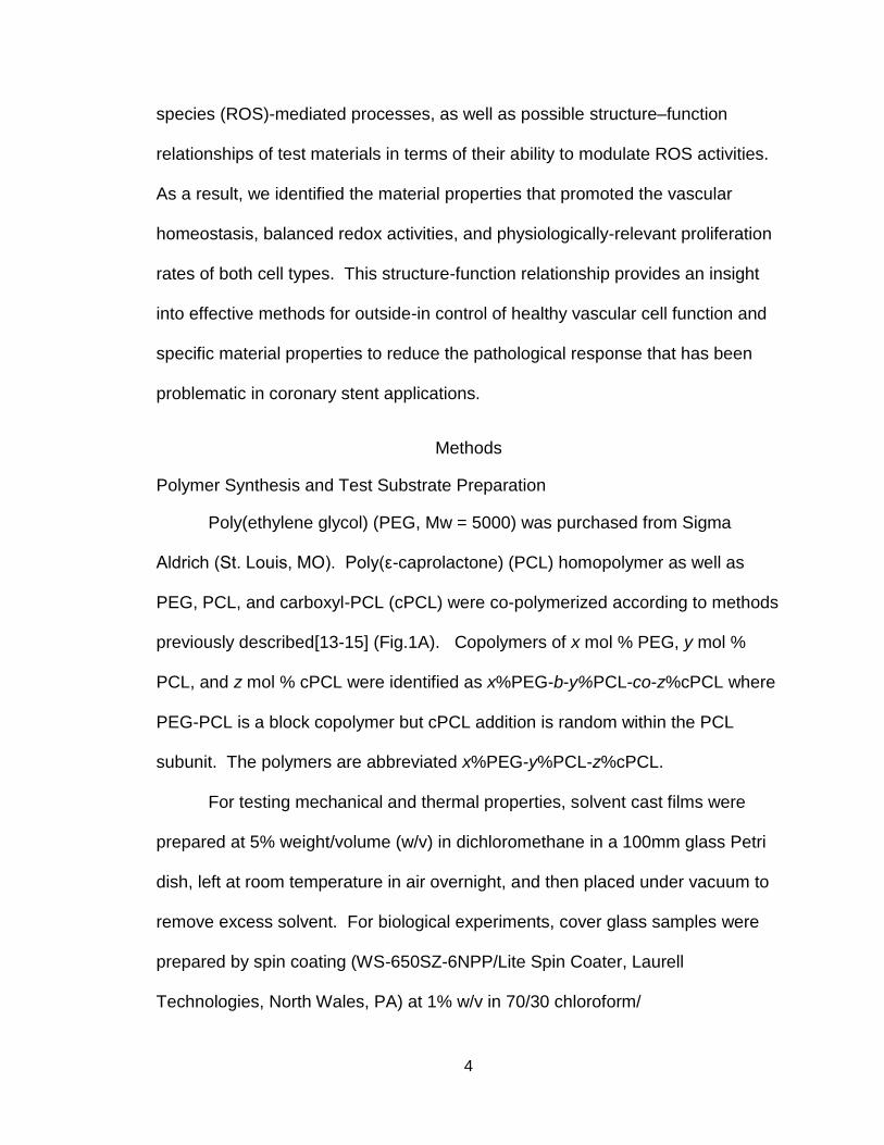
previously described[13-15] (Fig.1A). Copolymers of x mol % PEG, y mol %
PCL, and z mol % cPCL were identified as x%PEG-b-y%PCL-co-z%cPCL where
PEG-PCL is a block copolymer but cPCL addition is random within the PCL
subunit. The polymers are abbreviated x%PEG-y%PCL-z%cPCL.
For testing mechanical and thermal properties, solvent cast films were
prepared at 5% weight/volume (w/v) in dichloromethane in a 100mm glass Petri
dish, left at room temperature in air overnight, and then placed under vacuum to
remove excess solvent. For biological experiments, cover glass samples were
prepared by spin coating (WS-650SZ-6NPP/Lite Spin Coater, Laurell
Technologies, North Wales, PA) at 1% w/v in 70/30 chloroform/
5
dimethylformamide onto 15mm glass cover slips at 4,000 RPM for 30 seconds.
Samples were placed under vacuum for at least two days before use. Samples
for cellular interaction experiments were sterilized under UV light for 1 hour.
Thermal and Mechanical Properties
Differential scanning calorimetry (DSC, Q1000, TA Instruments, New
Castle, DE) was performed with sample mass between 5 and 10 mg in aluminum
pans with tops. The procedure included two runs from -80°C to 100°C with a
ramp rate of 10°C/minute. The values from the second run were reported such
that thermal history was erased (n = 3). Thermogravimetric analysis (TGA-1000,
Figure 1. Polymers synthesis. Schematic representation of polymer synthesis. The random copolymer is identified as x%PEG-b-y%PCL-co-z%cPCL where x, y, and z represent the respective molar ratio. PCL-PEG is a block copolymer, but the addition of cPCL is random within the PCL subunit. The polymers are abbreviated x%PEG-y%PCL-z%cPCL.

6
Instrument Specialist Inc., Twin Lakes, WI) was performed using a heating rate
of20 oC/min to a final temperature of 600 oC.
Dynamic mechanical analysis (DMA, Q800 DMA, TA Instruments, New
Castle, DE) was performed with samples that were soaked in dH2O at 37°C for 2
days prior to testing. Wet stress and strain were recorded using a submersion
clamp containing dH2O at room temperature. A preload force of 0.1 N was
applied to each sample and force was increased at a rate of 0.1 N/minute until
failure. The average Young’s Modulus was measured (n = 3). For temperature
sweeps, a tension clamp was used with dry samples in air. The procedure
included two runs from -80°C to 50°C with a ramp rate of 20 °C/minute and a
strain of 30μm at 1Hz. All values were calculated using Universal Analysis
software provided by TA Instruments.
Molecular Weight and Degradation
Gel permeation chromatography (GPC, Shimadzu Corp., Kyoto, Japan)
with an inline Wyatt miniDAWN TREOS light scattering detector (Wyatt
Technology Corp., Santa Barabara, CA) was used to measure Mn based upon
dn/dC for each polymer type (n=4). Degradation properties of polymers were
characterized by measuring Mn over time (i.e., 0, 4, 7, and 28 days) after
incubation of polymer samples in phosphate buffer saline (PBS) at 37°C.
7
Surface Chemical Properties
The sessile drop method was used to measure contact angle with an in-
house goniometer. One 10 μL drop of dH2O was placed on each solvent-cast
film, pictures were taken immediately, and the angles on both sides of the drop
were measured to represent “dry” contact angles. Samples were then incubated
with dH2O drops for 2 hours at 37°C and measurements were taken to represent
“wet” contact angles. All contact angles were analyzed through imaging and
image analysis using ImageJ software (National Institutes of Health, Bethesda,
MD) (n = 3).
In order to characterize the negative surface charge driven by free
carboxyl groups of cPCL, polymer-coated cover glasses were incubated with 1%
v/v carboxylate-terminated, fluorescence-conjugated polystyrene microspheres
(Sigma) in water overnight at 37°C. Carboxyl groups generate a negative charge
on the microsphere surface and microspheres are therefore repelled more by the
polymer surface as cPCL % increases. Test samples were washed three times to
remove repelled microspheres from the test surfaces and the fluorescence
intensity of remaining microspheres on the test sample was measured with a
plate reader (infinite F500, Tecan Group Ltd., Mannedorf, Switzerland) (n = 4).
Cell Culture
Human coronary artery vascular smooth muscle cells (HCASMCs,
passages 6-8) were cultured in Dulbecco’s Modified Eagle’s Medium (DMEM,
Gibco Cell Culture, Carlsbad, CA) supplemented with10% heat-inactivated fetal
8
bovine serum (FBS, Gibco), 1% penicillin-streptomycin (Gibco), and 1% L-
glutamine (Gibco). Human coronary artery endothelial cells (HCAECs, passage
7) were cultured in MesoEndo Growth Medium (Cell Applications, Inc., San
Diego, CA) supplemented with 10% FBS and 1% penicillin-streptomycin. Cells
were purchased from Cell Applications, Inc. (San Diego, CA). Cells were
cultured for three days on test polymer samples before end point experiments.
Immunofluorescence Staining
Cells were fixed in 4% paraformaldehyde (Sigma) in dH2O and incubated
with primary antibody (1:100) overnight at 4°C. To measure proliferation, cells
were incubated with 5-bromo-2’-deoxyuridine (BrdU, Sigma) at 20 μM for 16
hours. Incorporated BrdU in proliferating cells were detected by staining with
primary rat anti-human BrdU antibodies (Abcam, Cambridge, MA), followed by
addition of secondary DyLight594-conjugated goat anti-rat (Jackson
Immunoresearch, West Grove, PA) antibodies. To evaluate a healthy contractile
phenotype in HCASMCs expression of smooth muscle myosin heavy chain
(smMHC) was detected by staining with primary mouse anti-human smMHC
antibodies (Abcam), followed by addition of secondary TRITC-conjugated goat
anti-mouse (Abcam) antibodies. To evaluate inflammatory action of HCAECs,
expression of vascular cell adhesion molecule (VCAM)-1 was detected by
staining with APC-conjugated anti-human VCAM-1 antibodies (CD 106,
BioLegend, San Diego, CA). Cell nuclei were counterstained with Hoechst
33258 (Sigma) in all the aforementioned types of fluorescence staining. Cells
9
were imaged under a Nikon Eclipse Ti inverted fluorescence microscope (Nikon
Instruments Inc, Melville, NY). Relative protein expression was quantified by
measuring fluorescence intensity from antibody staining, which was normalized
to cell number from Hoechst nucleus staining using ImageJ. Cell proliferation
was presented by the percent of BrdU-positive cells in the total number of cells
(%) (n=12 pictures/polymer).
Cell Activities
To measure intracellular superoxide and hydrogel peroxide, cells were
incubated with dihydroethidium (DHE, Invitrogen) and dichlorofluorescein
diacetate (DCFDA, Invitrogen), respectively for 30 minutes at 5 μg/mL following
the previously reported method [4]. To measure cell viability, cells were stained
with Calcein AM (Invitrogen, 1 μg/mL). All cells were counterstained with
Hoechst nucleus staining (5 μg/mL) to measure the total number of cells.
Fluorescence intensity of each staining (i.e., DHE, DCFDA, and Calcein AM) was
measured with a plate reader (Tecan) and were normalized to the corresponding
cell number. To measure total protein content, cells were lysed, proteins were
harvested and quantified by a colorimetric assay (BioRad, Hercules, CA). For
morphological analysis, HCASMC were stained with Texas Red-X phalloidin
(Invitrogen) and cell circularity was measured using ImageJ (n = 80) [16].
Degree of circularity is a 0-10 scale defined as 0 being an elongated morphology
and 10 representing a perfect circle.
10
Statistical Analysis
In all experiments, results are presented as means ± standard error mean
(SEM). Comparisons between individual sample groups were performed using
an unpaired Student’s t-test. For all statistics, p < 0.05 was considered
statistically significant.
Results
Synthesis
A subset of six polymers were synthesized, characterized and evaluated for
cellular interaction: 100%PCL, 4%PEG-96%PCL, 8%PEG-92%PCL, 4%PEG-
86%PCL-10%cPCL, and 8%PEG-82%PCL-10%cPCL. PCL polymers were
synthesized by ring-opening polymerization and, in the case of copolymers, were
extended from PEG (Fig. 1A). Terpolymers (e.g. polymers containing all three
subunits) were synthesized according to the method reported previously [13], but
with x%PEG-b-y%PCL as the starting material. Mn values ranged from 60 –
140kDa, as determined by GPC.
Degradation
Mn was measured by GPC following incubation of each polymer sample in
PBS at 37 C at the indicated time points over 28 days. The Mn of 100%PCL
decreased by ~13% at 28 days post incubation (Fig. 1B). With the addition of
10%cPCL to the PCL homopolymer (i.e., 90%PCL-10%cPCL), the polymer
degraded more quickly (~19%) and this effect was enhanced (~25%) upon
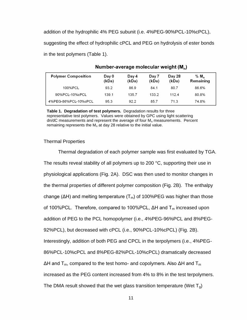
11
addition of the hydrophilic 4% PEG subunit (i.e. 4%PEG-90%PCL-10%cPCL),
suggesting the effect of hydrophilic cPCL and PEG on hydrolysis of ester bonds
in the test polymers (Table 1).
Thermal Properties
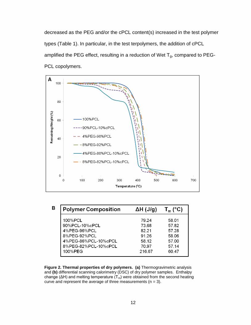
Thermal degradation of each polymer sample was first evaluated by TGA.
The results reveal stability of all polymers up to 200 °C, supporting their use in
physiological applications (Fig. 2A). DSC was then used to monitor changes in
the thermal properties of different polymer composition (Fig. 2B). The enthalpy
change (ΔH) and melting temperature (Tm) of 100%PEG was higher than those
of 100%PCL. Therefore, compared to 100%PCL, ΔH and Tm increased upon
addition of PEG to the PCL homopolymer (i.e., 4%PEG-96%PCL and 8%PEG-
92%PCL), but decreased with cPCL (i.e., 90%PCL-10%cPCL) (Fig. 2B).
Interestingly, addition of both PEG and CPCL in the terpolymers (i.e., 4%PEG-
86%PCL-10%cPCL and 8%PEG-82%PCL-10%cPCL) dramatically decreased
ΔH and Tm, compared to the test homo- and copolymers. Also ΔH and Tm
increased as the PEG content increased from 4% to 8% in the test terpolymers.
The DMA result showed that the wet glass transition temperature (Wet Tg)
Table 1. Degradation of test polymers. Degradation results for three representative test polymers. Values were obtained by GPC using light scattering dn/dC measurements and represent the average of four Mn measurements. Percent remaining represents the Mn at day 28 relative to the initial value.
12
decreased as the PEG and/or the cPCL content(s) increased in the test polymer
types (Table 1). In particular, in the test terpolymers, the addition of cPCL
amplified the PEG effect, resulting in a reduction of Wet Tg, compared to PEG-
PCL copolymers.
Figure 2. Thermal properties of dry polymers. (a) Thermogravimetric analysis and (b) differential scanning calorimetry (DSC) of dry polymer samples. Enthalpy change (ΔH) and melting temperature (Tm) were obtained from the second heating curve and represent the average of three measurements (n = 3).
13
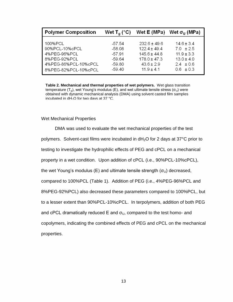
Wet Mechanical Properties
DMA was used to evaluate the wet mechanical properties of the test
polymers. Solvent-cast films were incubated in dH2O for 2 days at 37°C prior to
testing to investigate the hydrophilic effects of PEG and cPCL on a mechanical
property in a wet condition. Upon addition of cPCL (i.e., 90%PCL-10%cPCL),
the wet Young’s modulus (E) and ultimate tensile strength (σU) decreased,
compared to 100%PCL (Table 1). Addition of PEG (i.e., 4%PEG-96%PCL and
8%PEG-92%PCL) also decreased these parameters compared to 100%PCL, but
to a lesser extent than 90%PCL-10%cPCL. In terpolymers, addition of both PEG
and cPCL dramatically reduced E and σU, compared to the test homo- and
copolymers, indicating the combined effects of PEG and cPCL on the mechanical
properties.
Table 2. Mechanical and thermal properties of wet polymers. Wet glass transition temperature (Tg), wet Young’s modulus (E), and wet ultimate tensile stress (σU) were obtained with dynamic mechanical analysis (DMA) using solvent casted film samples incubated in dH2O for two days at 37 °C.

14
Water Contact Angle and Surface Charge
Using the sessile drop method, water contact angle was assessed to
evaluate the ability of PEG and cPCL subunits to modulate surface hydrophilicity
(Fig. 3A). The “dry” sessile contact angle was not significantly different among
the test polymer types. Following incubation for 2 hours at 37 °C with 95%
humidity, contact angles decreased noticeably, compared to dry sessile contact
angles. In particular, the “wet” contact angle decreased as the cPCL and/or PEG
content increased in the test polymer types, indicating the contributions of the
PEG and cPCL hydrophilic subunits. The wet contact angles were 0° on the test
terpolymers, indicating that the test surfaces fully absorbed the drop.
Relative negative surface charge was evaluated by employing the concept
of charge-charge repulsion between free carboxyl groups on microspheres and
Figure 3. Surface chemistry. (a) Advancing contact angle analysis for dry and wet polymer film surfaces (n=3). Wet polymer samples were incubated with one drop of dH2O for 2 hours at 37°C. (b) Polymer-coated glass cover slips were incubated with carboxylate-terminated, fluorescence-conjugated polystyrene microspheres to evaluate surface chemistry. Negative surfaces exhibit a lower fluorescence signal due to charge repulsion (n = 4).
15
the polymer surface. A strong fluorescence intensity indicates a surface with a
low negative charge due to reduced charge-charge repulsion. The relative
fluorescence intensity of polymer-coated cover glass was measured following
overnight incubation with carboxylate-terminated fluorescent microspheres (Fig.
3B). The 100%PCL surface exhibited the highest fluorescence intensity,
indicating the least negative charge. The addition of cPCL reduced the
fluorescence intensity significantly, as compared to the test polymers that do not
contain cPCL, confirming the presence of surface charge derived from cPCL.
The lowest fluorescence intensities were observed for the test terpolymers,
indicating an additional repellent effect of PEG.
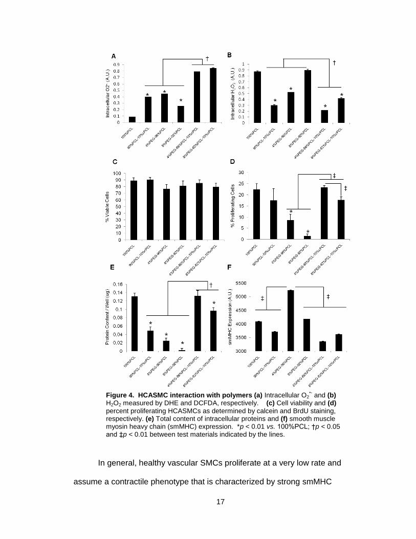
Cellular Interaction
- HCASMC Response
To determine the optimum composition of polymers for coronary stent
applications, responses of HCASMCs to test polymers were investigated first.
Balance of cellular oxidative mechanisms is crucial for maintaining vascular
homeostasis and preventing pathogenesis [17-21]. Therefore, we measured
intracellular reactive oxygen species (ROS) levels, specifically superoxide (O2•-)
and hydrogen peroxide (H2O2), and found them to vary significantly between test
polymer types (Fig. 4A-B). Intracellular O2•- levels correlated inversely with
intracellular H2O2 levels except in the case of 4%PEG-96%PCL (Fig. 4A). For
example, 8%PEG-92%PCL displayed a low level of O2•-, but a high level of H2O2;
conversely, terpolymer test samples displayed the highest levels of O2•- and the
16
lowest levels of H2O2. In the test co- and terpolymers, increasing PEG molar
ratios resulted in higher intracellular H2O2 levels, but this effect was counteracted
by the addition of negatively-charged cPCL (Fig. 4B). HCASMCs grown on the
test terpolymers showed statistically significant differences in both H2O2 and O2•-
levels relative to other test copolymers (p < 0.05).
HCAMSCs in all polymer groups maintained viability (> 70%), but
proliferation varied significantly (Fig. 4C-D). Percentages of proliferating
HCASMCs correlated proportionally with total protein contents for each group
(Fig. 4d-e), indicating that protein synthesis is required for cells to undergo
proliferation. Cells grown on terpolymers resulted in significantly different
percentages of proliferation and total protein content (p < 0.05). Interestingly,
proliferation percentages correlated inversely with intracellular H2O2 levels except
in the case of 100%PCL (Fig. 4A).
17
In general, healthy vascular SMCs proliferate at a very low rate and
assume a contractile phenotype that is characterized by strong smMHC
Figure 4. HCASMC interaction with polymers (a) Intracellular O2•- and (b)
H2O2 measured by DHE and DCFDA, respectively. (c) Cell viability and (d) percent proliferating HCASMCs as determined by calcein and BrdU staining, respectively. (e) Total content of intracellular proteins and (f) smooth muscle myosin heavy chain (smMHC) expression. *p < 0.01 vs. 100%PCL; †p < 0.05 and ‡p < 0.01 between test materials indicated by the lines.
18
expression, and a spindle-like morphology [22, 23]. In contrast, unhealthy,
“dedifferentiated” SMCs assume a circular cobble stone-like, synthetic phenotype
in which smMHC expression is significantly downregulated [22]. In order to test
the ability of the different polymers to discourage a phathogenic, synthetic
phenotype, we stained for smMHC and found that 4%PEG-96%PCL promoted a
statistically greater level of smMHC expression in HCASMCs relative to all other
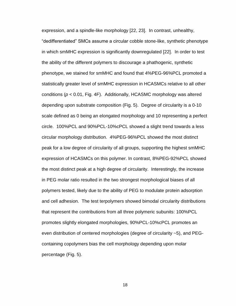
conditions (p < 0.01, Fig. 4F). Additionally, HCASMC morphology was altered
depending upon substrate composition (Fig. 5). Degree of circularity is a 0-10
scale defined as 0 being an elongated morphology and 10 representing a perfect
circle. 100%PCL and 90%PCL-10%cPCL showed a slight trend towards a less
circular morphology distribution. 4%PEG-96%PCL showed the most distinct
peak for a low degree of circularity of all groups, supporting the highest smMHC
expression of HCASMCs on this polymer. In contrast, 8%PEG-92%PCL showed
the most distinct peak at a high degree of circularity. Interestingly, the increase
in PEG molar ratio resulted in the two strongest morphological biases of all
polymers tested, likely due to the ability of PEG to modulate protein adsorption
and cell adhesion. The test terpolymers showed bimodal circularity distributions
that represent the contributions from all three polymeric subunits: 100%PCL
promotes slightly elongated morphologies, 90%PCL-10%cPCL promotes an
even distribution of centered morphologies (degree of circularity ~5), and PEG-
containing copolymers bias the cell morphology depending upon molar
percentage (Fig. 5).

19
- HCAEC Response
We then evaluated the responses of HCAECs to test polymer substrates.
Vascular smooth muscle cells and endothelial cells comprise the majority of the
vasculature and understanding how polymer properties can modulate the
response of each cell type differently is critical to designing a vascular stent
Figure 5. HCASMC morphology. (a) Histograms of circularity distribution for HCASMCs grown on test substrates (n = 80 cells analyzed / polymer). Degree of circularity ranges from 0 (elongated) to 10 (perfect cirle). (b) Fluorescence images of HCASMCs stained with phalloidin (red) and Hoechst (blue) on four different polymer types. Scale bars = 100 μm.
20
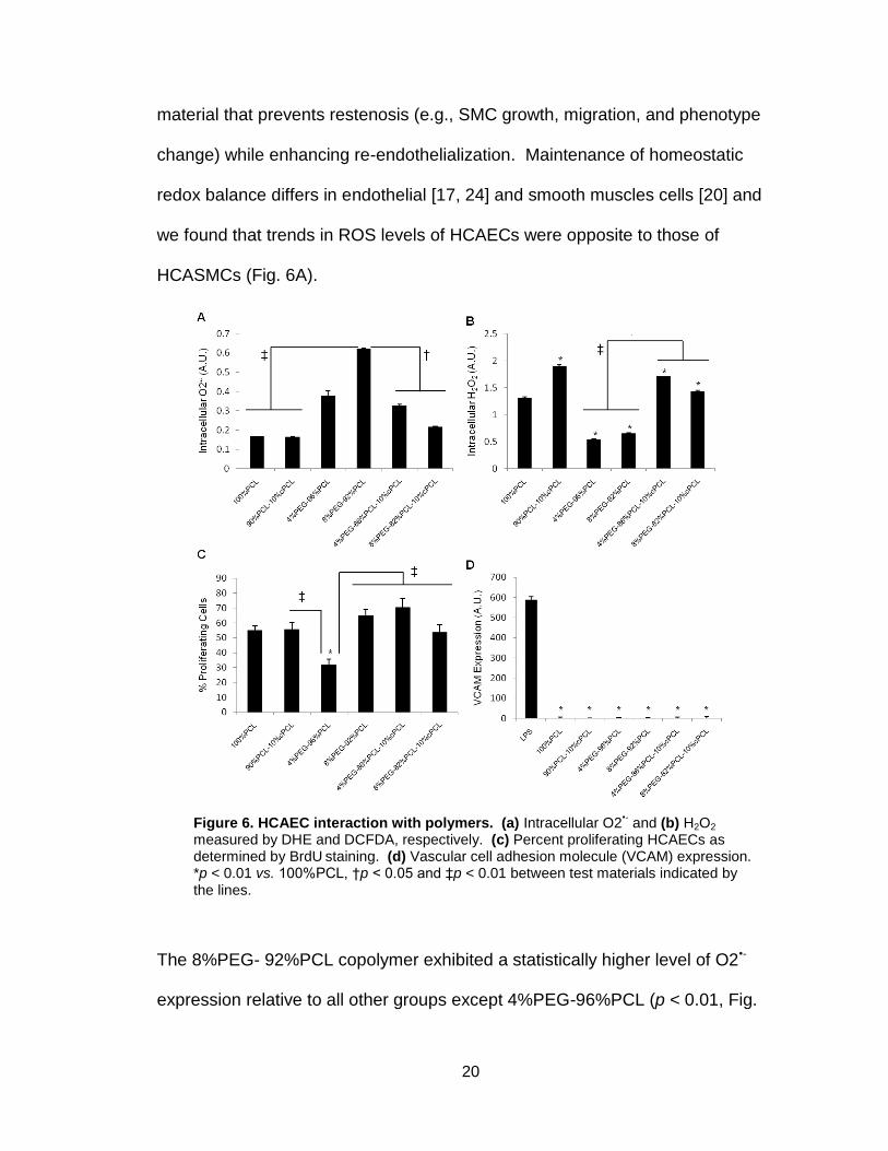
material that prevents restenosis (e.g., SMC growth, migration, and phenotype
change) while enhancing re-endothelialization. Maintenance of homeostatic
redox balance differs in endothelial [17, 24] and smooth muscles cells [20] and
we found that trends in ROS levels of HCAECs were opposite to those of
HCASMCs (Fig. 6A).
The 8%PEG- 92%PCL copolymer exhibited a statistically higher level of O2•-
expression relative to all other groups except 4%PEG-96%PCL (p < 0.01, Fig.
Figure 6. HCAEC interaction with polymers. (a) Intracellular O2•- and (b) H2O2
measured by DHE and DCFDA, respectively. (c) Percent proliferating HCAECs as determined by BrdU
staining. (d) Vascular cell adhesion molecule (VCAM) expression.
*p < 0.01 vs. 100%PCL, †p < 0.05 and ‡p < 0.01 between test materials indicated by the lines.
21
6A). Intracellular H2O2 levels of HCAECs correlated inversely with O2•-(Fig. 6B),
which was similar to the trends seen in HCASMCs (Fig. 4A and B). HCAEC
proliferation was high (> 50%) in all groups except 4%PEG-96%PCL (20-30%),
which was significantly lower than 100%PCL (p < 0.01) and all other groups (p <
0.05, Fig. 6C). To evaluate if any of the polymers stimulate pro-inflammatory
activation of HCAECs, VCAM-1 expression of HCAECs was measured on test
polymers. The condition with lipopolysaccharide (LPS) treatment was used as a
control (Fig. 6D). VCAM-1 expression in ECs is stimulated by pathoglogical cues,
resulting in recruitment of inflammatory cells but its expression is extremely low
in normal ECs [25]. We found that none of the polymers stimulated VCAM
expression, thereby further supporting their applicability as biocompatible
cardiovascular biomaterials, in particular as vascular stent materials (Fig. 6D).
Discussion
The purpose of this study was to synthesize and characterize a new class of
copolymers with tunable mechanical and physical properties for biomedical
applications, particularly coronary artery stents. Poly(ε-caprolactone) (PCL) was
used as the primary component due to its biocompatibility, hydrophobicity, and
slow degradation rate [2, 8, 9, 26]; poly(ethylene glycol) (PEG) to promote
hydrophilicity, water absorption and to modulate repulsion of proteins and cells
[4]; and carboxyl-PCL (cPCL) for increased hydrophilicity and exposure of a
negative surface charge that was found to reduce the repellent effect of PEG [4,
13]. The three subunits exhibit distinct characteristics and, when copolymerized
22
at varying molar ratios, the resulting polymer can be tailored to modulate cellular
response. Investigations with two primary human vascular cell types revealed
the structure-function relationships that control cell proliferation and phenotypic
changes in a reactive-oxygen species (ROS)-dependent manner.
PCL is a semi-crystalline, hydrophobic polymer with slow degradation
kinetics in aqueous environments [8, 9]. Addition of the random hydrophilic cPCL
subunit or PEG block improved water absorption, facilitated hydrolytic cleavage,
and resulted in a faster degradation rate (Fig. 1B). In dry conditions, adding
cPCL reduced enthalpy change and melting temperature due to decreased chain
regularity (Fig. 2B). Inclusion of the PEG block, however, increased these values
by promoting chain packing and crystallinity, which is supported by the values for
100%PEG. In wet conditions, addition of either cPCL or PEG reduced glass
transition temperature (Tg), Young’s modulus (E), and ultimate tensile strength
(σU) (Table 1). This effect was synergistically amplified in the test terpolymers
due to the increased water absorption by the hydrophilic subunits which
decreased crystallinity, weakened the overall polymer structure, and reduced
mechanical properties. Additionally, polymer surfaces composed of increasing
molar ratios of PEG and cPCL proportionally absorbed more water, as seen by
contact angle (Fig. 3A). Surface charge analysis using carboxylate-terminated
microspheres further confirmed these results (Fig. 3B). A decrease in
fluorescence signal (i.e. more negative charge) was observed for test samples
containing PEG and/or cPCL. The rise in fluorescence intensity for 8%PEG
polymers relative to 4%PEG can be explained by the formation of hydrogen
23
bonds between the carboxylate-terminated microspheres and the dense block of
ether groups at the surface [27, 28].
Homeostasis of vascular SMCs and endothelial cells (VEC) is dependent
upon effective redox mechanisms [22]. Imbalances in ROS, such H2O2 and O2•-,
result in abnormal cell proliferation, changes in phenotype, and the progression
of cardiovascular diseases, including hypertension, atherosclerosis, and
restenosis [19, 21, 29, 30]. VSMCs and VECs produce ROS at a basal level that
is crucial for cell signaling. However, excess ROS, either from intracellular
production or those released by inflammatory cells in pathological states, have
been implicated in disease progression, specifically causing the “dedifferentiation”
of VSMCs [22] and hyperproliferation of VECs [17]. Interestingly, H2O2 and O2•-
have been shown to stimulate opposite responses in VSMC and EC: VSMC
proliferation is promoted by H2O2 and arrested by O2•- [20], but EC proliferation is
stimulated by O2•- and arrested by H2O2 [17, 24]. Healthy VSMCs are
differentiated, quiescent, and maintain a contractile phenotype, which is
characterized by an elongated spindle-like morphology and expression of
smMHC [18, 22]. However, unhealthy VSMCs dedifferentiate, proliferate,
assume a synthetic phenotype, a circular morphology, and display low smMHC
expression. Healthy VECs proliferate slowly in vivo and hyperproliferation of
VECs can lead to vessel blockage [24, 31]. These competitive signaling events
reveal a complex framework in which maintenance of two healthy cell types
requires a specific ROS balance. We therefore investigated the role of these
polymers in modulating intracellular ROS production, cell proliferation, and

24
phenotypic changes in HCASMCs and HCAECs, and identified structure-function
relationships that provide healthy versus pathological environments.
As seen in Figure 4, HCASMC intracellular ROS varied dramatically in
response to different polymer substrates. In most groups, a high level of O2•-
correlated with a low level of H2O2, indicating the activity of superoxide dismutase
(SOD) in converting O2•- to H2O2 [24]. PEG-containing substrates have been
shown to promote intracellular H2O2 production, as seen for 4%PEG-96%PCL
and 8%PEG-92%PCL (Fig. 4B) [3, 4]. In the test terpolymers, the addition of
cPCL counteracted the effects of PEG and resulted in the lowest H2O2 levels.
Although cells on all polymers maintained intact cell viability (Fig. 4C),
differences in proliferation rates and total protein content reflect the ability of O2•-
and H2O2 to promote and arrest HCASMC proliferation, respectively (Fig. 1D-E).
In most groups, increasing levels of H2O2 corresponded with decreased
proliferation rate and protein content whereas O2•- levels correlated with
increased proliferation. However, HCASMC on 100%PCL showed high
intracellular H2O2 and low intracellular O2•-, yet maintained a high proliferation
rate and protein content. Because this is the only purely hydrophobic substrate,
we believe that the cell adhesive surface of hydrophobic 100%PCL promoted
proliferation of this mesenchymal cell type and that this effect was more
influential than the competing ROS-induced arrest of proliferation [2].
Expression of smMHC was statistically higher in HCASMCs grown on
4%PEG-96%PCL relative to all other groups (Fig. 4F). Morphological analysis
revealed that cells on 4%PEG-96%PCL showed the strongest bias towards an
25
elongated spindle-like phenotype, which is a typical phenotype of healthy
HCASMCs (Fig. 5). Interestingly, in these cells, both intracellular ROS types
stayed within a moderate range that, along with strong smMHC expression,
suggests an effective redox balance that maintained a healthy contractile
phenotype. In contrast, cells grown on the test terpolymer substrates displayed
high levels of intracellular O2•- coupled with sustained proliferation rates and the
lowest levels of smMHC expression in HCASMCs. Cell circularity of HCASMCs
on the test terpolymers assumed a bimodal distribution, suggesting a
heterogeneous population of contractile and synthetic phenotypes on these
surfaces (Fig. 5). HCAMSCs on 8%PEG-92%PCL had high intracellular H2O2
and reduced proliferation, but low protein content (Fig. 4B-D), indicating that the
repellent effect of PEG prevented cell attachment and cell-matrix interaction.
Together these results indicate that 4%PEG-96%PCL is the most appropriate
substrate to maintain a healthy HCASMC phenotype, as characterized by strong
smMHC expression, a low proliferation rate, and an effective redox balance.
We then assessed the response of HCAEC on polymer substrates in order
to understand how the different polymeric subunits can influence the two cell
types differently. By monitoring the response of two complementary coronary
artery cell types, we can identify the most appropriate polymer(s) for a coronary
stent application. We found that trends in intracellular ROS levels of HCAEC
were opposite to those of HCASMC with respect to the substrate composition
(Fig. 6A-B). Intracellular H2O2 was minimized on 4%PEG-96%PCL and 8%PEG-
92%PCL whereas intracellular O2•- was increased. HCAECs on the test
26
terpolymers displayed high levels of O2•- and low levels of H2O2. Proliferation
rates agreed with intracellular ROS patterns except for HCAECs on 8%PEG-
92%PCL, which displayed high intracellular O2•- and low H2O2, but maintained a
high proliferation rate (~65%). As with HCASMC, the 8%PEG-92%PCL exhibited
a strong repellent effect that prevented cell-matrix interactions, but for HCAECs,
the dominant cell-cell interactions promoted proliferation in an ROS-independent
manner. HCAECs grown on all polymers except 4%PEG-96%PCL showed
proliferation rates in a pathophysiological range ( >40%), making them less ideal
for cardiovascular application. Due to intermediate production of ROS and
maintenance of a physiologically-relevant proliferation rate, 4%PEG-96%PCL is
the most appropriate material for applications that require contacts with HCAECs
in vivo.
Polymer substrates were shown to modulate cellular activity in an ROS-
dependent manner. Analysis of the relationships between polymer properties
and the subsequent cellular response revealed structure-function relationships
that may provide insight into manipulating cellular activities by exploiting specific
polymer characteristics. Intracellular H2O2 in HCASMCs followed the same trend
as the polymer mechanical properties (e.g. E and σU) whereas intracellular O2•-
followed the same trend as the surface charge. We believe that increased
surface charge stimulates outside-in transport of O2•- that results in an abundance
of intracellular O2•-. Because production of H2O2 reflects HCASMC proliferation
potential and HCASMC are normally quiescent , stiffer substrates likely resemble
a pathophysiological environment that promotes proliferation, such as that found
27
in atherosclerosis [32]. For interactions with HCASMC, 4%PEG-96%PCL
provided the best substrate because of its moderate surface charge and
mechanical properties, as well as promotion of a balanced ROS production. This
combination of properties promoted a healthy, contractile phenotype. Cellular
behavior can also be modulated by PEG domains at the polymer surface, such
as with 8%PEG-92%PCL. The HCASMC from the mesenchymal lineage are
cell-matrix interactive and did not grow well on this substrate; however, the
HCAEC from the endothelial lineage are cell-cell interactive and flourished. Both
of these results disagreed with the trends of all other groups and therefore
provide an interesting insight to control competitive cell-cell versus cell-matrix
interactions.
Conclusion
In this study, we have synthesized and characterized a new class of
copolymers with tunable properties for biomedical applications. Polymer
properties are influenced by the molar ratios of the individual subunits and, by
varying their specific contributions, the resulting bulk and surface properties can
be controlled. Investigations of the responses of two primary human coronary
artery cell types revealed an ability of the substrate to modulate intracellular
reactive oxygen species, which directed changes in phenotype and related
cellular activities of the test cell types. Our study indicates that the polymer
composition-dependent material properties can directly influence cellular
response, in this case through ROS-mediated proliferation and phenotypic
28
changes. Therefore, by expanding our class of polymers, we will develop new
methods to control cell behavior and health by modulating outside-in cell
signaling.
29
REFERENCES
1. Lutolf MP, Hubbell JA. Synthetic biomaterials as instructive extracellular microenvironments for morphogenesis in tissue engineering. Nat Biotechnol 2005 Jan;23(1):47-55. 2. Sung HJ, Su J, Berglund JD, Russ BV, Meredith JC, Galis ZS. The use of temperature-composition combinatorial libraries to study the effects of biodegradable polymer blend surfaces on vascular cells. Biomaterials 2005 Aug;26(22):4557-4567. 3. Sung HJ, Chandra P, Treiser MD, Liu E, Iovine CP, Moghe PV, et al. Synthetic polymeric substrates as potent pro-oxidant versus anti-oxidant regulators of cytoskeletal remodeling and cell apoptosis. J Cell Physiol 2009 Mar;218(3):549-557. 4. Sung H-J, Luk A, Murthy S, Liu E, Jois M, Joy A, et al. Poly(ethylene glycol) as a sensitive regulator of cell survival fate on polymeric biomaterials: the interplay of cell adhesion and pro-oxidant signaling mechanisms. Soft Matter 2010;6(20):5196-5205. 5. Mani G, Feldman MD, Patel D, Agrawal CM. Coronary stents: A materials perspective. Biomaterials 2007 Mar;28(9):1689-1710. 6. Finn AV, Nakazawa G, Joner M, Kolodgie FD, Mont EK, Gold HK, et al. Vascular responses to drug eluting stents - Importance of delayed healing. Arteriosclerosis Thrombosis and Vascular Biology 2007 Jul;27(7):1500-1510. 7. Onuma Y, Ormiston J, Serruys PW. Bioresorbable Scaffold Technologies. Circulation Journal 2011 Mar;75(3):509-520. 8. Zhang LL, Xiong CD, Deng XM. Biodegradable polyester blends for biomedical application. J Appl Pol Sci 1995;56:103-122. 9. Tang ZG, Black RA, Curran JM, Hunt JA, Rhodes NP, Williams DF. Surface properties and biocompatibility of solvent-cast poly[-caprolactone] films. Biomaterials 2004 Aug;25(19):4741-4748. 10. Bergstrom K, Holmberg K, Safranj A, Hoffman AS, Edgell MJ, Kozlowski A, et al. Reduction of fibrinogen adsorption on PEG-coated polystyrene surfaces. J Biomed Mater Res 1992 Jun;26(6):779-790. 11. Deschamps AA, van Apeldoorn AA, Hayen H, de Bruijn JD, Karst U, Grijpma DW, et al. In vivo and in vitro degradation of poly(ether ester) block copolymers based on poly(ethylene glycol) and poly(butylene terephthalate). Biomaterials 2004 Jan;25(2):247-258. 12. Sun G, Zhang XZ, Chu CC. Effect of the molecular weight of polyethylene glycol (PEG) on the properties of chitosan-PEG-poly(N-isopropylacrylamide) hydrogels. J Mater Sci Mater Med 2008 Aug;19(8):2865-2872. 13. Gimenez S, Ponsart S, Coudane J, Vert M. Synthesis, properties and in vitro degradation of carboxyl-bearing PCL. Journal of Bioactive and Compatible Polymers 2001 Jan;16(1):32-46. 14. Dong CM, Qiu KY, Cu ZW, Feng XD. Synthesis of star-shaped poly(epsilon-caprolactone)-b-poly(DL-lactic acid-alt-glycolic acid) with multifunctional initiator and stannous octoate catalyst. Macromolecules 2001 Jul 3;34(14):4691-4696. 15. Sosnik A, Cohn D. Poly(ethylene glycol)-poly(epsilon-caprolactone) block oligomers as injectable materials. Polymer 2003 Nov;44(23):7033-7042. 16. Sung HJ, Eskin SG, Sakurai Y, Yee A, Kataoka N, McIntire LV. Oxidative stress produced with cell migration increases synthetic phenotype of vascular smooth muscle cells. Ann Biomed Eng 2005 Nov;33(11):1546-1554. 17. Cai H. Hydrogen peroxide regulation of endothelial function: origins, mechanisms, and consequences. Cardiovasc Res 2005 Oct 1;68(1):26-36. 18. Clempus RE, Griendling KK. Reactive oxygen species signaling in vascular smooth muscle cells. Cardiovasc Res 2006 Jul 15;71(2):216-225. 19. Cai H, Harrison DG. Endothelial dysfunction in cardiovascular diseases: the role of oxidant stress. Circ Res 2000 Nov 10;87(10):840-844.
30
20. Li PF, Dietz R, vonHarsdorf R. Differential effect of hydrogen peroxide and superoxide anion on apoptosis and proliferation of vascular smooth muscle cells. Circulation 1997 Nov 18;96(10):3602-3609. 21. Elahi MM, Kong YX, Matata BM. Oxidative stress as a mediator of cardiovascular disease. Oxid Med Cell Longev 2009 Nov-Dec;2(5):259-269. 22. Su B, Mitra S, Gregg H, Flavahan S, Chotani MA, Clark KR, et al. Redox regulation of vascular smooth muscle cell differentiation. Circ Res 2001 Jul 6;89(1):39-46. 23. Rovner AS, Murphy RA, Owens GK. Expression of smooth muscle and nonmuscle myosin heavy chains in cultured vascular smooth muscle cells. J Biol Chem 1986 Nov 5;261(31):14740-14745. 24. Zanetti M, Zwacka R, Engelhardt J, Katusic Z, O'Brien T. Superoxide anions and endothelial cell proliferation in normoglycemia and hyperglycemia. Arterioscler Thromb Vasc Biol 2001 Feb;21(2):195-200. 25. Galkina E, Ley K. Vascular adhesion molecules in atherosclerosis. Arterioscler Thromb Vasc Biol 2007 Nov;27(11):2292-2301. 26. Sung HJ, Meredith C, Johnson C, Galis ZS. The effect of scaffold degradation rate on three-dimensional cell growth and angiogenesis. Biomaterials 2004 Nov;25(26):5735-5742. 27. Pasche S, Voros J, Griesser HJ, Spencer ND, Textor M. Effects of ionic strength and surface charge on protein adsorption at PEGylated surfaces. Journal of Physical Chemistry B 2005 Sep 22;109(37):17545-17552. 28. Heymann B, Grubmuller H. Elastic properties of poly(ethylene-glycol) studied by molecular dynamics stretching simulations. Chemical Physics Letters 1999 Jul 9;307(5-6):425-432. 29. Hashimoto Y, Yoshinoya S, Aikawa T, Mitamura T, Miyoshi Y, Muranaka M, et al. Enhanced endothelial cell proliferation in acute Kawasaki disease (muco-cutaneous lymph node syndrome). Pediatric Research 1986;20(10):943-946. 30. Newby AC, Zaltsman AB. Molecular mechanisms in intimal hyperplasia. J Pathol 2000 Feb;190(3):300-309. 31. Meurice T, Vallet B, Bauters C, Dupuis B, Lablanche JM, Bertrand ME. Role of endothelial cells in restenosis after coronary angioplasty. Fundam Clin Pharmacol 1996;10(3):234-242. 32. Tajaddini A, Kilpatrick DL, Vince DG. A novel experimental method to estimate stress-strain behavior of intact coronary arteries using intravascular ultrasound (IVUS). J Biomech Eng 2003 Feb;125(1):120-123.