Embed Size (px)
Citation preview
Cornell Institute for
Biology Teachers Copyright Cornell Institute for Biology Teachers, 2000. This work may be copied by the original recipient from CIBT to provide copies for users working under the direction of the original recipient. All other redistribution of this work without the written permission of the copyright holder is prohibited.
Lab issue/rev. date: 9/30/98
Title: The Broken Heart
Authors: Mary Colvard, Cobleskill-Richmondville High School Cobleskill, New York
Dr. Marguerite Frongillo, Cornell University, Ithaca, New York
Appropriate Level:
High School Biology: Regents, General, and Honors
Abstract: Part I: The Tell Tale Heart is an activity during which students familiarize themselves with the structure of the heart. They locate the atria, ventricles, and major blood vessels. Through “surgical” procedures, students perform coronary bypass surgery and correct patent ductus arteriosus. Human and dog hearts are compared in terms of common structure and defects.
Part II: Heartworm Poster has students research the life cycle, epidemiology, and diagnostic procedures and treatment of dog heartworm. Their work culminates with them presenting the information in the form of a poster. After they have completed their work they are given supplemental information and are asked questions about heartworm disease.
Part III: In Heartworm Microfilariae students examine a stained smear of dog blood containing microfilariae and count living microfilariae in a fresh blood sample.
Special Notes:
Part I requires a heart, dissecting equipment, and soda straws.
Part II requires access to references and a computer.
Part III requires a sample of dog blood containing heartworm microfilariae (see instructor notes for ordering information).
Time Required:
Part I: “The Tell Tale Heart” - approximately 60 minutes for the procedures
Part II: “Dog Heartworm Poster” - one period to start work and then a week outside of class as homework should be adequate. After the poster is complete, the additional information and questions can be given as a homework assignment.
Part III: “Heartworm Microfilariae ” - two periods: one for making and staining blood smears containing microfilariae, and one lab for examining and counting living microfilariae.

©2000 CIBT Tell Tale Heart – Teacher’s Section page 2
National Standards:
As a result of doing this investigation, students should:
• identify questions and concepts that guide scientific investigation.
• develop an understanding of the organization of living systems.
• develop understandings about science and technology.
• develop an understanding of personal and community health.
• develop an understanding of the nature of scientific knowledge and historical perspectives.

©2000 CIBT Tell Tale Heart – Teacher’s Section page 3
Additional Teacher Information
Part I: The Tell -Tale Heart
Have students work in surgical teams of two. Each group will need a fresh or preserved mammal heart. Sheep, beef, or deer hearts are all fine. Preserved hearts are easier to use because if students do not happen to complete the “surgery” during the first lab session, the heart can be packaged and saved until the students can return and complete the investigation.
Materials:
• one pair of dissecting scissors • one pair of forceps
• one dissecting pan • one pair of latex or vinyl gloves/student
• two probes • 2 straws cut in half
• masking or lab tape to label the straws • a “needle” and 12 inches of bright thread
• one 16-cm section of narrow rubber tubing (Aquarium tubing will work, so would another soda straw if tubing is not available.)
• a reference chart of the heart (this could be a large poster which clearly illustrates the chambers and major vessels.)
The “needles” will be used when simulating the repair of an opening in the patent ductus. Size 4 fish hooks that have had the barb flattened and the point pinched off work well. If fishhooks are not easily available or fixable, Christmas ornament hangers can be used. They are not as sturdy and the “eye” which must be threaded is on the large side and should be crimped.
Part II: Heartworm Poster
The idea of placing the various components of a research paper on separate sheets and then taping them together to form a poster works well. Students feel that each piece is more “do-able” and less overwhelming. The individual sheets can also be done using a computer to enhance their appearance. Somehow it seems easier to them than if they were assigned the same topic as a typical research paper.
In addition to making the assignment more palatable, the posters can be easily folded and stored in student portfolios or lab folders. When the poster is coupled with the investigation of the impact of various levels of ivermectin, it becomes a very real project.
©2000 CIBT Tell Tale Heart – Teacher’s Section page 4
Part III: Heartworm Microfilariae
In addition to teaching biology of heartworm disease, this lab can be used to teach about nematodes, microscope skills, blood smear and cell recognition skills, quantitation calculation skills, observer error, and distribution variability.
Materials:
• Dog blood containing heartworm microfilariae is needed for both periods. A single sample can be used for both periods. We have found that the mff remain motile for almost a week if the blood is kept refrigerated. It is important to bring the sample to room temperature for about 30 minutes before using it. The blood containing heartworm mff is available from TRS Labs, 295 Research Drive, Athens, GA 30605. Tel: 706 549-0764, fax: 706-353-3590. (Billing Address for TRS Labs is PO Box 5112, Athens, GA 30604)
The cost in 1998 was $50 for 1-10 ml blood, plus $30 shipping. As the blood is perishable, it is shipped with cold packs, by FedEx, overnight priority. It comes in a 10 ml blood collecting tube containing EDTA, a chelating agent which prevents the blood from clotting.
• Stain for blood smears is needed for the first period. We used DiffQuik, made by Baxter Healthcare Corp. (Scientific Products Division, McGraw Park, Il 60085-6787.) You can order this through Fisher Scientific, 52 Fadem Road, Springfield, NJ 07081. Tel: 800 766-7000.
DiffQuik is catalog # CS430D. The cost in 1998 was $75.00 in a kit containing 500 ml of each of the 3 component solutions. Only about 50 ml of each solution is needed to stain the slides for one lab, and the remainder of the 500 ml can be saved and reused.
• Diluent for the blood is needed for the second period. To make a 1:10 dilution of the blood, we used phosphate buffered saline (PBS.) You could make this up yourself from ingredients that you may already have on hand. It consists of an aqueous solution that is:
120 millimolar NaCl 2.7 millimolar KCl 10 millimolar phosphate buffer salts
If you'd rather buy PBS ready made, it is available from Ward's Biology, PO BOX 92912, Rochester, NY 14692-9012. Tel: 800 962-2660. PBS is catalog # 37W5910 [pH 7.2]. The cost for 500 ml in 1998 was $7.55. It keeps indefinitely.
©2000 CIBT Tell Tale Heart – Teacher’s Section page 5
Background Information for Teacher
Patent Ductus Arteriosus in Dogs
The ductus arteriosus is a normal embryonic connection between the pulmonary artery and the aorta. It is located distal to the bracheocephalic artery that supplies the forelimbs, but proximal to the subclavian and other arteries supplying the rear of the body.
The most common congenital disorder of the heart occurs when the ductus fails to close. In dogs this is a polygenic inherited trait that occurs in certain breeds, and is four times more common in females than in males.
Patent ductus arteriosus serves to increase the pressure of blood in the pulmonary artery, leading to pulmonary edema. This adversely affects gas exchange in the lungs. Small breeds with PDA typically die within a year, while large breeds may live for several years with the condition. Even in large dogs, it is important to act early and surgically correct the condition before the left ventricle becomes enlarged in response to pumping more blood than normal. This enlargement causes permanent damage to lung vessels and leads to enlargement of the valve between the left atrium and ventricle, which in turn permits backwards currents of blood from the left ventricle into the left atrium, the enlargement of the left atrium, and ultimately left atrial fibrillation and death.
The younger the animal at the time of the operation, the better the reported success rate for surgical repair. PDA repair has been done successfully in puppies weighing only 0.5 kg, although such a small size makes the operation a challenge for the surgeon.
Patent Ductus Arteriosus in Humans
In the human fetus, a pair of umbilical arteries provides blood flow to the placenta. These arise from the internal iliac arteries and enter the umbilical cord. Oxygen and nutrients are brought to the fetus via blood that is returned from the placenta in the umbilical vein. This vein delivers blood to the capillaries within the developing liver and to the inferior vena cava through the ductus venosus.
The circulatory development of the fetus reflects the fact that it must extract oxygen not from inspired air but across the placenta. Also, during embryonic and fetal life, its lungs are collapsed. The capillaries are compressed and little blood flows through the lungs.
The interatrial and interventricular septa of the heart develop early. However, the interatrial partition remains functionally incomplete until the time of birth. The interatrial opening is called the foramen ovale and has a flap that acts as a valve. Blood is permitted to flow freely from the right atrium to the left atrium. Any backflow will close the valve and isolate the two chambers. Blood enters the fetal heart at the right atrium and bypasses the pulmonary circuit. Another short cut exists between the pulmonary and aortic trunks. This connection is called the ductus arteriosis and consists of a short, muscular vessel.
During diastole blood enters the right atrium and flows into the right ventricle. But about 25% of it also passes into the left atrium via the foramen ovale bypassing the pulmonary circuit. Also, over 90% of the

©2000 CIBT Tell Tale Heart – Teacher’s Section page 6
blood leaving the right ventricle passes through the ductus arteriosis and enters the systemic circuit rather than going to the lungs.
At birth, when the infant takes its first breath, the lungs expand and so do the pulmonary vessels. Blood rushes into the expanded vessels and with a few seconds, increasing oxygen levels stimulate the constriction of the ductus arteriosis. This serves to isolate pulmonary and aortic trunks. Also, as pressure rises in the left atrium, the flap closes the foramen ovale. It is permanently sealed after 48 hours. If the sealing process does not occur, the foramen remains open or patent. This causes circulation to the lungs to be reduced and blood oxygenation to be low. The newborn soon becomes starved for oxygen and cyanosis develops causing the infant to look blue. This is one factor that can produce a “blue baby.”
In adults, the interarterial septum has a shallow depression at the site of the foramen ovale. This depression is called the fossa ovalis. A fibrous cord is all that remains of the ductus arteriosis. It is referred to as the ligamentum arteriosum.
If the ductus arteriosis is not stimulated to constrict completely, a large volume of blood will bypass the lungs. This is referred to as patent ductus arteriosis and also results in cyanosis. Ventricular septal defect, atrioventricular defect, transposition of the great vessels, and a complex group of heart and circulatory defects called tetralogy of Fallot are all congenital circulatory problems that can produce a “blue baby.” This lab is primarily addressing patent ductus arteriosis since it involves students locating pulmonary vessels and the aorta.
Heartworm
This exercise is intended to reinforce students' knowledge of heart anatomy by asking students to consider the effects of adult heartworms on the pulmonary artery. addition it can be used to teach about the biology of nematodes.
Here are some key facts teachers may want to know about heartworms:
Latin Name: Dirofilaria immitis
Classification: PHYLUM NEMATODA, ORDER SPIRURIDA: SUPERFAMILY FILARIOIDEA
Distribution: Europe, North and South America (particularly in coastal areas and river valleys)
Host range: most prolific in canids; survives to adulthood in felids, ferrets; can infect primates but die as larvae in these hosts
Life cycle: Heartworm adults prefer to live in the pulmonary artery. Their offspring are prelarvae called microfilariae (mff.) These are about 308 micrometers in length, and are released into the blood to circulate throughout the body. Individual mff can remain alive and in circulation for about 2.5 years. Mosquitoes ingest mff when taking a blood meal from an infected dog. Once inside the mosquito, mff migrate to the Malpighian tubules and then to the head, developing through the first larval stages and accumulating as L3 in the salivary glands and proboscis by about 2 weeks after infection. When the mosquito takes another blood meal, these infective L3 actively migrate out of the mosquito's proboscis and into the bite wound.

©2000 CIBT Tell Tale Heart – Teacher’s Section page 7
In a dog the L3, now about 1.2 mm in length, remain in the skin a few days and then by 9 - 12 days after entering the dog molt to L4's. At this time they have reached a length of 1.6 mm. On average this stage takes an additional 60 days, during which larvae make a long migration through the body of the dog. Immature heartworms just after molting to the adult stage are about 2 cm in length. These adolescents are more vigorous migrators than the previous stages and can penetrate muscle; they move to the upper body, enter veins and migrate towards the heart. The earliest arrival at the heart is recorded as 68 days after entering the dog, but all arrive there by 90 days. Once at their final destination, they become sexually mature adults at about 6 months after entering the dog. Eventually they reach their full size, up to 28 cm. In dogs, heartworms can live about 7.5 years.
Infection route: L3 enter vertebrate host through mosquito bite wound
Location in host: adults found in pulmonary artery; in heavier infections, heart and vena cava as well
Prepatent period: (time from initial infection of the dog, until microfilariae appear in that dog's blood) 6 months
Signs of infection: Adult worms cause traumatic irritation of the linings of the vessels of the heart. The endothelial surface becomes roughened, with cells becoming disorganized allowing the vessels to become leaky; occasionally the damage is severe enough to rupture the artery. The artery wall forms villi that project into the lumen, disrupting and reducing blood flow. Similar changes in smaller arteries restrict both blood flow and the resiliency of the artery walls; villi in irritated veins also diminish venous circulation. These changes cause hypertension in the pulmonary artery and reduced blood flow to the lung, resulting in edema and inflammation.
Dogs with this congestive heart failure will cough, have trouble breathing, and will not be able to exercise. This can be fatal if not treated. However treatment itself may be dangerous as dead heartworms may also cause vessel damage. The severity of the disease is related to the size of the animal and the number and location of adult worms present. In a 25-kg dog, if there are fewer than 25 adults, they are in the pulmonary arteries in the lung, and that is where the vessel damage occurs. As the population increases, to 26 - 50 adults, some are found in the right ventricle, and with populations above 50, worms are also found in the right atrium. With further population increase, worms will be found in the vena cava. (Over 200 worms have been found in a single dog!) Such heavily infected dogs may develop caval syndrome: extreme lethargy, weakness, and concomitant hemoglobinuria, leading rapidly to death. In a 10-kg dog, as few as 36 adult worms can cause signs of dirofilariasis. In smaller animals such as ferrets, even a couple of worms could be fatal, because vessel size and heart capacity is so small.
In cats, the most severe signs occur at about 5 to 6 months after infection when young L5 reach the pulmonary artery. Tachypnea and coughing, as well as chronic otherwise unexplained vomiting, are common signs of heartworm infection in cats. As in dogs, there is worm-induced arteritis and hypertension of pulmonary arteries, but it usually does not result in heart failure. Heartworm related damage to the lung is usually due to hyperplasia of alveolar cells, leading to respiratory distress that mimics asthma. Another serious problem is emboli from dead worms which have a life span of only 3 years in cats. Such emboli can cause sudden death. Additionally, heartworm L4 can migrate aberrantly; these can be found in the CNS or in body cavities in cats more often than in dogs.
©2000 CIBT Tell Tale Heart – Teacher’s Section page 8
Antemortem Diagnosis: If diethylcarbamazine is to be started as a preventive in a dog over 6 months old, it is mandatory to test for mff. In dogs over 6 months old and not on monthly preventive, the Knott's test or other filter test demonstrating mff can be used. If the same dog is negative for mff, it can be checked for occult infection with antigen ELISA tests; adults can be seen in an echo cardiogram. In both dogs and cats radiographs are extremely useful in diagnosing heartworm disease. In cats, antigen tests have a high failure rate; instead, an ELISA test for anti-adult antibody. Note that mff are rarely seen in infected cats, because microfilaremia lasts only about 1 month in these hosts.
Postmortem Diagnosis: presence of adult worms in cardiac vessels or heart
Treatment in Dogs: Melarsomine, an arsenical, can be used with caution against adults. Thromboembolism from dead worms may cause fever, cough, hemoptysis, or right heart failure. Therefore, activity must be limited in the first month after treatment and anti-inflammatory steroids may be needed. An older arsenical, thiacetarsamide, is also available but has more adverse side effects and is harder to administer.
If dogs have caval syndrome, prompt surgical removal of the worms is recommended followed by adulticide therapy after recovery from surgery. Even in affected dogs without caval syndrome, surgical removal of worms from the pulmonary artery before administering adulticide can diminish subsequent signs of pulmonary thromboembolism.
After adulticide therapy in dogs, mff can be killed fairly rapidly with eight times the prophylactic dose of ivermectin. Also, the normal prophylactic dose of ivermectin will gradually reduce the circulating mff. The normal prophylactic dose of milbemycin will rapidly kill mff in dogs.
Treatment in Cats: Heartworm positive cats without apparent signs are given time to permit self cure, but are radiographed periodically to monitor the infection status. Lung signs associated with heartworm disease can be treated with prednisone; vomiting is eased with parenteral fluid therapy. Thiacetarsemide is used as an adulticide in cats that are in stable condition, but that have signs of dirofilariasis that can't be relieved by other therapy. Melarsomine has not yet been tested in cats.
Management: Fortunately for dogs, the L4 stage has been found to be extremely sensitive to ivermectin: 6 micrograms per kg dog body weight reliably kills heartworm L4's! The recommended dosage of ivermectin for killing L4s in cats is 4 times that for dogs. Milbemycin is also effective against this stage, but at higher doses which are the same for both dogs and cats. Either of these compounds can be given at monthly intervals during mosquito season to kill any entering larvae at the L4 stage.
©2000 CIBT Tell Tale Heart – Teacher’s Section page 9
Making a Stained Smear
Veterinarians use two forms of testing dogs' blood for heartworm. If an animal has not been protected by preventive medication with ivermectin or milbemycin, dogs first need to be checked for actual microfilariae circulating in their blood.
Filtration methods exist, but they are not as accurate as the Knott's test in differentiating between the 2 local species of filariid worms in dogs. In lieu of a Knott's test, which involves formaldehyde, we ask you to have your students make and stain blood smears. If your schedule does not allow you to do this with your students, you can make a set of stained smears ahead of time, and students can examine them in lab.
Making a blood smear:
Refer to student instructions; expect each student to try this several times before achieving a usable slide, so have plenty of slides available. If you wish you can prepare a set of slides for future use by mounting a large (22 x 30 mm or longer) coverslip on dry, stained slides. Use a resin such as Permount for mounting the coverslips.
Staining a smear with DiffQuik :
Refer to student instructions. A convenient way to get the stain set up is to pour about 50 ml of each solution into a separate 50 ml screw-cap conical tube. These are just wide enough for inserting a slide. When you're done, cap the tubes and the solutions will keep until you use them again.
What You See in a Stained Smear
The microfilariae should stain dark purple as do the white cells. Red cells are round, pale pink, and smaller than the white cells. Dog red cells average around 5 micrometers in diameter, and heartworm microfilariae are slightly wider than this. If you had Dipetalonema mff, they would appear narrower than the diameter of the average red cell.
Counting Live Microfilariae
a) Dilute the blood 1:10. Add 9 ml Phosphate Buffered Saline (PBS) to an empty tube. Then mix your blood sample by gently inverting the tube about 10 times. Withdraw 1 ml of blood and add it to the 9 ml PBS in the tube. Cap the tube and mix by inversion. Make sure the diluted sample is mixed immediately before students receive their drops.
b) If you don't want students to do this step, mark their plastic coverslips. With an extra fine black permanent felt tip marker, draw a line dividing the slip in half (2 equal rectangles.) then divide each of these in half again with 2 more parallel lines; rotate the coverslip again and repeat.

©2000 CIBT Tell Tale Heart – Teacher’s Section page 10
Note to instructor: when the authors did a 1:10 dilution of our sample and took a 25 microliter drop from a pipettor, they counted 45 mff on one coverslip with the following distribution:
3 5 6 5
2 3 3 4
5 4 0 3
2 0 0 0
Using an approximately 18-microliter drop from a glass pasteur pipette, the authors counted 36 mff on another coverslip with the following distribution:
1 1 1 4
3 2 1 0
5 5 2 1
7 4 1 0
If you wish, you could ask the students to consider why there is variability in the distribution of the mff within the grid, and how they could measure the variability with statistics.
Answers to Questions
The Tell-Tale Heart
2. Why is patent ductus a dangerous condition? Use your illustration to assist you when writing your explanation.
It is a congenital disorder of the heart that occurs when the ductus fails to close. Thus it reduces the oxygen available to cells.
3. Which side of the heart should be called the “low O2” side? Why?
The right side of the heart is the “low O2” side. This is because the blood has just returned from the body where O2 and CO2 exchange has occurred.
4. Where does the blood in the pulmonary vein come from?
The blood comes from the lungs.

©2000 CIBT Tell Tale Heart – Teacher’s Section page 11
5. Name and describe three circulatory disorders not mentioned in this lab.
There are a large number of circulatory disorders students could research. Some might be: sickle cell anemia, hypertension, stroke, varicose veins, atherosclerosis, coronary thrombosis, heart murmur, etc.
Heartworm Poster
1. What advantage is it to the heartworm to be a parasite? Consider basic needs such as obtaining food, eliminating waste, getting exercise, social life, reproduction, and dispersal.
The heartworm gets to have all the food (blood) it wants, particularly if the population of adults is not more than about 50. They do quite well in blood that is relatively de-oxygenated, so they must not demand much oxygen. Waste is wafted away by the blood. They get plenty of exercise because they have to swim against the current 24 hours a day to remain in the same place. They also had quite an adventurous workout migrating around in the dog to arrive in the pulmonary artery during their "childhood." As long as the population is numerous enough, they have no problem finding others of their own kind, socializing and reproducing, all in the dark. It is quite lonely for the individual heartworms that are stranded in some remote dog with only members of the same sex, however. They just keep on swimming and presumably hoping that a mosquito bearing larvae of the opposite sex will bite their dog before they're too old to care. As long as reproduction has occurred, dispersal is arranged by mosquitoes picking up mff from the dog's blood. In temperate places like New York State, this is limited to the warmer months of the year. The mff keep circulating within the dog's blood for up to two and a half years after leaving their mother's body, so they have a chance of being picked up even if they were produced at an unfavorable time of year.
2. Are there any advantages to the dog in having heartworm parasites?
No, but if the dog has a huge number of circulating mff they could kill the mosquitoes that bite this dog.
3. How does ill health of each host, the intermediate and the definitive, affect the survival of the heartworm species?
(discussion) If the mosquito infected with heartworm larvae is too sick to bite another dog, those larvae will not contribute to the next heartworm generation. If the dog infected with adult heartworms dies rapidly (dying within a year of infection would be a rapid death for an infected animal), those adults will have been able to make a limited contribution to the next heartworm generation.
4. Which of these hosts is more important for the survival of the heartworm species?
(discussion) One could argue that the dog is because it has the potential for producing more individuals, and heartworms spend more of their lives within the dog. One could argue that mosquitoes are, because if there were no mosquitoes heartworm could not spread to new dogs.
©2000 CIBT Tell Tale Heart – Teacher’s Section page 12
5. How important is the health of the dog to the survival of the heartworm species?
(discussion) One could argue that as long as the disease takes a fairly long time to progress, the health of the dog doesn't matter much because heartworms would have a chance to have at least some of their mff taken up before the dog died. One could argue that it is much better to have a low level infection that will keep the dog relatively healthy relatively long, because this will increase the number of mff than can be dispersed.
6. Why didn't we mention the role of male mosquitoes in the transmission of heartworm disease?
Male mosquitoes don't take blood meals. They are vegetarians.
7. How does the adolescent growth spurt of heartworms compare to that of humans, and how could this dramatic spurt benefit the lifestyle of the heartworm?
(discussion) Arguing that adolescent humans could grow about 12 inches or about 0.17 of their adult height, we see that heartworm females grow about 26 cm or about 0.93 of their adult length, in their adolescence. It is probably much easier for heartworms to migrate through the dog's body without causing enough damage to be detected and eliminated while they are still small.
Stained Blood Smear
1. Observe the stained mff, looking for the rounded "head" end and the tapered tail. The dark purple dots are nuclei. These mff are prelarvae that have functional muscular, nervous, and excretory systems. Would you expect them to have a working digestive system?
The mff have no digestive system, probably absorbing nutrients through their cuticle.
2. Comparing the width of the mff at its widest diameter to the diameter of the average dog red cells, which is wider?
The mff appear to be about the same width as the average red cell.
3. Describe the distribution of the mff within your blood smear. Are the mff from other students’ smears distributed the same way yours are?
We found that the mff were usually at the edges of the smear furthest from the original drop. We would expect that all students would have a similar result unless there were variations in smearing technique.

©2000 CIBT Tell Tale Heart – Teacher’s Section page 13
Counting Live Mff
1. Were the mff evenly distributed across your coverslip?
In our experience, the mff were not evenly distributed.
2. How many mff were there in your 20 microliter drop of blood that had been diluted 1:10?
In our sample there were approximately 40 mff.
3. How would you account for differences between you and your partner's counts?
Mff might have moved before the second student counted them; there may be differences in eyesight or microscope technique, etc., between the students.
4. Find out the number of mff found by each of the other sets of lab partners in your lab, and report the range and mean of the class results: Range:______ Mean: ________
How would you account for the variability in counts between groups?
Drop size may have varied between groups; mff may have settled out if diluted blood was not mixed immediately before students obtained their blood; if people counted without waiting a minute, some of the mff may have been obscured by red cells, etc.
5. Calculate how many mff are in 1 ml of undiluted blood, based on your answer to #2 above.
Multiply the answer to question 1, number of mff per drop, times 50, = number in 1 ml of the 1:10 dilution. This amount is multiplied by 10 to yield the number per ml of undiluted blood; we calculated approximately 200,000/ml.
6 If a mosquito can take only 30 mff before getting sick, and could drink 200 microliters of blood, would she get sick if she took her whole meal from the dog whose blood this is?
Multiply the answer to #3 above by 0.2. In our case, she would have gotten 40,000. Even if, as some researchers claim, the mosquito can somehow avoid infection by half of the mff she ingests, this would still be a lethal dose for the mosquito.
References
Bowman, DD, 1995. Georgis' Parasitology for Veterinarians. 6th ed. Saunders, Phila.
Jackson, RF, 1987. Microfilaricides. Sem Vet Med and Surg, Sm Anim. 1987:44-47.
Orihel, TC, 1961. Morphology of the Larval Stages of Dirofilaria immitis in the Dog. J Parasit. 47:251-262.
Rawlings, CA, 1986. Heartworm Disease in Dogs and Cats. Saunders, Phila.
Todaro, WS, Morris, CD, and Heacock, NA, 1977. Dirofilaria immitis and its Potential Mosquito Vectors in Central New York State. Am J Vet Res. 38:1197-1200.

©2000 CIBT Tell Tale Heart – Student Section page 1
The Tell Tale Heart Student
Laboratory Exercise
CIBT
Name _________________________
Introduction to the Heart
The main function of the heart is to keep blood constantly moving throughout the body. In mammals the heart is a large organ composed of cardiac muscle cells rich in mitochondria. Mammal hearts have four chambers. The two upper chambers, known as atria or auricles, are thin-walled and designed to receive blood. The two lower chambers, known as ventricles, have much thicker walls and perform more work than the atria. Additionally, since the left ventricle must do even more work than the right, the mammal heart seems somewhat lopsided. It is critical that the heart remains healthy and undamaged.
There are constant assaults on the vigor and efficiency of the heart. For centuries scientists have been working on finding the causes of (and solutions to) many cardiovascular problems in humans and other mammals. As early as the 1600’s, heart movement and blood circulation was studied in dogs. In 1665 the first transfusion was done using quills and silver tubes to transfer the blood from one dog to another. Since its cardiovascular and respiratory system closely resembles that of a small human, the dog has served as a model in the development of treatments that have successfully saved the lives of many dogs and humans. Surgical procedures have been developed to open narrowed arteries in the neck or leg, and diseased and damaged arteries in the heart can be surgically by-passed. Many congenital defects (those one is born with) can be repaired.
Parasitic worms may reduce heart function in dogs and may even result in death. Mosquitoes are known vectors of this nasty, life-threatening parasite, commonly called the dog heartworm. Heart anatomy will be examined as it relates to heartworm infection and also to a common congenital defect found in humans, dogs, and cats known as patent ductus arteriosis (PDA). Symptoms and methods of treatment are similar in all three species. In humans this condition is commonly referred to as “blue baby”. You will become familiar with the basic structure of the heart and the repair of both patent ductus arteriosis and an acquired defect that requires bypass surgery. You will examine the structure of the mammalian heart to learn how the heart functions and perform surgery to “repair” damaged parts.
In a normal fetus, the ductus arteriosus diverts blood from the pulmonary artery around the undeveloped lungs to the aorta. This is because the fetus does not get oxygen from the air but across the placenta from the mother’s blood. The lungs do not expand until the baby is born and takes its first breath. The increase in oxygen in the blood causes the

©2000 CIBT Tell Tale Heart – Student Section page 2
ductus to constrict and shut down. In dogs and cats, the ductus arteriosis is short. It is usually only about one centimeter long and one centimeter wide. In the human, it is much larger. The congenital defect, PDA, results when the connection between the aorta and the pulmonary artery fails to close after birth and blood does not follow the pathway it should in the normal adult organism. The word “patent” means open. Thus the term “patent ductus arteriosis” means that the connection, the ductus arteriosis, remains open when it should be closed off.
PDA occurs in about 1:750 live births in dogs. It is much less common in cats. The disorder is hereditary and more common in poodles, German shepherds, Shetland sheep dogs, collies, Pomeranians, and spaniels. The problem is often detected when the dog or cat is brought to the veterinarians for vaccinations. When using a stethoscope during a routine check of the heart, a murmur can be heard. When coupled with other symptoms, the veterinarian can make a diagnosis of ductus arteriosus. The murmur results from damage done to a heart valve when the ductus arteriosis does not close. (Humans have heart murmurs, too. These are often due to the backflow of blood as the result of a faulty heart valve. It is often not related to this problem.) If the disease is untreated, the left side of the animal’s heart will enlarge and eventually fail to function. There will be gradual and increased damage to the pulmonary vessels, followed by valve failure and ultimately death. A pet owner should watch for symptoms such as a lack of energy, irregular or rapid breathing, coughing, fainting, and poor growth. Afflicted animals often have weak rear legs due to the way the blood flow is incorrectly directed. This is because the blood supply to the abdomen and rear legs is incompletely oxygenated, while the upper part of the body is normal.
In a human baby, the heart sounds would also be abnormal. There could be a heart murmur just as with a puppy or kitten. Cyanosis (a color change) also results. In dogs the lack of a healthy pink flesh tone can be detected by examining the gums. In humans, the baby takes on a bluish color due to a lack of oxygen. This is why the disease is sometimes called “blue baby.”
Another heart problem you will repair is acquired. Coronary circulation is important in maintaining the health of the heart muscle itself. Sometimes arteries feeding blood to the heart narrow and become blocked with plaque (cholesterol). One way of combating this problem is through coronary bypass surgery. A vein removed from the patient’s leg is typically used as the bypass vessel. It diverts some of the blood leaving the left side of the heart to a coronary artery, bypassing the blocked section.

©2000 CIBT Tell Tale Heart – Student Section page 3
Materials:
• mammal heart (pig or sheep), preserved or fresh
• disposable latex gloves
• 2 drinking straws cut in half at the middle
• masking or lab tape
• dissecting equipment (pan, scissors, scalpel, forceps, probe)
• bright thread
• 16 cm section of plastic tubing • “needle”
Procedure:
Which way is up?
To perform heart surgery, you want the patient on his or her back. The ventral surface of the heart should be facing you. The difference during this operation is that the patient is not with you! You are working only on the heart.
NOTE: Always wear protective gloves when handling animal tissue.
1. To decide which way the heart should be positioned on your dissecting pan, first find the two small, pale, floppy ear-like structures at the top of the heart. These are the atria or auricles. The term auricle is just another way of saying “little ears.”
2. Examine Figure 1. Feel the heart at points A and B. One side should feel much tougher or harder than the other. The tougher/harder side is made of a thicker wall of muscle. It is the left ventricle. The other side is thinner and does not feel as hard.
3. Position the heart so that the left ventricle is on your right and the atria are away from you. Look for a large, distinct coronary artery. It seems to separate the heart into two sections. Use the following illustration to guide you in positioning your specimen on the dissecting pan.

©2000 CIBT Tell Tale Heart – Student Section page 4
Getting to know you.
4. With the scalpel, cut the heart approximately along line A and then along line B. Cut deep enough to pass all the way through the muscle. Do not join the cut you make at A with B. Keep the two incisions as separate openings into the heart chambers. You should be able to see that one wall is much thicker than the other wall.
5. Look inside the right and left ventricles. Notice the cords stretching from the floor of each ventricle to the atrium above it. These cords are associated with the valves. Also notice the solid wall that separates the right and left sides of the heart.
6. Push your index finger through the cut you made in the right ventricle. Move your finger around until you can feel it enter and then see it move around in the right atrium.
7. Move your finger in the right atrium until you see it going into a large blood vessel. This vessel is the vena cava.
8. Remove your finger and cut away some of the fat and connective tissue surrounding the vena cava; it will make it easier to see the vessel. Put a straw in the vena cava. Work it back through the right auricle and into the right ventricle. With a piece of tape, label the straw—vena cava/right side.
9. Again put your finger into the right ventricle and push your finger out of a large blood vessel (do not go back into the right atrium). This new vessel should be the pulmonary artery. Push a straw through the ventricle into the pulmonary artery and label it—pulmonary artery/right.
Figure 1
cut - along dotted line B
left auricle
aorta
A B
right auricle
left ventricleright ventricle
cut - along dotted line A
coronary artery
septum (not visible from the

©2000 CIBT Tell Tale Heart – Student Section page 5
10. Next put your finger into the left ventricle. Move your finger up but do not go into the left atrium. Find the blood vessel that leads out of the left ventricle. This is the aorta. Put a straw into the aorta and down into the left ventricle. With a piece of tape, label the straw—aorta/left side.
11. Clean away fat and connective tissue at the base of the aorta.
Correcting patent ductus arteriosus
12. Now that you have identified the aorta and the pulmonary artery, you are ready to correct patent ductus arteriosus in a puppy or “blue baby” syndrome in a human. Locate the thin bridge of ligament between the pulmonary artery and the aorta. In an individual with patent ductus arteriosus, this ligament would be an open vessel carrying blood still loaded with CO2 into the aorta and then through the body, bypassing the lungs.
13. A surgeon would now stitch the opening in the ligament between the vessels closed. To simulate this, thread the needle provided and make two or three looped “stitches” in the ligament separating the pulmonary artery and the aorta. When done, cut the “needle” from the tread, leaving the stitches in place. The flow of blood, once the opening is sealed, will follow the correct path. An adequate supply of oxygenated blood will travel to all parts of the body.
Follow the pathway of blood from the vena cava where it enters the heart, to the aorta where it finally leaves the heart to circulate through the rest of the body. Fill in the spaces with the names of the structures through which the blood must pass.
The vena cava brings blood loaded with CO2 to the (a)______________________, which delivers
the blood to the (b)______________________. This chamber pumps the CO2 loaded blood
through the (c)______________________ to the (d)______________________ where the blood
gives up its CO2 and takes in (e)______________________. The blood then returns to the heart
through the (f)______________________ and enters the (g)______________________. This
chamber delivers blood into the (h)______________________ which pumps it through the
(i)______________________ to all parts of the body.
©2000 CIBT Tell Tale Heart – Student Section page 6
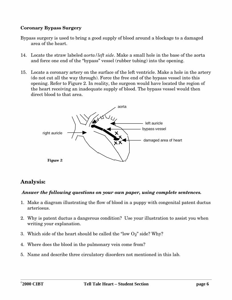
Coronary Bypass Surgery
Bypass surgery is used to bring a good supply of blood around a blockage to a damaged area of the heart.
14. Locate the straw labeled aorta/left side. Make a small hole in the base of the aorta and force one end of the “bypass” vessel (rubber tubing) into the opening.
15. Locate a coronary artery on the surface of the left ventricle. Make a hole in the artery (do not cut all the way through). Force the free end of the bypass vessel into this opening. Refer to Figure 2. In reality, the surgeon would have located the region of the heart receiving an inadequate supply of blood. The bypass vessel would then direct blood to that area.
right auricle
aorta
left auricle
Figure 2
bypass vessel
damaged area of heart
Analysis:
Answer the following questions on your own paper, using complete sentences.
1. Make a diagram illustrating the flow of blood in a puppy with congenital patent ductus arteriosus.
2. Why is patent ductus a dangerous condition? Use your illustration to assist you when writing your explanation.
3. Which side of the heart should be called the “low O2” side? Why?
4. Where does the blood in the pulmonary vein come from?
5. Name and describe three circulatory disorders not mentioned in this lab.

©2000 CIBT Dog Heartworm Poster – Student Section page 1
Dog Heartworm Poster
Student Laboratory Exercise
CIBT
Name: ______________________________________
Date Due: ___________________________________
The heartworm, Dirofilaria immitis, is a parasite that takes up residence in the pulmonary artery and right side of the heart of dogs, cats, ferrets, and closely related animals. Although heartworms can infect man and other animals, the worms die in these animals without reaching maturity.
The heartworm is a nematode worm with a complicated life cycle. It undergoes a series of stages in its development. Not all of these stages occur within the circulatory system of its dog host. Some very important events in a young heartworm's life occur in the digestive and excretory systems of many species of mosquitoes that serve as hosts for heartworms. The mosquito is also the vector for heartworms. In fact, heartworm transmission depends upon mosquitoes. The geographic prevalence of heartworm disease in untreated animals corresponds with the distribution of mosquitoes.
After completing this poster exercise, you will be able to:
1. discuss the location and nature of damage done by heartworm to the vessels of the dog's heart
2. describe the life history of the dog heartworm
3. discuss aspects of heartworm evolutionary survival strategy
4. define what an intermediate host is
5. define what a definitive host is
For this project, you will construct a poster that provides certain information. You have just opened a research lab and need to alert responsible pet owners to the dangers of heartworm. You are also investigating a possible treatment for the infection. Your poster or brochure should include the following information:
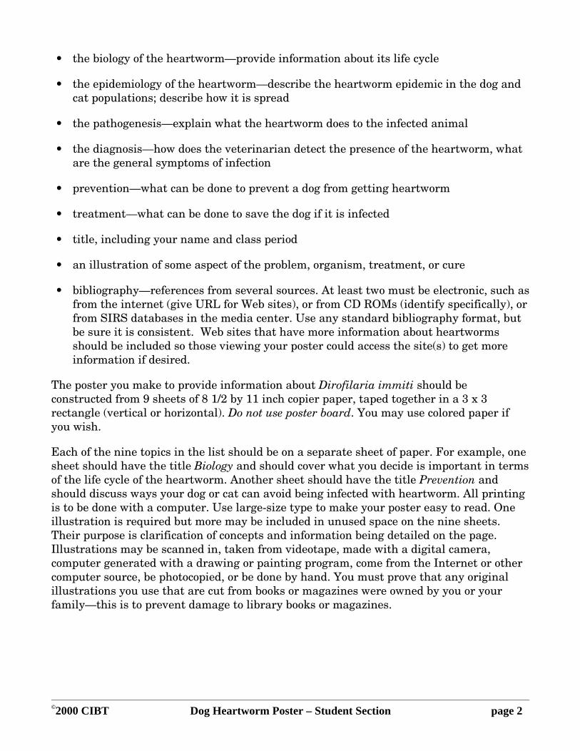
©2000 CIBT Dog Heartworm Poster – Student Section page 2
• the biology of the heartworm—provide information about its life cycle
• the epidemiology of the heartworm—describe the heartworm epidemic in the dog and cat populations; describe how it is spread
• the pathogenesis—explain what the heartworm does to the infected animal
• the diagnosis—how does the veterinarian detect the presence of the heartworm, what are the general symptoms of infection
• prevention—what can be done to prevent a dog from getting heartworm
• treatment—what can be done to save the dog if it is infected
• title, including your name and class period
• an illustration of some aspect of the problem, organism, treatment, or cure
• bibliography—references from several sources. At least two must be electronic, such as from the internet (give URL for Web sites), or from CD ROMs (identify specifically), or from SIRS databases in the media center. Use any standard bibliography format, but be sure it is consistent. Web sites that have more information about heartworms should be included so those viewing your poster could access the site(s) to get more information if desired.
The poster you make to provide information about Dirofilaria immiti should be constructed from 9 sheets of 8 1/2 by 11 inch copier paper, taped together in a 3 x 3 rectangle (vertical or horizontal). Do not use poster board. You may use colored paper if you wish.
Each of the nine topics in the list should be on a separate sheet of paper. For example, one sheet should have the title Biology and should cover what you decide is important in terms of the life cycle of the heartworm. Another sheet should have the title Prevention and should discuss ways your dog or cat can avoid being infected with heartworm. All printing is to be done with a computer. Use large-size type to make your poster easy to read. One illustration is required but more may be included in unused space on the nine sheets. Their purpose is clarification of concepts and information being detailed on the page. Illustrations may be scanned in, taken from videotape, made with a digital camera, computer generated with a drawing or painting program, come from the Internet or other computer source, be photocopied, or be done by hand. You must prove that any original illustrations you use that are cut from books or magazines were owned by you or your family—this is to prevent damage to library books or magazines.

©2000 CIBT Dog Heartworm Poster – Student Section page 3
Questions on Dog Heartworm Poster Research
Name: ______________________________________ Date Due_____________________
Please use our own notes on your poster research as well as the following information to answer the questions below.
Heartworm Havoc and Habits
A. Havoc
Heartworm disease occurs in wild and domestic dogs, ferrets, and cats, and occasional infections can occur in man as well. It is due to the presence of adult worms in the large vessels and heart. Adult worms cause traumatic irritation of the linings of the vessels of the heart. Constant rubbing of heartworms against the normally smooth vessel lining induces damage similar to early stages of human arteriosclerosis, but without deposits of cholesterol or other lipids.
The endothelial surface becomes roughened, with cells becoming disorganized, allowing the vessels to become leaky; occasionally the damage is severe enough to rupture the artery. The artery wall grows abnormal projections called villi that project into the lumen, disrupting and reducing blood flow. Similar changes in smaller arteries restrict both blood flow and the resiliency of the artery walls. Similar villi diminish blood flow in irritated veins.
These changes cause high pressure in the pulmonary artery and reduced blood flow to the lung. There is damage to the lung tissue, including an increase in tissue fluid, called edema, and inflammation. Dogs with this congestive heart failure will cough, have difficulty breathing, and will not be able to exercise. This can be fatal if not treated. However treatment itself may be dangerous, as dead heartworms may also cause vessel damage.
The severity of the disease is related to the size of the animal and the number and location of adult worms present. In a 25-kg dog, if there are fewer than 25 adults, they are in the pulmonary arteries in the lung; that is where the vessel damage occurs. As the population increases to 26-50 adults, some are found in the right ventricle, and with populations above 50 worms are also found in the right atrium. With further population increase, worms will be found in the vena cava. Over 200 worms have been found in a single dog!
In a 10-kg dog, as few as 36 adults can cause disease signs. In smaller animals such as ferrets, even a couple of worms could be fatal because vessel size and heart capacity is so small.
©2000 CIBT Dog Heartworm Poster – Student Section page 4
Fortunately, humans are a dead end host. Immature worms die in the lung on their attempted journey to the heart and never reach the life-threatening adult stage.
B. Habits
The nematodes that cause heartworm disease are in the suborder Filarioidea, order Spirurida, in the phylum Nematoda. Nematodes have long, unsegmented bodies with an external skeleton or "cuticle;" they are completely equipped with a nervous system, muscular system, excretory system, and digestive system. They do not need a circulatory or a respiratory system, as their body fluid circulates passively through their pseudocoelom. The phylum Nematoda is large and diverse; there are many free-living nematodes, plenty of plant parasitic nematodes, and many important animal parasitic nematodes including such famous examples as the whipworms, hookworms, and roundworms.
Most gravid female nematodes lay eggs, but some produce larvae; the filariids including the heartworm produce prelarvae, or microfilariae. Heartworm microfilariae are released into the bloodstream and circulate in the blood. This is the stage we will be studying in today's lab.
Nematodes have 5 other worm-like stages that are creatively named L1, L2, L3, L4, and L5. Stages L1-4 are the larval stages, while L5 is the adult. Parasitic nematodes can rarely complete their whole life within one host; it is much more common for them to have an obligatory period outside their definitive host. (The definitive host is the one in which sexual reproduction occurs.) In some nematodes this is a free-living existence, but in others such as the heartworm this period of development must be completed in a mosquito intermediate host. Heartworms spend their time as L1-L3 stages in the mosquito which ingests microfilariae when it takes a blood meal from an infected dog.
Microfilariae (mff) are about 308 micrometers in length and average heartworm infections typically produce about one thousand to one hundred thousand mff per ml of blood. A single Aedes aegypti female needs about 200 microliters of blood before she is fully engorged. A comfortable burden of mff is about 30; if she ingests too many more than this she may experience illness and possibly death from the infection. Mosquitoes can actually restrict the number of mff they ingest by excluding up to half of the mff present in a meal sized volume of circulating blood. Once inside the mosquito, mff migrate and develop to the infective stage for dogs, accumulating in the salivary glands and proboscis by about 2 weeks after infection. When the mosquito takes her next blood meal, these infective L3 actively migrate out of the mosquito's proboscis and into the bite wound.
If the host is a dog, the L3 now about 1.2 mm in length remain in the skin a few days. Then by 9-12 days after entering the dog they molt to L4s. At this time they have reached a length of 1.6 mm. On average this stage takes an additional 60 days, during which larvae make a long migration through the body of the dog.

©2000 CIBT Dog Heartworm Poster – Student Section page 5
Fortunately for dogs, the L4 stage has been found to be extremely sensitive to ivermectin, a macrocyclic lactone compound originally isolated from fungus, Streptomyces avermitilis, that was cultured from a soil sample collected near a golf course in Japan. It can affect sensitive nematodes by altering interneuron synaptic transmission resulting in paralysis. A dose of only 6 micrograms per kg dog body weight reliably kills heartworm L4's! A related compound, milbemycin, is also effective against this stage, but at higher doses. Veterinarians recommend protecting uninfected dogs by giving them a monthly dose of one of these compounds to kill any entering larvae at the L4 stage.
In unprotected dogs, adolescent heartworms just after molting to the fifth stage are about 2 cm in length. These adolescents migrate towards the heart. The earliest arrival at the heart is recorded as 68 days after entering the dog, but all arrive there by 90 days. Once at their final destination, they become sexually mature adults, with fertilized females releasing microfilariae into the blood at about 6 months after entering the dog. Eventually they reach their full size, 16 cm for males and 28 cm for females.
Questions on Heartworm Poster Research
1. What advantage is it to the heartworm to be a parasite? Consider basic needs such as obtaining food, eliminating waste, getting exercise, social life, reproduction, and dispersal.
2. Are there any advantages to the dog in having heartworm parasites?
3. How does ill health of each host, the intermediate and the definitive, affect the survival of the heartworm species?
4. Which of these hosts is more important for the survival of the heartworm species?
5. How important is the health of the dog to the survival of the heartworm species?
6. Why didn't we mention the role of male mosquitoes in the transmission of heartworm disease?
7. How does the adolescent growth spurt of heartworms compare to that of humans, and how could this dramatic spurt benefit the lifestyle of the heartworm?

©2000 CIBT Heartworm Microfilariae – Student Section page 1
Heartworm Microfilariae Student
Laboratory Exercise
CIBT
Name___________________________________ Date:______________
Materials:
• dog blood with microfilariae • slides
• capillary tubes • DiffQuik or similar blood stain
Procedure:
1. Make a blood smear:
Note: the whole procedure takes just a few seconds, and is best done smoothly (expect to make lots of mistakes at first). Each step should take about 2 seconds each; we've described the step 4 in slow motion, but the combined 4a, 4b, and 4c should only take about 2 seconds!
a. Put 2 clean slides on the counter in front of you.
b. Mix your blood sample by gentle inversion and immediately fill a capillary tube at least 3/4 full, place your finger over the dry end, and wipe the wet outside of the capillary tube with a tissue.
c. Holding the capillary tube horizontally over the two slides, release your finger just enough to place a 1-mm diameter drop near the label end of each slide.
d. Pick up one of the 2 slides with your first 3 fingers on one wide edge and your thumb on the opposite edge: this is your "spreader." Hold the spreader at a 30-45° angle, with the short edge nearest your thumb touching the end of the other, stationary slide that's furthest from its blood drop.
Steady the stationary slide by placing one finger of your other hand behind the short edge near the label.
Smoothly draw the spreader back towards the label, through the drop of blood until the drop has spread nearly the width of the slide, then push it smoothly forward

©2000 CIBT Heartworm Microfilariae – Student Section page 2
away from the label. Note, for smooth motion on this crucial step, RELAX YOUR SPREADER WRIST!!
e. Repeat step 4, using the first spreader slide as the new stationary slide, and spreading with the old stationary slide.
2. Fix and stain your blood smear with DiffQuik:
a. Dip the smeared slide and gently agitate for 5 seconds in the methanol fixative, which is clear; drain briefly on a paper towel.
b. Dip the slide and gently agitate for 5 seconds in the orange dye; drain briefly on a paper towel.
c. Dip the slide and gently agitate for 5 seconds in the purple dye; then immediately rinse by agitating it in a cup of clean water for a few seconds.
d. Let the slide dry by standing in on a short edge on a paper towel; you may use a slide warmer, but take care to keep the smear up (don't touch the smear with anything, or it will come off the slide!)
3. Examine your smear
When the slide is dry, examine it with the 10x objective lens to locate the mff; then examine this area with your high power objective lens (40 or 45x objective.)
Questions About Stained Blood Smear
1. Observe the stained mff, looking for the rounded "head" end and the tapered tail. The dark purple dots are nuclei. These mff are prelarvae that have functional muscular, nervous, and excretory systems. Would you expect them to have a working digestive system?
2. Comparing the width of the mff at its widest diameter to the diameter of the average dog red cells, which is wider?
3. Describe the distribution of the mff within your blood smear. Are other students’ mff distributed the same way yours are?
©2000 CIBT Heartworm Microfilariae – Student Section page 3
Part IIIb: Student Laboratory Exercise: Counting live microfilariae
Materials:
• dog blood with mff, diluted 1:10 with phosphate buffered saline solution
• fine glass-marking pen (such as Extra fine point permanent marker), black
• capillary tubes • coverslips, plastic, 22mm2
• pipettor (20-200ml) or pasteur pipettes and dropper bulbs
Procedure:
1. Mark your coverslip:
With an extra fine black permanent marker, draw a line dividing the slip in half (2 equal rectangles,) then divide each of these in half again with 2 more parallel lines; rotate the coverslip 90o again and repeat.
2. Calibrate your drop size (skip this step if you have an adjustable pipettor):
a. Using a graduated microfuge tube add water one drop at a time, counting how many drops you put in, until you have exactly 1 ml. Make sure you hold your pipette perpendicularly so that the drops fall straight down from its mouth, not touching anything with the mouth of the pipette.
b. Calculate how many microliters per drop using this formula:
(1 ml/ # drops) (1000) =_____ ml per drop
3. Prepare your slide:
a. Place a drop (the approximate volume of which you know from step 2) or 20 ml of the 1:10 diluted blood sample on your slide,
b. Place a marked plastic coverslip on it and allow it to stand 1 minute before counting.
4. Count your mff:
a. Using the low power (4x) objective, count the mff you see in each of your marked squares and enter the data into the grid below. Make sure to enter "0" of you see no microfilariae in one of the boxes in your grid. In order to count all the mff fairly, and not count any twice, use the following rule. If mff are under the top or left side marker lines of a box, count them as in that box. If the mff are under the bottom or right side marks, do not count them as in that box.

©2000 CIBT Heartworm Microfilariae – Student Section page 4
FIRST OBSERVER'S COUNT:
Total number under coverslip: _______________
b. For verification, have your partner make a second count as soon as you are done. Finish the counting within 10 minutes of making the preparation so that the edges don't dry.
SECOND OBSERVER'S COUNT:
Total number under coverslip: ______
Questions About Counting Live mff
1. Were the mff evenly distributed across your coverslip?

©2000 CIBT Heartworm Microfilariae – Student Section page 5
2. How many mff were there in your 20-ml drop of blood that had been diluted 1:10?
3. How would you account for differences between you and your partner's counts?
4. Find out the number of mff found by each of the other sets of lab partners in your lab, and report the range and mean of the class results:
Range:__________ Mean: ____________
How would you account for the variability in counts between groups?
5. Calculate how many mff are in 1 ml of undiluted blood, based on your answer to #2 above. Show your calculation.
6. If a mosquito can take only 30 mff before getting sick, and could drink 200 microliters of blood, would she get sick if she took her whole meal from the dog whose blood this is?