Embed Size (px)
Citation preview
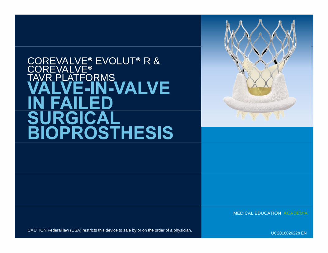
COREVALVE® EVOLUT® R &COREVALVE®TAVR PLATFORMSTAVR PLATFORMS
MEDICAL EDUCATION ACADEMIA
UC201602622b ENCAUTION Federal law (USA) restricts this device to sale by or on the order of a physician.
MEDICAL EDUCATION ACADEMIA
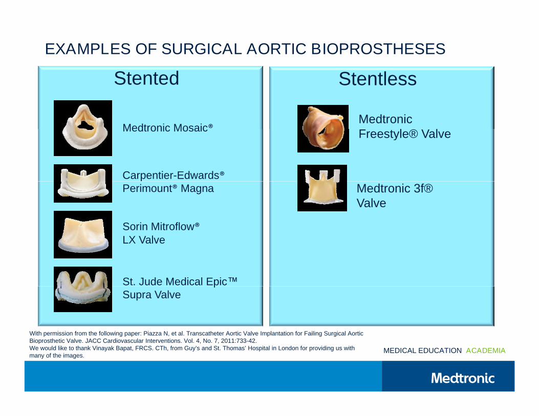
EXAMPLES OF SURGICAL AORTIC BIOPROSTHESES
Stented StentlessStented Stentless
Medtronic Mosaic®MedtronicFreestyle® Valve
Medtronic Mosaic®
Carpentier-Edwards®
Freestyle® Valve
Medtronic 3f®®
Perimount® Magna
Sorin Mitroflow®LX Valve
Medtronic 3f®Valve
LX Valve
St. Jude Medical Epic™St. Jude Medical Epic™Supra Valve
With permission from the following paper: Piazza N, et al. Transcatheter Aortic Valve Implantation for Failing Surgical AorticBioprosthetic Valve. JACC Cardiovascular Interventions. Vol. 4, No. 7, 2011:733-42.
MEDICAL EDUCATION ACADEMIA
Bioprosthetic Valve. JACC Cardiovascular Interventions. Vol. 4, No. 7, 2011:733-42.We would like to thank Vinayak Bapat, FRCS. CTh, from Guy’s and St. Thomas’ Hospital in London for providing us withmany of the images.
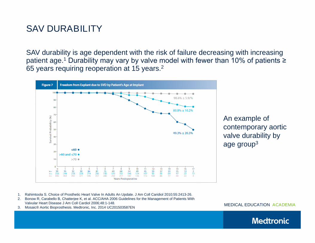
SAV DURABILITY
SAV durability is age dependent with the risk of failure decreasing with increasingpatient age.1 Durability may vary by valve model with fewer than 10% of patients ≥ 65 years requiring reoperation at 15 years.2
An example ofcontemporary aorticvalve durability byvalve durability byage group3
1. Rahimtoola S. Choice of Prosthetic Heart Valve In Adults An Update. J Am Coll Caridiol 2010;55:2413-26.2. Bonow R, Carabello B, Chatterjee K, et al. ACC/AHA 2006 Guidelines for the Management of Patients With
MEDICAL EDUCATION ACADEMIA
2. Bonow R, Carabello B, Chatterjee K, et al. ACC/AHA 2006 Guidelines for the Management of Patients WithValvular Heart Disease J Am Coll Cardiol 2006;48:1-148.
3. Mosaic® Aortic Bioprosthesis. Medtronic, Inc. 2014 UC201503587EN
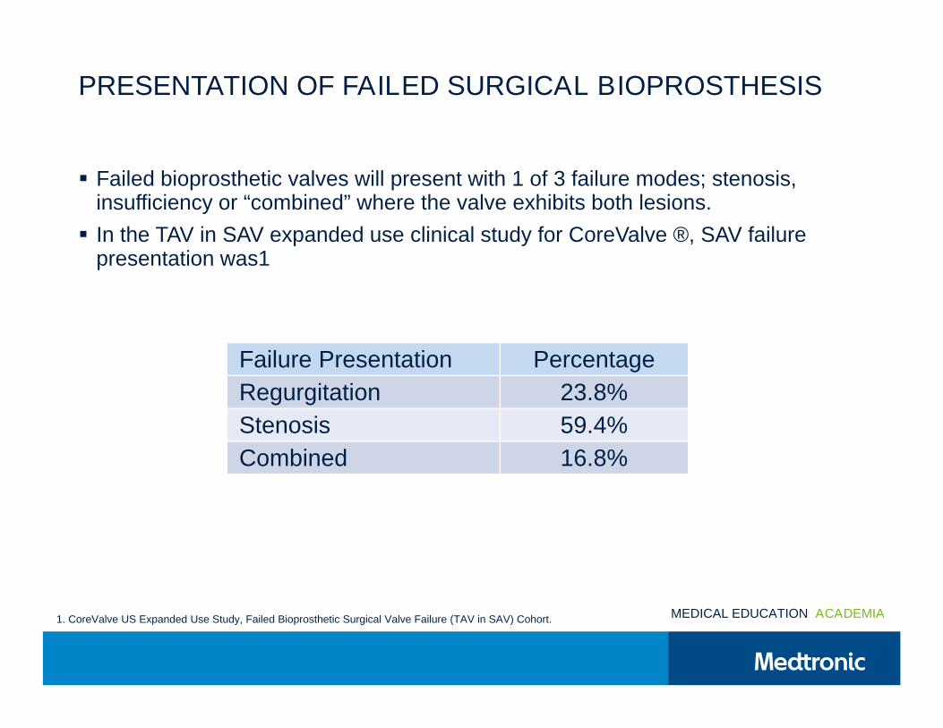
PRESENTATION OF FAILED SURGICAL BIOPROSTHESIS
Failed bioprosthetic valves will present with 1 of 3 failure modes; stenosis,insufficiency or “combined” where the valve exhibits both lesions.
In the TAV in SAV expanded use clinical study for CoreValve ®, SAV failure In the TAV in SAV expanded use clinical study for CoreValve ®, SAV failurepresentation was1
Failure Presentation Percentage
Regurgitation 23.8%
Stenosis 59.4%
Combined 16.8%
MEDICAL EDUCATION ACADEMIA1. CoreValve US Expanded Use Study, Failed Bioprosthetic Surgical Valve Failure (TAV in SAV) Cohort.
MEDICAL EDUCATION ACADEMIA
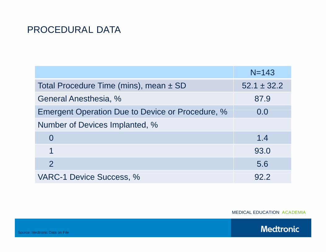
PROCEDURAL DATA
N=143
Total Procedure Time (mins), mean ± SD 52.1 ± 32.2
General Anesthesia, % 87.9
Emergent Operation Due to Device or Procedure, % 0.0Emergent Operation Due to Device or Procedure, % 0.0
Number of Devices Implanted, %
0 1.40 1.4
1 93.0
2 5.6
VARC-1 Device Success, % 92.2VARC-1 Device Success, % 92.2
MEDICAL EDUCATION ACADEMIA
Source: Medtronic Data on File
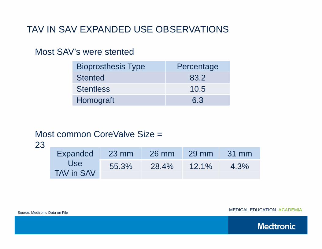
TAV IN SAV EXPANDED USE OBSERVATIONS
Bioprosthesis Type Percentage
Stented 83.2
Most SAV’s were stented
Stented 83.2
Stentless 10.5
Homograft 6.3
Most common CoreValve Size =23
ExpandedUse
TAV in SAV
23 mm 26 mm 29 mm 31 mm
55.3% 28.4% 12.1% 4.3%
23
TAV in SAV
MEDICAL EDUCATION ACADEMIASource: Medtronic Data on File
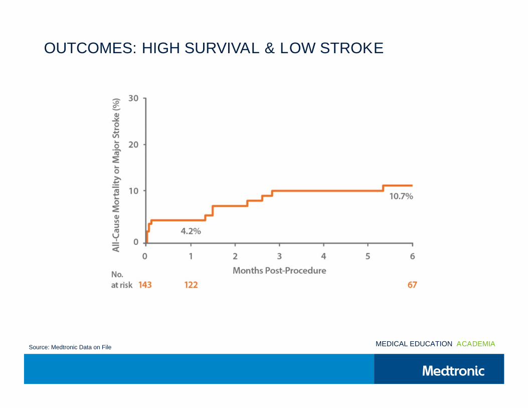
OUTCOMES: HIGH SURVIVAL & LOW STROKE
MEDICAL EDUCATION ACADEMIASource: Medtronic Data on File
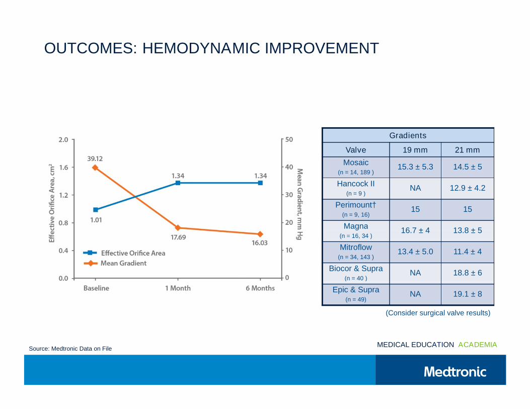
OUTCOMES: HEMODYNAMIC IMPROVEMENT
Gradients
Valve 19 mm 21 mm
Mosaic(n = 14, 189 )
15.3 ± 5.3 14.5 ± 5
Hancock II(n = 9 )
NA 12.9 ± 4.2
Perimount†(n = 9, 16)
15 15
Magna16.7 ± 4 13.8 ± 5
Magna(n = 16, 34 )
16.7 ± 4 13.8 ± 5
Mitroflow(n = 34, 143 )
13.4 ± 5.0 11.4 ± 4
Biocor & Supra(n = 40 )
NA 18.8 ± 6( = 40 )
Epic & Supra(n = 49)
NA 19.1 ± 8
(Consider surgical valve results)
MEDICAL EDUCATION ACADEMIASource: Medtronic Data on File
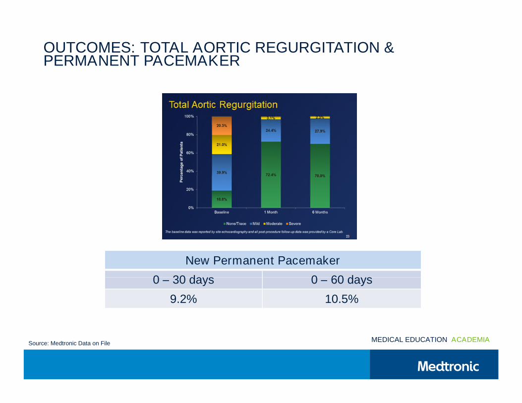
OUTCOMES: TOTAL AORTIC REGURGITATION &PERMANENT PACEMAKER
New Permanent Pacemaker
0 – 30 days 0 – 60 days0 – 30 days 0 – 60 days
9.2% 10.5%
MEDICAL EDUCATION ACADEMIASource: Medtronic Data on File
MEDICAL EDUCATION ACADEMIA
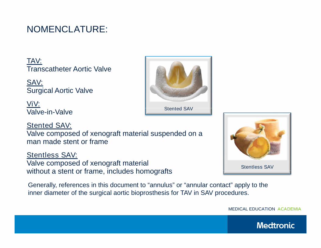
NOMENCLATURE:
TAV:Transcatheter Aortic Valve
SAV:Surgical Aortic Valve
ViV:Valve-in-Valve
Stented SAVValve-in-Valve
Stented SAV:Valve composed of xenograft material suspended on aman made stent or frame
Stented SAV
man made stent or frame
Stentless SAV:Valve composed of xenograft materialwithout a stent or frame, includes homografts
Stentless SAVwithout a stent or frame, includes homografts
Generally, references in this document to “annulus” or “annular contact” apply to theinner diameter of the surgical aortic bioprosthesis for TAV in SAV procedures.
MEDICAL EDUCATION ACADEMIA
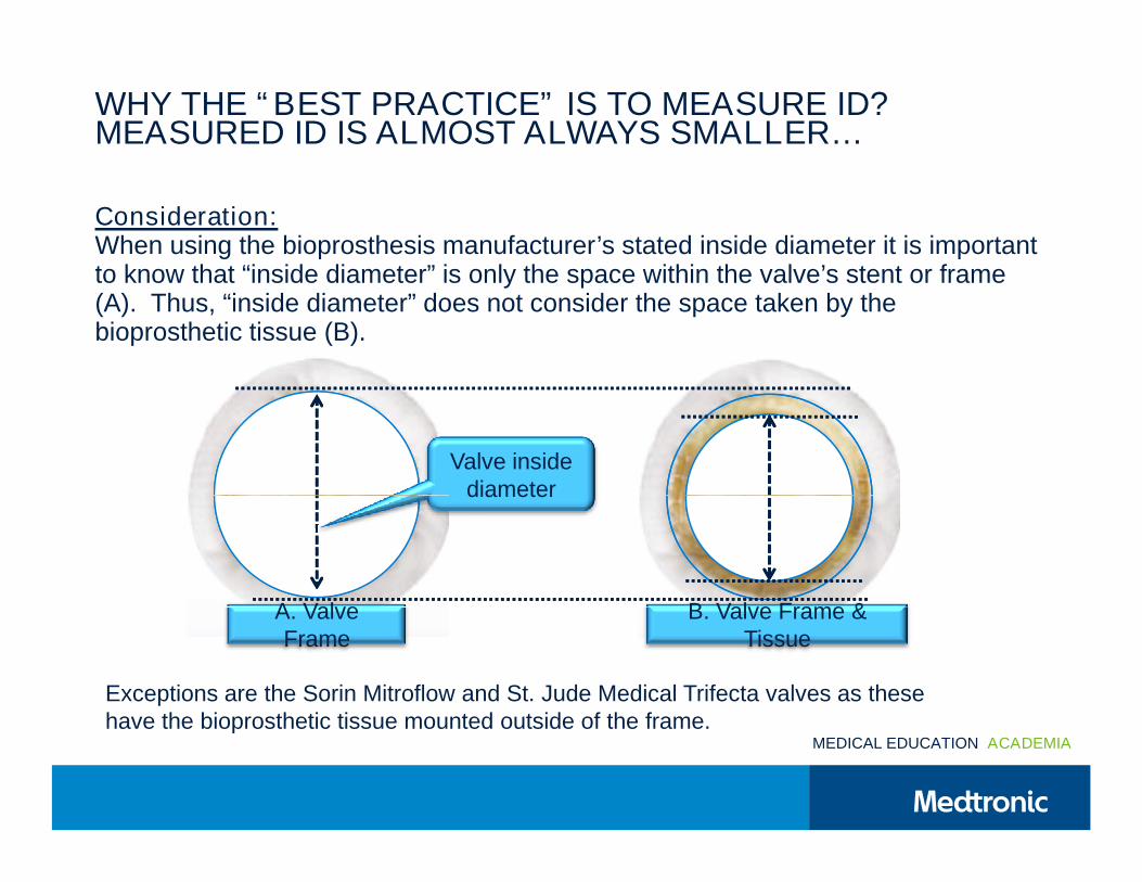
WHY THE “BEST PRACTICE” IS TO MEASURE ID?MEASURED ID IS ALMOST ALWAYS SMALLER…
Consideration:When using the bioprosthesis manufacturer’s stated inside diameter it is importantto know that “inside diameter” is only the space within the valve’s stent or frameto know that “inside diameter” is only the space within the valve’s stent or frame(A). Thus, “inside diameter” does not consider the space taken by thebioprosthetic tissue (B).
Valve insidediameterdiameter
A. Valve B. Valve Frame &
Exceptions are the Sorin Mitroflow and St. Jude Medical Trifecta valves as thesehave the bioprosthetic tissue mounted outside of the frame.
A. ValveFrame
B. Valve Frame &Tissue
MEDICAL EDUCATION ACADEMIA
have the bioprosthetic tissue mounted outside of the frame.
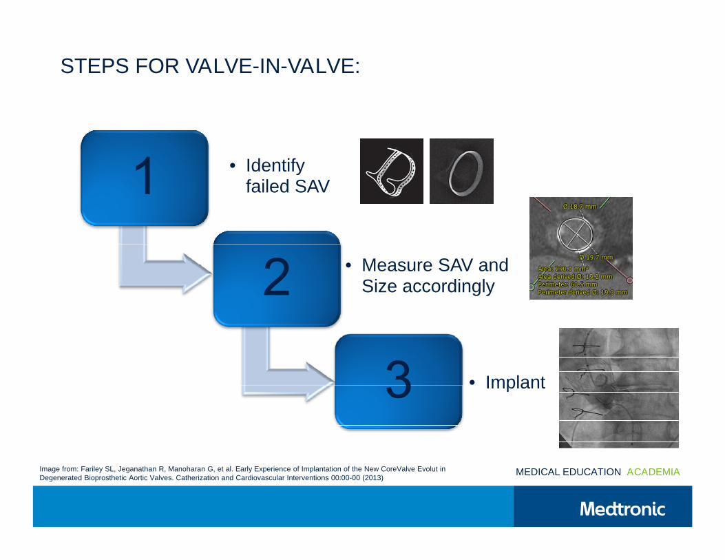
STEPS FOR VALVE-IN-VALVE:
• Identify• Identifyfailed SAV
• Measure SAV andSize accordingly
• Implant• Implant
MEDICAL EDUCATION ACADEMIAImage from: Fariley SL, Jeganathan R, Manoharan G, et al. Early Experience of Implantation of the New CoreValve Evolut inDegenerated Bioprosthetic Aortic Valves. Catherization and Cardiovascular Interventions 00:00-00 (2013)

MEDICAL EDUCATION ACADEMIA
IMPORTANT DISCLAIMERS
The content in this presentation was created with detailed input, review andapproval from Medtronic proctors. It is intended to be a resource to supportheart teams in their training, planning for, and performing procedures and is in noheart teams in their training, planning for, and performing procedures and is in noway intended to constitute medical advice or in any way replace the independentmedical judgment of a licensed and trained physician with respect to any patientneeds or circumstances.
The physician is solely responsible for all decision and medical judgmentsrelating to the treatment of their patients.
Please see the complete Instructions for Use, including all product indications,contraindications, precautions, warnings, and adverse events.contraindications, precautions, warnings, and adverse events.
MEDICAL EDUCATION ACADEMIA
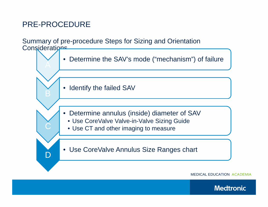
PRE-PROCEDURE
Summary of pre-procedure Steps for Sizing and OrientationSummary of pre-procedure Steps for Sizing and OrientationConsiderations
A• Determine the SAV’s mode (“mechanism”) of failure
A
B• Identify the failed SAV
B• Identify the failed SAV
• Determine annulus (inside) diameter of SAV
C
• Determine annulus (inside) diameter of SAV• Use CoreValve Valve-in-Valve Sizing Guide• Use CT and other imaging to measure
D• Use CoreValve Annulus Size Ranges chart
MEDICAL EDUCATION ACADEMIA
A. DETERMINE THE SAV’S MECHANISM OF FAILURE
Best Practice:Determine whether the failure mode of the SAV is:
Stenosis Stenosis
Insufficiency
Combined stenotic/Insufficient lesions
Considerations:
Implanting a CoreValve bioprosthesis in a degenerated SAV should be avoided ifthe there is significant concomitant perivalvular leak (between the prosthesis andthe there is significant concomitant perivalvular leak (between the prosthesis andthe native annulus)
The procedural impact of aortic regurgitation should be appreciated
MEDICAL EDUCATION ACADEMIA
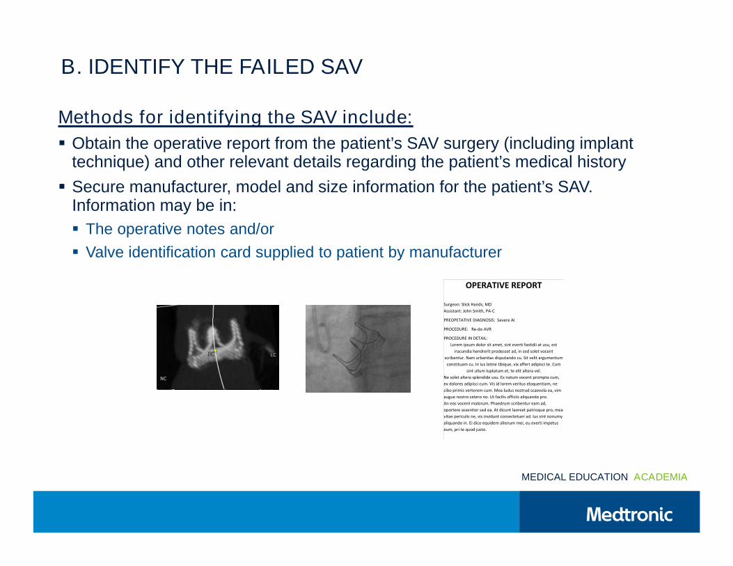
B. IDENTIFY THE FAILED SAV
Methods for identifying the SAV include:
Obtain the operative report from the patient’s SAV surgery (including implanttechnique) and other relevant details regarding the patient’s medical history
Secure manufacturer, model and size information for the patient’s SAV.Information may be in:
The operative notes and/or
Valve identification card supplied to patient by manufacturer Valve identification card supplied to patient by manufacturer
OPERATIVE REPORT
Surgeon: Slick Hands, MD
Assistant: John Smith, PA-C
PREOPETATIVE DIAGNOSIS: Severe AIPREOPETATIVE DIAGNOSIS: Severe AI
PROCEDURE: Re-do AVR
PROCEDURE IN DETAIL:
Lorem ipsum dolor sit amet, sint everti fastidii at usu, est
iracundia hendrerit prodesset ad, in sed solet vocent
scribentur. Nam urbanitas disputando cu. Sit velit argumentum
constituam cu. In ius latine tibique, vix affert adipisci te. Cum
sint ullum luptatum et, te elit altera vel.
Ne solet altera splendide usu. Ex natum vocent prompta cum,
ex dolores adipisci cum. Vis id lorem veritus eloquentiam, ne
cibo primis verterem cum. Mea ludus nostrud scaevola ea, vimcibo primis verterem cum. Mea ludus nostrud scaevola ea, vim
augue nostro cetero no. Ut facilis officiis aliquando pro.
An eos vocent malorum. Phaedrum scribentur eam ad,
oportere assentior sed ea. At dicunt laoreet patrioque pro, mea
vitae periculis ne, vis invidunt consectetuer ad. Ius sint nonumy
aliquando in. Ei dico equidem alienum mei, eu everti impetus
eum, pri te quod justo.
MEDICAL EDUCATION ACADEMIA
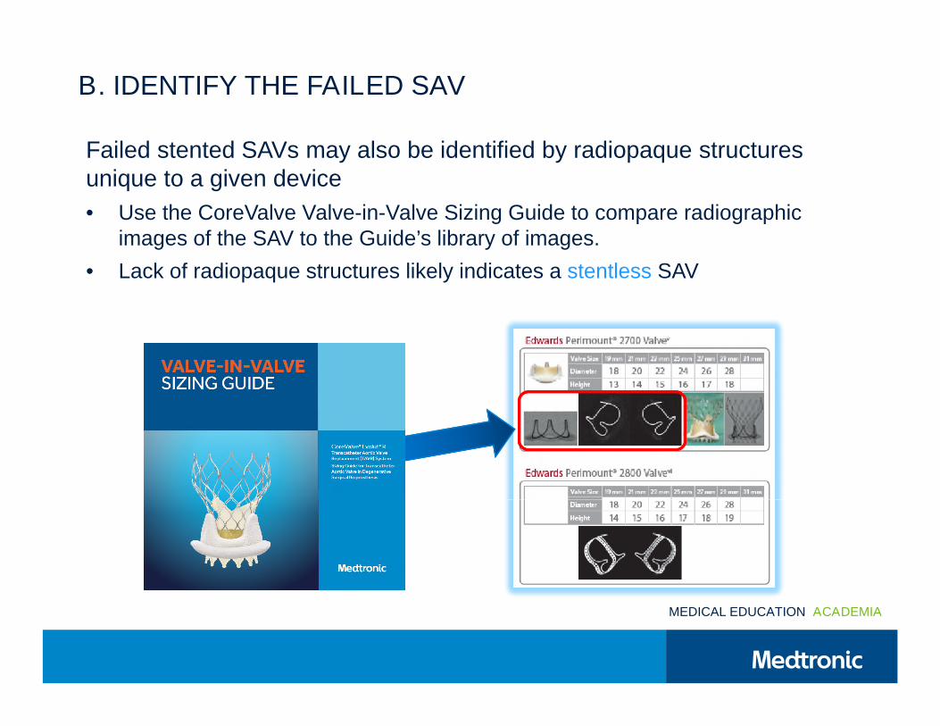
B. IDENTIFY THE FAILED SAV
Failed stented SAVs may also be identified by radiopaque structuresunique to a given device
• Use the CoreValve Valve-in-Valve Sizing Guide to compare radiographicimages of the SAV to the Guide’s library of images.
• Lack of radiopaque structures likely indicates a stentless SAV
MEDICAL EDUCATION ACADEMIA
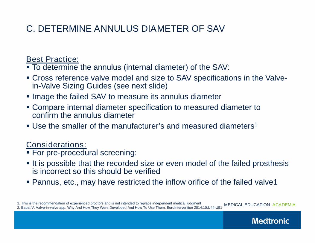
C. DETERMINE ANNULUS DIAMETER OF SAV
Best Practice: To determine the annulus (internal diameter) of the SAV:
Cross reference valve model and size to SAV specifications in the Valve- Cross reference valve model and size to SAV specifications in the Valve-in-Valve Sizing Guides (see next slide)
Image the failed SAV to measure its annulus diameter
Compare internal diameter specification to measured diameter to Compare internal diameter specification to measured diameter toconfirm the annulus diameter
Use the smaller of the manufacturer’s and measured diameters1
Considerations: For pre-procedural screening:
It is possible that the recorded size or even model of the failed prosthesisis incorrect so this should be verified It is possible that the recorded size or even model of the failed prosthesis
is incorrect so this should be verified
Pannus, etc., may have restricted the inflow orifice of the failed valve1
MEDICAL EDUCATION ACADEMIA1. This is the recommendation of experienced proctors and is not intended to replace independent medical judgment2. Bapat V. Valve-in-valve app: Why And How They Were Developed And How To Use Them. EuroIntervention 2014;10:U44-U51
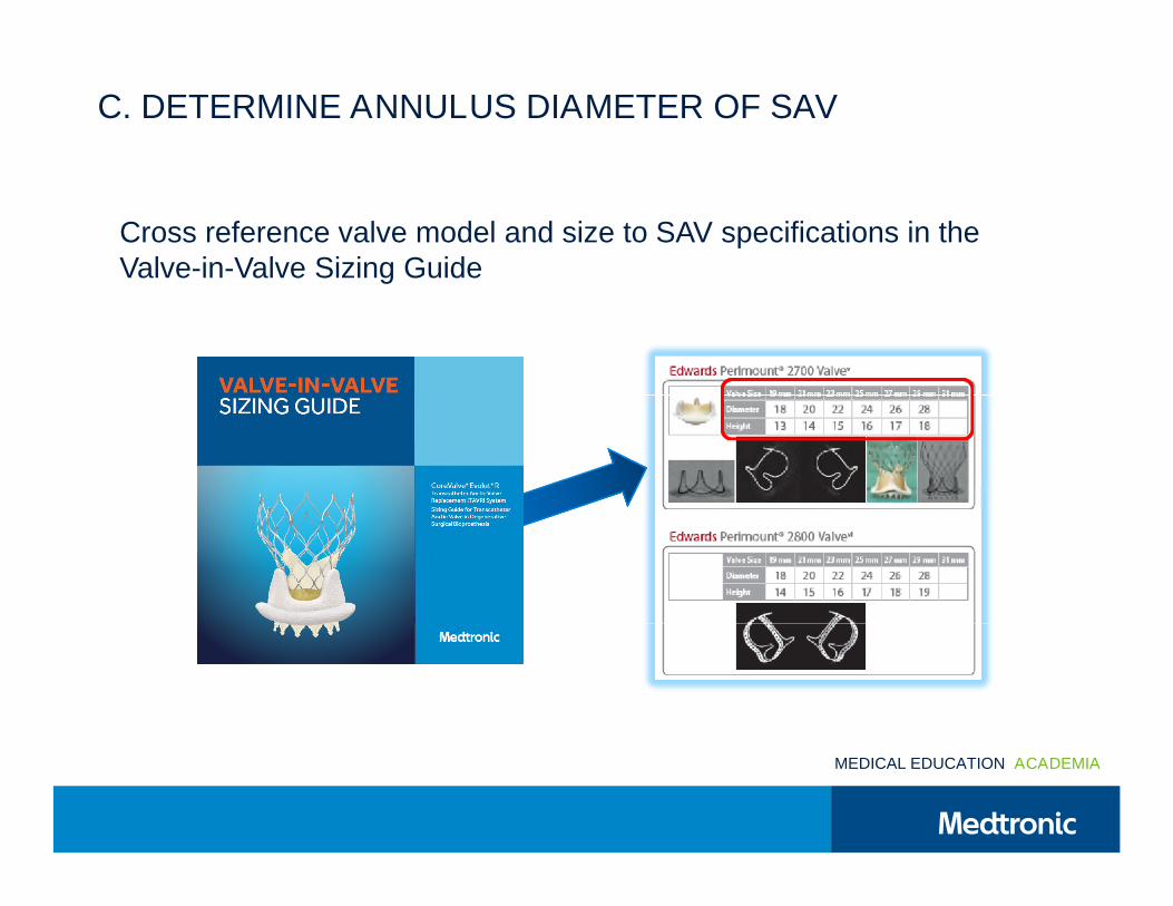
C. DETERMINE ANNULUS DIAMETER OF SAV
Cross reference valve model and size to SAV specifications in theValve-in-Valve Sizing GuideValve-in-Valve Sizing Guide
MEDICAL EDUCATION ACADEMIA
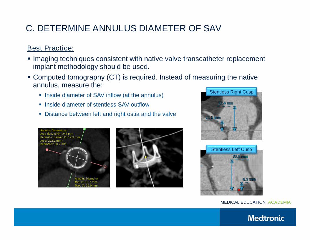
C. DETERMINE ANNULUS DIAMETER OF SAV
Best Practice:Best Practice:
Imaging techniques consistent with native valve transcatheter replacementimplant methodology should be used.
Computed tomography (CT) is required. Instead of measuring the native Computed tomography (CT) is required. Instead of measuring the nativeannulus, measure the:
Inside diameter of SAV inflow (at the annulus)
Inside diameter of stentless SAV outflow
Stentless Right Cusp
Inside diameter of stentless SAV outflow
Distance between left and right ostia and the valve
Stentless Left Cusp
MEDICAL EDUCATION ACADEMIA
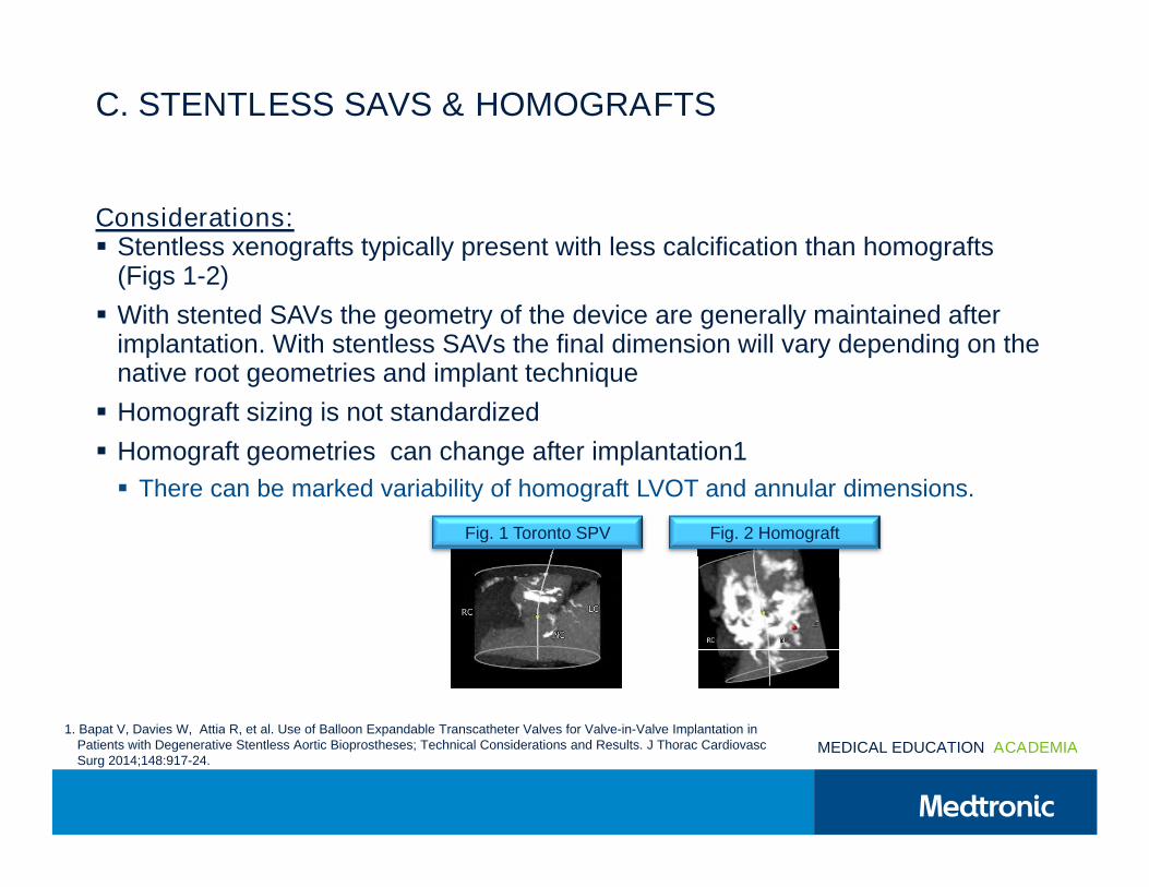
C. STENTLESS SAVS & HOMOGRAFTS
Considerations: Stentless xenografts typically present with less calcification than homografts
(Figs 1-2)(Figs 1-2)
With stented SAVs the geometry of the device are generally maintained afterimplantation. With stentless SAVs the final dimension will vary depending on thenative root geometries and implant techniquenative root geometries and implant technique
Homograft sizing is not standardized
Homograft geometries can change after implantation1
There can be marked variability of homograft LVOT and annular dimensions. There can be marked variability of homograft LVOT and annular dimensions.
Fig. 2 HomograftFig. 1 Toronto SPV
1. Bapat V, Davies W, Attia R, et al. Use of Balloon Expandable Transcatheter Valves for Valve-in-Valve Implantation in
MEDICAL EDUCATION ACADEMIA1. Bapat V, Davies W, Attia R, et al. Use of Balloon Expandable Transcatheter Valves for Valve-in-Valve Implantation in
Patients with Degenerative Stentless Aortic Bioprostheses; Technical Considerations and Results. J Thorac CardiovascSurg 2014;148:917-24.
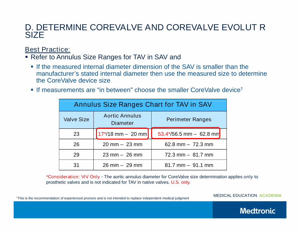
D. DETERMINE COREVALVE AND COREVALVE EVOLUT RSIZE
Best Practice: Refer to Annulus Size Ranges for TAV in SAV and
If the measured internal diameter dimension of the SAV is smaller than themanufacturer’s stated internal diameter then use the measured size to determinemanufacturer’s stated internal diameter then use the measured size to determinethe CoreValve device size
If measurements are “in between” choose the smaller CoreValve device†
Annulus Size Ranges Chart for TAV in SAVAnnulus Size Ranges Chart for TAV in SAV
Valve SizeAortic Annulus
DiameterPerimeter Ranges
23 17*/18 mm – 20 mm 53.4*/56.5 mm – 62.8 mm
26 20 mm – 23 mm 62.8 mm – 72.3 mm
29 23 mm – 26 mm 72.3 mm – 81.7 mm
31 26 mm – 29 mm 81.7 mm – 91.1 mm
*Consideration: ViV Only - The aortic annulus diameter for CoreValve size determination applies only toprosthetic valves and is not indicated for TAV in native valves. U.S. only.
MEDICAL EDUCATION ACADEMIA†This is the recommendation of experienced proctors and is not intended to replace independent medical judgment
PROCEDURE HIGHLIGHTS
In most cases predilatation (BAV) is not necessary
Considerations for ViV include differences between SAV’s and native valves.Stented SAV’s:Stented SAV’s:
Present a stiffer non-expandable landing zone
Have less ellipticity than native anatomy
The contact between TAV and SAV is different than between TAV and native valve The contact between TAV and SAV is different than between TAV and native valveduring deployment.
Anticipate movement towards the ventricle
Implant depth below the SAV annulus: Implant depth below the SAV annulus:
CoreValve: Target 4 mm
CoreValve Evolut R 3-5 mm
MEDICAL EDUCATION ACADEMIA
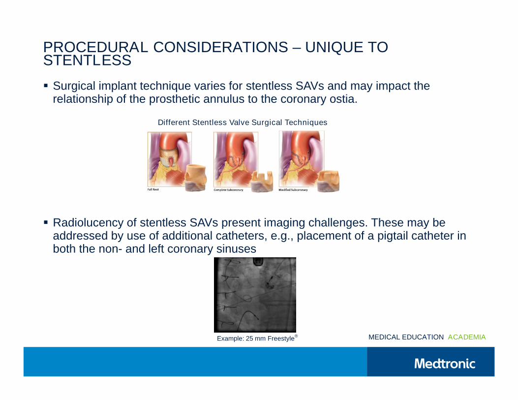
PROCEDURAL CONSIDERATIONS – UNIQUE TOSTENTLESS
Surgical implant technique varies for stentless SAVs and may impact therelationship of the prosthetic annulus to the coronary ostia.
Different Stentless Valve Surgical TechniquesDifferent Stentless Valve Surgical Techniques
Radiolucency of stentless SAVs present imaging challenges. These may be Radiolucency of stentless SAVs present imaging challenges. These may beaddressed by use of additional catheters, e.g., placement of a pigtail catheter inboth the non- and left coronary sinuses
MEDICAL EDUCATION ACADEMIAExample: 25 mm Freestyle®
BEST PRACTICES: POST-PROCEDURE
Confirm gradient across the valve with invasive measurement.
Perform a post-implant aortogram with the reference pigtail to ensure coronarypatency and assess aortic regurgitations.patency and assess aortic regurgitations.
MEDICAL EDUCATION ACADEMIA
POST-PROCEDURE (POST DILATATION)
Note: In the event that valve function or sealing is impaired due to excessivecalcification or incomplete expansion, a postimplant balloon dilatation of thebioprosthesis may improve valve function and sealing. To ensure patient safety,bioprosthesis may improve valve function and sealing. To ensure patient safety,valve size and patient anatomy must be considered when selecting the size ofthe balloon. The balloon size chosen for dilatation should not exceed thediameter of the native aortic annulus or, for surgical bioprosthetic valves, themanufacturer's labeled inner diameter.manufacturer's labeled inner diameter.
Note: Bench testing has only been conducted to confirm compatibility withNuMED Z-MED II™ Balloon Aortic Valvuloplasty catheters where CoreValveEvolut R and CoreValve bioprosthesis device performance was maintained afterEvolut R and CoreValve bioprosthesis device performance was maintained afterdilatation.
MEDICAL EDUCATION ACADEMIA
MEDICAL EDUCATION ACADEMIA
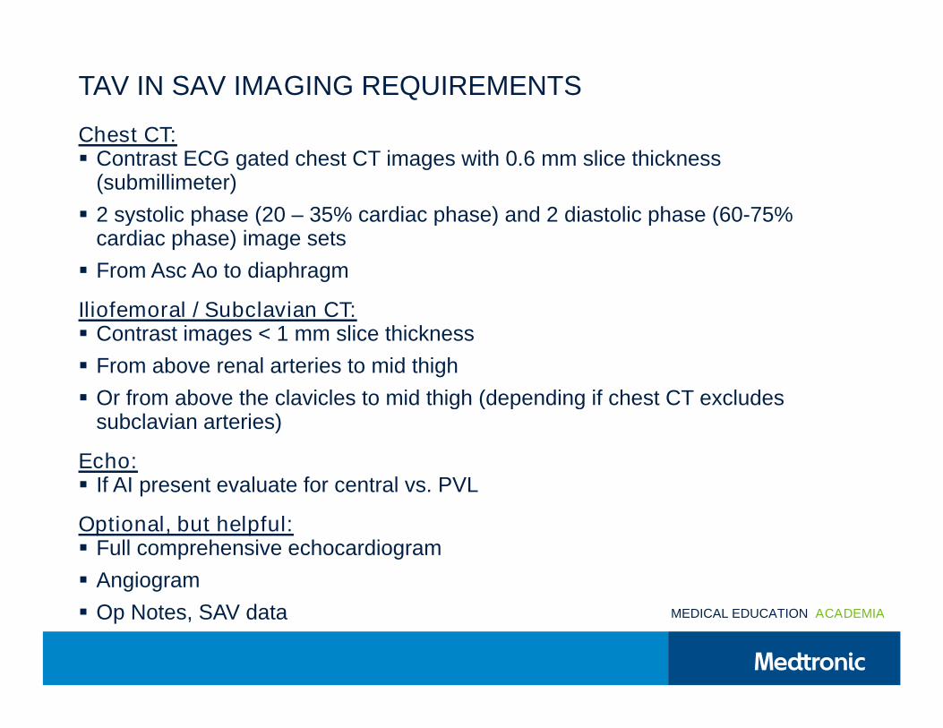
TAV IN SAV IMAGING REQUIREMENTS
Chest CT:Chest CT: Contrast ECG gated chest CT images with 0.6 mm slice thickness
(submillimeter)
2 systolic phase (20 – 35% cardiac phase) and 2 diastolic phase (60-75% 2 systolic phase (20 – 35% cardiac phase) and 2 diastolic phase (60-75%cardiac phase) image sets
From Asc Ao to diaphragm
Iliofemoral / Subclavian CT:Iliofemoral / Subclavian CT: Contrast images < 1 mm slice thickness
From above renal arteries to mid thigh
Or from above the clavicles to mid thigh (depending if chest CT excludes Or from above the clavicles to mid thigh (depending if chest CT excludessubclavian arteries)
Echo: If AI present evaluate for central vs. PVL
Optional, but helpful: Full comprehensive echocardiogram
Angiogram
MEDICAL EDUCATION ACADEMIA Op Notes, SAV data

INDICATIONS The Medtronic CoreValve and CoreValve Evolut R systems are indicated for use in patients with symptomatic heart disease due toeither severe native calcific aortic stenosis or failure (stenosed, insufficient, or combined) of a surgical bioprosthetic aortic valve who are judged
antegrade direction. However, use caution when moving the bioprosthesis in the antegrade direction. EnVeo R DCS Only: If a misload is detected,unsheath the bioprosthesis and examine the bioprosthesis for damage (for example, permanent frame deformation, frayed sutures, or valveeither severe native calcific aortic stenosis or failure (stenosed, insufficient, or combined) of a surgical bioprosthetic aortic valve who are judged
by a heart team, including a cardiac surgeon, to be at high or greater risk for open surgical therapy (ie, Society of Thoracic Surgeons predicted riskof operative mortality score ≥8% or at a ≥15% risk of mortality at 30 days).
CONTRAINDICATIONS The CoreValve and CoreValve Evolut R systems are contraindicated for patients presenting with any of the followingconditions: known hypersensitivity or contraindication to aspirin, heparin (HIT/HITTS) and bivalirudin, ticlopidine, clopidogrel, Nitinol (Titanium orNickel), or sensitivity to contrast media, which cannot be adequately premedicated; ongoing sepsis, including active endocarditis; preexistingmechanical heart valve in aortic position.
WARNINGS General Implantation of the CoreValve and CoreValve Evolut R systems should be performed only by physicians who have receivedMedtronic CoreValve training. This procedure should only be performed where emergency aortic valve surgery can be performed promptly.Mechanical failure of the delivery catheter system and/or accessories may result in patient complications. Transcatheter Aortic Valve(Bioprosthesis) Accelerated deterioration of the bioprosthesis may occur in patients presenting with an altered calcium metabolism.
unsheath the bioprosthesis and examine the bioprosthesis for damage (for example, permanent frame deformation, frayed sutures, or valvedamage). Do not attempt to reload a damaged bioprosthesis. Do not load the bioprosthesis onto the catheter more than 2 times or after it hasbeen inserted into a patient. EnVeo R DCS Only: Use the deployment knob to deploy and recapture the bioprosthesis. Do not use the trigger fordeploying or recapturing because it could cause inaccurate placement of the bioprosthesis. EnVeo R DCS Only: Once the radiopaque capsulemarker band reaches the distal end of the radiopaque paddle attachment (point of no recapture), retrieval of the bioprosthesis from the patient isnot recommended. Retrieval after the point of no recapture may cause mechanical failure of the delivery catheter system, aortic root damage,coronary artery damage, myocardial damage, vascular complications, prosthetic valve dysfunction (including device malposition), embolization,stroke, and/or emergent surgery. EnVeo R DCS Only: During deployment, the bioprosthesis can be advanced or withdrawn as long as annularcontact has not been made. Once annular contact is made, the bioprosthesis cannot be advanced in the retrograde direction; recapture until thebioprosthesis is free from annular contact, and then reposition in the retrograde direction. If necessary, and the radiopaque capsule marker bandhas not yet reached the distal end of the radiopaque paddle attachment, the bioprosthesis can be withdrawn (repositioned) in the antegradedirection. However, use caution when moving the bioprosthesis in the antegrade direction. While the catheter is in the patient, ensure theguidewire is extending from the tip. Do not remove the guidewire from the catheter while the catheter is inserted in the patient. Use the handle(Bioprosthesis) Accelerated deterioration of the bioprosthesis may occur in patients presenting with an altered calcium metabolism.
Precautions General The safety and effectiveness of the CoreValve and CoreValve Evolut R systems have not been evaluated in the pediatricpopulation. The safety and effectiveness of the bioprosthesis for aortic valve replacement have not been evaluated in the following patientpopulations: with a native valve lesion which does not meet the criteria for severe aortic stenosis (aortic valve area ≤1.0 cm2 or aortic valve area index ≤0.6 cm2/m2, a mean aortic valve gradient of ≥40 mm Hg, or a peak aortic-jet velocity ≥4.0 m/s); who are at moderate or low surgical risk (predicted perioperative mortality risk of <15%); with untreated, clinically significant coronary artery disease requiring revascularization; with apreexisting prosthetic heart valve with a rigid support structure in either the mitral or pulmonic position if either the preexisting prosthetic heartvalve could affect the implantation or function of the bioprosthesis or the implantation of the bioprosthesis could affect the function of thepreexisting prosthetic heart valve; with cardiogenic shock manifested by low cardiac output, vasopressor dependence, or mechanicalhemodynamic support. The safety and effectiveness of a CoreValve or CoreValve Evolut R bioprosthesis implanted within a failed preexistingtranscatheter bioprosthesis has not been demonstrated. Implanting a CoreValve or CoreValve Evolut R bioprosthesis in a degenerated surgical
guidewire is extending from the tip. Do not remove the guidewire from the catheter while the catheter is inserted in the patient. Use the handleof the delivery system to reposition the bioprosthesis. Do not use the outer catheter sheath. Once deployment is complete, repositioning of thebioprosthesis (e.g., use of a snare and/or forceps) is not recommended. Repositioning of a deployed valve may cause aortic root damage,coronary artery damage, myocardial damage, vascular complications, prosthetic valve dysfunction (including device malposition), embolization,stroke, and/or emergent surgery. Do not attempt to retrieve or to recapture (EnVeo DCS only) a bioprosthesis if any one of the outflow struts isprotruding from the capsule. If any one of the outflow struts has deployed from the capsule, the bioprosthesis must be released from the catheterbefore the catheter can be withdrawn. Ensure the capsule is closed before catheter removal. When using a separate introducer sheath, ifincreased resistance is encountered when removing the catheter through the introducer sheath, do not force passage. Increased resistance mayindicate a problem and forced passage may result in damage to the device and/or harm to the patient. If the cause of resistance cannot bedetermined or corrected, remove the catheter and introducer sheath as a single unit over the guidewire, and inspect the catheter and confirmthat it is complete. Clinical long-term durability has not been established for the bioprosthesis. Evaluate bioprosthesis performance as neededduring patient follow-up. Postprocedure, administer appropriate antibiotic prophylaxis as needed for patients at risk for prosthetic valve infectiontranscatheter bioprosthesis has not been demonstrated. Implanting a CoreValve or CoreValve Evolut R bioprosthesis in a degenerated surgical
bioprosthesis [transcatheter aortic valve in surgical aortic valve (TAV in SAV)] should be avoided in the following conditions. The degeneratedsurgical bioprosthesis presents with a: significant concomitant perivalvular leak (between the prosthesis and the native annulus), is not securelyfixed in the native annulus, or is not structurally intact (eg, wireform frame fracture); partially detached leaflet that in the aortic position mayobstruct a coronary ostium; stent frame with a manufacturer’s labeled inner diameter <17 mm. The safety and effectiveness of the bioprosthesisfor aortic valve replacement have not been evaluated in patient populations presenting with the following: blood dyscrasias as defined:leukopenia (WBC <1000 cells/mm3), thrombocytopenia (platelet count <50,000 cells/mm3), history of bleeding diathesis or coagulopathy, orhypercoagulable states; congenital bicuspid or unicuspid valve; mixed aortic valve disease (aortic stenosis and aortic regurgitation withpredominant aortic regurgitation [3-4+]); moderate to severe (3-4+) or severe (4+) mitral or severe (4+) tricuspid regurgitation; hypertrophicobstructive cardiomyopathy; new or untreated echocardiographic evidence of intracardiac mass, thrombus, or vegetation; native aortic annulussize <18 mm or >29 mm for CoreValve and <18 mm or >26 mm for CoreValve Evolut R per the baseline diagnostic imaging or surgical bioprostheticaortic annulus size <17 mm or >29 mm for CoreVavle and <17 mm or >26 mm for CoreValve Evolut R; transarterial access not able to
during patient follow-up. Postprocedure, administer appropriate antibiotic prophylaxis as needed for patients at risk for prosthetic valve infectionand endocarditis. Postprocedure, administer anticoagulation and/or antiplatelet therapy per physician/clinical judgment. Excessive contrast mediamay cause renal failure. Preprocedure, measure the patient’s creatinine level. During the procedure, monitor contrast media usage. Conduct theprocedure under fluoroscopy. The safety and efficacy of a CoreValve or CoreValve Evolut R bioprosthesis implanted within the initial transcatheterbioprosthesis have not been demonstrated. However, in the event that a CoreValve or CoreValve Evolut R bioprosthesis must be implanted withinthe initial transcatheter bioprosthesis to improve valve function, valve size and patient anatomy must be considered before implantation of thebioprosthesis to ensure patient safety (for example, to avoid coronary obstruction). In the event that valve function or sealing is impaired due toexcessive calcification or incomplete expansion, a postimplant balloon dilatation of the bioprosthesis may improve valve function and sealing. Toensure patient safety, valve size and patient anatomy must be considered when selecting the size of the balloon used for dilatation. The balloonsize chosen for dilatation should not exceed the diameter of the native aortic annulus or, for surgical bioprosthetic valves, the manufacturer’slabeled inner diameter. Refer to the specific balloon catheter manufacturer’s labeling for proper instruction on the use of balloon catheterdevices. Note: Bench testing has only been conducted to confirm compatibility with NuMED Z-MED IITM Balloon Aortic Valvuloplasty cathetersaortic annulus size <17 mm or >29 mm for CoreVavle and <17 mm or >26 mm for CoreValve Evolut R; transarterial access not able to
accommodate an 18-Fr sheath or the 14-Fr equivalent EnVeo R InLine sheath; sinus of valsalva anatomy that would prevent adequate coronaryperfusion; moderate to severe mitral stenosis; severe ventricular dysfunction with left ventricular ejection fraction (LVEF) <20%; end-stage renaldisease requiring chronic dialysis or creatinine clearance <20 cc/min; symptomatic carotid or vertebral artery disease; severe basal septalhypertrophy with an outflow gradient.
Prior to Use Exposure to glutaraldehyde may cause irritation of the skin, eyes, nose, and throat. Avoid prolonged or repeated exposure to thevapors. Damage may result from forceful handling of the catheter. Prevent kinking of the catheter when removing it from the packaging.Thisdevice was designed for single patient use only. Do not reuse, reprocess, or resterilize this product. Reuse, reprocessing, or resterilization maycompromise the structural integrity of the device and/or create a risk of contamination of the device, which could result in patient injury, illness,or death.The bioprosthesis size must be appropriate to fit the patient’s anatomy. Proper sizing of the device is the responsibility of the physician.Refer to Instructions for Use for available sizes. Failure to implant a device within the sizing matrix could lead to adverse effects such as thoselisted below. Patients must present with access vessel diameters of ≥6 mm for the CoreValve system and ≥5 mm for the CoreValve Evolut R
devices. Note: Bench testing has only been conducted to confirm compatibility with NuMED Z-MED IITM Balloon Aortic Valvuloplasty catheterswhere CoreValve or CoreValve Evolut R bioprosthesis device performance was maintained after dilation. Data on File..
POTENTIAL ADVERSE EVENTS Potential risks associated with the implantation of the CoreValve or CoreValve Evolut R transcatheter aortic valvemay include, but are not limited to, the following: • death • myocardial infarction, cardiac arrest, cardiogenic shock, cardiac tamponade •coronary occlusion, obstruction, or vessel spasm (including acute coronary closure) • cardiovascular injury (including rupture, perforation, tissueerosion, or dissection of vessels, ascending aorta trauma, ventricle, myocardium, or valvular structures that may require intervention) • emergentsurgical or transcatheter intervention (for example, coronary artery bypass, heart valve replacement, valve explant, percutaneous coronaryintervention [PCI], balloon valvuloplasty) • prosthetic valve dysfunction (regurgitation or stenosis) due to fracture; bending (out-of-roundconfiguration) of the valve frame; underexpansion of the valve frame; calcification; pannus; leaflet wear, tear, prolapse, or retraction; poor valvecoaptation; suture breaks or disruption; leaks; mal-sizing (prosthesis-patient mismatch); malposition (either too high or too low)/malplacement •prosthetic valve migration/embolization • prosthetic valve endocarditis • prosthetic valve thrombosis • delivery catheter system malfunctionresulting in the need for additional re-crossing of the aortic valve and prolonged procedural time • delivery catheter system componentlisted below. Patients must present with access vessel diameters of ≥6 mm for the CoreValve system and ≥5 mm for the CoreValve Evolut R
system or an ascending aortic (direct aortic) access site ≥60 mm from the basal plane for both systems. Implantation of the bioprosthesis should be avoided in patients with aortic root angulation (angle between plane of aortic valve annulus and horizontal plane/vertebrae) of >30° for rightsubclavian/axillary access or >70° for femoral and left subclavian/axillary access. Use caution when using the subclavian/axillary approach inpatients with a patent LIMA graft or patent RIMA graft. For direct aortic access, ensure the access site and trajectory are free of patent RIMA or apreexisting patent RIMA graft.
During Use For direct aortic and subclavian access procedures, care must be exercised when using the tip-retrieval mechanism to ensure adequateclearance to avoid advancement of the catheter tip through the bioprosthesis leaflets during device closure. For direct aortic access procedures,use a separate introducer sheath; do not use the EnVeo R InLine sheath. Adequate rinsing of the bioprosthesis with sterile saline, as described inthe Instructions for Use, is mandatory before implantation. During rinsing, do not touch the leaflets or squeeze the bioprosthesis. If a capsulebecomes damaged during loading or the capsule fails to close, replace the entire system (bioprosthesis, catheter, and CLS). Do not use a catheter
resulting in the need for additional re-crossing of the aortic valve and prolonged procedural time • delivery catheter system componentmigration/embolization • stroke (ischemic or hemorrhagic), transient ischemic attack (TIA), or other neurological deficits • heart failure • cardiacfailure or low cardiac output • ancillary device embolization • individual organ (for example, cardiac, respiratory, renal [including acute kidneyfailure]) or multi-organ insufficiency or failure • major or minor bleeding that may require transfusion or intervention (including life-threatening ordisabling bleeding) • vascular access-related complications (eg, dissection, perforation, pain, bleeding, hematoma, pseudoaneurysm, irreversiblenerve injury, compartment syndrome, arteriovenous fistula, stenosis) • mitral valve regurgitation or injury • conduction system disturbances (forexample, atrioventricular node block, left-bundle branch block, asystole), which may require a permanent pacemaker • infection (includingsepticemia) • hypotension or hypertension • hemolysis • peripheral ischemia • bowel ischemia • abnormal lab values (including electrolyteimbalance) • allergic reaction to antiplatelet agents, contrast medium, or anesthesia • exposure to radiation through fluoroscopy and angiography• permanent disability.
07/2015, © 2015 Medtronic, Inc. All rights reserved. Please reference the CoreValve and CoreValve Evolut R Instructions for Use for more
CAUTION – Investigational device. Limited by United States law to investigational use. 3333
becomes damaged during loading or the capsule fails to close, replace the entire system (bioprosthesis, catheter, and CLS). Do not use a catheterwith a damaged capsule. After a bioprosthesis has been inserted into a patient, do not attempt to reload that bioprosthesis on the same or anyother catheter. AccuTrak DCS Only: During implantation, if resistance to deployment is encountered (e.g., the micro knob starts clicking or is tightor stuck), apply upward pressure to the macro slider while turning the micro knob. If the bioprosthesis still does not deploy, remove it from thepatient and use another system. AccuTrak DCS Only: Once deployment is initiated, retrieval of the bioprosthesis from the patient (e.g., use of thecatheter) is not recommended. Retrieval of a partially deployed valve using the catheter may cause mechanical failure of the delivery cathetersystem, aortic root damage, coronary artery damage, myocardial damage, vascular complications, prosthetic valve dysfunction (including devicemalposition), embolization, stroke, and/or emergent surgery. AccuTrak DCS Only: During deployment, the bioprosthesis can be advanced orwithdrawn as long as annular contact has not been made. Once annular contact is made, the bioprosthesis cannot be advanced in the retrogradedirection; if necessary, and the frame has only been deployed ≤2/3 of its length, the bioprosthesis can be withdrawn (repositioned) in the
07/2015, © 2015 Medtronic, Inc. All rights reserved. Please reference the CoreValve and CoreValve Evolut R Instructions for Use for moreinformation regarding indications, warnings, precautions and potential adverse events.
CoreValve is a registered trademark of Medtronic CV Luxembourg S.a.r.l.Evolut and Freestyle are registered trademarks of Medtronic 3f is a registered trademark of Medtronic 3F Therapeutics, Inc.Toronto SPV and Toronto Root are trademarks of St. Jude Medical, Inc. Prima Plus is a trademark of Edwards LifeSciences Corrp.Z-MED is a trademark of NuMED Inc.
CAUTION Federal law (USA) restricts this device to sale by or on the order of a physician.