Embed Size (px)
Citation preview

i
v
Copyright by
Jonathan Yen
2015

MATERIALS AND BIOLOGICAL APPROACH TO GENE DELIVERY IN HUMAN
EMBRYONIC STEM CELLS
BY
JONATHAN S Y YEN
DISSERTATION
Submitted in partial fulfillment of the requirements
for the degree of Doctor of Philosophy in Bioengineering
in the Graduate College of the
University of Illinois at Urbana-Champaign, 2015
Urbana, Illinois
Doctoral Committee:
Associate Professor Jianjun Cheng, Chair
Professor Ning Wang
Assistant Professor Kristopher Kilian
Assistant Professor Jian Ma
Assistant Professor Gregory Underhill

ii
Abstract
Gene delivery is an important tool used in the study and manipulation of human
pluripotent stem cells for regenerative medicine purposes. However current methods of
transient gene delivery are still highly inefficient. Using materials and biologically based
concepts, I aim to develop new methods and protocols to enhance the efficiency of gene
delivery. For the materials aspect, diblock copolymers consisting of poly(ethylene glycol)-
block-poly(γ-4-(((2-(piperidin-1-yl)ethyl)amino)methyl)benzyl-L-glutamate) (PEG-b-
PVBLG-8) were synthesized and evaluated for their ability to mediate gene delivery in
hard-to-transfect cells, such as IMR-90 human fetal lung fibroblasts and human embryonic
stem cells (hESCs). The PEG-b-PVBLG-8 contained a membrane-disruptive, cationic,
helical polypeptide block (PVBLG-8) for complexing with DNA and a hydrophilic PEG
block to improve the biocompatibility of the gene delivery vehicle. PEG-b-PVBLG-8
diblock polymers with a high degree of polymerization have a greater transfection
efficiency and lower toxicity in IMR-90 cells than the commercial reagent Lipofectamine
2000. The usefulness of PEG-b-PVBLG-8 was further demonstrated via the successful
transfection of hESCs without a measured loss in cell pluripotency markers. This system
proved to be inefficient for hESCs, thus I designed a system that uses the combination of
a cell specific and materials approach. Plasmid DNA was condensed with PVBLG-8 to
form nanocomplexes, which were further coated with hyaluronic acid. PVBLG-8 has
proven to be an effective gene delivery material in certain cell lines, due to its membrane
disruptive properties. Yet in more sensitive cell lines, like hESCs, it proves to be toxic and

iii
thus ineffective. Hyaluronic acid not only shields the positive charges from the helical
peptides, but also acts as a targeting moiety for cell surface receptor CD44, which binds
and facilitates the internalization of hyaluronan for degradation. Despite the negative
charged surface, the gene transfection of the cells increased by 1.5 fold with reduced
toxicity. I demonstrated that the increased transfection efficiency is due to the CD44
mediated targeting delivery of DNA by HA coating nanocomplex. In addition, this
nanocomplex system can be further activated through the endosomal specific degradation
of HA by hyaluronidase to expose PVBLG-8. From the biological aspect, a small molecule
that selectively inhibits the Rho-associated kinase inhibitor (Y-27632) was discovered to
transiently alter the hESC morphology to induce spreading and reduced membrane tension.
These morphological changes allowed the increase of plasmid transfection, siRNA
transfection and nanoparticle uptake to increase substantially. Cells were also able to
recover after treatment back to normal pluripotent stem cell morphology and express
important pluripotency markers. These new methods expands the field of gene delivery in
human pluripotent stem cells, which can be further applied to other biomedical
applications.

iv
For my family, who have always been there for me.

v
ACKNOWLEDGEMENTS
I would like to thank my advisor, Professor Jianjun Cheng for giving me the
opportunity to complete my PhD training in his lab and for all the guidance and support.
Thank you for your patience, as I was able to explore a whole new field. I am inspired by
the high standards that you set for yourself and of those around you, your attention to detail,
and dedication and commitment to science. Thank you for always encouraging me to be
better and not give up.
I would also like to thank the other members of my thesis committee, Dr. Kris
Kilian, Dr. Jian Ma, Dr. Gregory Underhill, and Dr. Ning Wang for contributing their
valuable experience, intelligence, and time.
I would like to thank former and current Cheng lab members for all their support,
intellectual discussions, and great memories. Special thanks to Dr. Li Tang, Dr. Fei Wang,
Dr. Yanfeng Zhang, Dr. Menghua Xiong and Dr, Lichen Yin for your mentorship,
friendship, and advice. I would also like to thank, Hanze Ying, for reading through my
dissertation, Qian Yin, Ryan Baumgartner, and Ziyuan Song for your friendship, great
discussions, support, and great memories. I would also like to thank Linna Guan, who
worked closely with me during her undergraduate studies, for her hard work and friendship.
I would like to acknowledge my training grant under the National Science
Foundation Integrative Graduate Education and Research Traineeship in Cellular and
Molecular Mechanics and BioNanotechnology (Grant: 0965918). Special thanks to Laura
Miller for all your help during the program.

vi
I would also like to thank Professor Prashant Mali and Sarah Dowey for their
inspiration in the stem cell field, their friendship, advice, and support whenever I need it.
Finally, I would like to give special thanks to my parents, who have always fostered my
interest in the sciences, and my sister for being there for me and supporting me in
everything I have done. Baruch Hashem for giving me the opportunity to pursue my love
for science, medicine, and stem cells.

vii
Table of Contents
Chapter 1. Introduction and Background................................................................................... 1
1.1 References ............................................................................................................. 10
Chapter 2. Development of biocompatible, cationic, helical polypeptide for non-viral gene
delivery to stem cells .................................................................................................................. 17
2.1 Introduction ........................................................................................................... 17
2.2 Materials and Methods .......................................................................................... 19
2.3 Results ................................................................................................................... 27
2.4 Discussion ............................................................................................................. 32
2.5 Conclusion ............................................................................................................ 35
2.6 Figures................................................................................................................... 37
2.7 References ............................................................................................................. 53
Chapter 3. Coating of cationic, helical peptide with hyaluronic acid for smart, safe, and
targeted non-viral gene delivery .............................................................................................. 55
3.1 Introduction ........................................................................................................... 55

viii
3.2 Materials and Methods .......................................................................................... 56
3.3 Results ................................................................................................................... 62
3.4 Discussion ............................................................................................................. 69
3.5 Conclusion ............................................................................................................ 72
3.6.Figures................................................................................................................... 74
3.7.References ............................................................................................................. 94
Chapter 4. Enhancement of gene delivery into human embryonic stem cells .................... 98
4.1 Introduction ........................................................................................................... 98
4.2 Materials and Methods .......................................................................................... 99
4.3 Results ................................................................................................................. 108
4.4 Discussion ........................................................................................................... 115
4.5 Conclusion .......................................................................................................... 119
4.6 Figures................................................................................................................. 121
4.7 References ........................................................................................................... 140

1
CHAPTER 1
Introduction and Background
Human embryonic stem cells (hESCs) hold tremendous potential in the field of
regeneration due in particular to two characteristics: pluripotency and self-renewal. The
former allows these cells to differentiate into either the endoderm, mesoderm or ectoderm
lineages and therefore can transform into virtually any of the cells found in the human
body. The latter allows the cells to proliferate almost endlessly. This is unlike many of the
other cells in the human body, which are limited in their growth and expansion. This has
been a major hindrance in regenerative medicine thus far. In order for research in the lab
to be an applicable technique for biomedical purposes, it is necessary to obtain enough of
a certain type of cell in order to encapsulate it into three-dimensional scaffold for tissue
engineering uses. This second main property of embryonic stem cells solves this issue of
the source of the cells and allows one to grow a large quantity of the cells. The ability of
human pluripotent stem cells to differentiate into virtually any cell in the body is a double
edged- sword, where although it holds much promise, the control of the differentiation is a
major hurdle to be solved. Once accurate control is held over pluripotency as well as
growth, tissue engineering could hold much promise.
Despite the tremendous potential in the application of tissue engineering, there are
still many caveats and ethical issues in the use of embryonic stem cells. Embryonic stem

2
cells are derived from embryos discarded during in-vitro fertilization. Upon entry into the
blastocyst, the invasive procedure destroys the embryo and thus leads to the biggest ethical
issue pertaining to human embryonic stem cells; many people believe that the destruction
of an embryo is considered murder. Another problem that one faces with human embryonic
stem cells is its direct clinical application, although not of ethical nature. Since these cells
were not derived from the patient himself, the actual transplantation of the cell as well as
its differentiated lineages could lead to many immunological complications. For example,
due to an immunological incompatibility, the patient could contract what is known as graft-
versus-host disease, (GVHD), where the implanted cells are rejected and fail to be grafted
in the body.
There are various roads to pluripotency, the most obvious is when the sperm is
reprogrammed by the egg upon normal fertilization resulting in a totipotent cell that gives
rise to the entire embryo proper and to the extra-embryonic tissues. This is the process that
nature takes and is associated with near perfect efficiency of reprogramming. For somatic
cells however, this uphill ascent has classically been achieved by one of two means. For
Instance, one can introduce nuclei of somatic cells into oocytes. In this process, typically
only 5% of cloned embryos develop to term. Nevertheless one can get surviving off-spring,
as in the remarkable case of Dolly the sheep. Alternatively, one can do a simple cell fusion
of somatic cells to ES cells, but the resultant cells, although multipotent by various
criterion, have tetraploid nuclei. In both scenarios, the oocytes/ES cells provide undefined

3
trans-acting factors that actively remodel the somatic genome back to a pluripotent state.
While the former process is saddled by serious ethical and technical concerns, the latter
process of cell fusion results in the formation of tetraploid cells, which have limited clinical
potential. What would be ideal is an in vitro reprogramming approach where simple
expression of few factors could directly convert somatic cells into pluripotent stem cells.
Until recently this had been merely fantasy. Now, the two main problems in using
embryonic stem cells can also be easily overcome with in-vitro reprogrammed induced
pluripotent stem cells. These cells have been found to look and behave like human
embryonic stem cells in all forms.
Viral Gene Delivery
Both viral and non-viral gene delivery have been explored extensively for basic
research as well as therapeutic applications. In the pursuit of regenerative medicine, gene
delivery is an important tool to manipulate and control cell fate. One of the newest
applications of gene delivery is the reprogramming of terminally differentiated cells into
induced pluripotent stem cells (iPSCs) or, more recently, induced neurons, blood
progenitors and cardiomyocytes [3-5]. In the pioneering work of Yu et al., a recombinant
lentivirus carrying the genes for OCT4, SOX2, NANOG and LIN28 was used to reprogram
IMR-90 fetal lung fibroblasts into iPSCs [6]. While the resulting iPSCs were
characteristically and functionally pluripotent, they nonetheless retained remnants of viral
DNA as a result of lentiviral integration. This could have serious implications if tissue

4
derived from such iPSCs was used therapeutically. Non-viral approaches to achieve safe,
virus-free reprogramming of IMR-90 cells using synthetic polymers and lipids have been
fruitless as existing gene delivery materials have proven to be inefficient at mediating
effective gene delivery in these cells.
The genetic manipulation of hESCs is also an important tool in regenerative
medicine. The control and overexpression of specific genes afforded by gene delivery is
valuable not only in efforts to control stem cell fate, but also to study cell behavior in
differentiation and gene targeting studies [7]. Lentiviral transduction has been established
as an effective method for gene delivery to hESCs because of their consistently high
transfection efficiency and capability to maintain stable transgene expression[8].
Viral gene delivery is typically more efficient than its non-viral counterpart but
poses increased risks of immunogenicity, insertional mutagenesis, and viral integration into
the host genome [9]. While the integration event results in permanent transgene
expression—which may not be desirable for all applications—it also provides a route for
the prolonged expression of undesired viral components in host cells. As non-viral gene
delivery relies on synthetic polymers or lipids and DNA that are explicitly free from viral
components, it is generally considered a safer alternative than viral gene therapy.
Therefore, there is a push to develop non-viral gene delivery materials to match the
efficiency of viral vectors [10-19].

5
Non-Viral Gene Delivery
Non-viral gene delivery, often characterized by its desired biocompatibility and
minimal immunogenicity, provides an ideal alternative to viral gene delivery [10-13,16-
24]. Nevertheless, non-viral systems applied to human embryonic stem cell colonies are
hindered by low transfection efficiency, which limits their applications [22,24-26]. There
have been several materials developed for non-viral gene delivery into a variety of cells,
such as poly-beta amino esters [25] and cationic helical peptides, like PVBLG-8 [27,28],
but very few are effective in hESCs. But, there are many issues that plague non-viral gene
delivery in hESCs, such as low efficiency and high cytotoxicity. Due to the physiology of
hESCs, non-viral gene delivery nanoparticles have low uptake efficiency. After the
nanoparticles are taken up, the DNA/plasmid needs to reach the nucleus for transcription,
but can be trapped in the endosomes and thus tagged for degradation. Active targeting of
various cell surface receptors has been found to be quite effective for nanoparticle uptake,
due to receptor mediated endocytosis. After the nanoparticles are endocytosed, they need
to quickly escape the endosome and release their DNA cargo. Therefore, it will be very
practical to develop a smart delivery system with active targeting motif coated on the
complex, which can enhance the uptake of the gene delivery vehicles and shield the
cationic polymers to reduce cytotoxicity, and also can quickly escape from the endosome
after rapid deshelling of the charge shielding moiety in the endosome.

6
Polypeptides were among the first set of materials examined as non-viral gene
delivery agents [29-31]. Given the simplicity in synthesis and formulation with anionic
DNA, cationic poly(L-Lysine) (PLL) was one of the most intensively studied gene delivery
polypeptides. However, as a DNA delivery vector, unmodified PLL suffered from low
transfection efficiency. Even following modification with functional moieties like
saccharide [32,33], imidazole [34], and guanidinium groups [35], PLL has proven to be a
largely ineffective gene delivery vector. Nonetheless, there have been numerous attempts
to create novel gene delivery vehicles with modified polypeptides, like
poly(glycoamidoamine)s [36] and HPMA-oligolysines [37].
Many biologically active peptides share facially amphipathic helical domains as a
common structural motif [38,39]. Peptides that possess this structure are often able to
interact with and destabilize the lipid bilayers of cell membranes. In terms of gene delivery,
cell membrane destabilization can facilitate cell internalization and escape from endocytic
vesicles [40,41]. PLL and modified PLL, however, adopt random coil structures because
strong intramolecular side-chain charge repulsion prohibits α-helix formation. As such,
PLL functions as a conventional polyelectrolyte in gene delivery studies and exhibits
limited membrane activity.
Cell Morphology

7
Low transfection efficiency can be largely attributed to the distinct physiology of
hESCs. hESCs are mildly intrinsically stiff in structure due to the fact that they grow in
tight colonies and in rounded up shapes [42]. Because of such tight two-dimensional
colonies, cells in the center are often compressed by the surrounding cells [43] and
exposure of centered cells to exogenous materials is greatly limited, which prevents
effective internalization of gene delivery materials and thus leads to low transfection
efficiency. Such cases have been widely noted in previous gene delivery studies by [24]
and [44-46], which demonstrates that the outer edge of the hESCs have notably higher
uptake efficiency. These physical properties of the hESC colony growth pose a large
limitation in gene delivery that may not be able to be solved through the material design of
the delivery vector. To this end, I am seeking alternative strategies to increase the gene
delivery efficiency by manipulating the cellular state and physiology of hESCs.
It has been discovered that pluripotent stem cells have two main states, naïve and
primed states [1]. Mouse embryonic stem cells (mESC) derived from the inner cell mass
of developing blastocysts are capable of indefinite maintenance in the pluripotent state.
These mESCs are termed naïve cells, in which they are capable of chimeric embryo
contribution. There is another type of pluripotent cell lineage that is derived from
postimplantation epiblast of mouse embryos termed epiblast stem cells (mEpiSCs), which
are distinct molecularly and epigenetically from mESCs. These mEpiSCs are incapable of
chimeric contribution, but able to generate teratomas, demonstrating their pluripotent

8
potential, thus termed primed cells. Naïve cells and primed cells demonstrate tremendous
difference in their growth condition and morphology. Naïve mESCs depend on LIF/Stat3
signaling and grow in a packed dome colony, while primed mEpiSCs depend on bFGF and
TGFb/Activin signaling and grow in a flattened 2D morphology. In addition, it has been
found that in contrast to naïve mESCs, primed EpiSCs are intolerant to single cell
passaging. Gene delivery into naïve ESCs have been found to be highly efficient in contrast
to the primed EpiSCs. It is possible for the primed mEpiSCs to revert back to the naïve
mESC state with specific overexpression and growth factor treatments. It has been shown
that human pluripotent stem cells, both hESCs and hiPSCs behave identically to the primed
mEpiSCs. In a recent report, it was demonstrated that naïve human embryonic stem cells
could be rewired to behave like mESCs through gene over expression and controlled
growth factors. In addition, they were able to produce naïve human induced pluripotent
stem cells [2]. There have been supplemental studies to further efficiently convert the
primed hESCs to naïve hESCs for more effective therapeutic applications due to their
robustness and ease of manipulation. Currently, the process is complex and inefficient.
Scope and Organization
The aim of my PhD research is to develop a new and more effective gene delivery
system for hESCs through both a materials approach and a more biologically cell based
approach to control cell fate. In the following four chapters, I will describe unique methods
developed to overcome obstacles of non-viral gene delivery systems based on the cationic

9
helical peptide, PVBLG-8, and the small molecule Y-27632. The organization of my thesis
is briefly described below. Chapter 2 describes the chemical modification of PVBLG-8
with PEG to reduce cytotoxicity from the charges. By establishing that reduced charges
can reduce cytotoxicity, and still have some gene transfection efficiency, Chapter 3
discusses other non-chemical modifications of the PVBLG-8 system to retain the low
cytotoxicity while increasing the transfection efficiency. Acknowledging the importance
of the physiological state of hESC colonies, Chapter 4 explores coupling a biological
approach to increasing transfection efficiency, through the transient alteration of the cells
with Y-27632.

10
1.1 References:
[1] Nichols J, Smith A. Naive and primed pluripotent states. Cell Stem Cell
2009;4(6):487-92.
[2] Hanna J, Cheng AW, Saha K, Kim J, Lengner CJ, Soldner F, et al. Human embryonic
stem cells with biological and epigenetic characteristics similar to those of mouse ESCs.
Proceedings of the National Academy of Sciences 2010;107(20):9222-27.
[3] Pfisterer U, Kirkeby A, Torper O, Wood J, Nelander J, Dufour A, et al. Direct
conversion of human fibroblasts to dopaminergic neurons. Proceedings of the National
Academy of Sciences 2011;108(25):10343.
[4] Ambasudhan R, Talantova M, Coleman R, Yuan X, Zhu S, Lipton SA, et al. Direct
reprogramming of adult human fibroblasts to functional neurons under defined
conditions. Cell Stem Cell 2011.
[5] Szabo E, Rampalli S, Risueño RM, Schnerch A, Mitchell R, Fiebig-Comyn A, et al.
Direct conversion of human fibroblasts to multilineage blood progenitors. Nature
2010;468(7323):521-26.
[6] Yu J, Vodyanik MA, Smuga-Otto K, Antosiewicz-Bourget J, Frane JL, Tian S, et al.
Induced pluripotent stem cell lines derived from human somatic cells. Science
2007;318(5858):1917.
[7] Zou J, Maeder ML, Mali P, Pruett-Miller SM, Thibodeau-Beganny S, Chou BK, et al.
Gene targeting of a disease-related gene in human induced pluripotent stem and
embryonic stem cells. Cell Stem Cell 2009;5(1):97-110.

11
[8] Ye Z, Yu X, Cheng L. Lentiviral gene transduction of mouse and human stem cells.
Methods in Molecular Biology 2008;430:243-53.
[9] Dave UP, Jenkins NA, Copeland NG. Gene therapy insertional mutagenesis insights.
Science 2004;303(5656):333.
[10] Leong KW, Mao HQ, Truong-Le VL, Roy K, Walsh SM, August JT. DNA-
polycation nanospheres as non-viral gene delivery vehicles. J Controlled Release
1998;53(1-3):183-93.
[11] Kizjakina K, Bryson JM, Grandinetti G, Reineke TM. Cationic glycopolymers for
the delivery of pDNA to human dermal fibroblasts and rat mesenchymal stem cells.
Biomaterials 2012;33(6):1851-62.
[12] McLendon PM, Fichter KM, Reineke TM. Poly(glycoamidoamine) Vehicles
Promote pDNA Uptake through Multiple Routes and Efficient Gene Expression via
Caveolae-Mediated Endocytosis. Mol Pharm 2010;7(3):738-50.
[13] Srinivasachari S, Reineke TM. Versatile supramolecular pDNA vehicles via "click
polymerization" of beta-cyclodextrin with oligoethyleneamines. Biomaterials
2009;30(5):928-38.
[14] Bak XY, Lam DH, Yang J, Ye K, Wei EL, Lim SK, et al. Human embryonic stem
cell-derived mesenchymal stem cells as cellular delivery vehicles for prodrug gene
therapy of glioblastoma. Hum Gene Ther 2011;22(11):1365-77.

12
[15] Yang F, Green JJ, Dinio T, Keung L, Cho SW, Park H, et al. Gene delivery to
human adult and embryonic cell-derived stem cells using biodegradable nanoparticulate
polymeric vectors. Gene Ther 2009;16(4):533-46.
[16] Shim MS, Kwon YJ. Controlled delivery of plasmid DNA and siRNA to
intracellular targets using ketalized polyethylenimine. Biomacromolecules
2008;9(2):444-55.
[17] Shim MS, Kwon YJ. Acid-transforming polypeptide micelles for targeted nonviral
gene delivery. Biomaterials 2010;31(12):3404-13.
[18] Shim MS, Kwon YJ. Dual mode polyspermine with tunable degradability for
plasmid DNA and siRNA delivery. Biomaterials 2011;32(16):4009-20.
[19] Jiang XA, Zheng YR, Chen HH, Leong KW, Wang TH, Mao HQ. Dual-Sensitive
Micellar Nanoparticles Regulate DNA Unpacking and Enhance Gene-Delivery
Efficiency. Adv Mater 2010;22(23):2556-60.
[20] Yin LC, Song ZY, Kim KH, Zheng N, Tang HY, Lu H, et al. Reconfiguring the
architectures of cationic helical polypeptides to control non-viral gene delivery.
Biomaterials 2013;34(9):2340-49.
[21] Yang F, Cho SW, Son SM, Bogatyrev SR, Singh D, Green JJ, et al. Genetic
engineering of human stem cells for enhanced angiogenesis using biodegradable
polymeric nanoparticles. Proc Natl Acad Sci USA 2010;107(8):3317-22.
13
[22] Yang F, Green JJ, Dinio T, Keung L, Cho SW, Park H, et al. Gene delivery to
human adult and embryonic cell-derived stem cells using biodegradable nanoparticulate
polymeric vectors. Gene Ther 2009;16(4):533-46.
[23] Gabrielson NP, Lu H, Yin LC, Li D, Wang F, Cheng JJ. Reactive and Bioactive
Cationic a-Helical Polypeptide Template for Nonviral Gene Delivery. Angew Chem Int
Ed 2012;51(5):1143-47.
[24] Yen J, Zhang YF, Gabrielson NP, Yin LC, Guan LN, Chaudhury I, et al. Cationic,
helical polypeptide-based gene delivery for IMR-90 fibroblasts and human embryonic
stem cells. Biomaterials Science 2013;1(7):719-27.
[25] Green JJ, Zhou BY, Mitalipova MM, Beard C, Langer R, Jaenisch R, et al.
Nanoparticles for Gene Transfer to Human Embryonic Stem Cell Colonies. Nano Lett
2008;8(10):3126-30.
[26] Kobayashi N, Rivas-Carrillo JD, Soto-Gutierrez A, Fukazawa T, Chen Y, Navarro-
Alvarez N, et al. Gene delivery to embryonic stem cells. Birth Defects Res C Embryo
Today Rev 2005;75(1):10-8.
[27] Yen J, Zhang Y, Gabrielson NP, Yin L, Guan L, Chaudhury I, et al. Cationic, helical
polypeptide-based gene delivery for IMR-90 fibroblasts and human embryonic stem
cells. Biomaterials Science 2013.
[28] Gabrielson NP, Lu H, Yin LC, Li D, Wang F, Cheng JJ. Reactive and Bioactive
Cationic a-Helical Polypeptide Template for Nonviral Gene Delivery. Angewandte
Chemie-International Edition 2012;51(5):1143-47.

14
[29] Monsigny M, Roche AC, Midoux P, Mayer R. Glycoconjugates as carriers for
specific delivery of therapeutic drugs and genes. Adv Drug Del Rev 1994;14(1):1-24.
[30] Wagner E, Curiel D, Cotten M. Delivery of Drugs, Proteins and Genes into Cells
Using Transferrin as a Ligand for Receptor-Mediated Endocytosis. Advanced Drug
Delivery Reviews 1994;14(1):113-35.
[31] Cho SK, Kwon YJ. Polyamine/DNA polyplexes with acid-degradable polymeric
shell as structurally and functionally virus-mimicking nonviral vectors. Journal of
Controlled Release 2011;150(3):287-97.
[32] Ferkol T, Perales JC, Mularo F, Hanson RW. Receptor-mediated gene transfer into
macrophages. Proceedings of the National Academy of Sciences 1996;93(1):101-05.
[33] Erbacher P, Bousser MT, Raimond J, Monsigny M, Midoux P, Roche AC. Gene
transfer by DNA/glycosylated polylysine complexes into human blood monocyte-derived
macrophages. Human Gene Therapy 1996;7(6):721-29.
[34] Putnam D, Gentry CA, Pack DW, Langer R. Polymer-based gene delivery with low
cytotoxicity by a unique balance of side-chain termini. Proceedings of the National
Academy of Sciences 2001;98(3):1200=05.
[35] Okuda T, Sugiyama A, Niidome T, Aoyagi H. Characters of dendritic poly (-lysine)
analogues with the terminal lysines replaced with arginines and histidines as gene carriers
in vitro. Biomaterials 2004;25(3):537-44.
15
[36] Ingle NP, Malone B, Reineke TM. Poly (glycoamidoamine) s: a broad class of
carbohydrate-containing polycations for nucleic acid delivery. Trends in Biotechnology
2011:443-53.
[37] Johnson RN, Chu DSH, Shi J, Schellinger JG, Carlson PM, Pun SH. HPMA-
oligolysine copolymers for gene delivery: Optimization of peptide length and polymer
molecular weight. Journal of Controlled Release 2011:303-11.
[38] Robertson DE, Farid RS, Moser CC, Urbauer JL, Mulholland SE, Pidikiti R, et al.
Design and synthesis of multi-haem proteins. Nature 1994(368):425-32.
[39] Muñoz V, Serrano L. Elucidating the folding problem of helical peptides using
empirical parameters. Nature Structural & Molecular Biology 1994;1(6):399-409.
[40] Stewart KM, Horton KL, Kelley SO. Cell-penetrating peptides as delivery vehicles
for biology and medicine. Organic & biomolecular chemistry 2008;6(13):2242-55.
[41] Deshayes S, Morris M, Divita G, Heitz F. Cell-penetrating peptides: tools for
intracellular delivery of therapeutics. Cellular and Molecular Life Sciences
2005;62(16):1839-49.
[42] Thomson JA, Itskovitz-Eldor J, Shapiro SS, Waknitz MA, Swiergiel JJ, Marshall
VS, et al. Embryonic stem cell lines derived from human blastocysts. Science
1998;282(5391):1145-7.
[43] Hammerick KE, Huang ZB, Sun N, Lam MT, Prinz FB, Wu JC, et al. Elastic
Properties of Induced Pluripotent Stem Cells. Tissue Engineering Part A 2011;17(3-
4):495-502.

16
[44] Xiong C, Tang DQ, Xie CQ, Zhang L, Xu KF, Thompson WE, et al. Genetic
engineering of human embryonic stem cells with lentiviral vectors. Stem Cells Dev
2005;14(4):367-77.
[45] Xia X, Zhang Y, Zieth CR, Zhang SC. Transgenes delivered by lentiviral vector are
suppressed in human embryonic stem cells in a promoter-dependent manner. Stem Cells
Dev 2007;16(1):167-76.
[46] Xia X, Zhang SC. Genetic modification of human embryonic stem cells. Biotechnol
Genet Eng Rev 2007;24:297-309.
17
Chapter 2
Development of biocompatible, cationic, helical polypeptide for non-viral gene delivery to stem cells
Significant portions of this chapter were published as “Cationic, helical polypeptide-based
gene delivery for IMR-90 fibroblasts and human embryonic stem cell" Jonathan Yen,
Yanfeng Zhang, Nathan Grabrielson, Lichen Yin, Linna Guan, Isthier Chaudhury, Hua Lu,
Fei Wang, and Jianjun Cheng, Biomaterials Science, 2013, 1, 719 - 727.
2.1 Introduction
Viral transfections have proven to be quite effective in gene transfection, but none
of the traditional polymeric gene delivery methods have proven quite as effective in hESCs.
Therefore a new class of gene transfection material is required for hESCs. Recently, we
reported a new cationic polypeptide possessing a pH-, ionic- and temperature-stable
cationic helical structure [1]. Traditionally, charged polypeptides adopt random coil
orientations because strong intramolecular side-chain charge repulsion prohibits α-helix
formation. In our reported polypeptide, termed PVBLGn-8 where n is the degree of
polymerization, the helical structure is stabilized by increasing the distance between the
charged side chain groups and the polypeptide backbone. This has the net effect of both
minimizing the effect of charge repulsion while simultaneously stabilizing the helix
through hydrophobic interaction between the pendent side chain groups.

18
Our prior characterization of PVBLGn-8 peptides as non-viral gene delivery
materials revealed that the helical structure facilitates strong, disruptive interactions with
cell membranes which aid the endosomal escape of the endocytosed complexes of
polypeptide and DNA [2]. Although gene delivery with PVBLGn-8 proved to be effective
in COS-7 and HEK 293 cells with comparable performance to the commercial transfection
agent Lipofectamine 2000 [2], it was largely ineffective in IMR-90 fibroblasts and hESCs
due to its substantial toxicity in these cells. However, I believe that the cationic helicity of
PVBLG-8 is an important characteristic that can increase transfection efficiency into hard
to transfect cells, though it is also responsible for the cytotoxicity in the cells. Therefore,
before the material can be effectively used in IMR90s and hESCs, the charges need to be
effectively reduced, while still maintaining some of the cationic helical structure. Thus, I
have designed and synthesized a diblock copolymer incorporating polyethylene glycol
(PEG) to the PVBLGn-8 polypeptide. I demonstrated that the addition of PEG to PVBLGn-
8 markedly decreases the toxicity of PVBLGn-8 yet preserves the biological activity and
gene delivery efficiency of the polypeptide. Moreover, with its reduced toxicity, PEG-b-
PVBLG-8 is able to effectively transfect IMR-90 fibroblasts and human embryonic stem
cells (Scheme 2.1), which suggests that it may be possible to ultimately achieve higher-
reprogramming efficiency of fibroblasts with a safer non-viral vector.

19
2.2 Materials and Methods
2.2.1 General
All chemicals were purchased from Sigma-Aldrich (St. Louis, MO, USA) and used
as received unless otherwise specified. Anhydrous dimethylformamide (DMF) was dried
by a column packed with 4Å molecular sieves and stored in a glovebox. Tetrahydrofuran
(THF) and hexane were dried by a column packed with alumina and stored in a glove box.
OptiMEM and Lipofectamine 2000 were purchased from Invitrogen (Carlsbad, CA, USA).
pEGFP-N1 was obtained from Elim Biopharmaceuticals (Hayward, CA, USA). The human
embryonic stem cell line H1 was cultured in mTeSR 1 medium from Stem Cell
Technologies (Vancouver, Canada). Milli-Mark™ Anti-SSEA-4-PE were purchased from
EMD Millipore (Billerica, MA, USA). IMR-90 fetal lung fibroblast cells were purchased
from ATCC (Manassas, VA, USA) and cultured in MEM containing Earle’s balanced salt
solution supplemented with 10% fetal bovine serum. L-glutamic acid copper(II) complex
copper(II) salt tetrahydrate[3] and -(4-vinylbenzyl)-L-glutamate N-carboxyanhydride
(VB-Glu-NCA)[1,4] were prepared by following previously reported procedures.
2.2.2 Instrumentation
NMR spectra were recorded on a Varian UI400 MHz, a UI500NB MHz or a VXR-
500 MHz spectrometer. Tandem gel permeation chromatography (GPC) experiments were
performed on a system equipped with an isocratic pump (Model 1100, Agilent Technology,
20
Santa Clara, CA, USA), a DAWN HELEOS 18-angle laser light scattering detector (also
known as multi-angle laser light scattering (MALLS) detector, Wyatt Technology, Santa
Barbara, CA, USA) and an Optilab rEX refractive index detector (Wyatt Technology, Santa
Barbara, CA, USA). The detection wavelength of HELEOS was set at 658 nm. Separations
were performed using serially connected size exclusion columns (100, 500, 103 and 104 Å
Phenogel columns, 5 µm, 300 × 7.8 mm, Phenomenex, Torrance, CA, USA) at 60 °C using
DMF containing 0.1 M LiBr as the mobile phase. The MALLS detector was calibrated
using pure toluene with no need for external polymer standards and was used for the
determination of the absolute molecular weights. The molecular weights (MWs) of all
polymers were determined based on the dn/dc value of each sample, calculated offline by
using the internal calibration processed by the ASTRA V software (version 5.1.7.3, Wyatt
Technology, Santa Barbara, CA, USA). Infrared spectra were recorded on a Perkin Elmer
100 serial FTIR spectrophotometer equipped with universal attenuated total reflectance
(ATR), which enabled the analysis of polymer samples in powder form. Circular dichroism
(CD) measurements were carried out on a JASCO J-700 or a JASCO J-720 CD
Spectrometer. Ozone was produced by an OZV-8S ozone generator manufactured by
Ozone Solutions Inc. (Hull, IA, USA). Lyophilization was performed on a FreeZone
lyophilizer (Labconco, Kansas City, MO, USA). Flow cytometry analysis was conducted
on a BD FACSCanto 6 color flow cytometry analyzer (Becton Dickinson, Franklin Lakes,
NJ, USA). Cells were visualized with a Zeiss Axiovert 40 CFL fluorescence microscope
equipped with a 20 objective (Thornwood, NY, USA). Zeta potential and particle size

21
were analyzed with a Malvern Zetasizer (Worcestershire, UK).
2.2.3 General procedure for the polymerization of VB-Glu-NCA
Following previously established procedure to synthesize and polymerize NCAs to
prepare polypeptides [5-8]. In a glove box, VB-Glu-NCA (56 mg, 0.2 mmol) was dissolved
in DMF (1 mL) followed by the addition of PEG-amine and 1,5,7-triazabicyclo[4.4.0]dec-
5-ene (TBD) at various monomer:amine:TBD ratios (Table 2.1). The polymerization
solutions were stirred at room temperature for 24-60 h until VB-Glu-NCA was consumed.
Aliquots of the polymerization solutions were diluted to 10 mg polymer/mL using DMF
containing 0.1 M LiBr and analyzed by GPC. The real-time concentration of NCA was
quantified by measuring the intensity of the anhydride band at 1784 cm-1 by FTIR. The
conversion of VB-Glu-NCA was determined by comparing the VB-Glu-NCA
concentration in the polymerization solution versus the initial VB-Glu-NCA concentration.
When the polymerization was complete, the majority of the DMF was removed under
vacuum and the polymer was precipitated with ether (15 mL). The resulting PEG-b-
PVBLG polymer was sonicated in ether for 5 min and centrifuged to remove remaining
solvent. After the sonication-centrifugation steps were repeated two more times, PEG-b-
PVBLG was collected and dried under vacuum (44 mg and 75% yield, and 35 mg and 68%
yield for PEG113-b-PVBLG76 and PEG113-b-PVBLG287, respectively). 1H NMR (TFA-d,
500 MHz): 7.53 (d, 2H, J =7.0 Hz, ArH), 7.39 (d, 2H, J =7.0 Hz, ArH), 6.84 (dd, 1H, J1

22
=11.0 Hz, J2 =18.0 Hz C6H4CH=CH2), 5.91 (d, 1H, J =18.0 Hz, C6H4CH=CH2), 5.43 (d,
1H, J =11.0 Hz, C6H4CH=CH2), 5.26 (m, 2H, ArCH2), 4.80 (m, 1H, CHCH2CH2COOCH2),
4.13 (m, -OCH2CH2- in PEG), 2.68 (m, 2H, CHCH2CH2COO), 2.30 (m, 1H,
CHCH2CH2COO), 2.12 (m, 1H, CHCH2CH2COO).
2.2.4 General procedure for the synthesis of poly(ethylene glycol)-block-poly(γ-(4-
aldehydebenzyl)-L-glutamate) (PEG-b-PABLG)
PEG-b-PVBLG (40 mg) was dissolved in chloroform (30 mL) at -78oC. Oxygen
was then bubbled into the solution for 1 min followed by the bubbling of ozone until the
solution became blue. The ozone was then replaced by oxygen, which was bubbled into
the solution for 2 min until the solution became colorless. The solution was degassed and
back filled with nitrogen. Dimethyl sulfide (1 mL) was then added and the solution was
stirred at room temperature overnight. Afterwards, the solvent was removed under vacuum
and the resulting PEG113-b-PABLG76 was purified by sonicating the polymer in methanol
(3 × 15 mL) and collected by centrifugation. PEG113-b-PABLG76 was dried under vacuum
(33 mg, 82% yield). PEG113-b-PABLG287 was synthesized from PEG113-b-PVBLG287 by
following the similar procedure for synthesis of PEG113-b-PABLG76 with 86% yield. 1H
NMR (TFA-d, 500 MHz): 10.31 (1H, CHOC6H4), 8.40 (d, 2H, J =7.0 Hz, ArH), 7.96 (d,
2H, J =7.0 Hz, ArH), 5.71 (2H, CHOC6H4CH2), 5.21 (1H, CHCH2CH2CO2CH2), 4.10 (m,
-OCH2CH2- in PEG), 3.12 (2H, CHCH2CH2), 2.75 (1H, CHCH2CH2), 2.56 (1H,

23
CHCH2CH2).
2.2.5 General procedure for the preparation of PEG-b-PVBLG-8
PEG-b-PABLG (20 mg), N-(2-aminoethyl)piperidine (5 molar equiv relative to the
Glu repeating unit of PEG-b-PABLG) and borane-pyridine complex (5 molar equiv) were
mixed in DMF (3 mL) and stirred at 50oC for 48 h (Table 2). The mixture was poured into
3 M HCl (3 mL) and dialyzed against water for 48 h. The resulting PEG-b-PVBLG-8 was
lyophilized. The yields of PEG-b-PVBLG-8 copolymers were between 60 and 70%, with
grafting efficiencies greater than 95%, which was determined as previously reported [2].
2.2.6 General procedure for the analysis of polypeptide conformations by circular
dichroism (CD)
Circular dichroism studies were performed on JASCO J-700 and J-720 CD
spectrometers. Samples were prepared at polymer concentrations of 0.01-0.1 mg/mL
unless otherwise specified. In a representative experiment, the sample solution was placed
in a quartz cell with a path length of 0.5 cm and the mean residue molar ellipticity of the
polymer was calculated based on the measured apparent ellipticity according to the
equation: Ellipticity ([θ] in deg·cm2·dmol-1) = (millidegrees × mean residue weight)/(path
length in millimeters × concentration of polypeptide in mg·mL-1) [9]. For helix-

24
temperature dependency studies, the temperature of the sample chamber containing the
quartz cell was varied from 4 to 70oC using a water bath. A minimum of 10 min was
allowed for sample temperature equilibration prior to collecting CD measurements. The α-
helix contents of the polypeptides were calculated using the following equation: % α-helix
= (-[]222 + 3000)/39,000 [10].
2.2.7 Agarose gel retardation
A solution of DNA (0.5 µg) was prepared in OptiMEM (50 µL). Separately, a
solution of polypeptide in OptiMEM (50 µL) was prepared to achieve the desired
polypeptide:DNA weight ratio. Following mixing of the two solutions, complexes were
incubated at room temperature for 20 min, after which an aliquot (20 µL) was withdrawn
and loading dye (4 µL) was added. The mixture was then run on a 2% agarose gel (100 V,
60 min). DNA was stained with ethidium bromide and visualized on a Gel Doc imaging
system (Biorad, Hercules, CA, USA)
2.2.8 Characterization of polymer/DNA complex with zeta potential and dynamic light
scattering
Solutions of DNA (25 µg) were prepared in OptiMEM (400 µL). Separately, a
solution of polypeptide (1 mg) was prepared in OptiMEM (400 µL). A solution of
Lipofectamine 2000 (50 µL, 1 mg/mL) in OptiMEM (400 µL) was also prepared as a

25
control. The DNA solution was then mixed with either the polypeptide or Lipofectamine
2000 solution and allowed to incubate at rt for 20 min. The size and surface charge of the
resulting polyplexes were analyzed by dynamic light scattering (DLS) and zeta potential.
2.2.9 Transfection of IMR-90 with Lipofectamine 2000 and PVBLG-8 Polymers
IMR-90 cells were seeded at 50,000 cells per well in 24-well plates one day prior
to transfection. On the day of transfection, plasmid pEGFP-N1 DNA (1 μL, 1 mg/mL) was
diluted with OptiMEM (50 μL). Separately, Lipofectamine 2000 (2 μL, 1 mg/μL) or the
polymer solution (10-80 µL, 1 mg/mL) was diluted with OptiMEM (50 μL). The individual
solutions were then mixed gently and allowed to incubate for 5 min at rt, after which they
were combined and allowed to incubate at rt for another 20 min. The cell media was then
aspirated and replaced with pre-warmed (37°C) OptiMEM (500 μL). The complex solution
was added dropwise to the cells. The cells were then incubated at 37°C with 5% CO2 for 4
h, after which the cell media was replaced with normal culture media (500 μL). After
incubation for a total of 48 h at 37°C with 5% CO2, the cells were imaged with a fluorescent
microscopy. The EGFP transfection efficiency was quantified by flow cytometry.
2.2.10 Sample preparation and flow cytometry analysis
Prior to analysis by flow cytometry, transfected cells on the 24-well plate were
washed with 1 PBS (500 μL for each well) to remove any residual serum, dead cells and

26
debris. Next, trypsin (100 µL) was added and incubated for 5-10 min to detach the cells
from the plate. PBS (100 µL) was then added and pipetted up and down to break up cell
clumps. A solution of 4% paraformaldehyde (100 µL) was added to fix the cells. Samples
were kept in covered flow cytometry tubes until analysis (BD FACSCanto, Franklin Lakes,
NJ, USA).
2.2.11 MTT assay of polymers
For MTT assays, 10,000 cells were seeded in each well of a 96-well plate one day
before transfection. The cells were then transfected as described above, save for an 80%
reduction in volume and reagent quantity to accommodate the reduced well volume. The
cells were incubated for 4 h at 37°C in the transfection mix before being returned to fresh
growth media. After 48 h, the cells were washed with PBS and MTT solution was added.
Following 4-h incubation at 37°C, MTT solubilization solution (10% Triton X-100 in
acidic (0.1M HCl) isopropanol) was added to the cells and the absorbance of 570 nm light
was quantified on a Perkins Elmer plate reader (Waltham, MA, USA).
2.2.12 hESC transfection
hESCs were seeded in Matrigel-coated 24-well plates. Plasmid DNA (1 µL,
1mg/mL) was diluted in OptiMEM (50 µL). The polymer solution (10-40 µL, 1 mg/mL)

27
was diluted with OptiMEM (50 µL). The two solutions were then vortexed gently and
allowed to incubate for 5 min at rt, after which they were combined and allowed to incubate
for another 20 min at rt. Next, the mixtures were added to the cells dropwise and allowed
to incubate at 37°C for 4 h. The media was then aspirated and fresh media was added. After
48 h, the cells were stained with DAPI (250 µL, 3 nM) and SSEA-4 –PE (250 µL, 0.02
mg/mL), a pluripotency cell marker, for 30 min at 37 °C.
2.3 Results
2.3.1 Synthesis and characterization of PEG-b-PVBLG-8 (PEV)
-(4-Vinylbenzyl)-L-glutamate N-carboxyanhydride (VB-Glu-NCA) was prepared
by following previously reported methods [1,2,4]. The ring-opening polymerization of VB-
Glu-NCA with PEG-amine as the macroinitiator yielded PEG-block-poly(-(4-
vinylbenzyl)-L-glutamate) (PEG-b-PVBLG) with controlled molecular weights (MWs)
and narrow molecular-weight distributions (Scheme 2.2). At the VB-Glu-NCA/PEG-amine
ratio of 100, the obtained Mn of 20.7 × 10³ gmol-1 agreed well with the theoretical Mn of
24.6 × 10³ gmol-1 and had a narrow molecular weight distribution of 1.21 (entry 1, Table
2.1). Two PEG-b-PVBLG copolymers were prepared with degrees of polymerization (DP)
of 76 (PEG-b-PVBLG76) and 287 (PEG-b-PVBLG287) of the PVBLG block (Table 2.1).
The ozonation of PEG-b-PVBLG yielded PEG-b-poly(-(4-aldehydebenzyl-L-glutamate)
(PEG-b-PABLG), which served as the reactive intermediate that, through subsequent

28
hydroamination and reduction with N-(2-aminoethyl)piperidine, yielded the desired PEG-
b-PVBLG76-8 (PEV-L) and PEG-b-PVBLG287-8 (PEV-H). Grafting efficiencies of greater
than 95% were achieved for both PEV-L and PEV-H (Table 2.2).
Both PEV-L and PEV-H are highly soluble in water at pH 1-10 (> 50 mg/mL),
which is drastically different from the corresponding parental (PEG-b-PVBLG) and
intermediate polymers (PEG-b-PABLG) that are insoluble in water. The excellent water
solubility of PEV-L and PEV-H is clearly related to their charged side groups, which make
it possible for the applications of PEV at physiological pH. Both PEV-L and PEV-H
showed the characteristic CD spectra of an -helix with two minima at 208 and 222 nm
(Fig. 2.1a), consistent with our previously reported α-helical conformation of PVBLG-8
[2,11]. Helical contents of greater than 90% were observed for both PEV-L and PEV-H at
pH 3 when the side chain amine groups are protonated (Table 2.2). As expected, the charge
repulsion of the side groups had minimal effect on helix stability because the charged
amine groups were placed far away from the polypeptide backbone. Furthermore, the
helicity—as measured by the value of -[]222—was shown to be stable against pH and salt
changes in the surrounding environment. For example, the -[]222 values of PEV-L and
PEV-H remained unchanged when the solution pH was increased from 1 to 9 (Fig. 2.1b).
The helices of PEV-L and PEV-H were also fairly stable in concentrated denaturing
conditions, such as in 1M NaCl (Fig. 2.1c) and 2M urea (Fig. 2.1d) aqueous solutions.
These observations suggested that PEVs would maintain their helical conformation in
29
various extracellular and intracellular environments with well-preserved properties
throughout the gene transfection processes.
2.3.2 Complex formation with PEG-b-PVBLG-8 with DNA
The ability of PEG-b-PVBLG-8 to bind and complex with DNA was examined
using a gel retardation assay. Polymer was mixed with plasmid DNA at DNA:polymer
weight ratios between 1:1 and 1:60 and run on an agarose gel. The results for PEV-L can
be seen in Fig. 2.2. The addition of polymer in excess of a 1:2 (DNA:polymer weight ratio)
resulted in the formation of stable complexes which prohibited the migration of DNA under
an electrophoretic force. Interestingly, at a 1:2 DNA:polymer weight ratio, the DNA could
still be seen in the loading well, indicating incomplete condensation. However, the DNA
was no longer visible when sufficient polymer was added to achieve a 1:10 DNA:polymer
weight ratio, indicating complete condensation. As can be seen in Fig. 2.1a, DNA binding
does not affect the α-helicity of the peptide.
2.3.3 Dynamic light scattering
Dynamic light scattering revealed complexes formed between DNA and
Lipofectamine 2000 at a 1:2 DNA:Lipofectamine 2000 weight ratio to be approximately
574 nm in diameter. Meanwhile, the hydrodynamic diameter of complexes of PEV-L and
PEV-H at a 1:40 DNA:polymer weight ratio were substantially smaller—about 107 nm

30
and 246 nm for PEV-L and PEV-H, respectively (Fig. 2.3). Testing of DNA:polymer
weight ratios less than and greater than 1:40 did not dramatically change the measured
diameter—provided a minimum amount of polymer was added to achieve complexation.
For example, the diameter of complexes made with DNA:PEV-L weight ratios of 1:10,
1:20, 1:40 and 1:80 were 117 nm, 115 nm, 107 nm, and 84 nm, respectively. This suggests
that once a minimum amount of polypeptide is present—presumably enough to achieve a
1:10 DNA:polymer weigh ratio based on Fig. 2.2—the excess polymer is not incorporated
into the complex and exists freely in the solution. Furthermore, despite the rod-like
structure of the helical polypeptide, complexes formed between DNA and PEG-PVBLG-8
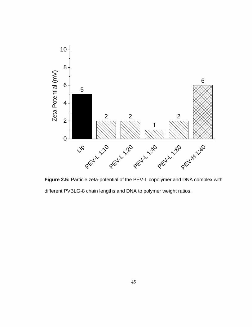
possessed a globular structure under TEM (Fig. 2.4). Zeta potential measurements were
performed to ensure that the complex formed between PEV-L (or PEV-H) and DNA were
overall positively charged. PEV/DNA complexes formed possessed zeta potentials
between 2 and 10 mV (Fig. 2.5), while PVBLG-8/DNA complexes possessed zeta
potentials between 20 and 30 mV (data not shown). The addition of PEG block to the
polypeptide shielded the cationic charge of the PVBLG-8 block.
2.3.4 Toxicity
The toxicity of the PEV-H was compared with PVBLG-8 homopolymer (P0) and
Lipofectamine 2000 via MTT assays in IMR-90 cells. PVBLG-8 (P0) was found to be
notably toxic to IMR-90 cells. At 1:40 DNA:polymer weight ratio, a viability of 6% was

31
observed. The new diblock polymers of both low (PEV-L) and high (PEV-H) molecular
weight showed substantially reduced toxicity to IMR-90 cells (Fig. 2.6). Increasing
DNA:polymer ratio resulted in increased toxicity for both PEV-L and PEV-H polymers.
PEV-H/DNA complexes showed slightly reduced toxicity compared to PEV-L/DNA
complex, but both PEV-L- and PEV-H polymers were less toxic than Lipofectamine 2000
under similar condition.
2.3.5 Transfection
Transfection experiments were performed with the diblock co-polymers to
determine if their reduced toxicity impacted their ability to effectively delivery genes to
IMR-90 cells. As shown in Fig. 2.7, the commercial reagent Lipofectamine 2000 resulted
in approximately 19% of treated cells expressing the delivered GFP transgene. Unmodified
PVBLG-8 (P0), on the other hand, had a transfection efficiency of only 1.4% with low cell
viability. While PVBLG-8 performed comparable to Lipofectamine 2000 in COS-7 and
HeLa cells in previous studies, its poor performance as shown in Fig. 7 is likely due to the
excessive toxicity of the polypeptide in IMR-90 cells. Reducing the toxicity of PVBLG-8
through the addition of PEG blocks resulted in an increased IMR-90 transfection efficiency
to 9.4% and 21.4% for PEV-L and PEV-H, respectively. Because of the reduced toxicity
of PEV-L and PEV-H, the DNA dosage could be increased from 1 µg to 2 µg, which
resulted in slightly higher transfection efficiencies, 13% and 27%, for PEV-L and PEV-H

32
formulations, respectively (data not shown). Although this increase in efficiency was
modest, it should be highlighted that the diblock polymers appeared to have reduced
toxicity. For example, even though both Lipofectamine 2000- and PEV-H-transfected cells
expressed similar amount of GFP in Figure 2.8, the cells transfected with PEV-H possessed
an overall healthier phenotype with flat and elongated shapes as opposed to cells
transfected with Lipofectamine 2000 that appeared to be sparse and rounded.
To further demonstrate the application of PEG-b-PVBLG-8, I next evaluated the
gene delivery efficiency to the H1 hESCs. PEV-H transfection in separated H1 cells
resulted in higher transfection efficiency than in cells plated as colonies (Fig. 2.9, 2.10).
Moreover, the polymer was shown to have no impact on hESC pluripotency 48 h post-
transfection. This was evidenced by the similar expression of stage specific embryonic
antigen-4 (SSEA-4) both before and after transfection in colonies and single cells (Fig.
2.11). Further evidence of pluripotency was shown through a western blot of the OCT4
pluripotency transcription factor before and after transfection with PEV-H (Fig. 2.12).
Combined with its efficiency, the mild cell impact of the PEV-H made it a promising
reagent to manipulate the gene expression of human stem cells.
2.4 Discussion
Transfection efficiency is often limited due to the toxicity of vectors. Generally, the
more efficient the transfection material, the greater impact it may have on cell health. This

33
is largely due to the requirements of effective gene delivery—namely, a high polycationic
charge to condense DNA and enable cell surface binding and membrane-lytic properties to
facilitate intracellular escape from endocytic vesicles. While good for the delivery of
nucleic acids, highly cationic materials can also bind and interfere with the function of
necessary proteins within the cell. Moreover, the membrane lytic effect of materials can be
unspecific and may act in desirable as well as undesirable manners. Effective gene delivery
materials are able to balance their positive and negative tendencies so that they are efficient
enough to allow macromolecules like DNA to enter the cells but safe enough that the cells
are not irreparably damaged during the process. In my dissertation, I focus on the
previously described PVBLG-8 materials and try to reduce their overall toxicity while
maintaining their effective gene delivery performance.
Previous characterization of the helical polypeptide PVBLG-8 revealed that it can
operate as an effective delivery vector for both DNA and siRNA [2]. In both cases, its
performance was demonstrated to be tied to its ability to form stable helices that cause pore
formation within membranes. In the case of DNA delivery, the membrane lytic potential
was essential to the escape of DNA-polypeptide complexes from endocytic vesicles. In the
case of siRNA delivery, the helical PVBLG-8 caused pore formation within cells
membranes to allow the non-endocytic diffusion of siRNA into the cell cytosol. Therefore,
in our desire to reduce the toxicity of the PVBLG-8 materials, it was also essential to retain
their helical structure. Unfortunately, just as helicity makes the materials effective delivery

34
agents, it is also a contributing factor to overall toxicity. As such, I sought a strategy to
append charge-shielding materials to the helical PVBLG-8. By shielding the positive
charge, the toxicity of the material should be minimized by reducing its electrostatic
attraction with negatively charged cell membranes. At the same time, since the helicity of
the PVBLG-8 block was maintained (Fig. 2.1a), I believed that the material would retain
its ability to effectively escape the endosome and mediate effective gene delivery (Scheme
2.1).
To test the feasibility of incorporation of charge shielding groups, PEG was
covalently conjugated with PVBLG-8 to yield the diblock polymer PEG-b-PVBLG-8. The
PEG used had a MW of 5000 Da (DP of 113) and was conjugated to helical PVBLG-8 with
DPs of 76 and 287 to yield the diblock materials PEV-L and PEV-H, respectively (Scheme
2.2). Circular dichroism experiments revealed that the incorporation of PEG did not alter
the presence and stability of the helices even after the DNA is bound and condensed by the
peptide (Fig. 2.1a). Moreover, the materials were also able to bind and condense plasmid
DNA into spherical particles with diameters on the order of 100 nm (Fig. 2.2–2.4). Despite
the inclusion of PEG, these particles were demonstrated to retain an overall positive surface
charge similar to commercial materials like Lipofectamine 2000 by analyzing their surface
zeta potential (Fig. 2.5).
Toxicity measurements with the diblock materials revealed that the inclusion of a

35
PEG block substantially increased the biocompatibility of the materials in IMR-90 cells.
For example, treatment with the unmodified PVBLG-8 left only approximately 5% of
IMR-90 cells viability. However, with the addition of a PEG block, cell viability increased
dramatically and was less toxic than Lipofectamine 2000 (Fig. 2.6). With its improved
toxicity profile, PEG-b-PVBLG-8 was able to mediate effective transfection in IMR-90
cells. Previously, the toxicity of the materials was so extreme that only 1.4% of cells were
successfully transfected. With the reduced toxicity, that number was increased to
approximately 20%—an increase of approximately 14-fold. With its improved safety
profile, PEG-b-PVBLG-8 was mild enough to transfect H1 human embryonic stem cells
without affecting the expression of cell pluripotency markers (Fig. 2.9-2.12).
2.5 Conclusion
I prepared diblock copolymers consisting of poly(ethylene glycol)-block-poly(γ-4-
(((2-(piperidin-1-yl)ethyl)amino)methyl)-benzyl-L-glutamate) (PEG-b-PVBLG-8) and
evaluated their capability to mediate gene delivery in IMR-90 human fetal lung fibroblasts
and human embryonic stem cells (hESCs). The PEG-b-PVBLG-8 contained a membrane-
disruptive, cationic, helical polypeptide block (PVBLG-8) for complexing with DNA and
a hydrophilic PEG block to improve the biocompatibility of the gene delivery vehicle.
PEG-b-PVBLG-8 copolymers with low (n = 76) and high (n = 287) degrees of
polymerization (n) of the PVBLG-8 block were synthesized. I found that the incorporation
of PEG effectively reduced the toxicity of the helical PVBLG-8 block without dramatically

36
compromising the complexation of copolymers with DNA and their transfection
efficiencies. PEG-b-PVBLG-8 diblock polymer with a high degree of polymerization had
a greater transfection efficiency and lower toxicity in IMR-90 cells than the commercial
reagent Lipofectamine 2000. The usefulness of PEG-b-PVBLG-8 was further
demonstrated via the successful transfection of hESCs without a measured loss in cell
pluripotency markers. In contrast to many other polymer- and lipid-based transfection
systems which utilize the proton sponge mechanism (e.g. polyethylenimine) or lipid mixing
(e.g. DOTAP, DOPE, etc.) to facilitate endosomal escape, the peptides described here make
use of a novel pore formation mechanism. As endocytic escape is generally considered
one of the most challenging aspects of gene delivery, this alternative endosomolytic
mechanism may prove useful when working with cell lines not readily amenable to
transfection by current methods (i.e. IMR-90 and hES cells). This method of the PEG
modification onto the PVBLG-8 backbone proved to be quite tedious and hard to control,
to scale up the diblock polypeptide is quite costly and difficult. Therefore, an alternative
gene delivery system that uses the cationic helical peptide was developed to take advantage
of the endosomal escape of the nanocomplexes.

37
2.6 Figures
Scheme 2.1: Plasmid DNA condensation by PEG-b-PVBLG-8 and the uptake
and release of DNA inside the cell.
++
+
+
++ DNA of Interest
-- -- -
-- -- -

38
Scheme 2.2: The chemical route for preparation of PEG-b-PVBLG-8 from VB-Glu-
NCA. (i) mPEG113-NH2, TBD, nitrobenzene; (ii) benzyl chloroformate, diisopropyl
ethyl amine, tetrabutylammonium floride; (iii) O3, CHCl3, -78°C, Me2S; (iv) x,
Borane-pyridine; (v) HCl

39
Table 2.1. PEG113-NH2 Initiated Polymerization of VB-Glu-NCA.
Product M:Amine:
TBDa Time (h) Conv. (%)
Mn (Mn*) (×
103 g/mol)b MWD
PEG113-b-PVBLG76 100:1:0.1 24 80 20.7 (24.6) 1.21
PEG113-b-PVBLG287 400:1:0.1 60 78 75.3 (81.4) 1.29 aTBD: 1,5,7-triazabicyclo[4.4.0]dec-5-ene; b the MW obtained (theoretical MW = Mn,
PEG + 245.27 × conv. × [M]/[I]).

40
Table 2.2. Synthesis and conformation analysis of PEG-b-PVBLG-8.a
Starting Polymer Product Grafting Eff. (%)b
[θ]222 (103deg cm2/dmol)c
Helical Content
(%)d
PEG113-b-PABLG76 PEG113-b-PVBLG76-8
(PEV-L) > 95 32.8 91.8
PEG113-b-PABLG287 PEG113-b-PVBLG287-
8 (PEV-H) > 95 34.6 96.4
a Reducing reagent (5 molar equiv) was used. Reaction was carried out for 48 h at 50oC.
bThe grafting efficiency was determined by 1H NMR analysis; cThe mean residue molar
ellipticity was calculated by following literature-reported formulas: Ellipticity ([θ]222 nm in cm2
deg dmol−1) = (millidegrees × mean residue weight)/(path length in millimeters ×
concentration of polypeptide in mg mL−1); dThe α-helix contents of the polypeptides were
calculated using the following equation: % α-helix = (-[]222 + 3000)/39,000 [10].

41
Figure 2.1: Demonstration of helicity of PEV (a) CD spectra in water of PEV-
L,PEV-H, DNA/PEV-L, and DNA/PEV-H at 1:40 weight ratio at pH 3. (b) The pH
dependence of the residue molar ellipticity at 222 nm for PEV-L and PEV-H at
0.05 mg/mL. (c) Salt dependence of residue ellipticity at 222 nm for PEV (d) The
helical stabilities of PEV-L and PEV-H at pH 3 and 0.05 mg mL−1 in the
presence of urea.
0 2 4 6 8 10
0
10
20
30
40
50
-[] 2
22 (
10
-3 d
eg c
m2/d
mol)
pH
PEV-L
PEV-H
0.0 0.2 0.4 0.6 0.8 1.0 1.2
0
10
20
30
40
50
-[] 2
22 (
10
-3 d
eg
cm
2/d
mo
l)
NaCl (M)
PEV-L
PEV-H
0.0 0.5 1.0 1.5 2.0
0
10
20
30
40
50
-[] 2
22 (
10
-3 d
eg
cm
2/d
mo
l)
Urea (M)
PEV-L
PEV-H
b) a)
c) d)
200 210 220 230 240 250-50
-25
0
25
Wavelength (nm)
[]
(10
-3 d
eg c
m2/d
mol)
PEV-L
PEV-H
DNA/PEV-L with 1:40 weight ratio
DNA/PEV-H with 1:40 weight ratio

42
Figure 2.2: DNA/Polymer complex analysis: Gel Retardation of the PEV-L at
different DNA to polymer weight ratios.
Ladder

43
Figure 2.3: Particle size analysis of the PEV-L copolymer and DNA complex with
different PVBLG-8 chain lengths and DNA to polymer weight ratios.
574
117 115 10784
246
Lip
PEV-L
1:1
0
PEV-L
1:2
0
PEV-L
1:4
0
PEV-L
1:8
0
PEV-H
1:4
0
0
200
400
600
800
1000
Part
icle
Dia
mete
r (n
m)

44
Figure 2.4: TEM image of PEV-L with DNA nanocomplex. Scale bar: 200 nm
45
Figure 2.5: Particle zeta-potential of the PEV-L copolymer and DNA complex with
different PVBLG-8 chain lengths and DNA to polymer weight ratios.
5
2 2
1
2
6
Lip
PEV-L
1:1
0
PEV-L
1:2
0
PEV-L
1:4
0
PEV-L
1:8
0
PEV-H
1:4
0
0
2
4
6
8
10
Zeta
Pote
ntial (m
V)

46
Figure 2.6: In-vitro analysis in IMR90 cells. MTT cell viability assay of the
DNA/polymer nanocomplex with different polymers and at different weight ratios in
IMR90.
Con
trol
Lip
1:2
P0
1:40
PEV-L
1:2
0
PEV-L
1:4
0
PEV-L
1:6
0
PEV-L
1:8
0
PEV-H
1:2
0
PEV-H
1:4
0
PEV-H
1:6
0
PEV-H
1:8
0
0
20
40
60
80
100
120%
Ce
ll V
iab
ility

47
Figure 2.7: Initial testing for PEV-L along with Lipofectamine 2000 (Lip) and
PVBLG-8 (P0) with varying pEGFP-N1 plasmid and polymer amount. Transfection
efficiency was analyzed 48 hours post transfection with flow cytometry.
18.6
1.4
3.9 5
.2
9.4
21.4
20.2
19.5
Lip
1:2
P0
1:40
PEV-L
1:4
0
PEV-L
1:6
0
PEV-L
1:8
0
PEV-H
1:4
0
PEV-H
1:6
0
PEV-H
1:8
0
0
10
20
% G
FP
Po
sitiv
e

48
Figure 2.8: Fluorescent images of the transfection using Lipofectamine and
PEV-H. Scale bar: 250 µm
Lip PEV-L PEV-H
Bright field
GFP

49
6.5
4.95.5
7.7
Lip 1:2
PEV-H 1:10
PEV-H 1:20
PEV-H 1:40
0
2
4
6
8
10%
GF
P P
ositiv
e
Figure 2.9: EGFP plasmid transfection efficiency using Lipofectamine 2000 and
PEV-H of hESC H1 as small colonies

50
54.7
18.1
30.7
50.0
Lip 1:2
PEV-H 1:10
PEV-H 1:20
PEV-H 1:40
0
10
20
30
40
50
60
70
% G
FP
Po
sitiv
e
Figure 2.10: EGFP plasmid transfection efficiency using Lipofectamine 2000 and
PEV-H of hESC H1 as single cells as analysed by flow cytometry.

51
Negative
PEV-H
Colonies Single Cells
Bright field
Hoechst
SSEA-4
GFP
Negative
PEV-H
Fig
ure
2.1
1:
Brigh
t fie
ld a
nd
flu
ore
scen
ce
im
agin
g o
f P
EV
-H tra
nsfe
ctio
n o
f E
GF
P p
lasm
id into
hE
SC
as
co
lon
ies a
nd s
ingle
ce
lls. C
olo
nie
s w
ere
sta
ine
d w
ith
Ho
ech
st a
nd
plu
rip
ote
ncy m
ark
er
SS
EA
-4 a
ntib
od
y
co
nju
ga
ted
with
PE
. S
ca
le b
ar:
250
µm

52
Figure 2.12: Western blot of cells isolated 72 h post transfection demonstrating
the protein expression of OCT4 in hESC H1 cells.
Oct4
Alpha Tubulin

53
2.7 References
[1] Lu H, Bai YG, Wang J, Gabrielson NP, Wang F, Lin Y, et al. Ring-Opening
Polymerization of gamma-(4-Vinylbenzyl)-L-glutamate N-Carboxyanhydride for the
Synthesis of Functional Polypeptides. Macromolecules 2011;44(16):6237-40.
[2] Gabrielson NP, Lu H, Yin LC, Li D, Wang F, Cheng JJ. Reactive and Bioactive
Cationic a-Helical Polypeptide Template for Nonviral Gene Delivery. Angewandte
Chemie-International Edition 2012;51(5):1143-47.
[3] Vanheeswijk WAR, Eenink MJD, Feijen J. An Improved Method for the Preparation
of Gamma-Esters of Glutamic-Acid and Beta-Esters of Aspartic-Acid. Synthesis-Stuttgart
1982(9):744-47.
[4] Lu H, Wang J, Bai YG, Lang JW, Liu SY, Lin Y, et al. Ionic polypeptides with unusual
helical stability. Nat Commun 2011;2:206.
[5] Lu H, Wang J, Lin Y, Cheng J. One-pot synthesis of brush-like polymers via integrated
ring-opening metathesis polymerization and polymerization of amino acid N-
carboxyanhydrides. J Am Chem Soc 2009;131(38):13582-83.
[6] Lu H, Cheng J. N-trimethylsilyl amines for controlled ring-opening polymerization of
amino acid N-carboxyanhydrides and facile end group functionalization of polypeptides. J
Am Chem Soc 2008;130(38):12562-63.
[7] Lu H, Cheng JJ. Hexamethyldisilazane-mediated controlled polymerization of alpha-
Amino acid N-carboxyanhydrides. J Am Chem Soc 2007;129(46):14114-15.

54
[8] Bai YG, Lu H, Ponnusamy E, Cheng JJ. Synthesis of hybrid block copolymers via
integrated ring-opening metathesis polymerization and polymerization of NCA. Chem
Commun 2011;47(38):10830-32.
[9] Greenfield NJ. Using circular dichroism spectra to estimate protein secondary structure.
Nat Protoc 2006;1(6):2876-90.
[10] Morrow JA, Segall ML, Lund-Katz S, Phillips MC, Knapp M, Rupp B, et al.
Differences in stability among the human apolipoprotein E isoforms determined by the
amino-terminal domain. Biochemistry-Us 2000;39(38):11657-66.
[11] Gabrielson NP, Lu H, Yin L, Kim KH, Cheng J. A Cell-penetrating Helical Polymer
For siRNA Delivery to Mammalian Cells. Mol Ther 2012;20(8):1599-609.

55
Chapter 3:
Coating of cationic, helical peptide with hyaluronic acid for
smart, safe, and targeted non-viral gene delivery
Significant portions of this chapter were published as "Enhanced Non-Viral Gene Delivery
to Human Embryonic Stem Cells via Small Molecule-Mediated Transient Alteration of
Cell Structure" Jonathan Yen, Lichen Yin, and Jianjun Cheng, J. Mat. Chem. B., 2014, 2,
8098-8105.
3.1 Introduction
PVBLG-8 has been demonstrated as a gene delivery system with high membrane
activity and endosomal escape, which can be attributed to the cationic helicity and rigid
structure[1]. Although PVBLG-8 showed some gene transfection in hESCs, it also
exhibited remarkable cytotoxicity to the cells due to the cationic helical characteristics of
the peptide[1]. There is a fine balance between the transfection efficiency and toxicity of
cationic helical peptides. There have been chemical methods to modify the cationic helical
peptide to reduce its toxicity, like structural reconfiguration[2], conjugations[3-6], and
triggered degradation of the material [7].These modifications can effectively reduce the
toxicity through the reduction of the cationic charge density on the peptide, while retaining
its transfection efficiency. Yet, the conjugation of PEG onto PVBLG-8 as I described
earlier was tedious and not easily tuneable.
56
Herein, I decided to take a formulative approach to develop a new gene delivery
system based on self-assembled DNA/PVBLG-8 nanocomplexes coated by negatively
charged hyaluronic acid (HA). HA can target and specifically bind to CD44, a known
receptor specific to HA with high expression level in hESCs[8]. HA has been used in
nanomedicine to target tumors, which also have high CD44 expression levels[9-14]. By
coating the nanocomplexes with HA, it not only shields the charges to decrease the toxicity,
but also acts as a targeting moiety for receptor-based endocytosis through HA/CD44
interactions. Furthermore, the HA can be deshielded through the pH drop and actions of
endogenous hyaluronidase after internalization to expose the cationic helical peptides,
allowing rapid and efficient endosomal release and expression of DNA plasmids. The HA
coated nanocomplex demonstrates three important characteristics that enhance gene
transfection efficacy in hESCs; an outer shell with active targeting moiety against CD44,
an enzyme and pH sensitive targeting shell that can be released in the endosome exposing
the cationic helical peptides, and the cationic helical peptides which can effectively allow
for endosomal escape (Scheme 3.1).
3.2. Materials and Methods
3.2.1 General
hESCs H1 (hESC-H1) was cultured in E8 medium from Stem Cell Technologies
(Vancouver, Canada). Y-27632 was purchased from Stemgent (Cambridge, MA, USA).

57
YOYO-1 was purchased from Invitrogen (Carlsbad, CA, USA). pEGFP-N1 was obtained
from Elim Biopharmaceuticals (Hayward, CA, USA). Milli-Mark™ Anti-SSEA-4-PE was
purchased from EMD Millipore (Billerica, MA, USA). PVBLG-8, a helical cationic
polypeptide with a polymerization degree of 200, was synthesized following our reported
procedures [15].
3.2.2 Instrumentation
Flow cytometry analysis was conducted on a BD LSRII flow cytometry analyzer
(Becton Dickinson, Franklin Lakes, NJ). Cells were visualized with a Zeiss Axiovert 40
CFL fluorescence microscope equipped with a 10 and 20x objective (Thornwood, NY).
Zeta potential and size analysis was conducted on the Malvern Zetasizer (Worcestershire,
UK).
3.2.3 Nanocomplex formation and characterization
Various non-viral vectors were used for the transfection analyses. Plasmid DNA (1
µL, 1mg/mL) was diluted in water (25 µL). PVBLG-8 (7.5μL, 1 mg/mL) was diluted with
water (25 µL). For the PVBLG-8 and coated nanocomplex procedures, the PVBLG-8 and
plasmids were mixed at various w:w ratios and were incubated for 20 m at rt for complex
formation. Hyaluronic acid (2.5-50μL,1mg/mL), dissolved in water, was added to the
nanocomplex mixture and incubated for another 20 m at rt. For the mixed system, the DNA
and hyaluronic acid were first mixed together at various w:w ratios and then the PVBLG-

58
8 was added at various ratios. The mixture was incubated at rt for 30 m for complex
formation. Dynamic light scattering (DLS) and zeta potential analysis was conducted on
the samples with a Malvern Zetasizer (Worcestershire, UK).
The nanocomplexes were subjected to electrophoresis in 1% agarose gel at 100 V
for 45 minutes to evaluate DNA condensation by the polypeptides in terms of DNA
migration.
3.2.4 In-vitro Gene Transfection
Cells were seeded in matrigel coated 24 well plates as single cells using accutase
and incubated for 24 hours with rho-kinase inhibitor Y-27632 (10 μM). Using fresh low
protein and serum free E8 medium, nanocomplexes as described earlier were added at 1μg
DNA/well. After incubation for 4 h, the medium was replaced with fresh E8 medium and
cells were cultured for 48 h. The cells were collected with accutase, fixed with
paraformaldehyde and analyzed using flow cytometry to determine transfection efficiency.
3.2.5 In-vitro HA blocking
hESCs were incubated with free hyaluronic acid (300kDa, 1-5mg/mL) for 1 h prior
to transfection. The cells were then washed 1x with PBS and placed back with fresh E8
media. The nanoparticles were added to the cells at 1 µg DNA/well. After incubation for 4
h, the medium was replaced with fresh E8 medium and cells were cultured for 48 h. The
59
cells were collected with accutase, fixed with paraformaldehyde and analyzed using flow
cytometry to determine transfection efficiency.
3.2.6 pH and hyaluronidase exposure of nanocomplex
Nanocomplexes were formulated as previous described. HA coated nanocomplexes
(400 µL, 100 µg/mL DNA) were incubated with hyaluronidase (100 µL, 0.5 mg/mL) in
phosphate and citric acid buffer with a pH of 7.4, 6.8, 6.2 or 5.6 for 0, 30, or 120 m at 37°C.
The HA coated nanocomplexes (100 µL) were then diluted in their corresponding buffer
and the zeta potential of the resulting particles were analysed with the Zetasizer.
To test the membrane activity of the nanoparticles, Large Unilamellar Vesicles by
Extrusion Technique (LUVETS)[16] loaded with 8-aminonaphthalene-1,3,6-trisulfonic
acid (ANTS) and p-xylene-bis-pyridinium bromide (DPX) were formulated[17].
Briefly,DOPC (100 µL, 25 mg/mL CHCl3) and POPC (1 mL, 10 mg/mL CHCl3) were
mixed in round bottom flask and evaporated with a rotary evaporator to create dry thin
film. The film was allowed to dry overnight under vacuum. The lipid film was rehydrated
by 5 mL of ANTS/DPX solution (12.5 mM ANTS, 45 mM DPX, 10 mM phosphate buffer,
pH 7.4) for 30 m at rt. The solution was then put through 5 freeze thaw cycles in liquid
nitrogen and like warm water. The solution was then extruded 15 times through 0.4 µM
polycarbonate membranes. Any extravesicular components were removed through a
Sephadex G-50 gel filtration column.
60
Nanocomplexes were formulated as previous described. HA coated nanocomplexes
(400 µL, 100 µg/mL DNA) were incubated with hyaluronidase (100 µL, 0.5 mg/mL) in
phosphate and citric acid buffer with a pH of 7.4 or 6.8 for 0 or 30 m at 37°C. The HA
coated nanocomplexes (10 µL) and the ANTS/DPX LUVETs (5 µL) were mixed diluted
in a final solution of the corresponding buffer of 100 µL. Triton x was used as the positive
control. The leaked ANTS dye was measured using a plate reader at 360 nm excitation and
530 nm emission. The final leakage percentage was calculated using the ratio of
fluorescence of the sample with the positive control after background subtraction.
3.2.7 Cell viability
Cells were seeded in matrigel coated 96 well plates as single cells using accutase
and incubated for 24 hours with y-27632 (10 µM). Using fresh low protein and serum free
E8 medium, nanocomplexes were added at 0.2µg DNA/well. After incubation for 4 h, the
medium was replaced with fresh E8 medium and cells were cultured for 48 hours. Cell
viability was evaluated through a MTT assay. Cells without complex treatment served as
the control and results were expressed as percentage viability of control cells.
3.2.8 Intracellular Uptake Studies
DNA (1 mg/mL) was mixed and labeled with YOYO-1 (20 µM) at one dye
molecule per 50bp of DNA. The HA coated nanocomplexes were then formed with

61
YOYO-1/DNA as discussed before at a DNA:PVBLG-8:HA weight ratio of 1:7.5:7.5.
hESCs were plated on matrigel coated 24-well plate and allowed to grow to medium sized
colonies. The complexes were added to the wells at 1 µg of YOYO-1-DNA per well and
incubated for 4 h at 37°C. The cells were then quantified through flow cytometry to
quantify YOYO-1 uptake. Results are expressed as mean fluorescence levels.
To elucidate the mechanisms regarding the cellular internalization of
DNA/PVBLG-8/HA complexes, the uptake study was performed at 4 °C or in the presence
of various endocytic inhibitors for 2 h. Cells were pre-treated with chlorpromazine (10
µg/mL), genistein (200 µg/mL), dynasore (80 µM), and wortmannin (50 nm) for 30 min
before the complexes were added and throughout the experiment for 2 h at 37°C. Results
are expressed as percentage mean GFP fluorescence level at 37 °C in control cells in the
absence of endocytic inhibitors.
3.2.9 Western blot analysis and SSEA staining
After 72 h, the cells were stained with DAPI (250 µL, 3 nM) and SSEA-4–PE (250
µL, 0.02 mg/mL), a pluripotency cell marker, for 30 min at 37 °C. After 5 d, the cells were
collected with RIPA buffer and mixed with Laemmli buffer supplemented with 2-
Mercaptoethanol and heated for 5 m at 100°C to denature the proteins and then put on ice.
The samples were ran on 10% SDS PAGE Gel at 120 V for 1.5 h, and wet transferred to
the nitrocellulose membrane using the AMRESCO Rapid Western Blot Kit per

62
manufacturer’s instructions. The membrane was stained with OCT4 and α-Tubulin primary
antibodies and then with HRP-tagged secondaries.
3.3. Results
3.3.1 Characterization of DNA:PVBLG-8:HA Nanocomplexes
DNA condensation by PVBLG-8 followed by HA coating was evaluated by gel
retardation assay. As shown in Fig. 3.1, DNA can be bind tightly to PVBLG-8 with a
weight feed ratio at 1:7.5,and the coating of HA on the nanocomplex did not disrupt or
displace the DNA in the nanocomplexes even when the feeding ratio of DNA to PVBLG-
8 to HA at 1:7.5:50. This result indicated that the complexes are relatively stable through
the coating method, and the negative charges on the HA does not interfere in the assembly
or disassemble the nanocomplexes. Yet, when the HA was mixed directly with the DNA
and PVBLG-8, the complex was unstable and some DNA leakage was detected (Fig. 3.2).
The size and zeta potential of the DNA/PVBLG-8 nanocomplexes at 1:7.5 weight
ratios with varying HA weight ratio coatings were characterized using the DLS and
zetasizer. Without HA coating, the nanocomplex size was 57.9 nm in diameter. After the
coating of HA, at between 1:7.5:5 and 1:7.5:15 weight ratios, the effective diameters
increased to around 170 nm. With further addition of HA to 1:7.5:50, the diameter
increased to 270 nm. The zeta potential of the nanocomplex without HA, measured to be
19.2 mV, decreased to 11.1 mV with HA coating at 1:7.5:1 ratio indicating that the

63
nanocomplexes retained their positive charges. The zeta potential then decreases to
between -18 and -25 mV when the ratio of HA was between 1:7.5:5 and 1:7.5:15 and then
drops to -42 mv at 1:7.5:50. The increase and stabilization of the size and zeta potential
demonstrates that the nanocomplexes were successfully coated with HA (Fig. 3.3).
The stability of the nanocomplexes was tested through the use of the dynamic light
scattering to determine the size of the nanoparticles over time. The particles were tested at
30 min and 4 h after formulation at 37°C. As previously demonstrated, the size of the
nanocomplexes with HA coating ranged from 150-180 nm and less than a hundred nm
without the HA coating. Thus, if the HA coating were to come off or be destabilized, the
size of the nanocomplex would become smaller. The sizes of nanocomplex remained
constant in the different mediums, thus indicating that the nanocomplexes are quite stable
(Fig. 3.4).
3.3.2 hESC Transfection with HA Coated DNA/PVBLG-8 Nanocomplexes
Through the optimization of various DNA:PVBLG-8 weight ratios, 1:7.5 is the
optimal ratio for the highest transfection efficiency (Fig. 3.5). Thus, the 1:7.5 ratio was
used with various ratios of HA coating on the nanocomplexes. Without HA coating, the
nanocomplexes achieved 22% transfection efficiency, while with the increased ratio of HA
coated on the nanocomplexes, the transfection efficiency went up to 28%, 27% and 30%
for 7.5:2.5, 7.5:5, 7.5:7.5 of PVBLG-8:HA ratios, respectively. As the amount of HA was
64
increased to 15 and 30, the transfection efficiency dropped significantly to 20% and 17%,
respectively (Fig. 3.6).This drop in transfection efficiency can be attributed to the excess
HA in the system that did not coat the nanocomplexes, which compete for the CD44
receptors. PVBLG-8 by itself has also proven to be quite toxic to the cells, (Fig. 3.7, 3.17),
where the cells are unhealthy and littered with cell debris. On the other hand, with HA
coating the cells demonstrated lower toxicity and higher transfection. The HA coating not
only acts as a targeting moiety, but also significantly counters the charges from the
PVBLG-8 thus decreasing the overall toxicity in hESCs.
To demonstrate that the targeting of the HA coated nanocomplexes was due to the
CD44 binding of the HA coated on the surface, pre-treatment of free HA was applied to
the cells for 30 min and then washed off before transfection. With the treatment of 2.5 and
5 mg/mL of free HA pre-treated on the cells , a drop of transfections to 8% and 6% were
shown, respectively, from 35% without pre-treatment (Fig. 3.8). This indicates that free
HA has bound to the cell surface CD44 receptors and blocked them from further uptake,
indicating that the CD44/HA interactions are an important aspect to the increased
transfection efficacy of the nanocomplex. To further demonstrate the importance of the
CD44 receptor for the system, the HA coated nanocomplexes were transfected in other
CD44 positive and negative cells, human mesenchymal stem cells (hMSCs) and COS-7
cells, respectively. For CD44 positive hMSCs, HA coated nanocomplexes showed almost
twice the transfection efficiency than those without HA coating. On the other hand, in

65
CD44 negative COS-7 cells, without the HA coating, the transfection efficiency was 36%
and it dropped down to 15% after HA coating (Fig. 3.9). These trends demonstrate the
important roles the CD44 receptors play in the transfection, which may due to the increased
uptake of the HA coated nanocomplexes.
3.3.3 Mechanistic Studies of DNA/PVBLG-8/HA Nanocomplexes
To demonstrate that the enhanced gene transfection is due to the increased uptake
through the CD44 receptor mediated pathway, the total uptake of DNA was evaluated.
Using DNA tagged with YOYO-1, an uptake study was done to determine the amount of
DNA/YOYO-1 uptake into the cells. It can be seen that without HA coating, the mean
YOYO-1 fluorescence level measured to be about 61%, while with the HA coating at
1:7.5:7.5 ratio, the mean YOYO-1 fluorescence was significantly higher at 67% (Fig 3.10).
In addition, with the treatment of free HA, the mean YOYO-1fluorescence level declined
significantly to around 44%, indicating a reduced nanocomplex uptake. Alternatively, the
uptake of DNA/YOYO-1 in CD44 negative COS-7 cells is reduced significantly after the
coating of HA, further demonstrating that the CD44 receptor mediated the uptake of HA
coating nanocomplex (Fig 3.11).
To show that the HA coated nanoparticles were indeed targeting the CD44
receptors, hESCs were stained with CD44 antibodies and incubated with HA coated
DNA/YOYO-1/PVBLG-8nanocomplexes for 2 h at 4°C. Through confocal imaging, CD44

66
is stained uniformly across the surface of the hESCs. The DNA/YOYO-1 can be visualized
on the cell surface co-localized with the CD44 receptors, indicating the importance of the
roles of the CD44/HA interactions (Fig 3.12).
To elucidate the mechanisms underlying the cellular internalization of the HA
coated nanocomplexes, an uptake study at 4°C or in the presence of various endocytic
inhibitors was performed. Energy dependent endocytosis was blocked at 4°C; clathrin
mediated endocytosis was blocked by chlorpromazine, which triggers dissociation of the
clathrin lattice; caveolae was inhibited by genistein and mβCD by inhibiting tyrosine
kinase and depleting cholesterol, respectively; macropinocytosis was inhibited by
wortmannin by inhibiting phosphatidyl inositol-3-phosphate[18,19]. When the cells were
incubated at 4°C, the uptake dropped to almost 3% of the control, while chlorpromazine
dropped to 62%, genistein dropped to 22%, and wortmannin to 91% of the control (Fig.
3.13). The dramatic drop of transfection efficiency at 4°C indicates the process is energy
dependent, and with the drop in uptake with the treatment of chlorpromazine and genistein,
further demonstrates that the nanocomplexes enters the cells through receptor mediated
endocytosis.
Due to the quick membrane disruption ability of PVBLG-8, the YOYO-1-DNA can
quickly escape the endosomes and permeate into the cell cytoplasm after 2 h incubation at
37°C (Fig. 3.14). Therefore a substitute, poly-lysine, a cationic peptide with lower

67
membrane disruption ability was used to visualize the uptake and localization of the HA
coated complexes. Through confocal imaging, there is significant co-localization of the
HA coated YOYO-1-DNA/PLL complex with the lysotracker red stain as seen through
CLSM confocal imaging (fig 4b). It demonstrates that HA coating helps the nanocomplex
uptake through receptor mediated endosomal pathway. But it also showed that PVBLG-8
nanocomplex induces faster and more efficient endosomal escape, which is beneficial for
gene transfection[1]. PVBLG-8 is exposed to allow for membrane disruption and
endosomal escape once the nanocomplex reaches the endosomes.
3.3.4 Exposure of PVBLG-8 of HA Coated Nanocomplex for Endosomal Escape
To demonstrate the endosome triggerable membrane disruption of HA coated
nanocomplexes for rapid endosomal escape, the nanocomplex zeta potentials and
membrane disruption abilities were evaluated after treatment with hyaluronidase (HAase)
at varying endosomal pH. At the initial time point of incubation of the HAase with varying
pH of 7.4, 6.8, 6.2 and 5.6, the zeta potential remains negative at -21, -17.6, -16.8, and -
10.3 mV, respectively. After 30 min incubation, the zeta potential at pH of 7.4 and 6.8
increased but remained negative at -18.2 and -8.32 mV, respectively. On the other hand,
the zeta potential at pH of 6.2, and 5.6, all increased to the positive range of 1.34 and 9.39
mV, respectively. Finally, after 120 min the zeta potential at pH of 7.4 stays negative at -
9.3 mV. But, for the pH at 6.8, 6.2 and 5.6, the zeta potential increases to positive of 1.69,
7.2 and 11.8 mV, respectively. A pH of 7.4 is representative of the cytoplasmic pH, while
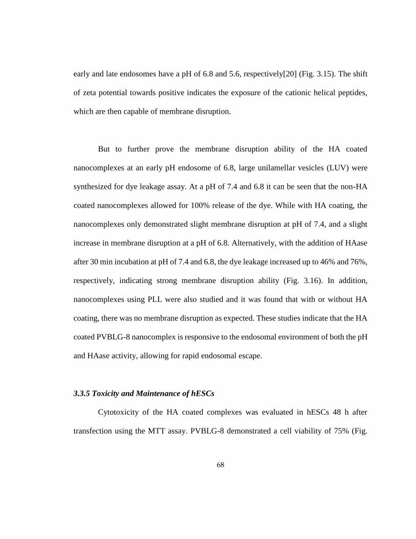
68
early and late endosomes have a pH of 6.8 and 5.6, respectively[20] (Fig. 3.15). The shift
of zeta potential towards positive indicates the exposure of the cationic helical peptides,
which are then capable of membrane disruption.
But to further prove the membrane disruption ability of the HA coated
nanocomplexes at an early pH endosome of 6.8, large unilamellar vesicles (LUV) were
synthesized for dye leakage assay. At a pH of 7.4 and 6.8 it can be seen that the non-HA
coated nanocomplexes allowed for 100% release of the dye. While with HA coating, the
nanocomplexes only demonstrated slight membrane disruption at pH of 7.4, and a slight
increase in membrane disruption at a pH of 6.8. Alternatively, with the addition of HAase
after 30 min incubation at pH of 7.4 and 6.8, the dye leakage increased up to 46% and 76%,
respectively, indicating strong membrane disruption ability (Fig. 3.16). In addition,
nanocomplexes using PLL were also studied and it was found that with or without HA
coating, there was no membrane disruption as expected. These studies indicate that the HA
coated PVBLG-8 nanocomplex is responsive to the endosomal environment of both the pH
and HAase activity, allowing for rapid endosomal escape.
3.3.5 Toxicity and Maintenance of hESCs
Cytotoxicity of the HA coated complexes was evaluated in hESCs 48 h after
transfection using the MTT assay. PVBLG-8 demonstrated a cell viability of 75% (Fig.

69
3.17). But with the HA coated nanoparticles, the cell viability increased to approximately
90% with the HA coated at 1:7.5:7.5 weight ratios.
To ensure that the materials did not alter the cell’s behavior or cause any
differentiation, the cells were stained with SSEA-4 antibodies, stage specific embryonic
antigen-4, 72 h after transfection, in which the cells all stained positive (Fig. 3.18). In
addition, cell lysate were collected 5 days after transfection and a western blot was
conducted to confirm the continued expression of the pluripotent factor, OCT4 (Fig. 3.19).
The expression of SSEA-4 and OCT4 demonstrates that this system does not cause
undesired differentiation or interferes with the pluripotency of the hESCs.
3.4. Discussion
Here, I developed a novel gene transfection system that contains an outer negatively
charged cell targeting shell consisting of HA, with an inner core made from a cationic
helical peptide that is able to condense DNA and facilitate endosomal escape. The system
utilizes the charge interactions between the outer negatively charged shell and positive
inner shell to be stable at neutral pH, but allows for the deshelling and exposure of the
positive helical peptides when exposed to a lower pH and HAase with in the endosome.
With the HA coating of the nanocomplexes, the hESC transfection efficiency of
DNA/PVBLG-8 nanocomplexes increases significantly, despite the alteration of the
overall charge of positive to negative. HA is a negatively charged biocompatible

70
polysaccharide that is found naturally in the body and has been used as the backbone of
many hydrogel systems for stem cell engineering[8]. A few glycosaminoglycans, except
for hyaluronic acid, have been shown to disrupt cationic polypeptide gene
transfections[21]. The reversal of charge indicates the loss of the initial membrane
disruption effect of the cationic helical peptide, which in theory should have decreased
transfection. However, the negative nanocomplexes increased the transfection efficiency,
indicating that the HA on the nanocomplexes is an integral part to the transfection
efficiency, in which they target the CD44 receptors that are expressed on hESC surfaces.
At up to 1:7.5:7.5 weight ratios, the transfection efficiency increases, then drops with
additional HA coating. This drop is likely to be caused by the excess HA that were not
coated on the nanocomplex. This can be seen by the DLS and zetapotential, that after a
certain ratio, the size and zeta potential of the nanocomplex stays constant. Thus, the excess
free HA could be competitively binding with the CD44 receptor against the nanoparticles,
demonstrated by the drastic drop in transfection efficiency with the pre-treatment of HA.
The drop demonstrates that the free HA has bound to the cell surface CD44 receptors and
blocked them from further uptake, indicating that the CD44/HA interactions are an
important aspect to the increased efficacy of the nanocomplex. The HA coated
nanocomplexes also showed a higher efficacy in CD44 positive hMSCs, yet in CD44
negative cells, COS-7 cells dropped significantly. The drop in transfection efficiency in
CD44 negative COS-7 cells with HA coated nanocomplexes, indicates that the CD44
receptor is indeed an important factor for the uptake of the HA coated nanocomplexes.

71
To understand the mechanism of uptake, I conducted uptake studies of DNA tagged
with YOYO-1 dye and found that the cells did significantly increase the uptake of the
nanoparticles with the HA coating. Through the study with various endocytosis inhibitors,
the uptake was drastically inhibited at 4°C, indicating that the uptake of the particles was
highly energy dependent. In addition, the decrease of the uptake due to the chlorpromazine,
demonstrated that some of the particles were taken up through clathrin dependent
endocytosis. On the other hand, genistein also had a large decrease in uptake of the YOYO-
1/DNA, thus indicating that some of the uptake is due to clathrin independent pathways
like caveolae mediated uptake. This denotes the importance of the CD44 receptor mediated
endocytosis. Both CD44[22] receptors and caveolin[23] have been known to be present in
lipid rafts domains, which are the sites of endocytosis in caveolae mediated endocytosis,
indicating the localization of the HA nancomplexes at CD44 containing lipid rafts
domains[24].
Commonly used transfection materials like polyethylenimine and polylysine have
the ability to condense DNA for non-viral gene delivery, but PEI demonstrates slower
endosomal release through the proton sponge effect, while polylysine demonstrates no
endosomal escape[25,26]. Our nanocomplex system transforms to exhibit membrane
disruptive properties in the endosomal environment of low pH and HAase activity. The
exposure of PVBLG-8 in the system enhances the endosomal release of the nanocomplex

72
and thus the DNA. When the materials were switched to poly-L-lysine, a non-membrane
disrupting polymer, there was little endosomal escape, and the nanocomplexes were mainly
retained in the endosomes. In early endosomes, at a low pH and with some HAase activity,
the HA coated nanocomplexes demonstrated increased LUV dye leakage due to membrane
disruption. Our system is superior to other materials, due to our highly available and active
membrane disruption activity that is initially shielded to reduce cytotoxicity to the cells,
but is revealed when it enters the endosomes.
Not only did the HA coated nanocomplexes increase transfection efficiency, they
also lowered the toxic effect of the cationic helical peptide. It also has little effect on the
cells pluripotency, for the hESCs still proved to be both OCT4 and SSEA4 positive. Thus,
the use of a targeting moiety for a receptor on hESCs is a step in looking at non-viral gene
delivery into hESCs differently, in which a cell’s specific mechanism is exploited to
enhance transfection efficiency.
3.5 Conclusion
Here I report a new targeting and triggerable gene delivery system for human
embryonic stem cells based on the PVBLG-8 peptide, a cationic helical peptide with high
membrane disruption properties. The DNA/PVBLG-8 nanocomplex by itself proves to be
toxic to the cells due to the high membrane disruption properties. Using HA as a coating
for the nanocomplexes not only decreases the toxicity from the cationic helical peptides,

73
but allows a specific targeting moiety for hESCs that increases the uptake and release of
DNA, which in turn increases the transfection efficiency. But after the material is taken up
through endocytosis, the low pH and HAase are able to degrade and the HA on the
nanocomplex is removed exposing the positive peptides and allowing for endosomal
escape through membrane disruption. This provides a promising approach for
manipulation of hESCs or pluripotent stem cells through transient gene delivery,
overcoming major hurdles towards development of many biomedical applications.
Increased gene transfection efficiency reduces the need for enrichment and sorting of the
manipulated pluripotent stem cells. These preliminary studies lead to further possibilities
of selective gene delivery to undifferentiated hESCs and potential further modification of
the HA coating for PVBLG-8 based in-vivo gene delivery to other CD44 positive cells. The
use of HA to target and enhance the non-viral gene delivery system provides an alternative
method that does not just rely on the chemical characteristics of the material for gene
delivery, but also targets and takes advantage of the biological properties of hESCs. Given
this information, I then looked further into other physiological properties of hESCs I could
use to further enhance gene transfection efficiency.

74
3.6 Figures
Scheme 3.1: Formulation and uptake route of the hyaluronic acid coated
PVBLG-8/DNA nanocomplexes
CD44
Receptor
Plasmid
DNAPVBLG-8
-
-
-
- +
+
+
+
+
+
+
+
+
+
-
-
-
-
-
-
- -Hyaluronic
Acid

75
Figure 3.1: Gel Retardation of HA coated nanocomplexes at varying
DNA:PVBLG-8 weight ratios.
DNA (w) 1 1 1 1 1 1 1 1 1 1
PV-BLG-8 (w) 0 7.5 7.5 7.5 7.5 7.5 7.5 7.5 7.5 7.5
HA (w) 0 0 1 2.5 5 7.5 10 15 25 50

76
Figure 3.2: Gel Retardation of HA coated nanocomplexes at varying
DNA:PVBLG-8 weight ratios.
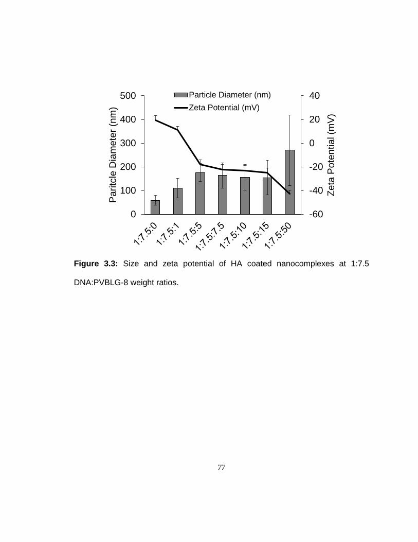
77
Figure 3.3: Size and zeta potential of HA coated nanocomplexes at 1:7.5
DNA:PVBLG-8 weight ratios.
-60
-40
-20
0
20
40
0
100
200
300
400
500
Ze
ta P
ote
ntia
l (m
V)
Pa
ritc
le D
iam
ete
r (n
m)
Particle Diameter (nm)
Zeta Potential (mV)

78
Figure 3.4: Stability of HA coated nanocomplexes at 1:7.5:7.5 DNA:PVBLG-8:HA
weight ratios in water, PBS and E8 medium as determine by size change
measured by DLS.
0
100
200
300
400
After formation 30 min (37°C) 4 hour (37°C)
Com
ple
x S
ize (
nm
)
Water PBS E8

79
Figure 3.5: Gene transfection optimization at varying DNA:PVBLG-8:HA coating
ratios (w:w:w).
0
5
10
15
20
25
30
35
40
1:x:0 1:x:2.5 1:x:5 1:x:7.5 1:x:10 1:x:15 1:x:30
% G
FP
Po
sit
ive
x=5
x=7.5
x=10
x=12.5
x=15

80
Figure 3.6: Transfection of hESCs with varying ratios of HA coating on
DNA:PVBLG-8 nanocomplexes.
0
10
20
30
40
% G
FP
Positiv
e
DNA:PVBLG-8:HA (w:w:w Ratio)
**

81
Figure 3.7: Fluorescence images of H1 hESCs 48 h post transfection with and
without HA coated nanocomplexes and with 5 mg/mL free HA pretreatment. Scale
bar: 250 µm
ControlNo HA
coating
HA
coating
Pre-
treatment
BF
GFP
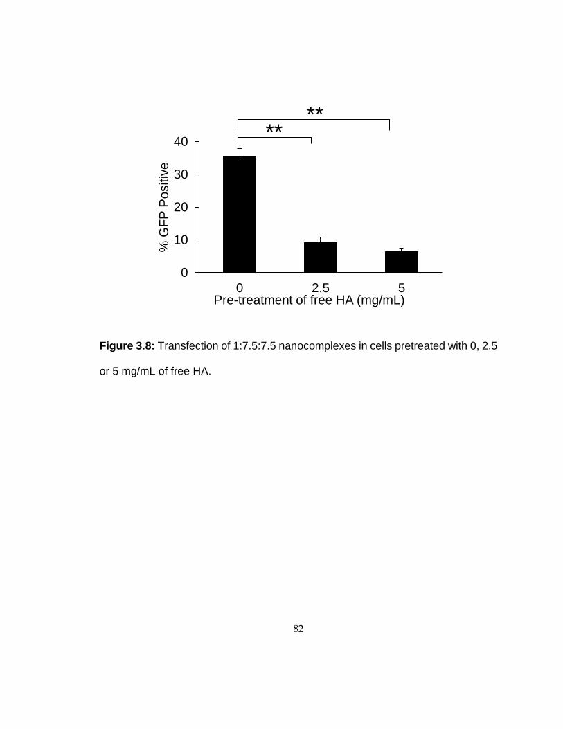
82
Figure 3.8: Transfection of 1:7.5:7.5 nanocomplexes in cells pretreated with 0, 2.5
or 5 mg/mL of free HA.
0
10
20
30
40
0 2.5 5
% G
FP
Po
sitiv
e
****
Pre-treatment of free HA (mg/mL)
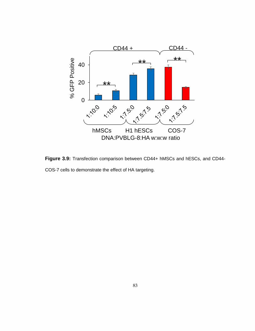
83
Figure 3.9: Transfection comparison between CD44+ hMSCs and hESCs, and CD44-
COS-7 cells to demonstrate the effect of HA targeting.
0
20
40
% G
FP
Po
sitiv
e
**
H1 hESCs COS-7
** **
hMSCs
CD44 + CD44 -
DNA:PVBLG-8:HA w:w:w ratio
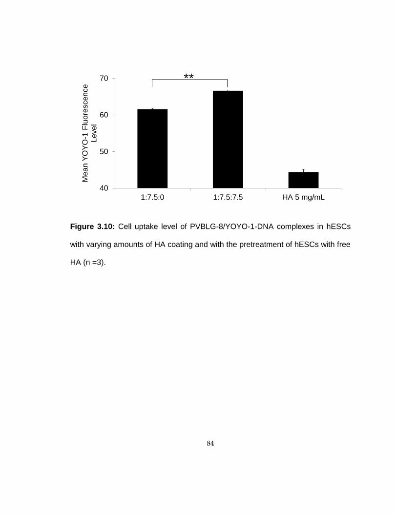
84
Figure 3.10: Cell uptake level of PVBLG-8/YOYO-1-DNA complexes in hESCs
with varying amounts of HA coating and with the pretreatment of hESCs with free
HA (n =3).
40
50
60
70
1:7.5:0 1:7.5:7.5 HA 5 mg/mL
Mean Y
OY
O-1
Flu
ore
scence
Level
**

85
Figure 3.11: Cell uptake level of YOYO-1-DNA /PVBLG-8 complexes with and
without HA coating in CD44 negative COS-7 cells at 1:7.5 weight ratios (n =3).
0
5
10
15
20
25
30
35
40
45
No coating HA coating
Flu
ore
sce
nce
Un
it L
eve
l p
er
50
,00
0 c
ells
(x1
00
0)
*

86
Figure 3.12: CLSM images of hESCs incubated with HA coated PVBLG-8/YOYO-
1-DNA complexes at 4° C for 1 h with CD44 antibody staining. Scale bar: 10 µm
Control
Hoechst CD44 YOYO-1/DNA Merge
HA coated
PVBLG-
8/YOYO-1-
DNA
b
.

87
Figure 3.13: Cell uptake mechanisms HA coated PVBLG-8/YOYO-1-DNA
complexes in hESCs through the pre-treatment and treatment of various
endocytotis inhibitors (n=3).
0
25
50
75
100%
Mean
Flu
ore
scence L
evel
of
Contr
ol
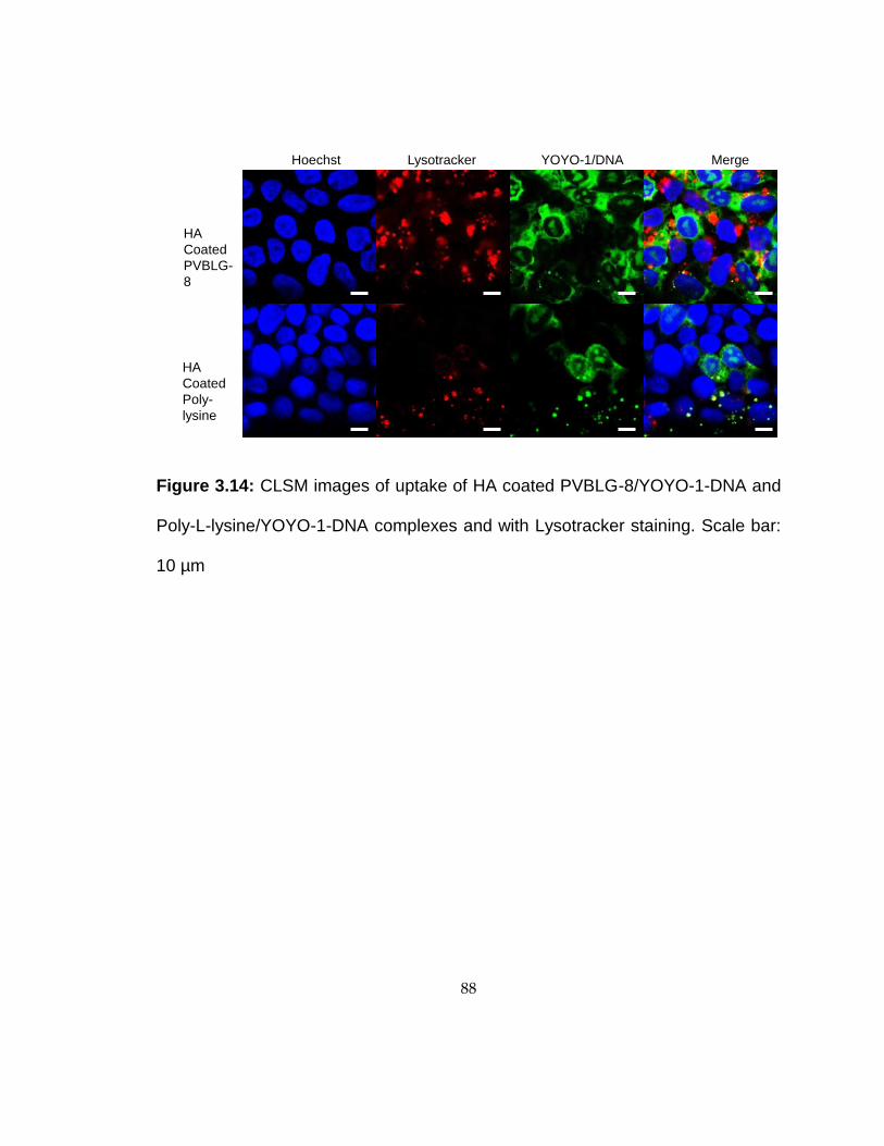
88
Figure 3.14: CLSM images of uptake of HA coated PVBLG-8/YOYO-1-DNA and
Poly-L-lysine/YOYO-1-DNA complexes and with Lysotracker staining. Scale bar:
10 µm
Hoechst YOYO-1/DNALysotracker Merge
HA
Coated
PVBLG-
8
HA
Coated
Poly-
lysine
b.
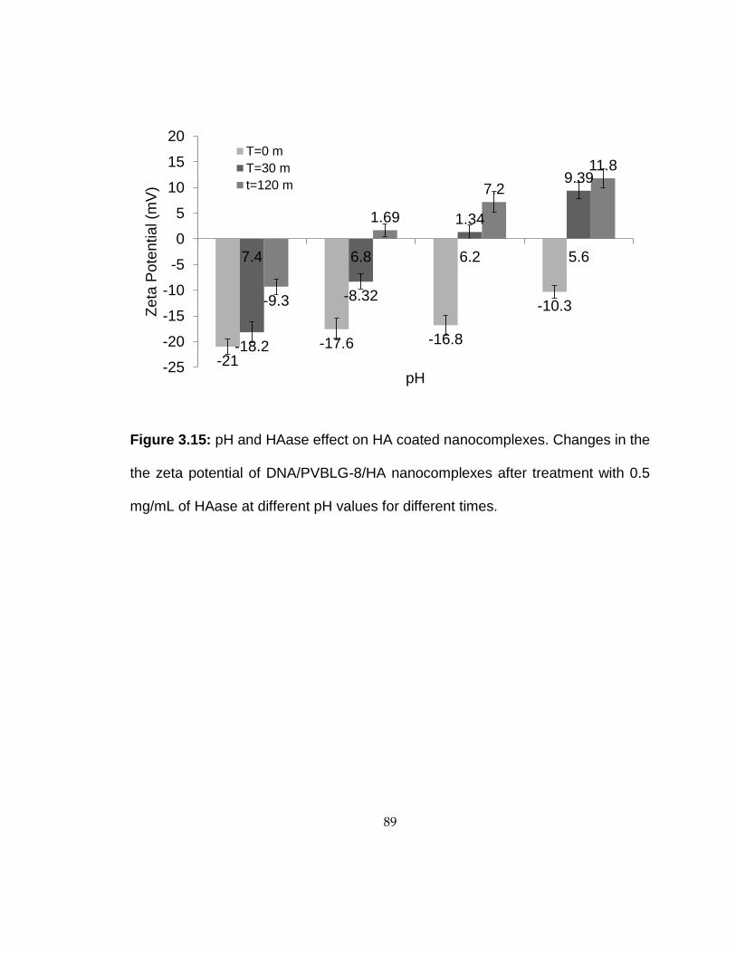
89
Figure 3.15: pH and HAase effect on HA coated nanocomplexes. Changes in the
the zeta potential of DNA/PVBLG-8/HA nanocomplexes after treatment with 0.5
mg/mL of HAase at different pH values for different times.
-21-17.6 -16.8
-10.3
-18.2
-8.32
1.34
9.39
-9.3
1.69
7.2
11.8
-25
-20
-15
-10
-5
0
5
10
15
20
7.4 6.8 6.2 5.6
Ze
ta P
ote
ntia
l (m
V)
pH
T=0 m
T=30 m
t=120 m

90
Figure 3.16: pH and HAase effect on HA coated nanocomplexes. ANTS/DPX
loaded LUVET leakage study of different nanocomplexes with and without the
HAase treatment at different pH values.
0
20
40
60
80
100
120
7.4 6.8
Pe
rce
nt o
f D
ye
Le
aka
ge
pH
DNA/PV8 DNA/PV8/HADNA/PV8/HA 0 h HAase DNA/PV8/HA 1/2 h HAaseDNA/PLL DNA/PLL/HA

91
Figure 3.17: Cell viability of hESCs at varying ratios of HA coating of
nanocomplexes as determined by MTT assay
0
20
40
60
80
100
120
1:7.5:0 1:7.5:5 1:7.5:7.5 1:7.5:10 1:7.5:15
Cell
Via
bili
ty (
%)
DNA:PVBLG-8:HA weight ratio
**

92
Figure 3.18: DAPI and SSEA4 staining patterns of hESCs 72 h after transfected
with HA coated nanocomplexes. Scale bar: 250 µm
HA CoatingControl
SSEA-4
Hoechst

93
Figure 3.19: Western blot analysis on the OCT4 expression in hESCs 5 days post
HA coated nanocomplexes transfection.
α-Tubulin
OCT4
94
3.7 References
[1] Gabrielson NP, Lu H, Yin LC, Li D, Wang F, Cheng JJ. Reactive and Bioactive
Cationic a-Helical Polypeptide Template for Nonviral Gene Delivery. Angewandte
Chemie-International Edition 2012;51(5):1143-47.
[2] Yin L, Song Z, Kim KH, Zheng N, Tang H, Lu H, et al. Reconfiguring the
architectures of cationic helical polypeptides to control non-viral gene delivery.
Biomaterials 2013;34(9):2340-9.
[3] Yin LC, Song ZY, Kim KH, Zheng N, Gabrielson NP, Cheng JJ. Non-Viral Gene
Delivery via Membrane-Penetrating, Mannose-Targeting Supramolecular Self-
Assembled Nanocomplexes. Adv Mater 2013;25(22):3063-70.
[4] Zheng N, Yin LC, Song ZY, Ma L, Tang HY, Gabrielson NP, et al. Maximizing gene
delivery efficiencies of cationic helical polypeptides via balanced membrane penetration
and cellular targeting. Biomaterials 2014;35(4):1302-14.
[5] Zhang RJ, Yin LC, Cheng JJ. Effect of side chain profiles on helical polypeptide
mediated non-viral gene transfection efficiency. Abstracts of Papers of the American
Chemical Society 2013;246.
[6] Yen J, Zhang Y, Gabrielson NP, Yin L, Guan L, Chaudhury I, et al. Cationic, helical
polypeptide-based gene delivery for IMR-90 fibroblasts and human embryonic stem
cells. Biomaterials Science 2013.
[7] Yin LC, Tang HY, Kim KH, Zheng N, Song ZY, Gabrielson NP, et al. Light-
Responsive Helical Polypeptides Capable of Reducing Toxicity and Unpacking DNA:

95
Toward Nonviral Gene Delivery. Angewandte Chemie-International Edition
2013;52(35):9182-86.
[8] Gerecht S, Burdick JA, Ferreira LS, Townsend SA, Langer R, Vunjak-Novakovic G.
Hyaluronic acid hydrogel for controlled self-renewal and differentiation of human
embryonic stem cells. Proceedings of the National Academy of Sciences
2007;104(27):11298-303.
[9] Cho HJ, Yoon HY, Koo H, Ko SH, Shim JS, Lee JH, et al. Self-assembled
nanoparticles based on hyaluronic acid-ceramide (HA-CE) and Pluronic (R) for tumor-
targeted delivery of docetaxel. Biomaterials 2011;32(29):7181-90.
[10] Cho HJ, Yoon IS, Yoon HY, Koo H, Jin YJ, Ko SH, et al. Polyethylene glycol-
conjugated hyaluronic acid-ceramide self-assembled nanoparticles for targeted delivery
of doxorubicin. Biomaterials 2012;33(4):1190-200.
[11] Choi KY, Chung H, Min KH, Yoon HY, Kim K, Park JH, et al. Self-assembled
hyaluronic acid nanoparticles for active tumor targeting. Biomaterials 2010;31(1):106-14.
[12] Choi KY, Min KH, Na JH, Choi K, Kim K, Park JH, et al. Self-assembled
hyaluronic acid nanoparticles as a potential drug carrier for cancer therapy: synthesis,
characterization, and in vivo biodistribution. J Mater Chem 2009;19(24):4102-07.
[13] Choi KY, Min KH, Yoon HY, Kim K, Park JH, Kwon IC, et al. PEGylation of
hyaluronic acid nanoparticles improves tumor targetability in vivo. Biomaterials
2011;32(7):1880-89.

96
[14] Kim EJ, Shim G, Kim K, Kwon IC, Oh YK, Shim CK. Hyaluronic acid complexed
to biodegradable poly L-arginine for targeted delivery of siRNAs. J Gene Med
2009;11(9):791-803.
[15] Lu H, Wang J, Bai YG, Lang JW, Liu SY, Lin Y, et al. Ionic polypeptides with
unusual helical stability. Nat Commun 2011;2:206.
[16] Bang EK, Gasparini G, Molinard G, Roux A, Sakai N, Matile S. Substrate-Initiated
Synthesis of Cell-Penetrating Poly(disulfide)s. J Am Chem Soc 2013;135(6):2088-91.
[17] Smolarsky M, Teitelbaum D, Sela M, Gitler C. A simple fluorescent method to
determine complement-mediated liposome immune lysis. J Immunol Methods
1977;15(3):255-65.
[18] Khalil IA, Kogure K, Akita H, Harashima H. Uptake pathways and subsequent
intracellular trafficking in nonviral gene delivery. Pharmacol Rev 2006;58(1):32-45.
[19] Gratton SEA, Ropp PA, Pohlhaus PD, Luft JC, Madden VJ, Napier ME, et al. The
effect of particle design on cellular internalization pathways. Proc Natl Acad Sci USA
2008;105(33):11613-18.
[20] Kneipp J, Kneipp H, Wittig B, Kneipp K. One-and two-photon excited optical pH
probing for cells using surface-enhanced Raman and hyper-Raman nanosensors. Nano
Lett 2007;7(9):2819-23.
[21] Mislick KA, Baldeschwieler JD. Evidence for the role of proteoglycans in cation-
mediated gene transfer. Proc Natl Acad Sci USA 1996;93(22):12349-54.

97
[22] Ponta H, Sherman L, Herrlich PA. CD44: from adhesion molecules to signalling
regulators. Nature reviews Molecular cell biology 2003;4(1):33-45.
[23] Hooper NM. Detergent-insoluble glycosphingolipid/cholesterol-rich membrane
domains, lipid rafts and caveolae (review). Molecular membrane biology
1999;16(2):145-56.
[24] Dufaÿ Wojcicki A, Hillaireau H, Nascimento TL, Arpicco S, Taverna M, Ribes S, et
al. Hyaluronic acid-bearing lipoplexes: physico-chemical characterization and in vitro
targeting of the CD44 receptor. Journal of Controlled Release 2012;162(3):545-52.
[25] Akinc A, Thomas M, Klibanov AM, Langer R. Exploring polyethylenimine-
mediated DNA transfection and the proton sponge hypothesis. J Gene Med
2005;7(5):657-63.
[26] Sonawane ND, Szoka FC, Verkman AS. Chloride accumulation and swelling in
endosomes enhances DNA transfer by polyamine-DNA polyplexes. J Biol Chem
2003;278(45):44826-31.
98
Chapter 4:
Enhancement of gene delivery into human embryonic stem
cells
4.1 Introduction
Considering the colony-forming properties of hESCs that limit non-viral gene
delivery, I hypothesized that increasing cell spreading would increase cellular uptake and
thus the gene transfection efficiency. Growing hESCs on stiffer substrates has shown to
disperse cells and promote cell spreading[1,2], and it has also been shown in other cell
types that an increase in the substrate stiffness can lead to higher transfection efficiency[3].
However, such an approach not feasible for hESCs, mainly due to the sensitive nature of
hESCs to their external environment. It has been reported that hESCs grown on stiff
substrates will start differentiating[4], but it indicates that cellular uptake is intrinsic to the
stiffness and structure of the cells, and a decrease in membrane tension and contractility
would lead to increased cellular spreading[5] and stimulate endocytosis[6].
Dissociation of hESCs into single cells or small colonies can potentially facilitate
cell spreading. However, since hESCs are considered to be in the primed state, they are
intolerant to single cell passaging and usually exhibit <1% clonal efficiency due to
apoptosis upon cellular detachment and dissociation[7]. Rho-associated kinase (ROCK)
inhibitor has been used to diminish dissociation-induced apoptosis, resulting in increased

99
survival of individual hESCs[7]. By pre-treatment of the hESCs with Y-27632 (10 µM) for
an hour before single cell dissociation, the cloning efficiency increased to 27%. Cells
treated with Y-27632 maintain their pluripotency and morphology for at least 5 passages
and are able to differentiate into neural cells[7]. Therefore, with an attempt to facilitate
hESC spreading without causing apoptosis, the selective ROCK inhibitor, Y-27632, was
used to alter the cell’s myosin fibers and mimic the cell's structure when grown on a stiffer
substrate by inhibiting non-myosin IIA[8,9]. Y-27632 is also known to decrease cell-
generated tension[10]. I hypothesized that Y-27632 would facilitate the spreading and
flattening of hESCs colonies on plates due to decreased membrane tension, which would
then increase the surface area exposure to allow for a more efficient uptake of gene delivery
vectors and promote the gene transfection (Scheme 4.1). A broad spectrum of non-viral
gene delivery vectors, including commercially available reagents (Lipofectamine 2000
(LPF), Fugene HD (FHD), poly-L-lysine (PLL), poly-L-arginine (PLR), and
polyethyleneimine (PEI)), were evaluated in terms of their transfection efficiencies in the
presence or absence of Y-27632. Mechanistic analysis into the effect of Y-27632 on cell
spreading, cell uptake property, and pluripotency of hESCs was also performed. The
treatment of hESCs with Y-27632 was revealed to significantly increase transfection
efficiency of all the tested materials by 1.5 to 2 fold in hESCs, establishing an alternative
use of Y-27632 as a tool for enhanced non-viral gene delivery.
4.2. Materials and Methods

100
4.2.1. General
Human embryonic stem cell line H1 (hESC-H1) was cultured in mTeSR 1 medium
from Stem Cell Technologies (Vancouver, Canada). Fugene HD (FHD) was purchased
from Promega (Madison, WI, USA). Y-27632 was purchased from Stemgent (Cambridge,
MA, USA). Opti-MEM, Lipofectamine 2000 (LPF), and YOYO-1 were purchased from
Invitrogen (Carlsbad, CA, USA). pEGFP-N1 was obtained from Elim Biopharmaceuticals
(Hayward, CA, USA). Milli-Mark™ Anti-SSEA-4-PE was purchased from EMD
Millipore (Billerica, MA, USA). PLL, PLR, and PEI were purchased from Sigma-Aldrich
(St. Louis, MO, USA).
4.2.2. Instrumentation
Flow cytometry analysis was conducted on a BD LSRII color flow cytometry
analyzer (Becton Dickinson, Franklin Lakes, NJ). Cells were visualized with a Zeiss
Axiovert 40 CFL fluorescence microscope equipped with a 10x and 20x objective
(Thornwood, NY). Fluorescence imaging was done using the GE InCell Analyzer 2000
from GE Healthcare Sciences (Piscataway, NJ, USA). Zeta potential and size analysis were
conducted on a Malvern Zetasizer (Worcestershire, UK).
4.2.3 Preparation and characterization of nanocomplexes
Various non-viral vectors were used for the transfection analyses. Plasmid DNA (1
µL, 1 mg/mL) was diluted with water (50 µL). The transfection reagent, polyarginine (10

101
μL, 1 mg/mL), polylysine (10 μL, 1 mg/mL), or polyethylenimine (5 μL, 1 mg/mL) was
diluted with water (50 µL). The two solutions were then vortexed gently and allowed to
incubate for 20 min at rt, after which they were combined and allowed to incubate for
another 20 min at rt.
Plasmid DNA (1 µL, 1 mg/mL) was diluted with mTeSR (50 µL). The transfection
reagent, LPF (2 μL, 1 mg/mL) was diluted with mTeSR (50 µL). The two solutions were
then vortexed gently and allowed to incubate for 20 min at rt, after which they were
combined and allowed to incubate for another 20 min at rt.
Various non-viral vectors were used for the transfection analyses. Plasmid DNA (1
µL, 1 mg/mL) was diluted with mTeSR (50 µL). The transfection reagent, FHD (3.5 μL, 1
mg/mL) was added to the DNA solution. The solution was then vortexed gently and
allowed to incubate for 20 min at rt, after which they were combined and allowed to
incubate for another 20 min at rt.
Solutions of plasmid DNA (5-25 µg/mL) were prepared in water or medium.
Separately, a solution of 50 µg of the vectors was prepared in 400 µL water. A solution of
50 µl LPF and FHD in 400 µL media was also prepared as a control. The DNA solution
was then mixed with the vectors, LPF, or FHD solution and allowed to incubate at room

102
temperature for 20 min. Dynamic light scattering (DLS) and zeta potential analysis were
conducted on the samples with a Malvern Zetasizer.
4.2.4 hESC Transfection with Y-27632
hESCs were seeded on Matrigel-coated 24-well plates and cultured in mTeSR
medium for 24 h. For the full treatment group, Y-27632 was added into the culture medium
at a final concentration of 0, 10, 30 or 50 µM for 4 h prior to the transfection.
Nanocomplexes prepared from pEGFP-N1 plasmid and different transfection vectors were
added dropwise into the media and cells were cultured at 37°C for 4 h. For the pre-treatment
group, cells were treated with Y-27632 (50 µM) for 4 h at 37°C prior to transfection. The
cells were then washed with PBS and fresh media was added prior to the addition of the
nanocomplex and further incubation at 37°C for 4 h. For the post-treatment group, cells
were treated with Y-27632-free medium for 4 h prior to transfection. Directly prior to
transfection, the medium was replaced with Y-27632 (50 µM) containing medium, and
cells were transfected with various vectors at 37°C for 4 h. In all three cases, 4 h after
treatment with the nanocomplexes, the media was replaced with fresh mTeSR and cultured
for 48 h before measurement of the transfection efficiency by flow cytometry.
Alternatively, 10 µM of blebbistatin, a non-muscle myosin IIA inhibitor, downstream of
the rho-associated protein kinase, was added to the cells prior to and during transfection.

103
4.2.5 Intracellular Uptake Studies
DNA (1 mg/mL) was labeled with YOYO-1 (20 µM) at one dye molecule per 50bp
of DNA[11], and was allowed to form nanocomplexes with transfection reagents as
described above. hESCs were plated on matrigel coated 24-well plate and cultured until
they reached medium (50 cells) sized colonies. Nanocomplexes were added at 1 µg YOYO-
1-DNA/well and cells were incubated at 37°C for 4 h. Cells were then harvested, re-
suspended in PBS, and subjected to flow cytometry analysis to quantify the cellular uptake
level of YOYO-1-DNA.
To elucidate the mechanisms underlying the cellular internalization of FHD/DNA
nanocomplexes, the uptake study was performed at 4 °C or in the presence of various
endocytic inhibitors. Cells were pre-treated with chlorpromazine (10 µg/mL), genistein
(200 µg/mL), methyl-β-cyclodextrin (mβCD, 50 µM), dynasore (80 µM), or wortmannin
(50 nM) for 30 min before addition of the nanocomplexes and throughout the uptake study
at 37°C for 2 h. Results were expressed as percentage uptake level of control cells that were
treated with the nanocomplexes at 37 °C for 2 h in the presence of Y-27632 (50 µM) while
in the absence of endocytic inhibitors.
4.2.6 hESC spreading and transfection analysis
hESC H1 were plated on matrigel coated 96-well plates as medium sized colonies
and cultured for 24 h. Y-27632 was added at various concentrations (0, 10, 20, and 50 µM)

104
and after 4 h incubation, the cell nuclei were stained with Hoechst 33258 and the cytoplasm
was stained with CellTracker Red CMTPX per manufacturer’s protocols. Five wells were
imaged with 9 fields each using the GE InCell Analyzer 2000 in the Hoechst and Texas
Red channel (Fig. 4.17). The images were then analyzed using the GE InCell Analyzer
workstation. The nuclei were identified using top-hat segmentation in the Hoechst channel,
and the cell area was analyzed and calculated through the analysis in the CMPTX channel
using multiscale top-hat. The cytoplasmic area was then normalized to the number of nuclei
counted.
hESC H1 were seeded on matrigel-coated 24-well plates and cultured in mTeSR
medium for 24 h. Before transfection, Y-27632 was added into the culture medium at a
final concentration of 0, 10, 30 or 50 µM, and incubated at 37 °C for 4 h. Nanoparticles
from pEGFP-N1 and FHD were added dropwise into the culture medium and cells were
further incubated at 37°C for 4 h. The medium was then replaced with fresh mTeSR
medium, and cells were further cultured for 48 h. After 48 h, the cell nuclei were stained
with Hoechst 33258. Forty-five fields were imaged using the GE InCell Analyzer 2000 in
the Hoechst and GFP channel (Fig. 4.18). Using the InCell Analyzer Workstation, the
nuclei were identified using top-hat segmentation in the Hoechst channel and the numbers
of cells were then calculated. The identified nuclei were then further segmented by
multiscale top-hat in the GFP signal to determine the number of cells with positive GFP

105
signals. The transfection efficiency was then determined by dividing the number of GFP
positive cells by the total number of cells.
4.2.7 siGLO Green Delivery into hESC
Two wells of a 6 well plate of hESC H1s or WA01 OCT4 GFP KI H1 were plated
into a 24 well plate coated with Matrigel a day or two before transfection. Y-27632 at 0
μM, 10 μM or 30 μM was added to the cells 4 hours before transfection. For transfection
with Lipofectamine 2000 with incubation, siGlo Green (30 picomoles) was added to
Optimem (50 μL) and Lipofectamine (1 μL, 1 mg/mL) was added to another tube of
Optimem (50 μL). The two solutions were mixed and incubated at room temperature for
20 minutes and added directly to the cells. For the alternative method, instead of mixing
the two solutions, the polymer solution was added directly to the cells first and then the
solution containing the siGlo Green was added separately to the cells right after. The cells
were incubated for either 48 hours and collected and analyzed with flow cytometry.
4.2.8 RITC Silica nanoparticle synthesis
Methanol (1.0 mL), DI water (0.27 mL), and concentrated ammonia (0.24 mL) were
mixed. TEOS (62.5 μL, 0.28 mmol) was then added to the solvent mixture followed by the
addition of a DMSO solution (20 μL) of trimethylorthosilicate (2 mg, 4.3 μmol).
Fabrication of monodisperse RITC-NCs with other sizes can be similarly achieved by
tuning the concentrations of TEOS, water, and ammonia. After the reaction was complete,

106
without isolating the NCs, a methanol solution of RITC-trimethylorthosilicate (10 mg/mL,
100 μL) was added to the silica NC solution. The mixture was stirred for 12 h in the dark.
A methanol solution of mPEG-sil (10 mg/mL, 100 μL) was added. RITC-NCs were
collected by centrifugation at 15k rpm, washed with ethanol (3 × 1 mL), and redispersed
in DI water or 1× PBS buffer before use. [12]
4.2.9 hESC RITC-SNP Uptake
Two wells of a 6 well plate of hESC H1s or WA01 OCT4 GFP KI H1 were plated
into a 24 well plate coated with Matrigel a day or two before transfection. Y-27632 at 0
μM or 3 μM was added to the cells 4 hours before transfection. The RITC-SNP (30-40 μg)
was added to the wells and incubated for 2h. The cells were then washed with PBS (3x)
and analyzed with flow cytometry.
4.2.10 Sample preparation and flow cytometry analysis
Prior to analysis by flow cytometry, transfected cells on the 24-well plate were
washed with PBS (3 × 500 μL) to remove any residual serum, dead cells, and debris.
Accutase (100 µL) was added to detach the cells from the plate, and PBS (100 µL) was
then added to re-suspend the cells. An aqueous solution of paraformaldehyde (4%, 100 µL)
was added to fix the cells, which were then subject to flow cytometry analysis.

107
4.2.11 Cell viability
For the MTT assays cytotoxicity assessment, 1 6-well well of confluent hESCs was
plated into 24 wells of a matrigel coated 48-well plate one day before transfection. The
cells were then pre-treated for 4 h with 0, 10, 30, and 50 µM of Y-27632 for 4 h and
transfected with FHD as described previously. The cells were further incubated for 4 h at
37°C in the transfection mix before removal of the transfection reagent and replacement of
fresh media. After 48 h, the cell viability was monitored by the MTT assay and was
represented as percentage viability of control cells that did not receive any transfection
treatment. For the MTT assay, the cells were washed with PBS and MTT solution was
added. Following 4-h incubation at 37°C, MTT solubilization solution (10% Triton X-100
in acidic (0.1M HCl) isopropanol) was added to the cells and the absorbance of 570 nm
light was quantified on a Perkins Elmer plate reader (Waltham, MA, USA).
4.2.12 Western blot analysis and SSEA-4 staining
After 72 h, the cells were stained with DAPI (250 µL, 3 nM) and SSEA-4–PE (250
µL, 0.02 mg/mL), a pluripotency cell marker, for 30 min at 37 °C. The cells were imaged
using the GE InCell Analyzer 2000.
After 4 d treatment of 50 µM Y-27632 and FHD transfection, the cells were lysed
with the RIPA buffer, mixed with Laemmli buffer supplemented with 2-Mercaptoethanol,
and heated at 100 °C for 5 min to denature the proteins. After being cooled in ice, the

108
samples were subjected to electrophoresis on a 10% SDS PAGE Gel at 120 V for 1.5 h,
and wet transferred to the nitrocellulose membrane using the AMRESCO Rapid Western
Blot Kit per manufacturer’s instructions. The membrane was stained with OCT4 and α-
Tubulin primary antibodies and then with HRP-tagged secondary.
4.3 Results
4.3.1 Inhibition of Rho-associated kinase facilitates non-viral gene transfection
Prior to the transfection assessment, the pEGFP-N1 plasmid nanocomplexes with
the gene transfection vectors were evaluated by dynamic light scattering and zetasizer to
determine their complexation capacities. I confirmed that all tested materials were able to
form nanocomplexes with DNA, as reported by many other prior studies. Upon
complexation with DNA, the nanocomplexes of DNA with PLR, PLL and PEI gave sizes
around 60-70 nm while DNA complexes with LPF and FHD following the standard
protocol afforded much larger particle size (above 300 nm, Fig. 4.1). PLR, PLL, and PEI
are cationic polymers with sufficiently long backbones so that they can well condense the
anionic DNA into compact nanocomplexes via electrostatic interaction as well as
intermolecular entanglement. In comparison, LPF and FHD are cationic lipid based
materials, and their short molecular length would prevent sufficient entanglement with the
DNA molecules, thus leading to relatively larger complexes.

109
Transfection efficiency of each material was then evaluated in the presence (50
µM) or absence of Y-27632 by monitoring the EGFP expression, attempting to probe the
effect of Y-27632 on gene transfection. As shown in Fig. 4.2, PLR was unable to mediate
transfection in H1 hESCs (transfection efficiency < 1%), which was consistent with
previous findings in multiple cell lines[13,14]. Upon the treatment of Y-27632, all other
materials significantly increased their transfection efficiency of hESCs as measured by
flow cytometry. Specifically, the PLL transfection efficiency with Y-27632 increased from
3.0 ± 0.8% to 5.8 ± 0.8%. For PEI mediated transfection, the transfection efficiency
increased from 7.7 ± 0.8% to 13.7 ± 1.3% in the presence of Y-27632. Similar enhancement
of gene transfection efficiencies was observed with the use of Y-27632 when LPF and FHD
were used as transfection agents. Their transfection efficiencies improved from 8.8 ± 0.8%
to 15.3 ± 1.2% and 21.5 ± 0.9% to 37.0 ± 1.0%, respectively. These studies clearly
demonstrate that Y-27632 treatment can universally increase the transfection efficiency by
approximately 1.7 to 1.9 fold in non-viral gene delivery to hESCs.
4.3.2 Y-27632 treatment increases cell spreading
To further investigate the treatment effect of Y-27632, transfection studies were
performed on H1 hESC colonies that were treated with varying concentrations of Y-27632
for 4 h prior to and during transfection. DNA/FHD complexes, which displayed the highest
transfection efficiency among all tested systems, were selected for further studies. With no

110
treatment, the EGFP transfection efficiency was 14.4 ± 0.9%. With 10 µM treatment of Y-
27632, the transfection efficiency increased to 17.6 ± 1.4%. When the concentration of Y-
27632 was increased to 30 µM and 50 µM, the respective efficiency increased to 22.7 ±
0.8% and 26.4 ± 1.1% (Fig. 4.3-4.4). To confirm that the mechanism of increased
transfection efficiency was due to the spreading and actin-myosin interactions, the cells
were treated with blebbistatin, a small molecule that acts downstream of Y-27632[15],
inhibits non-muscle myosin IIA, reduces actin/myosin interactions and alters the
intracellular structure and morphology. Treatment of blebbistatin at 10 µM also resulted in
increased transfection efficiency of DNA/FHD complex up to 17.8 ± 0.4%, indicating a
strong correlation between cell structure and transfection efficiency (Fig. 4.3).
When hESC colonies were treated with Y-27632 for 4 h at varying concentrations
(10, 30, and 50 µM), significant morphological changes of the cells were observed. In the
absence of Y-27632, the cells maintained a two-dimensional cobble stone like colony
morphology, and the cells were tight and rounded up (Fig. 4.5). At 10 µM, the colonies
started to spread and lose their rounded-up structure. At 30 and 50 µM, the cells were even
more spread out and elongated, further indicating that they had relaxed their original
structure. This increased the surface area and decreased the surface membrane tension. To
further validate the Y-27632-mediated cell spreading, the cytoplasm and nuclei of the Y-
27632-treated cells were respectively labelled before cells were imaged and analysed using

111
the GE InCell Analyzer (Fig S4.1). Without Y-27632 treatment, the cells were calculated
to have a cytoplasmic area of 445 ± 29.8 µm2 per cell. When treated with Y-27632 at 10,
30, and 50 µM, the cytoplasmic area per cell increased to 516 ± 8.0, 563 ± 32.7 and 589 ±
18.6 µm2, respectively (Fig. 4.6). The increased cytoplasmic area thus substantiated the
promotion of cell spreading upon Y-27632 treatment. Through further analysis of the cell
spreading effect on transfection efficiency, the GFP efficiency was also measured using
the GE InCell Analyzer and InCell Workstation (Fig S4.2). With the increased spreading,
the GFP transfection efficiency increases from about 6.3 ± 3.4% at 0 µM to 10.9 ± 4.4%,
22.8 ± 0.6% and 30.6 ± 1.7% at 10, 30, and 50 µM of Y-27632 treatment, respectively (Fig
4.6). Through the GE InCell imaging analysis of the hESCs cytoplasmic area and GFP
expression, correlation between the cell spreading and GFP transfection upon the treatment
of Y-27632 was noted.
4.3.3 Sequential treatment of Y-27632 and uptake inhibition study
To study the importance of morphological changes, hESCs were treated with Y-
27632 in three different stages, with FHD as the model vector. When the cells were treated
with Y-27632 for 4 h and removed before transfection, the transfection efficiency was
28.9%. When the cells were treated with Y-27632 only during the transfection for 4 h, the
transfection efficiency decreased to 25.4%. Finally, when the cells were treated for 4 h
before and during the transfection with Y-27632, the transfection efficiency increased to

112
31.3% (Fig. 4.7). The increase in transfection when the hESCs were pretreated with Y-
27632 indicates the importance of altering the cell morphology before the transfection.
A further uptake study of the YOYO-1-DNA was conducted at various
concentrations of Y-27632. In the absence of Y-27632, 67% of the cells had taken up the
FHD/YOYO-1-DNA nanocomplexes. When the concentration of Y-27632 was increased
from 10 to 50 µM, the uptake level continued to increase to 95%, in consistence with the
transfection trend (Fig. 4.8). Such observation indicated that Y-27632 promoted the overall
cellular uptake of the nanocomplexes as a result of facilitated colony spreading, which
ultimately potentiated the transfection efficiency.
The internalization mechanism is often closely related to the intracellular fate and
ultimately the transfection efficiency. To this end, the effect of Y-27632 on the intracellular
kinetics was further probed by monitoring the cell uptake in the presence of different
endocytic inhibitors. Energy dependent endocytosis was blocked at 4°C; clathrin mediated
endocytosis was blocked by chlorpromazine by triggering dissociation of the clathrin
lattice; caveolae was inhibited by genistein and mβCD by inhibiting tyrosine kinase and
depleting cholesterol, respectively; clathrin mediated endocytosis and caveolae were
inhibited by dynasore by inhibiting dynamin; macropinocytosis was inhibited by
wortmannin by inhibiting phosphatidyl inositol-3-phosphate[16,17]. As shown in Fig. 4.9,
through the different inhibition of endocytotic pathways, the uptake of YOYO-1-DNA did

113
not diminish through the use of the inhibitors. On the other hand, at 4°C, there was a
decrease in uptake efficiency. This suggests that the FHD nanocomplexes entered the cells
partially via energy dependent endocytosis, but not through clathrin and caveolae mediated
endocytosis or macropinocytosis. Interestingly, there was an increase of uptake at 4°C from
50% to 75% in the Y-27632 treated cells. Potentially, this denotes the decreased energy
requirement for endocytosis with the treatment of Y-27632.
4.3.4 Combination of Y-27632 treatment with HA coated nanocomplexes
To demonstrate the combined effect of Y-27632 treatment and HA coated
DNA/PVBLG-8 nanocomplexes, the two methods were combined in their transfection into
H1 hESCs. Using the optimal transfection ratios of 1:7.5 (DNA:PVBLG-8 weight ratios)
and 1:7.5:7.5 (DNA:PVBLG-8:HA weight ratios), the transfection efficiency increases
from 11.2% to 15.4% GFP positive for PVBLG-8 only transfection with and without Y-
27632 treatment, respectively. Finally, using the HA coated nanocomplexes the
transfection efficiency increased to 16.8% and 24.5% GFP positive with and without Y-
27632 treatment, respectively. This indicates that coupling the two systems does
significantly increase the transfection efficiency.
4.3.5 siSRNA siGLO Green uptake
Transfection of siRNA siGlo Green was performed using Lipofectamine 2000 with
the treatment of Y-27362 at 0 µM, 10 µM, and 30 µM concentrations with uptake
114
efficiencies of 55.67%, 75.4% and 85.92%, respectively, again demonstrating the effects
of transfection efficiency enhancement of Y-27632 (Fig. 4.11).
4.3.6 Nanoparticle uptake
In the initial study, 3 sizes of silica nanoparticles tagged with RITC (RITC-SNP)
were synthesized using the Stober method [12], 200, 51 and 25 nm. Two sets of RITC-SNP
were synthesized one with and one without PEG surface modifications. These RITC-SNP
were incubated for 2 h with the cells treated with 0 μM or 30 μM Y-27632. For the 200,
50, 25 nm sized RITC-SNP with no surface modifications, the transfection efficiencies
without Y-27632 treatment and incubated for 2 h were 1.75%, 1.23% and 0.65%,
respectively. The transfection efficiencies for cells treated with 30 μM Y-27632 were 9.5%,
6.5% and 3.5% (Fig. 4.12). For the 200, 50, 25 nm sized RITC-SNP with no surface
modifications the transfection efficiencies without Y-27632 treatment and incubated for 5
h were 70.03%, 86.54% and 86%, respectively. The transfection efficiencies for cells
treated with 3x Y-27632 were 83.61%, 92.65% and 94.53% (Fig. 4.13). These trends
demonstrated higher uptake with the treatment or Y-27632 in hESC, especially with the
use of non-surface modified RITC-SNP.
4.3.7 Treatment of Y-27632 maintains pluripotency of hESCs
Treatment of hESCs with Y-27632 at the concentration of 50 µM significantly
increases non-viral transfection efficiency. The treated cells can also revert back to their

115
normal hESC morphology 48 h post-treatment (Fig. 4.4). This study demonstrates that Y-
27632 only transiently alters the hESC morphology to facilitate improved gene delivery.
To further confirm whether the hESCs were only transiently altered by the treatment of Y-
27632 and FHD transfection, extracellular stage specific embryonic antigen 4 (SSEA-4)
was stained with PE conjugated antibodies and imaged 72 h post transfection with 50 µM
Y-27632 treatment (Fig. 4.14). The cells stained uniformly positive for SSEA-4, indicating
well-maintained pluripotency. A western blot assay of the hESCs conducted 4 days post
FHD/DNA transfection with Y-27632 also indicated unaltered expression of OCT4, a gene
indicator of hESC pluripotency. With the treatment of Y-27632 at all concentrations, there
was no change in the OCT4 expression. However, when treated with 10, 30, and 50 µM of
Y-27632 in the presence of FHD, a slight reduction in the OCT4 expression level was
observed, indicating that Y-27632 itself did not affect pluripotency and the minor change
of OCT4 expression could be induced by the transfection reagent. These experiments
substantiated that gene transfection and Y-27632 treatment were transient and the cells
were able to revert back to their natural pluripotent state after removal of Y-27632 (Fig.
4.15). The treatment of hESCs with Y-27632 also showed no cytotoxicity to the cells at
any treatment concentrations (Fig. 4.16). The Y-27632 treatment at any of the
concentrations for transfection demonstrated only transient alteration of the cells with well-
retained pluripotency and low cytotoxicity.
4.4 Discussion

116
In the current study, I demonstrated that with the treatment of Y-27632, transfection
efficiencies of a variety of non-viral gene delivery materials in hESC, including PLR, PLL,
PEI, LPF, and FHD, were markedly augmented. Y-27632 allowed the hESC colonies to
effectively transform their internal mechanical structure by inhibiting the actin-mysoin
contractility and cell-to-cell adhesion, thus facilitating spreading of the cells. As such, the
exposed surface area was increased and the membrane tension was decreased, ultimately
leading to an increase in the internalization level of exogenous genetic materials. Using
one of the most efficient commercial transfection reagents, FHD, I demonstrated a dose
dependent effect of Y-27632 small molecule treatment in hESCs. An increase in the Y-
27632 concentration from 0 to 50 µM correlated to a 1-fold increment in the transfection
efficiency. To ensure that the increased transfection efficiency was due to the actin-myosin
inhibition effect on the spreading or morphology change of the cells and not the other
pathways associated with the rho-associated protein kinase pathway, the cells were also
treated with 10 µM blebbistatin. Blebbistatin acts downstream of the rho-associated protein
kinase, and directly inhibits non-muscle myosin IIA. This inhibition directly decreases the
affinity of myosin with actin, indicating that the Y-27632’s role in the cell morphological
change is responsible for the increased gene transfection.
From the treatment of varying concentrations of Y-27632, the cell’s morphology
changes from a rounded up structure to an elongated flat morphology and disassociation
from the colony. This flattening and spreading of the cells increased the cell surface area

117
that was exposed to exogenous nanocomplexes, and thus the nanocomplexes were more
readily taken up by the cells. A higher Y-27632 concentration resulted in enhanced cell
spreading and ultimately increased transfection efficiency. The important role of Y-27632
in increasing cell adhesion and survival also helped the cells to maintain physiological
functions, thus contributing to the transfection process[1]. In addition, through the
spreading and elongation of the cells, the cell membrane could have also decreased
membrane tension, allowing for a higher rate of endocytosis [5,6]. With decreased
membrane tension, the energy required for endocytosis is lowered [6], which is shown in
the 4°C FHD nanocomplex uptake experiment, in which the uptake efficiency increased
with the treatment of Y-27632.
It is important that the cells be transiently altered before transfection, so that the
spreading of the cells and decrease of the membrane tension could allow for efficient
nanocomplex uptake. In our detailed study on the adding sequence of Y-27632, pretreated
cells showed significantly higher transfection efficiency than post-treated cells, indicating
the importance of morphological change prior to the transfection process. The altered
morphology primes the cells by increasing the cytoplasmic area and facilitating the uptake
of the nanocomplexes during transfection. In consistence with our findings that cells in
smaller colonies afforded higher transfection efficiencies (data not shown), it was further
demonstrated that Y-27632-mediated cell spreading and larger cell surface area is
attributed to the decreased surface tension and de-clumping of the cell colonies.

118
Not only does the pre-treatment with Y-27632 enhance DNA plasmid and siRNA
nanocomplexes, but it also can enhance the uptake of other nanoparticles, like unmodified
silica nanoparticles at a varying sizes. As expected, there is a correlation between the
uptake efficiency and nanoparticle size. For smaller nanoparticles (20-25 nm), the higher
the uptake efficiency, while larger nanoparticles (200 nm) have much lower uptake
efficiency. When increasing time of incubation of the nanoparticles, there is a drastic
increase of nanoparticle uptake. Yet in both cases, the treatment of Y-27632 increases does
significantly increase the uptake efficiency, indicating that the system works for the uptake
of all kinds of nanoparticles.
I have also demonstrated that the system of transient change of cell morphology
with the smart and targeting system can overall significantly increase gene transfection
efficiency. With the treatment of Y-27632 there is an increase in the transfection of
PVBLG-8 nanocomplexes, but with also increased cytotoxicity (data not shown). Then
with the further addition of the HA coating, the transfection efficiency is significantly
increased, indicating that Y-27632 treatment does not affect caveolae and receptor
mediated endocytosis pathways, indicating that the system can be coupled together. But
further optimization is required to bring the two systems together effectively to reach the
optimal transfection efficiency applicable to stem cell engineering applications.

119
As previously reported [7], the use of Y-27632 does not permanently affect the
pluripotency and hESC cell state. In accordance with this finding, the cells recovered to
their natural, tight, two-dimensional colony morphology 72 h after removal of Y-27632.
Expression of SSEA-4 and OCT4 were also observed, suggesting reversible alteration of
cell physiology and maintenance of cell pluripotency.
4.5 Conclusion
I studied and adapted a new approach to increase the non-viral gene delivery to
hESCs by transiently altering the colony structure to increase their susceptibility for uptake
of nanocomplexes. Treatment of hESCs with Y-27632 prior to and during transfections
effectively increases cell spreading and decreases cell membrane tension, which increases
cell uptake and thus potentiates the gene transfection. The hESC colonies were able to
return to their original morphology and maintain their pluripotency within hours after
removal of Y-27632. While most of the current studies in non-viral gene delivery focus on
the material design, this study opens a new window to control gene transfection in hESCs
on the cellular side. Therefore it provides a promising approach for manipulation of
pluripotent stem cells through transient gene therapy, overcoming a big hurdle against
controlling and studying pluripotent stem cell differentiation and development toward
various biomedical applications. With the increase in the gene transfection efficiency, an
increase in differentiation efficiency of hESCs can be expected, which would reduce the
need for enrichment and sorting of desired cells. This new system has also been shown that

120
it can be coupled with the HA targeting nanocomplex system, which can be further
optimized to bring non-viral gene delivery into hESCs to levels that can be used instead of
viral systems for stem cell engineering applications. The preliminary findings of the current
study also unravel the possibility of manipulating the cellular states and properties in future
designs of intracellular delivery vehicles into hESCs. A lot of stem cell engineering
research is heavily reliant on viral gene delivery systems, including reprogramming,
differentiation, gene targeting/editing, and mechanistic studies, but are then limited due to
safety concerns. But with these advances towards safe and effective non-viral gene
delivery, these stem cell engineering applications are one step closer to clinical therapies.
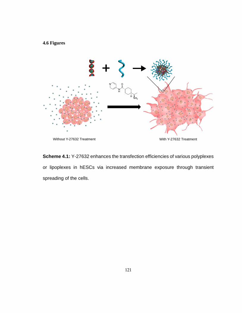
121
4.6 Figures
Scheme 4.1: Y-27632 enhances the transfection efficiencies of various polyplexes
or lipoplexes in hESCs via increased membrane exposure through transient
spreading of the cells.
Without Y-27632 Treatment With Y-27632 Treatment

122
Figure 4.1: Size and zeta potential of various nanocomplexes (w:w): PLR (1:10),
PLL (1:10), PEI (1:5), LPF (1:2), and FHD (1:3.5)
0
10
20
30
40
0
100
200
300
400
500
PLR PLL PEI LPF FHD
Zeta
Pote
ntial (m
V)
Part
icle
Dia
me
ter
(nm
)Particle Size Zeta Potential

123
Figure 4.2: Transfection efficiencies of various reagents in hESCs in the presence
of absence of Y-27632 (50 μM). Cells were pre-treated with Y-27632 cells for 4 h
before removal of Y-27632 and addition of various complexes.
0
10
20
30
40
PLR PLL PEI LPF FHD
% G
FP
Positiv
e0 µM Y-27632
50 µM Y-27632

124
Figure 4.3: Transfection scheme and transfection efficiencies of Fugene HD/DNA
complexes in H1 hESCs in the presence of Y-27632 or Blebbistatin at various
concentrations. (* p <0.05, ** p<0.01)
Plate cells4 h
Addition of
Y-27632
Transfection
Starts
4 h16 h
Replace with
Fresh Media
0
10
20
30
0 10 30 50 10
% G
FP
Po
sitiv
e
****
*
Y-27632 (µM) Blebbistatin (µM)
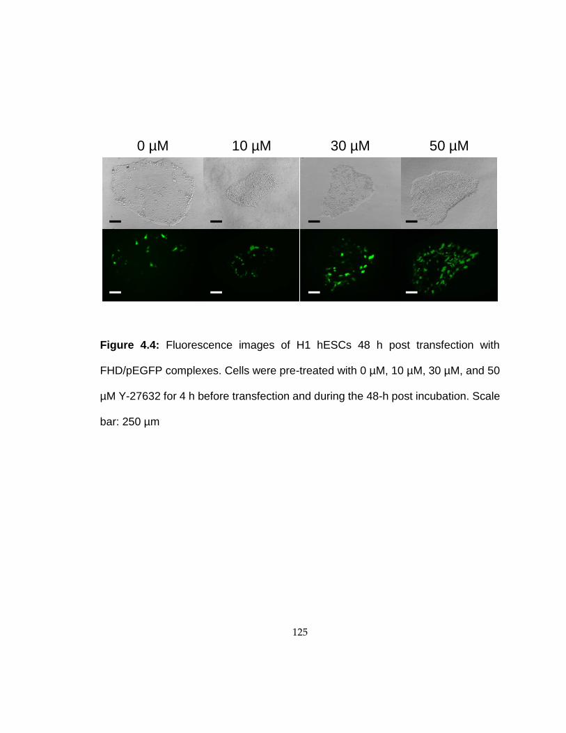
125
Figure 4.4: Fluorescence images of H1 hESCs 48 h post transfection with
FHD/pEGFP complexes. Cells were pre-treated with 0 µM, 10 µM, 30 µM, and 50
µM Y-27632 for 4 h before transfection and during the 48-h post incubation. Scale
bar: 250 µm
0 µM 10 µM 50 µM30 µM

126
Figure 4.5: Bright-field imaging showing the morphological change of hESCs after
4-h treatment with Y-27632 at various concentrations. Scale bar: 250 µm
0 µM 10 µM
30 µM 50 µM

127
Figure 4.6: Alteration of the cytoplasm area (unit) per nucleus of hESCs following
treatment with Y-27632 of various concentrations. Images were taken and
analyzed with the GE InCell Analyzer. (n=5)
0
10
20
30
40
400
500
600
0 10 30 50
% G
FP
Positiv
e
Cyto
pla
sm
Are
a (
un
it)
per
nu
cle
us
Y-27632 (µM)
Cytoplasm Area Transfection efficiency

128
Figure 4.7: Transfection efficiency of FHD in hESCs in the presence of Y-27632.
Y-27632 was applied to the cells for 4 h before transfection, during the 48-h
transfection period, or a combination thereof.
EGFP Transfection Efficiency Dependence on the Presence
of Y-27632 either before and/or during the Transfection Step
Pre-incubation (4 h) Transfection (4 h) % GFP Positive
--- --- 19.6 0.8
--- 50 µM 25.4 1.9
50 µM --- 28.9 1.8
50 µM 50 µM 31.3 3.6

129
Figure 4.8: Cell uptake level of FHD/YOYO-1-DNA complexes in hESCs in the
presence of various concentrations of Y-27632 (n =3). (* p <0.05, ** p<0.01)
0
25
50
75
100
0 10 30 50
YO
YO
-1 F
luore
sce
nce
Level
Y-27632 (µM)
**
****

130
Figure 4.9: Cell uptake mechanisms of FHD/YOYO-1-DNA complexes in hESCs
in the presence or absence of Y-27632. Results were expressed as percent
uptake level of control cells without low temperature (4 oC) or endocytic inhibitor
treatment (n=3).
0
25
50
75
100
% o
f C
ontr
ol Y
OY
O-1
D
NA
Up
take
0 µM Y-27632
50 µM Y-27632*
**
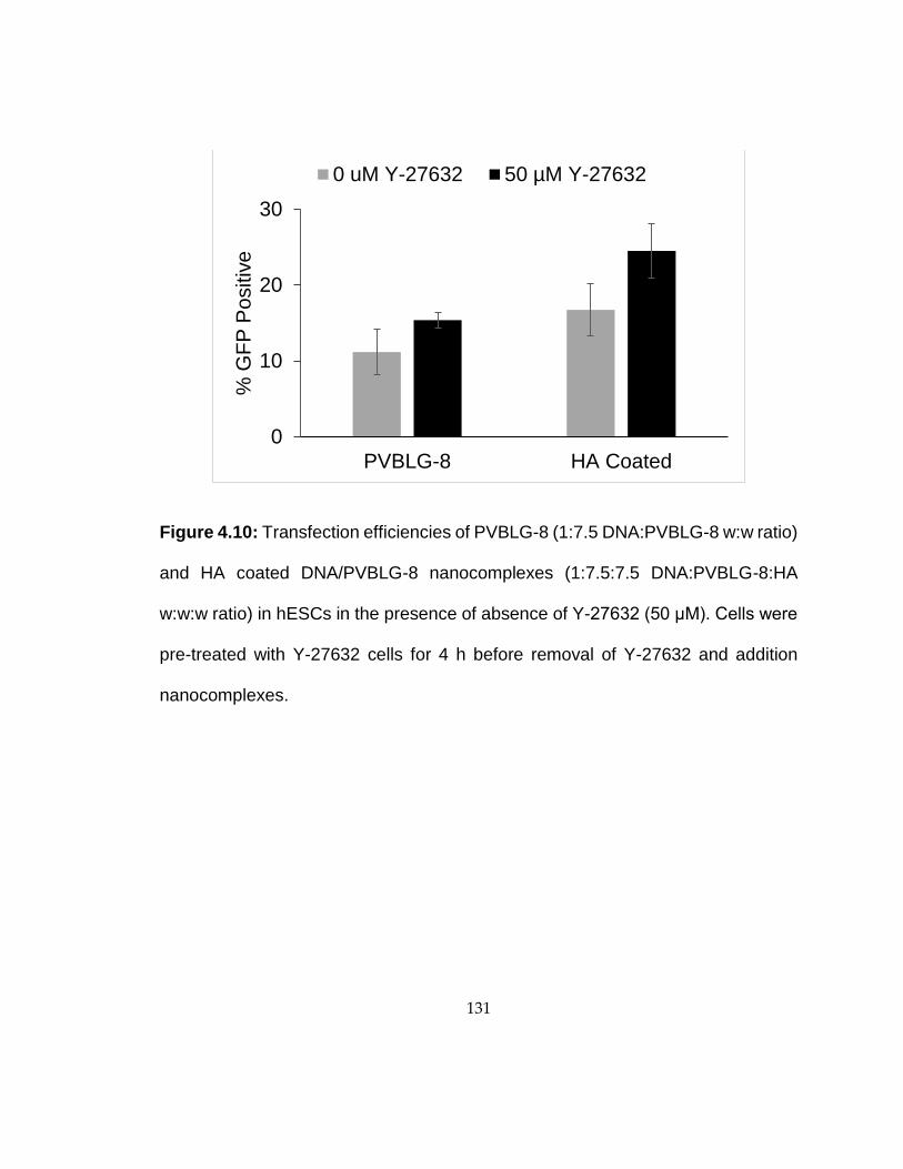
131
Figure 4.10: Transfection efficiencies of PVBLG-8 (1:7.5 DNA:PVBLG-8 w:w ratio)
and HA coated DNA/PVBLG-8 nanocomplexes (1:7.5:7.5 DNA:PVBLG-8:HA
w:w:w ratio) in hESCs in the presence of absence of Y-27632 (50 μM). Cells were
pre-treated with Y-27632 cells for 4 h before removal of Y-27632 and addition
nanocomplexes.
0
10
20
30
PVBLG-8 HA Coated
% G
FP
Po
sitiv
e
0 uM Y-27632 50 µM Y-27632

132
Figure 4.11: Flow Cytometry of siGlo Green (siRNA analog) using Lipofectamine,
after 3 h Y-27632 incubation.
55.67
75.4
85.92
0
10
20
30
40
50
60
70
80
90
100
Lip 0X Lip 1X Lip 3X
% S
iglo
gre
en
up
take

133
Figure 4.12: Flow Cytometry data analysis of hESC with and without treatment of
Y-27632 with different sizes of SNP tagged with RITC without surface modification
after 2 h incubation.
1.751.23
0.65
9.5
6.5
3.5
0
2
4
6
8
10
12
25 50 200
% R
ITC
SN
P u
pta
ke
RITC SNP size (nm)
0x Rho Inhibitor
3x Rho Inhibitor

134
Figure 4.13: Flow Cytometry data analysis of hESC with and without treatment of
Y-27632 with different sizes of SNP tagged with RITC without surface modification
after 5 h incubation.
86.01 86.54
70.03
94.53 92.65
83.61
0
20
40
60
80
100
120
20 50 200
0x Rho Inhibitor
3x Rho inhibitor
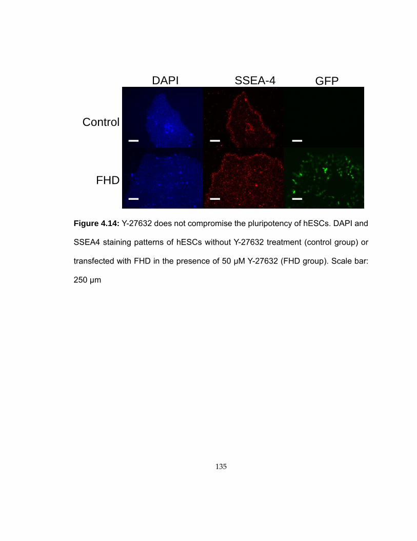
135
Figure 4.14: Y-27632 does not compromise the pluripotency of hESCs. DAPI and
SSEA4 staining patterns of hESCs without Y-27632 treatment (control group) or
transfected with FHD in the presence of 50 µM Y-27632 (FHD group). Scale bar:
250 µm
Control
FHD
DAPI SSEA-4 GFP

136
Figure 4.15: Western blot analysis on the OCT4 expression in hESCs 5 days post
FHD transfection and treatment of Y-27632 at various concentrations.
50 kD
32 kD
Alpha Tubulin
Oct4
Y-27632 (µM):
Transfection: - + - + - + - +
0 0 10 10 30 30 50 50
Ladder

137
Figure 4.16: Cytotoxicity of H1 hESCs 48 h after transfection with FHD at various
concentrations MTT cell viability assay of the FHD transfection with varying
concentrations (0-50 µM) of Y-27632 treatment in H1 hESCs after 48 h.
0
20
40
60
80
100
0 10 30 50
% C
ell
Via
bili
ty (
MT
T)
Y-27632 (µM)

138
Figure 4.17: Representative images of the alteration of the hESC cytoplasmic area
following treatment with Y-27632 of various concentrations (0, 10, 30 and 50 µM).
First row represents the Hoechst channel and the second row represents CMTPX
cell tracking channel. The third row represent the software generated
segmentation used for analysis. The blue outline represents the identified nuclei
and the green outline is the identified CMTPX stain of the cellular cytoplasm from
which the cell area was calculated.
0 µM 10 µM 30 µM 50 µM
Hoechst
CMTPX
Cell Tracker
Computer
Segmentation

139
Figure 4.18: Representative images of the transfection of hESCs after alteration
of the cytoplasm area with the treatment of Y-27632 at various concentrations (0,
10, 30 and 50 µM). First row represents the Hoechst channel and the second row
represents the GFP channel. The third row represent the software generated
segmentation used for analysis. Blue outline represents the identified nuclei and
green outline is the determined cell cytoplasm as extrapolated by the nuclei and
GFP channel.
0 µM 10 µM 30 µM 50 µM
Hoechst
GFP
Computer
Segmentation

140
4.7 References
[1] Harb N, Archer TK, Sato N. The Rho-Rock-Myosin Signaling Axis Determines Cell-
Cell Integrity of Self-Renewing Pluripotent Stem Cells. Plos One 2008;3(8).
[2] Lakins JN, Chin AR, Weaver VM. Exploring the link between human embryonic
stem cell organization and fate using tension-calibrated extracellular matrix
functionalized polyacrylamide gels. Progenitor Cells: Springer; 2012. p. 317-50.
[3] Kong HJ, Liu JD, Riddle K, Matsumoto T, Leach K, Mooney DJ. Non-viral gene
delivery regulated by stiffness of cell adhesion substrates. Nature Materials
2005;4(6):460-64.
[4] Zoldan J, Karagiannis ED, Lee CY, Anderson DG, Langer R, Levenberg S. The
influence of scaffold elasticity on germ layer specification of human embryonic stem
cells. Biomaterials 2011;32(36):9612-21.
[5] Raucher D, Sheetz MP. Cell spreading and lamellipodial extension rate is regulated
by membrane tension. J Cell Biol 2000;148(1):127-36.
[6] Dai J, Sheetz MP. Regulation of endocytosis, exocytosis, and shape by membrane
tension. Cold Spring Harbor Symp Quant Biol 1995;60:567-71.
[7] Watanabe K, Ueno M, Kamiya D, Nishiyama A, Matsumura M, Wataya T, et al. A
ROCK inhibitor permits survival of dissociated human embryonic stem cells. Nat
Biotechnol 2007;25(6):681-86.
[8] Kaunas R, Nguyen P, Usami S, Chien S. Cooperative effects of Rho and mechanical
stretch on stress fiber organization. Proc Natl Acad Sci USA 2005;102(44):15895-900.
141
[9] McBeath R, Pirone DM, Nelson CM, Bhadriraju K, Chen CS. Cell shape, cytoskeletal
tension, and RhoA regulate stem cell lineage commitment. Dev Cell 2004;6(4):483-95.
[10] Narumiya S, Ishizaki T, Uehata M. Use and properties of ROCK-specific inhibitor
Y-27632. Regulators and Effectors of Small Gtpases, Pt D 2000;325:273-84.
[11] Gabrielson NP, Cheng JJ. Multiplexed supramolecular self-assembly for non-viral
gene delivery. Biomaterials 2010;31(34):9117-27.
[12] Tang L, Fan TM, Borst LB, Cheng J. Synthesis and Biological Response of Size-
Specific, Monodisperse Drug–Silica Nanoconjugates. ACS nano 2012;6(5):3954-66.
[13] Yin L, Song Z, Kim KH, Zheng N, Tang H, Lu H, et al. Reconfiguring the
architectures of cationic helical polypeptides to control non-viral gene delivery.
Biomaterials 2013;34(9):2340-9.
[14] Tang HY, Yin LC, Kim KH, Cheng JJ. Helical poly(arginine) mimics with superior
cell-penetrating and molecular transporting properties. Chemical Science
2013;4(10):3839-44.
[15] Ohgushi M, Matsumura M, Eiraku M, Murakami K, Aramaki T, Nishiyama A, et al.
Molecular pathway and cell state responsible for dissociation-induced apoptosis in human
pluripotent stem cells. Cell Stem Cell 2010;7(2):225-39.
[16] Khalil IA, Kogure K, Akita H, Harashima H. Uptake pathways and subsequent
intracellular trafficking in nonviral gene delivery. Pharmacol Rev 2006;58(1):32-45.
142
[17] Gratton SEA, Ropp PA, Pohlhaus PD, Luft JC, Madden VJ, Napier ME, et al. The
effect of particle design on cellular internalization pathways. Proc Natl Acad Sci USA
2008;105(33):11613-18.