Embed Size (px)
Citation preview

Copyright
by
Christopher A. Allen
2007
The Dissertation Committee for Christopher A. Allen Certifies that this is the
approved version of the following dissertation:
The Effects of Low-Shear Modeled Microgravity on Streptococcus
pneumoniae and Adherent-Invasive Escherichia coli
Committee:
Alfredo Torres, Ph.D., Supervisor
David Niesel, Ph.D.
Ashok Chopra, Ph.D., C.Sc.
Duane Pierson, Ph.D.
Raymond Stowe, III, Ph.D.
_________________________
Dean, Graduate School
The Effects of Low-Shear Modeled Microgravity on Streptococcus
pneumoniae and Adherent-Invasive Escherichia coli
by
Christopher A. Allen, B.S., M.S.
Dissertation
Presented to the Faculty of the Graduate School of
The University of Texas Medical Branch
in Partial Fulfillment
of the Requirements
for the Degree of
Doctor of Philosophy
The University of Texas Medical Branch
July, 2007

Dedication
To my parents, James and Barbara, and my wife, Amy, whose unending support gave me
the strength to persevere.

v
Acknowledgements
I would like to acknowledge my mentors Drs. Alfredo Torres and David Niesel
whose guidance and continued support helped me grow and develop as a scientist under their supervision. I wish to also thank my supervisory committee members Drs. Ashok Chopra, Duane Pierson, and Ray Stowe, III for their time, support, and valuable input towards my research progress. I would like to extend a special thanks to all the members of the “Torres Lab Family,” both past and present, whose assistance in my research allowed me to complete the challenging and dynamic journey through my graduate studies. Lastly, I would like to thank all of my friends and fellow colleagues whose friendship, encouragement, and support proved crucial over the years to help me carry on through the highs and lows of graduate school.
vi
The Effects of Low-Shear Modeled Microgravity on Streptococcus
pneumoniae and Adherent-Invasive Escherichia coli
Publication No._____________
Christopher A. Allen, Ph.D.
The University of Texas Medical Branch, 2007
Supervisor: Alfredo G. Torres
The effects of low-shear modeled microgravity (LSMMG) were investigated on
Streptococcus pneumoniae global gene expression and on adherent-invasive Escherichia
coli (AIEC) physiology and colonization properties. Habitation in space exposes both
humans and microbes to microgravity conditions which are characterized by reductions
in fluid shear forces. Areas of low-shear stress are also encountered in physiologically
relevant regions of the body including the respiratory, gastrointestinal, and urogenital
tracts. The LSMMG environment impacts both bacterial physiology and virulence
properties and can be modeled using rotating-wall bioreactors known as high-aspect ratio
vessels (HARVs).
Previous studies have evaluated the global transcriptional profiles of Gram-
negative bacteria; however, no Gram-positive species have been examined. Microarray
analysis of S. pneumoniae strain TIGR4 (serotype 4), after growth under LSMMG,
revealed a dramatic down-shift in gene expression based on cluster analysis. Within this
group of responsive genes, statistical analyses revealed that the expression of 81 genes
vii
was significantly altered. These genes were found to be associated with 7 different
functional categories, including many which were uncharacterized. Several gene groups
shared common functional operons and regulons such as those involved in competence
induction, antimicrobial peptide production, and carbohydrate uptake.
While previous studies examining the effects of LSMMG on bacteria have
focused on well-characterized strains of both commensal and pathogenic species, there is
limited information regarding the effects of LSMMG on clinical isolates associated with
Crohn’s Disease, an inflammatory bowel pathology. Analysis of wild-type AIEC strain
O83:H1 and an isogenic rpoS mutant (CAA001), after growth under LSMMG, revealed
alterations in environmental stress resistance and increased adherence. Altered
resistances to thermal and osmotic stresses were observed by LSMMG-grown AIEC
O83:H1, while resistance to oxidative and acid stresses appeared to be rpoS-dependent.
Further, CAA001 displayed a hyper-adherent phenotype while grown under LSMMG.
TnphoA mutagenesis was used to abolish the hyper-adherent phenotype of CAA001
under LSMMG, and the insertion was mapped within the tnaB gene, encoding tryptophan
permease. Complementation of the tnaB gene in the rpoS tnaB double-mutant restored
adherence capabilities. These findings extend our understanding of how mechanical
forces (e.g. LSMMG) can affect the functions of Gram-positive and Gram-negative
species.
viii
Table of Contents
List of Tables ......................................................................................................... xi
List of Figures ....................................................................................................... xii
List of Abbreviations ........................................................................................... xiv
INTRODUCTION 1
Chapter 1 Low-Shear Modeled Microgravity and Bacteria....................................1 Microbial Responses to Mechanical Stimuli .................................................1 Modeling Low-Shear Modeled Microgravity (LSMMG) Conditions with High Aspect Ratio Vessels (HARVs) .............................................................4 Microbial Responses to the LSMMG Environment .......................................6
Chapter 2 Streptococcus pneumoniae ...................................................................10 Clinical Features and Epidemiology.............................................................10 Virulence Factors and Pathogenesis .............................................................11 Streptococcus pneumoniae Genomic Characterization ................................14
Chapter 3 Adherent-Invasive Escherichia coli .....................................................17 Inflammatory Bowel Disease........................................................................17 Microbial Flora and Crohn's Disease............................................................18 E. coli Contributions to Crohn's Disease ......................................................19 Crohn's Disease-Associated E. coli Virulence Properties ............................20
Objectives of this Dissertation Study.....................................................................26
MATERIALS AND METHODS 28 Bacterial strains and growth conditions........................................................28 Microarray analysis.......................................................................................30 Quantitative real-time PCR analysis.............................................................32 Recombinant DNA techniques .....................................................................33 TnphoA mutagenesis.....................................................................................33 Cosmid library construction..........................................................................34
ix
Construction of AIEC O83:H1 rpoS isogenic mutant by allelic exchange ..34 Transcomplementation of rpoS mutant.........................................................35 SDS-PAGE and western blot analysis ..........................................................36 Growth kinetics under LSMMG ...................................................................36 Environmental stress assays..........................................................................36 Bacterial adhesion and invasion assays ........................................................37 Tryptophanase enzyme assays ......................................................................38
RESULTS AND DISCUSSION 40
Chapter 4 The Effects of Low-Shear Modeled Microgravity on Streptococcus pneumoniae Global Gene Expression....................................................................40
Introduction...................................................................................................40 Results and Discussion .................................................................................40
Cluster analysis reveals similar expression patterns in LSMMG- responsive genes in S. pneumoniae......................................................40 Identification of significantly altered genes under LSMMG by SAM analysis.................................................................................................41 Global transcriptional profile of S. pneumoniae in response to LSMMG compared to 1 x g control conditions ...................................45
Chapter 5 The Effects of Low-Shear Modeled Microgravity on Adherent-Invasive Escherichia coli Physiology and Colonization Potential.......................................55
Introduction...................................................................................................55 Results and Discussion .................................................................................55
The impact of LSMMG on AIEC O83:H1 growth kinetics and RpoS protein expression ................................................................................55 Altered environmental stress resistance in AIEC O83:H1 and rpoS mutant strain CAA001 under LSMMG ...............................................59 Enhanced adherence by AIEC O83:H1 to Caco-2 cells after growth under LSMMG.....................................................................................64 Characterization of the TnphoA isolate CAA003 exhibiting reduced adherence phenotype under LSMMG conditions ................................68

x
CONCLUSIONS 75
Bibliography ..........................................................................................................77
Vita……….............................................................................................................91
xi
List of Tables
Table 1: Bacterial strains and plasmids...........................................................29
Table 2: Quantitative Real-Time RT-PCR S. pneumoniae Genes and
Primers ..............................................................................................32
Table 3: Significance table showing delta values generated by SAM for S.
pneumoniae grown under 1 x g vs LSMMG conditions...................43
Table 4: Functional categories of Streptococcus pneumoniae genes
differentially expressed under LSMMG ...........................................46
Table 5: Real-time RT-PCR confirmation of representative genes down-
regulated under LSMMG conditions in S. pneumoniae using
microarrays .......................................................................................49
xii
List of Figures
Figure 1: Theoretical model illustrating a potential mechanism used by
microbes to sense changes in aqueous shear forces............................3
Figure 2: Operational positions of High-Aspect Ratio Vessels (HARVs) in
LSMMG and control (1 x g) orientations ...........................................5
Figure 3: Current model for AIEC participation in CD pathogenesis..............25
Figure 4: Hierarchical clusters of genes from 1 x g and LSMMG-grown S.
pneumoniae .......................................................................................42
Figure 5: Significance Analysis of Microarrays (SAM) plots for 1 x g vs.
LSMMG-grown S. pneumoniae........................................................44
Figure 6: Growth kinetics of AIEC O83:H1 and rpoS mutant strain CAA001
grown under LSMMG.......................................................................57
Figure 7: RpoS protein expression is not altered under LSMMG....................57
Figure 8: Altered resistance to environmental stresses by LSMMG- grown
AIEC O83:H1 and rpoS mutant (CAA001)......................................61
xiii
Figure 9: Adherence and invasion of AIEC O83:H1 and rpoS mutant CAA001
to Caco-2 cells...................................................................................65
Figure 10: Adherence and invasion of CAA003 to Caco-2 cells .......................69
Figure 11: Complementation of strain CAA003 with tnaCAB operon restores
adherence under LSMMG.................................................................71
Figure 12: Tryptophanase enzymatic activity in AIEC O83:H1, rpoS mutant
CAA001, and rpoS, tnaB mutant CAA003.......................................73
xiv
List of Abbreviations
AIEC Adherent-invasive E. coli
ANOVA Analysis of Variance
Ap Ampicillin
BIP Bacteriocin-inducing peptide
BLAST Basic Local Alignment Search Tool
BLP Bacteriocin-like peptide
CbpA Choline-binding protein A
CD Crohn’s Disease
CFU Colony forming units
Cm Chloramphenicol
CPS Capsular polysaccharide
CSP Competence-stimulating peptide
Cy3 cyanine 3 – dUTP
Cy5 cyanine 5 – dUTP
xv
DMEM Dulbecco’s Modified Eagle’s medium
ECL Electrochemiluminescence
EDTA Ethylenediamine tetraacetic acid
EHEC Enterohemmorhagic E. coli
EIEC Enteroinvasive E. coli
EPEC Enteropathogenic E. coli
ETEC Enterotoxigenic E. coli
FDR False Detection Ratio
Fur Ferric uptake regulator
GAS Group A Streptococcus
GCB Granulomatous colitis boxer dog model
HARV High-aspect ratio vessel
HPr Histidine-containing PTS phosphotransferase
HtrA High-temperature requirement A class protein
IBD Inflammatory Bowel Disease
xvi
IgR Immunoglobulin receptor
IL Interleukin
Km Kanamycin
LB Luria Bertani media
LDH Lactate dehydrogenase
LPS Lipopolysaccharide
LSMMG Low-shear modeled microgravity
MAPKK Mitogen-activated protein kinase kinase
M-cell Microfold cell
MOPS Neidhardt’s MOPS-based defined media supplemented
with glucose
MscL Mechanosensitive channel of large conductance
OD Optical density
PAF Platelet-activating factor
PBS Phosphate-buffered saline
xvii
PBST Phosphate-buffered saline supplemented with Tween 20
PCR Polymerase chain reaction
PEP Phosphophenol pyruvate
PFGRC Pathogen Functional Genome Resource Center
PMN Polymorphonuclear neutrophil
PsaA Pneumococcal surface adhesin A
PspA Pneumococcal surface protein A
PspC Pneumococcal surface protein C
PTS Phosphotransferase system
PVDF Polyvinylidene fluoride
RT-PCR Reverse transcription-PCR
SAM Significance Analysis of Microarrays
SDS-PAGE Sodium dodecyl sulfate-polyacrylamide gel electrophoresis
SOPC S-o-nitrophenyl-L-cysteine
Tc Tetracycline
xviii
THY Todd Hewitt media supplemented with yeast extract
TIGR The Institute for Genomic Research
TNF-α Tumor necrosis factor-α
TnphoA Transposon containing alkaline phosphatase gene phoA
TSA Trypticase Soy agar
UC Ulcerative Colitis
UspA Universal stress protein A
XP 5-bromo-4-chloro-3-indolylphosphate
1
INTRODUCTION
Chapter 1: Low-Shear Modeled Microgravity and Bacteria
Microorganisms inhabit a vast range of diverse ecological niches. In order for
bacteria to successfully survive and persist in such dynamic environments, they must be
able to adapt to constantly changing conditions such as temperature, pH, oxygen and
nutrient availability, and to changes in physical forms of environmental stresses such as
fluid shear forces.
MICROBIAL RESPONSES TO MECHANICAL STIMULI
“Mechanotransduction” refers to the process by which cells sense and respond to
mechanical signals. There is increasing evidence supporting mechanical forces as
important signaling regulators in cellular processes involving changes in biochemistry,
gene expression, and tissue development. Eukaryotic cell molecules associated with
these responses include cytoskeletal structures, transmembrane integrin receptors, and
adhesins which interact with the extracellular matrix (70). Integrins and surface adhesion
molecules (e.g. cadherins and selectins) function as receptors which sense changes in
mechanical stresses outside the cell surface. These “mechanoreceptors” then transmit
these signals from the membrane into the cell, inducing cytoskeletal rearrangements,
which alter cell structure and mechanics. In contrast, information is limited regarding the
responses of prokaryotic cells to mechanically-based stresses. Previous studies have
shown that bacteria do contain specialized structures such as transmembrane channels
which allow them to respond to environmental changes impacting cellular mechanics,
such as shifts in osmotic conditions. Sukharev et al. characterized the molecular
mechanism of action for the mechanosensitive channel of large conductance (MscL)
found in Mycobacterium tuberculosis and Escherichia coli (133). The MscL channel
functions as a cytoplasmic gate which opens to relieve turgor pressure in response to
2
stress tension applied to the membrane due to hypo-osmotic conditions encountered in
the environment. This mechanosensitive channel represents the first example of an
isolated bacterial molecule shown to respond to mechanical stimuli by going through a
conformational change, resulting in the opening of a large aqueous pore (132).
Other prokaryotic mechanosensitive structures include the collagen receptor of
Staphylococcus aureus and the FimH adhesin of E. coli, both of which have previously
been shown to alter the adherence properties of these two bacterial genera in response to
changes in fluid shear forces. Using a parallel plate-flow chamber system, Mascari et al.
showed that the detachment rate by collagen adhesin (CNA) of S. aureus decreases in
response to increased fluid wall-shear rates (90). Flow chamber systems are essentially
composed of two microscope slides stacked on top of one another and separated by a
rubber or silicone gasket creating a space between the slides to act as the flow chamber.
Inlet and outlet ports are secured into opposite ends of the chamber, where holes are pre-
drilled into the upper slide and function in the perfusion of liquids and/or suspensions of
cells via a mechanical syringe drive. Flow rates are adjustable by altering the speed of
the plunger or by the use of syringes with different capacity limitations. Adherence /
detachment rates are visually recorded with a video camera-mounted microscope and
mathematically calculated based on counts of adherent cells at different times (53). The
authors suggested that the decreased adherence, in response to increased fluid shear
forces, could be attributed to unique intrinsic properties of the specific CNA bond,
resulting in a strengthened bond. In a similar study, Thomas et al. elucidated the
molecular mechanism of action by the FimH subunit (adhesin) of E. coli in response to
increased fluid shear forces generated in a comparable flow chamber model (140). The
FimH adhesin is one of three terminal subunits (FimF and FimG) located at the end of
type I fimbrae associated with mannose-based adhesion. FimH is comprised of two
domains: the lectin domain involved in the binding of mannose and the pilin domain
which anchors the adhesin to the fimbrial tip. A ten-fold increase in fluid shear forces
was shown to enhance bacterial adhesion to guinea pig erythrocytes by altering the
conformation of FimH. The conformational change involved the extension of an
interdomain linker chain connecting the lectin and pilin domains of the adhesin.
3
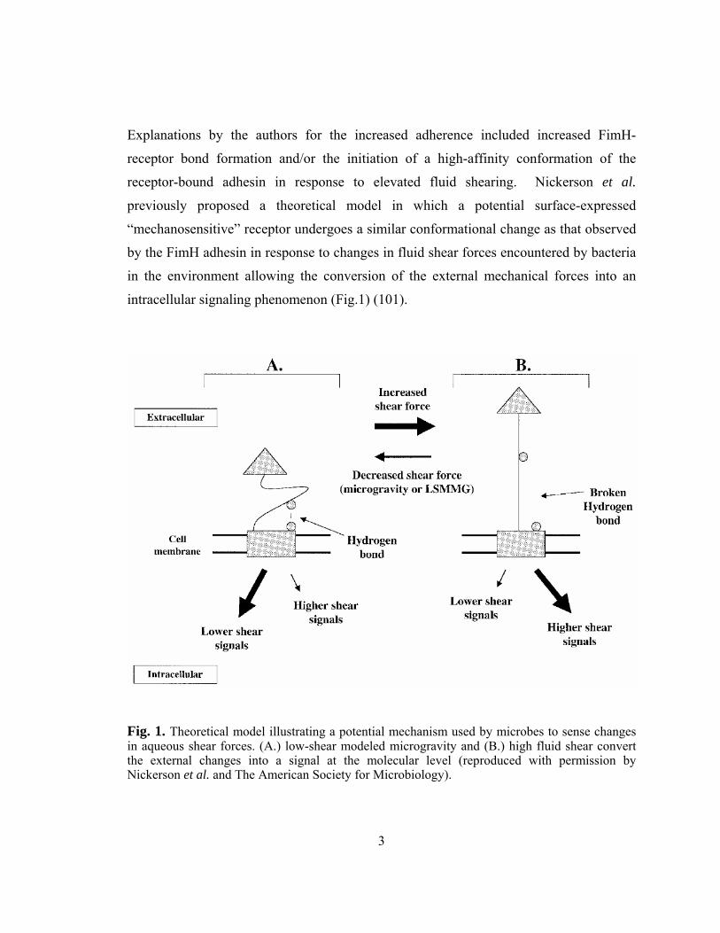
Explanations by the authors for the increased adherence included increased FimH-
receptor bond formation and/or the initiation of a high-affinity conformation of the
receptor-bound adhesin in response to elevated fluid shearing. Nickerson et al.
previously proposed a theoretical model in which a potential surface-expressed
“mechanosensitive” receptor undergoes a similar conformational change as that observed
by the FimH adhesin in response to changes in fluid shear forces encountered by bacteria
in the environment allowing the conversion of the external mechanical forces into an
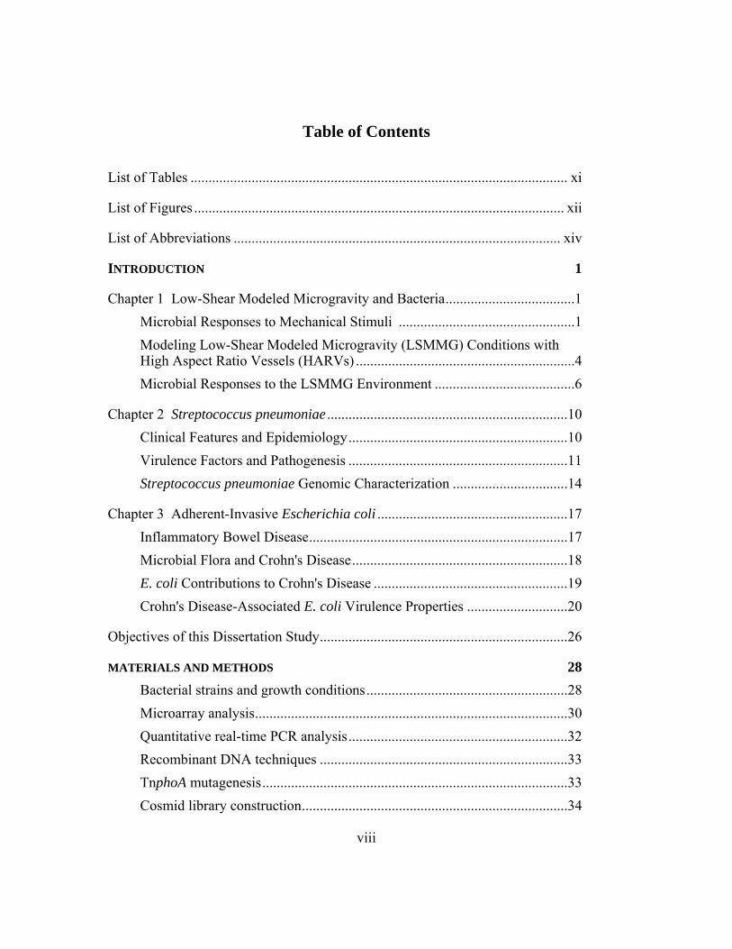
intracellular signaling phenomenon (Fig.1) (101).
Fig. 1. Theoretical model illustrating a potential mechanism used by microbes to sense changes in aqueous shear forces. (A.) low-shear modeled microgravity and (B.) high fluid shear convert the external changes into a signal at the molecular level (reproduced with permission by Nickerson et al. and The American Society for Microbiology).
4
MODELING LOW-SHEAR MODELED MICROGRAVITY (LSMMG) CONDITIONS WITH HIGH ASPECT RATIO VESSELS (HARVS)
Changes in fluid shear forces are commonly encountered by pathogenic bacteria
during the colonization of different niches within the human host. These fluctuations can
range from low shear forces (< 1 dyne/cm2), like those encountered in utero and between
brush border microvilli of epithelial cells to higher forces (4-50 dynes/cm2), as
encountered along the walls of blood vessels (31, 64, 130). A number of different
rotating-wall bioreactors have been utilized as model systems to generate environments
characterized by low shear and low turbulence to cultivate cell cultures in a suspended
state. Within this family of bioreactors are the high-aspect ratio vessels (HARVs) which
were originally designed by the NASA Biotechnology Group based at the Johnson Space
Center, Houston, TX, for the purpose of studying cell cultures under microgravity
conditions (66).
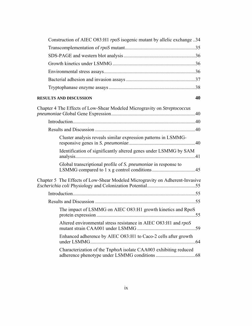
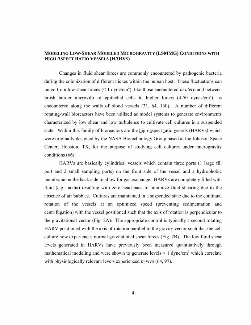
HARVs are basically cylindrical vessels which contain three ports (1 large fill
port and 2 small sampling ports) on the front side of the vessel and a hydrophobic
membrane on the back side to allow for gas exchange. HARVs are completely filled with
fluid (e.g. media) resulting with zero headspace to minimize fluid shearing due to the
absence of air bubbles. Cultures are maintained in a suspended state due to the continual
rotation of the vessels at an optimized speed (preventing sedimentation and
centrifugation) with the vessel positioned such that the axis of rotation is perpendicular to
the gravitational vector (Fig. 2A). The appropriate control is typically a second rotating
HARV positioned with the axis of rotation parallel to the gravity vector such that the cell
culture now experiences normal gravitational shear forces (Fig. 2B). The low fluid shear
levels generated in HARVs have previously been measured quantitatively through
mathematical modeling and were shown to generate levels < 1 dyne/cm2 which correlate
with physiologically relevant levels experienced in vivo (64, 97).
5
Fig. 2. Operational positions of High-Aspect Ratio Vessels (HARVs) in LSMMG and control (1 x g) orientations. In the LSMMG orientation the axis of rotation is perpendicular to the gravitational vector (A) while in the control (1 x g) orientation the axis of rotation is parallel to the gravitational vector (B) allowing growing cultures to experience normal gravitational shearing conditions.
6
MICROBIAL RESPONSES TO THE LSMMG ENVIRONMENT
Previous studies utilizing HARVs to cultivate different bacterial genera under
LSMMG conditions have reported changes in many aspects of microbial biology, such as
alterations in physiology, gene and protein expression, and pathogenesis. Some of the
earliest studies examining the effects of LSMMG on bacteria showed that cultures grown
in the HARVs undergo alterations in certain aspects of secondary metabolism such as
antibiotic production and localization. The production of antimicrobial peptides such as
cephalosporin, microcin B17, and rapamycin produced by Streptomyces clavuligerus, E.
coli, and S. hygroscopicus, respectively, were found to be inhibited by cultures grown
under LSMMG conditions, whereas the production of gramicidin S by Bacillus brevis
was found to be unaltered (44-47, 56). Other observed metabolic changes under
LSMMG conditions include the absence of glycerol-based repression of gramicidin S
and microcin B17 production by B. brevis and E. coli normally observed in shaker flasks
(45, 46). Changes in antibiotic localization have also been observed as a specific
response to LSMMG conditions. Microcin B17 and rapamycin were found to accumulate
in the media of HARV-grown cultures in contrast to the intracellular production of these
antimicrobials by shaker flask cultures (44). The authors attributed the extracellular
localization of these products to be dependent on fluid shear levels experienced by the
bacteria, since introduction of a single glass bead into the HARV reversed the effects.
The addition of synthetic beads of different sizes has been shown to quantitatively
increase fluid shear forces generated within the HARVs (97).
In addition to alterations in the production of microbial products, LSMMG
conditions have also been shown to alter growth kinetics and the ability of bacteria to
resist different environmental stresses. Salmonella enterica serovar Typhimurium growth
kinetics revealed a decrease in generation time when grown in minimal media under
LSMMG conditions, in comparison to growth in rich media such as Luria-Bertani broth
(LB) (147). An increase in dry-cell weight was observed by E. coli grown in minimal
media in the LSMMG environment (44). The increased growth kinetics under LSMMG
7
indicates that the proliferation of certain microbes is enhanced. This could contribute to
more efficient colonization and subsequent infection in the host. Wilson et al. previously
showed that S. Typhimurium demonstrated an enhanced ability to resist thermal, osmotic,
and acid stress conditions after growth under LSMMG, while resistance to oxidative
stress was found to be diminished (147). A diminished ability to resist thermal and acid
stresses was also observed in S. pneumoniae strain TIGR4 after growth under LSMMG
(2). In contrast, E. coli strain AMS6 was found to exhibit an increased resistance to both
osmotic and acid stress conditions after growth under LSMMG (85). Interestingly, in
both the S. Typhimurium and E. coli studies, mutant strains in the rpoS gene were
generated to investigate the impact of this global stress regulator under LSMMG
conditions. While environmental stress resistance was not altered between wild-type and
rpoS mutant strains of S. Typhimurium, the E. coli mutant strain demonstrated a
decreased resistance upon entry into stationary phase in the HARVs. The observed
difference in stress resistance between the wild-type and E. coli mutant strains could be
correlated to RpoS activity levels in relation to growth phase. Optimal RpoS activity is
generally experienced during the latter stages of growth such as the transition from late-
log to stationary phase. These findings indicate the impact and importance of low-shear
environments on different physiological components within bacteria, and suggest that
while growth under LSMMG conditions can induce metabolic changes in microbes; these
changes are not universal and vary between different bacteria.
Changes at both the genomic and proteomic levels have also been reported in
which both global transcriptional and translational activities of bacteria have been altered
after growth in the LSMMG environment. Following microarray analysis, S.
Typhimurium strains 14028 (22 responsive genes) and χ3339 (163 responsive genes)
were shown to exhibit altered global gene profiles in a strain-specific manner after
growth under LSMMG (23, 148). LSMMG-responsive genes were found to be
differentially expressed (up- or down-regulated) in response to LSMMG compared to
those in 1 x g control cultures. In both studies, the majority of LSMMG-responsive
genes were down-regulated. Characterization of responsive genes indicated that they
represented various functional categories physically located throughout the genome. A
8
number of the genes identified in the χ3339 strain were found in the same transcriptional
operon or in physically-linked clusters. The expression of lipopolysaccharide (LPS)
biosynthetic genes was found to be down-regulated and corresponded to decreased LPS
protein expression under LSMMG (148). Wilson et al. also reported the presence of
binding sites for the iron uptake regulator, Fur, in the upstream regions of several
LSMMG-responsive genes. Further characterization of wild-type and fur mutant strains
revealed the participation of Fur in acid stress resistance under LSMMG conditions.
Interestingly, this study revealed that known virulence genes were either non-responsive
or down-regulated in response to the LSMMG conditions. In contrast, Chopra et al.
reported genomic alterations in S. Typhimurium strain 14028 under LSMMG conditions
and found the up-regulation of certain stress-related and virulence-associated genes (23).
The stress-response gene dnaK, which encodes the heat-shock protein Hsp70, was up-
regulated under LSMMG and previously found to function in the stabilization of RpoS
translation in response to environmental stresses (121). The Salmonella virulence gene
virK was also found to be up-regulated. This gene has been found to function in
membrane remodeling in the host and in actin-based motility (35, 96).
Additional studies investigating the impact of LSMMG on the transcriptional
profiles of bacteria have also included investigations with E. coli K-12 strain MG1655 (1,
142). In a recent study, Tucker et al. found that growth of E. coli MG1655 in either rich
(LB) or chemically defined (Neidhardt’s MOPS-based defined media supplemented with
0.2% glucose; MOPS) media under LSMMG conditions revealed no specific gene groups
which were strictly responsive to LSMMG conditions regardless of media type.
LSMMG-cultures exhibiting altered gene expression in LB were found to be associated
with cell envelope processes, while altered genes in MOPS participated in translational
processes. In both studies, overall changes in transcriptional activity were reflected by
the down-regulation of the majority of LSMMG-responsive genes.
Changes in protein profiles have been reported in S. Typhimurium (14028 and
χ3339) and enteropathogenic E. coli (EPEC) strain E2348/69 in response to growth in the
LSMMG environment (23, 99). In both studies the expression of a number of functional
proteins were found to be altered (repressed or enhanced), in addition to the induction of
9
proteins expressed only in the LSMMG environment. Alterations in the two-dimensional
protein profile of S. pneumoniae TIGR4 have also been observed in response to growth
under LSMMG (unpublished data). These findings demonstrate that the responses
bacteria undergo in the LSMMG environment at the transcriptional and translational
levels not only differ between different genera, but can also differ in a strain-specific
manner involving regulatory strategies and different molecular pathways.
Changes in virulence potential and pathogenesis have also been observed in
bacterial cultures grown under LSMMG conditions. Infection of BALB/c mice with
LSMMG-grown S. Typhimurium χ3339, via oral administration, resulted in a decreased
time-of-death, decreased LD50, and an increase in colonization efficiency in different
tissues (99). In addition, these cultures also exhibited an increased ability to resist acid
stress and survive within macrophages. The ability to invade and stimulate the
production of tumor necrosis factor α (TNF-α; in macrophages) and stress-associated
mitogen-activated protein kinase kinase 4 (MAPKK4; in epithelial cells) was enhanced
by LSMMG-grown S. Typhimurium 14028 (23). Responses to growth in the LSMMG
environment also resulted in virulence-based alterations of pathogenic strains of E. coli,
including increased production of heat-labile toxin by enterotoxigenic E. coli (ETEC)
strain 180, increased TNF-α production in macrophages infected by EPEC strain E2348-
69, and enhanced expression of the adhesin intimin, in enterohemorrhagic E. coli (EHEC)
strain 86-24 (20, 23). LSMMG-grown S. pneumoniae TIGR4 have been shown to exhibit
increased capabilities to adhere to and invade respiratory epithelia as well as a decrease in
LD50 in interperitoneally infected mice (unpublished data). While these various findings
support the enhancement in virulence potential among different bacterial strains, there is
currently little evidence explaining what changes are occurring at the molecular and
physiological levels in bacteria to induce such phenotypic changes under LSMMG
conditions. Furthermore, the majority of bacterial virulence genes found to be altered
under these conditions have been found to be down-regulated.
10
Chapter 2: Streptococcus pneumoniae
CLINICAL FEATURES AND EPIDEMIOLOGY
Streptococcus pneumoniae is a Gram-positive, non-motile, facultative anaerobic,
encapsulated diplococcus, which replicates forming variable chain lengths in liquid and
on solid medium. Laboratory diagnostic features include the presentation of α-hemolysis
on blood agar, the absence of catalase activity, solubility in bile salts, and optochin
susceptibility (11, 69). S. pneumoniae colonies can undergo phase variation taking on
two different forms of opacity. Opaque colonies are characterized by large, white,
uniform domed-shaped colonies in comparison to the less-defined transparent colony
phenotype. Transparent variants express higher levels of surface phosphorylcholine
(component of teichoic acid; cell wall constituent) which promote increased adherence to
host cells expressing cellular platelet-activating factor (PAF) receptor leading to
enhanced colonization capabilities in areas such as the nasopharynx (59, 95). In contrast,
opaque phenotypes exhibit increased capsule production which allows for increased
survival in the bloodstream. Most S. pneumoniae clinical isolates express an anti-
phagocytic capsule, though non-encapsulated isolates have been isolated (conjunctivitis
cases). The capsule consists of repeating oligosaccharides covalently bound to the cell
wall constituents’ peptidoglycan and C polysaccharide (CPS). Differences in capsule
polysaccharide composition contribute to the identification of 90 different S. pneumoniae
serotypes. In addition to phase variation and capsular differences, S. pneumoniae also
exhibits the natural ability to internalize DNA from other bacteria (competence), allowing
the acquisition of new phenotypic traits (transformation), such as new virulence factors,
antibiotic resistance, and the production of new serologically different capsules (capsule
switching).
S. pneumoniae is a member of the normal flora which colonizes upper respiratory
niches such as the nasopharynx and can be isolated from both healthy children (20-40%)
and adults (5-10%) (95). In immunocompromised individuals, however, S. pneumoniae
is opportunistic and can cause mild-to-severe infections and diseases. The global rate of
11
invasive pneumococcal disease is 15 in 100,000 individuals annually, with higher
incidences of infection found among newborns, infants up to 2 years of age, and the
elderly (49). S. pneumoniae is a major cause of bacterial meningitis and otitis media
(common ear infection) as well as the leading cause of community-acquired pneumonia
(95). Previous infections (e.g. respiratory viral infections) contribute to the establishment
of pneumococcal disease onset by disrupting clearance mechanisms which normally
eliminate bacterial colonization (e.g. congestion in the opening of the eustachian tube
and/or fluid accumulation in paranasal sinus cavities).
VIRULENCE FACTORS AND PATHOGENESIS
S. pneumoniae contains a number of virulence factors, including a capsule,
adhesins, antigenic cell wall constituents, and toxins, which contribute to disease. The
anti-phagocytic capsule plays a crucial role as a virulence factor in immunoevasion.
Besides the obvious role of allowing the bacteria to avoid opsonophagocytosis, the
numerous variations in the chemical structure of the CPS capsule allow for up to 90
different pneumococcal serotypes helping avoid antibody-mediated immunity. As
previously discussed, differences in the content and amount of capsule expressed on the
bacterial surface can promote survival in different niches within the host. Lower capsular
levels allow for greater adherence and invasion potential, as observed by transparent
variants in the nasopharynx where opaque variants expressing higher amounts of capsule
can avoid opsonophagocytosis more effectively within the bloodstream (59, 72, 95). One
of the main pathological characteristics of pneumococcal disease is the initiation of
aggressive inflammatory responses. Cell wall components such as peptidoglycan and
teichoic acid contribute to the antigenic stimulation of these responses when released
upon bacterial lysis by activating the complement pathway and stimulating the secretion
of macrophage activators Interleukin-1 (IL-1) and TNF-α (57). In addition, complement
activation leads to the production of pathway components (i.e. C3a, C4a, and C5a) which
function as chemotactic factors. Complement clearance mechanisms, such as
phagocytosis and action by the membrane attack complex, are neutralized by the
12
presence of CPS-disrupting interactions between surface-bound C3b and phagocytic cells
due to the presence of the thickened cell wall (25, 117). Phosphorylcholine is expressed
on the bacterial surface in two states: a free form and a form bound by choline-binding
proteins. Of the bound forms of phosphorylcholine, choline-binding protein A (CbpA)
was the first surface adhesin described and the most abundantly expressed. CbpA plays
an important role in bacterial adhesion to human cells and has a key role in penetrating
the blood-brain barrier. During initial nasopharyngeal colonization, S. pneumoniae binds
by unknown bacterial lectins to epithelial cell surface carbohydrates (N-acetyl-D-
glucosamine-β1-3 and β1−4 galactose) and sialic acid. CbpA binds polymeric
immunoglobulin receptors (IgR) on mucosal epithelial cells and PAF receptors of
activated endothelial cells to facilitate transport of bacteria into the blood stream and
across the blood-brain barrier, respectively (111, 112, 115). Additional Cbps, such as
Cbp D, E, and G, also play roles in adhesion (62).
Other choline-binding proteins involved in bacterial carriage and virulence
include pneumococcal surface adhesin A (PsaA), pneumococcal surface protein C
(PspC), pneumococcal surface protein A (PspA), and autolysin (59, 72, 95). PsaA shares
homology with lipoprotein adhesins in other streptococci and functions as a permease in
the manganese transport system. PsaA also functions in the regulation of pneumococcal
adherence and virulence. PspC interacts with complement through the binding of factor
H, leading to C3b degradation and preventing complement activation. PspC can also
bind and neutralize secretory IgA antibodies. PspA is an important virulence determinant
that functions to prevent C3b activation and subsequent complement activation by
interacting with factor B. PspA also functions as a lactoferrin receptor for iron
acquisition. Autolysin is involved in growth phase-dependent autolysis which is
triggered as pneumococcal growth transitions from late-exponential to stationary phase.
Lysis of the bacteria contributes to the inflammatory responses through the release of cell
wall components and intracellular toxins such as pneumolysin. In addition to reports of
growth phase-dependent induction, there have been others indicating autolytic induction
through the action of pneumolysin, produced by the pneumococcus, and by human
13
lysozyme produced as a defense factor in response to infection and the onset of
inflammation (9, 27). S. pneumoniae produces several enzymes which aid in colonization and invasion.
Hyaluronidase functions to degrade connective tissue to enhance bacterial dissemination
into the blood stream through the disruption of the blood-brain barrier. S. pneumoniae
also produces two neuraminidases, NanA and B. These enzymes function to enhance
adherence and colonization by cleaving N-acetyl-neuraminic acid from mucin and sialic
acid residues from glycoconjugates of host tissues, to decrease mucus viscosity and
expose additional receptors for pneumococcal adhesion. Secretory IgA1 protease
degrades host IgA1, which protects bacteria from type-specific antibody. The protease
cleaves antibodies at the hinge region generating one Fc fragment and two Fab fragments.
This activity allows the bacteria to avoid mucociliary clearance by coating the bacteria
with Fab fragments, which prevents the binding of intact antibodies containing the Fc
segment which binds mucus for removal (74). The production and subsequent release of
the cytotoxin, pneumolysin, during cell lysis functions as a thiol-activated cytolysin
which binds target cell membranes, oligomerizes, and forms pores in the membrane of
host-cells resulting in cell lysis. Additional effects of pneumolysin include the disruption
of ciliary motion on epithelia, prevention of mucociliary clearance, the inhibition of
lymphocyte proliferation and antibody synthesis, as well as reduced neutrophil migration
and bactericidal activity (51, 52, 104).
S. pneumoniae infections can remain localized or develop into more severe forms
of disease such as meningitis. Infections are initiated from upper respiratory colonization
sites (e.g. nasopharynx) and can disseminate into the middle ear and sinuses and access
the bloodstream leading to bacteremia, and the subsequent breaching of the blood-brain
barrier resulting in the infection of the choroid plexus. Upon dissemination into the
subarachnoid space, an inflammatory response is initiated which is key to the disease
etiology of meningitis (72, 95). Studies using rabbit infection models suggest that
bacterial cell wall components, such as peptidoglycan and teichoic acid, elicit cerebral
spinal fluid abnormalities associated with meningitis through inflammatory mediators
such as IL-1, IL-6, TNF-α, and C5a (109). Symptoms associated with pneumococcal
14
pneumonia include fever, chills, shortness of breath, fatigue, sweats, and cough.
Pneumonia occurs from the successful invasion of bacteria into the alveolar spaces where
replication occurs, and the immune responses, characterized by complement activation
and generation of vasoactive substances, result in accumulation of exudative fluid and
white blood cells in the alveoli and septa of the lungs. Fluid filling the lungs can be
radiographically viewed as consolidated areas. Complications, such as empyema
(collection of pus within pleural space between lungs and pleura membrane), can arise
from pneumococcal pneumonia, resulting in mortality exceeding 30% in some
documented hospital cases despite aggressive therapy. Additional syndromes, which are
rare, yet can still occur from pneumococcal infection, include peritonitis, endocarditis,
osteomyelitis, epidural and brain abscesses as well as soft tissue infections (95).
STREPTOCOCCUS PNEUMONIAE GENOMIC CHARACTERIZATION
Bacterial pathogens can adapt to different anatomical niches within the human
host through mediating differential gene expression in response to changing
environmental stimuli. Microarray technology has become a useful tool in characterizing
how these changes alter the global genomic profiles of diverse bacterial generas such as
B. subtilis, M. pneumoniae, S. gordonii, and Borrelia burgdorferi in response to
numerous environmental changes such as alterations in salinity, oxygen availability,
temperature, pH, and in vivo conditions, respectively (110, 129, 144, 146, 149).
Microarray analysis of different pneumococcal species such as S. pneumoniae and S.
pyogenes have demonstrated changes in global transcriptional activity in response to
different environmental variables including temperature, antibiotics, competence-
induction, and species/strain variability (65, 103, 107, 113, 127). Studies of group A
Streptococcus (GAS) and S. pneumoniae have revealed that the expression of a number
of genes, which function in different physiological processes, can be altered in a
temperature-dependent manner (103, 127). GAS cultures grown at 29°C demonstrated
altered gene expression by 9% of genes, including those genes which encode
transcriptional regulators, transporters, and iron homeostasis proteins. Though the
15
expression of few known GAS virulence genes was altered, 28 genes encoding proteins
with different secretion signals were altered. The altered expression of these genes can
subsequently affect the extracellular proteome which could include proteins used in host
cell interactions thus contributing to the survival and virulence of the bacterium. In S.
pneumoniae, Pandya et al. also reported changes in the expression of genes distributed
throughout the genome in response to incubation at different temperatures (21, 29, 33,
and 40°C). Hierarchical cluster analysis revealed gene groups exhibiting correlating
changes in expression in a temperature-specific manner. The relevance of such findings
demonstrates that during colonization of the host, bacteria, such as Streptococci, can
respond to changing environmental stimuli, such as temperature, within different parts of
the body (e.g. nasopharynx, 33°C; lungs, 37°C; 40°C blood stream during febrile
response), through mediating differential gene expression. By altering the expression of
certain genes, bacteria can subsequently alter the expression of different proteins which
can aid in processes such as colonization, cell division, virulence, and immune system
evasion to enhance their ability to survive and persist in the host.
Microarray analysis has been employed in the identification of genes responsible
for antibiotic resistance by S. pneumoniae. Studies using wild-type and mutant S.
pneumoniae strains with disruptions in the operon encoding the VncRS two-component
system (aids in vanomycin resistance) have revealed VncS-dependent regulation of gene
clusters involved in the import of “death peptide” signals resulting in autolysis in
response to antibiotics (113). Besides antibiotic resistance others have reported
differences in transcriptional patterns when using global analyses to study other S.
pneumoniae processes (e.g. the induction of competence and when studying phenotypic
differences) among various clinical isolates. Peterson et al. used microarray analysis to
characterize the kinetics of S. pneumoniae competence gene expression during
exponential growth in vitro (107). Results of the study revealed the induction times of
different genes involved in the onset of natural competence, while 8 additional loci were
also identified and found to be induced during the competence process. In an effort to
study intra- and interspecies genomic variations, Hakenbeck et al. used microarray
analysis to compare different isolates of S. pneumoniae with reference strains addressing
16
the acquisition of antibiotic resistance and variations in capsule serotypes (65). Up to
10% of the genes were altered between isolates and the reference strains and 10 clusters
were identified which accounted for half of the altered genes. Most of the genes were
associated with transposases and mosaic genes housing antibiotic resistance determinants.
In addition, distinct antigenic profiles were observed upon overall comparison with
commensal strains such as S. mitis and S. oralis. These reports indicate the importance of
genomic profiling in understanding S. pneumoniae responses to different environmental
stimuli as well as distinguishing the characteristics among different species and strains.
17
Chapter 3: Adherent-Invasive Escherichia coli
INFLAMMATORY BOWEL DISEASE
Inflammatory bowel disease (IBD) is characterized by the inflammation of the
gastrointestinal tract and can be accompanied by a range of symptoms, including fever,
abdominal pain and cramping, and repeating episodes of severe watery or bloody
diarrhea. IBD is typically manifested by one of two different disease forms: ulcerative
colitis (UC) or Crohn’s disease (CD). UC is mainly localized to the inner lining of the
rectum and the colon (large intestine). UC inflammation begins at the rectum and lower
intestine (sigmoid area) and progresses upward, affecting continuous sections of the inner
lining of the colon. These inflamed tissues can develop bleeding ulcers secreting mucus
and pus. While there is currently no cure for UC, drug therapies can be implemented to
reduce symptoms, and in severe cases surgical resectioning of the colon can be carried
out to remove diseased sections of tissue (92).
Unlike UC which rarely involves the small intestine, CD inflammation typically
begins at the lower region of the small intestine (ileum), but can also extend into
additional areas including the rectum, stomach, esophagus, and mouth. CD is initially
manifested as a scattering of small sores along the ileal lining which can become
inflamed and develop into ulcers. Ulceration in the mouth and throat can complicate
eating and swallowing, causing pain to patients. In contrast to UC, separate patches of
inflamed sores can occur simultaneously in different regions leaving areas of healthy
tissue in between. Large volumes of water and salt can be secreted by affected areas
overwhelming the absorptive capacity of the colon, resulting in the development of
frequent and repetitive diarrheal episodes. The continual inflammation and ulceration of
bowel tissues can lead to the swelling and thickening of bowel walls and scar tissue
disrupting the movement of fecal materials through the bowel and leading to abdominal
cramping and pain. In addition, the growth of the sores into large ulcers can lead to
penetration into, and sometimes through, digestive tract walls. As with UC, there is no
18
cure for CD, and options include drug therapies and surgical alternatives in severe cases.
The onset incidence of IBD has been found to be highest among populations living in
cities of industrialized countries, with equal representation among men and women. In
the U.S. alone there are currently over 500,000 cases of UC, targeting individuals
between the ages of 15 and 40 years of age. There are more than 100,000 cases of CD in
children, typically diagnosed around the ages of 10 and 11, in the U.S. (92).
There is no known cause for either UC or CD; however contributing factors
include hereditary factors, immune system dysfunction, environmental factors, and the
use of antibiotics. Typically, 20% of CD patients have an immediate relative (parent,
sibling, child) with some form of IBD, which suggests a hereditary link to the
predisposition (92). High frequencies of CD patients have been shown to have mutations
in the NOD2/CARD15 gene which is thought to be associated with the early onset of
symptoms and high risk of post-surgical relapse (13). Aberrant and unregulated
hypersensitivity by the immune system to either invading enteric pathogens or to normal
flora within the gut is also thought to contribute to the chronic state of inflammation
associated with IBD. Environmental factors thought to contribute to IBD pathology
include the presence of bacterial, viral, and/or food antigens which can repeatedly
stimulate immune responses leading to chronic inflammation. Lastly, the use of
antibiotics is thought to contribute by disrupting the balance of microbial flora in the
intestinal tract leading to the overgrowth of species which could contribute to colitis, such
as that observed from overgrowth of Clostridium difficile (50). While all of these factors
have the potential to contribute to the onset of IBD, increasing evidence suggests that
abnormal immune responses to microflora in the gut contributes to the chronic
inflammation of IBD and will be further discussed.
MICROBIAL FLORA AND CROHN’S DISEASE
CD has been characterized by the presence of chronic transmural, segmental, and
granulomatous inflammation within the intestinal tract (43). The features presented in
CD patients correlate with those of various microbial infections in the gut and can include
19
ulcerations, microabscesses, fissures, fistulas, granulomas, and lymphangitis (29).
Pathogens such as Mycobacterium sp., Yesinia enterocolitica, and Chlamydia
trachomatis have been shown to induce inflammatory responses similar to that exhibited
by CD patients (119). Previous studies have found evidence which suggests that the
microbial flora within the gastrointestinal tract of CD patients could be functioning as an
antigenic stimulus contributing to the chronic state of inflammation through unregulated
immune responses by the host. Clinical studies conducted with CD patients have shown
that a decrease in luminal floral concentrations in response to intestinal lavage and
antibiotic treatment resulted with symptomatic improvements (116, 134). In rodent
models of intestinal inflammation, symptoms were shown to be attenuated or absent
when the animals were kept under germ-free conditions (122, 136). Cave et al. found
that mice developed granulomas when inoculated with gastrointestinal homogenates
prepared from CD patients (21). Various studies have shown correlations with the
presence of bacterial pathogens prior to and during the manifestation of CD in patients.
A small percentage of patients during epidemics involving Shigella sp., Yersinia sp., and
Salmonella sp. developed IBD without prior evidence of bacterial infection (108). In
other cases, CD patients have either tested positive for various bacterial antigens (e.g.
Listeria, E. coli, Streptococcus sp.) or had bacterial isolates (e.g. M. tuberculosis)
successfully removed from tissues (22, 83).
E. COLI CONTRIBUTIONS TO CROHN’S DISEASE
E. coli is a motile, Gram-negative, aerobic bacillus which comprises a major part
of the normal flora within the gastrointestinal tract. While the presence of non-
pathogenic species of E. coli play an important role in the maintenance of normal
intestinal physiology and luminal flora stability, there are a variety of pathotypes which
cause a number of diarreheagic and extraintestinal diseases through the expression of
different virulence factors affecting a range of cellular processes. While several different
bacterial genera have been implicated in their contribution to CD etiology, increasing
evidence suggests E. coli as a candidate in the contribution of disease. While altered

20
luminal flora concentrations have been found in CD patients, the neoterminal ileum has
been found to be colonized by abnormally high levels of E. coli (81). Surgical sampling
results have shown that the frequency of isolating E. coli from intestinal serosa and
mesenteric lymph nodes was 27 and 33% higher in CD patients, respectively, in
comparison to controls (4, 78). Analysis of the ileal mucosa of CD patients showed E.
coli recovery frequency was 65% from chronic lesions and 100% from early lesions,
while overall predominance was abnormally high (50-100%) (30).
In addition, clinical studies have shown E. coli antibody titers to be higher in CD
patients compared to those of control groups; however no specific serogroups were
associated with disease (17, 135). Using immunocytochemistry, Cartun et al. reported
that 69% of CD patient mucosal samples were positive for E. coli antibodies, while Liu et
al. reported that in 57% of CD cases, positively labeled tissues were located in the lamina
propria, along fissures, and on macrophages localized beneath ulcers (19, 83). While no
single serogroup or specific strain of E. coli has been identified from CD patients,
Masseret et al. reported that CD isolates did share common ribotype patterns (91). These
profiles have been found in both healthy and ulcerated mucosa suggesting uniform
colonization regardless of the state of the tissue and the possibility of a common
evolutionary ancestry.
CROHN’S DISEASE-ASSOCIATED E. COLI VIRULENCE PROPERTIES
Pathogenic E. coli classes contain well-characterized virulence factors which aid
in the progression of infection and disease. These factors include a variety of adhesins,
invasins, toxins, and secretion systems which aid in the attachment and infection of host
cells. Clinical E. coli strains removed from stool samples and biopsies of CD patients
have been investigated for the presence of virulence-factor encoding genes common to
these pathogenic classes, and none of these genes have been identified in the isolates (30,
120). However, these clinical isolates have demonstrated intrinsic adherent and invasive
properties, which play an important role in colonization and infection of host tissues and
thus have been categorized in a new putative class of E. coli known as adherent-invasive
21
E. coli (AIEC) (29). Several studies have demonstrated the adhesive potential of AIEC to
different epithelial cell lines. AIEC strains isolated from 53-62% of stool samples taken
from CD patients have exhibited adherence to buccal cells compared to 5-6% of isolates
from controls (18, 58). Comparative studies investigating the adherence properties of
AIEC strains isolated from the ileal mucosa of CD patients reported that the strains
preferentially adhere to differentiated Caco-2 cells, which are representative of mature
intestinal epithelia (30). This preferential adherence trend correlates with findings in CD
patients which show that crypt epithelia, which are representative of immature intestinal
epithelial cells, are rarely involved in early lesion formation (118).
The primary lesions that develop in patients are indicative of early stages of CD
(e.g. aphthous ulcers develop from necrotizing M-cells) and mainly occur in Peyer’s
patches (54). The formation of these lesions is similar to those developed during
infections by enteric pathogens, such as Shigella and Salmonella, in which invasion of
host cells plays an important role in their virulence. AIEC strains isolated from early and
chronic ileal lesions of CD patients have been shown to exhibit efficient invasion
capabilities. AIEC strain LF82 was previously characterized for its invasive potential
and found to effectively invade a number of different epithelial cell lines in vitro,
including Caco-2, HCT-8, HEp-2, and Intestine-407 cells (15). Comparative analysis of
the invasive properties of AIEC LF82 revealed unique properties which differ from those
of other invasive pathogens. Most invasive bacterial strategies investigated such as those
of enteroinvasive E. coli (EIEC) and S. flexneri are dependent on actin microfilament
functioning, but are independent of microtubules (26, 41). In contrast, AIEC LF82 uptake
depends on both functional actin microfilaments and microtubules (15). The uptake
mechanism resembles a macropinocytosis-like process which involves the elongation of
host cell membrane extensions around the bacterial cell. Upon entry into the host cell,
the bacteria lyse the endocytic vesicle and successfully survive and replicate within the
host cell cytoplasm. The invasion process carried out by AIEC strains is unique in that
none of the genetic invasive determinants possessed by other invasive pathogens have
been found in these strains. Boudeau et al. previously investigated the invasion process
carried out by AIEC LF82 and found that type 1 pili-mediated adherence played a key
22
role in the invasion of host cells (14). AIEC LF82 invasion was found to be inhibited by
the presence of D-mannose or methyl-α-D-mannopyranoside. Adherence by type 1 pili
to host cells is dependent on the binding of D-mannose moieties on the host cell surface.
Non-invasive mutants generated by TnphoA mutagenesis were found to contain
disruptions in genes comprising the fim operon, encoding the type 1 pili, and were unable
to induce membrane extensions upon adherence to host cells. The induction of
membrane extensions and subsequent invasion by the mutant strains was restored upon
trancomplementation with cloned fim operon of non-pathogenic strain E. coli K-12.
Interestingly, complementation of non-invasive laboratory strain JM109 with the fim
operon from LF82 did not transfer invasive capabilities, indicating that the type 1 pili
must be expressed in the AIEC genetic background to confer invasive ability. In addition
to type 1 pili, bacterial flagella were also found to function in the co-operation of AIEC
LF82 adhesion and invasion directly through active motility and indirectly through the
regulation of type 1 pili expression, respectively (7). More recently, absence of the
expression of lipoproteins NlpI and YfgL were found to diminish AIEC LF82 adherence
and invasion abilities. The contribution of NlpI expression was suggested to function in
the regulatory pathways mediating the expression of flagella and type 1 pili, because an
nlpI mutant AIEC LF82 strain was unable to express flagella and exhibited small
amounts of type 1 pili (8). A yfgL mutant strain of AIEC LF82 was shown to exhibit a
highly decreased ability to invade epithelia and release outer membrane vesicles which,
previously, have been shown to function in the intercellular transport of virulence factors
to the host cell (114).
Intracellular pathogens have been the main focus in identifying potential
microbial sources contributing to CD due to their ability to evade phagocytosis-based
killing, survive within macrophages, and function as antigenic stimuli for T cells and
macrophages, in eliciting the chronic inflammatory state of the gut. One of the main
histological hallmarks of CD is the development of granulomatous inflammation in the
lymph nodes and epithelial lining of the intestinal tract. This etiology has also been
found during infections by intracellular bacteria and fungi (29). Previous studies have
detected E. coli antigens in lymph nodes, granulomas, and in macrophages of CD patients
23
(19, 83). Glasser et al. have previously reported AIEC strains to successfully invade,
survive, and replicate within macrophages without inducing cell death (61). In these
studies intracellular replication increased up to 74-fold when compared to initial infection
and escape from phagosomal vesicles was not found to be a required step as seen in
epithelial cells. Instead, the fusion of these early vesicles into larger vacuoles was
observed during macrophage invasion as seen in Salmonella and Y. enterocolitica
infections (3, 63). This activity has been thought to promote survival through the dilution
and attenuation of toxic compounds within the phagosomes. In a recent study, Bringer et
al. found that the high-temperature requirement A class (HtrA) protein played an
important role in survival during intramacrophagic replication (16). Inactivation of the
htrA gene in an AIEC mutant resulted in a decreased ability to resist oxidative stress and
diminished growth capabilities under acidic and nutrient poor conditions, as experienced
in the microenvironment of phagosomes. Thus, AIEC appears to contain specific genes,
such as htrA, which play a key role in survival in response to unfavorable environments
such as those encountered upon cell invasion, e.g., the acidic environment of macrophage
phagosomes. One unique feature exhibited by AIEC strains is the ability to avoid the
induction of macrophage cell death upon invasion, as opposed to other invasive bacteria.
Classical indicators of cell death including DNA fragmentation/degradation, cell
detachment, and lactate dehydrogenase (LDH) release have not been observed in AIEC-
infected macrophages even up to 24 hours post-infection (61). The absence of
macrophage cytotoxicity and death by AIEC uptake differ greatly from that observed by
most of the diarrheagic E. coli pathovars which elicit cytotoxic events and apoptosis in
infected macrophages (79, 80).
AIEC strains have been shown to be capable of the continual stimulation of
infected macrophages to produce large amounts of pro-inflammatory cytokines (e.g.
TNF-α) which can contribute to the chronic inflammatory state of the gastrointestinal
tract of CD patients (61). The continual activation and production of TNF-α
demonstrates that not only are the infected macrophages still active, but that this constant
activation can be sustained through the replication of the intracellular bacteria residing in
the fused vacuoles of the macrophages. The current working model for the contribution
24
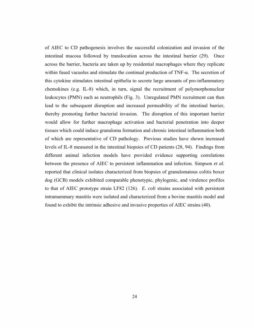
of AIEC to CD pathogenesis involves the successful colonization and invasion of the
intestinal mucosa followed by translocation across the intestinal barrier (29). Once
across the barrier, bacteria are taken up by residential macrophages where they replicate
within fused vacuoles and stimulate the continual production of TNF-α. The secretion of
this cytokine stimulates intestinal epithelia to secrete large amounts of pro-inflammatory
chemokines (e.g. IL-8) which, in turn, signal the recruitment of polymorphonuclear
leukocytes (PMN) such as neutrophils (Fig. 3). Unregulated PMN recruitment can then
lead to the subsequent disruption and increased permeability of the intestinal barrier,
thereby promoting further bacterial invasion. The disruption of this important barrier
would allow for further macrophage activation and bacterial penetration into deeper
tissues which could induce granuloma formation and chronic intestinal inflammation both
of which are representative of CD pathology. Previous studies have shown increased
levels of IL-8 measured in the intestinal biopsies of CD patients (28, 94). Findings from
different animal infection models have provided evidence supporting correlations
between the presence of AIEC to persistent inflammation and infection. Simpson et al.
reported that clinical isolates characterized from biopsies of granulomatous colitis boxer
dog (GCB) models exhibited comparable phenotypic, phylogenic, and virulence profiles
to that of AIEC prototype strain LF82 (126). E. coli strains associated with persistent
intramammary mastitis were isolated and characterized from a bovine mastitis model and
found to exhibit the intrinsic adhesive and invasive properties of AIEC strains (40).
25
Fig. 3. Current model for AIEC participation in CD pathogenesis. Colonization precedes invasion into epithelia followed by uptake into residential macrophages. Intracellular replication stimulates the subsequent production of TNF-α which stimulates epithelia to secrete potent chemokine attractants such as IL-8 initiating polymorphonuclear (PMN) leukocyte recruitment.
26
Objectives of this Dissertation Study
The use of HARVs as a model system to study the effects of LSMMG on bacteria
has produced important findings which show that bacteria are, indeed, responsive to
mechanical stimuli in addition to other physiological forms of stress. These findings
have been shown to impact various aspects of prokaryotic physiology, virulence
potential, and activity at both the transcriptional and translational levels. The work
presented here utilizes HARVs as a model system to characterize the effects of LSMMG
on two very different bacterial genera, adherent-invasive E. coli (AIEC) and S.
pneumoniae.
In Chapter 2, the use of microarray technology is discussed regarding its potential
as a tool to better understand how S. pneumoniae adapts to various environmental factors
which begins at the genomic level and can be observed through alterations of the
genomic profile of the bacterium. As discussed in Chapter 1, microarray analyses of
different bacteria generas to LSMMG conditions have thus far been limited to
investigations of Gram-negative enteric strains such as S. Typhimurium and E. coli. My
work investigates, for the first time, the effects of the LSMMG environment on the global
transcriptional profile of an upper respiratory, Gram-positive pathogen. This genomic
analysis and characterization will provide a better understanding of how mechanically-
based stresses also affect the global transcriptional activity of respiratory pathogens.
These findings will allow for further studies which can pursue the identification of key
regulators and putative molecular mechanisms modulating the responses observed by S.
pneumoniae to LSMMG conditions.
As discussed in chapters 1 and 3, the effects of LSMMG have been found to alter
many different properties of both commensal and pathogenic strains of E. coli; however,
there is presently no knowledge of how AIEC isolates respond to different forms of
mechanical stress, such as low-fluid shearing, as encountered within the gastrointestinal
tract of the host where colonization occurs. HARVs were utilized to characterize basic
and fundamental properties of AIEC O83:H1 grown under LSMMG conditions such as

27
growth kinetics, stress resistance, and colonization potential as well as some of the
molecular regulatory components associated with these processes.
28
MATERIALS AND METHODS
Bacterial strains and growth conditions.
Strains and plasmids are listed in Table 1. E. coli strains were routinely grown in
LB broth or on LB agar at 37°C while S. pneumoniae TIGR4 (139) was grown in Todd
Hewitt broth supplemented with 0.5 % yeast extract (THY) or on Trypticase Soy agar
(TSA) supplemented with 5% sheep blood at 33oC in a candle extinction jar. M9
minimal media (M9 minimal salts (Gibco BRL), 1 mM MgSO4, 0.05% NaCl, 0.2%
glucose, 0.1 mM CaCl2; pH 7.0) was used to culture E. coli strains in the HARVs while
THY was used for S. pneumoniae cultures at 37°C in 5% CO2 (86). Isolated colonies
from plates were used to seed overnight cultures (5 ml). Fresh media (100 ml) was
inoculated (1:100) using overnight cultures and aseptically added to two sterile HARV
vessels (50 ml / vessel) (Synthecon, Inc.). Additional media was added to each HARV to
completely fill each vessel and eliminate the presence of any air bubbles. Vessels were
oriented into either the LSMMG or 1 x g control orientations (Fig.2) and incubated at
37°C at rotational speeds of 15 rpm for S. pneumoniae or 25 rpm for E. coli. The
rotational speed for S. pneumoniae was previously optimized in the laboratory of Dr.
David Niesel to prevent gravitational sedimentation and centrifugation of bacteria. The
rotational speed used for E. coli has been previously optimized by Nickerson et al. (85,
99) for Salmonella and Lynch et al. (96) for E. coli. For microarray studies a second 1 x g
static control HARV was included to function as an additional filter to exclude genes
which were found to be differentially expressed between 1 x g rotating and 1 x g static
conditions. Allen et al. have previously shown that gene expression can be altered
between static and rotating 1 x g controls independent of LSMMG conditions (1).
Therefore, genes found to be differentially expressed between rotating and static controls
were determined not to be directly impacted by the LSMMG environment and were
excluded from further analysis. S. pneumoniae HARV cultures were monitored by
optical density (OD600nm) until cells entered mid-logarithmic phase (OD = 0.5) after
29
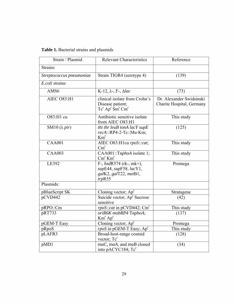
Table 1. Bacterial strains and plasmids
Strain / Plasmid Relevant Characteristics Reference
Strains:
Streptococcus pneumoniae Strain TIGR4 (serotype 4) (139)
E.coli strains:
AMS6 K-12, λ-, F-, ∆lac (73)
AIEC O83:H1 clinical isolate from Crohn’s Disease patient; Tcr Apr Smr Cmr
Dr. Alexander Swidsinski Charite Hospital, Germany
O83:H1 cu Antibiotic sensitive isolate from AIEC O83:H1
This study
SM10 (λ pir) thi thr leuB tonA lacY supE recA::RP4-2-Tc::Mu-Km; Kmr
(125)
CAA001 AIEC O83:H1cu rpoS::cat; Cmr
This study
CAA003 CAA001::TnphoA isolate 1; Cmr Kmr
This study
LE392 F-, hsdR574 (rk-, mk+), supE44, supF58, lacY1, galK2, galT22, metB1, trpR55
Promega
Plasmids:
pBlueScript SK Cloning vector; Apr Stratagene pCVD442
Suicide vector; Apr Sucrose sensitive
(42)
pRPO::Cm rpoS::cat in pCVD442; Cmr This study pRT733
oriR6K mobRP4 TnphoA; Kmr Apr
(137)
pGEM-T Easy Cloning vector; Apr Promega pRpoS rpoS in pGEM-T Easy; Apr This study pLAFR3
Broad-host-range cosmid vector; Tcr
(128)
pMD1 tnaC, tnaA, and tnaB cloned into pACYC184; Tcr
(34)
30
which samples were collected for experimentation. Harvesting S. pneumoniae cultures at
mid-logarithmic phase avoids cultures from undergoing natural stationary phase-induced
autolysis. E. coli HARV cultures were harvested after 12 h of growth and then
quantitated based on spectrophotometric analysis. Concentrations were then adjusted for
experimentation. When required, growth media was supplemented with antibiotics at the
following concentrations: chloramphenicol (Cm) 30 μg ml-1; kanamycin (Km) 50 μg ml-1;
ampicillin (Ap) 100 μg ml-1; and tetracycline (Tc) 25 μg ml-1.
Microarray analysis.
S. pneumoniae HARV cultures were grown to an optical density of 0.5, harvested
and neutralized in a 2:1 volume (reagent to culture) of RNAprotect bacterial reagent
(Qiagen, Valencia, CA) to stabilize RNA, cells were pelleted, and stored at -80°C. Prior
to RNA isolation, bacterial cells were resuspended in lysis buffer (10 mM Tris HCl, 1
mM EDTA, pH 8.0, 1 mg/ml lysozyme) then lysed by mechanical disruption using a
Mini BeadBeater™ (Biospec Products, Inc.) for 5 minutes at maximum speed with 0.1
mm zirconia/silica beads (Biospec Products, Inc.). RNA was isolated with RNeasy
columns (Qiagen, Valencia, CA) and DNase treated using Turbo DNase-Free (Ambion,
Austin, TX), according to manufacturers’ instructions. Purified RNA was used to
generate cDNA which was labeled and applied to S. pneumoniae strain TIGR4 genome
cDNA microarrays, developed by The Institute for Genomic Research (TIGR, Rockville,
MD), based on the TIGR4 strain genomic sequence, and provided by the Pathogen
Functional Genome Resource Center (PFGRC) of the National Institutes of Health. Each
microarray consisted of polymerase chain reaction (PCR) products representing segments
of 2131 open reading frames (95% of predicted coding regions) from strain TIGR4.
The generation and labeling of cDNA, microarray hybridization and processing,
and intensity value generation was performed by the UTMB Molecular Genomics Core
facility (UTMB, Galveston, TX) as previously described (103). To generate reproducible
and statistically significant data, duplicate microarray slides were used for each
individual experiment. The cDNA generated from experimental and control samples was
31
labeled by the incorporation of either cyanine3-dUTP (Cy3; green fluorescence) or
cyanine5-dUTP (Cy5; red fluorescence) fluorescently labeled nucleotides to distinguish
competitive hybridization of array spots. In contrast to affymetrix array chips, each
microarray slide was hybridized with differentially labeled cDNA from both control and
LSMMG cultures. Three independent HARV experiments were performed comparing 1 x
g control cultures with LSMMG while two independent experiments were carried out
comparing different gravitational controls (1 x g rotating and 1 x g static conditions). The
microarray data were analyzed using GenePix Pro 6.0 (Molecular Devices Corporation,
Union City, CA), Spotfire DecisionSite 7.3 (Spotfire, Somerville, MA), Significance
Analysis of Microarrays (SAM; Stanford University, Stanford, CA), and analysis of
variance (ANOVA). Genepix Pro 6.0 is a standard microarray image analysis software
suite which can be used to visualize the spots on microarrays, generate signal intensity
values for each spot, and subtract background noise from arrays. Spotfire DecisionSite
7.3 is a software package containing a number of different analytic techniques and
methods to assess and analyze microarray data as well as illustrate the data in different
graphical forms (i.e. heat maps). SAM is a software program developed to identify
statistically significant genes in microarray data (143). This is carried out by the
assimilation of gene-specific t tests. Individual genes are assigned scores based on
expression changes relative to standard deviations of repeated measurements. The
expression of genes is deemed significant based on an adjustable threshold and the
stringency of this threshold can be correlated with a false detection ratio (FDR)
calculated as a percentage from permutations of the repeated measurements.
ANOVA is a multivariate statistic used to estimate the variance of a multiple-
group population to determine statistical significance. Spotfire DecisionSite 7.3 was used
to carry out ANOVA analyses. Gene expression changes of 1.5-fold or greater and with
a p value of ≤ 0.05 were considered significant. Only genes whose expression was found
significant by all four analysis techniques were considered to minimize the inclusion of
false positives in the data. Computational hierarchical cluster analysis was carried out
using CLUSFAVOR 6.0 (Baylor College of Medicine, Houston, TX), and Spotfire
32
DecisionSite 7.3 (Spotfire, Somerville, MA). A detailed description of these methods has
been previously published (55).
Quantitative Real Time RT-PCR Analysis.
Results from microarray analyses were confirmed by performing quantitative
real-time reverse transcription-PCR (RT-PCR) on seven representative genes found to be
differentially expressed by LSMMG above threshold levels established for detection.
Reactions were performed on the LightCycler thermal cycler system (Roche Diagnostics,
Indianapolis, IN) using SYBR Green I dye (Qiagen, Valencia, CA) according to
manufacturer’s instructions. Reactions were prepared containing 200 ng of RNA, 1 μg of
each primer, 10 μl SYBER Green PCR master mix, and 0.2 μl of reverse transcriptase in
a total reaction volume of 20 μl. Standard thermocycler program settings included reverse
transcription at 55oC for 20 min and a denaturation step at 95oC for 15 min followed by
40 cycles with 95oC denaturation for 15 sec, 55oC annealing for 20 sec, and 72oC
extension for 20 sec. The temperature transition rate was set at 20oC per second.
Detection of the fluorescent product was performed at the end of the extension period at
80oC for 10 sec. To confirm amplification specificity, the PCR products were subjected
to a melting curve analysis. Negative controls containing water instead of RNA were
concomitantly run to confirm that the samples were not cross-contaminated. Targets
were normalized to reactions performed using the housekeeping DNA gyrase subunit B
(gyrB) gene amplimers, and fold change was determined using the comparative threshold
method (84). All of the experiments were performed at least in triplicate. Where
applicable, the data were plotted as arithmetic mean ± standard deviation, and Student’s t-
test (P ≤ 0.05) was used for data analysis.
Table 2. Quantitative Real-Time RT-PCR S. pneumoniae Genes and Primers
Locus Gene Primers
SP0553 nusA (forward) 5’-AATTCCACCCAGCTCGTTACG-3’
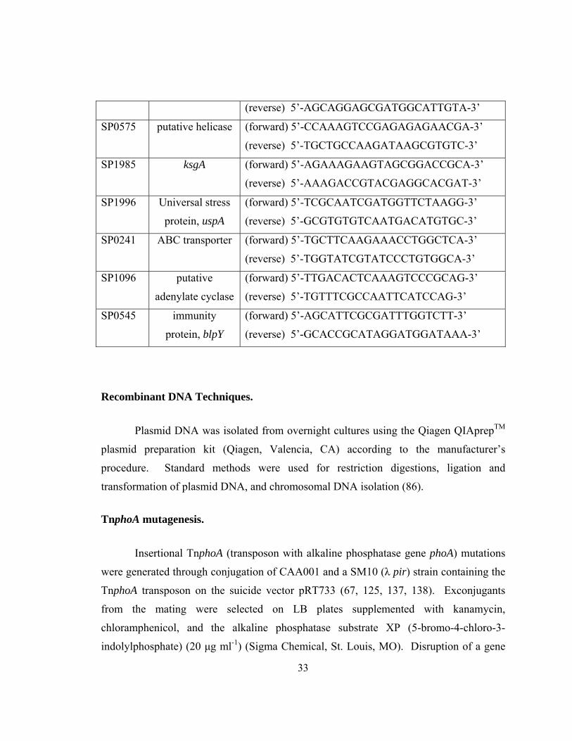
33
(reverse) 5’-AGCAGGAGCGATGGCATTGTA-3’
SP0575 putative helicase (forward) 5’-CCAAAGTCCGAGAGAGAACGA-3’
(reverse) 5’-TGCTGCCAAGATAAGCGTGTC-3’
SP1985 ksgA (forward) 5’-AGAAAGAAGTAGCGGACCGCA-3’
(reverse) 5’-AAAGACCGTACGAGGCACGAT-3’
SP1996 Universal stress
protein, uspA
(forward) 5’-TCGCAATCGATGGTTCTAAGG-3’
(reverse) 5’-GCGTGTGTCAATGACATGTGC-3’
SP0241 ABC transporter (forward) 5’-TGCTTCAAGAAACCTGGCTCA-3’
(reverse) 5’-TGGTATCGTATCCCTGTGGCA-3’
SP1096 putative
adenylate cyclase
(forward) 5’-TTGACACTCAAAGTCCCGCAG-3’
(reverse) 5’-TGTTTCGCCAATTCATCCAG-3’
SP0545 immunity
protein, blpY
(forward) 5’-AGCATTCGCGATTTGGTCTT-3’
(reverse) 5’-GCACCGCATAGGATGGATAAA-3’
Recombinant DNA Techniques.
Plasmid DNA was isolated from overnight cultures using the Qiagen QIAprepTM
plasmid preparation kit (Qiagen, Valencia, CA) according to the manufacturer’s
procedure. Standard methods were used for restriction digestions, ligation and
transformation of plasmid DNA, and chromosomal DNA isolation (86).
TnphoA mutagenesis.
Insertional TnphoA (transposon with alkaline phosphatase gene phoA) mutations
were generated through conjugation of CAA001 and a SM10 (λ pir) strain containing the
TnphoA transposon on the suicide vector pRT733 (67, 125, 137, 138). Exconjugants
from the mating were selected on LB plates supplemented with kanamycin,
chloramphenicol, and the alkaline phosphatase substrate XP (5-bromo-4-chloro-3-
indolylphosphate) (20 μg ml-1) (Sigma Chemical, St. Louis, MO). Disruption of a gene
34
encoding a membrane-associated protein (i.e. secreted or cytoplasmic membrane
proteins) would be indicated through the enzymatic activity of alkaline phosphatase
which cleaves a phosphate group from XP (colorless substrate) resulting with a blue
colored by-product and a subsequent blue colony. Disruption of the gene generates a
gene fusion composed of the amino-terminal of the target gene fused to the phoA gene
which encodes alkaline phosphatase which only exhibits high activity levels outside of
the cytoplasm when exposed to the periplasmic space.
Cosmid library construction.
To locate the transposon insertion sites, a cosmid library was generated using the
broad-range cosmid vector pLAFR3 (128). Genomic DNA was isolated by the modified
Marmur protocol (88) and partially digested with Sau3A1 according to Maniatis et al.
(87). Digests which yielded fragments of 9-12 kb in size on 1% agarose gels were used in
overnight ligation reactions (0.3 μg vector, 0.8 μg DNA, 60U ligase) containing BamH1
digested pLAFR3 and T4 DNA ligase (New England Biolabs) at 16°C. Ligation
reactions were introduced into E. coli strain LE392 by infecting the bacteria with phage
particles packaged in vitro using the Packagene Lambda DNA Packaging System
(Promega) according to the manufacturer’s instructions. Bacterial colonies were selected
on LB agar supplemented with Km and Tc. Cosmid DNA from isolates containing
TnphoA within the DNA cloned was sequenced by the UTMB Protein Chemistry
Laboratory using a reverse primer flanking the 5’ region of the transposon (5’-
GTTTTCCAGAACAGGGCAAA-3’). Sequence results were analyzed using the Basic
Local Alignment Search Tool program (BLAST) to compare against known nucleotide
sequences in the EMBL data bank (http://www.ncbi.nlm.nih.gov/BLAST/) (105).
Construction of AIEC O83:H1 rpoS isogenic mutant by allelic exchange.
The rpoS gene (Entrez Accession No. NC000913) was PCR amplified in two
segments: the 5’ portion of the gene as a 922-bp fragment containing flanking KpnI (5’
end) and SmaI (3’ end) restriction sites and the 3’ portion as a 904-bp fragment
35
containing flanking SmaI (5’ end) and BamHI (3’ end) restriction sites. The following
two primer sets were used to generate the two fragments: 5RPO (5’-
AACGGTACCAACAGCAAGC-3’) and 3SMRPO (5’-
GTCCCGGGTCAAACTTCTCTAC-3’) for the 5’ segment; and 5SMRPO (5’-
GACCCGGGACGTGGTTTCCGCTTCTCA-3’) and 3RPO (5’-
GTCCCGGGTCAAACTTCTCTAC-3’) for the 3’ fragment. Each fragment was
individually cloned into pBluescript SK, re-digested, and the two fragments ligated into
the same plasmid. The cloned gene was disrupted with the cat gene inserted into SmaI
restriction sites and the disrupted gene then subcloned into suicide vector pCVD442. The
construct was introduced into AIEC O83:H1cu by conjugation with donor strain SM10 (λ
pir) and allelic exchange was performed as previously described (141). Strain O83:H1cu
is an antibiotic sensitive strain and displayed an identical adherent and invasive
phenotype to the parental strain O83:H1 (data not shown). Candidate colonies which
were lactose positive on MacConkey plates and resistant to chloramphenicol were
screened for sensitivity to ampicillin and kanamycin. The presence of the cat cassette
within the chromosomal rpoS gene of strain CAA001 was confirmed by PCR.
Transcomplementation of rpoS mutant.
The rpoS gene and its promoter region were amplified from the chromosomal
DNA of AIEC O83:H1 using primers 5RPOS (5’-ACCAACAGCAAGCACAAC-3’) and
3RPOS (5’-TGAGACTGGCCTTTCTGAC-3’). The PCR product was gel extracted,
digested with EcoRI then ligated into EcoRI-linearized pGEM-T Easy. The resulting
construct pRpoS was used to complement TnphoA mutants generated from the RpoS
mutant background. This complementation was used to determine if the disrupted genes
in the identified TnphoA mutants, containing disruptions in genes encoding membrane-
associated proteins, were under the regulation of RpoS. Such mutants were identified by
the loss of alkaline phosphatase activity upon complementation with an intact rpoS gene
which resulted with the reversion of the blue colony phenotype (used to select TnphoA
mutants) back to a colorless colony phenotype.
36
SDS-PAGE and Western blot analysis.
Bacterial samples were removed from HARVs at 4, 12, and 24 hours, and cell
concentrations were standardized based on OD600 readings. Cells (4 x 10⁸) were pelleted,
suspended in Laemmli protein sample buffer (77), boiled for 5 minutes, and used for
sodium dodecyl sulfate-polyacrylamide gel electrophoresis (SDS-PAGE). Cell lysates
(10 μl/well) were loaded onto a 10% SDS polyacrylamide gel and run at 100 volts.
Samples were transferred onto polyvinyl fluoride (PVDF) Immobilon-P transfer
membrane (Millipore, Bedford, MA) and blocked in 5% non-fat milk overnight at 4°C.
Membranes were washed with phosphate-buffered saline (PBST) with 0.1% Tween 20
and probed with a polyclonal anti-RpoS antibody kindly provided by A. Matin (Stanford
University). Blots were developed with an ECL-Plus kit (Amersham, Piscataway, N.J.).
Twenty-four hour AIEC O83:H1 and rpoS mutant test tube cultures were included as
positive and negative controls.
Growth kinetics under LSMMG.
Cultures of E. coli AMS6, AIEC O83:H1, and the rpoS mutant were grown in M9
minimal media within HARVs oriented in LSMMG and 1 x g control positions as
described in Figure 1. Growth of both strains was monitored by measuring OD600 nm at
one hour increments for 12 hours. Growth was monitored quantitatively by serial
dilution and plating at 4, 8, and 12 hours.
Environmental stress assays.
HARV-grown cultures were harvested aseptically through the sampling ports of
the LSMMG and 1 x g control vessels after 12 hours of growth and culture densities were
determined by measuring optical density (OD600). Bacterial cultures were grown in
HARVs allowing an adequate amount of time to acclimate and take on phenotypic
characteristics in response to LSMMG and 1 x g control environments within the vessels
prior to stress challenges. A bacterial concentration of 1x10⁷ CFU (approximately 5-15
37
μl) was immediately exposed to one of the four different environmental stresses
investigated. Specific stress levels, concentrations and exposure periods tested were
comparable with those conditions previously tested for S. Typhimurium and E. coli
AMS6 HARV studies (85, 147). To reduce variability in the results obtained with the
HARV model system, E. coli K-12 strain AMS6, previously examined by Lynch et al.,
was included in all stress assays to function as a comparative bacterial control along with
the strains examined (85). Thermal stress was evaluated by adding bacterial aliquots to 1
ml of LB liquid media then transferring cultures to heating blocks set at 55°C to heat
shock the bacteria. Responses to acid, osmotic, and oxidative stresses were analyzed by
adding bacteria to 1 ml of filter-sterilized media which was either pre-adjusted to a pH of
2.0 with HCl, supplemented with NaCl to a concentration of 3.0 M, or supplemented with
H2O2 (Fisher Scientific) to a concentration of 10.0 mM. Stress exposure time was
evaluated at 30 min and 1 hour, followed by serial plating in duplicate on LB agar plates
to determine the percentage of survival. The use of LB as the stress diluent did not affect
the growth kinetics of the cultures during the challenge periods since similar results for
the assays were also observed using sterile phosphate buffered saline (PBS) as the diluent
in subsequent studies. Cultures from each HARV vessel were serial diluted and plated at
time zero prior to stress exposure to calculate percent survival. The percentage of
survival was calculated by dividing the number of colony forming units (CFU) at either
30 min or 1 hour by the CFU at time zero and multiplying by 100. A minimum of three
independent stress assays was carried out for each stress examined. Statistical
significance (p ≤ 0.05) was determined using Student’s t-Test.
Bacterial adhesion and invasion assays.
T-75 flasks of Caco-2 cells were grown to confluence in Dulbecco’s Modified
Eagle’s Medium (DMEM) (Cellgro) supplemented with 10% (v/v) fetal bovine serum at
37°C under 5% CO2. Cells were trypsinized, diluted, and seeded at 1x10⁵ cells ml-1 per
well in 24-well plates (Corning) and incubated for 48 hours prior to infection. Before
infection, monolayers were washed with sterile PBS (pH 7.4) and replenished with 1 ml
38
of DMEM without supplements. Monolayers were incubated with 1x10⁷ bacteria (MOI
100:1) per well for 1.5 hours at 37°C in 5% CO2. Infected monolayers were washed
twice with PBS to remove non-adherent bacteria followed by lysis with 200 μl of 0.1%
Triton X-100 in PBS to recover adherent bacteria. Infected monolayers were incubated
an additional hour in fresh media supplemented with gentamicin (50 μg/ml), to kill
extracellular bacteria, prior to harvesting. Incubation times were optimized from
standard 3 hour incubation periods to minimize time for culture re-adaptation outside of
the HARVs while allowing sufficient time for interaction between bacteria and tissue
culture cells. Supplementary assays carried out with incubation periods at 4°C confirmed
adherence and invasion trends under LSMMG conditions. Incubation assays at 4°C
excluded potential interference of metabolic and physiological changes occurring due to
re-adaptation outside of LSMMG environment. Lysates were serial diluted and plated on
LB agar to quantitate adherent bacterial numbers from triplicate wells as the mean.
Statistical significance (p ≤ 0.05) was determined using Student’s t-Test.
Tryptophanase enzyme assays.
Tryptophanase activity was determined by measuring the conversion of S-o-
nitrophenyl-L-cysteine (SOPC) to o-nitrothiophenylate (131). The assay procedure was
based on those described previously (5, 60). Cells were grown to mid-logarithmic phase
(OD600 = 0.5 - 0.6) in M9 minimal media, washed with and resuspended in 2 ml of cold
lysis buffer (100 mM potassium phosphate, pH 7.8, 50 μm pyridoxal phosphate, 10 mM
β-mercaptoethanol, and 2 mM ethylenediamine tetraacetic acid [EDTA]), and sonicated
for 1 minute on ice. Lysates were spun down (12,000 x g for 2 minutes) to remove cells
debris and protein concentrations from the supernatant determined using a Bio-Rad
Protein assay reagent (Bio-Rad, Hercules, CA) according to the manufacturer’s
instructions. Fifty microliters of sample supernatant was combined with 450 μl of assay
buffer (100 mM potassium phosphate (pH 7.8); 100 μm pyridoxal phosphate; 20 mM β-
mercaptoethanol; and 1 mg bovine serum albumin per ml) and incubated at 30°C for 5
minutes. The reaction was started by the addition of 500 μl of pre-warmed 0.66 mM
39
SOPC. Reactions were monitored at 370 nm with a spectrophotometer over the course of
3 minutes until the ∆A370 calculated was linear. Enzymatic activity was calculated using
an extinction coefficient of 1.86 M-1 cm-1 according to Suelter for o-nitrothiophenolate
(131). Tryptophanase activity was calculated as follows: (∆A370 x 1.0 ml reaction
volume)/{(1.86 μmol/ml) x ([time (min)] x [protein concentration (mg/ml)] x [cell extract
volume (ml)]} as described by Gish (60). One unit of tryptophanase-specific activity was
defined as 1 μmol of o-nitrothiophenolate produced per minute at room temperature
(~25°C). All extracts were assayed in duplicate.
40
RESULTS AND DISCUSSION
Chapter 4: The Effects of Low-Shear Modeled Microgravity on Streptococcus pneumoniae Global Gene Expression
INTRODUCTION
Bacteria grown under LSMMG conditions have been shown to exhibit alterations
in physiology and increased virulence potential (100, 101). To better understand the
genomic changes taking place influencing these changes in phenotype, microarray
analysis has been utilized to examine the global transcriptional profiles of different
enteric bacterial species including S. Typhimurium and E. coli (23, 142, 148). While
these genomic profiles have aided in allowing us to better understanding how changes in
gene expression under LSMMG conditions can affect Gram-negative enteric bacteria in
terms of virulence and physiological adaptations, the effects of this unique environment
have yet to be investigated in a Gram-positive species such as the upper respiratory
pathogen, S. pneumoniae. Previous findings from the laboratory of Dr. David Niesel
have shown that the growth of S. pneumoniae under LSMMG conditions can enhance
virulence potential and alter both physiology and protein profiles (unpublished data).
Therefore, microarray analysis was utilized to investigate the changes which take place at
the genomic level in S. pneumoniae after growth under LSMMG to identify genes which
are responsive to this environment and function in changes involving physiology, protein
expression, and virulence potential previously observed.
RESULTS AND DISCUSSION
Cluster analysis reveals similar expression patterns in LSMMG-responsive genes in S. pneumoniae.
In an effort to identify trends in the global gene expression of S. pneumoniae in
response to the LSMMG environment, hierarchical cluster analysis was carried out on
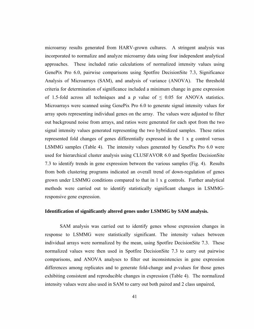
41
microarray results generated from HARV-grown cultures. A stringent analysis was
incorporated to normalize and analyze microarray data using four independent analytical
approaches. These included ratio calculations of normalized intensity values using
GenePix Pro 6.0, pairwise comparisons using Spotfire DecisionSite 7.3, Significance
Analysis of Microarrays (SAM), and analysis of variance (ANOVA). The threshold
criteria for determination of significance included a minimum change in gene expression
of 1.5-fold across all techniques and a p value of ≤ 0.05 for ANOVA statistics.
Microarrays were scanned using GenePix Pro 6.0 to generate signal intensity values for
array spots representing individual genes on the array. The values were adjusted to filter
out background noise from arrays, and ratios were generated for each spot from the two
signal intensity values generated representing the two hybridized samples. These ratios
represented fold changes of genes differentially expressed in the 1 x g control versus
LSMMG samples (Table 4). The intensity values generated by GenePix Pro 6.0 were
used for hierarchical cluster analysis using CLUSFAVOR 6.0 and Spotfire DecisionSite
7.3 to identify trends in gene expression between the various samples (Fig. 4). Results
from both clustering programs indicated an overall trend of down-regulation of genes
grown under LSMMG conditions compared to that in 1 x g controls. Further analytical
methods were carried out to identify statistically significant changes in LSMMG-
responsive gene expression.
Identification of significantly altered genes under LSMMG by SAM analysis.
SAM analysis was carried out to identify genes whose expression changes in
response to LSMMG were statistically significant. The intensity values between
individual arrays were normalized by the mean, using Spotfire DecisionSite 7.3. These
normalized values were then used in Spotfire DecisionSite 7.3 to carry out pairwise
comparisons, and ANOVA analyses to filter out inconsistencies in gene expression
differences among replicates and to generate fold-change and p-values for those genes
exhibiting consistent and reproducible changes in expression (Table 4). The normalized
intensity values were also used in SAM to carry out both paired and 2 class unpaired,
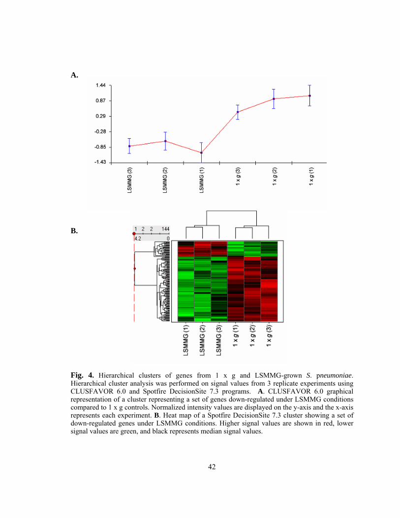
42
A.
B.
Fig. 4. Hierarchical clusters of genes from 1 x g and LSMMG-grown S. pneumoniae. Hierarchical cluster analysis was performed on signal values from 3 replicate experiments using CLUSFAVOR 6.0 and Spotfire DecisionSite 7.3 programs. A. CLUSFAVOR 6.0 graphical representation of a cluster representing a set of genes down-regulated under LSMMG conditions compared to 1 x g controls. Normalized intensity values are displayed on the y-axis and the x-axis represents each experiment. B. Heat map of a Spotfire DecisionSite 7.3 cluster showing a set of down-regulated genes under LSMMG conditions. Higher signal values are shown in red, lower signal values are green, and black represents median signal values.
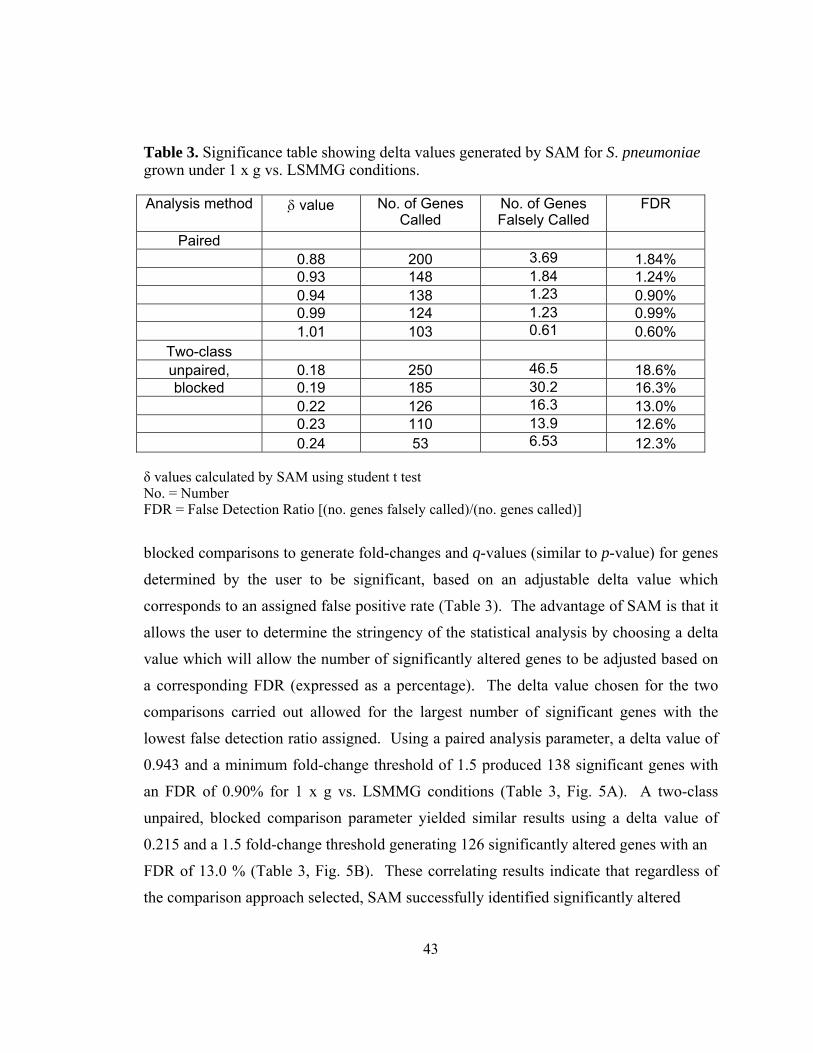
43
Table 3. Significance table showing delta values generated by SAM for S. pneumoniae grown under 1 x g vs. LSMMG conditions.
Analysis method δ value No. of Genes Called
No. of Genes Falsely Called
FDR
Paired 0.88 200 3.69 1.84% 0.93 148 1.84 1.24% 0.94 138 1.23 0.90% 0.99 124 1.23 0.99% 1.01 103 0.61 0.60%
Two-class unpaired, 0.18 250 46.5 18.6% blocked 0.19 185 30.2 16.3%
0.22 126 16.3 13.0% 0.23 110 13.9 12.6% 0.24 53 6.53 12.3% δ values calculated by SAM using student t test No. = Number FDR = False Detection Ratio [(no. genes falsely called)/(no. genes called)]
blocked comparisons to generate fold-changes and q-values (similar to p-value) for genes
determined by the user to be significant, based on an adjustable delta value which
corresponds to an assigned false positive rate (Table 3). The advantage of SAM is that it
allows the user to determine the stringency of the statistical analysis by choosing a delta
value which will allow the number of significantly altered genes to be adjusted based on
a corresponding FDR (expressed as a percentage). The delta value chosen for the two
comparisons carried out allowed for the largest number of significant genes with the
lowest false detection ratio assigned. Using a paired analysis parameter, a delta value of
0.943 and a minimum fold-change threshold of 1.5 produced 138 significant genes with
an FDR of 0.90% for 1 x g vs. LSMMG conditions (Table 3, Fig. 5A). A two-class
unpaired, blocked comparison parameter yielded similar results using a delta value of
0.215 and a 1.5 fold-change threshold generating 126 significantly altered genes with an
FDR of 13.0 % (Table 3, Fig. 5B). These correlating results indicate that regardless of
the comparison approach selected, SAM successfully identified significantly altered
44
A.
Paired
-8
-6
-4
-2
0
2
4
6
-5 -4 -3 -2 -1 0 1 2 3 4 5
Expected
Obs
erve
d
Significant: 138Median # false sig.: 1.22891
Delta 0.94300Fold Change 1.5
B.
Two-class unpaired, blocked
-2.5
-2
-1.5
-1
-0.5
0
0.5
1
1.5
2
-2 -1.5 -1 -0.5 0 0.5 1 1.5 2
Expected
Obs
erve
d
Significant: 126Median # false sig.: 16.32993
Delta 0.21470Fold Change 1.5
Fig. 5. Significance Analysis of Microarrays (SAM) plots for 1 x g vs. LSMMG-grown S. pneumoniae. SAM was used to compare signal values for S. pneumoniae grown under 1 x g and LSMMG conditions from each replicate (6 arrays). Data were analyzed in Excel using the paired (A) and two-class unpaired, blocked (B) options, with the additional requirement of at least a 1.5-fold change in gene expression. A. The delta value (0.943) chosen yielded the greatest number of significant genes (138) with the lowest false detection ratio (1.2/138 = 0.90%). B. The delta value (0.215) chosen yielded 126 significant genes with a false detection ratio of 13.0% (16.3/126). Significantly up-regulated genes are shown in red, and significantly down-regulated genes are shown in green.
45
LSMMG-responsive genes with modest changes in the FDR. The trend of down-
regulated transcriptional activity in response to LSMMG conditions was observed for all
analysis techniques utilized. In addition to the 1 x g rotating samples, cells grown under
static gravitational conditions were included to serve as secondary negative controls.
Any genes found to be differentially expressed between 1 x g rotating and static
gravitational controls represented variations that were not directly attributed to the
LSMMG environment and were thus excluded from further analyses.
Global transcriptional profile of S. pneumoniae in response to LSMMG compared to 1 x g control conditions.
The stringent 4-method analysis used identified only down-regulated genes in
bacteria grown under LSMMG conditions, compared to 1 x g control cultures.
Microarray analysis of S. pneumoniae cultures grown under LSMMG revealed the
differential expression of 81 genes, all of which were found to be down-regulated in
comparison to 1 x g control cultures (Table 4). LSMMG-responsive genes represented a
wide range of diverse functional categories, in addition to uncharacterized genes. The
differential expression of these various genes indicates that growth of S. pneumoniae
under LSMMG can affect many different physiological functions, including metabolism,
signaling, DNA repair/recombination, surface protein expression, and ion transport.
These differentially expressed genes were physically located throughout the genome,
which suggests that LSMMG conditions impact global transcriptional activity
encompassing the entire chromosome. There were, however, 17 genes identified by
microarray analysis that were up-regulated based on fold-change alone, but that did not
meet the additional threshold criteria established for significance. Most of these up-
regulated genes were found to be uncharacterized, but included the following
characterized genes: 3 ATP transporters (SP0092, SP0600-Vexp2 ATP-binding protein,
SP0912), 1 pyrimidine-nucleoside phosphorylase (SP0842), and 1 transcriptional
regulator (SP0246, DeoR family). DeoR and DeoR homologs have been shown to
function as transcriptional repressors for operons involved in deoxynucleoside catabolism
(24, 150).
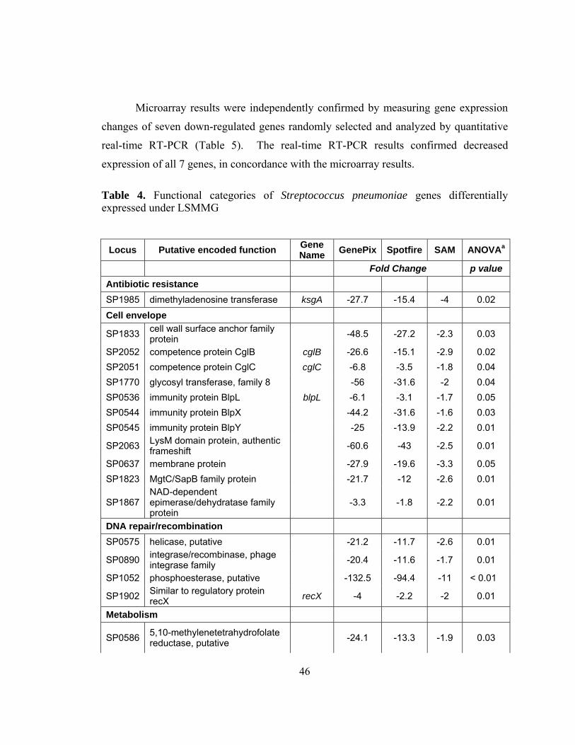
46
Microarray results were independently confirmed by measuring gene expression
changes of seven down-regulated genes randomly selected and analyzed by quantitative
real-time RT-PCR (Table 5). The real-time RT-PCR results confirmed decreased
expression of all 7 genes, in concordance with the microarray results.
Table 4. Functional categories of Streptococcus pneumoniae genes differentially expressed under LSMMG
Locus Putative encoded function Gene Name GenePix Spotfire SAM ANOVAa
Fold Change p value Antibiotic resistance SP1985 dimethyladenosine transferase ksgA -27.7 -15.4 -4 0.02
Cell envelope
SP1833 cell wall surface anchor family protein -48.5 -27.2 -2.3 0.03
SP2052 competence protein CglB cglB -26.6 -15.1 -2.9 0.02 SP2051 competence protein CglC cglC -6.8 -3.5 -1.8 0.04 SP1770 glycosyl transferase, family 8 -56 -31.6 -2 0.04 SP0536 immunity protein BlpL blpL -6.1 -3.1 -1.7 0.05 SP0544 immunity protein BlpX -44.2 -31.6 -1.6 0.03 SP0545 immunity protein BlpY -25 -13.9 -2.2 0.01
SP2063 LysM domain protein, authentic frameshift -60.6 -43 -2.5 0.01
SP0637 membrane protein -27.9 -19.6 -3.3 0.05 SP1823 MgtC/SapB family protein -21.7 -12 -2.6 0.01
SP1867 NAD-dependent epimerase/dehydratase family protein
-3.3 -1.8 -2.2 0.01
DNA repair/recombination SP0575 helicase, putative -21.2 -11.7 -2.6 0.01
SP0890 integrase/recombinase, phage integrase family -20.4 -11.6 -1.7 0.01
SP1052 phosphoesterase, putative -132.5 -94.4 -11 < 0.01
SP1902 Similar to regulatory protein recX recX -4 -2.2 -2 0.01
Metabolism
SP0586 5,10-methylenetetrahydrofolate reductase, putative -24.1 -13.3 -1.9 0.03
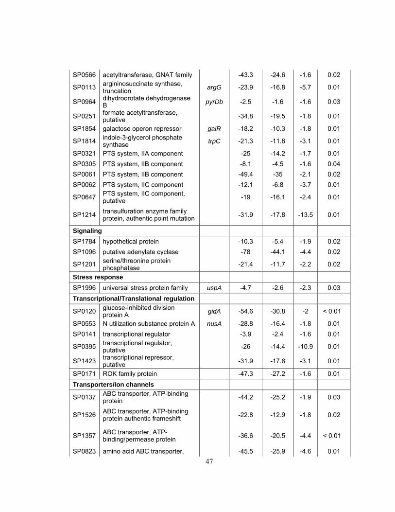
47
SP0566 acetyltransferase, GNAT family -43.3 -24.6 -1.6 0.02
SP0113 argininosuccinate synthase, truncation argG -23.9 -16.8 -5.7 0.01
SP0964 dihydroorotate dehydrogenase B pyrDb -2.5 -1.6 -1.6 0.03
SP0251 formate acetyltransferase, putative -34.8 -19.5 -1.8 0.01
SP1854 galactose operon repressor galR -18.2 -10.3 -1.8 0.01
SP1814 indole-3-glycerol phosphate synthase trpC -21.3 -11.8 -3.1 0.01
SP0321 PTS system, IIA component -25 -14.2 -1.7 0.01 SP0305 PTS system, IIB component -8.1 -4.5 -1.6 0.04 SP0061 PTS system, IIB component -49.4 -35 -2.1 0.02 SP0062 PTS system, IIC component -12.1 -6.8 -3.7 0.01
SP0647 PTS system, IIC component, putative -19 -16.1 -2.4 0.01
SP1214 transulfuration enzyme family protein, authentic point mutation -31.9 -17.8 -13.5 0.01
Signaling SP1784 hypothetical protein -10.3 -5.4 -1.9 0.02 SP1096 putative adenylate cyclase -78 -44.1 -4.4 0.02
SP1201 serine/threonine protein phosphatase -21.4 -11.7 -2.2 0.02
Stress response SP1996 universal stress protein family uspA -4.7 -2.6 -2.3 0.03
Transcriptional/Translational regulation
SP0120 glucose-inhibited division protein A gidA -54.6 -30.8 -2 < 0.01
SP0553 N utilization substance protein A nusA -28.8 -16.4 -1.8 0.01 SP0141 transcriptional regulator -3.9 -2.4 -1.6 0.01
SP0395 transcriptional regulator, putative -26 -14.4 -10.9 0.01
SP1423 transcriptional repressor, putative -31.9 -17.8 -3.1 0.01
SP0171 ROK family protein -47.3 -27.2 -1.6 0.01
Transporters/Ion channels
SP0137 ABC transporter, ATP-binding protein -44.2 -25.2 -1.9 0.03
SP1526 ABC transporter, ATP-binding protein authentic frameshift -22.8 -12.9 -1.8 0.02
SP1357 ABC transporter, ATP-binding/permease protein -36.6 -20.5 -4.4 < 0.01
SP0823 amino acid ABC transporter, -45.5 -25.9 -4.6 0.01
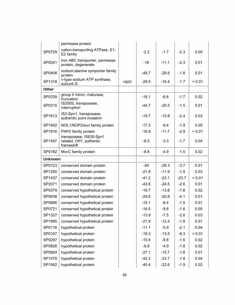
48
permease protein
SP0729 cation-transporting ATPase, E1-E2 family -2.2 -1.7 -2.3 0.05
SP0241 iron ABC transporter, permease protein, degenerate -16 -11.1 -2.3 0.01
SP0408 sodium:alanine symporter family protein -49.7 -28.6 -1.8 0.01
SP1318 v-type sodium ATP synthase, subunit G ntpG -29.5 -16.4 -1.7 < 0.01
Other
SP0339 group II intron, maturase, truncation -16.1 -8.8 -1.7 0.02
SP0315 IS200S, transposase, interruption -44.7 -25.5 -1.5 0.01
SP1613 IS3-Spn1, transposase, authentic point mutation -19.7 -10.8 -2.4 0.03
SP1402 NOL1/NOP2/sun family protein -17.3 -9.4 -1.9 0.05 SP1916 PAP2 family protein -16.8 -11.7 -2.9 < 0.01
SP1497 transposase, IS630-Spn1 related, Orf1, authentic frameshift
-6.5 -3.3 -1.7 0.04
SP0182 MccC family protein -8.8 -4.9 -1.5 0.02
Unknown SP0723 conserved domain protein -40 -28.3 -3.7 0.01 SP1250 conserved domain protein -21.8 -11.9 -1.5 0.03 SP1437 conserved domain protein -41.2 -23.1 -23.7 < 0.01 SP2071 conserved domain protein -43.6 -24.5 -2.6 0.01 SP0379 conserved hypothetical protein -19.7 -13.8 -1.8 0.02 SP0638 conserved hypothetical protein -29.6 -20.8 -4.1 0.01 SP0686 conserved hypothetical protein -15.1 -8.4 -1.5 0.01 SP0721 conserved hypothetical protein -16.5 -9.6 -1.6 0.05 SP1327 conserved hypothetical protein -13.9 -7.5 -2.6 0.03 SP1995 conserved hypothetical protein -21.9 -12.4 -1.8 0.01 SP0116 hypothetical protein -11.1 -5.9 -2.1 0.04 SP0167 hypothetical protein -19.3 -13.5 -6.3 < 0.01 SP0297 hypothetical protein -15.6 -8.8 -1.6 0.02 SP0826 hypothetical protein -6.8 -4.9 -1.8 0.02 SP0924 hypothetical protein -27.1 -15.7 -1.6 0.01 SP1078 hypothetical protein -42.2 -23.7 -1.8 0.04 SP1562 hypothetical protein -40.4 -22.6 -1.9 0.02
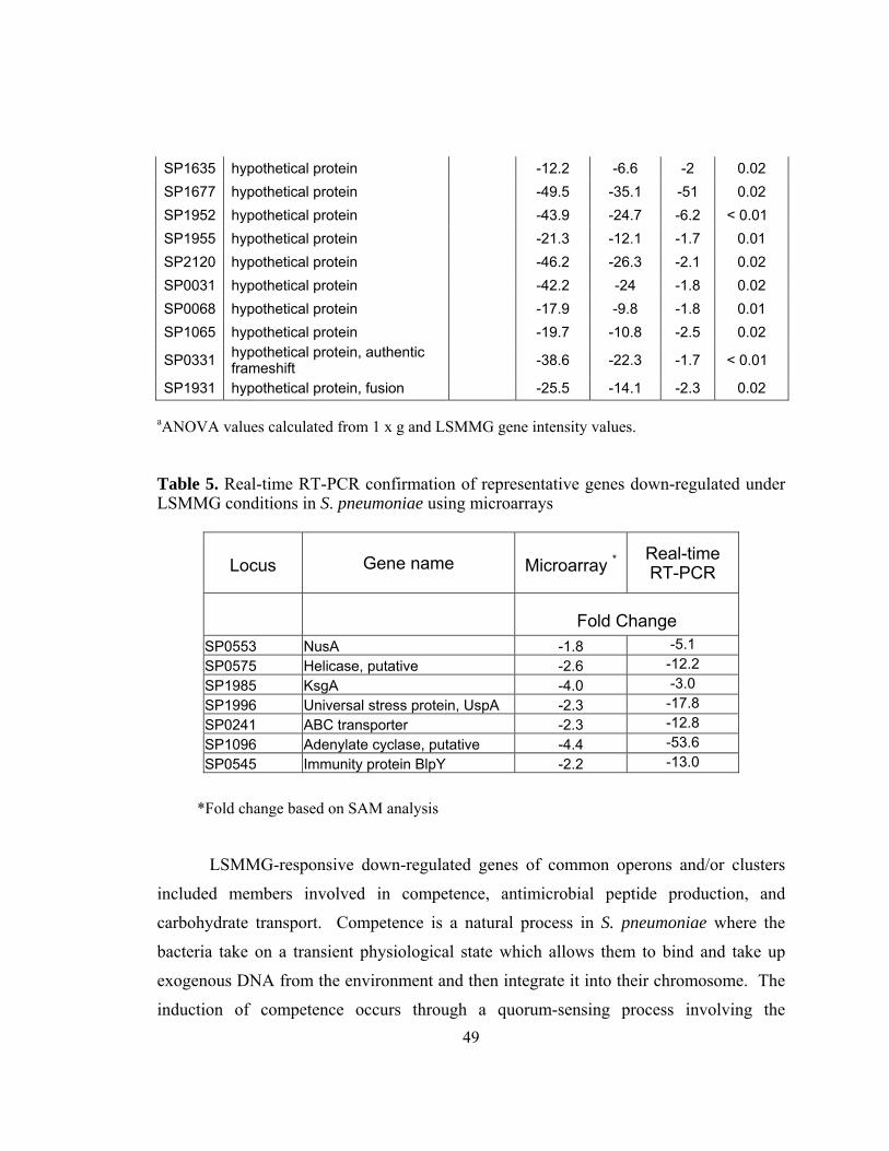
49
SP1635 hypothetical protein -12.2 -6.6 -2 0.02 SP1677 hypothetical protein -49.5 -35.1 -51 0.02 SP1952 hypothetical protein -43.9 -24.7 -6.2 < 0.01 SP1955 hypothetical protein -21.3 -12.1 -1.7 0.01 SP2120 hypothetical protein -46.2 -26.3 -2.1 0.02 SP0031 hypothetical protein -42.2 -24 -1.8 0.02 SP0068 hypothetical protein -17.9 -9.8 -1.8 0.01 SP1065 hypothetical protein -19.7 -10.8 -2.5 0.02
SP0331 hypothetical protein, authentic frameshift -38.6 -22.3 -1.7 < 0.01
SP1931 hypothetical protein, fusion -25.5 -14.1 -2.3 0.02
aANOVA values calculated from 1 x g and LSMMG gene intensity values. Table 5. Real-time RT-PCR confirmation of representative genes down-regulated under LSMMG conditions in S. pneumoniae using microarrays
Locus Gene name Microarray * Real-time RT-PCR
Fold Change SP0553 NusA -1.8 -5.1 SP0575 Helicase, putative -2.6 -12.2 SP1985 KsgA -4.0 -3.0 SP1996 Universal stress protein, UspA -2.3 -17.8 SP0241 ABC transporter -2.3 -12.8 SP1096 Adenylate cyclase, putative -4.4 -53.6 SP0545 Immunity protein BlpY -2.2 -13.0
*Fold change based on SAM analysis
LSMMG-responsive down-regulated genes of common operons and/or clusters
included members involved in competence, antimicrobial peptide production, and
carbohydrate transport. Competence is a natural process in S. pneumoniae where the
bacteria take on a transient physiological state which allows them to bind and take up
exogenous DNA from the environment and then integrate it into their chromosome. The
induction of competence occurs through a quorum-sensing process involving the
50
secretion of competence-stimulating peptide (CSP) to threshold concentrations of about
10 ng ml-1, which then leads to the activation of the two-component system ComDE.
Phosphorylation of ComE (response regulator) and ComD (histidine kinase) induces the
activation of the alternative sigma factor ComX, which, in turn, initiates the transcription
of several operons containing genes encoding a number of factors involved in DNA
uptake, processing, and recombination (71). Two down-regulated genes (cglB and cglC)
identified are members of the cgl operon, which is one of the late competence operons
induced during the onset of competence in the S. pneumoniae life cycle (75, 106). The
proteins encoded by these genes are involved in construction of the surface-expressed
translocasome (DNA uptake apparatus) that functions in DNA uptake and processing
during competence (75).
The microarray results revealed the down-regulation of three genes (blpL, blpX,
and blpY) which are members of the blp (bacteriocin-like peptide) regulon, another
quorum-sensing system which functions in the production of antimicrobial peptides and
immunity proteins which are co-transcribed for intra- and interspecies competition during
colonization (32, 33). Induction of bacteriocin production occurs through the detection of
bacteriocin-inducing peptide (BIP) which activates the two-component system BlpRH
and the subsequent activation of operons containing genes encoding bacteriocin structural
and immunity proteins (32). Bactericidal activity of bacteriocins generally involves
receptor-mediated mechanisms such as pore formation or membrane permeabilization.
Producer strains of bacteriocins are protected through the synchronous production of
immunity proteins which associate with the target surface receptors preventing pore
formation (98). The blpL, blpX, and blpY genes are members of operons which encode
the protective immunity proteins.
Knutsen et al. previously reported that the qsrAB operon, which encodes an
uncharacterized ABC transporter, can undergo cross-induction by either of the two
signaling pheromones (CPS or BIP) associated with the ComABCDE and BlpABCSRH
two-component signaling pathways, respectively, due to the presence of a hybrid direct
repeat motif which can recognize both response regulators (ComE and BlpR) (71). These
findings indicate the potential for cross-talk between these two independent quorum-
51
sensing systems which both contain genes responsive to the LSMMG environment. Such
responses may suggest that while the impact of LSMMG can affect many different
operons and gene clusters of various functions throughout the genome, some of these
groups could respond in a coordinated manner by interactions between pathways through
convergent regulatory mechanisms.
Another group of down-regulated genes included five members (SP0321-PTS
IIA, SP0305-PTS IIB, SP0061-PTS IIB, SP0062-PTS IIC, and SP0647-PTS IIC) of the
phosphotransferase systems (PTS), involved in carbohydrate uptake and metabolism.
The PTS system is generally composed of three-to-four functional components, IIA, IIB,
IIC, and IID which are generated as either individual subunits in a complex or as different
domains in a single polypeptide (e.g. IIAB). The first enzyme (EI) in the PTS cascade
acquires a phosphoryl group from phosphophenol pyruvate (PEP), resulting in the
dephosphorylation of PEP to pyruvate. The phosphotransfer chain continues as EI
phosphorylates histidine-containing PTS phosphotransferase (HPr), followed by IIA, IIB,
and finally IIC / IID, which contain sugar binding sites allowing phosphorylation and
uptake of different carbohydrates from the environment (124). Diep et al. recently
reported that the IICD membrane surface complex of the mannose PTS pathway can also
serve as a target receptor for class II bacteriocins and corresponding immunity proteins
(38). Thus, the down-regulation of SP0062 and SP0647 which encode the PTS IIC
components may serve as a survival strategy during colonization in low-shear areas of the
host to compete against other species. The genes reported above were found to exhibit
changes in expression at levels above the established threshold for significance of
additional genes within the competence. Additional members of the Blp and PTS
regulons were also found to be down-regulated, albeit at levels below the threshold
limits. These results suggest that LSMMG conditions generate an environment in which
S. pneumoniae phenotypic changes may be characterized by decreases in DNA
transformability, carbohydrate uptake, and the production of antimicrobial
peptide/immunity peptides.
While most of the remaining down-regulated genes do not share common operons
or clusters in the genome, several are functionally tied with the acquisition of antibiotic
52
resistance, stress resistance, and virulence. One of these genes encodes the rRNA
adenine dimethyltransferase, KsgA, which catalyzes the methylation of 16S rRNA (102).
Dimethylation of two adenosine bases in 16S rRNA is conserved across all prokaryotic
species, and inactivation of the ksgA gene has been shown to lead to bacterial resistance
to aminoglycoside antibiotics (e.g. kasugamycin) due to the loss of this modification.
Alterations in the gene expression of antibiotic resistant genes, such as ksgA, could
contribute to the trend of increased antibiotic resistance demonstrated by bacteria during
space flight (93). The gene encoding universal stress protein A (UspA) was also found to
be down-regulated under LSMMG. UspA is produced in response to a variety of internal
and external stresses and is thought to be a member of the RecA-dependent DNA
protection and repair system involved in the protection of DNA from UV damage (39).
Liu et al. recently reported that UspA of S. Typhimurium LT2 plays an important role in
conferring resistance to metabolic and oxidative stresses and also contributes to bacterial
virulence in mice (82). The down-regulation of uspA and other stress-related genes could
explain the increased sensitivity found in S. pneumoniae upon exposure to thermal and
acid stresses after growth under LSMMG conditions (2). Two other genes that were
found to be down-regulated under LSMMG conditions (SP1823 and a homolog of gidA)
are associated with virulence acquisition and regulation. SP1823 encodes an MgtC/SapB
family protein which has been demonstrated to function as a horizontally-acquired
virulence factor responsible for survival in macrophages, virulence in mice, and growth
under low Mg2+ conditions (12). The virulence regulator gidA (glucose-inhibited division
protein) mediates the expression of cytotoxic enterotoxin and subsequent virulence in
mice in the food-borne pathogen Aeromonas hydrophila (123). The repression of certain
virulence genes by bacteria grown under LSMMG may play an important functional role
in immune evasion, thus contributing to their survival and pathogenesis.
Previous studies in the laboratory of Dr. David Niesel have shown that S.
pneumoniae exhibits an increased ability to adhere to and invade A549 respiratory
epithelia after growth under LSMMG conditions (unpublished data). Our microarray
analysis revealed that the genes encoding the various factors associated with these
processes were not significantly affected by growth under LSMMG conditions. Thus, no
53
direct correlations were identified which could explain the altered physiological
properties observed. A similar trend was observed for S. Typhimurium strain χ3339 after
growth under LSMMG. While virulence, survival in macrophages, and stress resistance
were shown to be enhanced after growth under LSMMG, none of the genes encoding
known virulence factors were induced after growth in LSMMG (99, 147, 148). A
correlation was observed between the down-regulated genes involved in LPS
biosynthesis and LPS protein expression. SDS-PAGE analysis of LPS levels correlated
with array findings, indicating a decrease in protein expression compared to 1 x g control
cultures. While microarray analysis of strain χ3339 indicated no enhancement of
virulence gene expression, other reports have demonstrated increases in virulence and
environmental stress resistance by other bacterial strains and species after growth under
LSMMG (99, 147). LSMMG-grown S. Typhimurium strain 14028 has been shown to
demonstrate increased invasion potential, stimulate TNF-α production in infected
epithelia, and exhibit altered gene and protein expression (23). Microarray analysis of
strain 14028 showed a trend similar to that of χ3339, in that most of the differentially
expressed genes were down-regulated in response to LSMMG; however one gene
encoding a virulence factor, VirK (involved in membrane remodeling and motility), was
found to be induced, which might suggest strain-specific differences in response to this
environment.
In a recent study evaluating changes in the genomic profile of E. coli MG1655
after growth under LSMMG, Wilson et al. reported differential gene expression in
cultures grown in both rich media (LB) and minimal media (MOPS) (142). Results from
this study indicated that the majority of LSMMG-responsive genes were down-regulated
in cultures grown in both media and that the majority of these genes did not correlate to
those found to be responsive to LSMMG in strain χ3339. LSMMG-responsive genes (16
up-regulated, 19 down-regulated) from MOP cultures included the induction of genes
associated with chemotaxis, flagella synthesis, and acid stress tolerance while genes
involved in stress responses, transport, and envelope function were down-regulated.
Transcriptional responses in LB cultures (1 up-regulated, 14 down-regulated) included

54
the induction of accA (encodes carboxyl transferase subunit) and the down-regulation of
genes involved in biosynthesis, energy utilization, and ribosomal genes.
55
Chapter 5: The Effects of Low-Shear Modeled Microgravity on Adherent-Invasive Escherichia coli Physiology and Colonization Potential
INTRODUCTION
Clinical isolates from Crohn’s Disease (CD) patients have been categorized in a
new putative class of E. coli known as adherent-invasive E. coli (AIEC) based on their
unique properties which allow these strains to play a contributory role to the chronic
inflammatory state representative of the pathology of inflammatory bowel disease (29).
There is currently a limited amount of information on the factors and regulatory
mechanisms used by these strains to adhere to and invade host cells and stimulate
cytokine production. Additionally, no known virulence factors have been identified
which could classify these isolates in one of the major classes of pathogenic E. coli. In
an effort to understand how these strains respond and adapt to the environmental factors
encountered in the gut, HARVs were used as a model system to cultivate AIEC strain
O83:H1 in a low-shear, low-turbulence environment, as experienced in the
gastrointestinal tract. While previous reports have shown non-pathogenic commensal
strains of E. coli to undergo alterations in stress resistance and gene expression in
response to LSMMG, no clinical isolates such as AIEC isolates have been studied to
understand how mechanical stresses affect their physiology (85, 142). Reports have
shown conflicting evidence regarding a role for the global stress response regulator RpoS
in different enteric species grown under LSMMG, and nothing is known regarding this
regulator in clinical isolates grown in this unique environment (85, 147). Therefore, an
rpoS isogenic mutant was generated from AIEC O83:H1 to evaluate the putative role of
this global regulator in AIEC clinical isolates in response to the LSMMG environment.
RESULTS AND DISCUSSION
The impact of LSMMG on AIEC O83:H1 growth kinetics and RpoS protein expression.
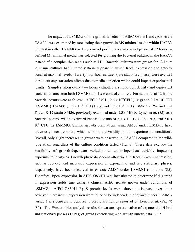
56
The impact of LSMMG on the growth kinetics of AIEC O83:H1 and rpoS strain
CAA001 was examined by monitoring their growth in M9 minimal media within HARVs
oriented in either LSMMG or 1 x g control positions for an overall period of 12 hours. A
defined M9 minimal media was selected for growing the bacterial cultures in the HARVs
instead of a complex rich media such as LB. Bacterial cultures were grown for 12 hours
to ensure cultures had entered stationary phase in which RpoS expression and activity
occur at maximal levels. Twenty-four hour cultures (late-stationary phase) were avoided
to rule out any starvation effects due to media depletion which could impact experimental
results. Samples taken every two hours exhibited a similar cell density and equivalent
bacterial counts from both LSMMG and 1 x g control cultures. For example, at 12 hours,
bacterial counts were as follows: AIEC O83:H1, 2.6 x 10⁹ CFU (1 x g) and 2.5 x 10⁹ CFU
(LSMMG); CAA001, 1.5 x 10⁹ CFU (1 x g) and 1.7 x 10⁹ CFU (LSMMG). We included
E. coli K-12 strain AMS6, previously examined under LSMMG by Lynch et al. (85), as a
bacterial control which exhibited bacterial counts of 7.3 x 10⁸ CFU, in 1 x g, and 7.0 x
108 CFU, in LSMMG. Similar growth correlations using AMS6 under LSMMG have
previously been reported, which support the validity of our experimental conditions.
Overall, only slight increases in growth were observed in CAA001 compared to the wild-
type strain regardless of the culture condition tested (Fig. 6). These data exclude the
possibility of growth-dependent variations as an independent variable impacting
experimental analyses. Growth phase-dependent alterations in RpoS protein expression,
such as reduced and increased expression in exponential and late stationary phases,
respectively, have been observed in E. coli AMS6 under LSMMG conditions (85).
Therefore, RpoS expression in AIEC O83:H1 was investigated to determine if this trend
in expression holds true using a clinical AIEC isolate grown under conditions of
LSMMG. AIEC O83:H1 RpoS protein levels were shown to increase over time;
however, increases in expression were found to be independent of growth under LSMMG
versus 1 x g controls in contrast to previous findings reported by Lynch et al. (Fig. 7)
(85). The Western blot analysis results shown are representative of exponential (4 hrs)
and stationary phases (12 hrs) of growth correlating with growth kinetic data. Our
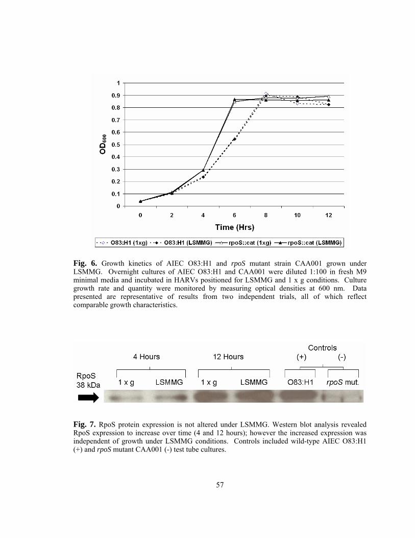
57
Fig. 6. Growth kinetics of AIEC O83:H1 and rpoS mutant strain CAA001 grown under LSMMG. Overnight cultures of AIEC O83:H1 and CAA001 were diluted 1:100 in fresh M9 minimal media and incubated in HARVs positioned for LSMMG and 1 x g conditions. Culture growth rate and quantity were monitored by measuring optical densities at 600 nm. Data presented are representative of results from two independent trials, all of which reflect comparable growth characteristics.
Fig. 7. RpoS protein expression is not altered under LSMMG. Western blot analysis revealed RpoS expression to increase over time (4 and 12 hours); however the increased expression was independent of growth under LSMMG conditions. Controls included wild-type AIEC O83:H1 (+) and rpoS mutant CAA001 (-) test tube cultures.
58
findings at late stationary phases of growth (24 hrs) are similar to the increased RpoS
expression observed at the 12 hr time point.
Results from our growth kinetics analysis of AIEC O83:H1 and the rpoS mutant
CAA001 grown in M9 minimal media under LSMMG conditions indicate that the mutant
strain enters exponential phase and reaches stationary phase at slightly faster rates when
compared to the wild-type strain (Fig. 6). This increased growth was found to be
independent of the LSMMG environment since each strain was found to grow at similar
rates when LSMMG and 1 x g cultures were compared. Lynch et al. reported that no
major differences were observed in wild-type E. coli AMS6 grown in M9 minimal media
under LSMMG or 1 x g controls; however, no information was given regarding changes
in growth rates by an isogenic rpoS mutant generated from the parental strain (85). The
growth kinetics of E. coli MG1655 were also previously evaluated in HARVS in both LB
and MOPS minimal media (142). In this study the lag phase of MOPS cultures was
longer than that of LB cultures irrespective of LSMMG and 1 x g control orientations. In
addition, exponential cultures grown in MOPS were found to grow at similar rates in
LSMMG and 1 x g control HARVs. This is in contrast to LB exponential cultures, which
exhibited slower growth under LSMMG compared to 1 x g conditions. Wilson et al.
demonstrated that wild-type S. Typhimurium strain χ3339 and an isogenic rpoS mutant
showed increased growth kinetics under LSMMG conditions compared to 1 x g control
cultures when grown in M9 minimal media. These effects were not observed when the
bacterial strains were grown in Lennox Broth (rich media) (147). These findings suggest
that the effects of LSMMG in combination with growth in minimal media can enhance
the growth kinetics in certain strains and species of bacteria, though the effect is not
universal. Environmental stresses such as low fluid shear and nutritional limitations
correspond to the natural conditions enteric species encounter in the gut. In addition,
RpoS does not appear to have a significant impact on the growth of AIEC O83:H1 and S.
Typhimurium under these conditions, based on our findings and those reported by Wilson
et al., since diminished growth rates were not observed in the mutant strains. It remains to
be determined however, if this observation holds true for commensal strains of E. coli
such as AMS6 and MG1655.
59
Lynch et al. previously reported that RpoS protein expression was increased in
stationary cultures of E. coli AMS6 grown under LSMMG and that responses to certain
stresses were RpoS-dependent during this growth phase. We, therefore, evaluated RpoS
expression in our clinical isolate to determine if this trend was conserved across different
strains. While RpoS protein expression was found to increase over time, as previously
established, this increased expression occurred in both LSMMG and 1 x g control
cultures, suggesting that expression changes are independent of the LSMMG
environment in AIEC O83:H1 (Fig. 7) (68). These findings suggest that any potential
RpoS-mediated effects on AIEC O83:H1 under LSMMG conditions do not occur
primarily due to changes in protein expression levels, but possibly at other levels of
regulation (e.g. transcriptional level).
Altered environmental stress resistance in AIEC O83:H1 and rpoS strain CAA001 under LSMMG.
AIEC O83:H1 and CAA001 were next examined to determine if growth under
LSMMG conditions altered survival to different environmental stresses compared to
growth under 1 x g controls. AIEC O83:H1 was evaluated for changes in resistance to
thermal, osmotic, acid, and oxidative stresses after growth under LSMMG to determine
whether this environment can alter the response of AIEC to different environmental
stresses. AIEC O83:H1 exhibited a significant increase in thermal stress resistance (Fig.
8A) after growth under LSMMG conditions compared to 1 x g control cultures.
Enhanced thermal stress resistance has also been observed in Salmonella after growth
under LSMMG (147). Increased resistance was also observed after exposure of AIEC
O83:H1 to oxidative stress for 30 minutes (Fig. 8B). While there appeared to be a slight
decrease in resistance to osmotic stress by LSMMG-grown AIEC O83:H1, no significant
differences were observed between LSMMG and 1 x g cultures in response to acid stress
(Figs. 8C and D).
E. coli strain AMS6 was included in the stress assays to function as an internal
bacterial control. The purpose of including this strain was to show that the use of the
60
HARV system was comparable to that by other laboratories and, in doing so, similar
trends in stress resistance (e.g. increased osmotic and acid stress resistance) were
successfully reproduced as previously published with this same strain by Lynch et al.
(Fig. 8) (85). In addition, the previous findings were expanded by including in the
analyses thermal and oxidative stresses, which revealed significant increases in thermal
stress resistance and oxidative stress sensitivity exhibited by the AMS6 strain after
growth under LSMMG.
It was observed that both the AMS6 and AIEC O83:H1 strains exhibited an
increased resistance to thermal stress (Fig. 8A), while displaying opposing trends in
resistance to osmotic and oxidative stresses (Fig. 8B and C). As previously reported, the
AMS6 strain exhibited an increase in acid stress resistance after growth under LSMMG,
while AIEC O83:H1 exhibited no major differences to this stressor regardless of the
growth environment (Fig. 8D). It was observed that the increase in oxidative stress
resistance by LSMMG-grown AIEC O83:H1 appeared to diminish between 30 minutes
and 1 hour of exposure (Fig. 8B), which could indicate the reversion of LSMMG-induced
phenotypic characteristics, whereas the resistance profiles observed during exposure to
the other three stressors persisted throughout both challenge time points.
Examination of the rpoS mutant strain CAA001 showed that growth under
LSMMG not only increased thermal stress resistance by the mutant, as observed in the
LSMMG-grown wild-type AIEC and AMS6 strains, but the enhanced resistance was also
at significantly higher levels (Fig. 8A). An increase in osmotic stress resistance was also
exhibited by LSMMG-grown CAA001 as observed in strain AMS6 (Fig. 8C). In
addition, both strains demonstrated a decreased resistance to oxidative stress after growth
under LSMMG conditions though the sensitivity by CAA001 was much higher to this
stressor (Fig. 8B). Neither CAA001 nor the wild-type strain exhibited LSMMG-
dependent differences to acid stress conditions in contrast to the enhanced resistance
exhibited by LSMMG-grown AMS6 (Fig. 8D).
Previous reports investigating E. coli strains AMS6 and MG1655 have indicated
contrasting trends in stress resistance after growth under LSMMG conditions in minimal
media. While the AMS6 strain was found to exhibit increased resistance to acid and
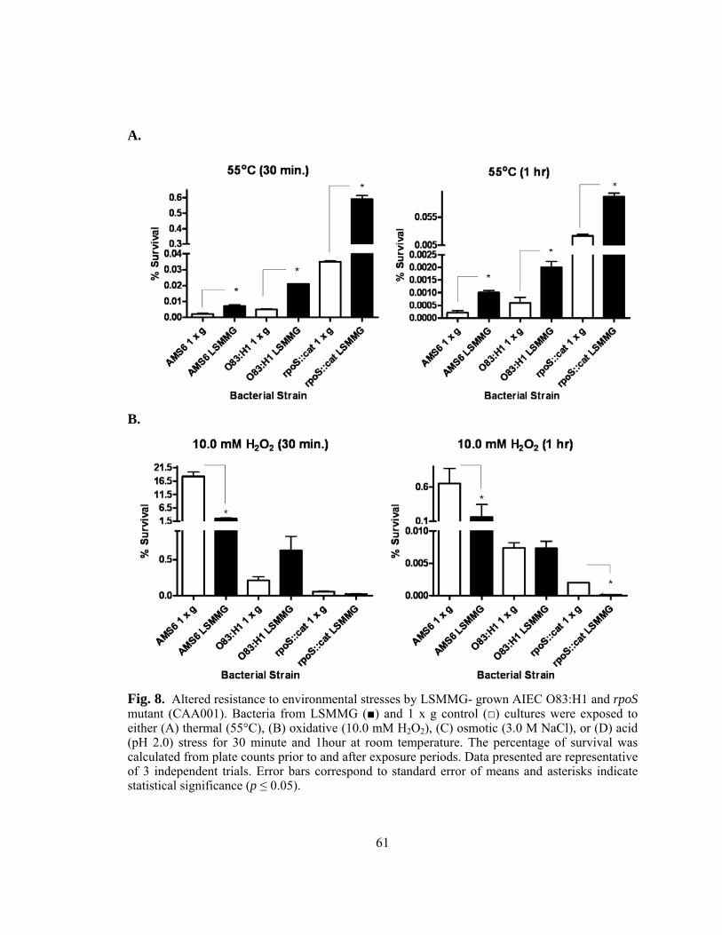
61
A.
B.
Fig. 8. Altered resistance to environmental stresses by LSMMG- grown AIEC O83:H1 and rpoS mutant (CAA001). Bacteria from LSMMG (■) and 1 x g control (□) cultures were exposed to either (A) thermal (55°C), (B) oxidative (10.0 mM H2O2), (C) osmotic (3.0 M NaCl), or (D) acid (pH 2.0) stress for 30 minute and 1hour at room temperature. The percentage of survival was calculated from plate counts prior to and after exposure periods. Data presented are representative of 3 independent trials. Error bars correspond to standard error of means and asterisks indicate statistical significance (p ≤ 0.05).
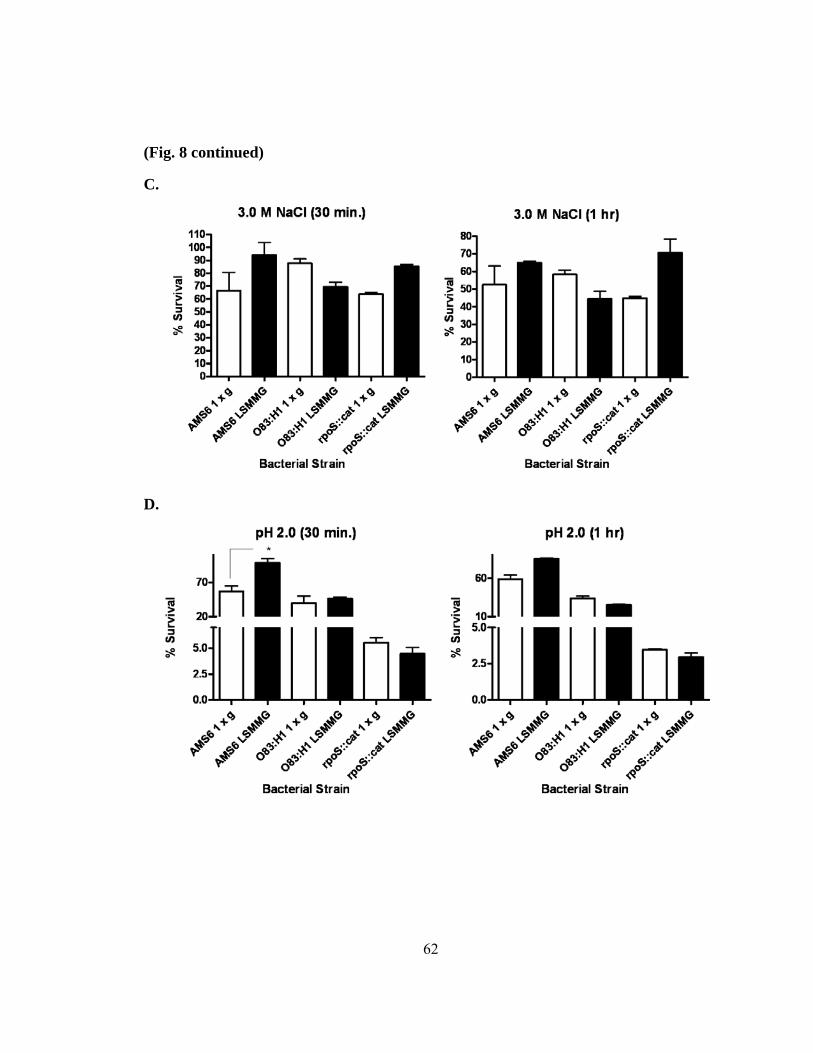
62
(Fig. 8 continued)
C.
D.
63
osmotic stresses after growth under LSMMG, the MG1655 strain did not show any
significant changes in stress resistance to not only these two stressors, but also towards
additional stresses including alkaline, thermal, oxidative, and ethanol stresses (85, 142).
The AIEC O83:H1 clinical isolate evaluated in this study demonstrated a combination of
both increases in resistance and sensitivity to the different environmental stresses
examined. In some of these cases, the effects appeared to be independent of growth in
the LSMMG environment. Stress resistance was also previously investigated in the
enteric pathogen S. Typhimurium after growth in minimal media under LSMMG
conditions. Results from this study demonstrated that while growth under LSMMG
resulted in a phenotype exhibiting enhanced resistance to thermal, osmotic, and acid
stresses, sensitivity to oxidative stress was also increased (147). These results suggest
that growth under conditions of LSMMG can alter bacterial physiology impacting the
ability of the bacterium to resist different stresses and that this is not a universal effect.
Furthermore, these changes not only vary between different bacterial genera, but also can
alter bacterial physiology in a strain-dependent manner.
The impact of RpoS on stress resistance under LSMMG conditions has been
shown to differ between different bacterial genera and in some cases also by growth
phase. In S. Typhimurium and E. coli AMS6, environmental stress resistance was found
to be independent of RpoS expression under LSMMG in stationary and exponential phase
cultures, respectively (85, 147). In contrast, both acid and osmotic stress resistances were
found to be diminished in LSMMG-stationary phase cultures of an E. coli AMS6 rpoS
mutant (85, 147). Therefore, the impact of RpoS was evaluated in AIEC O83:H1 under
LSMMG during stationary phases of growth where RpoS expression occurs at maximal
levels (Fig. 7). Comparisons between CAA001 and the wild-type strain revealed similar
trends in resistance to thermal (increased resistance) and acid (no change between
LSMMG and 1 x g) stresses under LSMMG conditions (Fig. 8 A and D). In contrast,
CAA001 exhibited slight increases in resistance to osmotic stress and decreased oxidative
stress resistance after growth in LSMMG compared to the wild-type strain (Fig. 8B and
C). While the presence of RpoS expression appears to be important for oxidative and
64
acid stress resistance under LSMMG, it is not clear as to how the absence in activity of
this critical stress response regulator can lead to increased thermal and osmotic resistance
in the LSMMG environment. Based on similar stress resistance patterns exhibited by
both LSMMG-grown wild-type and rpoS mutant S. Typhimurium strains, Wilson et al.
has suggested that environmental cues from the LSMMG environment may initiate
RpoS-independent changes in the physiology of bacteria allowing them to phenotypically
become pre-adapted to resist multiple environmental stresses (147). Perhaps cues from
the LSMMG environment function to elicit a combination of both RpoS-dependent and
independent effects in AIEC O83:H1 in a stress-specific manner. Alternatively, previous
reports have shown varying degrees of heterogeneity in RpoS functionality and
subsequent resistance toward different environmental stresses in different pathogenic
strains of E. coli (10).
Collectively, these findings demonstrate that growth under LSMMG can alter
bacterial resistance to different stresses and that these changes are not universal, but can
vary between different genera and strains. Furthermore, results from assays with the
CAA001 demonstrated an important role for RpoS in mediating protection against certain
stresses (e.g. oxidative and acid stresses) under LSMMG conditions during stationary
growth periods. The results presented here reveal, for the first time, the effects of
LSMMG and the impact of RpoS on the ability of an AIEC clinical isolate to tolerate
different environmental stresses.
Enhanced adherence by AIEC O83:H1 to Caco-2 cells after growth under LSMMG.
AIEC clinical isolates are characterized by their intrinsic ability to adhere to and
invade gastrointestinal epithelial cells and residential macrophages, resulting in the
production of TNF-α, which can contribute to pro-inflammatory responses (61).
Therefore, we examined the impact of LSMMG on these two central characteristics using
Caco-2 cells. Twelve-hour cultures of AIEC O83:H1 grown under LSMMG and 1 x g
control conditions were used to infect Caco-2 monolayers. AIEC O83:H1 LSMMG
cultures were found to exhibit significantly higher adherence levels to Caco-2
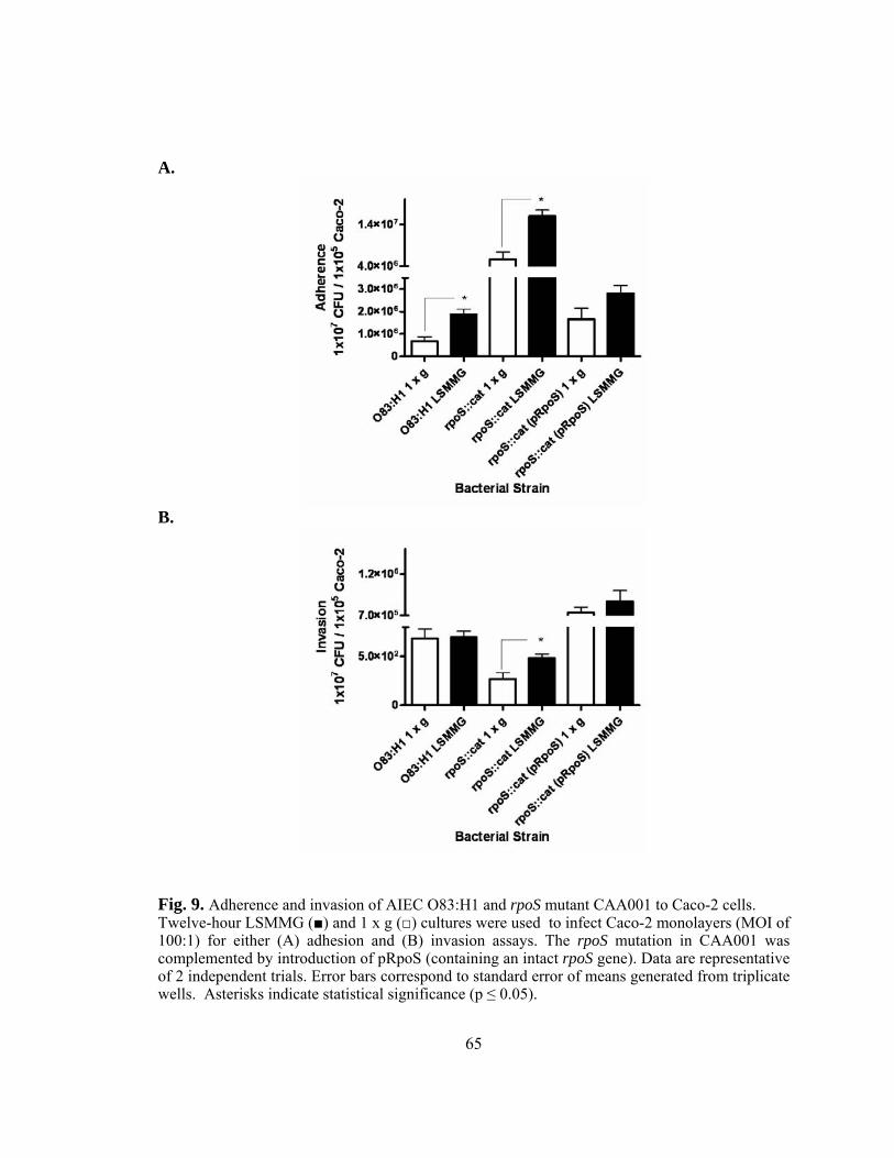
65
A.
B.
Fig. 9. Adherence and invasion of AIEC O83:H1 and rpoS mutant CAA001 to Caco-2 cells. Twelve-hour LSMMG (■) and 1 x g (□) cultures were used to infect Caco-2 monolayers (MOI of 100:1) for either (A) adhesion and (B) invasion assays. The rpoS mutation in CAA001 was complemented by introduction of pRpoS (containing an intact rpoS gene). Data are representative of 2 independent trials. Error bars correspond to standard error of means generated from triplicate wells. Asterisks indicate statistical significance (p ≤ 0.05).
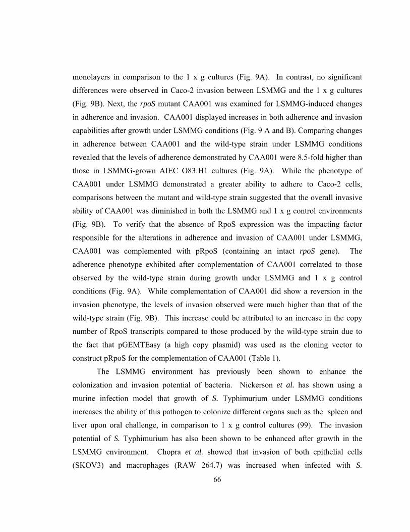
66
monolayers in comparison to the 1 x g cultures (Fig. 9A). In contrast, no significant
differences were observed in Caco-2 invasion between LSMMG and the 1 x g cultures
(Fig. 9B). Next, the rpoS mutant CAA001 was examined for LSMMG-induced changes
in adherence and invasion. CAA001 displayed increases in both adherence and invasion
capabilities after growth under LSMMG conditions (Fig. 9 A and B). Comparing changes
in adherence between CAA001 and the wild-type strain under LSMMG conditions
revealed that the levels of adherence demonstrated by CAA001 were 8.5-fold higher than
those in LSMMG-grown AIEC O83:H1 cultures (Fig. 9A). While the phenotype of
CAA001 under LSMMG demonstrated a greater ability to adhere to Caco-2 cells,
comparisons between the mutant and wild-type strain suggested that the overall invasive
ability of CAA001 was diminished in both the LSMMG and 1 x g control environments
(Fig. 9B). To verify that the absence of RpoS expression was the impacting factor
responsible for the alterations in adherence and invasion of CAA001 under LSMMG,
CAA001 was complemented with pRpoS (containing an intact rpoS gene). The
adherence phenotype exhibited after complementation of CAA001 correlated to those
observed by the wild-type strain during growth under LSMMG and 1 x g control
conditions (Fig. 9A). While complementation of CAA001 did show a reversion in the
invasion phenotype, the levels of invasion observed were much higher than that of the
wild-type strain (Fig. 9B). This increase could be attributed to an increase in the copy
number of RpoS transcripts compared to those produced by the wild-type strain due to
the fact that pGEMTEasy (a high copy plasmid) was used as the cloning vector to
construct pRpoS for the complementation of CAA001 (Table 1).
The LSMMG environment has previously been shown to enhance the
colonization and invasion potential of bacteria. Nickerson et al. has shown using a
murine infection model that growth of S. Typhimurium under LSMMG conditions
increases the ability of this pathogen to colonize different organs such as the spleen and
liver upon oral challenge, in comparison to 1 x g control cultures (99). The invasion
potential of S. Typhimurium has also been shown to be enhanced after growth in the
LSMMG environment. Chopra et al. showed that invasion of both epithelial cells
(SKOV3) and macrophages (RAW 264.7) was increased when infected with S.
67
Typhimurium cultures grown under LSMMG conditions compared to 1 x g controls (23).
The findings presented here show that LSMMG can also enhance the colonization
potential of clinical isolates of AIEC through physiological changes which increase the
capabilities of such strains to adhere to intestinal epithelia.
While RpoS is well-known for its role as an important global stress regulator in
response to various environmental stresses, it is less clear as to what regulatory role this
sigma factor plays in the process of colonization and virulence potential. Bhagwat et al.
has previously shown that there are various degrees of RpoS functional heterogeneity
between different E. coli serotypes and strains regarding adherence (10).
Complementation of different native O157:H7 strains with an rpoS-containing vector
appeared to increase adherence capabilities at different levels. In contrast, a decrease in
adherence was observed in an O26:H11 strain upon complementation, while little
difference was observed in the adherence abilities of an O111:H11 strain regardless of
complementation with rpoS. The authors suggested that these differences in adherence
could be attributed to differences in rpoS regulation of adherence mechanisms among
serotypes. Alternatively, other sigma factors and/or transcriptional regulators maybe
involved in these adherence mechanisms in the absence of RpoS. While studies such as
these typically utilize laboratory test tube-grown cultures, there is little information
known regarding the role of RpoS in the mediation of bacterial adherence in response to
mechanical stresses such as LSMMG. Salmonella virulence potential has been shown to
be dependent on RpoS regulation when grown under standard laboratory conditions (48).
However, previous studies by Wilson et al. have shown that both wild-type and rpoS
mutant strains of S. Typhimurium demonstrate an increased ability to tolerate different
physiological stresses and survive in macrophages after growth under LSMMG (147).
The authors suggest that the LSMMG environment can serve to pre-adapt S.
Typhimurium to resist multiple stress environments, encountered during the course of
infection, in an RpoS-independent manner. The findings presented here show, for the
first time that the absence of RpoS expression can result in a hyper-adherent phenotype in
an AIEC O83:H1 serotype grown under the conditions of LSMMG. To further
understand the correlation between the absence of RpoS activity and the enhanced AIEC
68
adherence phenotype in response to LSMMG, we next investigated the presence of
RpoS-regulated adhesins/adhesion-associated factors which participate in the phenotype
observed under LSMMG conditions.
Characterization of TnphoA isolate CAA003 exhibiting reduced adherence phenotype under LSMMG conditions.
The elevated adherence demonstrated by rpoS mutant strain CAA001 lead us to
investigate novel proteins contributing to this phenotype. To achieve this, we carried out
transposon mutagenesis on the rpoS mutant strain to identify genes encoding putative
adhesins and/or adhesion-associated proteins. We performed TnphoA mutagenesis and
identified 26 transposon mutants out of 10,000 colonies which were identified as dark
blue colonies on media containing the substrate XP (5-bromo-4-chloro-3-indolyl
phosphate). To look for mutants whose expression is mediated by RpoS, the 26 mutants
were complemented with the plasmid pRpoS (generated from pGEMTEasy, Table 1),
containing an intact copy of the rpoS gene, and screened again to look for the reversion
of blue colonies back to colorless colonies. This screening process yielded two mutant
strains which were further evaluated for changes in adhesion. A significant decrease in
adherence was observed in one of the two isolates, which was named CAA003
(CAA001::TnphoA isolate 1) (Fig. 10A). Strain CAA003 displayed reduced adherence in
comparison to wild-type AIEC O83:H1 and CAA001 strains after growth under LSMMG
(Fig. 10A). The decreased adherence suggested that the transposon had disrupted a gene
encoding a putative adhesion-associated protein in CAA003. While the invasive ability
of CAA003 was found to be comparable to that of the rpoS mutant under LSMMG
conditions (Fig. 10B), overall invasion by the two strains was lower than that of the wild-
type strain.
To further characterize the diminished adherence phenotype exhibited by
CAA003, identification of the TnphoA-disrupted gene within the genome of CAA003
was pursued. For this purpose, a cosmid library was generated and cosmids ligated with
fragments containing the transposon were isolated and sequenced. BLAST results
69
A.
B.
Figure 10. Adherence and invasion of CAA003 to Caco-2 cells. CAA003 (rpoS – tnaB –- ) was grown under 1 x g control (□) or LSMMG (■) conditions then examined for changes in the ability to (A) adhere and (B) invade Caco-2 monolayers. (A) Both adhesion and invasion by LSMMG-grown CAA003 was found to be decreased in comparison to that of O83:H1 and CAA001. Data are representative of 2 independent trials. Asterisks indicate statistical significance (p ≤ 0.05).
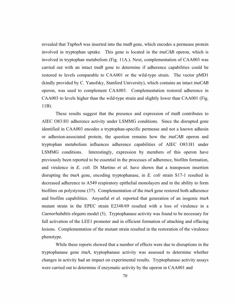
70
revealed that TnphoA was inserted into the tnaB gene, which encodes a permease protein
involved in tryptophan uptake. This gene is located in the tnaCAB operon, which is
involved in tryptophan metabolism (Fig. 11A.). Next, complementation of CAA003 was
carried out with an intact tnaB gene to determine if adherence capabilities could be
restored to levels comparable to CAA001 or the wild-type strain. The vector pMD1
(kindly provided by C. Yanofsky, Stanford University), which contains an intact tnaCAB
operon, was used to complement CAA003. Complementation restored adherence in
CAA003 to levels higher than the wild-type strain and slightly lower than CAA001 (Fig.
11B).
These results suggest that the presence and expression of tnaB contributes to
AIEC O83:H1 adherence activity under LSMMG conditions. Since the disrupted gene
identified in CAA003 encodes a tryptophan-specific permease and not a known adhesin
or adhesion-associated protein, the question remains how the tnaCAB operon and
tryptophan metabolism influences adherence capabilities of AIEC O83:H1 under
LSMMG conditions. Interestingly, expression by members of this operon have
previously been reported to be essential in the processes of adherence, biofilm formation,
and virulence in E. coli. Di Martino et al. have shown that a transposon insertion
disrupting the tnaA gene, encoding tryptophanase, in E. coli strain S17-1 resulted in
decreased adherence to A549 respiratory epithelial monolayers and in the ability to form
biofilms on polystyrene (37). Complementation of the tnaA gene restored both adherence
and biofilm capabilities. Anyanful et al. reported that generation of an isogenic tnaA
mutant strain in the EPEC strain E2348/69 resulted with a loss of virulence in a
Caenorhabditis elegans model (5). Tryptophanase activity was found to be necessary for
full activation of the LEE1 promoter and in efficient formation of attaching and effacing
lesions. Complementation of the mutant strain resulted in the restoration of the virulence
phenotype.
While these reports showed that a number of effects were due to disruptions in the
tryptophanase gene tnaA, tryptophanase activity was assessed to determine whether
changes in activity had an impact on experimental results. Tryptophanase activity assays
were carried out to determine if enzymatic activity by the operon in CAA001 and
71
A.
B.
Fig. 11. Complementation of strain CAA003 with the tnaCAB operon restores adherence under LSMMG. (A) The tryptophanase (tnaCAB) operon. The three genes making up the operon include tnaC which encodes for a leader peptide sequence involved with Rho-dependent antitermination, tnaA which encodes for the enzyme tryptophanase which breaks down tryptophan, and tnaB which encodes a tryptophan-specific permease (the transposon insertion is indicated by [▼]). The CAA003 isolate was complemented with vector pMD1 (containing an intact tnaCAB operon) and grown under 1 x g control (□) or LSMMG (■) conditions to determine if adherence to Caco-2 monolayers is restored. Data are representative of 2 independent trials. Asterisks indicate statistical significance (p ≤ 0.05).
72
CAA003 was altered in comparison to the wild-type strain. The assay involved the use of
S-o-nitrophenyl-L-cysteine as a chromogenic substrate which can be degraded in the
presence of tryptophanase forming o-nitrothiophenolate which can be measured
photometrically (Fig. 12A). Tryptophanase-specific activity levels were found to be
intact in both the CAA001 and CAA003 strains at levels exceeding the wild-type strain
(Fig. 12B), indicating that disruption of the tnaB gene was the main variable impacting
adherence activity. The elevated activity demonstrated by the rpoS mutant strains could
be attributed to the absence of RpoS expression which could result in unregulated and
enhanced tryptophanase activity by the operon. Lacour et al. has previously shown that
RpoS plays a regulatory role in mediating the expression of genes in the tnaCAB operon
and subsequent tryptophanase expression (76).
While there is evidence that members of the tryptophanase operon can alter
adherence properties, there remains the question of what is the mechanism by which
these effects are transduced. There is an increasing amount of evidence which indicates
that indole, one of the by-products of tryptophan metabolism, can function as an external
signaling molecule. Using lacZ fusions, previous studies have identified indole as the
extracellular signaling factor in conditioned media from stationary phase E. coli cultures
that was responsible for the activation of a number of different genes including tnaB (6,
145). The activity from the tnaB::lacZ fusion was found to be decreased greater than
70% after using conditioned media from a tnaA mutant that was unable to metabolize
tryptophan and produce indole. In a subsequent study, Di Martino et al. demonstrated that
exogenous supplementation of indole to an E. coli S17-1 tnaA mutant restored the ability
of the mutant to form biofilms on polystyrene (36). The authors suggested that indole
may act as a signaling molecule involved in mediating the expression of adhesion and
biofilm-promoting factors.
These findings along with the experimental results presented in this study show
that tnaB and other members of the tnaCAB operon play an important role in adherence
and colonization processes in E. coli. Under LSMMG conditions, the absence of tnaB
expression results with a decline in adherence capabilities by AIEC O83:H1 and
complementation restores this adherence phenotype. One possible explanation for this
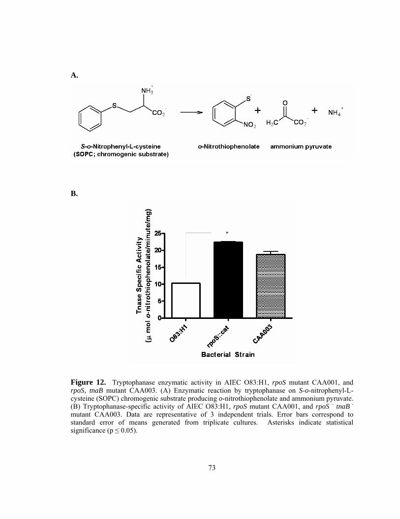
73
A.
B.
Figure 12. Tryptophanase enzymatic activity in AIEC O83:H1, rpoS mutant CAA001, and rpoS, tnaB mutant CAA003. (A) Enzymatic reaction by tryptophanase on S-o-nitrophenyl-L-cysteine (SOPC) chromogenic substrate producing o-nitrothiophenolate and ammonium pyruvate. (B) Tryptophanase-specific activity of AIEC O83:H1, rpoS mutant CAA001, and rpoS – tnaB - mutant CAA003. Data are representative of 3 independent trials. Error bars correspond to standard error of means generated from triplicate cultures. Asterisks indicate statistical significance (p ≤ 0.05).
74
trend is that the loss of tnaB expression prevents normal expression of the tryptophan
permease responsible for tryptophan uptake and import into the cell. A decrease in the
amount of intracellular tryptophan directly reduces the production of indole through
tryptophan metabolism, which can affect downstream signaling processes dependent on
the activation by this extracellular signaling molecule. These processes could include the
expression of genes encoding proteins which may play a role in adherence-based
processes.
75
CONCLUSIONS
The research findings presented in this dissertation show how mechanical
stresses, such as microgravity and low-shear stress, can alter the genomic profile of
Gram-positive bacteria (e.g. S. pneumoniae) as well as the physiology and colonization
properties of adherent-invasive E. coli isolates associated with Crohn’s Disease. The
overall trend of down-regulated genes observed by S. pneumoniae in response to
LSMMG conditions included genes physically located throughout the genome, which
represented multiple functional categories and included genes of common operons /
regulons. While previous work by Niesel et al. (unpublished data) has demonstrated an
increase in virulence potential by S. pneumoniae after growth under LSMMG, the
induction of known virulence genes was not observed to explain these findings. The
genomic responses to the LSMMG environment could suggest that the lack of virulence
gene expression might be contributing to immunoevasive strategies elicited by the
bacteria to avoid detection by the host immune system. Alternatively, LSMMG-mediated
responses could be promoting the induction of novel genes and mechanisms associated
with virulence function and / or the fine-tuning of characterized genes and mechanisms in
a different way to promote virulence-associated functions. Future investigations can be
pursued to correlate the gene expression trends presented here with changes at the protein
level. Determining the putative roles for such proteins in survival and virulence-
associated processes will help substantiate the importance of LSMMG-mediated
signaling and its influence on transcriptional regulation and subsequent protein
expression. In addition, the potential roles of LSMMG-responsive genes at the cellular
level can be determined through mutagenesis-based studies involving physiological
comparisons (e.g. stress resistance and virulence potential) between wild-type and mutant
strains in response to the LSMMG environment.
Physiological analyses of AIEC strain O83:H1 after growth under LSMMG
revealed both alterations in environmental stress resistance and colonization potential.
Alterations in stress resistance (e.g. increased stress resistance and sensitivity) appeared
to be both LSMMG and rpoS-dependent in a stress-specific manner. The absence of
76
rpoS expression under LSMMG conditions conferred a hyper-adherent phenotype which
involved participation of the tryptophan permease, encoded by the gene tnaB. Though no
genes were identified which encode known adhesins or adhesion-associated proteins, a
possible mechanism through which the tnaCAB operon could be contributing to
adherence is by the production of the tryptophan metabolite indole. This compound has
previously been shown to function as an extracellular signaling molecule associated with
the restoration of adhesion and biofilm formation capabilities in mutant strains (89, 145).
To better understand to potential contribution of indole in adherence function, future
studies should address changes in the global gene profiles of LSMMG-grown cultures
with and without indole supplementation. In addition, further analyses addressing RpoS
regulation in AIEC at the transcriptional, translational, and post-translational levels could
provide information which can be used to develop a more comprehensive understanding
of the molecular mechanism(s) and key mediators used by bacteria to respond and adapt
to mechanical forces such as LSMMG.

77
BIBLIOGRAPHY
1. Allen, C. A., C. L. Galindo, U. Pandya, D. A. Watson, A. K. Chopra, and D. W. Niesel. 2007. Transcription profiles of Streptococcus pneumoniae grown under different conditions of normal gravitation. Acta Astronautica 60:433-444.
2. Allen, C. A., Galindo, C., Williams, M., Pandya, U. Chopra, A.K., and D.
Niesel. 2006. A Global Transcriptional Analysis of Streptococcus pneumoniae in Response to Low-Shear Modeled Microgravity. Gravit Space Biol Bull 19:143-144.
3. Alpuche-Aranda, C., E. Racoosin, J. Swanson, and S. Miller. 1994.
Salmonella stimulate macrophage macropinocytosis and persist within spacious phagosomes. J. Exp. Med. 179:601-608.
4. Ambrose, N. S., Johnson, M., Burdon, D.W., Keighley, M.R. 1984. Incidence
of pathogenic bacteria from mesenteric lymph nodes and ileal serosa during Crohn's disease surgery. Br J Surg 71:623-625.
5. Anyanful, A., J. M. Dolan-Livengood, T. Lewis, S. Sheth, M. N. DeZalia, M.
A. Sherman, L. V. Kalman, G. M. Benian, and D. Kalman. 2005. Paralysis and killing of Caenorhabditis elegans by enteropathogenic Escherichia coli requires the bacterial tryptophanase gene. Mol Microbiol 57:988-1007.
6. Baca-DeLancey, R. R., M. M. T. South, X. Ding, and P. N. Rather. 1999.
Escherichia coli genes regulated by cell-to-cell signaling. Proc Natl Acad Sci 96:4610-4614.
7. Barnich, N., J. Boudeau, L. Claret, and A. Darfeuille-Michaud. 2003.
Regulatory and functional co-operation of flagella and type 1 pili in adhesive and invasive abilities of AIEC strain LF82 isolated from a patient with Crohn's disease. Mol Microbiol 48:781-794.
8. Barnich, N., M.-A. Bringer, L. Claret, and A. Darfeuille-Michaud. 2004.
Involvement of Lipoprotein NlpI in the Virulence of Adherent Invasive Escherichia coli Strain LF82 Isolated from a Patient with Crohn's Disease. Infect. Immun. 72:2484-2493.
9. Berry, A. M., J. Yother, D. E. Briles, D. Hansman, and J. C. Paton. 1989.
Reduced virulence of a defined pneumolysin-negative mutant of Streptococcus pneumoniae. Infect Immun 57:2037-42.
10. Bhagwat, A. A., J. Tan, M. Sharma, M. Kothary, S. Low, B. D. Tall, and M.
Bhagwat. 2006. Functional Heterogeneity of RpoS in Stress Tolerance of

78
Enterohemorrhagic Escherichia coli Strains. Appl. Environ. Microbiol. 72:4978-4986.
11. Bisno, A. L., and I. Van de Rijn. 2000. Classification of Streptococci, p. 2100-
2101. In M. G.L, B. J.E., and R. Dolin (ed.), Principles and Practice of Infectious Disease, 5 ed, vol. 2. Churchchill Livingstone, Philidelphia.
12. Blanc-Potard, A.-B., and B. Lafay. 2003. MgtC as a Horizontally-Acquired
Virulence Factor of Intracellular Bacterial Pathogens: Evidence from Molecular Phylogeny and Comparative Genomics. J Mol Evol 57:479-486.
13. Bonen, D. K., and J. H. Cho. 2003. The genetics of inflammatory bowel disease.
Gastroenterology 124:521-536. 14. Boudeau, J., N. Barnich, and A. Darfeuille-Michaud. 2001. Type 1 pili-
mediated adherence of Escherichia coli strain LF82 isolated from Crohn's disease is involved in bacterial invasion of intestinal epithelial cells. Mol Microbiol 39:1272-1284.
15. Boudeau, J., A.-L. Glasser, E. Masseret, B. Joly, and A. Darfeuille-Michaud.
1999. Invasive Ability of an Escherichia coli Strain Isolated from the Ileal Mucosa of a Patient with Crohn's Disease. Infect. Immun. 67:4499-4509.
16. Bringer, M.-A., N. Barnich, A.-L. Glasser, O. Bardot, and A. Darfeuille-
Michaud. 2005. HtrA Stress Protein Is Involved in Intramacrophagic Replication of Adherent and Invasive Escherichia coli Strain LF82 Isolated from a Patient with Crohn's Disease. Infect. Immun. 73:712-721.
17. Brown, W. R., Lee, E.M. 1973. Radioimmunologic measurements of naturally
occurring bacterial antibodies. Human serum antibodies reactive with Escherichia coli in gastrointestinal and immunologic disorders. Lab Clin Med 82:125-136.
18. Burke, D. A., Axon, A.T. 1988. Adhesive Escherichia coli in inflammatory
bowel disease and infective diarrhoea. BMJ 297:102-104. 19. Cartun, R. W., Van Kruiningen, H.J., Pedersen, C.A., Berman, M.M. 1993.
An immunocytochemical search for infectious agents in Crohn's disease. Mod Pathol 6:212-219.
20. Carvalho, H. M., L. D. Teel, G. Goping, and A. D. O'Brien. 2005. A three-
dimensional tissue culture model for the study of attach and efface lesion formation by enteropathogenic and enterohaemorrhagic Escherichia coli. Cell Microbiol 7:1771-1781.
79
21. Cave, D. R., Mitchell, D.N., Kane, S.P., Brooke, B.N. 1973. Further animal evidence of a transmissible agent in Crohn's disease. Lancet 2:1120-1122.
22. Chiodini, R. J., Van Kruiningen, H.J., Thayer, W.R., Merkal, R.S., Coutu,
J.A. 1984. Possible role of mycobacteria in inflammatory bowel disease. I. An unclassified Mycobacterium species isolated from patients with Crohn's disease. Dig Dis Sci 29:1073-1079.
23. Chopra, V., A. A. Fadl, J. Sha, S. Chopra, C. L. Galindo, and A. K. Chopra.
2006. Alterations in the Virulence Potential of Enteric Pathogens and Bacterial-Host Cell Interactions Under Simulated Microgravity Conditions. J. Toxicol. Environ. Health A 69:1345-1370.
24. Christensen, M., T. Borza, G. Dandanell, A.-M. Gilles, O. Barzu, R. A. Kelln,
and J. Neuhard. 2003. Regulation of Expression of the 2-Deoxy-D-Ribose Utilization Regulon, deoQKPX, from Salmonella enterica Serovar Typhimurium. J. Bacteriol. 185:6042-6050.
25. Chudwin, D. S., S. G. Artrip, A. Korenblit, G. Schiffman, and S. Rao. 1985.
Correlation of serum opsonins with in vitro phagocytosis of Streptococcus pneumoniae. Infect Immun 50:213-7.
26. Clerc, P., and P. J. Sansonetti. 1987. Entry of Shigella flexneri into HeLa cells:
evidence for directed phagocytosis involving actin polymerization and myosin accumulation. Infect. Immun. 55:2681-2688.
27. Cottagnoud, P., and A. Tomasz. 1993. Triggering of pneumococcal autolysis by
lysozyme. J Infect Dis 167:684-90. 28. Daig, R., Andus, T., Aschenbrenner, E., Falk, W., Scholmerich, J., Gross, V.
1996. Increased interleukin 8 expression in the colon mucosa of patients with inflammatory bowel disease. Gut 38:216-222.
29. Darfeuille-Michaud, A. 2002. Adherent-invasive Escherichia coli: a putative
new E. coli pathotype associated with Crohn's disease. Int. J. Med. microbiol. 292:185-193.
30. Darfeuille-Michaud, A. 1998. Presence of adherent Escherichia coli strains in
ileal mucosa of patients with Crohn's disease. Gastroenterology 115:1405-1413. 31. Davies, P. F. 1995. Flow-mediated endothelial mechanotransduction. Physiol.
Rev. 75:519-560.
80
32. Dawid, S., A. M. Roche, and J. N. Weiser. 2007. The blp Bacteriocins of Streptococcus pneumoniae Mediate Intraspecies Competition both In Vitro and In Vivo. Infect. Immun. 75:443-451.
33. de Saizieu, A., C. Gardes, N. Flint, C. Wagner, M. Kamber, T. J. Mitchell,
W. Keck, K. E. Amrein, and R. Lange. 2000. Microarray-Based Identification of a Novel Streptococcus pneumoniae Regulon Controlled by an Autoinduced Peptide. J. Bacteriol. 182:4696-4703.
34. Deeley, M. C., and C. Yanofsky. 1981. Nucleotide sequence of the structural
gene for tryptophanase of Escherichia coli K-12. J. Bacteriol. 147:787-796. 35. Detweiler, C. S., D. M. Monack, I. E. Brodsky, H. Mathew, and S. Falkow.
2003. virK, somA and rcsC are important for systemic Salmonella enterica serovar Typhimurium infection and cationic peptide resistance. Mol Microbiol 48:385-400.
36. Di Martino, P., R. Fursy, L. Bret, B. Sundararaju, and R. S. Phillips. 2003.
Indole can act as an extracellular signal to regulate biofilm formation of Escherichia coli and other indole-producing bacteria. Can J Microbiol 49:443-449.
37. Di Martino P, M. A., Phillips R, Orange N, Hulen C. 2002. Isolation of an
Escherichia coli strain mutant unable to form biofilm on polystyrene and to adhere to human pneumocyte cells: involvement of tryptophanase. Can J Microbiol 48:132-137.
38. Diep, D. B., M. Skaugen, Z. Salehian, H. Holo, and I. F. Nes. 2007. Common
mechanisms of target cell recognition and immunity for class II bacteriocins. Proc Natl Acad Sci USA 104:2384-2389.
39. Diez, A., N. Gustavsson, and T. Nystrom. 2000. The universal stress protein A
of Escherichia coli is required for resistance to DNA damaging agents and is regulated by a RecA/FtsK-dependent regulatory pathway. Mol Microbiol 36:1494-1503.
40. Dogan, B., S. Klaessig, M. Rishniw, R. A. Almeida, S. P. Oliver, K. Simpson,
and Y. H. Schukken. 2006. Adherent and invasive Escherichia coli are associated with persistent bovine mastitis. Vet Microbiol 116:270-282.
41. Donnenberg, M. S., Donohue-Rolfe, A., Keusch, G.T. 1990. A comparison of
HEp-2 cell invasion by enteropathogenic and enteroinvasive Escherichia coli. FEMS Microbiol Lett 57:83-86.
81
42. Donnenberg, M. S., and J. B. Kaper. 1991. Construction of an eae deletion mutant of enteropathogenic Escherichia coli by using a positive-selection suicide vector. Infect. Immun. 59:4310-4317.
43. Duchmann, R., Zeitz, M. 1999. Crohn's Disease, p. 1055-1080. In P. L. Ogra,
Mestecky, J., Lamm, M.E., Strober, W., Bienenstock, J., McGhee, J.R. (ed.), Mucosal Immunology. Academic Press, San Diego, CA.
44. Fang, A., D. Pierson, D. Koenig, S. Mishra, and A. Demain. 1997. Effect of
simulated microgravity and shear stress on microcin B17 production by Escherichia coli and on its excretion into the medium. Appl. Environ. Microbiol. 63:4090-4092.
45. Fang, A., D. L. Pierson, S. K. Mishra, and A. L. Demain. 2000. Relief from
glucose interference in microcin B17 biosynthesis by growth in a rotating-wall bioreactor. Lett Appl Microbiol 31:39-41.
46. Fang, A., D. L. Pierson, S. K. Mishra, D. W. Koenig, and A. L. Demain. 1997.
Gramicidin S Production by Bacillus brevis in Simulated Microgravity. Curr Microbiol 34:199-204.
47. Fang, A., D. L. Pierson, S. K. Mishra, D. W. Koenig, and A. L. Demain. 1997.
Secondary metabolism in simulated microgravity: β-lactam production by Streptomyces clavuligerus. J Ind Microbiol Biotechnol 18:22-25.
48. Fang, F., S. Libby, N. Buchmeier, P. Loewen, J. Switala, J. Harwood, and D.
Guiney. 1992. The Alternative {sigma} Factor KatF (RpoS) Regulates Salmonella Virulence. Proc Natl Acad Sci 89:11978-11982.
49. Fedson, D. S., Musher, D.M., Eskola, J. 1998. Pneumococcal vaccine. In S. A.
Plotkin, Orenstein, W.A. (ed.), Vaccines, 3rd ed. W.B. Saunders, Philadelphia, PA.
50. Fekety, R., and A. B. Shah. 1993. Diagnosis and treatment of Clostridium
difficile colitis. JAMA 269:71-75. 51. Feldman, C., T. J. Mitchell, P. W. Andrew, G. J. Boulnois, R. C. Read, H. C.
Todd, P. J. Cole, and R. Wilson. 1990. The effect of Streptococcus pneumoniae pneumolysin on human respiratory epithelium in vitro. Microb Pathog 9:275-84.
52. Ferrante, A., B. Rowan-Kelly, and J. C. Paton. 1984. Inhibition of in vitro
human lymphocyte response by the pneumococcal toxin pneumolysin. Infect Immun 46:585-589.
53. Forrester, J., and J. Lackie. 1984. Adhesion of neutrophil leucocytes under conditions of flow. J Cell Sci 70:93-110.
82
54. Fujimura, Y., R. Kamoi, and M. Iida. 1996. Pathogenesis of aphthoid ulcers in
Crohn's disease: correlative findings by magnifying colonoscopy, electron microscopy, and immunohistochemistry. Gut 38:724-732.
55. Galindo, C. L., J. Sha, D. A. Ribardo, A. A. Fadl, L. Pillai, and A. K. Chopra.
2003. Identification of Aeromonas hydrophila Cytotoxic Enterotoxin-induced Genes in Macrophages Using Microarrays. J. Biol. Chem. 278:40198-40212.
56. Gao, Q., A. Fang, D. L. Pierson, S. K. Mishra, and A. L. Demain. 2001. Shear
stress enhances microcin B17 production in a rotating wall bioreactor, but ethanol stress does not. Appl Microbiol. Biotechnol. 56:384-387.
57. Geelen, S., C. Bhattacharyya, and E. Tuomanen. 1993. The cell wall mediates
pneumococcal attachment to and cytopathology in human endothelial cells. Infect Immun 61:1538-43.
58. Giaffer, M. H., Holdsworth, C.D., Duerden, B.I. 1992. Virulence properties of
Escherichia coli strains isolated from patients with inflammatory bowel disease. Gut 33:646-650.
59. Gillespie, S. H., Balakrishnan, I. 2000. Pathogenesis of pneumococcal infection.
J Med Microbiol 49:1057-1067. 60. Gish K, Y. C. 1993. Inhibition of expression of the tryptophanase operon in
Escherichia coli by extrachromosomal copies of the tna leader region. J Bacteriol. 175:3380-3387.
61. Glasser, A.-L., J. Boudeau, N. Barnich, M.-H. Perruchot, J.-F. Colombel, and
A. Darfeuille-Michaud. 2001. Adherent Invasive Escherichia coli Strains from Patients with Crohn's Disease Survive and Replicate within Macrophages without Inducing Host Cell Death. Infect. Immun. 69:5529-5537.
62. Gosink, K. K., E. R. Mann, C. Guglielmo, E. I. Tuomanen, and H. R.
Masure. 2000. Role of novel choline binding proteins in virulence of Streptococcus pneumoniae. Infect Immun 68:5690-5695.
63. Grant, T., V. Bennett-Wood, and R. M. Robins-Browne. 1999.
Characterization of the Interaction between Yersinia enterocolitica Biotype 1A and Phagocytes and Epithelial Cells In Vitro. Infect. Immun. 67:4367-4375.
64. Guo, P., A. M. Weinstein, and S. Weinbaum. 2000. A hydrodynamic
mechanosensory hypothesis for brush border microvilli. Am J Physiol Renal Physiol 279:F698-712.
83
65. Hakenbeck, R., N. Balmelle, B. Weber, C. Gardes, W. Keck, and A. de Saizieu. 2001. Mosaic Genes and Mosaic Chromosomes: Intra- and Interspecies Genomic Variation of Streptococcus pneumoniae. Infect. Immun. 69:2477-2486.
66. Hammond, T. G., and J. M. Hammond. 2001. Optimized suspension culture:
the rotating-wall vessel. Am J Physiol Renal Physiol 281:F12-25. 67. Harel, J., Forget, C., Ngeleka, M., Jacques, M., and Fairbrother, J.M. 1992.
Isolation and characterization of adhesin-defective TnphoA mutants of septicaemic porcine Escherichia coli of serotype O115:K-:F165. J. Gen. Microbiol. 138:2337-2345.
68. Hirsch, M., and T. Elliott. 2005. Stationary-Phase Regulation of RpoS
Translation in Escherichia coli. J. Bacteriol. 187:7204-7213. 69. Holt, J. G., N. R. Krieg, P. H. A. Sneath, and D. Bergey. 1994. Bergey's
Manual of Determinative Bacteriology, 9 ed. Williams & Williams, Baltimore. 70. Ingber, D. E. 2003. Mechanobiology and diseases of mechanotransduction. Ann
Med 35:564-577. 71. Knutsen, E., O. Ween, and L. S. Havarstein. 2004. Two Separate Quorum-
Sensing Systems Upregulate Transcription of the Same ABC Transporter in Streptococcus pneumoniae. J. Bacteriol. 186:3078-3085.
72. Koedel, U., W. M. Scheld, and H.-W. Pfister. 2002. Pathogenesis and
pathophysiology of pneumococcal meningitis. Lancet Infect Dis 2:721-736. 73. Kopytek, S. J., J. C. D. Dyer, G. S. Knapp, and J. C. Hu. 2000. Resistance to
Methotrexate Due to AcrAB-Dependent Export from Escherichia coli. Antimicrob. Agents Chemother. 44:3210-3212.
74. Kornfeld, S. J., and A. G. Plaut. 1981. Secretory immunity and the bacterial IgA
proteases. Rev Infect Dis 3:521-534. 75. Lacks, S. A., S. Ayalew, A. G. de la Campa, and B. Greenberg. 2000.
Regulation of competence for genetic transformation in Streptococcus pneumoniae: expression of dpnA, a late competence gene encoding a DNA methyltransferase of the DpnII restriction system. Mol Microbiol 35:1089-1098.
76. Lacour, S., and P. Landini. 2004. {sigma}S-Dependent Gene Expression at the
Onset of Stationary Phase in Escherichia coli: Function of {sigma}S-Dependent Genes and Identification of Their Promoter Sequences. J. Bacteriol. 186:7186-7195.
84
77. Laemmli, U. K. 1970. Cleavage of Structural Proteins during the Assembly of the Head of Bacteriophage T4. Nature 227:680-685.
78. Laffineur, G., Lescut, D., Vincent, P., Quandalle, P., Wurtz, A., Colombel,
J.F. 1992. Bacterial translocation in Crohn's disease. Gastroenterol Clin Biol 16:777-781.
79. Lai, X.-H., S.-Y. Wang, and B. E. Uhlin. 1999. Expression of cytotoxicity by
potential pathogens in the standard Escherichia coli collection of reference (ECOR) strains. Microbiology 145:3295-3303.
80. Lai, X.-H., J.-G. Xu, S. Melgar, and B. E. Uhlin. 1999. An apoptotic response
by J774 macrophage cells is common upon infection with diarrheagenic Escherichia coli. FEMS Microbiol Lett 172:29-34.
81. Lederman, E., Neut, C., Desreumaux, P., Klein, O., Gambiez, L., Cortot, A.,
Quandalle, P., Colombel, J.F. 1997. Bacterial overgrowth in the neoterminal ileum after ileocolonic resection for Crohn's disease. Gastroenterology 112: A1023.
82. Liu, W.-T., M. H. Karavolos, D. M. Bulmer, A. Allaoui, R. D. C. E.
Hormaeche, J. J. Lee, and C. M. Anjam Khan. 2007. Role of the universal stress protein UspA of Salmonella in growth arrest, stress and virulence. Microb Pathog 42:2-10.
83. Liu, Y., van Kruiningen, H.J., West, A.B., Cartun, R.W., Cortot, A.,
Colombel, J.F. 1995. Immunocytochemical evidence of Listeria, Escherichia coli, and Streptococcus antigens in Crohn's disease. Gastroenterology 108:1396-1404.
84. Livak, K. J., and T. D. Schmittgen. 2001. Analysis of Relative Gene Expression
Data Using Real-Time Quantitative PCR and the 2-[Delta][Delta]CT Method. Methods 25:402-408.
85. Lynch, S. V., E. L. Brodie, and A. Matin. 2004. Role and Regulation of
{sigma}s in General Resistance Conferred by Low-Shear Simulated Microgravity in Escherichia coli. J. Bacteriol. 186:8207-8212.
86. Maniatis, T., Fritsch, E.F., and Sambrook, J. 1982. Molecular Cloning: A
Laboratory Manual. Cold Spring Harbor Press, Cold Spring Harbor, NY. 87. Maniatis, T., R. C. Hardison, E. Lacy, J. Lauer, C. O'Connell, D. Quon, G.
K. Sim, and A. Efstratiadis. 1978. The isolation of structural genes from libraries of eucaryotic DNA. Cell 15:687-701.
85
88. Marmur, J. 1961. A procedure for the isolation of deoxyribonucleic acid from microorganisms. J Mol Biol 3:208-218.
89. Martino PD, F. R., Bret L, Sundararaju B, Phillips RS. 2003. Indole can act as
an extracellular signal to regulate biofilm formation of Escherichia coli and other indole-producing bacteria. Can J Microbiol. 49:443-449.
90. Mascari, L., Ymele-Leki, P., Eggleton, C.D., Speziale, P., Ross, J.M. 2003.
Fluid shear contributions to bacteria cell detachment initiated by a monoclonal antibody. Biotechnol Bioeng 83:65-74.
91. Masseret, E., J. Boudeau, J. F. Colombel, C. Neut, P. Desreumaux, B. Joly, A.
Cortot, and A. Darfeuille-Michaud. 2001. Genetically related Escherichia coli strains associated with Crohn's disease. Gut 48:320-325.
92. McLemore, L. J. 2007. Inflammatory bowel disease. Radiol Technol 78:291-
305. 93. Mishra S, P. D. 1992. Space Flight, Effects on Microorganisms., vol. 4.
Academic Press. 94. Mitsuyama, K., Toyonaga, A., Sasaki, E., Watanabe, K., Tateishi, H.,
Nishiyama, T., Saiki, T., Ikeda, H., Tsuruta, O., Tanikawa, K. 1994. IL-8 as an important chemoattractant for neutrophils in ulcerative colitis and Crohn's disease. Clin Exp Immunol 96:432-436.
95. Musher, D. M. 2000. Streptococcus pneumoniae, p. 2128-2147. In G. L.
Mandell, Bennett, J.E., Dolin, R. (ed.), Mandell, Douglas, and Bennett's Principles and Practice of Infectious Diseases, 5th ed, vol. 2. Churchhill Livingstone Publishing, Philadelphia, PA.
96. Nakata, N., C. Sasakawa, N. Okada, T. Tobe, I. Fukuda, T. Suzuki, K.
Komatsu, and M. Yoshikawa. 1992. Identification and characterization of virK, a virulence-associated large plasmid gene essential for intercellular spreading of Shigella flexneri. Mol Microbiol 6:2387-2395.
97. Nauman, E. A., C. M. Ott, E. Sander, D. L. Tucker, D. Pierson, J. W. Wilson,
and C. A. Nickerson. 2007. Novel Quantitative Biosystem for Modeling Physiological Fluid Shear Stress on Cells. Appl. Environ. Microbiol. 73:699-705.
98. Nes, I. F., D. B. Diep, and H. Holo. 2007. Bacteriocin Diversity in Streptococcus
and Enterococcus. J. Bacteriol. 189:1189-1198. 99. Nickerson, C. A., C. M. Ott, S. J. Mister, B. J. Morrow, L. Burns-Keliher,
and D. L. Pierson. 2000. Microgravity as a Novel Environmental Signal
86
Affecting Salmonella enterica Serovar Typhimurium Virulence. Infect. Immun. 68:3147-3152.
100. Nickerson, C. A., C. M. Ott, J. W. Wilson, R. Ramamurthy, C. L. LeBlanc,
K. Honer zu Bentrup, T. Hammond, and D. L. Pierson. 2003. Low-shear modeled microgravity: a global environmental regulatory signal affecting bacterial gene expression, physiology, and pathogenesis. J Microbiol Methods 54:1-11.
101. Nickerson, C. A., C. M. Ott, J. W. Wilson, R. Ramamurthy, and D. L.
Pierson. 2004. Microbial Responses to Microgravity and Other Low-Shear Environments. Microbiol. Mol. Biol. Rev. 68:345-361.
102. O'Farrell, H. C., J. N. Scarsdale, and J. P. Rife. 2004. Crystal Structure of
KsgA, a Universally Conserved rRNA Adenine Dimethyltransferase in Escherichia coli. J Mol Biol 339:337-353.
103. Pandya, U., C. A. Allen, D. A. Watson, and D. W. Niesel. 2005. Global
profiling of Streptococcus pneumoniae gene expression at different growth temperatures. Gene 360:45-54.
104. Paton, J. C., R. A. Lock, and D. J. Hansman. 1983. Effect of immunization
with pneumolysin on survival time of mice challenged with Streptococcus pneumoniae. Infect Immun 40:548-52.
105. Pearson, W. R., and D. J. Lipman. 1988. Improved Tools for Biological
Sequence Comparison. Proc. Natl. Acad. Sci. USA 85:2444-2448. 106. Pestova, E. V., and D. A. Morrison. 1998. Isolation and Characterization of
Three Streptococcus pneumoniae Transformation-Specific Loci by Use of a lacZ Reporter Insertion Vector. J. Bacteriol. 180:2701-2710.
107. Peterson, S., R. T. Cline, H. Tettelin, V. Sharov, and D. A. Morrison. 2000.
Gene Expression Analysis of the Streptococcus pneumoniae Competence Regulons by Use of DNA Microarrays. J. Bacteriol. 182:6192-6202.
108. Powell, S. J., Wilmot, A.J. 1966. Ulcerative post-dysenteric colitis. Gut 7:438-
433. 109. Quagliarello, V., Scheld, W.M. 1992. Bacterial meningitis: Pathogenesis,
pathophysiology, and progress. N Engl J Med 327:864-872. 110. Revel, A. T., A. M. Talaat, and M. V. Norgard. 2002. DNA microarray analysis
of differential gene expression in Borrelia burgdorferi, the Lyme disease spirochete. Proc Natl Acad Sci USA 99:1562-1567.
87
111. Ring, A., and E. Tuomanen. 2000. Host cell invasion by Streptococcus
pneumoniae. Subcell Biochem 33:125-35. 112. Ring, A., J. N. Weiser, and E. I. Tuomanen. 1998. Pneumococcal trafficking
across the blood-brain barrier. Molecular analysis of a novel bidirectional pathway. J Clin Invest 102:347-60.
113. Robertson, G. T., J. Zhao, B. V. Desai, W. H. Coleman, T. I. Nicas, R.
Gilmour, L. Grinius, D. A. Morrison, and M. E. Winkler. 2002. Vancomycin Tolerance Induced by Erythromycin but Not by Loss of vncRS, vex3, or pep27 Function in Streptococcus pneumoniae. J. Bacteriol. 184:6987-7000.
114. Rolhion, N., N. Barnich, L. Claret, and A. Darfeuille-Michaud. 2005. Strong
Decrease in Invasive Ability and Outer Membrane Vesicle Release in Crohn's Disease-Associated Adherent-Invasive Escherichia coli Strain LF82 with the yfgL Gene Deleted. J. Bacteriol. 187:2286-2296.
115. Rosenow, C., P. Ryan, J. N. Weiser, S. Johnson, P. Fontan, A. Ortqvist, and
H. R. Masure. 1997. Contribution of novel choline-binding proteins to adherence, colonization and immunogenicity of Streptococcus pneumoniae. Mol Microbiol 25:819-29.
116. Rutgeerts P, H. M., Geboes K, Peeters M, Penninckx F, Aerts R, Kerremans
R. 1995. Controlled trial of metronidazole treatment for prevention of Crohn's recurrence after ileal resection. Gastroenterology 108:1617-1621.
117. Salyers, A. A., and D. D. Whitt. 1994. Chapter 27: Streptococcus pneumoniae.
ASM Press, Washington D.C. 118. Sankey, E. A., Dhillon, A.P., Anthony, A., Wakefield, A.J., Sim, R., More, L.,
Hudson, M., Sawyerr, A.M., Pounder, R.E. 1993. Early mucosal changes in Crohn's disease. Gut 34:375-381.
119. Sartor, R. B., Rath, H.C., Sellon, R. K. 1996. Microbial factors in chronic
intestinal inflammation. Curr Opin Gastroenterol 12:327-333. 120. Schultsz, C., Moussa, M., van Ketel, R., Tytgat, G.N., Dankert, J. 1997.
Frequency of pathogenic and enteroadherent Escherichia coli in patients with inflammatory bowel disease and controls. J Clin Pathol 50:573-579.
121. Segal, G., Ron, E.Z. 1998. Regulation of heat-shock response in bacteria. Ann
NY Acad Sci 851:147-151.
88
122. Sellon, R. K., S. Tonkonogy, M. Schultz, L. A. Dieleman, W. Grenther, E. Balish, D. M. Rennick, and R. B. Sartor. 1998. Resident Enteric Bacteria Are Necessary for Development of Spontaneous Colitis and Immune System Activation in Interleukin-10-Deficient Mice. Infect. Immun. 66:5224-5231.
123. Sha, J., E. V. Kozlova, A. A. Fadl, J. P. Olano, C. W. Houston, J. W.
Peterson, and A. K. Chopra. 2004. Molecular Characterization of a Glucose-Inhibited Division Gene, gidA, That Regulates Cytotoxic Enterotoxin of Aeromonas hydrophila. Infect. Immun. 72:1084-1095.
124. Siebold, C., K. Flukiger, R. Beutler, and B. Erni. 2001. Carbohydrate
transporters of the bacterial phosphoenolpyruvate: sugar phosphotransferase system (PTS). FEBS Letters 504:104-111.
125. Simon, R., U. Priefer, and A. Puhler. 1983. A Broad Host Range Mobilization
System for Invivo Genetic-Engineering - Transposon Mutagenesis in Gram-Negative Bacteria. Bio-Technology 1:784-791.
126. Simpson, K. W., B. Dogan, M. Rishniw, R. E. Goldstein, S. Klaessig, P. L.
McDonough, A. J. German, R. M. Yates, D. G. Russell, S. E. Johnson, D. E. Berg, J. Harel, G. Bruant, S. P. McDonough, and Y. H. Schukken. 2006. Adherent and Invasive Escherichia coli Is Associated with Granulomatous Colitis in Boxer Dogs. Infect. Immun. 74:4778-4792.
127. Smoot, L. M., J. C. Smoot, M. R. Graham, G. A. Somerville, D. E.
Sturdevant, C. A. L. Migliaccio, G. L. Sylva, and J. M. Musser. 2001. Global differential gene expression in response to growth temperature alteration in group A Streptococcus. Proc Natl Acad Sci U S A 98:10416-10421.
128. Staskawicz B, D. D., Keen N, Napoli C. 1987. Molecular characterization of
cloned avirulence genes from race 0 and race 1 of Pseudomonas syringae pv. glycinea. J Bacteriol. 169:5789-5794.
129. Steil, L., T. Hoffmann, I. Budde, U. Volker, and E. Bremer. 2003. Genome-
Wide Transcriptional Profiling Analysis of Adaptation of Bacillus subtilis to High Salinity. J. Bacteriol. 185:6358-6370.
130. Stock, U. A., and J. P. Vacanti. 2001. Cardiovascular Physiology During Fetal
Development and Implications for Tissue Engineering. Tissue Eng 7:1-7. 131. Suelter, C. H., J. Wang, and E. E. Snell. 1976. Direct spectrophotometric assay
of tryptophanase. FEBS Lett. 66:230-232. 132. Sukharev, S. 1999. Mechanosensitive channels in bacteria as membrane tension
reporters. FASEB J. 13:55-61.
89
133. Sukharev, S., M. Betanzos, C.-S. Chiang, and H. R. Guy. 2001. The gating
mechanism of the large mechanosensitive channel MscL. Nature 409:720-724. 134. Sutherland L, S. J., Sessions J, Hanauer S, Krawitt E, Rankin G, Summers
R, Mekhjian H, Greenberger N, Kelly M, et al. 1991. Double blind, placebo controlled trial of metronidazole in Crohn's disease. Gut 32:1071-1075.
135. Tabaqchali, S., O'Donoghue, D.P., Bettelheim, K.A. 1978. Escherichia coli
antibodies in patients with inflammatory bowel disease. Gut 19:108-113. 136. Taurog, J., J. Richardson, J. Croft, W. Simmons, M. Zhou, J. Fernandez-
Sueiro, E. Balish, and R. Hammer. 1994. The germfree state prevents development of gut and joint inflammatory disease in HLA-B27 transgenic rats. J. Exp. Med. 180:2359-2364.
137. Taylor, R. K., C. Manoil, and J. J. Mekalanos. 1989. Broad-host-range vectors
for delivery of TnphoA: use in genetic analysis of secreted virulence determinants of Vibrio cholerae. J. Bacteriol. 171:1870-1878.
138. Taylor, R. K., V. L. Miller, D. B. Furlong, and J. J. Mekalanos. 1987. Use of
phoA Gene Fusions to Identify a Pilus Colonization Factor Coordinately Regulated with Cholera Toxin. PNAS 84:2833-2837.
139. Tettelin, H., K. E. Nelson, I. T. Paulsen, J. A. Eisen, T. D. Read, S. Peterson,
J. Heidelberg, R. T. DeBoy, D. H. Haft, R. J. Dodson, A. S. Durkin, M. Gwinn, J. F. Kolonay, W. C. Nelson, J. D. Peterson, L. A. Umayam, O. White, S. L. Salzberg, M. R. Lewis, D. Radune, E. Holtzapple, H. Khouri, A. M. Wolf, T. R. Utterback, C. L. Hansen, L. A. McDonald, T. V. Feldblyum, S. Angiuoli, T. Dickinson, E. K. Hickey, I. E. Holt, B. J. Loftus, F. Yang, H. O. Smith, J. C. Venter, B. A. Dougherty, D. A. Morrison, S. K. Hollingshead, and C. M. Fraser. 2001. Complete Genome Sequence of a Virulent Isolate of Streptococcus pneumoniae. Science 293:498-506.
140. Thomas, W. E., E. Trintchina, M. Forero, V. Vogel, and E. V. Sokurenko.
2002. Bacterial Adhesion to Target Cells Enhanced by Shear Force. Cell 109:913-923.
141. Torres, A. G., N. T. Perna, V. Burland, A. Ruknudin, F. R. Blattner, and J. B. Kaper. 2002. Characterization of Cah, a calcium-binding and heat-extractable autotransporter protein of enterohaemorrhagic Escherichia coli. Mol. Microbiol. 45:951-966.
142. Tucker, D. L., Ott, M.C., Huff, S., Fofanov, Y., Pierson, D.L., Willson, R.C.,
Fox, G.E. 2007. Characterization of Escherichia coli MG1655 in a low-shear modeled microgravity environment. BMC Microbiol 7: 1-16.
90
143. Tusher, V. G., R. Tibshirani, and G. Chu. 2001. Significance analysis of
microarrays applied to the ionizing radiation response. Proc Natl Acad Sci 98:5116-5121.
144. Vriesema, A. J. M., J. Dankert, and S. A. J. Zaat. 2000. A Shift from Oral to
Blood pH Is a Stimulus for Adaptive Gene Expression of Streptococcus gordonii CH1 and Induces Protection against Oxidative Stress and Enhanced Bacterial Growth by Expression of msrA. Infect. Immun. 68:1061-1068.
145. Wang, D., X. Ding, and P. N. Rather. 2001. Indole Can Act as an Extracellular
Signal in Escherichia coli. J. Bacteriol. 183:4210-4216. 146. Weiner III, J., C.-U. Zimmerman, H. W. H. Gohlmann, and R. Herrmann.
2003. Transcription profiles of the bacterium Mycoplasma pneumoniae grown at different temperatures. Nucl. Acids Res. 31:6306-6320.
147. Wilson, J. W., C. M. Ott, R. Ramamurthy, S. Porwollik, M. McClelland, D.
L. Pierson, and C. A. Nickerson. 2002. Low-Shear Modeled Microgravity Alters the Salmonella enterica Serovar Typhimurium Stress Response in an RpoS-Independent Manner. Appl. Environ. Microbiol. 68:5408-5416.
148. Wilson, J. W., R. Ramamurthy, S. Porwollik, M. McClelland, T. Hammond,
P. Allen, C. M. Ott, D. L. Pierson, and C. A. Nickerson. 2002. Microarray analysis identifies Salmonella genes belonging to the low-shear modeled microgravity regulon. Proc. Natl. Acad. Sci. USA 99:13807-13812.
149. Ye, R. W., W. Tao, L. Bedzyk, T. Young, M. Chen, and L. Li. 2000. Global
Gene Expression Profiles of Bacillus subtilis Grown under Anaerobic Conditions. J. Bacteriol. 182:4458-4465.
150. Zeng, X., H. H. Saxild, and R. L. Switzer. 2000. Purification and
Characterization of the DeoR Repressor of Bacillus subtilis. J. Bacteriol. 182:1916-1922.
91
Vita
Christopher A. Allen was born on September 23, 1974 in Beaumont, TX, to James and Barbara Allen. Christopher attended Stephen F. Austin State University where he majored in Biology with a minor in Chemistry. He graduated with a Bachelor of Science degree in 1997 and remained at Stephen F. Austin State University where he completed a Master of Science degree in 1999. He was employed as a research assistant at The University of Texas Medical Branch from 1999 to 2001. Christopher began his graduate studies for his doctoral degree in the fall of 2001 at The University of Texas Medical Branch.
Permanent address: 4822 Fannin Dr., Galveston, TX 77551 This dissertation was typed by Christopher A. Allen.
Publications
(Peer-Reviewed Articles) Allen, C.A., and S.C. Wagner. 2000. Rhizosphere of Cattail (Typha latifolia) and Spike Rush (Eleocharis tuberculosa) Inhabiting an Oil Spill. Texas J. Sci. 52: 335-344. Pandya, U., Allen, C.A., Watson, D.A., and D.W. Niesel. 2005. Global Profiling of Streptococcus pneumoniae Gene Expression at Different Growth Temperatures. Gene. 360: 45-54. Allen, C.A., Galindo, C.L., Williams, N.D., Pandya, U., Chopra, A.K., and D.W. Niesel. 2006. A Global Transcriptional Analysis of Streptococcus pneumoniae in Response to Low-Shear Modeled Microgravity. Gravit Space Biol Bull 19: 143-144. Allen, C.A., Galindo, C.L., Pandya, U., Watson, D.A., Chopra, A.K., and D.W. Niesel. 2007. Transcriptional Profiles of Streptococcus pneumoniae Grown Under Different Conditions of Normal Gravitation. Acta Astronautica 60: 433-444. Sonnenfeld, G., Aviles, H., Butel, J.S., Shearer, W.T., Niesel, D.W., Pandya, U., Allen, C.A., Ochs, H.D., Blancher, A., and Michele Abbal. 2007. Bed Rest and Immunity. Acta Astronautica 60: 234-236.
92
Eaves-Pyles, T., Allen, C.A., Taormina, J., Swidsinski, A., Tutt, C.B., Jezek, G.E., Islas-Islas, M., and A. G. Torres. 2007. Escherichia coli isolated from a Crohn's disease patient adheres, invades, and induces inflammatory responses in polarized intestinal epithelial cells. Int J Med Microbiol (accepted for publication). Allen, C.A., Niesel, D.W., and A.G. Torres. The Effects of Low-Shear Stress on Adherent-Invasive Escherichia coli. (in preparation for publication)
(Book Chapters) Allen, C.A. and A.G. Torres. Host-Microbe Communication with the G.I.Tract. G.I. Microbiota and Regulation of the Immune System. Eds. G. Huffnagle and M. Noverr. Landes Bioscience. (in press: book chapter).
(Meeting Abstracts) Wagner, S.C., and C.A. Allen. 1997. Influence of Plant Amendments on Microbial Activity in Fluometuron Contaminated Soils. Abstracts of the 97th General Meeting of the American Society for Microbiology. A.S.M. Press, pg. 524. Allen, C.A., and S.C. Wagner. 1999. Rhizosphere Activity of Plants Inhabiting an Oil Spill in Roy E. Larsen Sandyland Sanctuary. Abstracts of the 99th General Meeting of the American Society for Microbiology. A.S.M. Press, pg. 609. Allen, C.A., Pandya, U., Watson, D.A., and D.W. Niesel. 2004. Global Transcriptional Profiling of Streptococcus pneumoniae in Response to Modeled Microgravity. Abstracts of the 104th General Meeting of the American Society for Microbiology. A.S.M. Press. Allen, C.A. and A.G. Torres. 2005. The Effect of Low-Shear Stress on Virulence Potential of Adherent-Invasive E. coli. Gulf Coast GI Research Forum. pg. P-48. Allen, C.A. and A.G. Torres. 2005. The Effect of Low-Shear Stress on Adherent-Invasive Escherichia coli Physiology and Virulence Potential. Cold Spring Harbor Laboratory: Microbial Pathogenesis & Host Response Meeting. pg.15. Allen, C.A., Galindo, C.L., Williams, N.D., Pandya, U., Chopra, A.K., and D.W. Niesel. 2005. A Global Transcriptional Analysis of Streptococcus pneumoniae in Response to Low-Shear Modeled Microgravity. Gravit Space Biol Bull 19: 20. Allen, C.A. and A.G. Torres. 2005. Analysis of Low-Shear Stress on the Physiology and Virulence Potential of Adherent-Invasive Escherichia coli. American Society for Microbiology Texas Branch and Missouri Valley Branch Fall Meeting. pg. 23.
93
Allen, C.A. and A.G. Torres. 2006. The Impact of RpoS on Adherent-Invasive E. coli Virulence Potential in Response to Low-Shear Stress. Abstracts of the 106th General Meeting of the American Society for Microbiology. A.S.M. Press. Allen, C.A. and A.G. Torres. 2006. A Role for RpoS in the Regulation of Adherent-Invasive Escherichia coli Under Conditions of Low-Shear Stress. Virulence Mechanisms of Bacterial Pathogens International Symposium. Iowa State University, Ames, Iowa. Allen, C.A., Niesel, D.W., and A.G. Torres. 2006. The Effect of Low-Shear Modeled Microgravity on Adherent Invasive Escherichia coli Physiology and Virulence Potential. Gravit Space Biol Bull 20(1): 16. Allen, C.A. and A.G. Torres. 2006. A Regulatory Role for RpoS in Adherent-Invasive Escherichia coli strain O83:H1 under Conditions of Low-Shear Stress. American Society for Microbiology Texas Branch Meeting. pg. 12. Allen, C.A., and A.G. Torres. 2007. RpoS regulation of AIEC O83:H1 physiology and colonization potential under low-phosphate conditions. Abstracts of the 107th General Meeting of the American Society for Microbiology. A.S.M. Press.


![Untitled-1 [failteromhat.com] · Of Acheson, Adams, Robert, . Adams, William, Al ban , Christopher, Alban, J Ohn, Alban, Alexander, Henry, Alexander, : Alexander, , Allen, Allen,](https://img.pdfslide.us/doc/110x75/5b9468f009d3f2bd1e8d451b/untitled-1-of-acheson-adams-robert-adams-william-al-ban-christopher.jpg)