Embed Size (px)
Citation preview
Copyright 2010, John Wiley & Sons, Inc.
Chapter 6
The Skeletal System
Copyright 2010, John Wiley & Sons, Inc.
End of Chapter 6
Copyright 2010 John Wiley & Sons, Inc.All rights reserved. Reproduction or translation of this work beyond that permitted in section 117 of the 1976 United States Copyright Act without express permission of the copyright owner is unlawful. Request for further information should be addressed to the Permission Department, John Wiley & Sons, Inc. The purchaser may make back-up copies for his/her own use only and not for distribution or resale. The Publishers assumes no responsibility for errors, omissions, or damages caused by the use of theses programs or from the use of the information herein.
Copyright 2010, John Wiley & Sons, Inc.
Bone Function Support Protection Assist in movements Mineral homeostasis Blood cell production
Hemopoiesis in red bone marrow Triglyceride storage
Copyright 2010, John Wiley & Sons, Inc.
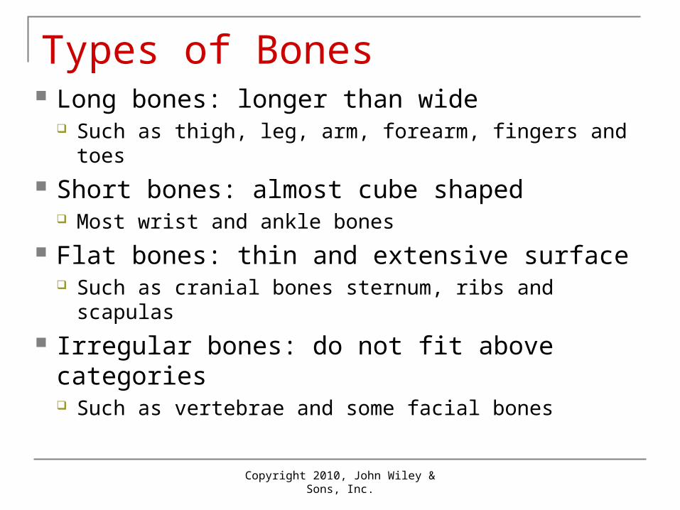
Types of Bones Long bones: longer than wide
Such as thigh, leg, arm, forearm, fingers and toes
Short bones: almost cube shaped Most wrist and ankle bones
Flat bones: thin and extensive surface Such as cranial bones sternum, ribs and scapulas
Irregular bones: do not fit above categories Such as vertebrae and some facial bones
Copyright 2010, John Wiley & Sons, Inc.
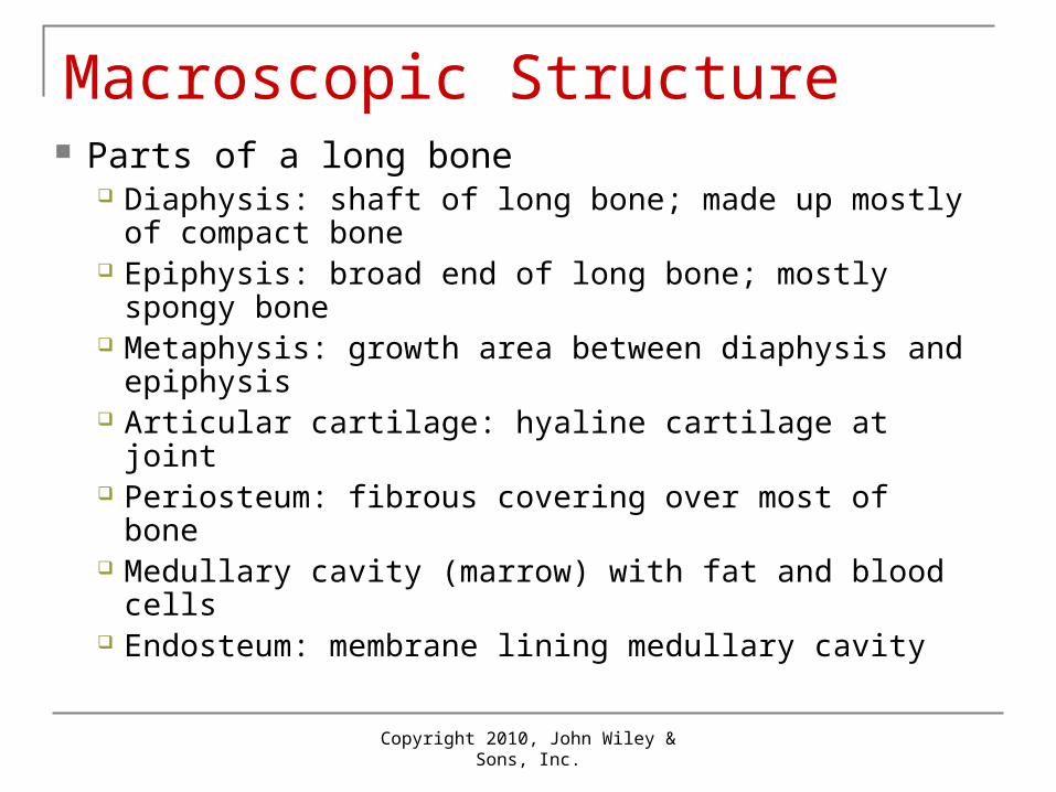
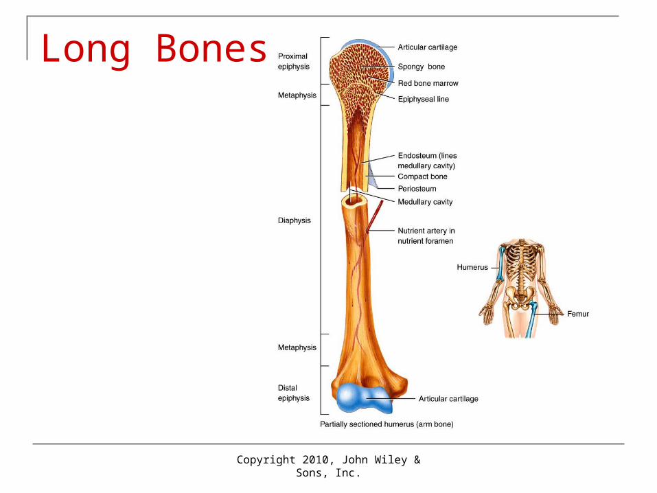
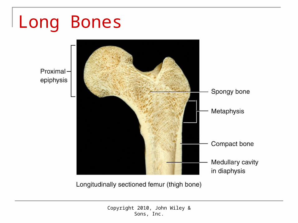
Macroscopic Structure Parts of a long bone
Diaphysis: shaft of long bone; made up mostly of compact bone
Epiphysis: broad end of long bone; mostly spongy bone
Metaphysis: growth area between diaphysis and epiphysis
Articular cartilage: hyaline cartilage at joint Periosteum: fibrous covering over most of bone Medullary cavity (marrow) with fat and blood cells Endosteum: membrane lining medullary cavity
Copyright 2010, John Wiley & Sons, Inc.
Long Bones
Copyright 2010, John Wiley & Sons, Inc.
Long Bones
Copyright 2010, John Wiley & Sons, Inc.
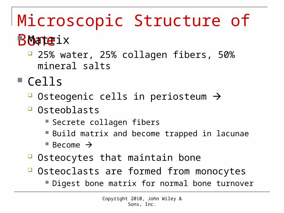
Microscopic Structure of Bone Matrix
25% water, 25% collagen fibers, 50% mineral salts
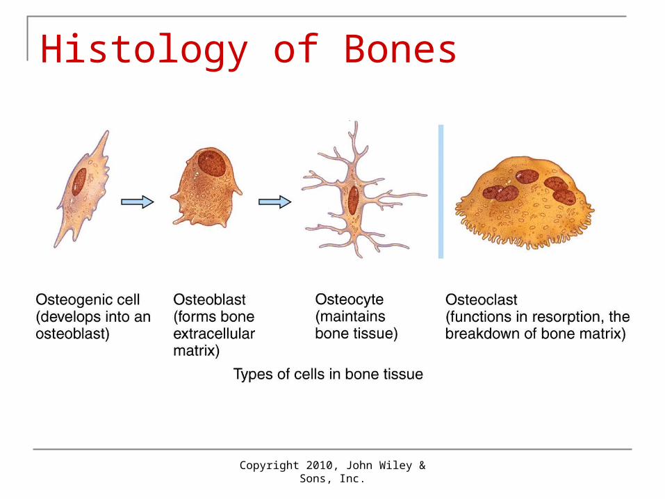
Cells Osteogenic cells in periosteum Osteoblasts
Secrete collagen fibers Build matrix and become trapped in lacunae Become
Osteocytes that maintain bone Osteoclasts are formed from monocytes
Digest bone matrix for normal bone turnover
Copyright 2010, John Wiley & Sons, Inc.
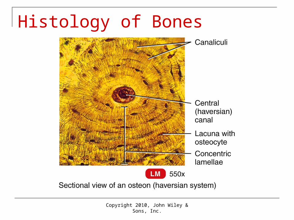
Histology of Bones
Copyright 2010, John Wiley & Sons, Inc.
Compact Bone Structure Arranged in osteons (haversian systems)
Cylinders running parallel to long axis of bone
Central canal through center of osteon Contains blood vessels, nerves, lymphatics
Concentric lamellae: layers of matrix Lacunae: “lakes” between lamellae
Contain osteocytes (bone cells)
Copyright 2010, John Wiley & Sons, Inc.
Compact Bone Structure Canaliculi (“little canals”)
Contain extensions of osteocytes Permit flow of ECF between central canal and
lacunae
Compact bone is covered by periosteum Perforating (Volkmann’s) canals
Carry blood and lymphatic vessels and nerves from periosteum
They supply central (Haversian) canals and also bone marrow
Copyright 2010, John Wiley & Sons, Inc.
Histology of Bones
Copyright 2010, John Wiley & Sons, Inc.
Spongy Bone Not arranged in osteons Irregular latticework of trabeculae
These contain lacunae with osteocytes and canaliculi
Spaces between trabeculae may contain red bone marrow
Spongy bone is lighter than compact bone, so reduces weight of skeleton
Copyright 2010, John Wiley & Sons, Inc.
Bone Dynamics and TissueInteractions Animation
Bone Dynamics and Tissue
You must be connected to the internet to run this animation.
Copyright 2010, John Wiley & Sons, Inc.
Bone Formation Known as ossification Timeline
Initial bone development in embryo and fetus Growth of bone into adulthood Remodeling: replacement of old bone Repair if fractures occur
Mesenchyme (early connective tissue) model This initial “skeleton” model will be replaced by
bone tissue beginning at 6 weeks of embryonic life
Copyright 2010, John Wiley & Sons, Inc.
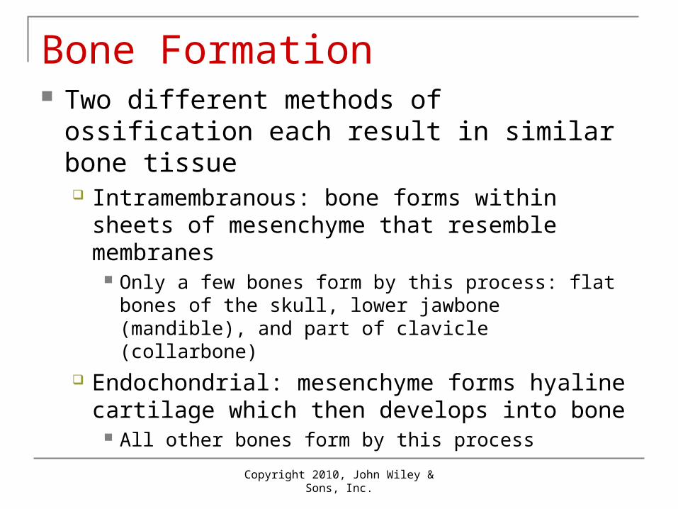
Bone Formation Two different methods of ossification each
result in similar bone tissue Intramembranous: bone forms within sheets of
mesenchyme that resemble membranes Only a few bones form by this process: flat bones of
the skull, lower jawbone (mandible), and part of clavicle (collarbone)
Endochondrial: mesenchyme forms hyaline cartilage which then develops into bone
All other bones form by this process
Copyright 2010, John Wiley & Sons, Inc.
1
Blood capillary
Ossification center
Mesenchymal cell
Osteoblast
Collagen fiber
Development of ossification center
Mandible
Flat boneof skull
1
Blood capillary
Ossification center
Mesenchymal cell
Osteoblast
Osteocyte in lacuna
Canaliculus
Osteoblast
Newly calcified bonematrix
Development of ossification center
Calcification
Mandible
Flat boneof skull
2
Collagen fiber
1
Blood capillary
Ossification center
Mesenchymal cell
Osteoblast
Development of ossification center
Calcification
Mandible
Flat boneof skull
2
Collagen fiber
Osteocyte in lacuna
Canaliculus
Osteoblast
Newly calcified bonematrix
Mesenchymecondenses
Blood vessel
Spongy bonetrabeculae
Osteoblast
Formation of trabeculae3
1
Blood capillary
Ossification center
Mesenchymal cell
Osteoblast
Mesenchymecondenses
Blood vessel
Spongy bonetrabeculae
Osteoblast
Periosteum
Spongy bone tissue
Compact bone tissue
Development of ossification center
Calcification Formation of trabeculae
Development of the periosteum
Mandible
Flat boneof skull
3
4
2
Collagen fiber
Osteocyte in lacuna
Canaliculus
Osteoblast
Newly calcified bonematrix
Copyright 2010, John Wiley & Sons, Inc.
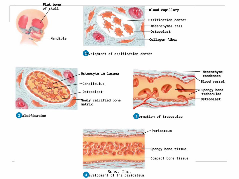
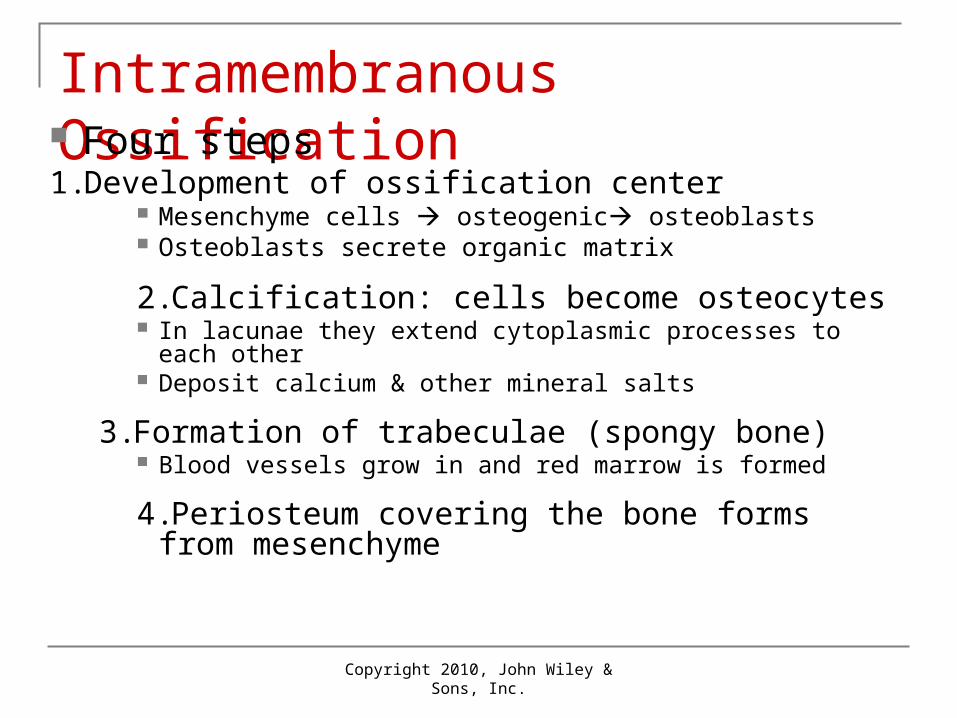
Intramembranous Ossification Four steps1. Development of ossification center
Mesenchyme cells osteogenic osteoblasts Osteoblasts secrete organic matrix
2.Calcification: cells become osteocytes In lacunae they extend cytoplasmic processes to each other Deposit calcium & other mineral salts
3.Formation of trabeculae (spongy bone) Blood vessels grow in and red marrow is formed
4.Periosteum covering the bone forms from mesenchyme
Copyright 2010, John Wiley & Sons, Inc.
1 Development ofcartilage model
Hyalinecartilage
Perichondrium
Proximalepiphysis
Distalepiphysis
Diaphysis
1 Development ofcartilage model
Growth ofcartilage model
2
Hyalinecartilage
Uncalcifiedmatrix
Calcifiedmatrix
Perichondrium
Proximalepiphysis
Distalepiphysis
Diaphysis
1 Development ofcartilage model
Development ofprimary ossificationcenter
Growth ofcartilage model
2 3
Hyalinecartilage
Uncalcifiedmatrix
Calcifiedmatrix
Nutrientartery
Perichondrium
Proximalepiphysis
Distalepiphysis
Diaphysis
Periosteum
Primaryossificationcenter
Spongybone
1
Hyalinecartilage
CalcifiedmatrixPeriosteum(covering compact bone)
Uncalcifiedmatrix
Calcifiedmatrix
Medullarycavity
Nutrientartery and vein
Nutrientartery
Perichondrium
Proximalepiphysis
Distalepiphysis
Diaphysis
Development ofcartilage model
Development ofprimary ossificationcenter
Development ofthe medullarycavity
Growth ofcartilage model
Periosteum
Primaryossificationcenter
2 3 4
Spongybone
Uncalcifiedmatrix
1 Development ofcartilage model
Development ofprimary ossificationcenter
Development ofthe medullarycavity
Growth ofcartilage model
2 3 4
Hyalinecartilage
CalcifiedmatrixPeriosteum(covering compact bone)
Uncalcifiedmatrix
Calcifiedmatrix
Medullarycavity
Nutrientartery and vein
Nutrientartery
Perichondrium
Proximalepiphysis
Distalepiphysis
Diaphysis
Periosteum
Primaryossificationcenter
Secondaryossificationcenter
Nutrientartery and vein
Uncalcifiedmatrix
Epiphysealartery andvein
Development of secondaryossification center
5
Spongybone
Uncalcifiedmatrix
1
Articular cartilage
Spongy bone
Epiphyseal plate
Secondaryossificationcenter
Nutrientartery and vein
Uncalcifiedmatrix
Epiphysealartery andvein
Formation of articular cartilageand epiphyseal plate
Development of secondaryossification center
Development ofcartilage model
Development ofprimary ossificationcenter
Development ofthe medullarycavity
Growth ofcartilage model
2 3 4
5 6
Hyalinecartilage
Uncalcifiedmatrix
CalcifiedmatrixPeriosteum(covering compact bone)
Uncalcifiedmatrix
Calcifiedmatrix
Medullarycavity
Nutrientartery and vein
Nutrientartery
Perichondrium
Proximalepiphysis
Distalepiphysis
Diaphysis
Periosteum
Primaryossificationcenter
Spongybone
Copyright 2010, John Wiley & Sons, Inc.
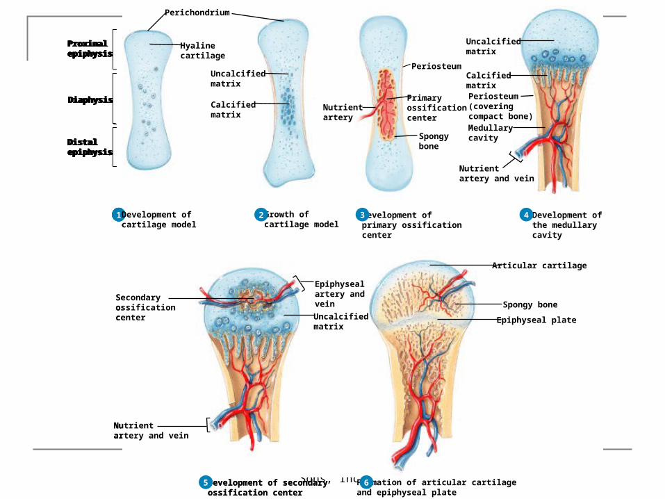
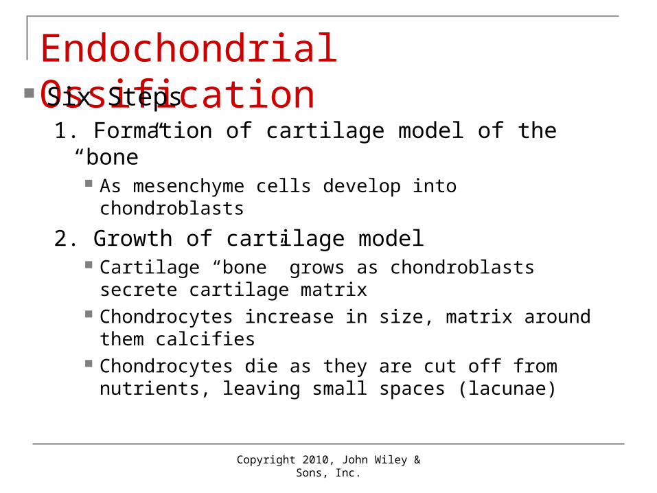
Endochondrial Ossification Six Steps
1. Formation of cartilage model of the “bone” As mesenchyme cells develop into chondroblasts
2. Growth of cartilage model Cartilage “bone” grows as chondroblasts secrete
cartilage matrix Chondrocytes increase in size, matrix around them
calcifies Chondrocytes die as they are cut off from nutrients,
leaving small spaces (lacunae)
Copyright 2010, John Wiley & Sons, Inc.
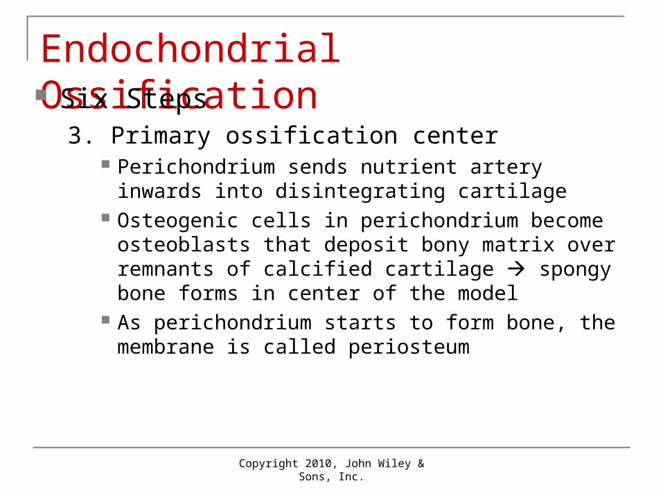
Endochondrial Ossification Six Steps
3. Primary ossification center Perichondrium sends nutrient artery inwards into
disintegrating cartilage Osteogenic cells in perichondrium become osteoblasts
that deposit bony matrix over remnants of calcified cartilage spongy bone forms in center of the model
As perichondrium starts to form bone, the membrane is called periosteum
Copyright 2010, John Wiley & Sons, Inc.
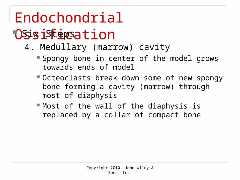
Endochondrial Ossification Six Steps
4. Medullary (marrow) cavity Spongy bone in center of the model grows towards
ends of model Octeoclasts break down some of new spongy bone
forming a cavity (marrow) through most of diaphysis Most of the wall of the diaphysis is replaced by a collar
of compact bone

Copyright 2010, John Wiley & Sons, Inc.
Endochondrial Ossification Six Steps
5. Secondary ossification center Similar to step 3 except that nutrient arteries enter ends
(epiphyses) of bones and osteoblasts deposit bony matrix spongy bone forms in epiphyses from center outwards
Occurs about time of birth
6. Articular cartilage and epiphyseal cartilage Articular cartilage at ends of epiphyses becomes
articular cartilage Epiphyseal (growth) plate of cartilage remains between
epiphysis and diaphysis until bone growth ceases
Copyright 2010, John Wiley & Sons, Inc.
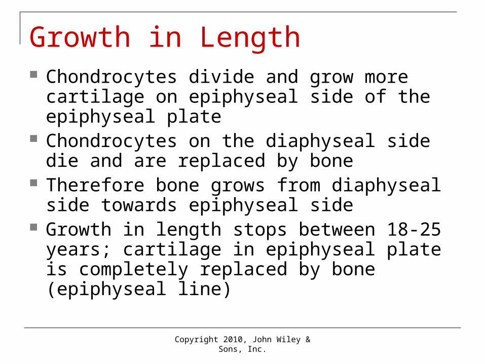
Growth in Length Chondrocytes divide and grow more cartilage
on epiphyseal side of the epiphyseal plate Chondrocytes on the diaphyseal side die and
are replaced by bone Therefore bone grows from diaphyseal side
towards epiphyseal side Growth in length stops between 18-25 years;
cartilage in epiphyseal plate is completely replaced by bone (epiphyseal line)
Copyright 2010, John Wiley & Sons, Inc.
Growth in Thickness As bones grow in length, they must
also grow in thickness (width) Perichondrial osteoblasts osteoblasts
lay down additional lamellae of compact bone
Simultaneously, osteoclasts in the endosteum destroy interior bone to increase width of the marrow
Copyright 2010, John Wiley & Sons, Inc.
Remodeling and Repair Remodeling in response to use
Resorption by osteoclasts and Deposition by osteoblasts
Repair after a fracture Dead tissue removed Chondroblasts fibrocartilage
spongy bone deposited by osteoblasts remodeled to compact bone
Copyright 2010, John Wiley & Sons, Inc.
Types of Fractures Partial: incomplete break (crack) Complete: bone broken into two or more
pieces Closed (simple): not through skin Open (compound): broken ends break skin
Copyright 2010, John Wiley & Sons, Inc.
Factors Affecting Growth Adequate minerals (Ca, P, Mg) Vitamins A, C, D Hormones
Before puberty: hGH + insulin-like growth factors Thyroid hormone and insulin also required Sex hormones contribute to adolescent growth
spurt Weight-bearing activity
Copyright 2010, John Wiley & Sons, Inc.
Calcium Homeostasis Blood levels of Ca2+ controlled Negative feedback loops Parathyroid hormone (PTH) increases
osteoclast activity + decreases loss of Ca2+ in urine
Calcitonin decreases osteoclast activity

Copyright 2010, John Wiley & Sons, Inc.
Negative Feedback
Copyright 2010, John Wiley & Sons, Inc.
Exercise & Bone Tissue Bone strengthened in response to use Bone resorbed during disuse; examples:
During prolonged bed rest Fracture with cast/immobilizer Astronauts without gravity
Copyright 2010, John Wiley & Sons, Inc.
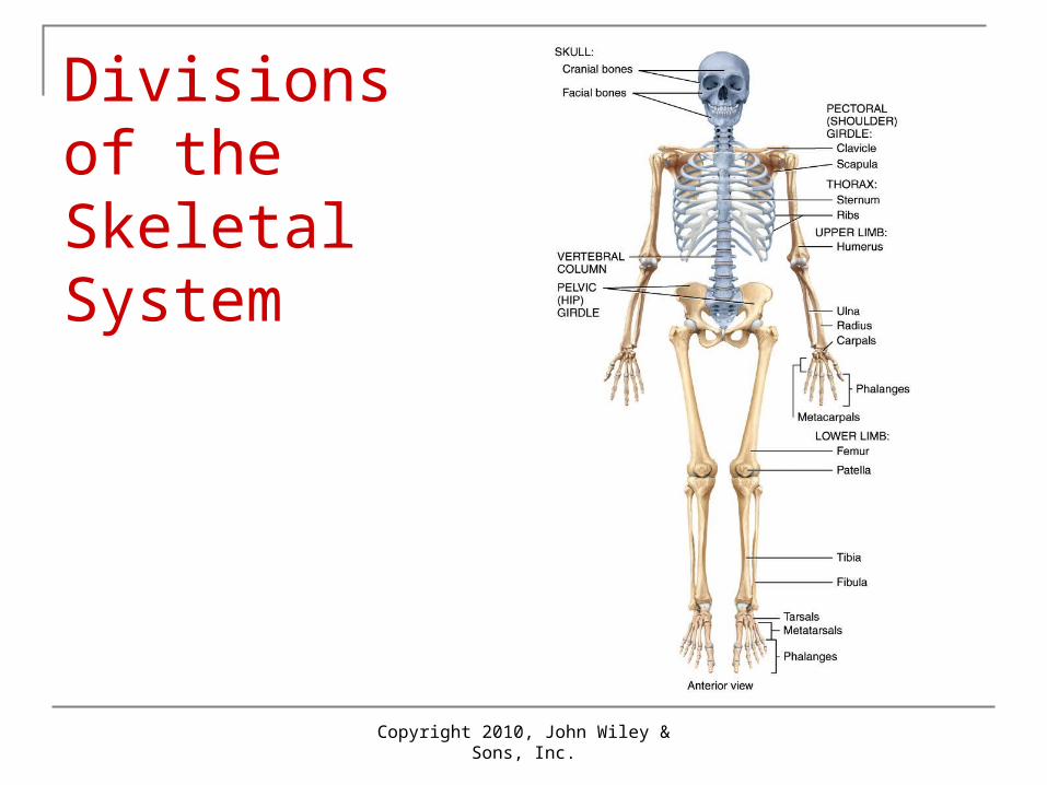
Divisions of Skeletal System Two divisions: axial and appendicular
Axial: bones around body axis Examples: skull bones, hyoid, ribs, sternum, vertebrae
Appendicular: bones of upper and lower limbs plus shoulder and hip bones that connect them Examples: collar bone (clavicle), arm (humerus),
forearm (radius and ulna), thigh bone (femur)
Copyright 2010, John Wiley & Sons, Inc.
Divisions of the Skeletal System
Copyright 2010, John Wiley & Sons, Inc.
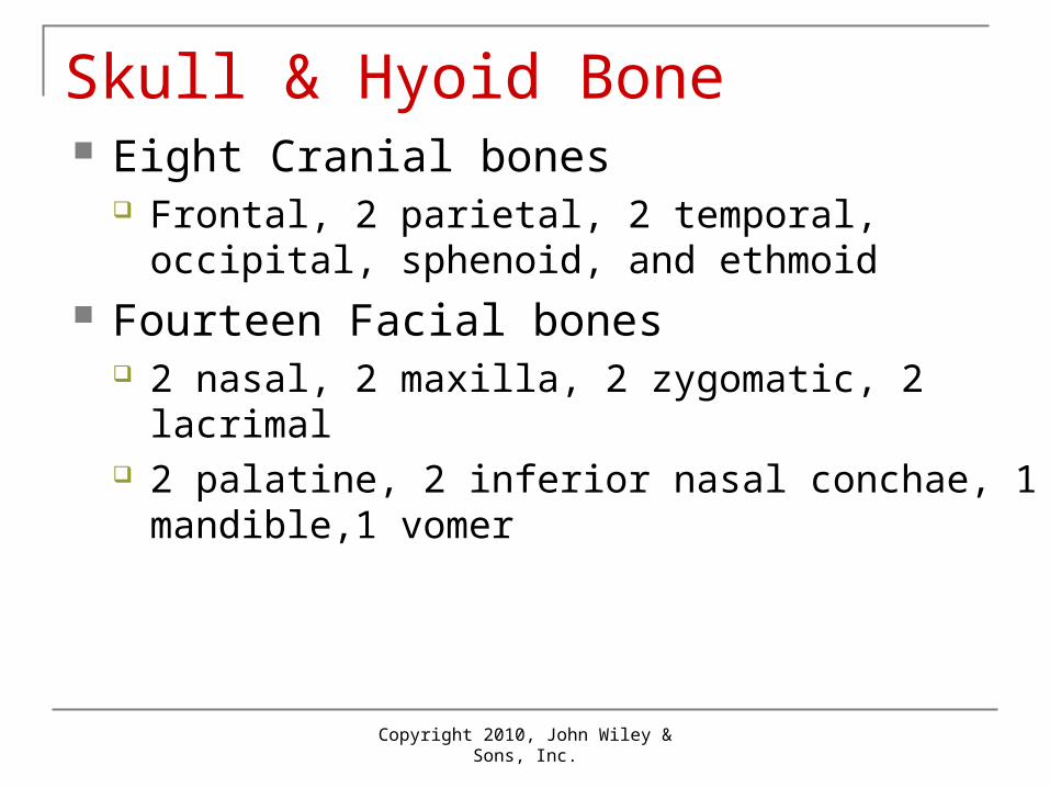
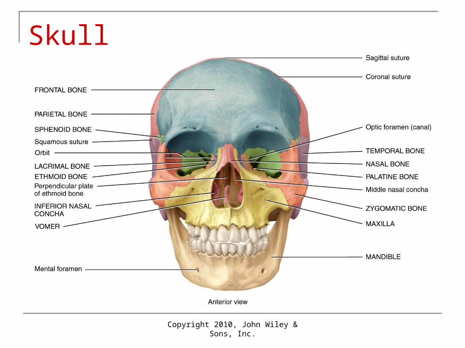
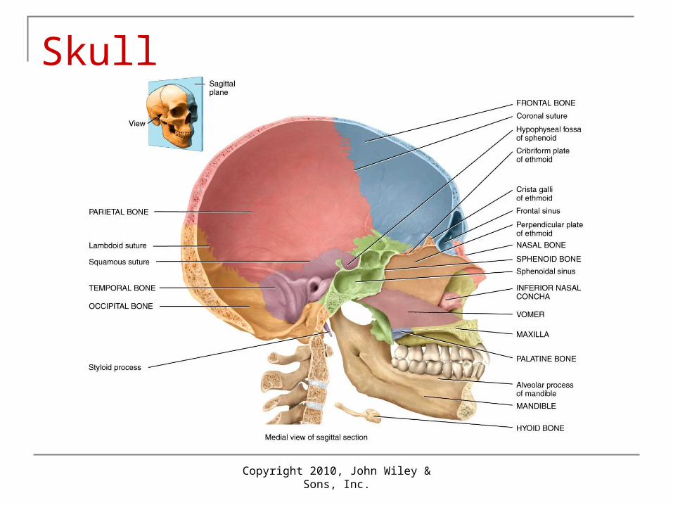
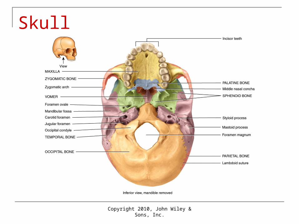
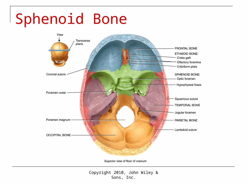
Skull & Hyoid Bone Eight Cranial bones
Frontal, 2 parietal, 2 temporal, occipital, sphenoid, and ethmoid
Fourteen Facial bones 2 nasal, 2 maxilla, 2 zygomatic, 2 lacrimal 2 palatine, 2 inferior nasal conchae, 1 mandible,1
vomer
Copyright 2010, John Wiley & Sons, Inc.
Skull

Copyright 2010, John Wiley & Sons, Inc.
Skull
Copyright 2010, John Wiley & Sons, Inc.
Skull
Copyright 2010, John Wiley & Sons, Inc.
Skull
Copyright 2010, John Wiley & Sons, Inc.
Sphenoid Bone
Copyright 2010, John Wiley & Sons, Inc.
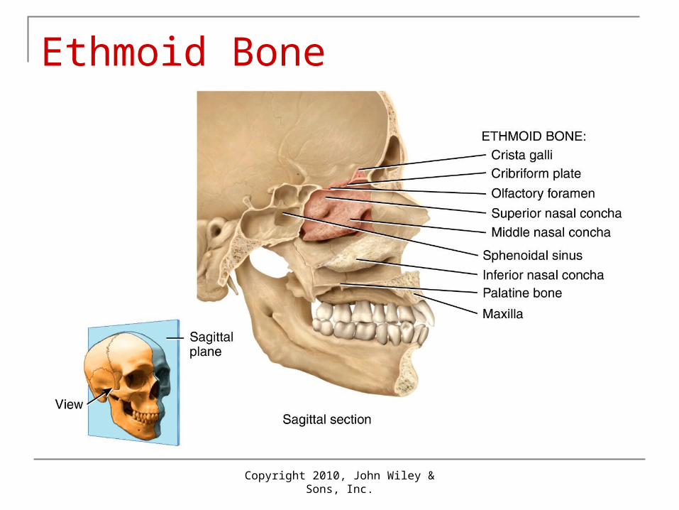
Ethmoid Bone
Copyright 2010, John Wiley & Sons, Inc.
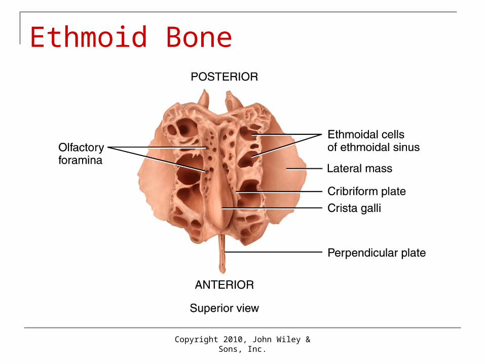
Ethmoid Bone
Copyright 2010, John Wiley & Sons, Inc.
Unique Features of Skull Sutures: immovable joint between skull
bones Coronal, sagittal, lambdoidal, squamous
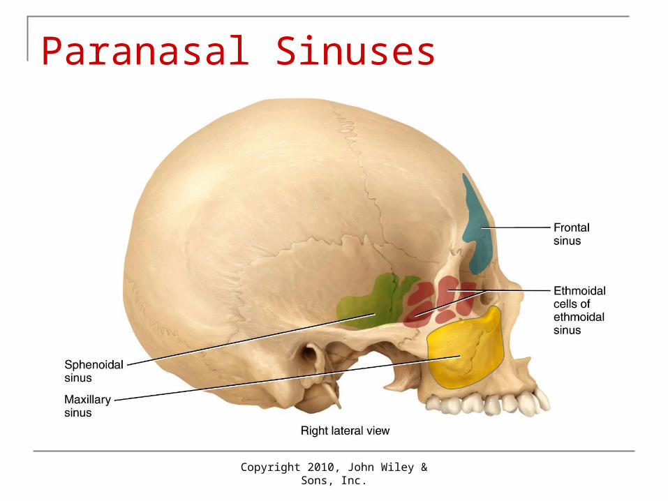
Paranasal sinuses: cavities Located in bones near nasal cavity
Fontanels: soft spot in fetal skull Allow deformation at birth Calcify to form sutures
Copyright 2010, John Wiley & Sons, Inc.
Paranasal Sinuses
Copyright 2010, John Wiley & Sons, Inc.
Vertebrae Functions
Encloses spinal cord Supports head Point of attachment for muscles of back, ribs
and pelvic girdle Regions (from superior to inferior)
7 cervical 12 thoracic 5 lumbar 1 sacrum and 1 coccyx
Copyright 2010, John Wiley & Sons, Inc.
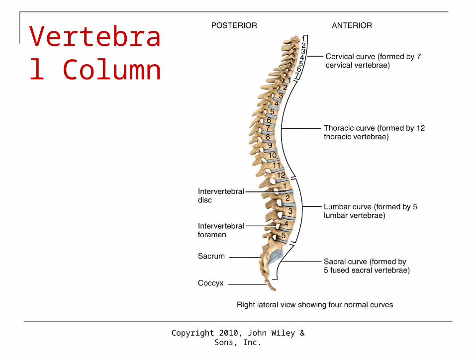
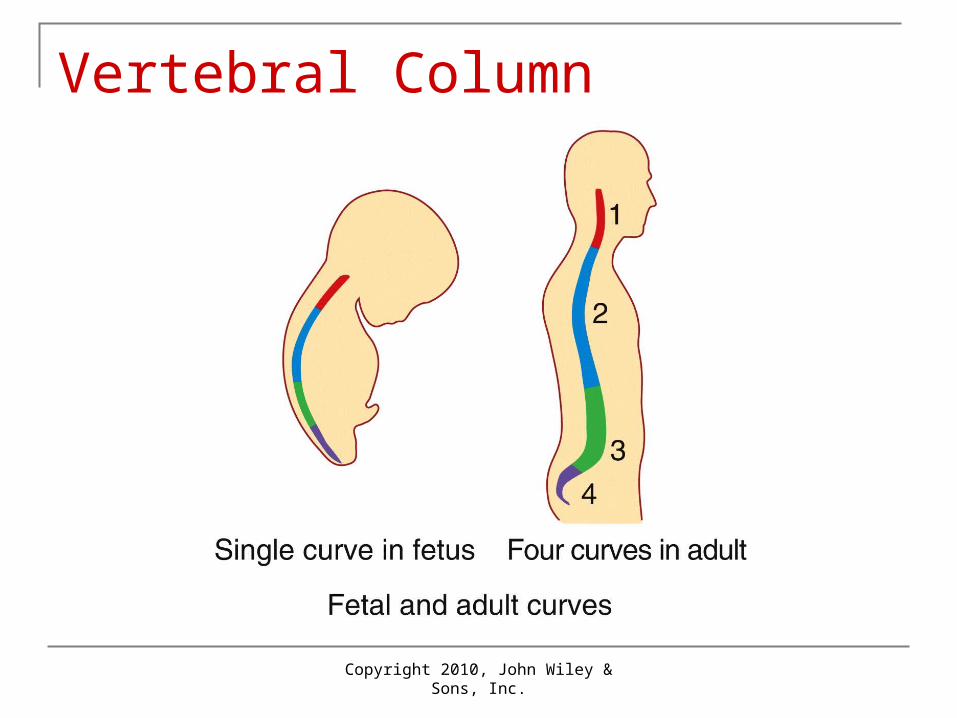
Normal Curves in Column Four normal curves
Cervical and lumbar curves are convex (bulge anteriorly)
Thoracic and sacral curves are concave (bulge posteriorly)
Curves increase strength, help in balance and absorb shocks
Copyright 2010, John Wiley & Sons, Inc.
Vertebral Column
Copyright 2010, John Wiley & Sons, Inc.
Vertebral Column
Copyright 2010, John Wiley & Sons, Inc.
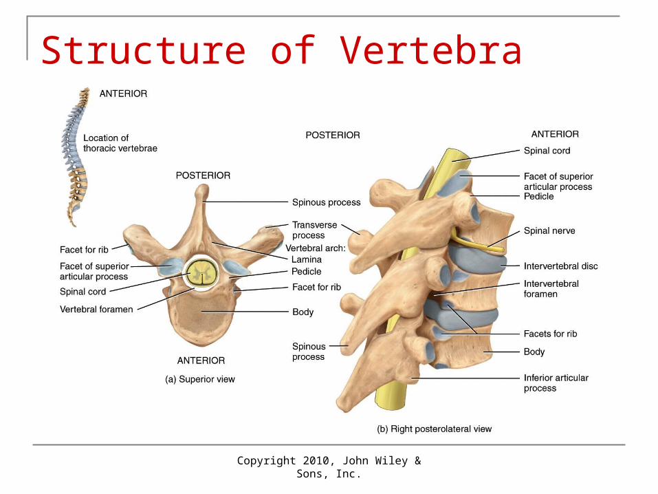
Structure of Vertebra Body: disc-shaped anterior portion Vertebral arch: posteriorly back from body
With the body, creates a hole called vertebral foramen Seven processes from this arch
Transverse process extending laterally on each side Spinous process extending dorsally Two each of superior and inferior articular processes
that form joints with vertebrae
Copyright 2010, John Wiley & Sons, Inc.
Structure of Vertebra
Copyright 2010, John Wiley & Sons, Inc.
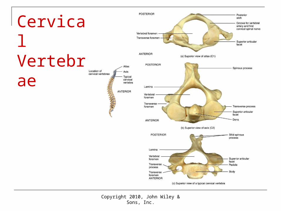
Cervical Area Cervical (C1-C7 from superior to inferior)
Spinous process often bifid with transverse foramina on transverse processes
C1: atlas Articulates with head, specialized to support head Lacks body and spinous process
C2: axis Has body and spinous process Called dens (“tooth”) that creates a pivot for head
rotation
Copyright 2010, John Wiley & Sons, Inc.
Cervical Vertebrae
Copyright 2010, John Wiley & Sons, Inc.
Other Vertebrae Thoracic (T1-T12 )
Larger than cervical Have facets for articulations with ribs
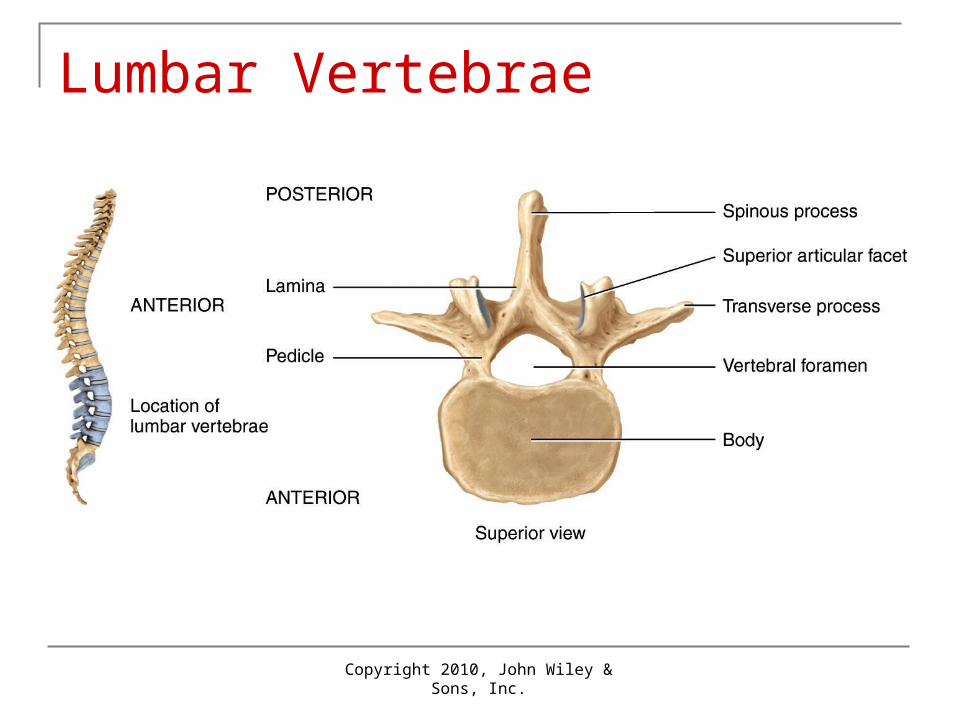
Lumbar (L1-L5) Largest and strongest; spinous processes short and
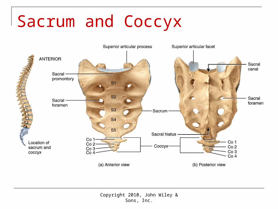
thick Sacrum (S1-S5 fused into one unit)
Foundation for pelvic girdle Contain sacral foramina for nerves and blood vessels
Coccyx: 4 coccygeal vertebrae fused into 1
Copyright 2010, John Wiley & Sons, Inc.
Lumbar Vertebrae
Copyright 2010, John Wiley & Sons, Inc.
Sacrum and Coccyx

Copyright 2010, John Wiley & Sons, Inc.
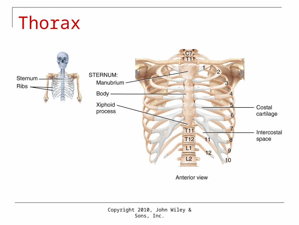
Thorax Thoracic cage: sternum, costal cartilages,
ribs and bodies of T1-T12 Sternum: form by 3 portions fused by about
age 25 years: Manubrium, body, xiphoid process
Ribs: 12 pairs True ribs are #1-7: articulate with sternum
directly by costal cartilages False ribs are #8-12: do not articulate with
sternum directly by costal cartilages
Copyright 2010, John Wiley & Sons, Inc.
Thorax
Copyright 2010, John Wiley & Sons, Inc.
Pectoral Girdle Function: attach bones of upper limbs to axial
skeleton Clavicles and scapulas: bilateral

Copyright 2010, John Wiley & Sons, Inc.
Right Pectoral (Shoulder) Girdle
Copyright 2010, John Wiley & Sons, Inc.
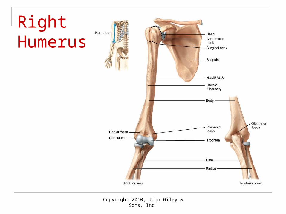
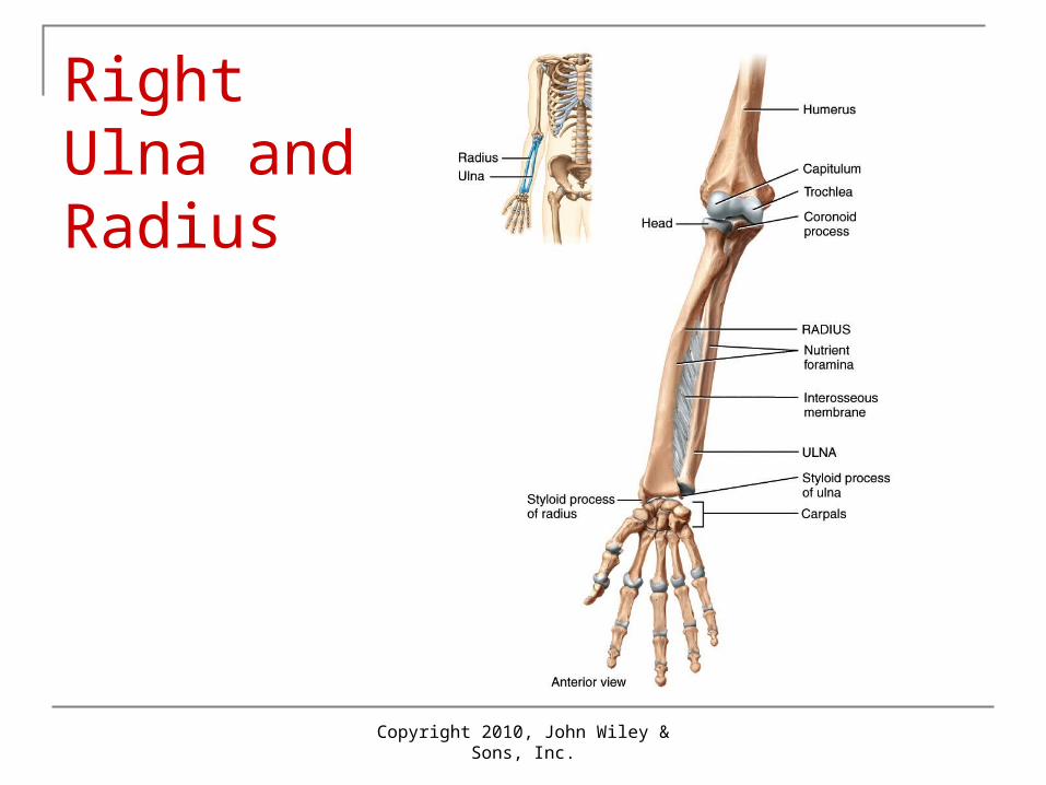
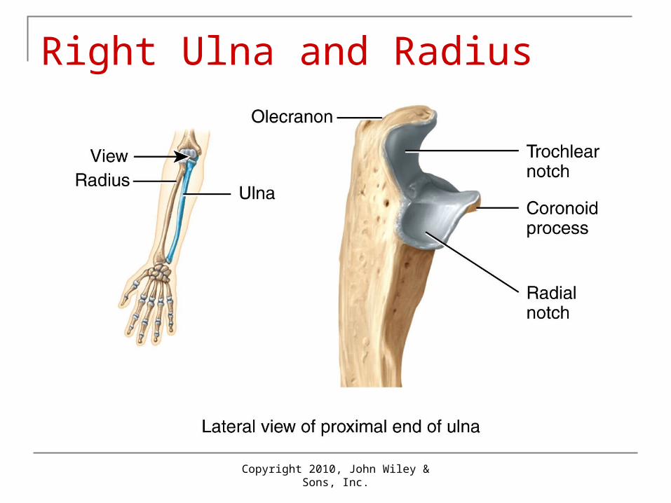
Upper Limb Humerus: arm bone
Articulates with scapula (glenoid cavity) at shoulder joint
Articulates with radius and ulna at elbow Ulna: medial bone Radius: lateral bone (thumb side)
Copyright 2010, John Wiley & Sons, Inc.
Right Humerus
Copyright 2010, John Wiley & Sons, Inc.
Right Ulna and Radius
Copyright 2010, John Wiley & Sons, Inc.
Right Ulna and Radius
Copyright 2010, John Wiley & Sons, Inc.
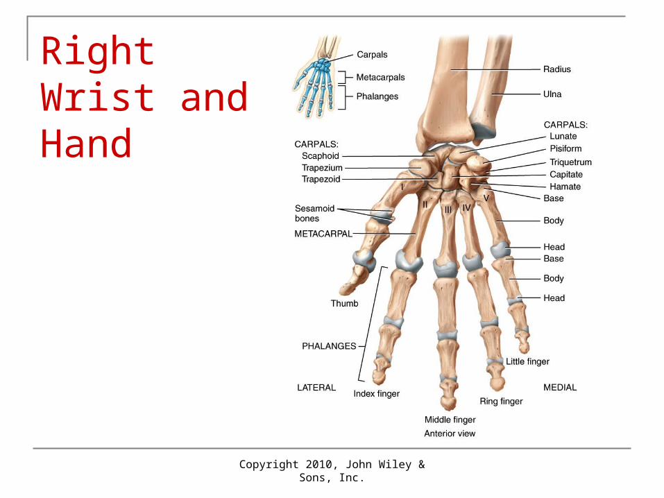
Wrist and Hand Carpus (wrist): 8 bones Metacarpals: 5 bones of palm of hand
Number 1-5 starting with thumb Phalanges: 14 bones of fingers
Numbered 1-5 metacarpals Each finger except the thumb has proximal,
middle and distal phalanges; thumb lacks middle phalanx
Copyright 2010, John Wiley & Sons, Inc.
Right Wrist and Hand
Copyright 2010, John Wiley & Sons, Inc.
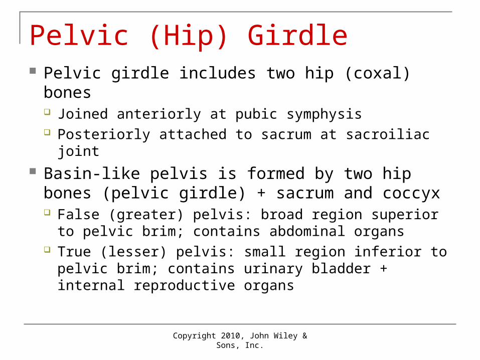
Pelvic (Hip) Girdle Pelvic girdle includes two hip (coxal) bones
Joined anteriorly at pubic symphysis Posteriorly attached to sacrum at sacroiliac joint
Basin-like pelvis is formed by two hip bones (pelvic girdle) + sacrum and coccyx False (greater) pelvis: broad region superior to
pelvic brim; contains abdominal organs True (lesser) pelvis: small region inferior to pelvic
brim; contains urinary bladder + internal reproductive organs
Copyright 2010, John Wiley & Sons, Inc.
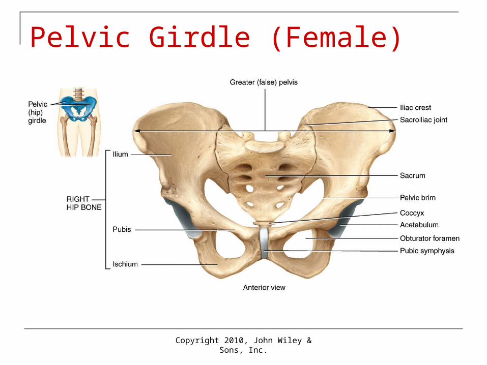
Pelvic Girdle (Female)
Copyright 2010, John Wiley & Sons, Inc.
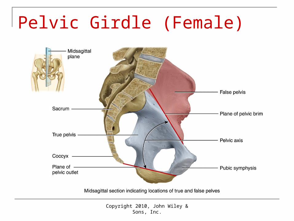
Pelvic Girdle (Female)
Copyright 2010, John Wiley & Sons, Inc.
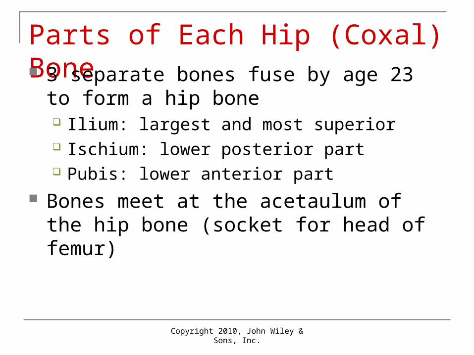
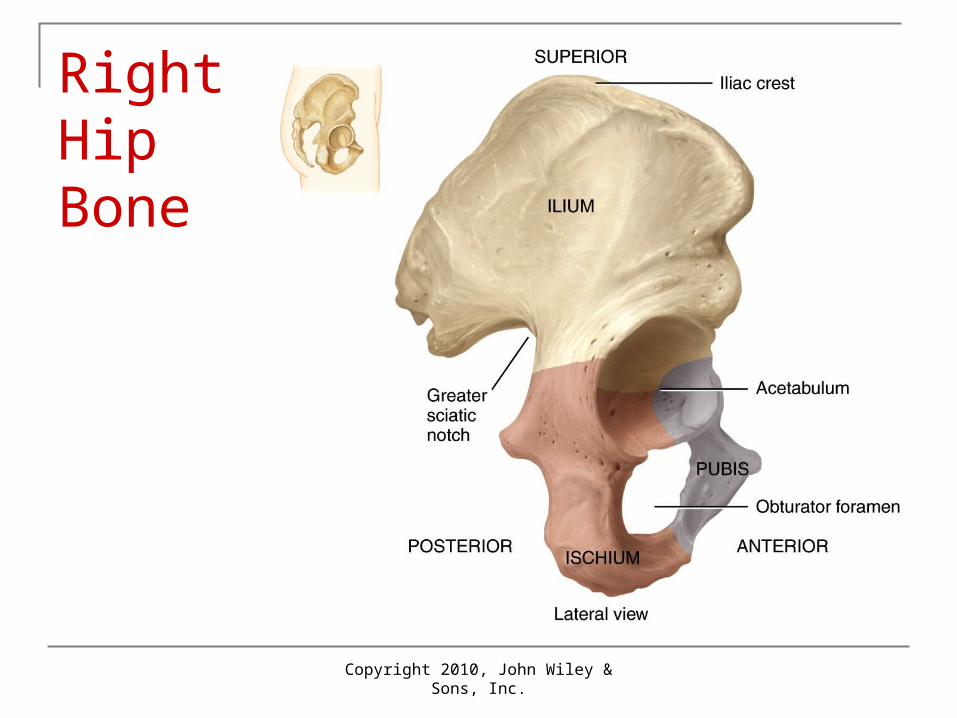
Parts of Each Hip (Coxal) Bone 3 separate bones fuse by age 23 to form a
hip bone Ilium: largest and most superior Ischium: lower posterior part Pubis: lower anterior part
Bones meet at the acetaulum of the hip bone (socket for head of femur)
Copyright 2010, John Wiley & Sons, Inc.
Right Hip Bone
Copyright 2010, John Wiley & Sons, Inc.
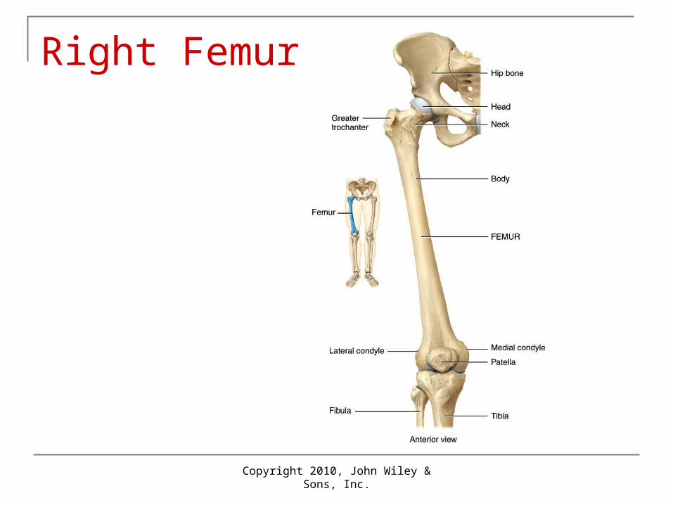
Lower Limb Femur (thigh bone): largest bone in the body
Articulates with hip proximally and with the tibia and patella distally
Head (fits into acetabulum) and greater trochanter at proximal end
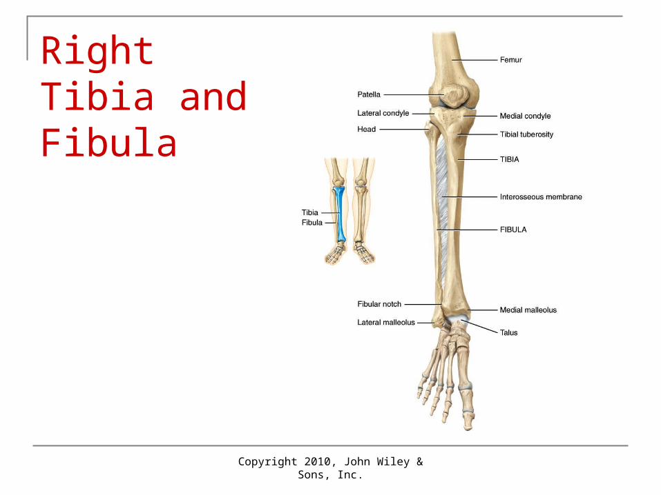
Patella: kneecap in anterior of knee joint Tibia: shin bone
Large medial, weight-bearing bone of leg
Fibula: longest, thinnest bone in body Lateral to tibia and smaller Does not articulate with femur
Copyright 2010, John Wiley & Sons, Inc.
Right Femur
Copyright 2010, John Wiley & Sons, Inc.
Right Tibia and Fibula
Copyright 2010, John Wiley & Sons, Inc.
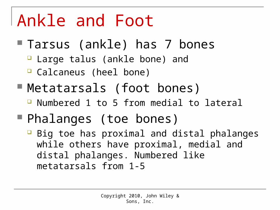
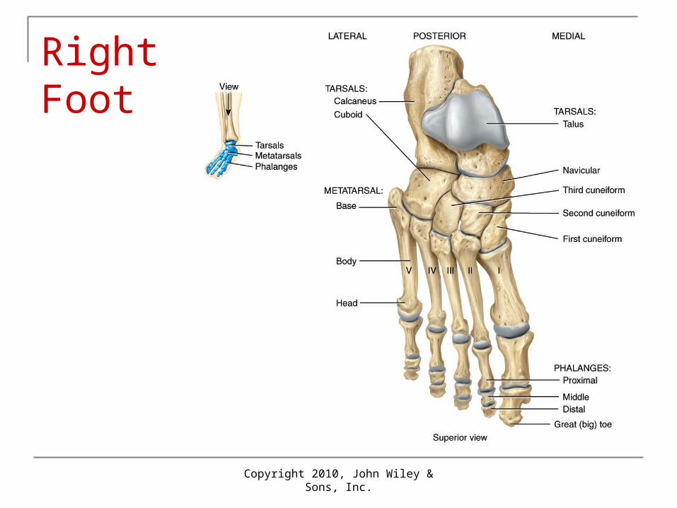
Ankle and Foot Tarsus (ankle) has 7 bones
Large talus (ankle bone) and Calcaneus (heel bone)
Metatarsals (foot bones) Numbered 1 to 5 from medial to lateral
Phalanges (toe bones) Big toe has proximal and distal phalanges while others
have proximal, medial and distal phalanges. Numbered like metatarsals from 1-5
Copyright 2010, John Wiley & Sons, Inc.
Right Foot
Copyright 2010, John Wiley & Sons, Inc.
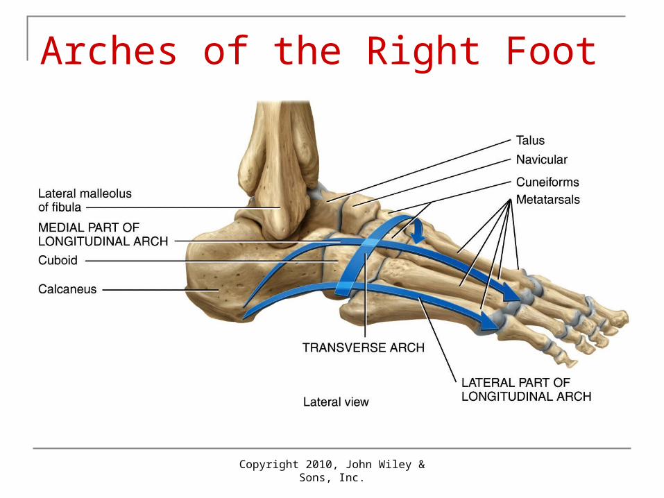
Arches of the Right Foot
Copyright 2010, John Wiley & Sons, Inc.
Male and Female Differences Males usually have heavier bones Related to muscle size and strength Female pelvis is wider and shallower than
male pelvis: allows for birth
Copyright 2010, John Wiley & Sons, Inc.
Aging and Skeletal System Birth through adolescence: more bone
formed than lost Young adults: gain and loss about equal As levels of sex steroids decline with age:
bone resorption > bone formation Bones become brittle and lose calcium
Copyright 2010, John Wiley & Sons, Inc.
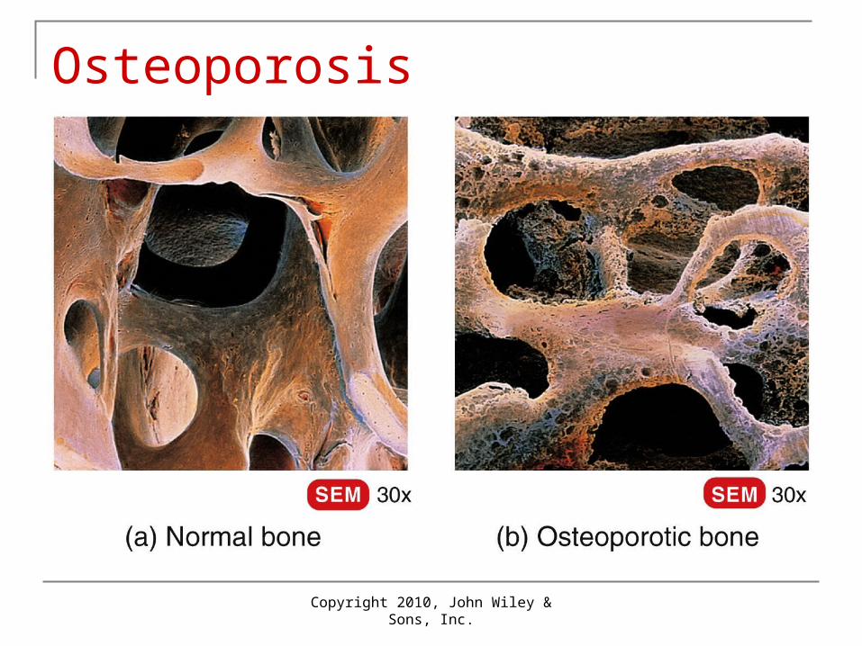
Osteoporosis