Embed Size (px)
Citation preview
Copyright © 2009 Pearson Education, Inc.
Figure 8.1 The structures of blood vessels in the human body.
Copyright © 2009 Pearson Education, Inc.
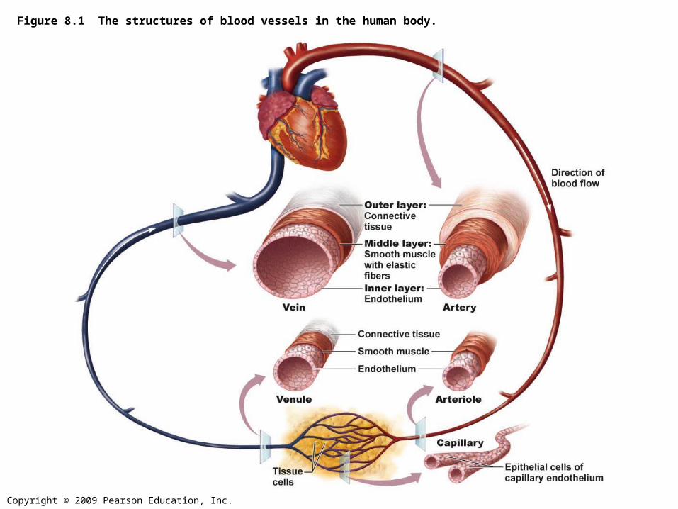
Figure 8.1 The structures of blood vessels in the human body.
Inner layer:_________
Middle layer:_________
Outer layer:_________
Connective tissue
Endothelium
______
________
________
________
_______
Direction ofblood flow
Tissuecells
_________
Smooth muscle
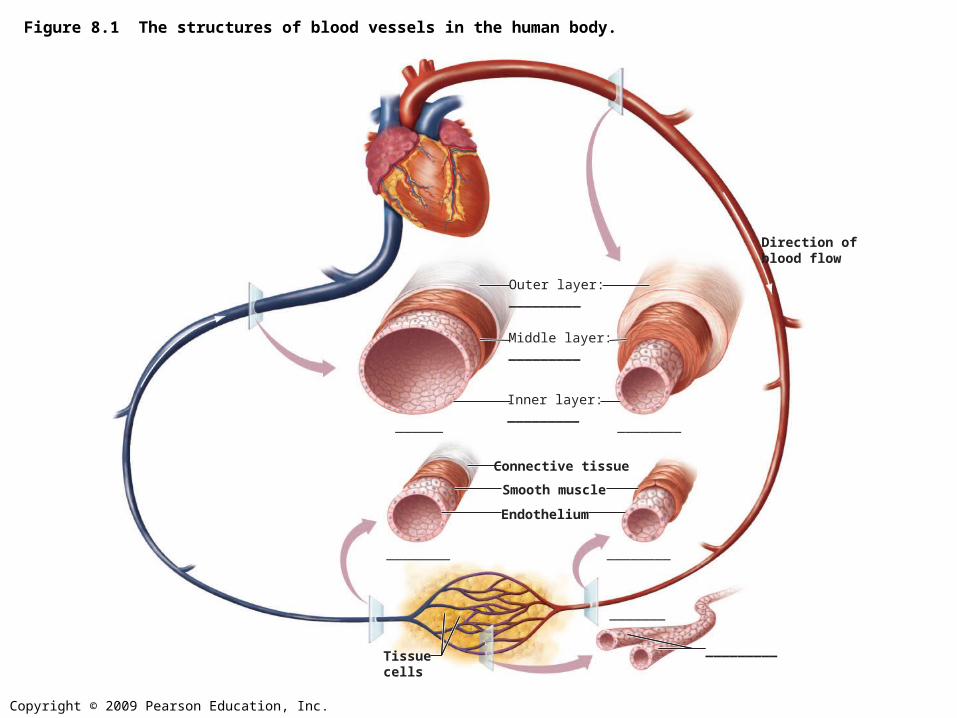
Tunics
• Tunica interna = tunica intima = inner layer– Endothelial layer that lines the lumen of all
vessels• Tunica media = middle layer
– Smooth muscle and elastic fiber layer, controls constriction/dilation of vessels
• Tunica externa = tunica adventitia = outer layer– Contains collagen fibers that protect and
reinforce vessels

Lumen
central blood-containing space surrounded by tunics
Copyright © 2009 Pearson Education, Inc.
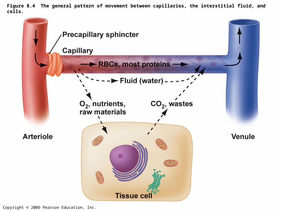
Figure 8.4 The general pattern of movement between capillaries, the interstitial fluid, and cells.
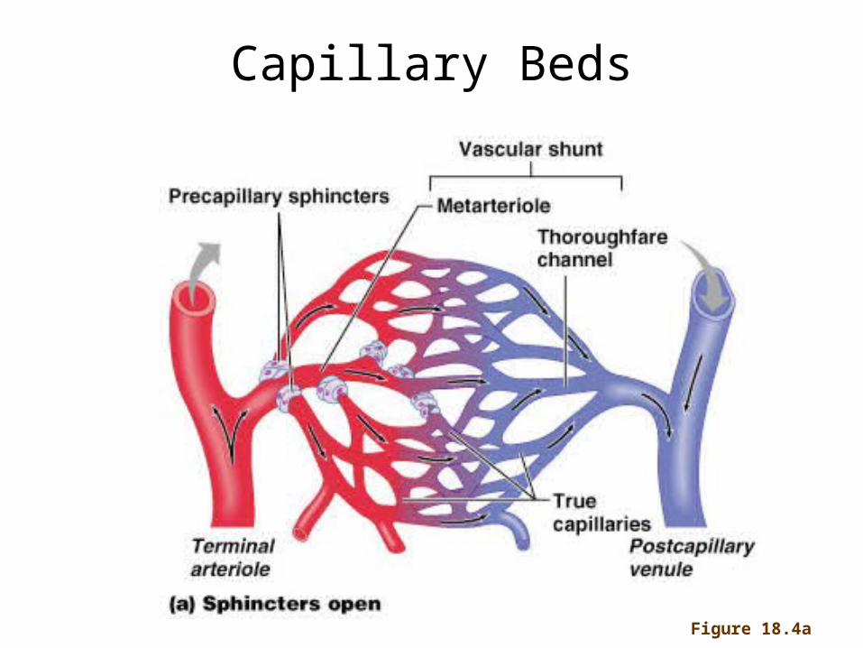
Capillary Beds
Figure 18.4a
Copyright © 2009 Pearson Education, Inc.
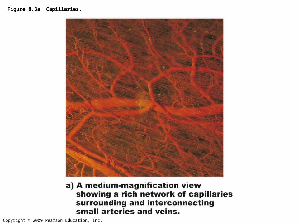
Figure 8.3a Capillaries.
Copyright © 2009 Pearson Education, Inc.
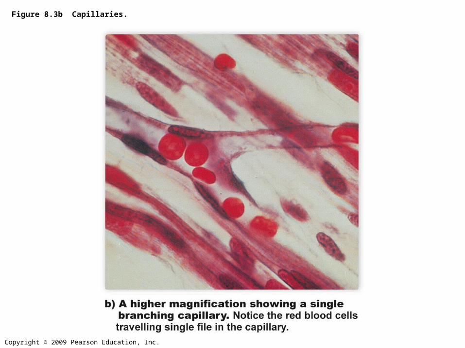
Figure 8.3b Capillaries.
Copyright © 2009 Pearson Education, Inc.
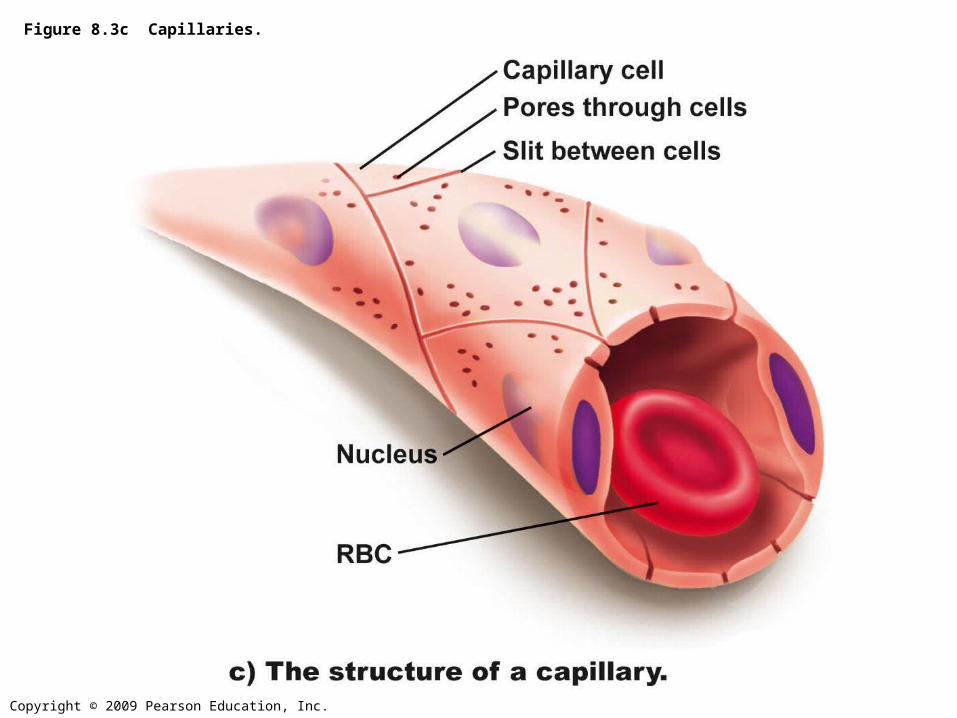
Figure 8.3c Capillaries.
• Veins have much lower blood pressure and thinner walls than arteries; larger lumen
• To return blood to the heart, veins have special adaptations
– Large-diameter lumens, offer little resistance to flow
– Valves which prevent backflow of blood – Respiratory “pump” – pressure changes created during
breathing suck blood toward the heart by squeezing local veins
– Muscular “pump” – contraction of skeletal muscles “milk” blood toward the heart
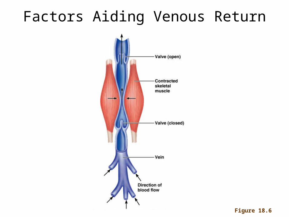
Factors Aiding Venous Return
Figure 18.6
Fetal Circulation 1
• Umbilical vein carries blood (highly oxygenated) from the placenta to the fetal heart.
• Umbilical arteries carry blood from fetal heart to placenta.
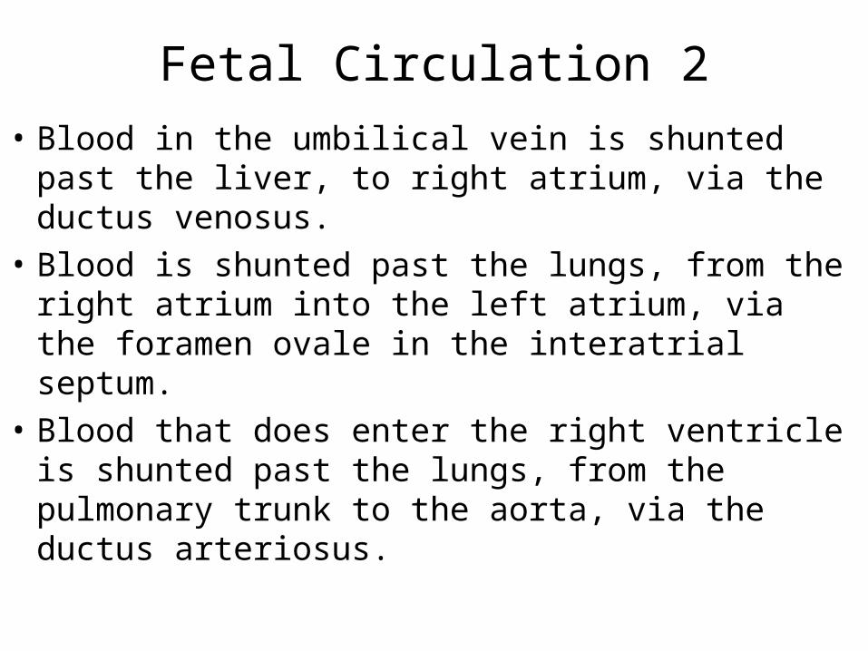
Fetal Circulation 2
• Blood in the umbilical vein is shunted past the liver, to right atrium, via the ductus venosus.
• Blood is shunted past the lungs, from the right atrium into the left atrium, via the foramen ovale in the interatrial septum.
• Blood that does enter the right ventricle is shunted past the lungs, from the pulmonary trunk to the aorta, via the ductus arteriosus.
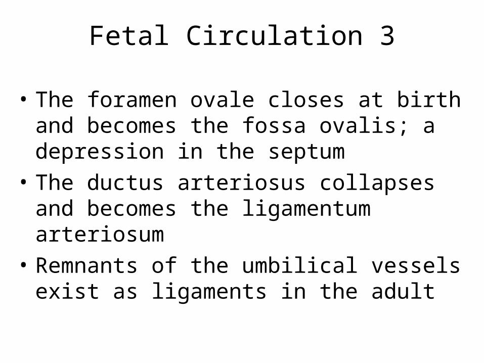
Fetal Circulation 3
• The foramen ovale closes at birth and becomes the fossa ovalis; a depression in the septum
• The ductus arteriosus collapses and becomes the ligamentum arteriosum
• Remnants of the umbilical vessels exist as ligaments in the adult

Microscope slides
Copyright © 2009 Pearson Education, Inc.
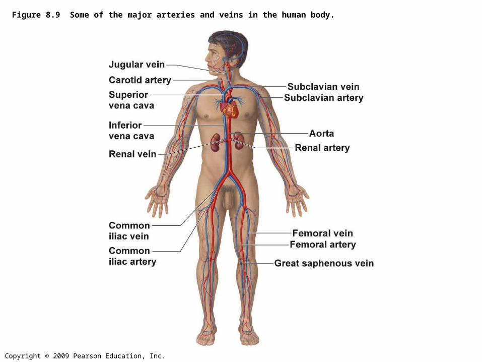
Figure 8.9 Some of the major arteries and veins in the human body.
Copyright © 2009 Pearson Education, Inc.
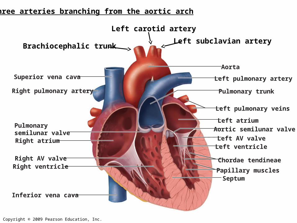
Superior vena cava
Pulmonarysemilunar valveRight atrium
Right AV valveRight ventricle
Inferior vena cava
Pulmonary trunk
Left pulmonary veins
Left atrium
Left AV valve
Aortic semilunar valve
Chordae tendineae
Papillary muscles
Left ventricle
Septum
Right pulmonary artery
Left pulmonary artery
Aorta
Brachiocephalic trunkLeft subclavian artery
Left carotid artery
Three arteries branching from the aortic arch
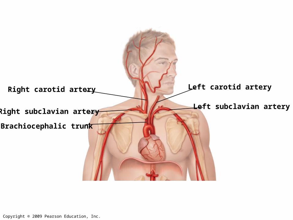
Copyright © 2009 Pearson Education, Inc.
Brachiocephalic trunk
Left carotid arteryRight carotid artery
Left subclavian arteryRight subclavian artery
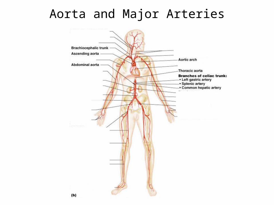
Aorta and Major Arteries
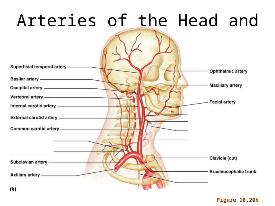
Figure 18.20b
Arteries of the Head and Neck
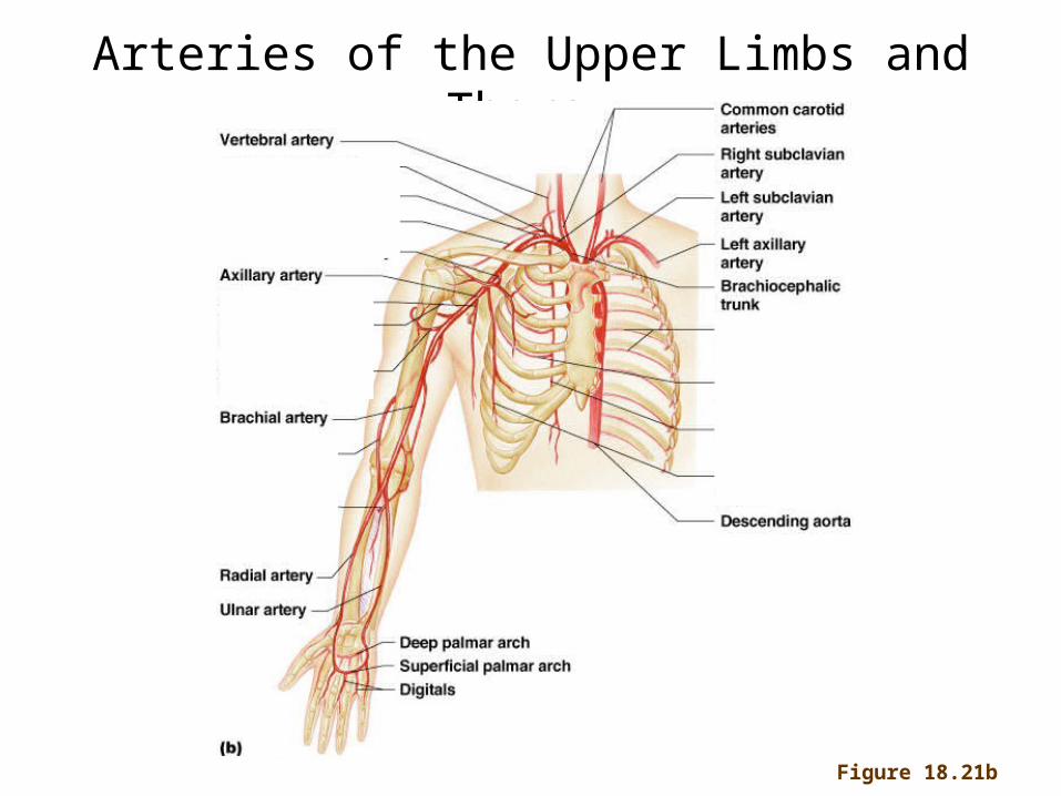
Figure 18.21b
Arteries of the Upper Limbs and Thorax
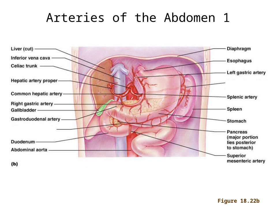
Figure 18.22b
Arteries of the Abdomen 1
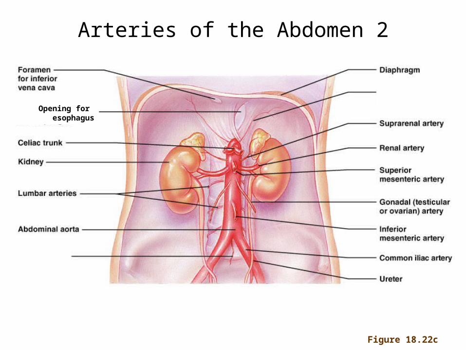
Figure 18.22c
Arteries of the Abdomen 2
Opening for esophagus
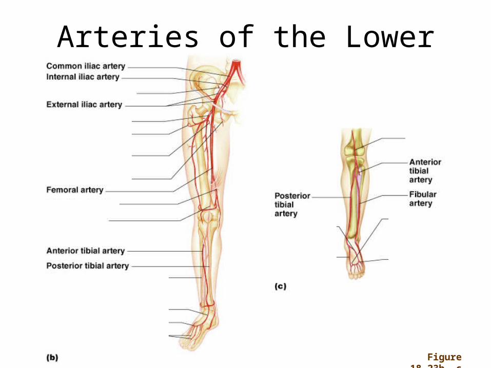
Arteries of the Lower Limbs
Figure 18.23b, c
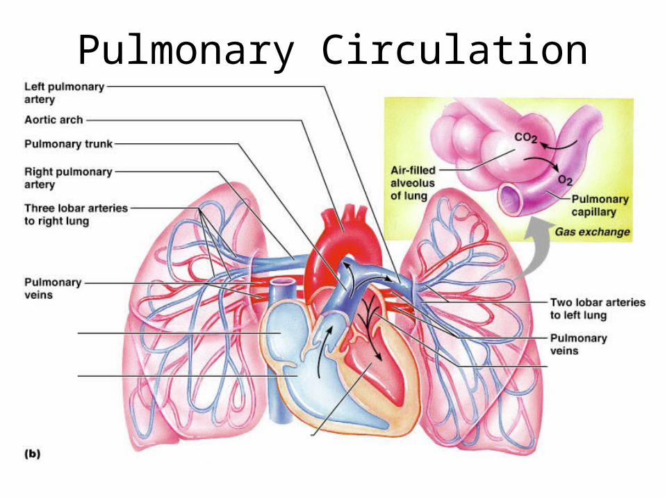
Pulmonary Circulation
Copyright © 2009 Pearson Education, Inc.
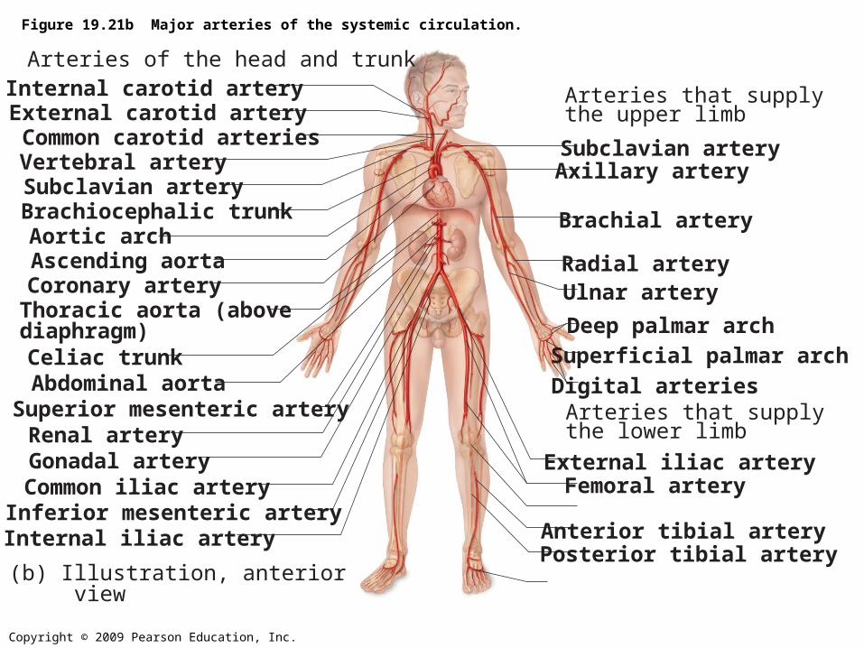
Figure 19.21b Major arteries of the systemic circulation.
Internal carotid artery
Common carotid arteries
Subclavian artery
Subclavian artery
Aortic archAscending aortaCoronary arteryThoracic aorta (abovediaphragm)
Renal artery
Superficial palmar arch
Radial arteryUlnar artery
Internal iliac artery
Deep palmar arch
Vertebral artery
Brachiocephalic trunk
Axillary artery
Brachial artery
Abdominal aortaSuperior mesenteric artery
Gonadal arteryCommon iliac artery
External iliac artery
Digital arteries
Femoral artery
Anterior tibial arteryPosterior tibial artery
(b) Illustration, anterior view
Inferior mesenteric artery
Celiac trunk
External carotid artery
Arteries of the head and trunk
Arteries that supply the upper limb
Arteries that supply the lower limb
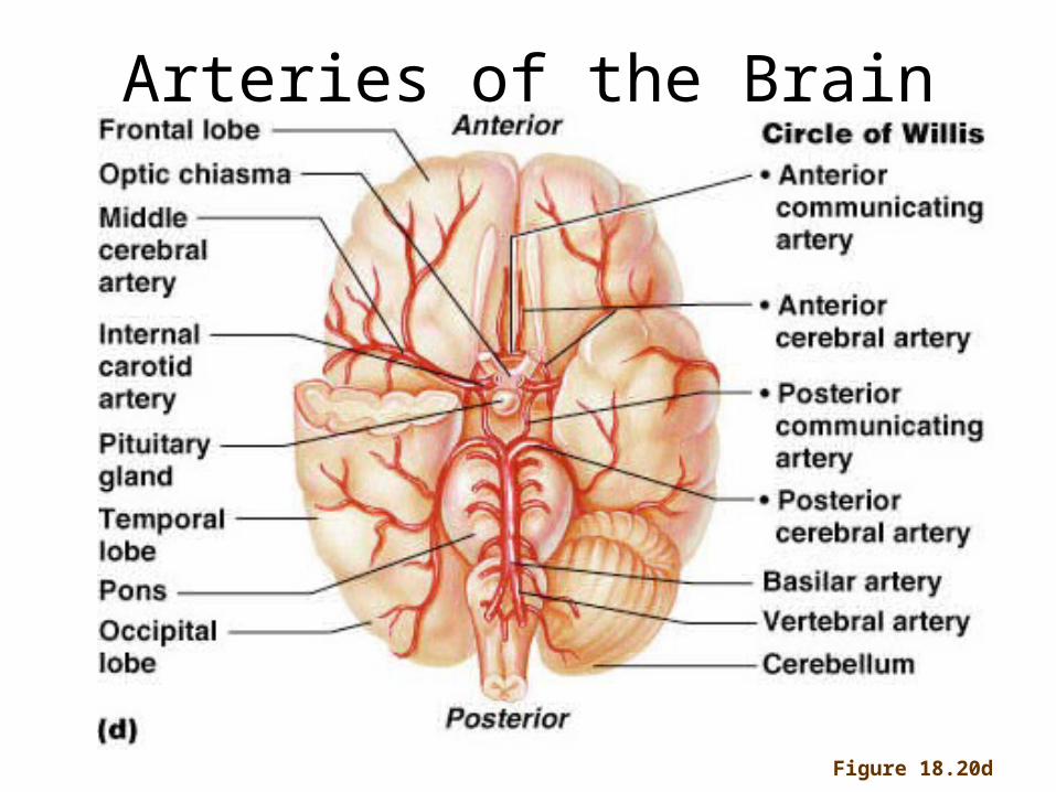
Figure 18.20d
Arteries of the Brain
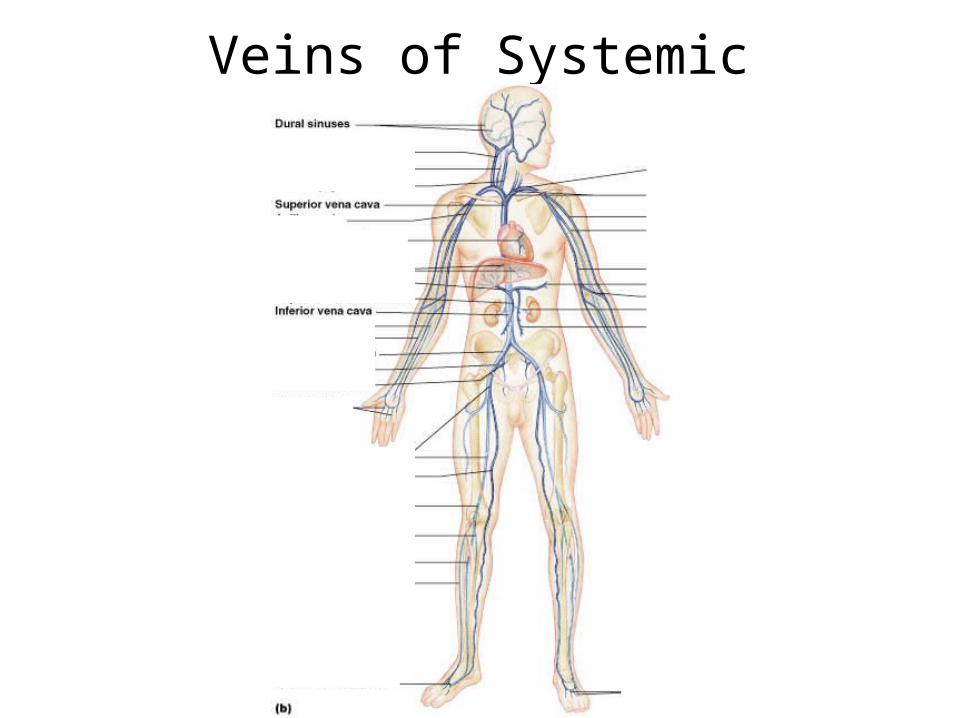
Veins of Systemic Circulation
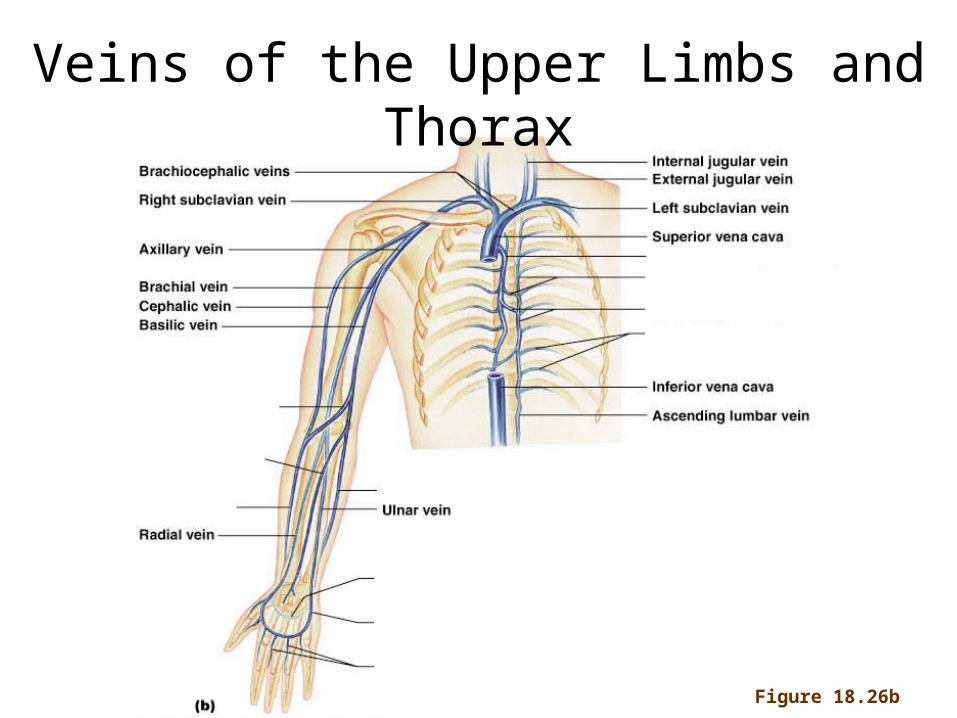
Figure 18.26b
Veins of the Upper Limbs and Thorax

The cephalic vein is an example of a badly named body part, and a thus confusing medical word, based on a translator’s mistake. The Canon of Avicenna ( 980-1037 CE) collected the medical knowledge of his times. This vein of the arm was named al-kifal, which means “the outer vein of the arm”. When this was translated into Latin in A.D. 1564, the translator thought al-kifal was related to the Latin word cephalicus, ‘pertaining to the head’. He was wrong, but the naming error stuck.
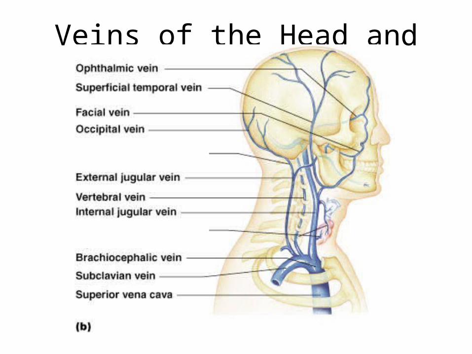
Veins of the Head and Neck
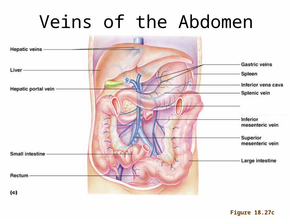
Figure 18.27c
Veins of the Abdomen
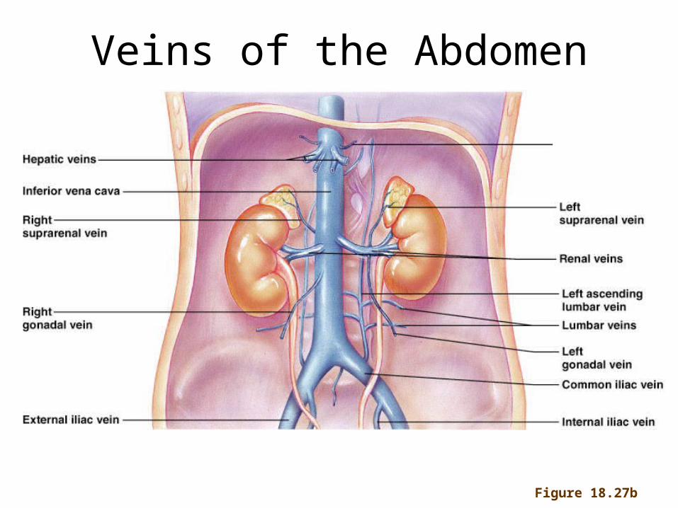
Figure 18.27b
Veins of the Abdomen
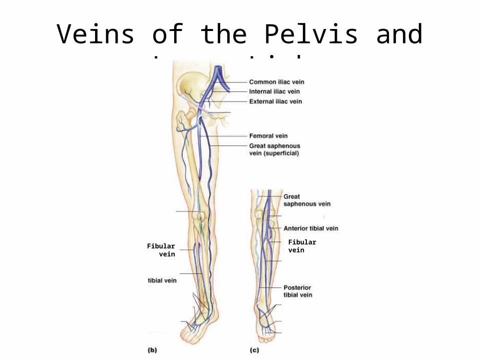
Veins of the Pelvis and Lower Limbs
Fibular veinFibular vein


![FIGURE 8.1 Process and controller. Curtis Johnson Process Control Instrumentation Technology, 8e] Copyright ©2006 by Pearson Education, Inc. Upper Saddle](https://img.pdfslide.us/doc/110x75/5515178e550346a80c8b5e70/figure-81-process-and-controller-curtis-johnson-process-control-instrumentation-technology-8e-copyright-2006-by-pearson-education-inc-upper-saddle.jpg)