Embed Size (px)
Citation preview
FEMS Microbiology Reviews 14 (1994) 121-138
Federation of European Microbiological Societies
Published by Elsevier
121
FEMSRE 00386
Copper resistance mechanisms in bacteria and fungi
Carlos Cervantes *+3 and Felix Gutierrez-Corona b
’ Institute de Investigaciones Quimico-Biologicas, Universidad Michoacana, 58240 Morelia, Mich., Mexico, and ’ Institute de Imestiga- cion en Biologia Experimental, Facultad de Quimica, Universidad de Guanajuato, Guanajuato, Gto., Mexico
(Received 5 July 1993; accepted 29 November 1993)
Abstract: Copper is both an essential micronutrient and a toxic heavy metal for most living cells. The presence of high
concentrations of cupric ions in the environment promotes the selection of microorganisms possessing genetic determinants for
copper resistance. Several examples of chromosomal and plasmid copper-resistance systems in bacteria have been reported, and
the mechanisms of resistance have started to be understood at the molecular level. Bacterial mechanisms of copper resistance are
related to reduced copper transport, enhanced efflux of cupric ions, or copper complexation by cell components. Copper tolerance
in fungi has also been ascribed to diverse mechanisms involving trapping of the metal by cell-wall components, altered uptake of
copper, extracellular chelation or precipitation by secreted metabolites, and intracellular complexing by metallothioneins and
phytochelatins; only the metallothionein chelation mechanism has been approached with molecular detail.
Key words: Copper resistance; Bacteria; Plasmid; Copper uptake and efflux; Fungi; Copper chelation
Introduction
Copper is an essential micronutrient for most, if not all, living organisms since it is the con- stituent of many metalloenzymes and proteins involved in electron transport, redox, and other important reactions [l]. The role of copper in redox reactions is due to its ability to undergo Cu(I) + Cu(I1) transitions which are dependent on the surrounding chemical environment [2]. Copper requirements by microorganisms are usu- ally satisfied with very low concentrations of the metal (in the order of l-10 PM). In contrast, copper present at higher levels in its free ionic form (Cu”> is toxic to microbial cells [3,4]. Cop-
* Corresponding author. Tel. (Fax): (043) 14 21 52.
SSDI 0168-6445(93)E0112-W
per toxicity is mainly due to its interactions with nucleic acids [5], to the alteration of enzyme active sites and to the oxidation of membrane components, processes that can be related to the ability of copper to generate toxic hydroxyl free- radicals [6,7]. On the other hand, organically complexed copper is relatively nontoxic to mi- croorganisms [8].
Given copper’s dual role of essentiality/ toxicity, microorganisms must possess delicate mechanisms to maintain intracellular copper within such a restricted level that neither inter- feres with normal metal homeostasis nor poses a risk of toxicity. Little information on copper transport in microorganisms has been reported, in part by lacking of a suitable radioisotope of the metal (the currently available 64Cu isotope has a half-life of only about 12 h). Copper properties as
122
an essential but toxic metal are shared by other micronutrients such as zinc, cobalt, and nickel, and are clearly distinguished from the unessential toxic cations of lead, mercury, and silver [9].
Copper and copper-containing compounds are widely used as bactericides [lo], algicides, fungi- cides, veterinary food additives, plant antibacte- rial agents, and for the preservation of natural and man-made materials 1111. Although copper is commonly encountered in the environment, toxic concentrations of the metal are only found associ- ated with certain mineral ores and industrial or agricultural discharges [121. High levels of copper generally exert a selective pressure on microor- ganisms that may result in the appearing of resis- tant variants possessing copper resistance genetic determinants 113-151. Bacteria and fungi are ubiquitous organisms with fundamental proper- ties as bioconverters, only recently considered as playing important roles in the biogeochemistry of heavy metals [16,17]. For this reason, understand- ing the responses of microorganisms to metals is of scientific interest and also may be relevant to devise biotechnological solutions for the recovery and environmental decontamination of valuable and/or toxic metals [18].
In this article we attempt to summarize the current knowledge on copper resistance in bacte- ria and fungi with emphasis on copper resistance mechanisms. Several aspects of resistance to cop- per in bacteria [15,18-301 and fungi [l&31-33,155] have been previously reviewed.
Copper resistance in bacteria
Chromosomal copper resistance
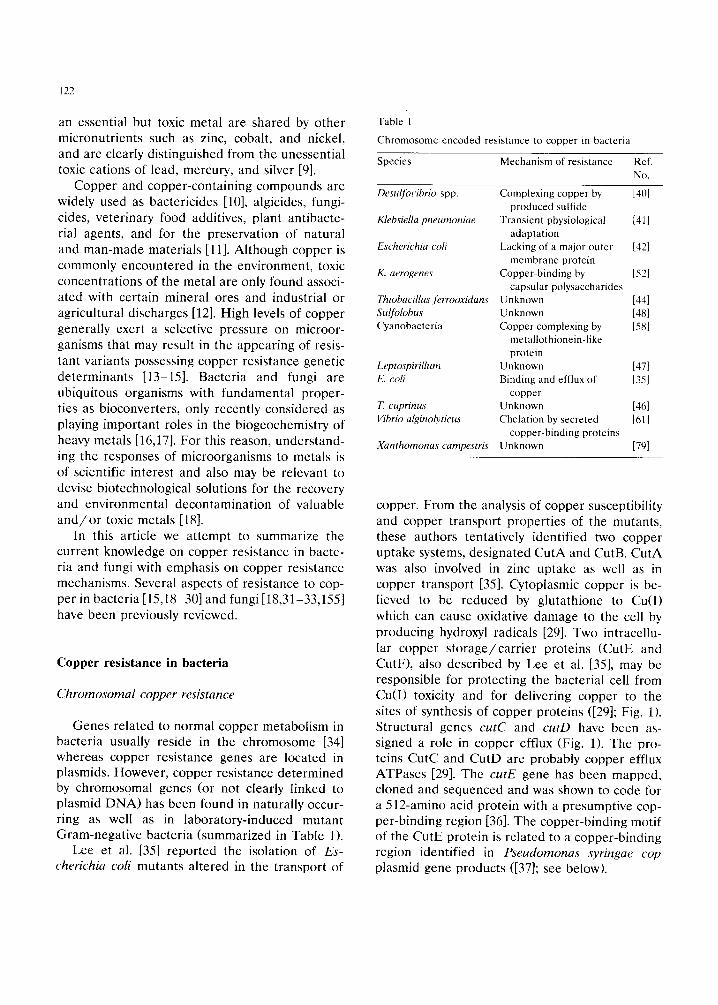
Genes related to normal copper metabolism in bacteria usually reside in the chromosome [34] whereas copper resistance genes are located in plasmids. However, copper resistance determined by chromosomal genes (or not clearly linked to plasmid DNA) has been found in naturally occur- ring as well as in laboratory-induced mutant Gram-negative bacteria (summarized in Table 1).
Lee et al. [351 reported the isolation of Es- cherichia coli mutants altered in the transport of
Table I
Chromosome-encoded resistance to copper in bacteria
Species Mechanism of resistance Ref
No.
Desulfolihrio spp.
Klehsiella tmeumoniae
Escherichia co/i
K. aerogenes
Thiobaciilus ferrooxidans Suifolobus Cyanobacteria
Leptospirillum E. coli
T. cuprinus Vibrio alginolyticus
Xanthomonas campestris
Complexing copper by
produced sulfide
Transient physiological
adaptation
Lacking of a major outer
membrane protein
Copper-binding by
capsular polysaccharides
Unknown
Unknown
Copper complexing by
metallothionein-like
protein
Unknown
Binding and efflux of
copper
Unknown
Chelation by secreted
copper-binding proteins
Unknown
1401
[411
[421
WI
[341 [481 Ml
1471 [%I
[4hl
[611
[791
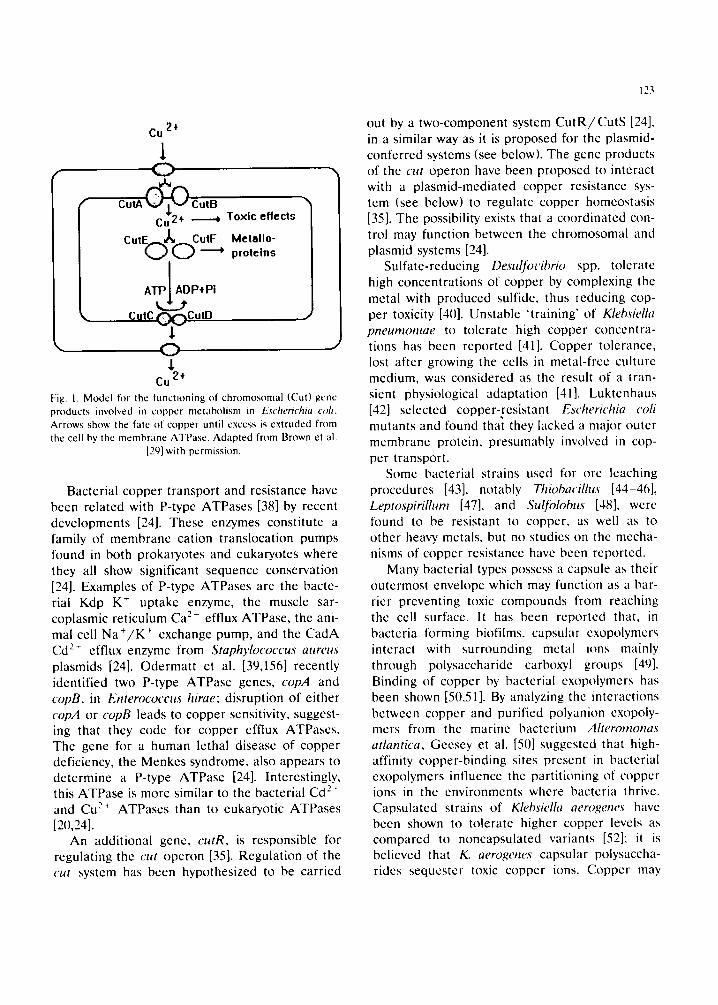
copper. From the analysis of copper susceptibility and copper transport properties of the mutants, these authors tentatively identified two copper uptake systems, designated CutA and CutB. CutA was also involved in zinc uptake as well as in copper transport [351. Cytoplasmic copper is be- lieved to be reduced by glutathione to Cu(I)
which can cause oxidative damage to the cell by producing hydroxyl radicals [29]. Two intracellu- lar copper storage/carrier proteins (CutE and CutF), also described by Lee et al. [351, may be responsible for protecting the bacterial cell from Cu(I) toxicity and for delivering copper to the sites of synthesis of copper proteins ([291; Fig. 1). Structural genes cutC and cutD have been as- signed a role in copper efflux (Fig. 1). The pro- teins CutC and CutD are probably copper efflux ATPases [29]. The c&E gene has been mapped, cloned and sequenced and was shown to code for a 512-amino acid protein with a presumptive cop- per-binding region [361. The copper-binding motif of the CutE protein is related to a copper-binding region identified in Pseudomonas syringae cop
plasmid gene products ([371; see below).
123
2,1. Cu
l
CutA ~ Curb ¢ -
+ ~ Toxic effects
CutEo~I~&utF Idetallo- proteins
I
ATP I AD P+ Pi |
~_ Cute ~(:~CutD
C)
Cu 2+ Fig. 1. Model for the functioning of chromosomal (Cut) genc products involved in copper metabolism in Excherichia colt. Arrows show the fate of copper until cxccss is extruded from the cell by the membrane ATPasc. Adapted from Brown et al.
[2q] with permission.
0 i
J
Bacterial copper transport and resistance have been related with P-type ATPases [38] by recent developments [24]. These enzymes constitute a family of membrane cation translocation pumps found in both prokaryotes and eukaryotcs where they all show significant sequence conservation [24]. Examples of P-type ATPases are the bacte- rial Kdp K- uptake enzyme, the muscle sar- coplasmic reticulum Ca 2- effiux ATPase, the ani- mal cell N a + / K + exchange pump, and the CadA Cd 2- efflux enzymc from Staphylococcus attreus plasmids [24]. Odermatt ct al. [39,156] recently idcntified two P-type ATPase genes, copA and copB, in Enterococcus hirae: disruption of either copA or copB leads to copper sensitivity, suggest- ing that they code for copper cfflux ATPascs. Thc gcne for a human lcthal diseasc of copper deficiency, the Menkcs syndrome, also appears to dctcrmine a P-type ATPasc [24]. Intcrcstingly, this ATPase is more similar to the bacterial Cd 2" and Cu 2' ATPases than to eukaryotic ATPases [20,24].
An additional gene, cutR, is responsible for regulating the cut operon [35]. Regulation of the cut system has been hypothesized to be carried
out by a two-component system C u t R / C u t S [24], in a similar way as it is proposed for the plasmid- conferred systems (see below). The gcne products of the cut operon have been proposed to interact with a plasmid-mediated copper resistance sys- tem (see below) to regulate copper homeostasis [35]. The possibility exists that a coordinated con- trol may function between the chromosomal and plasmid systems [24].
Sulfate-reducing Desulfocibrio spp. tolerate high concentrations of copper by complexing the metal with produced sulfide, thus reducing cop- per toxicity [4(I]. Unstable 'training' of Klebsiella pneurnoniae to tolerate high copper concentra- tions has been reported [41]. Copper tolerance, lost after growing the cells in metal-free culture medium, was considered as the result of a tran- sient physiological adaptation [41]. Luktenhaus [42] selected copper-resistant Escherichia colt mutants and found that they lacked a major outer membrane protein, presumably involved in cop- per transport.
Some bacterial strains used for ore leaching procedures [43], notably Thiobacillus [44-46], Leptospirillum [47], and Sulfolobus [48], were found to be resistant to copper, as well as to other heavy, metals, but no studies on the mecha- nisms of copper resistance have been reported.
Many bacterial types possess a capsule as their outermost envelope which may function as a bar- rier preventing toxic compounds from reaching the cell surface. It has been reported that, in bacteria forming biofi[ms, capsular cxopolymers interact with surrounding metal ions mainly through polysaccharide carboxy'l groups [49]. Binding of copper by bacterial exopolymcrs has been shown [50,51]. By analyzing the interactions between copper and purified polyanion exopoly- mers from the marine bacterium Alteromonas atlantica, Gcesey et al. [50] suggested that high- affinity copper-binding sites present in bacterial exopolymcrs influence the partitioning of copper ions in the environments where bacteria thrive. Capsulated strains of Klehsiella aerogenes have been shown to tolerate higher copper levels as compared to noncapsulated variants [52]; it is believed that K. aerogenes capsular polysaccha- rides sequester toxic copper ions. Copper may
J24
also be unspecifically bound by negatively charged groups on thc bacterial cell surface [53,54]. This property has been exploited as a means for detox- ifying copper-polluted waste streams by using bactcria [54-56]. Bacterial iron-binding sidcro- phorcs arc also known to bind copper [57].
Intraccllular accumulation of copper by a met- allothionein-likc protcin has bccn reported in cyanobactcria [58]; a cadmium-binding mctalloth- ionein analogue protein was previously found in a Pseudomonas strain [59]. The amino acid se- quence of a metaIlothionein (M'I') purified from the cyanobacterium Synechococcus sp. has been reported [58]. Huckle ct al. [60] recently cloned the structural gene for this MT from the Syne- chococcus chromosome and found that several ions (notably zinc, copper, and cadmium) specifi- cally stimulated transcription at the MT locus. Although the precise function of the prokaryotic MT is not known, by analogy to eukaryotic Ml's, it is believed to play roles in essential metal ion homeostasis and in resistance to non-essential rectal ions [611].
Harwood-Scars and Gordon [61] showed that thc marine bacterium Vibrio alginolyticus synthe- sizes extraeellular copper-binding proteins in rc- sponse to copper challenge; metal chelation was proposed as a mechanism of copper detoxifica- tion.
A copper resistance determinant from the chromosome of Xanthomonas campestris was re- cently cloned and will be discussed below with the plasmid systems.
Plasrnid-mediated copper resistance
Most adaptivc dispensable bacterial genes (e.g. those coding for antibiotic resistance, virulence properties and degradation of rare organic sub- strates) arc not usually located on the chromo- some but in extrachromosomal genetic elements called plasmids. Plasmid-eonferred copper rcsis- tancc determinants arc often capable of transfer- ring in viw) to new bacterial hosts by conjugation, or to othcr genetic elements by transposition [21]. These transfer properties represent a natural way of dispersing new adaptivc genetic abilities in bacterial populations inhabiting heavy-metal pol-
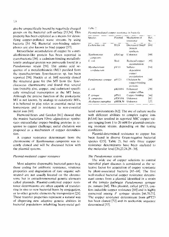
"1 able 2
Plasmid-mcdiatcd cuppcr rcsi:,tancc m bacteria
Spccics Plasmid Mcchani'ml ~I Rcl. resistance No.
I:M'herichia coh R 124
.¥unthonumus pXv( 'up camp(ts'tri3
h'. coil Rtsl
My<oha('tenum pVTl s( rof~daceum
IM'udomonas syringae pPT23
E. <oh pRJ 11){14
P. ~yrmgae pPS 11 X <ampestn.~ pXV 10A Ah'aligetles el~lrol~hU.~ pMOI.30
Decreased OmpF {St~[ protein s,,nthcsJs
t.'nkno~,n JSnl
Reduced copper [85] HCCII nl tl [~ll[ioI1
SLllfatc- is-l] dcpcndcnl copper precipitation
("hclation by 16S] copper-binding pr~Icins
Enhanced 178] copper el'flux
('oppcr-binding [~'~6] Unknown [Sl] Unknown [27]
luted environments [62]. The use of culture mcdia with different abilities to complex cupric ions [63,64] has resulted in reported MIC copper val- ues ranging from 1 to 20 mM for plasmid-contain- ing resistant strains, depending on the testing conditions.
Plasmid-determined resistance to copper has been found in diverse Gram-negative bacterial species ([15]: Tablc 2), but only threc copper resistance determinants have been analyzed to the molecular level [20,22,24,28-30].
The Pseudomonas system Thc wide use of copper solutions to control
microbial plant diseases is considered as the se- lective factor for acquisition of copper resistance by plant-associated bacteria [65-68]. The first well-studied bacterial copper resistance determi- nant comes from a plasmid identified in a strain of the tomato pathogen l{s'eudomonas syrmgae pv. tomato [68]. This plasmid, called pPT23, con- fers inducible copper resistance [69] and is highly conserved among I: .wringae strains [66,70,71]. The copper resistance determinant from pPT23 has been chined [72] and its nucleotide sequence determined [37].
Four structural genes, arranged in the cop operon, were initially identified; two of them, copA and copB, are necessary for expression of full copper resistance whereas plasmids with deletions in copC or copD still confer partial resistance [37]. From sequence analysis, a peptide of eight amino acids containing aspartic acid, histidine and mcthionine residues is repeated several times in both CopA and CopB, and is proposed to function as a copper-binding domain [37]. CopA also showed limited, but significant, sequence homology with the copper proteins azurin and plastocyanin [37]. The predicted polypeptides CopA and CopB generally lacked cysteine and thus do not pertain to the mctalloth- ionein class.
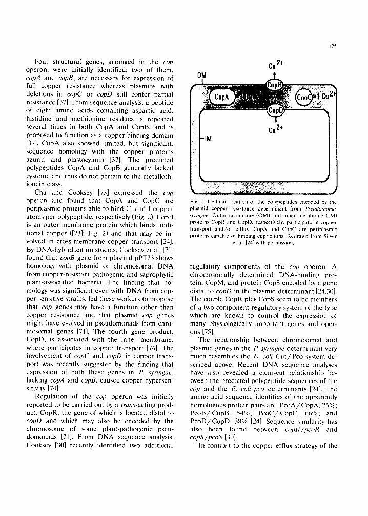
Cha and Cooksey [73] expressed the cop operon and found that CopA and CopC are periplasmic proteins able to bind 11 and 1 copper atoms per polypeptide, respectively (Fig. 2). CopB is an outer membrane protein which binds addi- tional copper ([73]; Fig. 2) and that may be in- volved in cross-membrane copper transport [24]. By DNA-hybridization studies, Cooksey et al. [71] found that copB gone from p[asmid pPT23 shows homology with plasmid or chromosomal DNA from copper-resistant pathogenic and saprophytic plant-associated bacteria. The finding that ho- mology was significant even with DNA from cop- per-sensitive strains, led these workers to propose that cop genes may have a function other than copper resistance and that plasmid cop genes might have evolved in pseudomonads from chro- mosomal genes [71]. The fourth gene product, CopD, is associated with the inner membrane, where participates in copper transport [74]. The involvement of copC and copD in copper trans- port was recently suggested by the finding that expression of both these genes in P. syringae, lacking copA and copB, caused copper hypersen- sitivity [74].
Regulation of the cop operon was initially reported to be carried out by a trans-acting prod- uct, CopR, the genc of which is located distal to copD and which may also be encoded by the chromosome of some plant-pathogenic pseu- domonads [71]. From DNA sequence analysis, Cooksey [30] recently identified two additional
125
Cu 2÷
cu2+ ?
2
Fig. 2. Cellular location of the polypeptides encoded by the plasmid copper resistance determinant from Pseu(h~monas wringae. Outer membrane (OM) and inner membrane (IM) proteins CopB and Copl), respectively, participate in copper transport and/or efflux. ('opA and ('opC are periplasmic proteins capable of binding cupric ions. Redrawn from Silver
ctal. [24] with permission.
regulatory components of the cop operon. A chromosomally determined DNA-binding pro- tein, CopM, and protein CopS encoded by a gene distal to copD in the plasmid determinant [24,30]. The couple CopR plus CopS seem to be members of a two-component regulatory system of the type which are known to control the expression ot" many physiologically important genes and oper- ons [75].
The relationship between chromosomal and plasmid genes in the P. syringae determinant very much resembles the E. coli C u t / P c o system de- scribed above. Recent DNA sequence analyses have also revealed a clear-cut relationship be- tween the predicted polypeptide sequences of the cop and the E. coli pco determinants [24]. The amino acid sequence identities of the apparently homologous protein pairs are: P c o A / C o p A , 76c7~,; P c o B / C o p B , 54%; P c o C / C o p C , 66c~; and P c o D / C o p D , 38% [24]. Sequence similarity has also been fl)und betwecn copR/pcoR and copS /pcoS [30].
In contrast to the copper-effiux strategy of the
12~
E. coil determinants (scc below), howcvcr, metal sequestration has been dcmonstratcd as the basis for copper resistance in plant-associatcd bacteria ([73]; Fig. 2). Increased copper accumulation was shown by Cooksey and Azad [76] in P. syringae with a cloncd cop operon as well as in related copper-resistant pscudomonads; some strains ac- cumulated as much as 12(1 mg of copper per gram (dry weight) of cells. The copper in resistant cells appears to bc in the cupric (Cu 2-) state since bacterial colonies still maintain the bluc color of these ions [66]. Copper complexing was highly spccific as copper-induced cells did not accumu- late other mctals [76]; a halt" of the ceil-bound coppcr was released by EI)TA, suggcsting that the metal was bound to the cell envelope, proba- bly through the outcr membrane protein CopB.
This is the first plasmid-dctermincd heavy metal resistance system that involves scquestra- tion of the toxic ion. Othcr heavy-metal resis- tance mechanisms result from reduced accumula- tion, cffiux, or chemical modification of the ions involved [21]. Coppcr complcxing, however, has bcen rcported for several chromosome-encoded resistancc systems (Table 1) and is commonly found in copper-rcsistant fungi (see below).
"Fhe E. coli system An E. coli copper resistance plasmid, pRJ 1004,
was found in a strain isolated from the effluent of a piggery [77] where copper sulfate, used as a food supplement, possibly acted as a selective agent. Inducible resistance to copper conferred by pRJl004 was found to be due to decreased metal uptake [78]. Rouch et al. [157] cloned the copper resistance determinant from pRJ1004 and four pco (for plasmid copper) genes, pcoARBC, were initially identified, and their corresponding gene products were assigned roles for the resis- tance phenotype. Further studies led to the find- ing of additional putative pco genes now ar- ranged as pcoABCDRSE [24,28]. As mentioned above, the pco genes show significant sequence homology to the cop genes, but the precise fun- cion of the Pco proteins is still unknown. The PcoA protein conserves the potential copper- binding domain of the CopA protein and thus appears to function as a copper-binding polypcp-
tide. The CopB protein, however, does not retain the potential copper-binding sites present in CopB and is proposed to have a function unre- lated to copper binding [29]. The PcoC protein shares a putative copper recognition motif with the CopB protein suggesting a role of PcoC in copper binding [29]. Sequence analysis showed that PcoD have potential t ransmembrane seg- ments and methionine and histidinc groups lo- cated on the outer face of the inner membrane. which are also conserved in CopD [29].
PooR is a repressor responsible for regulation of the pco operon; from sequence analysis, PooR was found to show homology with DNA-binding regulatory proteins of the two-component class, as it was mcntioncd above for thc pairs C u t R / CutS and C o p R / C o p S from the E. coli chromo- some and the Pseudomonas plasmid copper resis- tance determinants [24]. Further nucleotidc se- quence analysis of the pco operon showed the presence of another regulatory gene, named pcoS. From its deduced amino acid sequence, PcoS was predicted to be a membrane polypeptide with the attributes of an auto-phosphorylating protein which qualifies it as the sensor portion of the two-component P c o R / P c o S regulatory system [24]. Williams et al. [158] recently found pco homologs among copper-resistant enteric bacteria from pig farms in thc UK and Australia.
l,ee et al. [35] proposed a unique physical interaction between chromosomal (metabolic) and plasmid (resistance) genes in copper-resistant E. coil cells. Chromosomal genes cutA (uptake) and cutD (efflux) are required for the expression of copper resistance in E. coli, whereas plasmid gent products of pcoA and pcoB were proposed to interact with proteins CutC and CutD by form- ing a tbur-component efflux system [35]. More- over. chromosomal regulatory gene cutR re- pressed pco plasmid genes in the absence of a functional regulatory pcoR [35]. This genetic in- terplay would allow bacterial cells to regulate changing environmental copper concentrations according to cellular needs. As proposed by this model, chromosomal genes control normal low- copper situations, whereas plasmid genes will function when high (toxic) copper is present [35]. An additional, low-level copper resistance deter-
127
minant, called cdr, was found in the E. coil plasmid pRJl004 and a role in copper-damage repair was tentatively assigned [157]. This would be a distinct resistance mechanism since it does not deal with copper itself but with its toxic effects.
Other plasmid-mediated systems The chromosome of Xanthomonas campestris
pv. juglandis, a pathogen of walnuts, was the source of the third, still unpublished, sequenced copper resistance determinant [79]. Although of chromosomal origin, the four structural genes, copABCD, of the Xanthomonas determinant showed significant homology with both cop and pco gcncs [20]: surprisingly, the sequences were more similar for the Xanthomonas/E. coli dctcr- minants than wcrc for the Xanthomonas (a pscu- domonad)/Pseudomonas genes.
Copper resistance has also been encountered in strains of Xanthomonas campestris pv. cesica- toria, a pathogen of peppers and tomatoes, bear- ing large plasmids pXvCu [8(I] or pXVIOA [81]. The cloned copper resistance determinant from pXVIOA, which did not express in E. coli, showed strong nucleotide sequence homology with pXvCu and only slight similarity to P. syringae copper resistance genes [81]. Voloudakis et al. [82], how- ever, recently found significant homology be- tween copper resistance plasmid DNA from di- verse Xanthomonas strains and the copA gene from P. syringae, suggesting a possible common origin for the copper resistance determinants from these plant-associated bacteria. Garde and Ben- der [83] used a probe from pXV10A to detect copper resistance determinants from a variety of soil bacteria: genomic DNA from copper-sensi- tive strains failed to hybridize to the pXVIOA probe, thus allowing the authors to propose a non-endogenous origin of the Xanthomonas cop- per resistance genes. Since the t'. ~yringae resis- tance determinant is postulated of chromosomal origin ([83]; see above), exchange of plasmid DNA between pseudomonads and xanthomonads is suggested as responsible for the similarities be- tween the copper resistance determimmts of these bacteria [82]).
A distinct plasmid-cncoded copper resistance
mcchanism has bccn found in a strain of My- cobacterium scrofulaceum isolated from a pol- luted river [84]. Sulfate-dependent precipitation of the metal (as copper sulfide) was shown to bc rcsponsiblc for coppcr rcsistancc [84]. This mcch- anism resembles the one mentioned above for Desulfocibrio strains [40] that was probably not plasmid-linkcd.
In E. coli, R-factors Rtsl [85] and R124 [86] also confcr coppcr resistance. Cotter ct al. [87] reported that Rtsl-containing E. coli cells accu- mulated less coppcr than a plasmid-frec deriva- tive, although their copper uptake results wcrc somewhat masked by a rapid unspecific copper- binding. Copper resistance determined by plas- mid R124 was associated with a lower level of major outer mcmbranc protein OmpF [86]. To our knowledge, no further studies about copper resistance have bccn reported with these plas- mids.
Copper resistance in multiply heavy-metal-re- sistant Alcaligenes eutrophus isolatcs [88] is dctcr- mincd by large plasmids pMOI,85 [89] and pMOL30 [27]. To emphasize thcir rcsistancc properties, Mcrgcay ct al. [62] have proposed these strains to bc classified as Alcaligenes eutro- phus var. metallotolerans. Drcsslcr ct al. [90] re- ported the isolation of Alcaligenes and Pseu- domonas strains showing resistance to copper of probablc plasmid origin. Thcsc strains also con- tain genetic determinants for resistance to other toxic metals [90].
Copper resistance in fungi
Copper binding in fungal cells
It is known that the binding of copper in fungi, as well as that of other metals, comprises two phases. The first is a metabolism-independent surface binding pathway, whereas the second phase is an energy-dependent metal influx [31]. Binding o1" significant amounts of metals to the fungal cell wall is a very. rapid process, often taking place in a few minutes [91,92]. When cul- tured in the presence of copper, the cell walls of Penicillium italicum [93], Neurospora crassa
12b~
[93,94], P. ochro-chloron [95], Aureobasidium pul- lulans [96] and Mucor rouaii (Ramirez-Salgado el al., unpublished results) accumulated 5 -40% of the metal added to the culture medium, which, in some cases, gave rise to a blue-colored mycelium. Copper-tolerant strains of Rhizopus stolonifer, CunninghameUa blakesleeana [97] and M. rowcii (Ramirez-Salgado el at., unpublished) bound sig- nificantly more copper into the mycelial biomass than the correspondent copper-sensitive parental strains.
Germann and Lerch [98] reported that 'slime', a wall-less mutant of N. crassa [99], showed a reduced accumulation of copper when cultured in the presence of the metal as comparcd to the walled strain. This finding adds evidence about cell wall involvement in metal binding properties of fungal celts.
A copper-resistant strain of M. rouxii, cultured in thc presence of a low concentration (1.6 /,~M) of the metal, accumulated a ten-fold higher amount of copper in the cell wall fraction and in a mixture of cytosol and membranes, as com- pared to the metal-sensitive parental strain (Ta- ble 3). In the presence of a much higher copper concentration (3.2 mM), however, the differencc in metal accumulation was reduced to a 50% highcr amount of copper in the cell wall fraction, but not in the cy toso l /membrane fraction, of the resistant strain (Table 3). As previously suggested for other fungi [96,97], these findings point to the involvement of the cell wall in the development of copper resistance. However, it seems that, at
"Fable 3
Suhcellular distribution of copper in strains of Mucor nmxti
Copper (mM) Strain " Copper bound in growth (nmol /mg dry weight) medium
('ell walls Mixed traction h
(}.(X)I 6 Cup" 0.30 0.20 0.0016 ( 'up ' 2.80 2. I{) 3.2 ( 'up" 42.6 38.6 3.2 ( 'up ' 64.9 35.3
" ( 'up ' , copper-sensitive: ( 'up ~. copper-resislant. ~' Mixed membrane l'r:lction plus cytosolic fraction. l)ata from Ram~rez-Salgado el al., unpublished results.
- - ® . . .
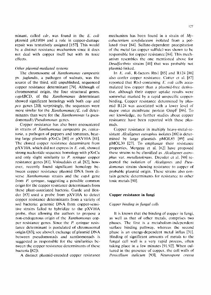
Fig. 3. Ultraslructural appearance of cell walls (CW) from a ( 'up ' strain of Trichoderma t'iride grown at low (A, 0.0016 raM) and high (B. 3.2 mM) concentration of copper. Bars: A. 100 nm: B. 2(~) nm. From CortcSs-Penagos et al.. unpublished.
least in M. rouxii, cytosolic a n d / o r membrane- associated components may also participate in metal tolerance processes.
The biopolymers of fungal cell walls, such as the polysaccharides chitosan and chitin, have been reported to be useful in the removal of valuable metals, particularly uranium [17,100]. The Phy- comycetes, such as R. stolonifer, C blakesleeana and M. rouxii, have a significant proportion of chitosan in the outer laycr of their vegetative cell walls [101]. It has been proposed that the copper-binding properties of mycelial cells of some fungi developed in the presence of the metal may be due to qualitative or quantitative changes in cell wall components, leading to an improved copper-binding ability and, thereby, to metal detoxification [94,102]. When cultured in the presence of toxic concentrations of copper, the cell wails of C. blakesleeana contain reduced amounts of chitosan and chitin and a higher proportion of a hydroxypro[ine-rich protein [103,104], which was absent in the cell walls of control cells. The changes in fungal cell walls by culturing in the presence of copper may be re- flected at the structural level [103,104]. Fig. 3 shows that a copper-resistant strain of Tricho- derma citqde, when cultured with toxic conccntra- fions of copper, produced about five-lk)ld thicker
129
cell walls (Cort6s-Penagos et al., unpublished re- suits).
It has been shown that, in addition to its important nutritional role, the presence of mycor- rhizal fungi in plants plays a protective role against the toxic effects of copper and other heavy metals [105,106]. The protective action of mycorrhizal fungi may be based on the adsortive properties of fungal surfaces [106] or on the se- questration of copper by cytoplasmic components such as polyphosphate granules [107,108].
Copper uptake in fungi
Energy-dependent copper uptake usually pro- ceeds at a slower rate than metal surface-binding and can be inhibited by low temperatures, glu- cose analogues, acidic pH, and metabolic in- hibitors and uncouplers (for a review see [31]). Energy-dependent uptake of copper was demon- strated in yeasts [109-112]. Measuring copper transport in filamentous fungi is complicated by the high level of metal binding to the cell walls: however, energy-depcndcnt uptake of copper has been demonstrated in protoplasts of the copper- tolerant fungus P. ochro-chloron [113] and in the polymorphic fungus A. pullulans. This latter fun- gus and the yeast Saccharomyces cerec&iae are similar in several aspects of copper transport, but they differ from the yeast Candida utilis [111]. In both A. pullulans [159] and S. cerecisiae [112], copper uptake is relatively less specific than in C. utilis since in this yeast the process is not inhib- ited by other cations [111]. In addition, a stoichio- metric exchange of 2 mol of potassium for 1 mol of copper was reported in S. cererisiae [112] and A. pullulans [159], whereas that exchange was not detected in C. utilis [111]. On the other hand, copper uptake in S. cererisiae is inhibited by CCCP [112], but this uncoupler had relatively little effect in C. utilis [111].
Several studies have suggested a correlation between copper tolerance and an altered metal uptake (reviewed in [31]). Metal-resistant yeast strains showed a decreased influx of copper, cad- mium and lithium [114-117]. It has also been reported that in a copper-tolerant strain of A. pullulans copper uptake was reduced as com-
pared to that of a sensitive strain [118]. By using protoplasts, Gadd et al. [117] demonstrated that decreased copper uptake by a copper-tolerant strain of S. ceret'isiae was due to changes in membrane transport properties rather than to alterations in cell wall permeability. Two separate mutant loci in Aspergillus nidulans, cupA and cupB, confer specific resistance to copper which was not due to an increased synthesis of copper- binding proteins [119]. A restricted uptake of copper was proposed as a mechanism responsible of copper resistance in A. nidulans [119].
Intracellular chelation of copper
A distinct mechanism of copper tolerance is the production of mctallothioneins. Thcsc are low-molecular-mass (< 10 kDa), cystcine-rich, metal-binding proteins inducibly synthesized in the presence of metals [120,121]. Metalloth- ioneins (MTs) are widely distributed throughout living organisms such as mammals (including hu- mans), plants, fungi [122,160], and cyanobacteria [58,123]. MTs have been proposed to be involved in a number of cellular processes including metal storage and detoxification, development, differ- entiation, control of metabolism, protection from free-radical toxicity, and UV response (reviewed in [124]).
Genetic studies in S. cere~'isiae showed that high levels of copper resistance could be at- tributed to a locus called CUP1 [125-127]. Cloning and molecular characterization of DNA se- quences conferring copper tolerance to S. cere- cisiae-sensitive strains showed that the resistance phenotype was due to a tandem amplification of a 2-kb genomic DNA segment of the CUP locus [126]. Further studies indicated that there exists a correlation between the levels of copper resis- tance and the extent of DNA amplification [32,121,128]. Copper-resistant strains of S. cere- t'isiae contain 2-14 copies of the 2-kb repeat unit in a tandem array (denoted CUPId), whereas copper-sensitive strains contain only one copy of the sequence (cupl ~) [129]. Transcription of CUP1 is 10-20-fold induced by the addition of copper but not of cadmium or zinc [130]. Copper-in- duced expression of the CUPI genc also requires
130
the participation of the ACEI (also called CUP2) transcription factor which is encoded by the CUP2 gene [131,132]. Sequence analysis of the A C E I gene revealed a predicted polypeptide possessing an N-terminal half rich in cysteine residues [133]; these cysteines form a copper-thiolate complex, similar to that described for yeast Cu-MT, which stabilizes ACEI for DNA binding [133]. A metal-activated transcription factor showing par- tial homology to the ACEI protein was reported in the yeast Candida glabrata [134]; this transcrip- tion factor showed ACEI properties when tested in S. ceret'isiae.
The CUP1 locus contains two open reading frames (ORFs), one of which codes for a cys- teine-rieh protein analogous to the we[I-char- acterized mammalian MTs; the second O R F en- codes a protein with no function ascribed as yet [121,128].
Hetcrologous MT genes were shown to be fully functiona[ in yeast [135,136]. For example, c I )NA of the monkey cadmium MT gene fused to promoter sequences of the yeast CUP1 gcnc on a multicopy plasmid conferred copper resistance in a metal-sensitive S. ceret'isiae strain with a dele- tion in the CUPl gene [135]. Similar results were obtained by Eckcr et al. [136] with a multicopy chimeric plasmid consisting of the yeast MT pro- moter and a fusion of the yeast and monkey MT structural genes. It has also been observed that high levels ot" expression of yeast CUP1 gene lead to cadmium resistance [136,137] although a lower ( 'UP1 level was required to confer copper resis- tance [137]. The potential of yeasts containing the amplified CUI'I gene to be used for detoxifiea- lion of metal-polluted discharges has been recog- nized [138].
The copper-inducible MT of S. cerecisiae has been purified [139,140], its low molecular-mass and high content of cysteine, qua[flied it to be designated as copperthionein [140], copper chelatin [139], yeast MT, or simply Cu-MT [32].
Other fungi in which copper-inducible cys- teine-rich proteins have been described include N. crassa [ 141 ] and Dactylium dendroides [ 142]. A pathogenic strain of Candida albicans was found to contain DNA sequences hybridizing with S. ceret'isiae CUP DNA and to synthesize copper-in-
ducible proteins similar to the S. ceret'isiae MT [128]. The pathogenic yeast ('. glahrata synthe- sizes a small protein in response to copper but no DNA sequence homology with the S. ceretisiae CUP gene was found [128]. Other workers were unable to detect hybridization of CUPI DNA probes with C. albicans DNA, although the pres- ence of low-molecular mass copper-binding pro- teins was demonstrated in this yeast [33].
Cano-Cancho[a et al. [143] have shown that the dimorphic Zygomycetes Mucor racemosus and M. rmL~ii both contain DNA sequences which hybridize with CUP DNA from S. ceret'isme: they showed that in M. twuxii there exists a CUI' multigene family. It was also found that DNA methylation controls CUP expression during de- velopment of Mucor cells [143].
By using a histochemical procedure, Morselt et a[. [144] 15ave reported that tolerance to heavy metals in ectomycorrhizal fungi is based on the presence of MT-like proteins which probably pro- tect the host plant in metal-polluted areas. Cop- per-tolerant strains of Trichoderma ciride, iso- lated from a copper mine, contain a 25-kDa pro- tein which is undetcctablc in Cu-sensitive strains, but no DNA homology was found with the ( 'UPI gene of S. cerecisiae (Cortt~s-Penagos, unpub- lished results).
Copper-inducible proteins, either containing or lacking cysteine, are produced in the fungus D. dendroides [142]. It is not clear whether they are similar to phytochelatins, metal-induced peptides of the general structure (y-Glu-Cys),,-Gly (n = 2- 11) that have been tkmnd in algae and higher phmts [145,146] and some fungi [33,147-149]. In the yeast C. glabrata, mctallothioneins and phy- tochelatins are produced in response to copper and cadmium, respectively [149]. Kneer ct al. [15(I] recently reported that the unre[ated fungi S. ceret'isiae and N. crassa, already known to synthesize MTs [32,33] also produce phy- toehclatins when exposed to cadmium; interest- ingly, the synthesis of phytochelatins in S. cere- t'isiae is induced by copper or zinc [15[)].
l:~tracellular chelation o f copper
It has been described that organic and i n o f ganic extrace[lular funga[ metabolites may detox-
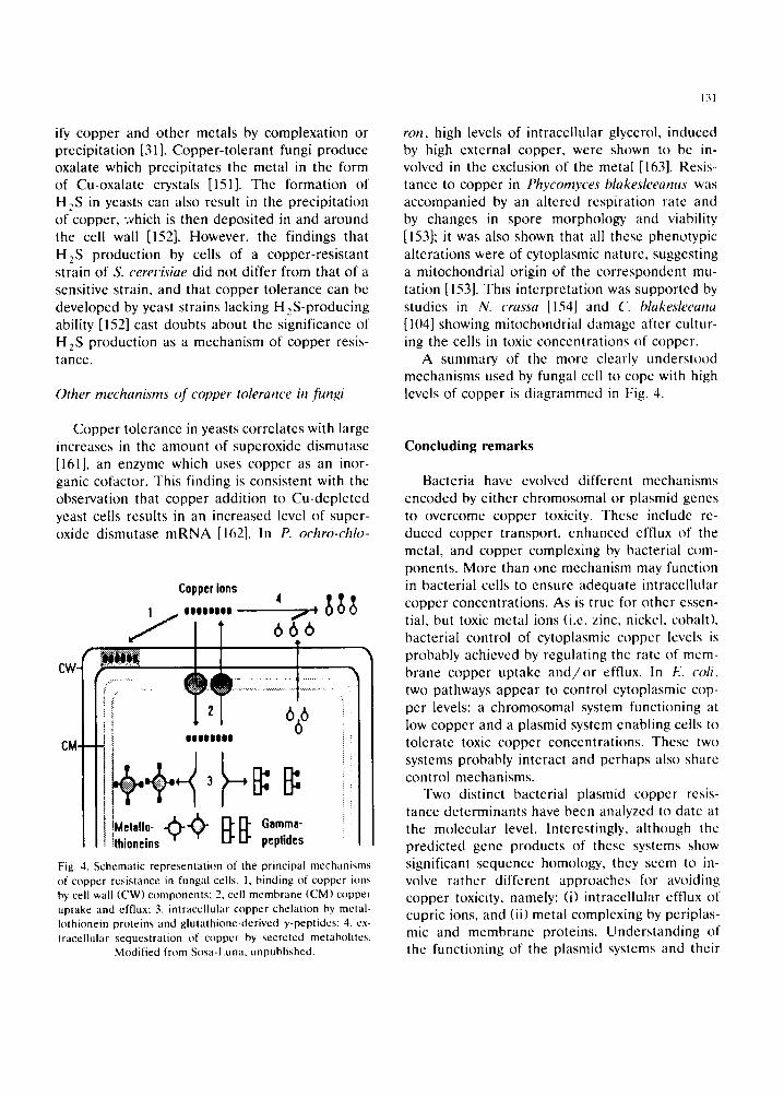
ify copper and othcr metals by complexation or prccipitation [31]. Copper-tolerant fungi produce oxalate which precipitates the metal in the form of Cu-oxalate crystals [151]. The formation of lq~S in yeasts can also result in the precipitation of copper, which is then deposited in and around the cell wall [152]. However, the findings that H2S production by cells of a copper-resistant strain of S. cererisiae did not differ from that of a sensitive strain, and that copper tolerance can be developed by yeast strains lacking H2S-producing ability [152] cast doubts about the significance of H2S production as a mechanism of copper resis- tancc.
Other mechanisms of copper tolerance in fungi
Copper tolcrancc in yeasts correlates with large incrcascs in thc amount of supcroxidc dismutase [161], an enzymc which uses coppcr as an inor- ganic eofactor. This finding is consistent with thc observation that copper addition to Cu-depletcd yeast cells rcsults in an increased lcvcl of super- oxidc dismutase m R N A [162]. In P. ochro-chlo-
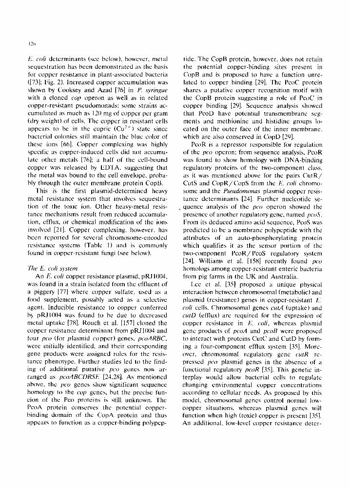
CW-
CM
Copper ions ' " ' " ' °
i~=! i!i!ii!!K ~ ~ .................................
] 00001000
I M e t a l l o - . ~ ~:~: Gamma- !thioneins peptides
Fig. 4. Schematic representation of the principal mechamsms of copper resistance in fungal cells. 1, binding of copper ions by cell wall (CW) components" 2, cell membrane (CM) copper uptake and efflux: 3, intraccllular copper chelation by mctal- Iothionein proteins and glutathionc-dcrivcd y-peptidcs; 4. ex- tracellular sequestration of copper by secreted metabolites.
Modified from Sos~l-l.una. unpublished.
131
ron, high levcls of intracellular glyccrol, induced by high external copper, were shown to be in- volvcd in thc cxclusion of the metal [163]. Resis- tance to copper in Phycomyces blakesleeanus was accompanied by an altered rcspiration rate and by changcs in spore morphology and viability [153]; it was also shown that all thcsc phenotypic alterations were of cytoplasmic naturc, suggcsting a mitochondrial origin of the correspondent mu- tation [153]. This intcrprctation was supported by studics in N. crassa [154] and C. blakesleeana [104] showing mitochondrial damagc after cultur- ing the cells in toxic conccntrations of copper.
A summary of the more clearly understood mechanisms used by fungal cell to cope with high lcvcls of copper is diagrammcd in Fig. 4.
Concluding remarks
Bacteria have evolved diffcrcnt mechanisms encoded by either chromosomal or plasmid genes to overcome copper toxicity. These include re- duced copper transport, enhanced effiux of the metal, and copper complexing by bacterial com- ponents. More than one mechanism may function in bacterial cells to ensure adequate intraccllular copper concentrations. As is truc for other essen- tial, but toxic mctal ions (i.e. zinc, nickcl, cobalt), bacterial control of cytoplasmic copper [cvcls is probably achieved by regulating the rate of mem- brane coppcr uptakc a n d / o r effiux. In E. coli, two pathways appear to control cytoplasmic cop- per levels: a chromosomal system functioning at low copper and a plasmid system enabling cells to tolerate toxic coppcr conccntrations. Thesc two systems probably intcract and perhaps also share control mechanisms.
Two distinct bacterial plasmid copper resis- tance determinants have been analyzed to date at the molccular level. Interestingly, although the predicted gcnc products of these systems show significant scqucncc homology, thcy sccm to in- volve rathcr different approachcs for avoiding copper toxicity, namely: (i) intracellular cfflux of cupric ions, and (ii) metal complcxing by pcriplas- mic and membranc proteins. Understanding of the functioning of the plasmid systems and their
132
relationship to chromosomal genes is still under way.
Similarly to bacteria, fungi have developed a variety of copper resistance mechanisms to sur- vive in the presence of toxic concentrations of cupric ions. These mechanisms include copper complexing by cell wall components, changes in membrane copper transport, synthesis of mtracel- lular copper-binding metallothioneins and phy- tochelatins, and production of extraccIlular cop- per-complexing or -precipitating metabolites. With the exception of some yeasts, the underlying genetic or molecular basis for copper resistance remains mostly unknown. Among these different copper-resistance mechanisms, only metalloth- ionein production has been tested through the employment of molecular genetic approaches.
Acknowledgements
We thank S. Silver for helpful suggestions on the manuscript and for making available unpub- lished inh)rmation, and L. Sosa-Luna for helpful comments. Work in the authors' laboratories was supported by Coordinaci6n de lnvestigaci6n Cientffica (Universidad Michoacana), DIGICSA (SEP), and CONACYT (0944-N9111 ).
References
I l,mile, R. (1984) Copper t)roleins and Copper En~'mes. ( 'R(" Press, Boca Raton. FI,.
2 Cotton, [.'.A. and Wilkinson, G. (1988;) Advanced Inor- ganic ('hcmistry. 5th. cdn. Wiley, New York, NY.
3 Macl,eod, R.A., Kuo, S.(.'. and Gelinas. R. (1967) Metabolic inju~, to bacteria. 11. Metabolic injury induced by distilled water or ( 'u :" in the plating diluent. J. Bacterio[. 93, 961-969.
4 I)omek. M.J., LeChavallier, M.W., Cameron. 5.('. and McFeters, G.A. (1984) Evidence h~r the role oI copper in the injury process of coliform bacteria in drinking water. Appl. t-nviron. Microbiol. 4~. 289-293.
5 Lippert. B. (1992) From cisplatin to artificial nt.cleases. The role of metal ion-nucleic acid interactions in biology. Biometals 5, 195-2118.
e, Simpson. J.A., Cheeseman, K.tI., Smith, S.E. and I)ean, R.'['. (198g) Free-radical gener~,tion by copper ions anti hydrogen peroxide. Biochcm. ,I. 254, 519 523.
7 Kobayashi, S., Ueda. K. and Komano. T. (1990) The
effects of metal ions tm the r)NA damage induced by hydrogen peroxide. Agric. Biol. Chem. 54, 69 76.
S Zevenhuizen, L.P.. Dolfing. J.. Eshuis, E.J. and Scholtcn- Koerselman, l.J. (1979) Inhib i tor effect of copper on bacteria related to the frcc ion concentration. Microb. Ecol. 5, 139-146.
9 Silver, 5. (1982) Bacterial interactions with mineral cations and anions: good ions and bad. In: Biomincralization and Biological Metal Accumulation (Westbrock, P. and E.W. de Jong, Eds.), pp. 439 457. Reidel, D.. Dordrecht.
10 ('ooksey, I).A. (1990) Genetics of bactericide resistance in plant pathogenic bacteria. Annu. Re','. Phytopathol. 28, 201-219.
11 Foye, W.(). (1977) Antimicrobial activities of mineral elements. In: Microorganisms and Minerals (Weinberg, E.I)., Ed), pp. 387-419. Marcel Dekker. New York, NY.
12 Forstner, U. (1984) Metal pollution of terrestrial waters. In: ( 'hanging Metal ('ycles and Human Health (Nriagu, J.()., lid.), pp. 71-94. Springer-Verlag, Berlin.
13 Ashida..I. (1965) Adaptation of fungi to metal toxicants. Annu. Rev. Phytopathol. 3, 153-174.
14 Dekker, J. (1976) Acquired resistance to fungicides. Annu. Rev. Phytopathol. 14,405-428.
15 Trevors, J.T. (1987) ( 'opper resistance in bacteria. Micro- biol. Sci. 4. 29-31.
16 Ehrlich, t l.[,. (1990) (;eomicrobiolog~,. 2nd. edn. Marcel 1)ekker. New York, NY.
17 Gahm, M., Keller, P.. Feldstein, I1., Galun, F,.. Siegel, S. and Siegel, B. (1983) Recover',' of uranium (VII from solution using fungi. 11. Release from uranium-loaded I'enicilliunr biomass. Water Air Soil Pollut. 20, 277--2S3.
l~ Jam. R.K. (199(1) Copper-resistant microorganisms and their rote in the environment. Worhl J. Microbiol. Bioteehnol. 6, 351~-365.
19 [a}elliveau, B.11., 5tarodub, M.E., ('otter, ('. and Trevors, .I.T. (1987) Metal resistance and accumulation in bacte- ria. Biotechnol. Adv. 5. 101-127.
2l) Silver, S. and Ji, G. (1994) Newer systems for bacterial resistances to toxic heavy metals. Environ. |[ealth Per- spect. (In press).
21 Silver. S. and Misra, T.K. (1988) Plasmid-mediated he~r,,3' metal resistances. Annu. Re','. Microbiol. 42. 717-743.
22 Silver. S. and Wakterhaug, M. (1992) Gene regulation ot plasmid- and chromosomal-determined inorganic ion transport in bacteria. Microbiol. Rev. 5tx 195 "~'~
23 Silver, S., 1,addaga, R.A. and Misra, T.K. (19~9) Plasmid-determined resistance Io nletal ions. In: Metal- Microbe Interactions (Peu~le, R.K. and Gadd, G.M.. Kds.), pp. 49-63. IRL Press. Oxlk~rd.
24 Silver. S., Lee, B.T.O., Brown, N.I,. and C k~ksey. I).A. (1993) Bacterial plasmid resistances to copper, cadmium and zinc. In: Chemistry' of Copper and Zinc Triads (Welch. A.J., Ed.), pp. 38 53. The Royal Society of Chemistry, l,tmdon.
25 ('crx'antcs, ('. (19901 Bacterial resistance to copper. Re',,. I,at.-Amer. Microbiol. 32. 321 325.
26 ('e~'antes. ('. and Silver, S. (1990) Inorganic cation and
anion transport systems of Pseudomonas. In: Pwu- domonas: Biotransfl~rmations, Pathogenicity and Evolv- ing Biotechnology (Silver, S.. Chakrabarty, A.M., Iglewski. B. and Kaplan, S., Eds.), pp. 359-372. American Society for Microbiology, Washington, DC.
27 Mergeay, M. (1991) Towards an understanding of the genetics of bacterial metal resistance. Trends Biotechnol. 9. 17-24.
28 Brown, N.L.. Rouch, D.A. and Lee, B.T.O. (19921 Cop- per resistance determinants in bacteria. Plasmid 27. 41- 51.
29 Brown, N.L., I,ee. B.T.O. and Silvcr, S. (1993) Bacterial transport and resistance to copper, ln: Metal Ions in Biological Systems, Vol. 30 (Siegel, 11., Ed.), pp. 405-435. Marcel Dekker, New York, NY.
3(I Cooksey. D.A. (1993) Copper uptake and resistance in bacteria. Mol. Microbiol. 7, 1-5.
31 Gadd, G.M. (19861 Fungal responses towards heavy met- als. In: Microbes in Extreme Environments (Herbert. R.A. and Codd, G.A.. Eds.). pp. $3-110. Academic Press. [amdon.
32 Butt, T.R. and Eckcr, D.J. (1987) Yeast mctallothionein and applications in biotcchnology. Microbiol. Rev. 51. 351-364.
33 Mchra, R.K. and Winge, D.R. (1991) Metal ion resis- tance in fungi: molecular mechanisms and their regulated expression. J. (_'ell Biochem. 45, 30-4(I.
34 Rouch, I)., Lee, B.T.O. and ( 'amakaris, J. (1989)Copper transport in Escheriehia coli. In: Metal Ion l lomeostasis: Molecular Bicflogy and Chemistry (Hamer, D.H. and Wingc, D.R.. Eds.), pp. 469-477. Alan R. Liss. New York, NY.
35 Lee, B.T.O., Brown. N.L., Rogers, S., Bergemann, A.. Camakaris, J. and Rouch, D. (19911) Bacterial response to copper in the environment: copper resistance in /:s- cherietmz coil as a model system. NAT() ASI Ser. G. 23, 625-632.
3¢~ Rogers. S.D., Bhave, M.R.. Mercer. J.F.B.. Camakaris, J. and l,cc, B.T.O. (1991) Cloning and characterization of cutE, a g c n c involved in copper transport in I(seheriehta coll. J. Bacteriol. 173, 6742-6748.
37 Mellano. M.A. and Cooksey. I).A. (1988) Nucleotidc sequence and organization of copper resistance genes from I{seudomonas ,~)'r#lgae pv. tomato. J. Bacteriol. 170, 2879-2883.
38 Silver, S.. Nucifora. G., ( 'hu, L. and Misra, T.K. (198t~) Bacterial resistance ATPases: primary pumps for export- ing toxic cations and anions. Trends Biochem. Sci. 14, 76-80.
39 Odermatt , A., St, tcr. It., Krapf, 1,?,. and Solioz. M. (1992) An ATPasc operon involved in copper resistance by l-nteroco~cus htrae. Ann. N.Y. Acad. Sci. 671. 484-486.
40 Temple. K.[,. and I,eRot,x. N.W. (1064) Syngenesis of sulfide ores: sulfate-reducing bacteria and copper toxic- it}'. Econ. Ecol. 59, 271-278.
41 Baldry. M.G.C., t togarth, D.S. and Dean, A.(.'.R. (19771 Chromium and copper sensitivity and tolerance in Kh'h- siella (Aerohaeter) aerogenes. Microbios I,ett. 4. 7-16.
133
42 Luktenhaus. J.F. (1977) Role of a major outer membrane protein in Escherwhia coll. J. Bacteriol. 131,631-637.
43 Brierley. C.L. (1982) Microbiological mining. Sci. Am. 247 (August), 42-51.
44 Brierley, C.[,. (1982) Bacterial leaching. Crit. Rev. Micro- biol. 6, 207-262.
45 Booth, G.H. and Mercer, S.J. (1963) Resistance to cop- per of some oxidizing and reducing bacteria. Nature (London) 19% fi22.
46 1 luber, 1 I. and Stctter. K.O. (1990) 77~iobacillus eupnnus sp. nov., a novel facultatively organotrophic metal-mobi- lizing bacterium. Appl. Environ. Microbiol. 56. 315-322.
47 Norris, P.R.. Barr, D.W. and Hinson, D. (1988) Iron and mineral oxidation by acidophilic bacteria: affinities for iron and attachment to pyrite. In: Biohydrometallurgy (Proceedings of the International Symposium, Warwick 1987) (Norris, P.R. and Kelly, D.P.. Eds.). pp. 43-49. Science and Technology Letters, Kew.
48 Norris, P.R. and Parrott. L. (19861 tligh temperature. mineral concentrate dissolution with Sulfolobus. In: Fun- damental and Applied Biohydrometallurgy (Lawrence, R.W.. Branion, R.M.R. and Ebner. H.G., Eds.), pp. 355 365. Elsevier, Amsterdam.
49 Geescy, G.G. and Jang, L. (19891 Interaction between metal ions and capsular polymers. In: Metal Ions and Bacteria (Bcvcridge. T.J. and Doyle, R..I.. Eds.). pp. 325-357. Wiley, New York, NY.
50 Gcesey, G.G., Bremer, P.J., Smith. J.J., Mueggc. M. and Jang, I_K. (1992)Two-phase model for describing the interaction between copper ions and exopolymcrs from AIteromona~ atkmtwa. Can. J. Microbiol. 38. 785-793.
51 Mitteh'nan, M.W. and Geesey, G.G. (19851 Copper-bintl- ing characteristics of exolx)lymcrs from a freshwater-sedi- ment bacterium. Appl. Environ. Microbiol. 49. 846-851.
52 Bitton. G. and Freihofer. V. ( 19781 Influence of extracel- lular polysaccharides on the toxicity of copper and cad- mium towards Klehsiella aerogenes. Microb. Ecol. 4. 119- 125.
53 Beveridge, TJ . . Forsberg. C.W. and Doyle. R.J. (1982) Major sites of metal binding in Bacillus fichen(formis walls. J. Bactcriol. 150, 1438-1448.
54 Dunn, G.M. and Bull, A.T. (19831 Bioaccumulation of copper by a defined community of activated sludge bacte- ria. Eur. J. Appl. Microbiol. Biotechnol. 17, 30-34.
55 Lcster, J.N.. Sterritt. R.M., Rudd, T. and Brown. M.J. (19841 Assessment of the rt)lc of bacterial extracellular polymers in controlling metal removal in biological waste water treatment. In: Microbiological Methods for Envi- ronmental Biotechnology, pp. 197-217. Society for Ap- plied Bacteriology.
56 Norbcrg, A.B. and Persson. II. (1984) Accumulation of heavy metal ions by Zooghma ramigera. Biotechnol. Bio- eng. 26, 239-246.
57 Clarkc. S.['.. Stuart. J. and Sandcrs-Loehr, J. (1978) Induction of siderophore activity in Anahaena spp. and its modulation of copper toxicity. Appl. Environ. Micro- biol. 53, 017-922.
58 Olafson. R.W. (19861 Physiological and chemical eharac-
134
terization of cyanobacterial metallothionein. Environ. I lealth Perspect. 65. 71-75.
59 l l igham. D., Sadlcr, P.J. and Scawen, M.D. (19841 Cad- mium-resistant t~'et~domona.~ putida synthesizes novel cadmium proteins. Science 225, 1043-1046.
6(I t luckle, J.W., Morby, A.P.. Turner , J.S. and Robinson, N.J. (19931 Isolation of a prokaryotic metallothionein and analysis of transcriptional control by trace metal ions. Mol. Microbiol. 7. 177.-187.
61 l larwood-Scars. V. and Gordon, A.S. (19t~11 Copper-in- duced production of copper-binding supernatant proteins by the marine bacterium Vibrio alginolvticus. Appl. Envi- ron. Microbiol. 56, 1327-1332.
¢"~2 Mergcay, M., Springael. D. and Top. E. (199(I) G e n t transfer in polluted soils. In: Bacterial Genetics in Natu- ral Environments (J.C. Fry and Day. M.J.. Eds.), pp. 152-171. Chapman and Hall, l ,ondon.
63 Bird, N.P., Chambers. J.G.. Lcech, R.W. and (_'ummins. D.A. (1985) A note on the use of metal species in microbiological tests involving growth media. J. Appl. Bacteriol. 59, 353 -355.
64 Ilughes, M.N. and Poole, R.K. (19911 Metal spcciation and microbial growth, the hard (and soft) facts. J. (}eu. Microbiol. 137, 725-734.
65 Sundin. G.W.. Jones. A.E. and Fulbright. D.W. (19891 Copper resistance in Pseudomonas syringae pv..~yr#:gae from chcrrs: orchards and its associated transfer in vitro anti in planta with a plasmid. Phytopathok)gy 79, 861-865.
66 Cooksey, D.A. (19911) Plasmid-dctcrmmcd copper resis- tance in IZseudomona,s syringae from impatiens. Appl. Environ. Microbiol. 56, 13 16.
67 Andersen, G.L., Menkissoglou, O. and I,indow. S.E. (1991) Occurrence and properties of copper-tolerant strains of Pseudomonas syringae isolated from fruit trees in California. Phytopathology 81,648-656.
68 Bender, C.L. and Cooksey. I).A. (19861 Indigenous plas- raids in P,s'eudomonas syringae pv. tomato: conjugativc transfer and role in copper resistance. ,1. Bactcriol. 165. 534. 541.
69 Mcllano, M.A. and Cookscy, I).A. (19881 Induction ol the copper resistance opcron of P~eudomonas s)'ringue. ,I. Bactcriol. 17(I. 4399-4401.
7(I ( 'ookscy, D.A. (1987) Characterization of a copper resis- tance plasmid conserved in copper-resist:.nt strains of Pseudomonus syringae pv. tomato. Appl. Environ. Micro- biol. 53. 454 456.
71 Cookscy, D.A., Azad. H.R., Cha, J. and l,im, C. (199(I) ( 'opper resistance gene homologs in pathogenic and saprophytic bacterial species from tomato. Appl. I'~nviron. Microbiol. 56. 431-435.
72 Bender. ('.L. and ('ooksey, I).A. (19871 Molecular cloning of copper resistance genes from Psettdon'lotla~ o'rt~tgae pv. tomato. J. Bacteriol. 169. 47(I-474.
73 ('hit, J.-S. and Cooksey, D.A. (19911 ( 'oppcr resistance in P.seudomonus syrmgae mediated by pcriplasn'tic and outer membrane proteins. Proc. Natl. Acad. Sci. USA S~,, 8915-8919.
74 Chii, J.-S. and ('ooksey, D.A. (19931 Copper hypersensi-
tivity and uptake in Pseudomonas syrit,gae containing cloned components of the copper resistance operon. Appl. Environ. Microbiol. 59, 1671-1674.
75 Parkinson, J.S. and Kofoid, E.C. (19921 ( 'ommunicat ion modules in bacterial signaling proteins. Annu. Roy. Genct. 26, 71-112.
76 Cooksey, D.A. and Azad, t t .R. (19921 Accumulation of copper and other metals by copper-resistant plant-patho- genic and saprophytic pseudomonads. Appl. Environ. Mi- crobiol. 5N, 274-278.
77 Tetaz, "I'.J. and Luke, RK.J . (19X3) Plasmid-controllcd resistance to copper in E.scherwhiu coli. J. Bactcriol. 154, 1263-. 1268.
78 Rouch. D., Camakaris, J., Lee, B.T.O. and Luke, R.K.J. (19851 Inducible plasmid-mediated copper resistance in Eseherichia coll. J. Gen. Microbiol. 131,939-943.
79 Lee, Y.A., l lendson, M. and Schroth, M.N. (1992)Cloning and characterization of copper-resistance genes from Xanthomonas campestri~ pv. juglandis. Phytopatholog~, X2, 1125.
N0 Stall, R.E., l,oschke, D.('. and Jones, .I.B. (1986) Linkage of copper resistance and avirulencc loci on a self-trans- missible plasmid in Xattthomonas eampestris pv. re.~ieato- ria. F'hytopathology 76, 240-243.
XI Bender, ('.L.. Ma[vick, D.K.. Conway, K.E., G~- -:~c, S. and Pratt, P. (199(I)Characterization of pXVIIIA, a cop- per resistance plasmid in Xanthomonas campestris pv. tesicatorm. Appl. Environ. Microbiol. 56. 17(I-175.
82 Voloudakis, A.E., Bendcr, (_'.I,. and Cooksey, I).A. (19931 Similarity between copper resistance genes from ,¥~ttt- thomonas eampe.stris and Pseudo/nona,s ,~3'ringae. Appl. Environ. Microbiol. 59. 1627-1634.
83 (-iarde, S. and Bcnder, C .L (19911 DNA probes for detection of copper resistance genes in Xanthomonas eampestris p,,. testcatoria. Appl. Environ. Microbiol. 57. 2435 -2439.
84 Erardi. F.X.. Failla. M.I,. and Falkinham II1, J.D. (19871 Plasmid-encodcd copper resistance and precipitation by Mvcobacterium serofidaceum. Appl. ~..viron. Microbiol. 53. 1951-1954.
85 lshihara, M., Kamio. Y. and Terawaki, Y. (19781 Cupric ion resistance as a new gene t i c marker of a temperature sensitive R plasmid, Rtsl in lLscherichia ~oli. Biochcm. Biophys. Rcs. Commun. N2, 74-8(I.
N6 Rossow, F.T. and Rowbur5', R.J. (19841 Effect of the resistance plasmid R124 on the level of the OmpF outer membrane protein on the response of l';seherichia eoh to environmental agents. J. Appl. Bacteriol. 56, 63-79.
X7 ('otter. ( .M. . l revors , J.T. and (]add, G.M. (19871 l)e- creased cupric ion uptake as the mechanism for cupric ion resistance in l';wherichia coli. FEMS Microbiol. Left. 48, 299-3113.
88 Mergeay, M., Nies, D., Schlegel, I I.G., Gerits, J., Charles, P. and Van Gijscgem, F. (19851 Alcaligenes eutrophus ('1-t34 is a faeu[tative chemolithotroph with plasmid- hound resistance to hea,,3, metals. J. Bacteriol. 162. 328- 334.
N9 l)iels, L., Sadouk, A. and Mergeay, M. (19891 Large
135
plasmids governing multiple resistances to heavy metals: a genetic approach. Toxicol. Environ. ( 'hem. 23. 79-8~L
911 Dressier. C., Kues, U.. Nits, D.11. and Friedrich. B. ( 1991 ) Determinants encoding resistance to several heavy metals in newly isolated copper-resistant bacteria. Appl. Environ. Mierobiol. 57, 31179-31185.
91 Rothstein, A. and l-ta~,es. A.D. 119561 The relationship of the cell surface to metabolism. Xlll . Thc cation-binding properties of the yeast cell surface. Arch. Biochem. Bio- phys. 63, 87-99.
92 Khovrychev, M.P. (19731 Absorption of copper ions by cells of ('andida utilis. Microbiology 42, 745-749.
93 Somers. E. (19631 The uptakc of copper by fungal cells. Ann. Appl. Biol. 51. 425-437.
94 Subramanyam, ('., Vcnkateswerlu, G. and Rao. S.I,.N. 119831 (_'ell wall composition of Neurospora cras~'a under conditions of copper toxicity. Appl. Environ. Microbiol. 46, 585-590.
t)5 Motohiro, F., Sunao, Y. and Shozo, T. (1983) Distribu- tion of copper in the cells of hea,,~, metal tolerant fungus, Penicillium ochro-ehloron, cultured in concentrated cop- per medium. Agric. Biol. Chem. 47. 1367-1369.
96 Gadd, G.M. 119841 Effect of c¢~ppcr on Aureobasidium pulhdans in solid medium: adaptation not necessary ti)r tolerant bchaviour. Trans. Br. Mycol. S¢~. 82, 546-549.
97 Garcia-Toledo, A.. Babich. II. and Stotzky. G. (19851 Training of Rhizopus stohm~fer and ('unninghamella hlakesleeana to copper: cotolerance to cadmium, cobalt. nickel, and lead. ( 'an. J. Microbiol. 31 ,485-492.
¢18 Germann. U.A. and l,erch. K. 119871 Copper accumula- tion in the cell-wall-deficient slime variant of Neurospora crassa. Comparison with a wild-type strain. Biochem J. 245. 479 .-484.
t,~9 Emerson. S. (1963) 'Slime'. a plasmodioid variant of Neurospora crassa. Genetica 34, 162- 182.
IIl~l Muzzarelli. R.A.A. (19851 Removal of uranium from solutions and brines by a derivative of chitosan and ascorbic acid. Carbohydr. Polym. 5 .85 -89 .
1111 t:lartnicki-(iarc[a, S. 11968) Cell wall chemistry, morpho- gcnesis and taxonomy in fungi. Annu. Re,,'. Microbiol. 22. ~ - 1(18.
102 Rao, S., Subramanyam, C. and Venkateswerlu, G. (1984) Nitrogen metabolism in the blue mycelia of Neuro.spora cra~sa isolated from copper toxic cultures. Curr. Micro- biol. Ill. 79-84.
1113 Venkateswerlu. G. and Stotzky, G. (19861 Copper atld cobalt alter the ccll wall composition of Cunninghamella hh~kesh'eana. ( 'an. J. Microbiol. 32, 654-662.
1114 Venkatcswerlu. G., Yoder, M.J. and Stotzky, G. (19891 Morphological, ultrastructural, and chemical changes in- duced in ('unninghamella blakesleeana by copper and cobah. Appl. Microbiol. Biotechm~l. 31. 204-210.
1115 Bradley. R., Burr, A.J. and Read, D.J. (19811 Mycorrhizal infection and resistance to heavy metal toxicity in Calluna culgans. Nature 292, 335-337.
1(16 Bradley, R., Burr, A.J. and Read, D.J. (19821 The biology
of Mycorrhiza in the Erieaceae. VIII. The role of mycor- rhizal infection in hea',,.'y metal resistance. New Phytol. 91, 197-209.
107 l,i. X.L.. Marschner. H. and George. E. (1991) Acquisi- tion of phosphorus and copper by VA mycorrhizal hy- phae and root to shoot transport in white clover. Plant Soil 136, 49-57.
108 Kohtari. S.K.. Marschner, It. and R/Smheld. V. 119911 Contribution of the VA mycorrhizal hyphae in acquisi- tion of phosphorus and zinc by maize grown in a calcare- ous soil. Plant Soil 131. 177- 185.
I119 Fuhrmann, G.F. and Rothstein. A. (19681 The transport of Zn 2". Co -' ' and Ni"" into yeast cells. Biochim. Biophys. Acla 163. 325-33[}.
1111 Ross, I.S. (1077) Effect of glucose on copper uptake and toxicity in Saccharomyees cerecistae. Trans. Br. Myc~[. Soc. 69, 77-81.
111 Ross, I.S. and Parkin, M.J. (1989) Uptake of copper by Candida utihs. Mycol. Res. 93, 33-37.
112 De Rome, L. and Gadd. G.M. 119871 Measurement of copper uptake in Saccharonlyces cerecisiae. F[ 'MS Micro- biol. i,ett. 43. 20;3-287.
I13 Gadd. G.M. and White, C. (19851 ('¢~pper uptake b~ lYnieillium oehro-chloron: influence of pt l on toxicity and demonstrat ion of energy-dependent copper influx using protoplasts. J. Gen. Microbiol. 131. 1875-1879.
114 Asensio. J., Ruiz-Argucso. T. and Rodrfguez-Navarro. A. (19761 Sensitivity of yeasts to l i th ium Antonie van I ,eeuwenhoek 42. 1-8.
115 Ross. 1.S. and Walsh. A.[,. (1~,~811 Resistance to copper in Saccharomyces cerecisiae. Trans. Br. Mycol. Soc. 77, 27- 32.
116 Joho. M.. Sukenobu, Y., Egashira. E. and Murayama. T. (19831 The correlation between (_'d z " scnsivity and Cd z " uptake in the strains of Saecharomyees c'erecisiae. Plant ('ell Physiol. 24. 389-394.
117 Gadd, G.M., Stewart. A., While, C. and Mowl. J.l,. (19841 Copper uptake by whole cells and protoplasts of a wild-type and copper-resistant strain of Saccharomyces cerecis'iae. FEMS Microbiol. Lett. 24, 231-234.
118 Gadd, G.M. and Griffiths. A.J. 1198111 Influence of pH on toxicity and uptake of copper in Aureobasidium pulh+hm.+. Trans. Br. M'¢col. Soc. 74. 387-392.
119 Phclan, A., Thurman , D.A. and Tomsett , A B . (1991))The isolation and characterization of copper-resistant mutants of Aq~ergillus nidulans. ( 'urr. Microbiot. 21. 255-260.
1211 Kagi, J.H.R. and Kojima. A.Y. (eds.) 11'1881 Metalloth- ionein I1. Birkhauser Vcrlag, Bascl.
121 Karin, M., Najarain, R., Haslinger. A.. Valenzucla, P , Welch, J. and Fogcl, S. (1¢-~841 Primary structure and transcription of an amplified genetic locus: the ('UPI locus of yeast. Proc. Natl. Acad. Sci. USA 81. 337 341.
122 l lamcr , D.tI., Thiele, D.J. and Lemontt . J.E. (1q851 Function and autorcgulation of yeast eopperthionein. Sci- cnce 228, 685-690.
123 Olafson. R.W., McCubbin. W.I). and Kay. C.M. (1t-~881
13t~
Primary and secondary-slructura] analysis of a unique prokaryolic metallothionein from a Synechocoeeus sp. cyanobacterium. Biochem. J. 251,691 -699.
124 Karin, M. (1985) Metallothioneins: proteins in search of function. Cell 41 .9 - 10.
125 Brenes-Pomales, A., Lmdegren, (i. and Lmdegren. ('.C. (19551 Gone control of copper sensitivity in Saccha- romyces. Naturc (I,ondon) 136, 841-842.
126 Fogel, S. and Welch. J.W. (1982) Tandem gone amplifica- tion mediates copper rcsistancc in yeast. Proc. Natl. Acad. Sci. USA 79. 5342-5346.
127 Welch, J.W., Fogel, S.. Cathala. G. and Karin, M. (1983) Industrial yeasts display tandem gen t iteration at the CUP1 region. Mol. Cell. Biol. 8. 1353-1361.
128 Butt, T.R., Sternberg. E.J., Gorman, J.A., Clark. P., I lamer. D., Rosenberg, M. and Crooke. S.T. (19841 ( 'op- per mctallothionein of yeast, structure of the gene and regulation of expression. Proc. Natl. Acad. Sei. USA 81. 3332-3336.
129 Fogel. S.. Welch, J.W. and Louis. [:,.J. (19841 Meiotic gene conversion mediates gene amplification. Cold Spring 1 larbor Syrup. Quant. Biol. 49. 55-65.
130 Gorman. J.A., Clark. P .E . Lee, M.('.. Debouck. C. and Rosenberg, M. (1986) Regulation of the yeast meta[Ioth- ionein gone. Gene 48, 13-22.
131 Szczypka, M. and "l'hiele, D.J. (19891 A cystcine-rieh nuclear protein activates yeast meta[Iothionein gone tran- scription. Mol. ('ell. Biol. 9. 421-429.
132 Welch. J.. Fogel, S.. Buchman, C. and Karin, M. (19891 The ('UP2 gen¢ produc! regulates the expression of the CUPI gent . coding for yeast metallolhionein. EMBO .1.8. 255-260.
133 Dameron. C.T., Winge, I).R.. George. GN. . Sansone, M., Hu. S, and t | amer , I). (1991) A copper-thiolatc polynuclear cluster in the ACEI transcription factor. Proc. Natl. Acad. Sci. USA 88. 6127-6131.
134 Zhou, P. and Thiele, l).J. (19911 isolalion of a metal- activated transcription factor gene from ('andida glabratu hy complemental ion in Saccharomyce3 cereuisiae. Proc. Natl. Acad. Sci. USA 88. 0112-6116.
135 Thiele, D.J., Walling, M.J. and Hamer. D.H. (1986) Mammalian melallothionein is functional in yeast. Sci- ence 231. 854-856.
136 ILcker, D.J., Butt, T.R., Siernbcrg, 1.5.J.. Necpcr, M.P., Debouck, C.. Gorman, J.A. and Crooke, S.T. (19861 Yeast metallothionein function in metal detoxification. J. Biol. (.7hem. 261, 16895-h39110
137 Jeyaprakash. A., Welch. J.W. and Fogcl, S. (19911 Multi- copy CUPI p[asmids enhance cadmium and copper resis- tance levels in yeast. Mol. Gen. Genet. 225, 363-368.
138 Welch. J. and Fogel, S. (19881 The yeast ( 'UPI gone: a model for biological detoxificalion of heaw metal efflu- ents. In: Environmental Biotechnology. Reducing Risks From Environmental Chemicals Through Biotechnology (('olwell. R., Chakrabarty, A.M., I,evin, M. and McCarty. P.. l 'ds.), p. 466. Plenum Press, New York. NY.
139 Premakumar, R . Winge. I).R.. Wiley. R D . and Ra- lagopalan. KV. (19751 Copper-chelatin: isolation from
various eukaryotic sources. Arch. Biochem. Biophys. 17(I. 278 -288.
140 Prinz, R. and Weser. U. (1975) Naturally occurring Cu- Thioncin in Saccharomyees cereuisiae. J. Physiol. ("hem. 356, 767 776.
141 Lerch, K. (19~¢0) Copper metallothklnein, a copper-bind- ing protein from Neurospora cras.~a. Nature (l ,ondon) 284. 368-37(I.
142 Shaizman, A.R. and Kosman. I).J. (19791 Characteriza- tion of two copper-binding components of the fungus Dactylmm dendroides. Arch. Biochem. Biophys. 194, 226- 235.
143 Cano-( 'anchola, ('.. Sosa-Luna. 1,.. Fonzi. W., Sypherd. P. and Ruiz-i lerrera, J. (1992) Developmental regulation of ( 'Ut' gene expression through DNA mcthy[ation in Mucor spp. J. Bacteriol. 174. 362- 366.
144 Morsell, A.F.W.. Smits. W.T.M. and [,imonard, T. (19861 1 listochemical demonstrat ion of heavy metal tolerance in ectomycorrhizal fungi. Plant Soil 96, 417-420.
145 Gri[I, E.. Wmnacker. E.L. and Zenk, M.H. (1985} Phy- tochelatins: the principal heavy-metal complexing pep- tides of higher plants. Science 23(I. 674- 676.
146 Gekeler. W.. (irill. E.. Winnacker. E.L. and Zenk. M.H. (19891 Survey of the plant kingdom for the ability to bind heavy metals through phytochelatins. Z. Naturforsch. 44c, 361-369.
147 Kondo, N., lsobe, M.. Imai, K. and (.]olo, T. (19<"44) Cadystin A and B, major unit pepiides comprising cad- mium binding pepiides induced in a fission yeast. Tetra- hedron l,ett. 25. 3869-3872.
148 ()rill, E., Winnacker, E.L. and Zenk, M.II. (19861 Synthe- sis of seven different homologous phytochelatins in metal exposed Schizosacchuromyce.~ pomhe cells. FEBS l,ctt. 197. 115-120.
149 Mehra, R.K., Tabor, lL.B., Gray, W.R. and Winge, I).R. (19881 Metal-specific synthesis of two mctallothioneins and y-glutamyl peptides in Candida ,~,,lubrata. Proc. Natl. Acad. Sci. USA 85, 8815-8819.
150 Kneer, R . Kutchan, T.M.. Hochberger, A. and Zenk. M.H. (19921 Saeeharomyces ceretisiae and Ncuro,v~ora cr(:s~a c(mtain hcaw metal sequestering phytoehelatin. Arch. Microbiol. 157, 305-310.
151 Murphy. R.J. and l,e',,'y..I.F. (1983) Production of copper oxalate by some copper tolerant fimgi. Trans. Br. Mycol. Soc. gl, 165-168.
152 Ashida, J., I | igashi, N. and Kikuehi. T. (19631 An clce- tronmicroscopic study on copper precipitation by copper-resistant yeast cells. Protoplasma 57, 27-32.
153 Arnau, J., Murillo, |:.J. and Torrcs-Martinez, S. (199(11 A cytoplasmica[ly inher!Led mutation in the fungus Ph.v- comyces blakesleeanu,~, J. (ien. Microhiol. 136, 1577-158 I.
154 Subramanyam. C. and Gupt,3. P.D. (1986) Glycogen de- position in Neurospora crassa under conditions ot copper toxicity: a correlative uhrastructural and biochemical study. Microbios 45. 55-62.
155 Gadd. G.M. (1993) Interactions of fungi with toxic met- als. New Phytol. 124. 25-60.
156 Oddermail. A., Surer, It., Krapf, R. and S(flioz, M. (19(131
Primary structure of two P-typc ATPascs involved in copper homeostasis in Enterococcus hirae. J. Biol. Chem. 268, 12775 12779.
157 Rouch, D., Lcc, B.T.O. and Camakaris, J. (1989) Genetic and molecular basis of copper resistance in Escherichia coli. In: Metal hm th,meostasis: Molecular Biology and Chemistry (Hamcr, D.H. and Winge, D.R., Eds.). pp. 439-446. Alan R. Liss, New York, NY.
158 Williams, J.R.. Morgan, A.. Rouch. D.A., Brown, N.L. and I,ee, B.T.O. (1993) Copper resistant enteric bacteria from United Kingdom and Australian piggeries. Appl. Environ. Microbiol. 5t,L 2531-2537.
159 Gadd, G.M. and Mowl, J.L. (1<185) Copper uptake by yeast-like cells, hyphac and chlamydosporcs of Aureoba- sidium pulhdans. Exptl. Mycol. 9, 230-240.
137
16(1 Foulkes, E.C. (19821 Biological Roles of Metallothionein. Elsevier. New York. NY.
161 Naiki, N. (1980) Role of supcroxide dismutasc in a cop- per-resistant strain of yeast. Plant Cell Physiol. 21, 775- 783.
162 Grceo, M.A., l lrab. D.I.. Magner. W. and Kosman, D.J. (1990) Cu, Zn superoxide dismutase and copper depriva- tion and toxicity in Saccharomyces ceretisiae. J. Bacteriol. 172, 317-325.
163 Gadd, G.M.. Chudck, J.A.. Foster. R. and Reed. R.H. (1984) The osmotic responses of Pemcillium ochro-chh~- ron: changes in internal solute levels in response to copper and salt stress. J. Gen. Microbiol. 13(I, 196t~ - 1975.