Embed Size (px)
Citation preview
Hydrobiologia 308 : 1 -11, 1995 .
1© 1995 Kluwer Academic Publishers . Printed in Belgium .
Copepods of the genus Halicyclops (Cyclopidae) from Belize
Carlos Eduardo Falavigna da RochaDepartamento de Zoologia, Instituto de Biociencias, Universidade de Sao Paulo, Caixa Postal 20520,01452-990-Sao Paulo, Brazil
Received 14 June 1994 ; in revised form 10 August 1994 ; accepted 18 August 1994
Key words: Taxonomy, Copepoda, Cyclopidae, Halicyclops, Carribean Fauna, Belize, brackish water
Abstract
Halicyclops tetracanthus sp .n . and H. venezuelaensis Lindberg were found in the Sittee River Estuary, Belize . Thenew species is distinguished from all its congeners by having a distal spine and two spiniform heterogeneouslyornamented setae on the inner margin of the terminal endopodal segment of legs 2 and 3 . Halicyclops venezuelaensis,previously known only from Venezuela is redescribed based on specimens of both sexes . Differences in theornamentation as well as in the relative length of some setae of the maxilla and maxilliped are pointed out asadditional useful characters in the taxonomy of Halicyclops . This is the first record of Halicyclops in Belize .
Introduction
The cyclopoid fauna of Belize still remains largelyunknown, contrasting with the knowledge about thesecopepods in other Caribbean areas, chiefly some ofthe Antilles . Apocyclops panamensis (Marsh, 1913)is the only species of the family Cyclopidae recordedin Belize, according to the faunistic list provided byReid (1990). More intensive collecting from brackishand freshwater habitats is needed to make known theactual species richness of the family, not only in Belizebut in nearby Central American countries as well. Inthe present paper the description of a new cyclopid(Halicyclops tetracanthus) is given, and Halicyclopsvenezuelaensis Lindberg, 1954 is redescribed .
Material and methods
The sample from the Sittee River was taken about 3miles from the mouth . The water seemed fresh; nei-ther salinity nor temperature were measured (F. Ferrari,personal communication) .
Whole specimens were examined in temporary lac-tic acid mounts. Fragments of a cover glass were usedto support the cover glass of the preparations . By mov-ing the cover glass slowly and carefully by hand, the
whole animal or a particular appendage was placedin different positions, making possible observation ofmorphological details . After examination, specimenswere returned and preserved in 70% ethanol .
Dissected and whole specimens were examined forvariation in the characters described as well as forpreparing and checking the drawings .
The figures were made using an oil immersion lensand a camera lucida on a Leitz Laborlux D phase-contrast microscope.
The material is deposited in the National Muse-um of Natural History, Smithsonian Institution, Wash-ington (USNM) and the Museum de Zoologia of theUniversidade de Sao Paulo, Sao Paulo (MZUSP) .
Taxonomy
Halicyclops tetracanthus sp . n. (Figs 1-17)
Material examined. Two females and 4 males from theSittee River Estuary (16 °48'N, 88 °17'W), 9 June1988, F. D. Ferrari col . Female holotype (USNM259774) and 4 paratypes (USNM 259775) depositedin the NMNH ; one dissected male in the author's col-lection .
2
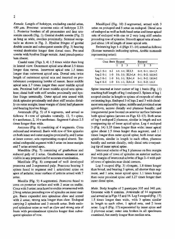
Female. Length of holotype, excluding caudal setae,400 µm. Prosome : urosome ratio of holotype 2.33 :1 . Posterior borders of all prosomites and first uro-somite smooth (Fig. 1) . Genital double somite (Fig. 2)as long as wide, swollen proximally. Seminal recep-tacle as shown in Fig. 2. Hyaline fringes of genitaldouble somite and subsequent somite (Fig . 3) bearingventral denticules longer than dorsal ones . Pre-analsomite with hyaline fringe serrate . Anal pseudopercu-lum absent.
Caudal rami (Figs 3, 4) 1 .5 times wider than longin lateral view. Outermost apical seta about 2 .5 timeslonger than ramus . Innermost apical seta 1 .5 timeslonger than outermost apical seta . Dorsal seta twicelength of outermost apical seta and inserted on pro-tuberance overpassing border of ramus . Inner middleapical seta 1 .7 times longer than outer middle apicalseta . Proximal half of inner middle apical seta spinu-lose ; distal half with stiff setules proximally and hya-line fringe terminally. Outer apical seta armed withthick spinules proximally and short stiff setules distal-ly on outer margin; inner margin of distal half plumoseand bearing hyaline fringe .
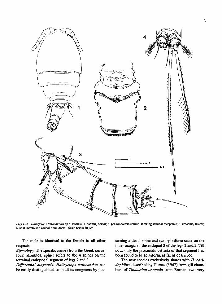
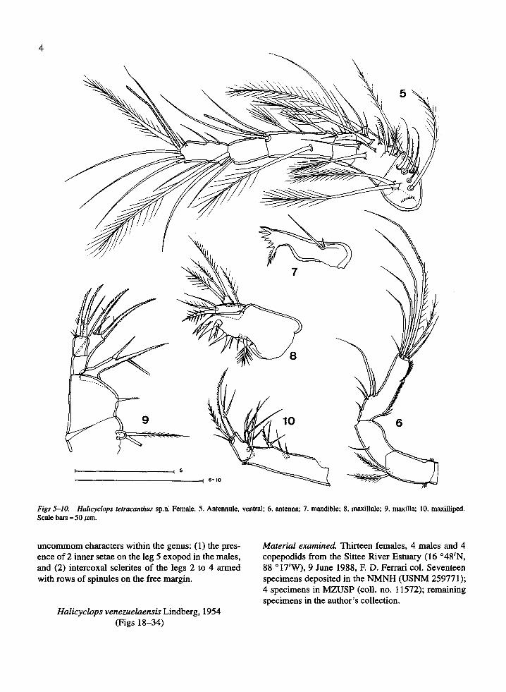
Antennule (Fig . 5) of 6 segments, and armed asfollows: 8 + row of spinules ventrally, 12, 5 + spine,6 + aesthetasc, 2, 10 + aesthetasc. Segment 4 about 2.5times longer than wide .
Antenna (Fig . 6) consisting of 4 segments . Coxareduced and unarmed . Basis with row of few spinuleson both inner and outer margins proximally, and 2 setaeat inner corner; seta representing exopod absent. Ter-minal endopodal segment with 5 setae on inner marginand 7 setae around apex .
Mandible (Fig . 7) consisting of gnathobase andreduced palp of 2 setae. Gnathobasic armament notvisible in any preparation for accurate examination .
Maxillule (Fig. 8) composed of well developedpraecoxa and 2-segmented palp . Praecoxa bearing 3spines fused to segment and 1 articulated spine onapex of arthrite ; inner surface of arthrite armed with 7setae.
Maxilla (Fig. 9) 4-segmented . Praecoxa fused tocoxa on posterior surface and with 2 setae on endite .Coxa with 3 setae ; seta fused to endite ornamented with2 long setules preceding row of spinules on inner mar-gin. Basis expanded into spinulose claw and armedwith 2 setae; strong seta longer than claw. Endopodcarrying 2 spinulose and 3 smooth setae . Both endo-pod spinulose setae as well as claw and strong seta ofbasis with proximalmost spinules longer than subse-quent spinules of row.
Maxilliped (Fig . 10) 2-segmented, armed with 3setae on protopod and 5 setae on endopod . Distal setaof endopod as well as both basal setae and inner apicalseta of endopod with one or 2 very long stiff setulespreceding row of spinules . Smooth apical seta of endo-pod about 1/4 of length of inner apical seta .
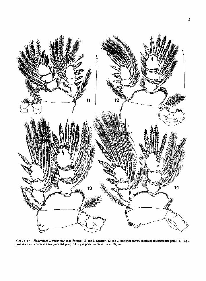
Swimming legs 1-4 (Figs 11-14) armed as follows(Roman numerals indicating spines, Arabic numeralsrepresenting setae) :
Spine inserted at inner corner of leg 1 basis (Fig . 11)reaching half length of leg 1 endopod 2 . Spines of leg 1exopod similar in length to spines of exopods of otherswimming legs . Endopod 3 of legs 2 and 3 with distal-most seta replaced by spine ; middle and proximal setaespiniform, serrate distally and plumose basally ; andintegumental pore located on anterior surface betweenboth apical spines (arrows on Figs 12-13) . Both setaeof leg 4 endopod 2 plumose, similar in length and notoverpassing tip of inner apical spine . Leg 4 endopod3 (Fig. 14) 1.35 times longer than wide ; inner apicalspine about 1 .5 times longer than segment, and 1 .1times longer than outer apical spine ; both inner setaespiniform, similar in length to each other, plumosebasally and serrate distally; only distal seta overpass-ing tip of inner apical spine .
Intercoxal sclerite of leg 1 plumose on free marginand with pair of rows of spinules on anterior surface .Free margin of intercoxal sclerite of legs 2-4 with pairof rows of spinules near distal corners .
Leg 5 exopod (Fig . 3) elongate, 1 .8 times longerthan broad, and bearing 3 spines, all shorter than seg-ment, and 1 seta ; inner apical spine 1 .1 times longerthan outer proximal spine and 1 .27 times longer thanouter distal spine.
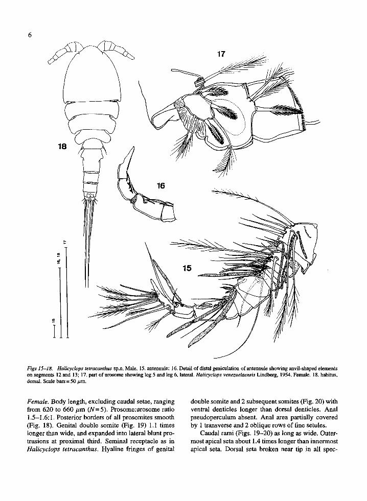
Male. Body lengths of 2 paratypes 335 and 340 µm .Urosome with 6 somites . Antennule of 14 segmentsand armed as in Figs 15 and 16 . Leg 5 exopod (Fig. 17)1.5 times longer than wide, with 3 spines similarin length to each other, 1 apical seta, and 2 innersetae . Leg 6 (Fig . 17) represented by inner spine and2 plumose setae; outer seta broken in all specimensexamined, but surely longer than median seta .
Coxa Basis Exopod Endopod1
2 3 1 2 3
Leg 1 0-1 1-1 I-I ; I-1 ; 111,2,3 0-1 ; 0-1 ; 1,1+1,3Leg 2 0-1 1-0 I-1 ; I-1 ; 111,1+1,4 0-1 ; 0-2 ; I,I1,1+2Leg 3 0-1 1-0 I-1 ; I-1 ; 11,1+1,4 0-1 ; 0-2 ; 1,11,1+2Leg 4 0-1 1-0 I-1 ; I-1 ; 11,1+1,4 0-1 ; 0-2; I,II,2
Figs 14. Halicyclops tetracmrhus sp.n. Female. 1. habitus, dorsal; 2. genital double somite, showing seminal receptacle; 3. urosome, lateral; 4. anal somite and caudal rami, dorsal. Scale bars = 50 pm.
The male is identical to the female in all other sessing a distal spine and two spiniform setae on the respects. inner margin of the endopod 3 of the legs 2 and 3. Till Etymology. The specific name (from the Greek fetrar, now, only the proximahnost seta of that segment had four; akanthos, spine) refers to the 4 spines on the been found to be spiniform, as far as described. terminal endopodal segment of legs 2 and 3. The new species exclusively shares with H. cari- Differential diagnosis. Halicyclops tetracanthus can dophilus, described by Humes (1947) from gill cham- be easily distinguished from all its congeners by pos- bers of Thalassina anomula from Borneo, two very
4
Figs 5-10. Halicyclops tetracanthus sp.n: Female. 5 . Antennule, ventral ; 6. antenna; 7. mandible; 8 . maxillule ; 9. maxilla; 10 . maxilliped .Scale bars = 50 µm .
uncommom characters within the genus : (1) the pres-ence of 2 inner setae on the leg 5 exopod in the males,and (2) intercoxal sclerites of the legs 2 to 4 armedwith rows of spinules on the free margin .
Halicyclops venezuelaensis Lindberg, 1954(Figs 18-34)
Material examined. Thirteen females, 4 males and 4copepodids from the Sittee River Estuary (16 °48'N,88 '17'W), 9 June 1988, F. D . Ferrari col . Seventeenspecimens deposited in the NMNH (USNM 259771) ;4 specimens in MZUSP (coil . no. 11572); remainingspecimens in the author's collection .
5
Figs 11-14. Halicyclops tetracanthus sp.n. Female. 11 . leg 1, anterior ; 12 . leg 2, posterior (arrow indicates integumental pore) ; 13 . leg 3,posterior (arrow indicates integumental pore) ; 14. leg 4, posterior. Scale bars= 50 um.
6
Figs 15-18. Halicyclops tetracanthus sp.n. Male. 15. antennule ; 16 . Detail of distal geniculation of antennule showing anvil-shaped elementson segments 12 and 13 ; 17. part of urosome showing leg 5 and leg 6, lateral . Halicyclops venezuelaensis Lindberg, 1954. Female. 18. habitus,dorsal. Scale bars = 50 µm .
Female . Body length, excluding caudal setae, rangingfrom 620 to 660 µm (N=5). Prosome :urosome ratio1 .5-1 .6 :1 . Posterior borders of all prosomites smooth(Fig. 18) . Genital double somite (Fig . 19) 1 .1 timeslonger than wide, and expanded into lateral blunt pro-trusions at proximal third . Seminal receptacle as inHalicyclops tetracanthus . Hyaline fringes of genital
double somite and 2 subsequent somites (Fig . 20) withventral denticles longer than dorsal denticles. Analpseudoperculum absent. Anal area partially coveredby 1 transverse and 2 oblique rows of fine setules .
Caudal rami (Figs . 19-20) as long as wide . Outer-most apical seta about 1 .4 times longer than innermostapical seta . Dorsal seta broken near tip in all spec-
7
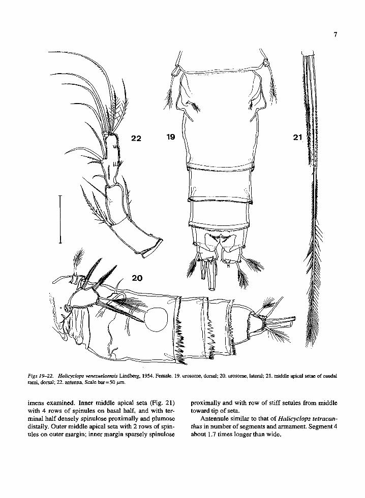
Figs 19-22. Halicyclops venezuelaensis Lindberg, 1954 . Female. 19 . urosome, dorsal ; 20 . urosome, lateral ; 21 . middle apical setae of caudalrami, dorsal ; 22 . antenna. Scale bar= 50 µm .
imens examined . Inner middle apical seta (Fig. 21)
proximally and with row of stiff setules from middlewith 4 rows of spinules on basal half, and with ter-
toward tip of seta.minal half densely spinulose proximally and plumose Antennule similar to that of Halicyclops tetracan-distally. Outer middle apical seta with 2 rows of spin- thus in number of segments and armament . Segment 4ules on outer margin ; inner margin sparsely spinulose
about 1.7 times longer than wide .
8
Antenna (Fig . 22) 4-segmented. Coxa reduced andunarmed. Basis with rows of few spinules on inner andouter margins proximally and 2 setae at inner corner ;seta representing exopod present . Terminal segmentbearing 5 setae on inner margin and 7 setae aroundapex .
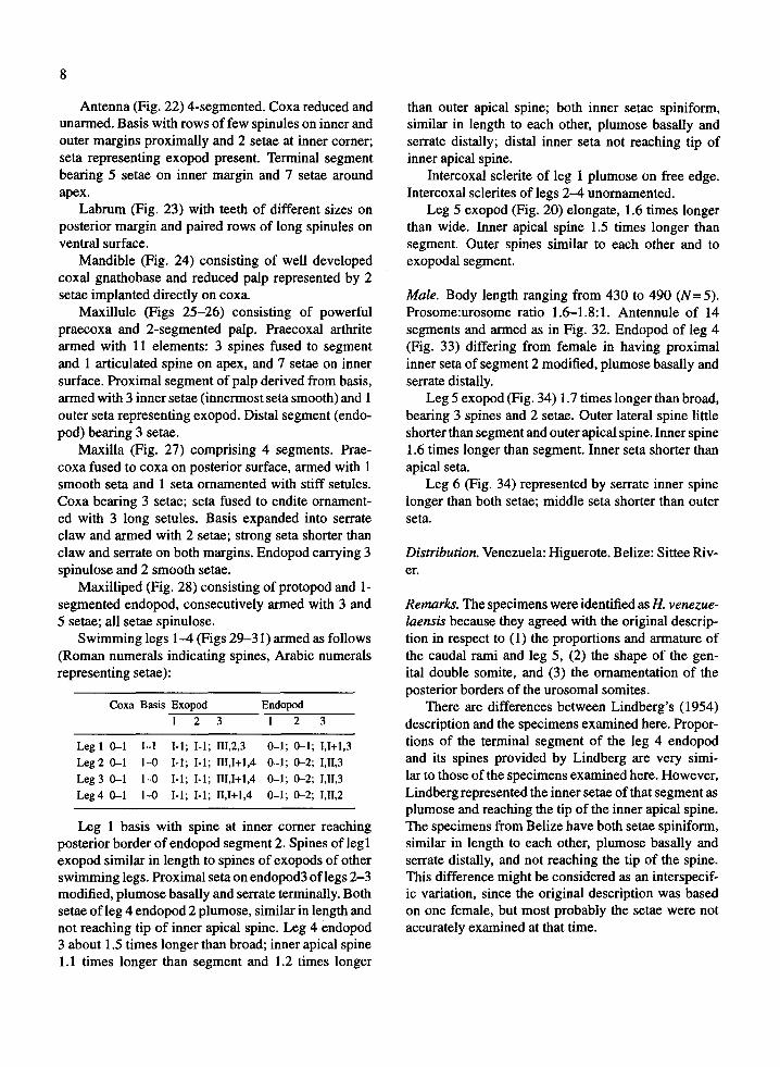
Labrum (Fig . 23) with teeth of different sizes onposterior margin and paired rows of long spinules onventral surface .
Mandible (Fig. 24) consisting of well developedcoxal gnathobase and reduced palp represented by 2setae implanted directly on coxa.
Maxillule (Figs 25-26) consisting of powerfulpraecoxa and 2-segmented palp. Praecoxal arthritearmed with 11 elements : 3 spines fused to segmentand 1 articulated spine on apex, and 7 setae on innersurface . Proximal segment of palp derived from basis,armed with 3 inner setae (innermost seta smooth) and 1outer seta representing exopod . Distal segment (endo-pod) bearing 3 setae .
Maxilla (Fig. 27) comprising 4 segments. Prae-coxa fused to coxa on posterior surface, armed with 1smooth seta and 1 seta ornamented with stiff setules .Coxa bearing 3 setae ; seta fused to endite ornament-ed with 3 long setules . Basis expanded into serrateclaw and armed with 2 setae ; strong seta shorter thanclaw and serrate on both margins . Endopod carrying 3spinulose and 2 smooth setae .
Maxilliped (Fig . 28) consisting of protopod and 1-segmented endopod, consecutively armed with 3 and5 setae ; all setae spinulose .
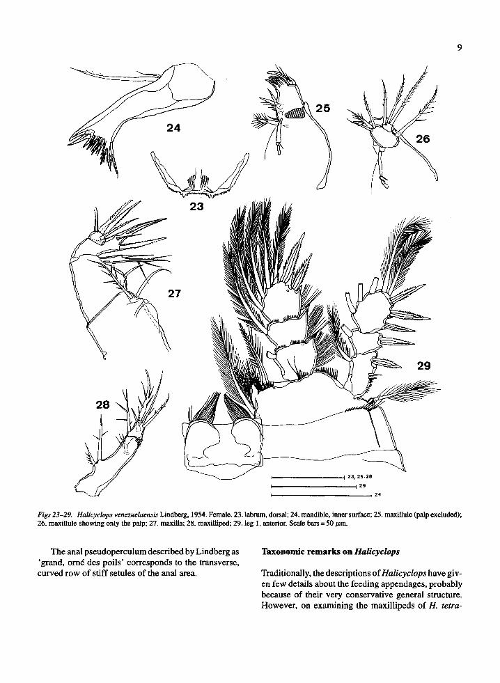
Swimming legs 1-4 (Figs 29-31) armed as follows(Roman numerals indicating spines, Arabic numeralsrepresenting setae) :
Leg 1 basis with spine at inner corner reachingposterior border of endopod segment 2 . Spines of leg Iexopod similar in length to spines of exopods of otherswimming legs . Proximal seta on endopod3 of legs 2-3modified, plumose basally and serrate terminally . Bothsetae of leg 4 endopod 2 plumose, similar in length andnot reaching tip of inner apical spine . Leg 4 endopod3 about 1.5 times longer than broad ; inner apical spine1 .1 times longer than segment and 1 .2 times longer
than outer apical spine ; both inner setae spiniform,similar in length to each other, plumose basally andserrate distally ; distal inner seta not reaching tip ofinner apical spine .
Intercoxal sclerite of leg 1 plumose on free edge .Intercoxal sclerites of legs 2-4 unornamented .
Leg 5 exopod (Fig . 20) elongate, 1.6 times longerthan wide . Inner apical spine 1 .5 times longer thansegment. Outer spines similar to each other and toexopodal segment.
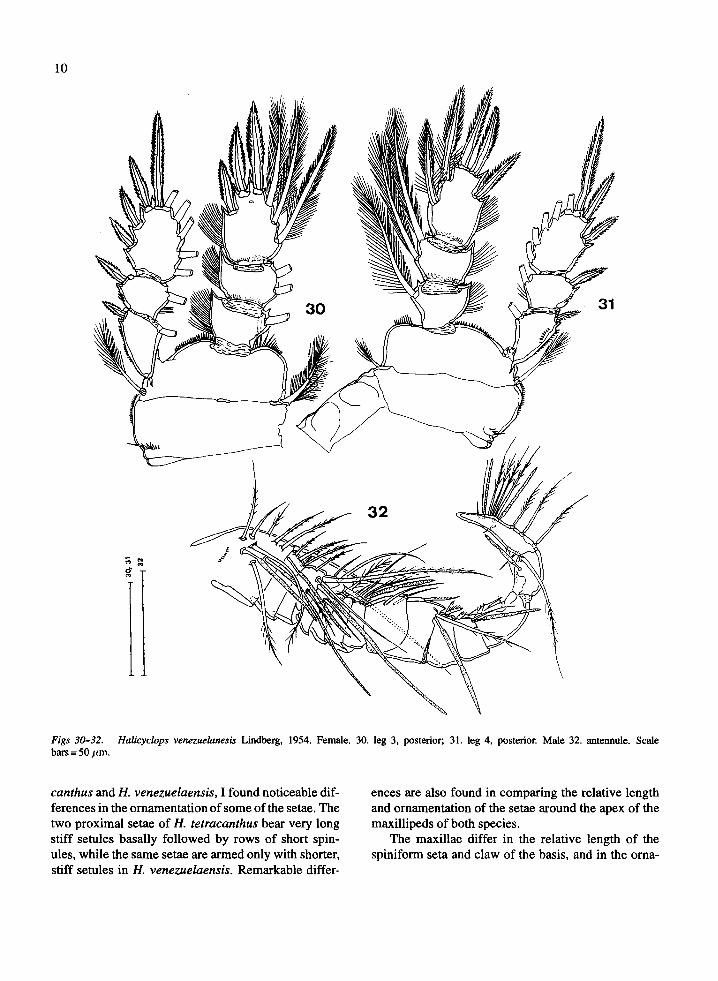
Male . Body length ranging from 430 to 490 (N=5) .Prosome:urosome ratio 1 .6-1.8 :1 . Antennule of 14segments and armed as in Fig. 32. Endopod of leg 4(Fig. 33) differing from female in having proximalinner seta of segment 2 modified, plumose basally andserrate distally.
Leg 5 exopod (Fig . 34) 1 .7 times longer than broad,bearing 3 spines and 2 setae. Outer lateral spine littleshorter than segment and outer apical spine . Inner spine1 .6 times longer than segment. Inner seta shorter thanapical seta .
Leg 6 (Fig. 34) represented by serrate inner spinelonger than both setae ; middle seta shorter than outerseta .
Distribution. Venezuela : Higuerote. Belize : Sittee Riv-er.
Remarks. The specimens were identified as H. venezue-laensis because they agreed with the original descrip-tion in respect to (1) the proportions and armature ofthe caudal rami and leg 5, (2) the shape of the gen-ital double somite, and (3) the ornamentation of theposterior borders of the urosomal somites .
There are differences between Lindberg's (1954)description and the specimens examined here . Propor-tions of the terminal segment of the leg 4 endopodand its spines provided by Lindberg are very simi-lar to those of the specimens examined here . However,Lindberg represented the inner setae of that segment asplumose and reaching the tip of the inner apical spine .The specimens from Belize have both setae spiniform,similar in length to each other, plumose basally andserrate distally, and not reaching the tip of the spine .This difference might be considered as an interspecif-ic variation, since the original description was basedon one female, but most probably the setae were notaccurately examined at that time .
Coxa Basis Exopod Endopod1 2 3 1 2 3
Leg 1 0-1 1-I I-1 ; I-1 ; 111,2,3 0-1 ; 0-1 ; 1,1+1,3Leg 2 0-1 1-0 I-1 ; I-1 ; 111,1+1,4 0-1 ; 0-2 ; I,II,3
Leg 3 0-1 1-0 I-1 ; I-1 ; 111,1+1,4 0-1 ; 0-2 ; 1,11,3Leg4 0-1 1-0 I-1 ; I-1 ; 11,1+1,4 0-1 ; 0-2 ; 1,11,2
Figs 23-29. Halicyclops venezuelaensis Lindberg, 1954 . Female . 23. labrum, dorsal ; 24 . mandible, inner surface ; 25 . maxillule (palp excluded) ;26. maxillule showing only the palp ; 27 . maxilla; 28 . maxilliped ; 29 . leg 1, anterior. Scale bars = 50 µm .
The anal pseudoperculum described by Lindberg as
Taxonomic remarks on Halicyclops`grand, ornd des poils' corresponds to the transverse,curved row of stiff setules of the anal area .
9
Traditionally, the descriptions of Halicyclops have giv-en few details about the feeding appendages, probablybecause of their very conservative general structure .However, on examining the maxillipeds of H. tetra-
10
Figs 30-32 . Halicyclops venezuelanesis Lindberg, 1954. Female . 30 . leg 3, posterior ; 31 . leg 4, posterior. Male 32 . antennule . Scalebars = 50 tim,
canthus and H. venezuelaensis, I found noticeable dif-
ences are also found in comparing the relative lengthferences in the ornamentation of some of the setae. The
and ornamentation of the setae around the apex of thetwo proximal setae of H. tetracanthus bear very long
maxillipeds of both species .stiff setules basally followed by rows of short spin-
The maxillae differ in the relative length of theules, while the same setae are armed only with shorter,
spiniform seta and claw of the basis, and in the orna-stiff setules in H. venezuelaensis . Remarkable differ-
Figs 33-34. Halicyclops venezuelaensis Lindberg, 1954 . Male .33 . endopod of leg 4, posterior; 34. part of urosome with leg 5 andleg 6, ventro-lateral . Scale bar=50 µm .
mentation of the longer seta on the terminal coxalendite .
I suggest that these two appendages be examinedwith more detail in future descriptions .
The morphology of the male antennule of Hali-cyclops is not usually detailed in descriptions, mostprobably due to the difficulty in seeing the segmen-tation and armament clearly. Comparison of the maleantennules of H. tetracanthus and H. venezuelaensisshowed the same number of segments in both species ;the only differences concerning the armament are inthe length of setae and aesthetascs .
Acknowledgments
The author is indebted to Dr Frank D . Ferrari who kind-ly made available the material for this study . Thanksare due to Drs Frank D . Ferrari and Janet L . W. Reidfor reading and commenting on the manuscript .
This is contribution #420 of the Caribbean CoralReef Ecosystems Program .
References
Humes, A . G ., 1947. A new cyclopoid copepod from a Bomeancrustacean . Trans . am. microsc. Soc . 66 : 293-301 .
Lindberg, K., 1954. Cyclopides (Crustaces Copdpodes) del'Amerique du Sud. Ark . Zool . 7 : 193-222 .
Reid, J . W., 1990. Continental and coastal free-living Copepo-ds (Crustacea) of Mexico, Central America and the CaribbeanRegion . In: D . Navarro L. & J . G. Robinson, (eds), DiversidadBiologica en la Reserva de la Biosfera de Sian Ka'an, QuintanaRoo, Mexico . Chetumal, Centro de Investigaciones de QuintanaRoo (CIQRO)/Program of Studies in Tropical Conservation, Uni-versity of Florida, 175-213 .
I1