Embed Size (px)
Citation preview
Coordination between Differentially RegulatedCircadian Clocks Generates Rhythmic Behavior
Deniz Top and Michael W. Young
Laboratory of Genetics, The Rockefeller University, New York, New York 10065
Correspondence: [email protected]
Specialized groups of neurons in the brain are key mediators of circadian rhythms, receivingdaily environmental cues and communicating those signals to other tissues in the organismfor entrainment and to organize circadian physiology. InDrosophila, the “circadian clock” ishoused in seven neuronal clusters, which are defined by their expression of the main circa-dian proteins, Period, Timeless, Clock, and Cycle. These clusters are distributed across the flybrain and are thereby subject to the respective environments associatedwith their anatomicallocations. While these core components are universally expressed in all neurons of thecircadian network, additional regulatory proteins that act on these components are differ-entially expressed, giving rise to “local clocks”within the network that nonetheless convergeto regulate coherent behavioral rhythms. In this review, we describe the communicationbetween the neurons of the circadian network and the molecular differences withinneurons of this network. We focus on differences in protein-expression patterns anddiscuss how such variation can impart functional differences in each local clock. Finally,we summarize our current understanding of how communication within the circadiannetwork intersects with intracellular biochemical mechanisms to ultimately specify behav-ioral rhythms. We propose that additional efforts are required to identify regulatory mecha-nisms within each neuronal cluster to understand the molecular basis of circadian behavior.
Circadian rhythms are behavioral and physi-ological responses that allow anticipation of
daily changes in the environment, such as day/night cycles. Indeed, circadian rhythms are en-trained by diurnal oscillations in light, tempera-ture, and food availability, each of which can beconsidered a “zeitgeber,” or “time giver.” Suchanticipatory behavior of daily events confersadaptive advantages on individuals that organizephysiology around planetary rhythms, as well aswithin a species as a whole, for purposes of mat-ing, cooperation, protection, etc. In a sense,
rhythmic anticipation can be thought of as aprimitivemechanismof planning and cognition.
The circadian clock, consisting of transcrip-tional negative feedback loops, underlies theregulation of a ∼24-h behavioral rhythm. Themolecular architecture of this feedback loop isconserved across the majority of multicellularorganisms. In animals, the “master clock” ishoused in specialized pacemaker neurons inthe brain that coordinate various “peripheralclocks” throughout the organism to regulatedaily physiological and behavioral changes.
Editors: Paolo Sassone-Corsi, Michael W. Young, and Akhilesh B. ReddyAdditional Perspectives on Circadian Rhythms available at www.cshperspectives.org
Copyright © 2017 Cold Spring Harbor Laboratory Press; all rights reservedAdvanced Online Article. Cite this article as Cold Spring Harb Perspect Biol doi: 10.1101/cshperspect.a033589
1
on August 28, 2018 - Published by Cold Spring Harbor Laboratory Press http://cshperspectives.cshlp.org/Downloaded from
Importantly, the various clocks can adapt tochanges in daily environmental rhythms, suchas the change in light/dark cycles through theseasons, while still maintaining their coordina-tion with each other. Thus, in addition to itsrole in anticipatory behavior, the master clockensures that various organs and tissues arephysiologically synchronized to respond coop-eratively to daily changes in the environment.
In this review, we focus on the circadian sys-tem ofDrosophila melanogaster. Themajority ofgenes involved in the regulation of circadianrhythms were first discovered in Drosophila,with subsequent analogous or orthologousgenes cloned in organisms ranging from fungito plants tomammals. The genetic tractability ofthe fly and its simpler circadian neuronal net-work have allowed us to begin to dissect thebiochemical mechanisms involved in regulatingbehavioral rhythms with molecular resolution.Here we review the latest developments in thebiochemistry of the Drosophila circadian clockas it relates to its neuronal network, and fit theknownmolecularmodel of the clock into the bio-chemical restrictions imposed upon each neuron.We will also highlight the molecular pathwaysoperating within the neurons that allow commu-nication between the clocks. Where appropriate,key gaps in our knowledge will be noted.
THE CIRCADIAN CLOCK NEURONALNETWORK
The circadian clock, also referred to as the mo-lecular oscillator in this review, is a transcrip-tional negative feedback loop that oscillateswith a∼24-h period to regulate rhythmic behav-ior and physiology. In Drosophila, the masterclock is housed in ∼150 neurons that composethe circadian clock neuronal network (CCNN).These neurons are defined by their expression ofthe core circadian proteins Clock (CLK), Cycle(CYC), Period (PER), and Timeless (TIM)(Houl et al. 2006; Kaneko and Hall 2000). CLKand CYC assemble into a DNA-binding tran-scriptional activator complex that regulates thetranscription of per and tim. After translation,PER and TIM proteins assemble into a tran-scriptional repressor complex (PER/TIM) that
binds to and blocks the activity of CLK/CYC,closing a negative feedback loop that oscillateswith ∼24-h periodicity (discussed in detail be-low). Loss-of-functionmutations in any of thesecore circadian proteins leads to arrhythmic be-havior, underscoring their central role withinthe molecular oscillator. Many clock neuronsexpress the light-sensing protein Cryptochrome(CRY) that communicates light signals to thecircadian clock, allowing light-mediated reset-ting of the oscillator and therefore rhythmic be-havior. The retina, which also expresses coreclock components, also participates in transmit-ting light information independently of CRY tothe CCNN. Neurons of the CCNN are groupedin seven clusters within the brain, and are namedby their anatomical location: small and largeventral lateral neurons (s-LNvs and l-LNvs, re-spectively), fifth s-LNv, dorsal lateral neurons(LNds), and three clusters of dorsal neurons(DNs 1–3) (Fig. 1A). In this section, we describesignaling between these clusters and how thiscommunication influences their internal bio-chemistry.
The Dual Oscillator Model
The rhythmic behavior pattern of the fly is cre-puscular, with peaks of activity during the tran-sitions between light and dark (i.e., sunrise andsunset). These peaks of activity are flanked byperiods of low activity reflecting sleep at night orsiesta in the day and define the periodicity ofrhythmic behavior. Exposure to light for longerperiods of time advances morning anticipationwhile simultaneously delaying evening anticipa-tion. The observation of this divergent anticipa-tory behavior led to the hypothesis of a dualoscillator model for regulating rhythmic behav-ior (Pittendrigh and Daan 1976). The dual os-cillatormodel proposed the existence of separatemorning and evening oscillators that convergeto regulate rhythmic behavior. Studies con-ducted over 10 years ago provided the first ana-tomical evidence to support the dual oscillatormodel. Genetic ablation of the s-LNvs and thel-LNvs leads to a loss of morning anticipation,while ablating the LNds, the fifth s-LNv, andsome DNs leads to a loss of evening anticipation
D. Top and M.W. Young
2 Advanced Online Article. Cite this article as Cold Spring Harb Perspect Biol doi: 10.1101/cshperspect.a033589
on August 28, 2018 - Published by Cold Spring Harbor Laboratory Press http://cshperspectives.cshlp.org/Downloaded from
sNP
FC
haIT
PN
PF
PD
F
PD
FR
AcC
h-R
GA
BA
A R
NP
F-R
1
DN
1D
N2
DN
3LN
dF
ifth
s-LN
vl-L
Nv
s-LN
v
AB
C
Paleommatidia
Yellowommatidia
R8 Rh6
R7 Rh4
R1-
6R
h1
R8
Rh5
R7 Rh3
R1-
6R
h1
PD
F
NP
F
CK
2αC
RY
DE
FG
AC
h
AC
hH
is
H-B
eyel
et
cAM
P
ITP
CR
Y
NP
F
Term
inal
PD
F
NP
F-R
1
TIM
PE
R
H
Abundance(not relative, a.u.)
Zei
tgeb
er ti
me
sNP
F
+
–
Figu
re1.(See
legend
onfollowingpage.)
Coordination between Differentially Regulated Circadian Clocks
Advanced Online Article. Cite this article as Cold Spring Harb Perspect Biol doi: 10.1101/cshperspect.a033589 3
on August 28, 2018 - Published by Cold Spring Harbor Laboratory Press http://cshperspectives.cshlp.org/Downloaded from
(Stoleru et al. 2004). Furthermore, restoring PERexpression to LNvs of per mutant flies restoresmorning but not evening anticipation, whereasrestoring PER in both the LNvs and LNds re-stores both anticipatory behaviors (Grima et al.2004). These studies established the LNvs as“morning cells” and the remaining cells as “eve-ning cells” (Grima et al. 2004; Stoleru et al. 2004,2005; Rieger et al. 2006; Guo et al. 2014). Morerecent genetic manipulations have suggestedthat four neurons, including three of the sixLNds and the fifth s-LNv, are sufficient to serveas evening cells (Guo et al. 2014). However, ma-nipulation of DNs can still alter evening antici-pation (Yao et al. 2016; Guo et al. 2016), suggest-ing that these clusters are nonetheless involvedin this behavior. Among morning cells (LNvs),the s-LNvs serve as master pacemaker neuronsbecause they drive rhythmic activity in constantdark conditions (Grima et al. 2004; Stoleru et al.2005; Rieger et al. 2006); without s-LNv activity,flies become arrhythmic. In the framework ofthe dual oscillator model, the morning cells (s-LNvs) dominate in the dark to drive coherentbehavioral rhythms and also govern the timingof evening anticipation by communicating tothe evening cells, placing the morning cells atthe top of a neuronal hierarchy.
Since the establishment of the dual oscillatormodel, the hierarchical organization of the cir-cadian neuronal clusters has been challengedand a more complex understanding of commu-nication within this network has begun toemerge. In constant light conditions that permitrhythmic behavior (i.e., in flies lacking the light-sensing protein CRY; cry0), the LNd/fifth s-LNvcells dominate, driving behavioral rhythmicity
when the s-LNvs are either arrhythmic or unableto communicate with the other clusters (Muradet al. 2007; Picot et al. 2007). This exception sug-gests that s-LNv dominance over the neuronalhierarchy is limited to constant dark conditions.The partitioned roles the different clusters playin morning and evening anticipation becomeimportant in seasonal changes of day length,where the evening cells dominate the CCNN inthe long days of summer, and the morning cellsdominate theCCNN in the longnights ofwinter,with communication between the two groupsmaintaining a 24-h behavioral rhythm (Stoleruet al. 2007). In molecular terms, longer light ex-posure accelerates the LNv clocks, resulting inadvanced morning anticipation, and deceleratesthe LNd/fifth s-LNv/DN clocks to delay eveninganticipation, as predicted by the dual oscillatormodel (Pittendrigh and Daan 1976). Anotherexception to the dual oscillator model comesfrom observed morning anticipation in dimlight in flies lacking functional morning cells(but possessing functional evening cells) (Bach-leitner et al. 2007; Rieger et al. 2009). Thus, theevening cells can compensate for the loss ofmorning cell activity in dim light conditions andoscillate without being driven by the s-LNvs. Asa result, the dual oscillator model is expanded.
The relationships within the CCNN are in-creasingly revealing higher orders of complexity.In addition to s-LNv (master clock) communi-cation to the DN1s, feedback signals from theDN1s back to the s-LNvs has also been identi-fied, suggesting there is bidirectional flow of in-formation between morning and evening cells(Guo et al. 2016). The LNds also communicatebidirectionally with s-LNvs (discussed in detail
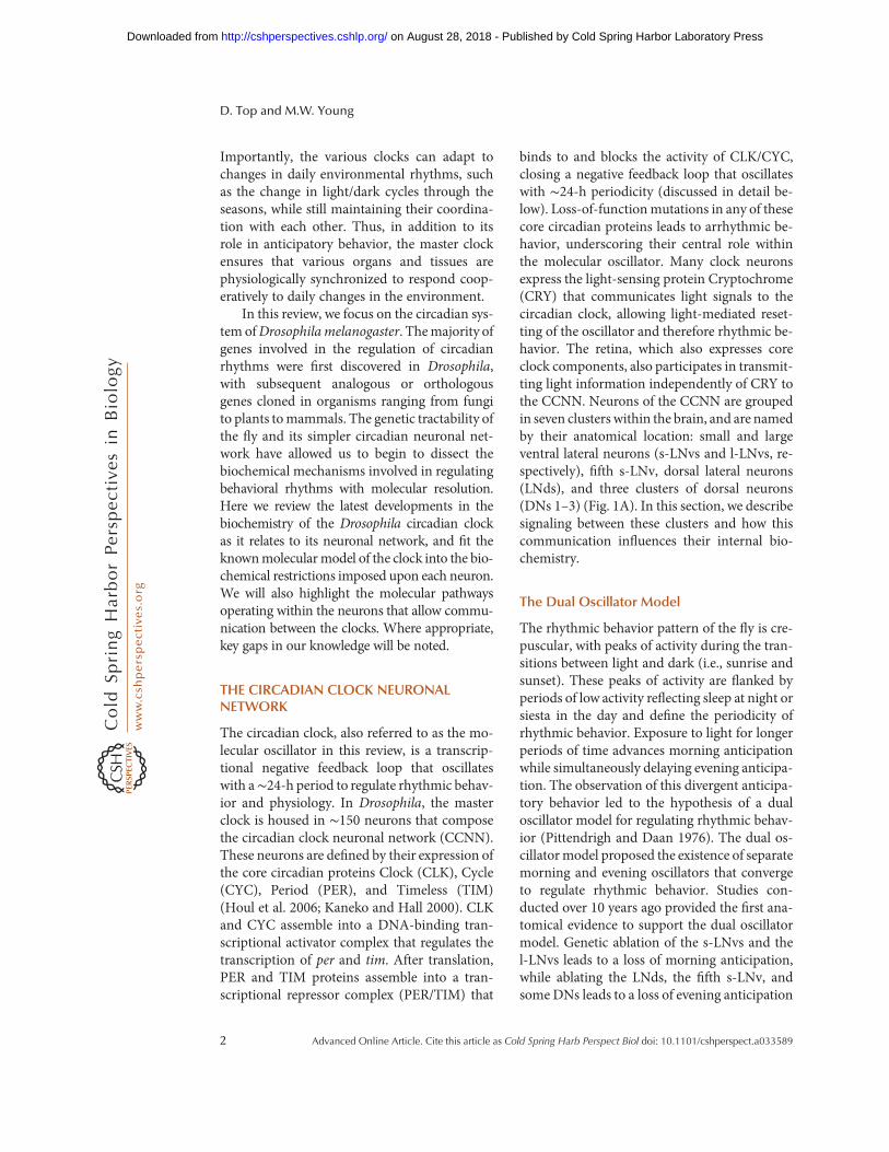
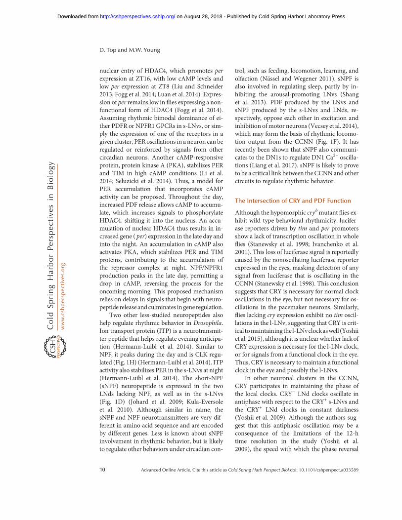
Figure 1.A schematic of the anatomical structure of the fly brain circadian clock neuronal network. (A) Locationand names of the seven neuronal clusters that make up the circadian neuronal network. (B) Structure of theommatidia that make up the fly retina, displaying the expression of rhodopsins (Rh) in receptor cells (R). Two ofthe receptor cells within R1-6 are removed from the schematic to reveal R7 and R8 for clarity. (C) The relativeposition of the receptor cells andH-B eyelet with respect to the LNvs in the brain. The positive and negative effectsof acetylcholine (ACh) and histamine (His) are shown in gray. (D) The expression pattern of neuropeptides ineach of the circadian neuronal clusters. (E) The expression pattern of receptors in each of the circadian neuronalcluster. (F) The sources and the targets of pigment-dispersing factor (PDF), neuropeptide F (NPF), and shortneuropeptide F (sNPF). (+) and (−) symbols represent the functional effect of the neuropeptides in nonspecifiedmotor neurons. (G) The expression pattern of CK2α and CRY in each neuronal cluster. (H ) A schematic of peakexpression of indicated proteins. Arrows represent the time of peak expression and are color-coded.
D. Top and M.W. Young
4 Advanced Online Article. Cite this article as Cold Spring Harb Perspect Biol doi: 10.1101/cshperspect.a033589
on August 28, 2018 - Published by Cold Spring Harbor Laboratory Press http://cshperspectives.cshlp.org/Downloaded from
below), and it is likely that other groups of neu-rons within the CCNN communicate similarly.Overexpression of kinases that selectively accel-erate or decelerate different clocks within theCCNN lead to split rhythms (a single behavioralrhythm split into behavioral components withshort and long periodicity), demonstrating thatrhythmic behavior is an emergent property ofthe communication of the various independentoscillators (Yao and Shafer 2014). Thus, inde-pendent molecular oscillators within each clus-ter are coordinated and synchronized to give riseto coherent behavioral rhythms.
Input from the Eye
Light is an important zeitgeber that helps en-train and reset the circadian clock. Light inputto the clock occurs through the deep brain lightreceptor CRY (Stanewsky et al. 1998; Emeryet al. 2000a; Dolezelova et al. 2007) and throughthe fly retina via rhodopsin activation (Helfrich-Förster et al. 2001). While light-activated CRYinduces the degradation of TIM to reset theclock in the brain (Stanewsky et al. 1998; Kohet al. 2006; Peschel et al. 2009), light-activatedrhodopsin in the retina triggers Gqα protein toactivate no receptor potential A (norpA), a phos-pholipase that depolarizes the cell and triggersrelease of the neurotransmitter histamine toinfluence the CCNN (Bloomquist et al. 1988;Montell 2012; Hardie and Juusola 2015). How-ever, the eye does not contribute to the circadianclock through CRY-dependent light input (Em-ery et al. 2000b; Bachleitner et al. 2007; Yoshiiet al. 2015), despite expressing CRY (Yoshii et al.2008), suggesting that these two light pathwaysconverge independently to synchronize the cir-cadian clock to the environment. More recently,a deep brain rhodopsin, Rh7, was demonstratedto be involved in light entrainment of pacemak-er neurons (Ni et al. 2017). Undoubtedly, futureworkwill uncover themolecularmechanisms bywhich Rh7 informs the circadian clock.
Different sections of the fly compound eyecommunicatewith circadian neurons to regulaterhythmic behavior. The retina of the compoundeye is composed of pale and yellow ommatidiamade up of clusters of receptor cells, designated
R1-8 (Fig. 1B) (Behnia and Desplan 2015). R7and R8 in the pale ommatidia express rhodopsin(Rh) 3 and Rh5, respectively, and regulate twi-light activity at dawn and dusk (Schlichting et al.2015). R8 in the yellow ommatidia expressesRh6 and R1-6 expresses Rh1; collectively theyregulate activity in dim light (e.g., moonlight)(Schlichting et al. 2014). Thus, rhodopsins with-in the receptor cells can communicate dim lightsignals to the brain to regulate activity. It is likelythat these cells can regulate morning anticipa-tion when flies lack functional s-LNvs. R1-6send projections from the retina into the laminaof the optic lobe, whereas R7 and R8 projectfurther into the brain, to the medulla, wherethey are in proximity to LNv dendrites (Fig.1C) (Helfrich-Förster et al. 2002). Despite thisanatomical proximity, there is as yet no evidenceof synaptic connections between the ommatidiaand the l-LNvs despite the influence of R1-6 onmoonlight activity (Schlichting et al. 2014).Thus, any R1-6 communication with the CCNNlikely occurs through nonsynaptic signaling.
On the other hand, a second structure in theeye, beneath the receptor cells, forms a synapticconnection with the CCNN. The Hofbauer–Buchner (H-B) eyelet, a structure located be-tween the retina and the lamina, communicateswith the LNvs (Fig. 1C) (Hofbauer and Buchner1989; Yasuyama andMeinertzhagen 1999; Mal-pel et al. 2002). The H-B eyelet is a remnant ofthe larval Bolwig organ, which plays a develop-mental role in the normal arborization of theLNvs in the medulla (Malpel et al. 2002). TheH-B eyelet expresses Rh6, similarly to R1-6, andis involved in light entrainment (Veleri et al.2007; Szular et al. 2012; Schlichting et al.2016). The H-B eyelet makes a synaptic connec-tion with the LNvs, communicating throughacetylcholine and histamine release (Helfrich-Förster et al. 2002;Malpel et al. 2002; Schlichtinget al. 2014, 2016; Muraro and Ceriani 2015).Blocking synaptic transmission between the H-B eyelet and the LNvs hinders behavioral phaseshifts, as well as inhibits neuronal synchrony ofTIM/PER oscillations (Veleri et al. 2007), sug-gesting that the H-B eyelet is important for com-municating light signals to the CCNN. Acetyl-choline signals likely underlie the transmission
Coordination between Differentially Regulated Circadian Clocks
Advanced Online Article. Cite this article as Cold Spring Harb Perspect Biol doi: 10.1101/cshperspect.a033589 5
on August 28, 2018 - Published by Cold Spring Harbor Laboratory Press http://cshperspectives.cshlp.org/Downloaded from
of light information to the CCNN because exog-enous addition of a cholinergic agonist triggers aspike in Ca2+ and cyclic adenosine monophos-phate (cAMP) levels in both LNvs (Wegeneret al. 2004; Lelito and Shafer 2012). Indeed, ace-tylcholine-mediated excitation of the l-LNvs ismediated by the compound eye (including theH-B eyelet) (Muraro and Ceriani 2015; Schlicht-ing et al. 2016), while histamine from the H-Beyelet (and/or other sources) inhibits l-LNv fir-ing (Schlichting et al. 2016). Although no evi-dence exists of acetylcholine receptor expressionin the s-LNvs, excitation of the H-B eyelet alsoleads to the excitation of the s-LNvs (Schlichtinget al. 2016), suggesting that the l-LNvs may relaysignals from the H-B eyelet. Together, thesefindings suggest that the H-B eyelet and photo-receptors communicate to the two LNv clustersthrough the l-LNvs. In summary, the fly retinacommunicates with the CCNN to stimulatecomplex Ca2+ and cAMP responses in theLNvs to influence behavioral rhythmicity.
It is important to note that, although signalsfrom the eye to the CCNN appear to be trans-mitted through the LNvs, our knowledge is in-complete. For example, flies lacking an oscillatorin theirmorning cells (both LNvs) exhibit rhyth-mic morning anticipation behavior in dim light(Rieger et al. 2009). Although dim light isthought to be mediated through a CRY-inde-pendent mechanism (Bachleitner et al. 2007),suggesting morning anticipation under theseconditions arises from retinal activity, recent ev-idence suggests that CRY may also respond toprolonged exposure to dim light in the brain(Vinayak et al. 2013). The recently discoveredRh7 also responds to dim light (Ni et al. 2017),but how it coordinates with the retinal responseis yet to be determined. In a retinal response,either the retina can also communicate to otherparts of theCCNN inways that have not yet beendetermined, or retinal communication throughthe LNvs does not require that the LNvs expressa functional oscillator. In a CRY response,morning anticipatory behavior may bypass theLNvs altogether and rely on dim light exposureof the evening cells. Future work will determinehow these mechanisms converge to regulatemorning anticipation.
The Ventral Lateral Neurons, Arousal,and Pigment-Dispersing Factor
The LNvs are the most studied of the circadianneuronal clusters and are easily identifiable bytheir expression of the neuropeptide pigment-dispersing factor (PDF) (Fig. 1D) (Helfrich-För-ster 1995). Elimination of either the LNvs orPDF causes a loss of morning anticipation andleads to arrhythmic behavior in constant darkconditions (Helfrich-Förster 1995, 1998; Rennet al. 1999; Stoleru et al. 2004). Therefore, PDFplays a central role in communication within theCCNN to maintain rhythmic activity in con-stant (dark) conditions. Although PDF expres-sion does not oscillate in the s-LNv cell bodies, itdepends on CLK activity for its expression, yetrhythmically (early morning) accumulates inthe s-LNv termini for release during the day(Fig. 1H) (Blau and Young 1999; Park et al.2000). On the other hand, PDF is not rhythmi-cally released from l-LNvs in constant condi-tions (Park et al. 2000), highlighting a functionaldifference in how the two clusters regulaterhythmic behavior through PDF.
The LNvs use PDF to play distinct roleswithin the CCNN to regulate rhythmic behav-ior. While the oscillations of the clock in the s-LNvs regulate morning anticipation and driverhythmic behavior in constant dark conditions(Grima et al. 2004; Stoleru et al. 2005), the l-LNvs are not responsible for either morning ac-tivity or evening activity. Instead, exposure tolight (sunrise) is communicated to the l-LNvneurons through the eye or by a response medi-ated by l-LNv CRY, triggering the l-LNvs to firerapidly and release PDF (Rieger et al. 2006).Neurons that express pdf receptor ( pdfr), a G-protein-coupled receptor (GPCR), respond tothe PDF released from the l-LNvs by up-regu-lating cAMP. Initially, identifying the neuronsin which pdfr is expressed proved difficult(Hyun et al. 2005; Lear et al. 2005b; Mertenset al. 2005). Later work involving epitope tag-ging PDFR, measuring pdfr mRNA expression,and critically, testing neuronal response to ex-ogenous PDF injection led to the conclusionthat s-LNvs express PDFR and the l-LNvs donot (Fig. 1E) (Shafer et al. 2008; Im and Taghert
D. Top and M.W. Young
6 Advanced Online Article. Cite this article as Cold Spring Harb Perspect Biol doi: 10.1101/cshperspect.a033589
on August 28, 2018 - Published by Cold Spring Harbor Laboratory Press http://cshperspectives.cshlp.org/Downloaded from
2010; Kula-Eversole et al. 2010; Im et al. 2011;Klose et al. 2016). Thus, PDF released from bothLNvs impacts only the s-LNvs (and other pdfr-expressing neurons in the CCNN), activatingthe cellular response leading to light-mediatedarousal (Sheeba et al. 2002, 2008a,b; Kula et al.2006; Parisky et al. 2008; Shang et al. 2008; Imet al. 2011; Guo et al. 2014; Schlichting et al.2016). Light-induced PDF release from thel-LNvs in turn triggers PDF release from thes-LNvs, and thereby helps to establish a PDFfeedback mechanism to reinforce PDF releasefrom the s-LNvs throughout the day (Pariskyet al. 2008). Elimination of the l-LNvs reducesthe efficacy of a light-pulse response in the latenight (Shang et al. 2008), and overexpression ofPDF in the l-LNvs induces arrhythmic activityin flies, phenocopying fly behavior in constantlight conditions (Rieger et al. 2006; Wülbecket al. 2008). Thus, the l-LNvs play a criticalrole in communicating light signals to theCCNN. This role in gating light signals to theCCNN also fits well with their function as anode for signals from the eye structure.
Two processes ensure strong PDF/PDFR-mediated communication in the s-LNvs in themorning. (1) The early morning peak in PDFaccumulation in the s-LNv termini is coincidentwith s-LNv hypersensitivity to PDF, asmediatedby the GTPase RalA (Park et al. 2000; Klose et al.2016). (2) The light-sensitive l-LNvs expressdimmed, which encodes a nonoscillating proteinthat amidates PDF, increasing its activity andhalf-life (Park et al. 2008). PDF-bound PDFRtriggers a cAMP spike in neurons (Mertenset al. 2005; Shafer et al. 2008), triggering furtherPDF release, strengthening the s-LNv response,and transitioning governance of rhythmic activ-ity from the evening to the morning cells (Choiet al. 2012). The s-LNv clock oscillationsdampen in pdf0 flies in constant darkness(Yoshii et al. 2009), underscoring the impor-tance of maintaining the PDF/PDFR feedback.Increased PDF sensitivity that the s-LNvs exhib-it in the morning, concurrent with amidatedPDF released by the l-LNvs, contribute to astronger morning arousal mechanism in re-sponse to anticipated light. It is also possiblethat amidated PDF released from the l-LNvs
may accentuate arousal at other times (e.g., lightpulse; see below). Because the s-LNvs exhibit astronger response to PDF released by the l-LNvsthan PDF released by the s-LNvs themselves, it islikely that the l-LNvs play a critical role in trig-gering arousal through PDF, contributing to re-setting the s-LNv clock in response to the lightsignal.
Arousal can also be triggered by other neu-rotransmitters. Dopamine (DA) has also beenimplicated in stimulating arousal (Kume et al.2005; Lebestky et al. 2009). Expression of D1-like DA receptor in the l-LNvs provides amolec-ular basis for the observedDA-mediated arousalinflies (Klose et al. 2016). In thefly,<200neuronssynthesize DAwith no evidence of overlap withclock neurons (Mao and Davis 2009). Thus, DAis a means for the CCNN to receive instructionfrom other circuits and does not appear to beinvolved in signaling from or within theCCNN. The s-LNv sensitivity to DA is synchro-nized with PDF sensitivity (Klose et al. 2016),raising the possibility that the neurons either re-spond directly to DA, or DA sensitivity is com-municated to the s-LNvs via PDF-release by thel-LNvs. Given that dopamine receptor expres-sion is low in the s-LNvs (Kula-Eversole et al.2010), it ismore likely that the s-LNvsare respon-sive to DA stimulation of the l-LNvs. Indeed,dopaminergic neurons make synaptic connec-tions with the LNvs in the fly brain, with a DAresponse observed in the l-LNvs (Shang et al.2011). Complicating matters, the cellular re-sponse of the s-LNvs to DA is distinct from itsresponse toPDF.Knockdownof adenylyl cyclaseAC3 eliminates PDF-mediated cAMP produc-tion with no effect on DA-mediated cAMP pro-duction in the s-LNvs of explanted brains thatare administered either neurotransmitter (Du-vall and Taghert 2013; Klose et al. 2016). Knock-ing down AC13E and ACX-A on the other handreduces cAMP response toDAbut does not alterthe response to PDF (Klose et al. 2016). Thus,l-LNv communication of a DA signal to thes-LNvs may be mediated through non-PDF sig-naling. Distinct adenylyl cyclases for eachGPCRnot only demonstrates that the PDF andDA sig-naling pathways are independent of each other,but also underscores how different receptors in-
Coordination between Differentially Regulated Circadian Clocks
Advanced Online Article. Cite this article as Cold Spring Harb Perspect Biol doi: 10.1101/cshperspect.a033589 7
on August 28, 2018 - Published by Cold Spring Harbor Laboratory Press http://cshperspectives.cshlp.org/Downloaded from
fluence the concentration of intermediary mole-cules such as cAMP differently, fine tuning aneuronal response.
How do the LNvs respond to a range of sig-nals while maintaining coherent molecular os-cillators that regulate rhythmic behavior? Al-though narrowing a large variety of signalsdown to a few intermediary molecules (such asCa2+ and cAMP)may seem like an overly simplemechanism for communicating complex infor-mation, small differences in the concentrationsand ratios of cAMP and Ca2+ can activate dif-ferent cellular processes to generate a range ofcomplex responses (Siso-Nadal et al. 2009). Theinternal environment of the LNvs will vary de-pending on the signals the neurons respond to.PDF released from the l-LNvs induces an in-crease in cAMP in the s-LNvs, without a corre-sponding increase in cytosolic Ca2+ (Yao et al.2012; Schlichting et al. 2016). In contrast, H-Beyelet signaling to the s-LNvs induces an in-crease in both cAMP and cytosolic Ca2+
(Schlichting et al. 2016). However, Ca2+ levelsoscillate in the s-LNvs in a light-independentmanner in constant dark conditions (Lianget al. 2016, 2017), indicating that Ca2+ is alsosubject to regulation by the s-LNv clock. Thus,depending on the time of day, PDF input, ami-dated PDF input, light input through the retinaand H-B eyelet, and signals from other sources,the cytosol will exhibit variations in cytoplasmiccAMP and Ca2+ that trigger a variety of re-sponses. The s-LNvs (and l-LNvs) may “know”where a signal is coming from and respondappropriately by sensing the variation in the in-termediary molecules. This phenomenon is im-portant when considering the different GPCRsthat participate in regulating rhythmic behavior.
The Dorsal Lateral Neurons and the Fifths-LNV
Evening anticipation is regulated by non-PDF(PDF−) neuronal clusters within the CCNN, asdiscussed above. Ablation of three CRY+ LNdsout of six, the fifth s-LNv and some DNs elim-inate evening anticipation, defining the eveningcells as CRY+ PDF− neurons (Stoleru et al.2004). Later work narrowed the definition fur-
ther by demonstrating that the CRY+ LNds andthe fifth s-LNv are sufficient to drive eveninganticipation (Rieger et al. 2006; Picot et al.2007; Guo et al. 2014).
The response of the evening cells to PDFreleased by themorning cells facilitatesmorninganticipation. Restoring PDFR in both morningand evening cells in a pdfr− genetic backgroundrestores wild-type activity in flies, while restor-ing PDFR only in the LNvs results in loss ofmorning anticipation similar to that of pdfr−
or pdf01 flies (Renn et al. 1999; Lear et al. 2009;Im and Taghert 2010). Because there is a re-ported lack of synaptic connections betweenthe LNvs and the LNds (Kim et al. 2013), theLNd response is dependent on PDF diffusion,which is sufficient for signaling (Jan and Jan1982; Nässel and Wegener 2011). Expressionof a membrane-tethered (nondiffusing) PDFpeptide is sufficient to restoremorning anticipa-tion when expressed in the evening cells (LNds),but not when expressed in the morning cells(Choi et al. 2012), suggesting that morning an-ticipation is regulated by the response of eveningcells to PDF. Split behavioral rhythms arise fromkinase overexpression in LNvs that correspondto the speed of the LNv (morning) clocks and theLNd (evening) clocks (Yao and Shafer 2014).Under the same conditions that lack PDFR,wild-type behavioral rhythms are restored, sug-gesting that instructions from the morning cellsare communicated through evening cells (Yaoand Shafer 2014). Others have similarly ob-served that overexpression of the CK2 mutantTimekeeper in the morning cells is masked in apdfr− genetic background (Lear et al. 2005b).Finally, ablation of the evening cells in flies leadsto a loss of rhythmic behavior in constant darkconditions, similar to flies with an arrhythmics-LNv (Stoleru et al. 2004). Together, these datasuggest that the LNv clocks (at least in part)regulate rhythmic behavior by communicatingthrough the evening cells through clock-regu-lated PDF release. Investigation of rhythmic be-havior of mutants in the ion channel narrow ab-domen (na), anoutputgeneof thecircadianclockthat regulates locomotion activity (Lear et al.2005a), points to a similar conclusion. Restoringa wild-type copy in the morning neurons of na
D. Top and M.W. Young
8 Advanced Online Article. Cite this article as Cold Spring Harb Perspect Biol doi: 10.1101/cshperspect.a033589
on August 28, 2018 - Published by Cold Spring Harbor Laboratory Press http://cshperspectives.cshlp.org/Downloaded from
mutant flies does not restore wild-type rhythmicbehavior, while restoring na in the evening neu-rons does (Lear et al. 2005a). Finally, interferingwith theneuronal clocks through theoverexpres-sion of a dominant-negative CLK mutant in theevening neurons severely weakens behavioralrhythmicity in a light/dark cycle and results inarrhythmic flies in constant dark conditions(Dissel et al. 2014). Therefore, the LNds, whileretaining some autonomy of their own clock, areresponsible for communicating instructionsfrom the s-LNvs (by way of PDF) to downstreamtargets to regulate rhythmic behavior.
The intracellular response of neurons to sig-nals such as PDF is cell-dependent. The LNdsrespond to PDF with an increase in intracellularcAMP (but not Ca2+) (Yao et al. 2012). Howev-er, the adenylyl cyclase that mediates PDF-de-pendent cAMP production in the LNds is dif-ferent than that in the LNvs (Duvall and Taghert2013). Therefore, the same GPCR responds toPDF to generate different cAMP responses inthe two clusters. This important distinction sug-gests that a PDF signal can induce differentcAMP responses depending on cell type. Be-cause both groups of neurons express the samecore clock components, the different adenylylcyclases may therefore tune the speed of eachlocal clock differently.
Other Neuropeptides and EveningAnticipation
The GPCRs and neuropeptides discussed so farregulatemorning anticipation and arousal. Neu-ropeptide F (NPF) and its receptor NPFR1, helpregulate evening anticipation. NPF is producedin two to three l-LNvs, three LNds, and the fifths-LNv (Hermann et al. 2012; Kim et al. 2013),with one study reporting production in all fours-LNvs (He et al. 2013), although not consistent-ly observed by others (Fig. 1D) (Hermann et al.2012; Kim et al. 2013). The s-LNvs do expressNPFR1 however, which associates with Gi toinhibit cAMP production (Garczynski et al.2002; Kim et al. 2013) that may antagonizePDFR or D1-like DA receptor production ofcAMP in these neurons (Fig. 1E). Therefore,PDF produced in the s- and l-LNvs communi-
cate to the LNds, fifth s-LNv, some DNs, as wellas feeding back to the s-LNvs, while NPF pro-duced in the LNds, fifth s-LNv, and some LNvscommunicate back to the s-LNvs in a neuronalfeedback loop (Fig. 1F). Flies expressing RNAithat targets npf or npfR1 exhibit a loss of eve-ning anticipation (He et al. 2013), mirroring theloss of morning anticipation in pdf0 or pdfr−
flies (Renn et al. 1999; Hyun et al. 2005; Learet al. 2005b; Mertens et al. 2005). NPF expres-sion peaks in the late day (Fig. 1H), correlatingwell with its regulatory role in evening anticipa-tion and contrastingwith the PDFpeak in s-LNvtermini in the early day (Park et al. 2000; Heet al. 2013). NPF and PDF also differently regu-late delays and advances in the morning andevening neurons. Loss of pdf modestly shortensrhythmic behavior in constant darkness, whileloss of npf modestly lengthens it, suggesting anantagonistic relationship between the morningand evening clocks (Fig. 1F) (Renn et al. 1999;Shafer and Taghert 2009; Hermann et al. 2012).The proposed antagonistic mechanism mirrorsthe advances and delays exhibited by morningand evening anticipation to light, discussedabove (Pittendrigh and Daan 1976). Finally,such a feedback between the morning and eve-ning neurons suggests that while morning an-ticipation is the response of evening cells to PDF,evening anticipation may be an s-LNv responseto NPF, although not necessarily exclusively bythese neurons.
NPF/PDF responses may regulate expres-sion of PER in the circadian clock by influencingcellular cAMP. Unlike PDF, NPF and NPFR1production is rhythmic, peaking in the mid- tolate day in an apparent reverse phase with TIMand PER protein production (Fig. 1H) (Hardinet al. 1992; Park et al. 2000; He et al. 2013; Her-mann-Luibl et al. 2014). If NPF/NPFR1 is dom-inant over PDF signaling in a target neuron suchas the s-LNvs, it would drive cAMP levels down.Conversely, if PDF signaling is dominant overNPF in a target neuron, cAMP levels would rise.It is not known whether cAMP regulates CLKactivity, but cAMP does influence the activity ofHDAC4, which is required for rhythmic pertranscription (Fogg et al. 2014). An increase incAMP levels induces the phosphorylation and
Coordination between Differentially Regulated Circadian Clocks
Advanced Online Article. Cite this article as Cold Spring Harb Perspect Biol doi: 10.1101/cshperspect.a033589 9
on August 28, 2018 - Published by Cold Spring Harbor Laboratory Press http://cshperspectives.cshlp.org/Downloaded from
nuclear entry of HDAC4, which promotes perexpression at ZT16, with low cAMP levels andlow per expression at ZT8 (Liu and Schneider2013; Fogg et al. 2014; Luan et al. 2014). Expres-sion of per remains low in flies expressing a non-functional form of HDAC4 (Fogg et al. 2014).Assuming rhythmic bimodal dominance of ei-ther PDFR or NPFR1 GPCRs in s-LNvs, or sim-ply the expression of one of the receptors in agiven cluster, PERoscillations in a neuron can beregulated or reinforced by signals from othercircadian neurons. Another cAMP-responsiveprotein, protein kinase A (PKA), stabilizes PERand TIM in high cAMP conditions (Li et al.2014; Seluzicki et al. 2014). Thus, a model forPER accumulation that incorporates cAMPactivity can be proposed. Throughout the day,increased PDF release allows cAMP to accumu-late, which increases signals to phosphorylateHDAC4, shifting it into the nucleus. An accu-mulation of nuclear HDAC4 thus results in in-creased gene ( per) expression in the late day andinto the night. An accumulation in cAMP alsoactivates PKA, which stabilizes PER and TIMproteins, contributing to the accumulation ofthe repressor complex at night. NPF/NPFR1production peaks in the late day, permitting adrop in cAMP, reversing the process for theoncoming morning. This proposed mechanismrelies on delays in signals that begin with neuro-peptide release and culminates ingene regulation.
Two other less-studied neuropeptides alsohelp regulate rhythmic behavior in Drosophila.Ion transport protein (ITP) is a neurotransmit-ter peptide that helps regulate evening anticipa-tion (Hermann-Luibl et al. 2014). Similar toNPF, it peaks during the day and is CLK regu-lated (Fig. 1H) (Hermann-Luibl et al. 2014). ITPactivity also stabilizes PER in the s-LNvs at night(Hermann-Luibl et al. 2014). The short-NPF(sNPF) neuropeptide is expressed in the twoLNds lacking NPF, as well as in the s-LNvs(Fig. 1D) (Johard et al. 2009; Kula-Eversoleet al. 2010). Although similar in name, thesNPF and NPF neurotransmitters are very dif-ferent in amino acid sequence and are encodedby different genes. Less is known about sNPFinvolvement in rhythmic behavior, but is likelyto regulate other behaviors under circadian con-
trol, such as feeding, locomotion, learning, andolfaction (Nässel and Wegener 2011). sNPF isalso involved in regulating sleep, partly by in-hibiting the arousal-promoting LNvs (Shanget al. 2013). PDF produced by the LNvs andsNPF produced by the s-LNvs and LNds, re-spectively, oppose each other in excitation andinhibition ofmotor neurons (Vecsey et al. 2014),which may form the basis of rhythmic locomo-tion output from the CCNN (Fig. 1F). It hasrecently been shown that sNPF also communi-cates to the DN1s to regulate DN1 Ca2+ oscilla-tions (Liang et al. 2017). sNPF is likely to proveto be a critical link between the CCNNand othercircuits to regulate rhythmic behavior.
The Intersection of CRY and PDF Function
Although the hypomorphic crybmutant flies ex-hibit wild-type behavioral rhythmicity, lucifer-ase reporters driven by tim and per promotersshow a lack of transcription oscillation in wholeflies (Stanewsky et al. 1998; Ivanchenko et al.2001). This loss of luciferase signal is reportedlycaused by the nonoscillating luciferase reporterexpressed in the eyes, masking detection of anysignal from luciferase that is oscillating in theCCNN (Stanewsky et al. 1998). This conclusionsuggests that CRY is necessary for normal clockoscillations in the eye, but not necessary for os-cillations in the pacemaker neurons. Similarly,flies lacking cry expression exhibit no tim oscil-lations in the l-LNv, suggesting that CRY is crit-ical tomaintainingthe l-LNvclockaswell (Yoshiiet al. 2015), although it is unclear whether lackofCRY expression is necessary for the l-LNv clock,or for signals from a functional clock in the eye.Thus, CRY is necessary to maintain a functionalclock in the eye and possibly the l-LNvs.
In other neuronal clusters in the CCNN,CRY participates in maintaining the phase ofthe local clocks. CRY− LNd clocks oscillate inantiphase with respect to the CRY+ s-LNvs andthe CRY+ LNd clocks in constant darkness(Yoshii et al. 2009). Although the authors sug-gest that this antiphasic oscillation may be aconsequence of the limitations of the 12-htime resolution in the study (Yoshii et al.2009), the speed with which the phase reversal
D. Top and M.W. Young
10 Advanced Online Article. Cite this article as Cold Spring Harb Perspect Biol doi: 10.1101/cshperspect.a033589
on August 28, 2018 - Published by Cold Spring Harbor Laboratory Press http://cshperspectives.cshlp.org/Downloaded from
occurs (within a day in the CRY− LNds) and anincreasing amplitude in reverse phase throughconstant darkness may suggest otherwise (Fig. 3in Yoshii et al. 2009). The phase-reversal phe-nomenon in the CRY− LNds is further compli-cated by the resynchronization of the CRY− LNdphasewith the s-LNv and CRY+ LNd phases in apdf0 background, suggesting that PDF plays arole in keeping the CRY− neurons in antiphase.It is not known whether exogenous CRY orPDFR expression in the CRY− LNds would syn-chronize their clocks with the CRY+ LNds.Echoing these data, all LNds in a pdfr−;;cryb ge-netic background display antiphasic PERcyclingwhen compared to cryb flies (Zhang et al. 2009),as does PER in the CRY+ LNds of cryb,pdf0 fliescompared to cryb or pdf0 flies alone (Cusumanoet al. 2009). In other words, antiphase oscilla-tions within the LNds depend both on CRY andPDF. This suggests that CRY and PDF convergeto direct the CRY− PDFR− LNds to oscillate inreverse phase in constant conditions. It remainsto be seen how CRY and PDF converge to regu-late the clocks in the two LNd subgroups.
THE ARCHITECTURE OF THE Drosophilamelanogaster CIRCADIAN CLOCK
The Negative Feedback Loop Model
The circadian clock is a collection of proteinsassembled into a delayed negative feedbackloop that maintains a 24-h rhythm of transcrip-tional regulation. Four feedback loops are cen-tered around the CLK/CYC activator complex(Fig. 2). In the primary feedback loop, the CLK/CYC complex mediates the transcription of perand tim by binding to an E-box sequence intheir promoters (Fig. 2A) (Hao et al. 1997,1999; Allada et al. 1998; Rutila et al. 1998;Dushay et al. 1990; Darlington et al. 2000;Menetet al. 2010). After a delay, the transcripts aretranslated and the PER and TIM proteins areassembled to form the repressor complex inthe cytoplasm. After a second delay, the PER/TIM complex is imported into the nucleus un-der regulation by the Shaggy (SGG) and caseinkinase II (CK2) kinases (Martinek et al. 2001;Smith et al. 2008; Top et al. 2016). In the nucle-us, the repressor complex binds to the activator
PER
TIM
DBTCK2 CRY
SGGPER TIM
CWO
CLK
clk
cyc
pdp1ε
vri
CYCPDP1ε
VRI
CLKCYC
CLKCYC
***
pertim
cwo
CWO
DBTA B
CK2PER TIM
CYCCLK
TIMPERDBTCK2 CRY
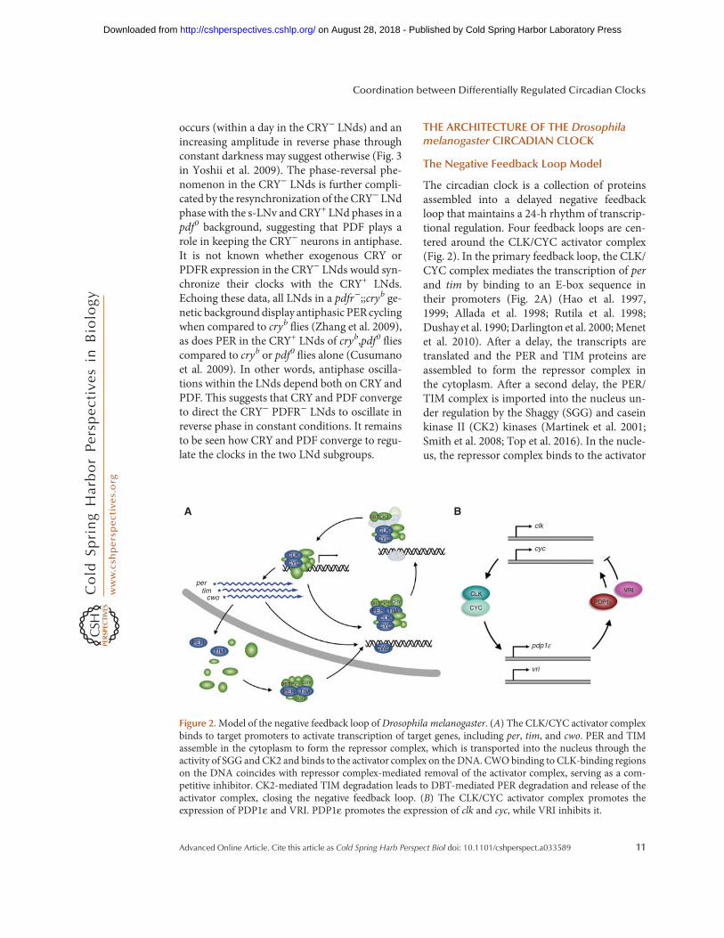
Figure 2.Model of the negative feedback loop of Drosophila melanogaster. (A) The CLK/CYC activator complexbinds to target promoters to activate transcription of target genes, including per, tim, and cwo. PER and TIMassemble in the cytoplasm to form the repressor complex, which is transported into the nucleus through theactivity of SGG and CK2 and binds to the activator complex on the DNA. CWObinding to CLK-binding regionson the DNA coincides with repressor complex-mediated removal of the activator complex, serving as a com-petitive inhibitor. CK2-mediated TIM degradation leads to DBT-mediated PER degradation and release of theactivator complex, closing the negative feedback loop. (B) The CLK/CYC activator complex promotes theexpression of PDP1ε and VRI. PDP1ε promotes the expression of clk and cyc, while VRI inhibits it.
Coordination between Differentially Regulated Circadian Clocks
Advanced Online Article. Cite this article as Cold Spring Harb Perspect Biol doi: 10.1101/cshperspect.a033589 11
on August 28, 2018 - Published by Cold Spring Harbor Laboratory Press http://cshperspectives.cshlp.org/Downloaded from
complex leading to CLK phosphorylation by as-yet-unknown mechanisms, followed by the re-moval of both complexes from the DNA, endingcircadian transcriptional activity of the CLK/CYC target genes, including tim and per (Har-din et al. 1990; Lee et al. 1999; Kim and Edery2006; Yu et al. 2006; Abruzzi et al. 2011). After athird delay, the repressor complex is degradedthrough the activity of CK2, casein kinase I/dou-bletime (DBT) and Nemo (NMO), releasing theCLK/CYC complex to initiate the next round oftranscription, and closing the negative feedbackloop (Kloss et al. 1998; Price et al. 1998; Glossopet al. 1999; Lin et al. 2002, 2005; Meissner et al.2008; Smith et al. 2008; Chiu et al. 2011; Yu et al.2011). The delays in the feedback ensure a clockoscillation with a ∼24-h frequency. The secondloop involves CLK/CYC-mediated expression ofClockwork Orange (CWO) that reinforces PER/TIM repressor activity. CWO cycles similarly toPER and binds to the CLK E-box sequences,competing with the activator complex and rein-forcing TIM/PER-mediated repression of tran-scription (Fig. 1H) (Kadener et al. 2007; Limet al. 2007; Matsumoto et al. 2007; Zhou et al.2016). In two other secondary feedback loops,the CLK/CYC activator complex mediates thetranscription of vrille (vri) and par domain pro-tein 1 ( pdp1ε), which are translated in the cyto-plasm (Fig. 2B) (Blau and Young 1999; Cyranet al. 2003). VRI represses CLK/CYC activity,whereas PDP1ε promotes it, helping tune the24-h oscillations of the circadian clock. Thefunction of the positive feedback loop is to eithermoderate the negative feedback, or to increasethe rate of CLK/CYC recovery as the repressorcomplex is removed. Further details of the cir-cadian clock, the secondary and tertiary struc-tures of the main proteins, and their interactionmotifs that mediate protein complex assemblyare reviewed elsewhere (Crane andYoung 2014).
The Oscillating Transcriptome
The circadian clock regulates the transcriptionof a large number of genes in Drosophila. CLKbinds to about 1500 sites in the Drosophila ge-nome, with more than 800 of these sites exhib-iting rhythmic CLK binding (Abruzzi et al.
2011). About 500 of these sites also display os-cillating RNA Pol II binding (Abruzzi et al.2011), which is in the range of the number oftranscripts previously estimated to fluctuateover the course of the day (150–750) (Wijnenet al. 2006). CLK/CYC binding to DNA peaks atnight, between ZT14 and ZT18 with exceptions,but always remains bound at lower levelsthroughout the day (Abruzzi et al. 2011). Thisresidual level of CLK/CYC E-box occupancyindicates that PER/TIM-mediated removal ofCLK/CYC from the DNA is incomplete, but isstill sufficient to ensure oscillations of the targettranscription. Residual occupancy also indicatesthat small variations in protein concentration,or critically, variations in protein affinity as me-diated by different posttranslational modifica-tions or protein partners, may differently tunegene expression. In support of this idea, micro-array data reveal variations in the onset of oscil-lating transcripts (Boothroyd et al. 2007; Na-goshi et al. 2010), suggesting that the activatorand repressor complexes do not necessarily actat the same time for each gene. Similarly, thereare tissue-specific differences in CLK-mediatedgene expression. For example, CLK binds to tworegions of the pdp1ε promoter in wild-type flyheads at ZT14, but binds to only one of theseregions in eyeless flies (Abruzzi et al. 2011), sug-gesting that the second site is eye-specific. Moregenerally, a need for differences in the regulationof CLK–DNA interactions at different times anddifferent cells is supported by microarray datathat reveal a wide range of gene expression dif-ferences across time and tissues (Nagoshi et al.2010). Thus, although expression levels of acti-vator and repressor are critical in maintainingthe timing of gene expression, they are not nec-essarily sufficient. It is very likely that the circa-dian clock uses a wide range of regulatory pro-teins to tightly regulate circadian protein activityand gene expression across the tissues in theorganism.
Regulatory Kinases of the Circadian Clockand Cryptochrome
The activity of the different core clock compo-nents is primarily regulated by protein interac-
D. Top and M.W. Young
12 Advanced Online Article. Cite this article as Cold Spring Harb Perspect Biol doi: 10.1101/cshperspect.a033589
on August 28, 2018 - Published by Cold Spring Harbor Laboratory Press http://cshperspectives.cshlp.org/Downloaded from
tions and posttranslational modifications. Dif-ferent accessory proteins interact with and in-fluence how the repressor and activator com-plexes are regulated. Here we will review thecurrent knowledge of some of these modifyingproteins that regulate timed activity, before dis-cussing their roles in specific tissues.
Doubletime/Casein Kinase I
Doubletime (Dbt) encodes casein kinase I(DBT), a serine/threonine kinase that phos-phorylates PER to regulate PER stability and nu-clear accumulation (Kloss et al. 1998; Price et al.1998; Cyran et al. 2005). Two mutant forms ofDBT are named for the manner in which theyalter behavioral rhythmicity, dbt short (dbtS)and dbt long (dbtL) (Kloss et al. 1998). Over-expression of either form of DBT in vivo resultsin a dominant change in behavior, while over-expression of the wild-type form has a modestlylonger (∼1 h) period (Muskus et al. 2007; Ven-katesan et al. 2015). The lack of a meaningfulchange in phenotype with DBT overexpressionsuggests that DBT activity is not a rate-limitingstep in the circadian clock. Of the DBTmutants,surprisingly, both long and short forms exhibitreduced kinase activity (Preuss et al. 2004; Kivi-mäe et al. 2008), suggesting that DBT may servetwo opposing roles in regulating rhythmicity.Indeed while DBTL and a DBT variant lackingATPase activity (DBTK38R) change the kineticsof PER degradation that reflect changes in be-havioral rhythmicity (i.e., delayed PER degrada-tion, longer behavioral periodicity), DBTS doesnot appear to alter the kinetics of PER degrada-tion at all (Preuss et al. 2004; Syed et al. 2011). IfDBTS is not impaired in PER degradation, butcan still impose a short behavioral rhythm onthe fly, then DBT must play an additional rolein regulating behavioral rhythmicity. One pos-sibility is that DBTS leads to early termination ofPER transcription inhibition (Bao et al. 2001;Syed et al. 2011). The presence of DBT, but notits kinase activity, is required to hyperphosphor-ylate and inactivate CLK to release it from DNA(Yu et al. 2009; Menet et al. 2010), which may behow dbtS flies exhibit a short behavioral rhythm.Because DBT cannot phosphorylate CLK in vi-
tro or interact with it directly, DBT likely medi-ates its effect on CLK through PER (Kim andEdery 2006; Yu et al. 2006; Kivimäe et al. 2008;Menet et al. 2010) by either being recruited tophosphorylate CLK, or through phosphoryla-tion of PER to influence its repressor function.Thus, while mutants like DBTL are defective intriggering timely degradation of PER protein(Syed et al. 2011), DBTS may be defective inregulating the activity of CLK protein.
DBT triggers and mediates a phosphoryla-tion cascade on PER, beginning at the PER shortdownstream region (PERSD), includes the clas-sic PER short (PERS) site, and culminates withmodification of S47 at the amino terminus (Ko-nopka and Benzer 1971; Kivimäe et al. 2008;Chiu et al. 2011; Garbe et al. 2013). Phosphor-ylation of S47 triggers physical association be-tween PER and Slimb, an F-Box/WD-40 repeatprotein that ubiquitinates PER to mediate itsdegradation (Grima et al. 2002; Ko et al. 2002;Chiu et al. 2008). Mutations that block phos-phorylation of S47 or the PERSD region stabilizePER and lengthen the periodicity of behavioralrhythms (Chiu et al. 2008; Kivimäe et al. 2008;Garbe et al. 2013). On the other hand, mutationof the PERS site, also involved in the phosphor-ylation cascade, destabilizes PER primarily inthe late night, resulting in a short behavioralrhythm (Konopka and Benzer 1971; Ederyet al. 1994), suggesting that modification ofthis site is required for PER stabilization. Thesedata suggest that PER may be alternatively sta-bilized and destabilized in different phases of thecircadian cycle. DBT physically associates withthe amino terminus of PER, and possibly a sec-ond, internal region spanning ∼50 residues inthe latter third of PER, remaining associatedwith the repressor complex until PER is de-graded (Kloss et al. 1998; Kim et al. 2007).PER is not immediately degraded however,because of its association with TIM, despite ac-cumulating phosphorylation events over time(Edery et al. 1994; Rothenfluh et al. 2000; Klosset al. 2001), which may mean that TIM influ-ences how PER is phosphorylated. Therefore,DBT activity in the nucleus and nuclear TIMdegradation form the basis of the temporalregulation of PER-mediated transcription re-
Coordination between Differentially Regulated Circadian Clocks
Advanced Online Article. Cite this article as Cold Spring Harb Perspect Biol doi: 10.1101/cshperspect.a033589 13
on August 28, 2018 - Published by Cold Spring Harbor Laboratory Press http://cshperspectives.cshlp.org/Downloaded from
pression (Kloss et al. 2001; Venkatesan et al.2015).
Casein Kinase II
Timekeeper (Tik) and Andante, respectively, en-code theα (catalytic) and β (regulatory) subunitsthat make up the heterotetrameric casein kinaseII (CK2), a kinase that phosphorylates PER andTIM to regulate protein stability and nuclearentry (Lin et al. 2002, 2005; Akten et al. 2003;Top et al. 2016). RNAi against α or β subunitscauses a long behavioral rhythm (Szabó et al.2013; Top et al. 2016). Overexpression of eachindividual subunit gives rise to a similar longrhythm phenotype (Lin et al. 2005; Szabó et al.2013; Top et al. 2016), an initially paradoxicalphenotype that has been more recently sug-gested to reflect a dominant-negative effectarising from altered CK2 stoichiometry and ti-tration of the functional complex (Top et al.2016). Consistent with this notion, overexpres-sion of both subunits yields a short behavioralrhythm suggestive of a CK2 gain-of-function(Top et al. 2016). CK2 physically associateswith and phosphorylates both PER and TIM toregulate rhythmic behavior (Lin et al. 2002,2005; Szabó et al. 2013; Top et al. 2016). Thus,CK2-mediated phosphorylation is a rate-limit-ing step in the circadian clock (Top et al. 2016).
CK2 modifies PER and TIM proteins to reg-ulate their stability. Overexpression of the CK2αmutant Tik in vivo results in a behavioralrhythm of ∼31 h, and correlates with the stabi-lization of PER and TIM proteins (Smith et al.2008). Accumulation of both PER and TIM isalso seen with the CK2β Andante mutation(Akten et al. 2003). RNAi depletion of CK2 inS2 cells stabilizes nuclear PER/TIM levels, whilelengthening behavioral period in flies to ∼33 h(Szabó et al. 2013; Top et al. 2016). Together,these data suggest that the activity of CK2 isrequired for normal oscillations of PER andTIM proteins. Mutation of CK2 target sites inPER do not lead to an accumulation of PERprotein above wild-type levels despite exhibitinglonger behavioral periods in flies (Lin et al.2005), but mutation of CK2 sites in TIM does(Top et al. 2016). Another allele of tim, tim ul-
tralong (timUL), carries a mutation at a serinesite that is predicted to block CK2-mediatedphosphorylation (Meissner et al. 2008), whichcauses TIM to persist in the nucleus and delayPER degradation (Rothenfluh et al. 2000). Thus,the evidence suggests that the main role for CK2is likely to regulate the stability of TIM. In ad-dition to its role in stabilizing the repressor com-plex, CK2 activity also contributes to nuclearaccumulation of the repressor complex (Smithet al. 2008; Top et al. 2016), and phosphorylatesCLK in a PER-dependent manner to stabilize it,contrasting DBT-mediated destabilization ofCLK (Szabó et al. 2013). The involvement ofCK2 in the stability and nuclear accumulationof the repressor complex and the stability of theactivator complex is likely to ensure that thesedifferent regulatory processes are biochemicallyand chronologically linked.
Shaggy/Glycogen Synthase Kinase 3
Shaggy encodes glycogen synthase kinase 3(SGG), a key regulator of nuclear accumulationof the repressor complex through modificationof PER and TIM (Martinek et al. 2001; Ko et al.2010; Top et al. 2016). In contrast to DBT andCK2, kinases that regulate stability of the repres-sor complex, SGG has no effect on the stabilityof PER or TIM (Top et al. 2016). Instead, SGGphysically associates with the repressor complex,with a preference for binding TIM over PER,phosphorylating TIM and triggering a CK2phosphorylation cascade to regulate PER/TIMnuclear accumulation (Top et al. 2016). Muta-tions that block SGG phosphorylation sites onTIM delay PER/TIM nuclear entry and lead to ahigh accumulation of cytoplasmic PER protein(Top et al. 2016), suggesting a link between themechanism that regulates nuclear entry and nu-clear degradation. It is likely that SGG-mediatedtriggering of nuclear entry of the TIM/PERcomplex is followed by CK2-mediated destabi-lization of TIM (Top et al. 2016), and then byDBT-mediated phosphorylation and destabili-zation of PER.
SGG is known to be regulated by variousextracellular signals. SGG is a target of signalingcascades that are activated byWnt or Hedgehog,
D. Top and M.W. Young
14 Advanced Online Article. Cite this article as Cold Spring Harb Perspect Biol doi: 10.1101/cshperspect.a033589
on August 28, 2018 - Published by Cold Spring Harbor Laboratory Press http://cshperspectives.cshlp.org/Downloaded from
reducing SGG activity (Harwood 2001; Doble2003; Zhang et al. 2005; Hur and Zhou 2010)and offering a potential biochemical link be-tween developmental signals and the circadianclock. SGG is also a target of signaling cascadesthat are activated by insulin or by growth factor,reducing SGG activity through the Akt/TORpathway (Harwood 2001; Zhang et al. 2006;Zheng and Sehgal 2010), offering another po-tential biochemical link between metabolic sig-nals and the circadian clock. In these cascades,SGG activity is modulated by phosphorylationas a function of cAMP levels (Fang et al. 2000).Thus, it is likely that the cAMP levels adjusted byPDFR, NPFR, and D1-like DA receptor modu-lates SGG activity through the signaling betweenneurons of the CCNN. Indeed, GSK3/SGG ac-tivity in the mouse has been shown to be reg-ulated by activation of dopamine receptors(Beaulieu et al. 2011). Thus, SGG represents apotential communication hub for extracellularsignals, and the biochemical state of the cell (e.g.,cAMP levels) to influence processes such as nu-clear entry of the repressor complex to regulatephase and synchrony of the different clocks.
Other Kinases
Nemo (NMO) is a proline-directed kinase thatphysically associates with and helps regulatePER and CLK stability (Yu et al. 2011). NMOis required for S596 phosphorylation on PER aspart of the DBT phosphorylation cascade thatregulates PER stability (Chiu et al. 2011; Garbeet al. 2013). S596 phosphorylation antagonizesDBT activity and helps stabilize PER, possiblyby delaying degradation (Chiu et al. 2011). Sur-prisingly, RNAi directed against nmo in fliesdoes not alter PER or TIM levels (Yu et al.2011), despite shortening behavioral rhythmic-ity (Chiu et al. 2011; Yu et al. 2011), conflictingwith the purported stabilizing role for S596phosphorylation. Because NMO interacts withCLK (Yu et al. 2011), NMOmay destabilize CLKand shorten behavioral rhythmicity througha mechanism of transcription termination.Whereas NMO stabilizes PER and destabilizesCLK, CK2 has the opposite effect on these pro-teins (Lin et al. 2005; Chiu et al. 2011; Yu et al.
2011), suggesting that the antagonistic action ofthese kinases on the repressor and activatorcomplexes may generate a more robust tran-scriptional negative feedback loop.
PKA is a kinase that communicates extracel-lular signals to the cell by phosphorylating targetproteins, activating, or inactivating them. Incircadian rhythms, PKA deficiencies lead to aloss of locomotor rhythmicity in flies (∼80%arrhythmic), while per RNA oscillation andeclosion rhythmicity remains intact (Majercaket al. 1997), indicating that PKA is involved inoutput mechanisms that specifically regulate lo-comotor rhythmicity. PKA activity is requiredto stabilize TIM in some clock neurons (LNdsand DN1s) and stabilize PER in other clockneurons (LNvs) (Li et al. 2014; Seluzicki et al.2014), offering a basis for observed behavioralarrhythmicity, but without a satisfactory expla-nation for how eclosion remains rhythmic. Themolecular basis for cell-type-specific differencesby which PKA stabilizes TIM and PER are notunderstood. While PKA is known to be acti-vated by cAMP levels, mechanisms by whichextracellular signals may contribute to differen-tial PKA activity in various circadian neuronsare poorly defined. Like SGG, PKA may trans-duce extracellular signals of the CCNN to influ-ence the stability of the repressor complex toregulate clock phase or synchrony. Whereas ex-tracellular signaling through SGG affects nucle-ar entry of the TIM/PER repressor complex andalters the onset of transcriptional repression(Martinek et al. 2001; Top et al. 2016), PKAsignaling alters TIM and PER stability, impact-ing the termination of transcriptional repression(Li et al. 2014; Seluzicki et al. 2014), suggestingthat these pathways may regulate target genes atdifferent times of day.
Cryptochrome
CRY is the primary photoreceptor for entrainingfly circadian rhythms (Stanewsky et al. 1998;Cashmore 2003). Flies that lack cry (cry01) orexpress the cry hypomorph (cryb) adjust tochanges in light regiments over a span of severaldays, instead of a single day (Stanewsky et al.1998; Dolezelova et al. 2007). In the presence
Coordination between Differentially Regulated Circadian Clocks
Advanced Online Article. Cite this article as Cold Spring Harb Perspect Biol doi: 10.1101/cshperspect.a033589 15
on August 28, 2018 - Published by Cold Spring Harbor Laboratory Press http://cshperspectives.cshlp.org/Downloaded from
of light, CRY binds to TIM and both proteinsare ubiquitinated for degradation by complexesinvolving Ramshackle (BRWD3)/JET/Cullin4and JET/Cullin3, respectively (Ceriani et al.1999; Koh et al. 2006; Peschel et al. 2009; Ozturket al. 2011, 2013). Several studies suggest thatlight triggers the reduction of the FAD coen-zyme bound within CRY, inducing a conforma-tional change that releases the CRY carboxy-ter-minal tail (CTT) (Dissel et al. 2004; Hoang et al.2008; Ozturk et al. 2011, 2008; Vaidya et al.2013; Ganguly et al. 2016). However, other ex-periments indicate that light causes CTT releasethrough a process that does not depend on flavinreduction (Ozturk et al. 2014). The release of theCRY CTT allows a TIM region of similar se-quence to bind to the exposed pocket in CRY,communicating the light signal to TIM (Vaidyaet al. 2013). Consistent with this model, removalof the CRY CTT promotes CRY-TIM bindingwithout the need for light activation, leadingto weaker entrainment in flies lacking theCTT (Busza et al. 2004). Despite a weakenedability to entrain to light, cry mutants lackingthe CTT can be entrained under low light con-ditions (Dissel et al. 2004), likely through theCRY-independent visual system, or the deepbrain photoreceptor Rh7 as described above(Rieger et al. 2003; Schlichting et al. 2014; Niet al. 2017).
RELATING THE CIRCADIAN CLOCK MODELTO THE CCNN AND CONFLICTS
Recent studies using luciferase and calcium re-porters in wild-type and mutant flies have chal-lenged the notion that the molecular architec-ture, rhythmic dynamics, and oscillatory phaseof core clock components are regulated the samein all pacemaker neurons (Roberts et al. 2015;Liang et al. 2016, 2017). The molecular modelof the circadian clock, however, is presented asa stereotyped model, often described as a fine-ly tuned mechanism that maintains a 24-hrhythm. If the model is correct, how does thecircadian clock oscillate with different frequen-cies in different neurons of the circadian system?To understand rhythmic behavior, our knowl-edge of intracellular mechanisms that regulate
the circadian clockmust be integratedwithin thecontext of extracellular signaling and synaptictransmission between pacemaker neurons. Inthis section, we will discuss “speeds” of clocksto refer to the period of the circadian clock with-in a given cluster of neurons, to distinguish an“advance” or “delay” in anticipatory behavior inmorning/evening cells.
The Effects of Different Kinases on the s-LNvand LNd Clocks
The s-LNvs have been described as master pace-maker cells that dominate behavior in constantdark conditions through the release of PDF(Renn et al. 1999; Grima et al. 2004; Stoleruet al. 2004, 2005). Speeding up the LNv clockthrough overexpression of SGG also speeds upthe LNd clock over a few days in constant dark-ness, suggesting communication between thetwo clusters (Stoleru et al. 2005). However,speeding up the LNv clock through overexpres-sion of DBTS speeds up the LNv clock, but failsto speed up the LNd/fifth s-LNv clocks. LNvsthat are slowed by DBTL overexpression failsto slow the fifth s-LNv (LNd clocks were notmeasured), contrasting the effect of DBTS over-expression (Yao et al. 2016). Focusing on thecontrasting results between SGG and DBTS
overexpression, it appears that the method bywhich the LNv clocks are sped up is importantin instructing the LNd clock speed.We speculatethat because DBT activity is required forreleasing transcriptional repression and SGGactivity is required for nuclear entry of PER/TIM to activate transcriptional repression, it ispossible that the differences observed in LNdclock speeds lie in the differences in the begin-ning or the ending of circadian transcriptionalregulation.
Kinase overexpression also affects morningand evening anticipation behaviors differently.Overexpressing DBTS in the LNvs advancesmorning anticipation with no effect on eveninganticipation, while overexpressing SGG in thesame neurons advances both morning and eve-ning anticipation (Stoleru et al. 2005; Yao et al.2016), reflecting their effect on speeds of theindividual molecular oscillators. Conversely,
D. Top and M.W. Young
16 Advanced Online Article. Cite this article as Cold Spring Harb Perspect Biol doi: 10.1101/cshperspect.a033589
on August 28, 2018 - Published by Cold Spring Harbor Laboratory Press http://cshperspectives.cshlp.org/Downloaded from
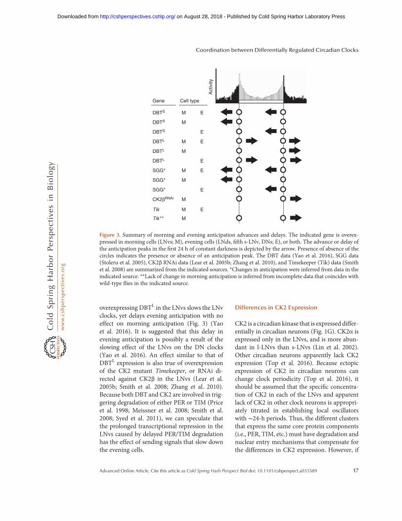
overexpressing DBTL in the LNvs slows the LNvclocks, yet delays evening anticipation with noeffect on morning anticipation (Fig. 3) (Yaoet al. 2016). It is suggested that this delay inevening anticipation is possibly a result of theslowing effect of the LNvs on the DN clocks(Yao et al. 2016). An effect similar to that ofDBTL expression is also true of overexpressionof the CK2 mutant Timekeeper, or RNAi di-rected against CK2β in the LNvs (Lear et al.2005b; Smith et al. 2008; Zhang et al. 2010).Because both DBT and CK2 are involved in trig-gering degradation of either PER or TIM (Priceet al. 1998; Meissner et al. 2008; Smith et al.2008; Syed et al. 2011), we can speculate thatthe prolonged transcriptional repression in theLNvs caused by delayed PER/TIM degradationhas the effect of sending signals that slow downthe evening cells.
Differences in CK2 Expression
CK2 is a circadian kinase that is expressed differ-entially in circadian neurons (Fig. 1G). CK2α isexpressed only in the LNvs, and is more abun-dant in l-LNvs than s-LNvs (Lin et al. 2002).Other circadian neurons apparently lack CK2expression (Top et al. 2016). Because ectopicexpression of CK2 in circadian neurons canchange clock periodicity (Top et al. 2016), itshould be assumed that the specific concentra-tion of CK2 in each of the LNvs and apparentlack of CK2 in other clock neurons is appropri-ately titrated in establishing local oscillatorswith ∼24-h periods. Thus, the different clustersthat express the same core protein components(i.e., PER, TIM, etc.) must have degradation andnuclear entry mechanisms that compensate forthe differences in CK2 expression. However, if
DBTS M E
DBTS M
EDBTS
DBTL M
DBTL
E
DBTL E
SGG* M E
SGG* M
SGG* E
CK2βRNAi M
Tik M EA
ctiv
ity
Gene Cell type
Tik ** M
M
Figure 3. Summary of morning and evening anticipation advances and delays. The indicated gene is overex-pressed in morning cells (LNvs; M), evening cells (LNds, fifth s-LNv, DNs; E), or both. The advance or delay ofthe anticipation peaks in the first 24 h of constant darkness is depicted by the arrow. Presence of absence of thecircles indicates the presence or absence of an anticipation peak. The DBT data (Yao et al. 2016), SGG data(Stoleru et al. 2005), CK2β RNAi data (Lear et al. 2005b; Zhang et al. 2010), and Timekeeper (Tik) data (Smithet al. 2008) are summarized from the indicated sources. *Changes in anticipation were inferred from data in theindicated source. **Lack of change in morning anticipation is inferred from incomplete data that coincides withwild-type flies in the indicated source.
Coordination between Differentially Regulated Circadian Clocks
Advanced Online Article. Cite this article as Cold Spring Harb Perspect Biol doi: 10.1101/cshperspect.a033589 17
on August 28, 2018 - Published by Cold Spring Harbor Laboratory Press http://cshperspectives.cshlp.org/Downloaded from
CK2 is necessary to regulate nuclear entry andnuclear degradation of TIM and PER, how isTIM/PER nuclear entry and degradation regu-lated in pacemaker neurons such as the LNds, ifCK2 is not expressed there? The CK2 phosphor-ylation sites identified on PER (Lin et al. 2005)and TIM (Top et al. 2016) may either be phos-phorylated by another kinase in the LNds, orPER and TIM degradation may be regulatedthrough an entirely different mechanism. It isalso possible that CK2 does not participate inregulating a mechanism of the circadian clockdespite being expressed in a neuronal cluster. Amutant form of TIM that delays CK2-mediatednuclear entry in s-LNvs does not delay nuclearentry in the l-LNvs despite a higher CK2α ex-pression in the l-LNvs (Lin et al. 2002; Top et al.2016). In summary, the LNds that express noCK2 must use another mechanism to regulatePER/TIM nuclear entry and degradation, andthe l-LNvs that express abundant CK2 but donot exhibit delayed nuclear entry when TIMphosphorylation sites are mutated likely useyet another mechanism to regulate PER/TIMnuclear entry. The differences LNvs exhibit inCK2 levels and the efficacy of mutations thatblock CK2 activity indicate that different mech-anisms are involved in regulating local clocks inthe CCNN.
Differences in NMO Expression
In the fly brain, NMO is expressed in two of fourl-LNvs, all s-LNvs, a subset of DN1s, at low lev-els in some LNds, and is not observed at all inDN2s or DN3s (Yu et al. 2011). An RNAi-me-diated decrease of NMO shortens rhythmic be-havior in constant dark conditions, advancingboth morning and evening anticipation (Chiuet al. 2011). The effect an nmo knockdown hason both anticipation events is consistent with itsexpression in morning and evening cells. How-ever, the variability of NMO expression in thedifferent circadian neurons raises questionssimilar to those in the case of CK2. Is PER reg-ulated by the same phosphorylation cascade inthe neurons lacking NMO? Do the clocks inthese neurons oscillate slightly longer or is therea different mechanism (i.e., another kinase) that
substitutes for the absence of NMO in theseneurons? Importantly, the absence of NMO insome neurons indicate that the model for regu-lating PER stability in the different neurons isnot universally applicable.
Differences in CRY Expression and LightResponse
Clock neurons differ in their expression of CRYand also in their responsiveness to light (Benitoet al. 2008; Yoshii et al. 2008). For example, TIMin the s-LNv clock is nonresponsive to a lightpulse at ZT 15, likely because of the low levels ofCRY in the cells at that time (Tang et al. 2010).Increasing CRY expression in these cells in-creases the degradation of TIM in response tolight and causes behavioral phase shifts (Tanget al. 2010). To speculate, this molecular andbehavioral response may be extrapolated to cir-cadian neurons that express low levels of CRY,suggesting a weakened or even absent light re-sponse in such low expressers. Indeed, a lightpulse differentially shifts the phase of each localclock (e.g., s-LNvs, LNds, and DN1s), but eachlocal clock returns to synchrony with each otherin less than 3 days, presumably through com-munication between these neurons (Robertset al. 2015). Responsiveness to light by the mo-lecular oscillator also depends on the proteinJetlag (JET). Increasing JET levels in s-LNvsaugments TIM degradation in response to alight pulse, similar to CRY overexpression(Tang et al. 2010). Yet, JET overexpressiondoes not elicit a behavioral response, in contrastto the observations made with CRY overexpres-sion (Tang et al. 2010). Thus, a single externalsignal can induce different phase shifts in eachclock that may manifest differently in rhythmicbehavior of the fly. Additionally, while thesefindings demonstrate that TIM degradation isimportant to reset the molecular oscillator, itappears that a behavioral phase response maybe regulated by CRY, not TIM degradation.
Monitoring clocks in different clusters whenanalyzing cry0 flies reveals that non s-LNvsphase shift rapidly to accommodate a phasechange in light regimen, while s-LNvs areslow to respond (Yoshii et al. 2015). This sug-
D. Top and M.W. Young
18 Advanced Online Article. Cite this article as Cold Spring Harb Perspect Biol doi: 10.1101/cshperspect.a033589
on August 28, 2018 - Published by Cold Spring Harbor Laboratory Press http://cshperspectives.cshlp.org/Downloaded from
gests that CRY-mediated light responsiveness iscritical in the s-LNvs and that non s-LNvs pos-sibly depend more on signals from the eye.These conclusions are supported by analysisof light-mediated TIM degradation in jetknockdown flies at ZT15 and ZT21, beforeand after TIM nuclear entry (Lamba et al.2014). A reduction of JET in the LNds at eithertime point has no effect on light-mediated TIMdegradation in either LNds or LNvs, nor does itinduce a phase response as compared to wild-type. JET reduction in the LNvs has no effect onLNv TIM degradation in response to light, butalters behavioral phase shifts. These data reaf-firm that behavioral phase response is likelymediated through CRY, and whether light-me-diated TIM degradation will occur depends onthe neuronal cluster in which TIM is expressed.Therefore, the sensitivity of s-LNvs to CRY-me-diated light detection, the different changes ofclock phase in the s-LNvs and LNds with a lightpulse, and the differences in CRY expression(and light sensitivity) in CRY-expressing neu-rons result in the different light responses byeach neuronal cluster.
Effects of PER on Different Clocks
The period gene is expressed in all clock neurons,in addition to many other tissues in the fly. PERis the key repressor of transcription in the circa-dian clock, with its expression directly correlatedwith behavioral rhythmicity (i.e., lower per ex-pression leads to longer rhythms) (Baylies et al.1992). Thus, any differences in PER concentra-tion in the different clusters would be expectedto affect the frequency of the local oscillator.Consistent with this model, the comparison ofperS with wild-type flies yields a range of differ-ences in calcium oscillations in the differentclusters (Liang et al. 2016). This observationsuggests that altering the stability of PER hasdifferent consequences on the oscillators in thedifferent clusters. This is also consistent withlong PER protein oscillations that the l-LNvsexhibit relative to the s-LNvs in wild-type flies(Fig. 4A inRoberts et al. 2015) that correlatewithlower per expression in the l-LNvs compared tothe s-LNvs (Kula-Eversole et al. 2010).
The regulation of PER function in specificneuronal clusters also relies on the spatial orga-nization of functional domains within the PERprotein. In the current model of PER function,DBT binds to the amino terminus of PER toregulate protein degradation (Kloss et al. 1998;Chiu et al. 2008), while CLK appears to interactwith PER at the carboxy-terminal end (Changand Reppert 2003), seemingly spatially separat-ing the degradation and repression activities ofPER. Consistent with this, deletions within thelast third of PER alter PER repressor activity(Chang and Reppert 2003; Sun et al. 2010) anda carboxy-terminal truncation of the last third ofPER (BG) cannot rescue the behavioral rhyth-micity of a per0 fly (Stanewsky et al. 1997). How-ever, despite demonstrating arrhythmic behav-ior, oscillation of PER in BG fly LNvs isindistinguishable from wild-type (Stanewskyet al. 1997). Therefore, the last third of PERthat encodes the “CLK-binding domain” is im-portant for regulating behavioral rhythmicity,but has no effect on the oscillation of the LNvclocks. Thus, it is tempting to speculate that afunctional CLK-binding domain is necessaryfor mediating rhythmic behavior, but dispensa-ble for the function of the LNv clock. Undoubt-edly, the resolution of this conflict will uncoverinterestingmechanistic insight into the functionof the LNv clocks.
Activity of Promoters
The mechanisms that regulate nuclear accumu-lation and nuclear stability of the repressor com-plex effectively titrate the repressor to regulategene expression. Screens of RNA expression inwhole heads or brains reveal a wide range ofpeaks and phases of oscillating transcripts (Cla-ridge-Chang et al. 2001; Ceriani et al. 2002; Uedaet al. 2002; Wijnen et al. 2006; Abruzzi et al.2011; Hughes et al. 2012). Genome-wide ChIPexperiments also reveal that CLK associationwith circadian-regulated promoters itself occurswith a circadian rhythm, with peak DNA-bind-ing phases spread throughout the circadiancycle (Abruzzi et al. 2011). Therefore, smallchanges in the accumulation of the activator orrepressor complex will have significant conse-
Coordination between Differentially Regulated Circadian Clocks
Advanced Online Article. Cite this article as Cold Spring Harb Perspect Biol doi: 10.1101/cshperspect.a033589 19
on August 28, 2018 - Published by Cold Spring Harbor Laboratory Press http://cshperspectives.cshlp.org/Downloaded from
quences for some genes, while leaving othergenes unaffected. Slight differences in SGG/CK2 expression, for example, could alter thetiming and the extent of nuclear accumulationof the repressor complex, effectively shifting theCLK-PER-binding equilibrium to alter the ex-pression of genes in a promoter-dependentmanner. In this respect, gene expression maybe tuned with modifications that alter bindingaffinity of proteins, which would, in turn, altertranscription activity. Additional mechanismsthat regulate protein stability, translation ratesand protein complex formation may give riseto functionally important differences in the dy-namics of circadian gene expression.
Understanding the Local Mechanisms thatRegulate Local Clocks
Together, the studies discussed here demon-strate that an understanding of specific mecha-nisms that regulate the circadian clock inindividual neuronal clusters is required to un-derstand the principles that govern behavior.Traditional overexpression studies have revealeda lot about circadian rhythms. However, manip-ulation of entire clusters by overexpressing ac-cessory components such as kinases makes twoassumptions: (1) that the intended target of thekinase is being manipulated, and (2) that thekinase is endogenous in that cluster and is beingsupplemented through overexpression. Neitherassumption is necessarily correct. For example,CK2 is not endogenously expressed in the LNds(Lin et al. 2002; Top et al. 2016). Therefore, abehavioral change as a result of CK2 overexpres-sion in the LNds may not necessarily reflect aneffect of phosphorylation of the intended tar-gets, because CK2 is involved a wide range ofother cellular processes that may alter variouscellular pathways and neuronal activity.
Cell-autonomous, loss-of-functionmanipu-lation of endogenous clock mechanism mayprovide a useful method to understand theproperties of individual clock neurons as wellas that of the network. Mutation of TIM thatblock SGG/CK2 phosphorylation of the proteindelays nuclear accumulation of the repressorcomplex in the s-LNvs (Top et al. 2016). In the
LNds, the delay is less severe, while in the l-LNvsthere is no delay. Thus, nuclear entry is regulatedby a different mechanism in the l-LNvs neurons(Top et al. 2016). It is not clear, however, wheth-er the TIM mutations delay nuclear entry of theLNd clock by blocking the nuclear entry mech-anism, or whether nuclear entry is delayed be-cause of the slower s-LNv clock. Additionally,the absence of a nuclear entry mechanism in-volving CK2 in the l-LNvs does not precludeother possible functions of CK2, such as degra-dation of TIM or PER.
Similarly, overexpressing or down-regulat-ing SGG in evening cells (LNds/fifth s-LNv/DNs) does not change behavioral rhythmicity,while the same manipulation in morning cells(LNvs), respectively, shortens or lengthens be-havioral rhythms (Stoleru et al. 2005; Fischeret al. 2016; Top et al. 2016). These data indicatethat SGG activity is limited to the morning cells,specifically the s-LNvs. However, manipulationof the morning cells by TIM mutants that blockSGG phosphorylation delay nuclear entry inevening cells (Top et al. 2016). Given that SGGoverexpression or down-regulation in eveningcells does not alter rhythmic behavior (Fischeret al. 2016), either the evening cells express adifferent kinase that targets the SGG sites onTIM or mutations at these sites are silent, andnuclear entry is regulated through anothermechanism that is likely dependent on signalingfrom s-LNvs. As shown by SGG and CK2, mu-tations alone are not predictive of behavioralphenotypes but rather by the combination ofthe mutation and the regulatory proteins thatmay be differentially expressed or regulatedwithin the CCNN.
In the example of another point mutation,the period short mutant ( perS) alters a DBTphosphorylation site that regulates PER stability(Chiu et al. 2008, 2011; Kivimäe et al. 2008;Garbe et al. 2013). As the name suggests, perS
flies exhibit shortened rhythms in DD, and anadvanced evening anticipation peak in LD withno effect on morning anticipation (Stanewskyet al. 1998; Renn et al. 1999; Emery et al.2000b; Li et al. 2014). Also, a perS, cryb dou-ble-mutant fly exhibits the same 19-h behavioralrhythm in constant light as a perS fly, conditions
D. Top and M.W. Young
20 Advanced Online Article. Cite this article as Cold Spring Harb Perspect Biol doi: 10.1101/cshperspect.a033589
on August 28, 2018 - Published by Cold Spring Harbor Laboratory Press http://cshperspectives.cshlp.org/Downloaded from
in which the LNds (i.e., evening cells) dominaterhythmic behavior (Emery et al. 2000a). Thissuggests that the “short” activity of perS affectsevening cells. Therefore, it is possible that phos-phorylation of the perS site is critical for theregulation of evening anticipation in the LNds.This does notmean that perS has no effect on theLNvs, however. Calcium peaks in each neuronalcluster are different, but critically, perS affectsthe peak in each cluster differently (Liang et al.2016). The DN1s and DN3s are advanced by∼2 h, while s-LNvs and LNds are advancedby 6 and 10 h, respectively, in LD conditions.These data suggest that while perS does not altermorning anticipation behavior, the neurons thatregulate morning anticipation (s-LNvs) can stillexhibit a large phase shift in peak cytoplasmiccalcium, possibly as a result of changes in thetiming of signals being received, albeit withoutaltering their influence on morning anticipa-tion. Future work will reveal the relationship ofCa2+ oscillations, clock speeds, timed anticipa-tion, and rhythmic behavior.
CONCLUSIONS
The Drosophila circadian rhythm field is in-creasingly becoming more molecular and atten-tion should increasingly be focused on cell-type-specific properties of neurons in the circadiannetwork. We now have a growing appreciationthat differences in the intracellular biochemicalpathways within these neurons alter the activityof the entire network and its behavioral outputs,yet many important questions remain unan-swered. What are the components of the activa-tor and repressor complexes in each of the clus-ters? If a regulatory kinase is absent, what is thesubstitute mechanism that ensures that the localclock runs smoothly? How does a local clockregulate the release of neurotransmitters to com-municate to other neuronal clusters? Whatmechanism of the clock is changed in responseto a neurotransmitter? Understanding the com-plex interplay between cytoplasmic secondarymessengers, core clock proteins, and their regu-lators that are differentially expressed within thecircadian neuron network is likely to provide
new insights into the relationships between bio-chemistry, neurobiology, and behavior.
REFERENCES
Abruzzi KC, Rodriguez J, Menet JS, Desrochers J, ZadinaA, Luo W, Tkachev S, Rosbash M. 2011. DrosophilaCLOCK target gene characterization: Implications forcircadian tissue-specific gene expression. Genes Dev25: 2374–2386.
Akten B, Jauch E, Genova GK, Kim EY, Edery I, Raabe T,Jackson FR. 2003. A role for CK2 in the Drosophila cir-cadian oscillator. Nat Neurosci 6: 251–257.
Allada R, White NE, So WV, Hall JC, Rosbash M. 1998. Amutant Drosophila homolog of mammalian Clock dis-rupts circadian rhythms and transcription of period andtimeless. Cell 93: 791–804.
Bachleitner W, Kempinger L, Wülbeck C, Rieger D, Hel-frich-Förster C. 2007. Moonlight shifts the endogenousclock ofDrosophila melanogaster. Proc Natl Acad Sci 104:3538–3543.
Bao S, Rihel J, Bjes E, Fan JY, Price JL. 2001. The Drosophiladouble-time S mutation delays the nuclear accumulationof period protein and affects the feedback regulation ofperiod mRNA. J Neurosci 21: 7117–7126.
Baylies MK, Vosshall LB, Sehgal A, Young MW. 1992. Newshort period mutations of the Drosophila clock gene per.Neuron 9: 575–581.
Beaulieu J-M, Del’guidice T, Sotnikova TD, Lemasson M,Gainetdinov RR. 2011. Beyond cAMP: The regulationof Akt and GSK3 by dopamine receptors. Front Mol Neu-rosci 4: 38.
Behnia R, Desplan C. 2015. Visual circuits in flies: Beginningto see the whole picture. Curr Opin Neurobiol 34: 125–132.
Benito J, Houl JH, Roman GW, Hardin PE. 2008. The blue-light photoreceptor CRYPTOCHROME is expressed in asubset of circadian oscillator neurons in the DrosophilaCNS. J Biol Rhythms 23: 296–307.
Blau J, Young MW. 1999. Cycling vrille expression is re-quired for a functionalDrosophila clock.Cell 99: 661–671.
Bloomquist BT, Shortridge RD, Schneuwly S, Perdew M,Montell C, Steller H, Rubin G, Pak WL. 1988. Isolationof a putative phospholipase C gene ofDrosophila, norpA,and its role in phototransduction. Cell 54: 723–733.
Boothroyd CE, Wijnen H, Naef F, Saez L, YoungMW. 2007.Integration of light and temperature in the regulation ofcircadian gene expression in Drosophila. PLoS Genet 3:e54.
BuszaA, Emery-LeM,RosbashM, EmeryP. 2004. Roles of thetwo Drosophila CRYPTOCHROME structural domains incircadian photoreception. Science 304: 1503–1506.
Cashmore AR. 2003. Cryptochromes. Cell 114: 537–543.Ceriani MF, Darlington TK, Staknis D, Más P, Petti AA,
Weitz CJ, Kay SA. 1999. Light-dependent sequestrationof TIMELESS by CRYPTOCHROME. Science 285: 553–556.
Ceriani MF, Hogenesch JB, Yanovsky M, Panda S, StraumeM, Kay SA. 2002. Genome-wide expression analysis in
Coordination between Differentially Regulated Circadian Clocks
Advanced Online Article. Cite this article as Cold Spring Harb Perspect Biol doi: 10.1101/cshperspect.a033589 21
on August 28, 2018 - Published by Cold Spring Harbor Laboratory Press http://cshperspectives.cshlp.org/Downloaded from
Drosophila reveals genes controlling circadian behavior. JNeurosci 22: 9305–9319.
Chang DC, Reppert SM. 2003. A novel C-terminal domainof Drosophila PERIOD inhibits dCLOCK:CYCLE-medi-ated transcription. Curr Biol 13: 758–762.
Chiu JC, Vanselow JT, Kramer A, Edery I. 2008. The phos-pho-occupancy of an atypical SLIMB-binding site onPERIOD that is phosphorylated by DOUBLETIMEcontrols the pace of the clock. Genes Dev 22: 1758–1772.
Chiu JC, Ko HW, Edery I. 2011. NEMO/NLK phosphory-lates PERIOD to initiate a time-delay phosphorylationcircuit that sets circadian clock speed. Cell 145: 357–370.
Choi C, Cao G, Tanenhaus AK, McCarthy EV, Jung M,Schleyer W, Shang Y, Rosbash M, Yin JCP, NitabachMN. 2012. Autoreceptor control of peptide/neurotrans-mitter corelease from PDF neurons determines allocationof circadian activity in Drosophila. Cell Rep 2: 332–344.
Claridge-Chang A, Wijnen H, Naef F, Boothroyd C, Rajew-sky N, Young MW. 2001. Circadian regulation of geneexpression systems in the Drosophila head. Neuron 32:657–671.
Crane BR, YoungMW. 2014. Interactive features of proteinscomposing eukaryotic circadian clocks. Annu Rev Bio-chem 83: 191–219.
Cusumano P, Klarsfeld A, Chélot E, Picot M, Richier B,Rouyer F. 2009. PDF-modulated visual inputs and cryp-tochrome define diurnal behavior in Drosophila. NatNeurosci 12: 1431–1437.
Cyran SA, Buchsbaum AM, Reddy KL, Lin MC, GlossopNRJ, Hardin PE, Young MW, Storti RV, Blau J. 2003.vrille, Pdp1, and dClock form a second feedback loop inthe Drosophila circadian clock. Cell 112: 329–341.
Cyran SA, Yiannoulos G, Buchsbaum AM, Saez L, YoungMW, Blau J. 2005. The double-time protein kinase regu-lates the subcellular localization of the Drosophila clockprotein period. J Neurosci 25: 5430–5437.
Darlington TK, Lyons LC, Hardin PE, Kay SA. 2000. Theperiod E-box is sufficient to drive circadian oscillation oftranscription in vivo. J Biol Rhythms 15: 462–471.
Dissel S, Codd V, Fedic R, Garner KJ, Costa R, Kyriacou CP,Rosato E. 2004. A constitutively active cryptochrome inDrosophila melanogaster. Nat Neurosci 7: 834–840.
Dissel S, Hansen CN, Özkaya Ö, Hemsley M, Kyriacou CP,Rosato E. 2014. The logic of circadian organization inDrosophila. Curr Biol 24: 1–10.
Doble BW. 2003. GSK-3: Tricks of the trade for a multi-tasking kinase. J Cell Sci 116: 1175–1186.
Dolezelova E, Dolezel D, Hall JC. 2007. Rhythm defectscaused by newly engineered null mutations in Drosophi-la’s cryptochrome gene. Genetics 177: 329–345.
Dushay MS, Konopka RJ, Orr D, Greenacre ML, KyriacouCP, Rosbash M, Hall JC. 1990. Phenotypic and geneticanalysis of Clock, a new circadian rhythmmutant inDro-sophila melanogaster. Genetics 125: 557–578.
Duvall LB, Taghert PH. 2013. E and M circadian pacemakerneurons use different PDF receptor signalosome compo-nents in Drosophila. J Biol Rhythms 28: 239–248.
Edery I, Zwiebel LJ, DembinskaME, RosbashM. 1994. Tem-poral phosphorylation of the Drosophila period protein.Proc Natl Acad Sci 91: 2260–2264.
Emery P, Stanewsky R, Hall JC, RosbashM. 2000a. A uniquecircadian-rhythm photoreceptor. Nature 404: 456–457.
Emery P, Stanewsky R, Helfrich-Förster C, Emery-Le M,Hall JC, Rosbash M. 2000b. Drosophila CRY is a deepbrain circadian photoreceptor. Neuron 26: 493–504.
Fang X, Yu SX, Lu Y, Bast RC, Woodgett JR, Mills GB. 2000.Phosphorylation and inactivation of glycogen synthasekinase 3 by protein kinase A. Proc Natl Acad Sci 97:11960–11965.
Fischer R,Helfrich-Förster C, Peschel N. 2016. GSK-3 β doesnot stabilize cryptochrome in the circadian clock of Dro-sophila. PLoS ONE 11: e0146571.
Fogg PCM, O’Neill JS, Dobrzycki T, Calvert S, Lord EC,McIntosh RLL, Elliott CJH, Sweeney ST, Hastings MH,Chawla S. 2014. Class IIa histone deacetylases are con-served regulators of circadian function. J Biol Chem 289:34341–34348.
GangulyA,ManahanCC, TopD, Yee EF, LinC, YoungMW,Thiel W, Crane BR. 2016. Changes in active site histidinehydrogen bonding trigger cryptochrome activation. ProcNatl Acad Sci 113: 10073–10078.
Garbe DS, Fang Y, Zheng X, Sowcik M, Anjum R, Gygi SP,Sehgal A. 2013. Cooperative interaction between phos-phorylation sites on PERIODmaintains circadian periodin Drosophila. PLoS Genet 9: e1003749.
Garczynski SF, Brown MR, Shen P, Murray TF, Crim JW.2002. Characterization of a functional neuropeptide Freceptor from Drosophila melanogaster. Peptides 23:773–780.
Glossop NR, Lyons LC, Hardin PE. 1999. Interlocked feed-back loops within the Drosophila circadian oscillator. Sci-ence 286: 766–768.
Grima B, Lamouroux A, Chélot E, Papin C, Limbourg-Bou-chon B, Rouyer F. 2002. The F-box protein slimb controlsthe levels of clock proteins period and timeless. Nature420: 178–182.
Grima B, Chélot E, Xia R, Rouyer F. 2004. Morning andevening peaks of activity rely on different clock neuronsof the Drosophila brain. Nature 431: 869–873.
Guo F, Cerullo I, Chen X, Rosbash M. 2014. PDF neuronfiring phase-shifts key circadian activity neurons in Dro-sophila. eLife 3: e02780.
Guo F, Yu J, Jung HJ, Abruzzi KC, Luo W, Griffith LC,Rosbash M. 2016. Circadian neuron feedback controlsthe Drosophila sleep–activity profile. Nature 536: 1–18.
Hao H, Allen DL, Hardin PE. 1997. A circadian enhancermediates PER-dependent mRNA cycling in Drosophilamelanogaster. Mol Cell Biol 17: 3687–3693.
Hao H, Glossop NR, Lyons L, Qiu J, Morrish B, Cheng Y,Helfrich-Förster C, Hardin P. 1999. The 69 bp circadianregulatory sequence (CRS) mediates per-like develop-mental, spatial, and circadian expression and behavioralrescue in Drosophila. J Neurosci 19: 987–994.
Hardie RC, Juusola M. 2015. Phototransduction in Dro-sophila. Curr Opin Neurobiol 34: 37–45.
Hardin PE, Hall JC, Rosbash M. 1990. Feedback of the Dro-sophila period gene product on circadian cycling of itsmessenger RNA levels. Nature 343: 536–540.
Hardin PE, Hall JC, RosbashM. 1992. Circadian oscillationsin period gene mRNA levels are transcriptionally regu-lated. Proc Natl Acad Sci 89: 11711–11715.
D. Top and M.W. Young
22 Advanced Online Article. Cite this article as Cold Spring Harb Perspect Biol doi: 10.1101/cshperspect.a033589
on August 28, 2018 - Published by Cold Spring Harbor Laboratory Press http://cshperspectives.cshlp.org/Downloaded from
Harwood AJ. 2001. Regulation of GSK-3: A cellular multi-processor. Cell 105: 821–824.
He C, Cong X, Zhang R, Wu D, An C, Zhao Z. 2013. Reg-ulation of circadian locomotor rhythm by neuropeptideY-like system inDrosophila melanogaster. Insect Mol Biol22: 376–388.
Helfrich-Förster C. 1995. The period clock gene is expressedin central nervous system neurons which also produce aneuropeptide that reveals the projections of circadianpacemaker cells within the brain of Drosophila mela-nogaster. Proc Natl Acad Sci 92: 612–616.
Helfrich-Förster C. 1998. Robust circadian rhythmicity ofDrosophila melanogaster requires the presence of lateralneurons: A brain-behavioral study of disconnected mu-tants. J Comp Physiol A 182: 435–453.
Helfrich-Förster C, Winter C, Hofbauer A, Hall JC, Stanew-sky R. 2001. The circadian clock of fruit flies is blind afterelimination of all known photoreceptors. Neuron 30:249–261.
Helfrich-Förster C, Edwards T, Yasuyama K, Wisotzki B,Schneuwly S, Stanewsky R, Meinertzhagen IA, HofbauerA. 2002. The extraretinal eyelet of Drosophila: Develop-ment, ultrastructure, and putative circadian function. JNeurosci 22: 9255–9266.
Hermann C, Yoshii T, Dusik V, Helfrich-Förster C. 2012.Neuropeptide F immunoreactive clock neurons modifyevening locomotor activity and free-running period inDrosophila melanogaster. J Comp Neurol 520: 970–987.
Hermann-Luibl C, Yoshii T, Senthilan PR, Dircksen H, Hel-frich-Förster C. 2014. The ion transport peptide is a newfunctional clock neuropeptide in the fruit fly Drosophilamelanogaster. J Neurosci 34: 9522–9536.
Hoang N, Schleicher E, Kacprzak S, Bouly JP, Picot M, WuW, Berndt A, Wolf E, Bittl R, Ahmad M. 2008. Humanand Drosophila cryptochromes are light activated by fla-vin photoreduction in living cells. PLoS Biol 6: e160.
Hofbauer A, Buchner E. 1989. Does Drosophila have seveneyes? Naturwissenschaften 76: 335–336.
Houl JH, Yu W, Dudek SM, Hardin PE. 2006. DrosophilaCLOCK is constitutively expressed in circadian oscillatorand non-oscillator cells. J Biol Rhythms 21: 93–103.
Hughes ME, Hong HK, Chong JL, Indacochea AA, Lee SS,Han M, Takahashi JS, Hogenesch JB. 2012. Brain-specif-ic rescue of clock reveals system-driven transcriptionalrhythms in peripheral tissue. PLoS Genet 8: e1002835.
Hur EM, Zhou FQ. 2010. GSK3 signalling in neural devel-opment. Nat Rev Neurosci 11: 1–13.
Hyun S, Lee Y, Hong ST, Bang S, Paik D, Kang J, Shin J, Lee J,Jeon K, Hwang S, et al. 2005. Drosophila GPCR Han is areceptor for the circadian clock neuropeptide PDF. Neu-ron 48: 267–278.
Im SH, Taghert PH. 2010. PDF receptor expression revealsdirect interactions between circadian oscillators in Dro-sophila. J Comp Neurol 518: 1925–1945.
Im SH, Li W, Taghert PH. 2011. PDFR and CRY signalingconverge in a subset of clock neurons to modulate theamplitude and phase of circadian behavior inDrosophila.PLoS ONE 6: e18974.
Ivanchenko M, Stanewsky R, Giebultowicz JM. 2001. Circa-dian photoreception in Drosophila: Functions of crypto-
chrome in peripheral and central clocks. J Biol Rhythms16: 205–215.
Jan LY, Jan YN. 1982. Peptidergic transmission in sympa-thetic ganglia of the frog. J Physiol (Lond) 327: 219–246.
Johard HAD, Yoishii T, Dircksen H, Cusumano P, Rouyer F,Helfrich-Förster C, Nässel DR. 2009. Peptidergic clockneurons in Drosophila: Ion transport peptide and shortneuropeptide F in subsets of dorsal and ventral lateralneurons. J Comp Neurol 516: 59–73.
Kadener S, Stoleru D, McDonald M, Nawathean P, RosbashM. 2007. Clockwork Orange is a transcriptional repressorand a new Drosophila circadian pacemaker component.Genes Dev 21: 1675–1686.
Kaneko M, Hall JC. 2000. Neuroanatomy of cells expressingclock genes in Drosophila: Transgenic manipulation ofthe period and timeless genes to mark the perikarya ofcircadian pacemaker neurons and their projections. JComp Neurol 422: 66–94.
Kim EY, Edery I. 2006. Balance between DBT/CKIε kinaseand protein phosphatase activities regulate phosphoryla-tion and stability ofDrosophilaCLOCKprotein. Proc NatlAcad Sci 103: 6178–6183.
Kim EY, Ko HW, Yu W, Hardin PE, Edery I. 2007. ADOUBLETIME kinase binding domain on the Drosoph-ila PERIOD protein is essential for its hyperphosphory-lation, transcriptional repression, and circadian clockfunction. Mol Cell Biol 27: 5014–5028.
Kim WJ, Jan LY, Jan YN. 2013. A PDF/NPF neuropeptidesignaling circuitry of male Drosophila melanogaster con-trols rival-induced prolonged mating. Neuron 80: 1190–1205.
Kivimäe S, Saez L, Young MW. 2008. Activating PER re-pressor through a DBT-directed phosphorylation switch.PLoS Biol 6: e183.
Klose M, Duvall LB, Li W, Liang X, Ren C, Steinbach JH,Taghert PH. 2016. Functional PDF signaling in the Dro-sophila circadian neural circuit is gated by Ral A-depen-dent modulation. Neuron 90: 781–794.
Kloss B, Price JL, Saez L, Blau J, Rothenfluh A, Wesley CS,YoungMW. 1998. TheDrosophila clock gene double-timeencodes a protein closely related to human casein kinaseIε. Cell 94: 97–107.
Kloss B, Rothenfluh A, YoungMW, Saez L. 2001. Phosphor-ylation of period is influenced by cycling physical associ-ations of DOUBLE-TIME, PERIOD, and TIMELESS inthe Drosophila clock. Neuron 30: 699–706.
Ko HW, Jiang J, Edery I. 2002. Role for Slimb in the degra-dation of Drosophila Period protein phosphorylated byDoubletime. Nature 420: 673–678.
Ko HW, Kim EY, Chiu J, Vanselow JT, Kramer A, Edery I.2010. A hierarchical phosphorylation cascade that regu-lates the timing of PERIOD nuclear entry reveals novelroles for proline-directed kinases and GSK-3β/SGG incircadian clocks. J Neurosci 30: 12664–12675.
Koh K, Zheng X, Sehgal A. 2006. JETLAG resets the Dro-sophila circadian clock by promoting light-induceddegradation of TIMELESS. Science 312: 1809–1812.
Konopka RJ, Benzer S. 1971. Clock mutants of Drosophilamelanogaster. Proc Natl Acad Sci 68: 2112–2116.
Kula E, Levitan ES, Pyza E, RosbashM. 2006. PDF cycling inthe dorsal protocerebrum of the Drosophila brain is not
Coordination between Differentially Regulated Circadian Clocks
Advanced Online Article. Cite this article as Cold Spring Harb Perspect Biol doi: 10.1101/cshperspect.a033589 23
on August 28, 2018 - Published by Cold Spring Harbor Laboratory Press http://cshperspectives.cshlp.org/Downloaded from
necessary for circadian clock function. J Biol Rhythms 21:104–117.
Kula-Eversole E, Nagoshi E, Shang Y, Rodriguez J, Allada R,Rosbash M. 2010. Surprising gene expression patternswithin and between PDF-containing circadian neuronsin Drosophila. Proc Natl Acad Sci 107: 13497–13502.
Kume K, Kume S, Park SK, Hirsh J, Jackson FR. 2005. Do-pamine is a regulator of arousal in the fruit fly. J Neurosci25: 7377–7384.
Lamba P, Bilodeau-Wentworth D, Emery P, Zhang Y. 2014.Morning and evening oscillators cooperate to reset circa-dian behavior in response to light input. Cell Rep 7: 601–608.
Lear BC, Lin J-M, Keath JR, McGill JJ, Raman IM, Allada R.2005a. The ion channel narrow abdomen is critical forneural output of the Drosophila circadian pacemaker.Neuron 48: 965–976.
Lear BC, Merrill CE, Lin J-M, Schroeder A, Zhang L, AlladaR. 2005b. A G protein-coupled receptor, groom-of-PDF,is required for PDF neuron action in circadian behavior.Neuron 48: 221–227.
Lear BC, Zhang L, Allada R. 2009. The neuropeptide PDFacts directly on evening pacemaker neurons to regulatemultiple features of circadian behavior. PLoS Biol 7:e1000154.
Lebestky T, Chang JSC, Dankert H, Zelnik L, Kim YC, HanKA, Wolf FW, Perona P, Anderson DJ. 2009. Two differ-ent forms of arousal in Drosophila are oppositely regulat-ed by the dopamine D1 receptor ortholog DopR via dis-tinct neural circuits. Neuron 64: 522–536.
Lee C, Bae K, Edery I. 1999. PER and TIM inhibit the DNAbinding activity of a Drosophila CLOCK-CYC/dBMAL1heterodimer without disrupting formation of the hetero-dimer: A basis for circadian transcription. Mol Cell Biol19: 5316–5325.
Lelito KR, Shafer OT. 2012. Reciprocal cholinergic andGABAergic modulation of the small ventrolateral pace-maker neurons of Drosophila’s circadian clock neuronnetwork. J Neurophysiol 107: 2096–2108.
Li Y, Guo F, Shen J, Rosbash M. 2014. PDF and cAMPenhance PER stability in Drosophila clock neurons. ProcNatl Acad Sci 111: E1284–E1290.
Liang X, Holy TE, Taghert PH. 2016. Synchronous Dro-sophila circadian pacemakers display nonsynchronousCa2+ rhythms in vivo. Science 351: 976–981.
Liang X, Holy TE, Taghert PH. 2017. A series of suppressivesignals within theDrosophila circadian neural circuit gen-erates sequential daily outputs.Neuron 94: 1173–1189.e4.
Lim C, Chung BY, Pitman JL, McGill JJ, Pradhan S, Lee J,Keegan KP, Choe J, Allada R. 2007. Clockwork orangeencodes a transcriptional repressor important for circa-dian-clock amplitude in Drosophila. Curr Biol 17: 1082–1089.
Lin JM, Kilman VL, Keegan K, Paddock B, Emery-Le M,Rosbash M, Allada R. 2002. A role for casein kinase 2αin the Drosophila circadian clock. Nature 420: 816–820.
Lin J-M, Schroeder A, Allada R. 2005. In vivo circadianfunction of casein kinase 2 phosphorylation sites in Dro-sophila PERIOD. J Neurosci 25: 11175–11183.
Liu Y, SchneiderMF. 2013. OpposingHDAC4 nuclear fluxesdue to phosphorylation by β-adrenergic activated protein
kinase A or by activity or Epac activated CaMKII in skel-etal muscle fibres. J Physiol 591: 3605–3623.
LuanB,GoodarziMO, PhillipsNG,GuoX, ChenYDI, Yao J,AllisonM, Rotter JI, Shaw R, MontminyM. 2014. Leptin-mediated increases in catecholamine signaling reduce ad-ipose tissue inflammation via activation of macrophageHDAC4. Cell Metab 19: 1058–1065.
Majercak J, Kalderon D, Edery I. 1997. Drosophila mela-nogaster deficient in protein kinase A manifests behav-ior-specific arrhythmia but normal clock function. MolCell Biol 17: 5915–5922.
Malpel S, Klarsfeld A, Rouyer F. 2002. Larval optic nerve andadult extra-retinal photoreceptors sequentially associatewith clock neurons duringDrosophila brain development.Development 129: 1443–1453.
Mao Z, Davis RL. 2009. Eight different types of dopaminer-gic neurons innervate the Drosophila mushroom bodyneuropil: Anatomical and physiological heterogeneity.Front Neural Circuits 3: 5.
Martinek S, Inonog S, Manoukian AS, Young MW. 2001. Arole for the segment polarity gene shaggy/GSK-3 in theDrosophila circadian clock. Cell 105: 769–779.
Matsumoto A, Ukai-Tadenuma M, Yamada RG, Houl J,Uno KD, Kasukawa T, Dauwalder B, Itoh TQ, TakahashiK, Ueda R, et al. 2007. A functional genomics strategyreveals clockwork orange as a transcriptional regulatorin the Drosophila circadian clock. Genes Dev 21: 1687–1700.
Meissner RA, Kilman VL, Lin JM, Allada R. 2008. TIME-LESS is an important mediator of CK2 effects on circadi-an clock function in vivo. J Neurosci 28: 9732–9740.
Menet JS, Abruzzi KC, Desrochers J, Rodriguez J, RosbashM. 2010. Dynamic PER repression mechanisms in theDrosophila circadian clock: From on-DNA to off-DNA.Genes Dev 24: 358–367.
Mertens I, Vandingenen A, Johnson EC, Shafer OT, Li W,Trigg JS, De Loof A, Schoofs L, Taghert PH. 2005. PDFreceptor signaling in Drosophila contributes to both cir-cadian and geotactic behaviors. Neuron 48: 213–219.
Montell C. 2012. Drosophila visual transduction. TrendsNeurosci 35: 356–363.
Murad A, Emery-Le M, Emery P. 2007. A subset of dorsalneurons modulates circadian behavior and light re-sponses in Drosophila. Neuron 53: 689–701.
Muraro NI, Ceriani MF. 2015. Acetylcholine from visualcircuits modulates the activity of arousal neurons in Dro-sophila. J Neurosci 35: 16315–16327.
Muskus MJ, Preuss F, Fan JY, Bjes ES, Price JL. 2007. Dro-sophila DBT lacking protein kinase activity produceslong-period and arrhythmic circadian behavioral andmolecular rhythms. Mol Cell Biol 27: 8049–8064.
Nagoshi E, Sugino K, Kula E, Okazaki E, Tachibana T,Nelson S, Rosbash M. 2010. Dissecting differential geneexpression within the circadian neuronal circuit of Dro-sophila. Nat Neurosci 13: 60–68.
Nässel DR,Wegener C. 2011. A comparative review of shortand long neuropeptide F signaling in invertebrates: Anysimilarities to vertebrate neuropeptide Y signaling? Pep-tides 32: 1335–1355.
D. Top and M.W. Young
24 Advanced Online Article. Cite this article as Cold Spring Harb Perspect Biol doi: 10.1101/cshperspect.a033589
on August 28, 2018 - Published by Cold Spring Harbor Laboratory Press http://cshperspectives.cshlp.org/Downloaded from
Ni JD, Baik LS, Holmes TC,Montell C. 2017. A rhodopsin inthe brain functions in circadian photoentrainment inDrosophila. Nature 545: 340–344.
OzturkN, Song SH, Selby CP, Sancar A. 2008. Animal type 1cryptochromes. Analysis of the redox state of the flavincofactor by site-directed mutagenesis. J Biol Chem 283:3256–3263.
Ozturk N, Selby CP, Annayev Y, Zhong D, Sancar A. 2011.Reaction mechanism of Drosophila cryptochrome. ProcNatl Acad Sci 108: 516–521.
Ozturk N, VanVickle-Chavez SJ, Akileswaran L, Van GelderRN, Sancar A. 2013. Ramshackle (Brwd3) promotes light-induced ubiquitylation of Drosophila Cryptochrome byDDB1-CUL4-ROC1 E3 ligase complex. Proc Natl AcadSci 110: 4980–4985.
Ozturk N, Selby CP, Zhong D, Sancar A. 2014. Mechanismof photosignaling by Drosophila cryptochrome: Role ofthe redox status of the flavin chromophore. J Biol Chem289: 4634–4642.
Parisky KM, Agosto J, Pulver SR, Shang Y, Kuklin E, HodgeJJL, Kang K, Liu X, Garrity PA, Rosbash M, et al. 2008.PDF cells are a GABA-responsive wake-promoting com-ponent of the Drosophila sleep circuit. Neuron 60: 672–682.
Park JH, Helfrich-Förster C, Lee G, Liu L, Rosbash M, HallJC. 2000. Differential regulation of circadian pacemakeroutput by separate clock genes in Drosophila. Proc NatlAcad Sci 97: 3608–3613.
Park D, Veenstra JA, Park JH, Taghert PH. 2008. Mappingpeptidergic cells in Drosophila: Where DIMM fits in.PLoS ONE 3: e1896.
Peschel N, Chen KF, Szabo G, Stanewsky R. 2009. Light-dependent interactions between the Drosophila circadianclock factors Cryptochrome, Jetlag, and Timeless. CurrBiol 19: 241–247.
Picot M, Cusumano P, Klarsfeld A, Ueda R, Rouyer F. 2007.Light activates output from evening neurons and inhibitsoutput frommorning neurons in theDrosophila circadianclock. PLoS Biol 5: e315.
Pittendrigh CS, Daan S. 1976. A functional analysis of cir-cadian pacemakers in nocturnal rodents. J Comp Physiol106: 223–252.
Preuss F, Fan JY, Kalive M, Bao S, Schuenemann E, Bjes ES,Price JL. 2004. Drosophila doubletime mutations whicheither shorten or lengthen the period of circadianrhythms decrease the protein kinase activity of caseinkinase I. Mol Cell Biol 24: 886–898.
Price JL, Blau J, Rothenfluh A, Abodeely M, Kloss B, YoungMW. 1998. double-time is a novel Drosophila clock genethat regulates PERIODprotein accumulation.Cell 94: 83–95.
Renn SC, Park JH, RosbashM, Hall JC, Taghert PH. 1999. Apdf neuropeptide gene mutation and ablation of PDFneurons each cause severe abnormalities of behavioralcircadian rhythms in Drosophila. Cell 99: 791–802.
Rieger D, Stanewsky R, Helfrich-Förster C. 2003. Crypto-chrome, compound eyes, Hofbauer–Buchner eyelets, andocelli play different roles in the entrainment and maskingpathway of the locomotor activity rhythm in the fruit flyDrosophila melanogaster. J Biol Rhythms 18: 377–391.
Rieger D, Shafer OT, Tomioka K, Helfrich-Förster C. 2006.Functional analysis of circadian pacemaker neurons inDrosophila melanogaster. J Neurosci 26: 2531–2543.
Rieger D, Wülbeck C, Rouyer F, Helfrich-Förster C. 2009.Period gene expression in four neurons is sufficient forrhythmic activity of Drosophila melanogaster under dimlight conditions. J Biol Rhythms 24: 271–282.
Roberts L, Leise TL, Noguchi T, Galschiodt AM, Houl JH,Welsh DK, Holmes TC. 2015. Light evokes rapid circadi-an network oscillator desynchrony followed by gradualphase retuning of synchrony. Curr Biol 25: 858–867.
Rothenfluh A, Young MW, Saez L. 2000. A TIMELESS-independent function for PERIOD proteins in the Dro-sophila clock. Neuron 26: 505–514.
Rutila JE, Suri V, Le M, So WV, Rosbash M, Hall JC. 1998.CYCLE is a second bHLH-PAS clock protein essential forcircadian rhythmicity and transcription ofDrosophila pe-riod and timeless. Cell 93: 805–814.
SchlichtingM, Grebler R, Peschel N, Yoshii T, Helfrich-För-ster C. 2014. Moonlight detection byDrosophila’s endog-enous clock depends on multiple photopigments in thecompound eyes. J Biol Rhythms 29: 75–86.
Schlichting M, Grebler R, Menegazzi P, Helfrich-Förster C.2015. Twilight dominates over moonlight in adjustingDrosophila’s activity pattern. J Biol Rhythms 30: 117–128.
Schlichting M, Menegazzi P, Lelito KR, Yao Z, Buhl E, DallaBenetta E, Bahle A, Denike J, Hodge JJ, Helfrich-FörsterC, et al. 2016. A neural network underlying circadianentrainment and photoperiodic adjustment of sleep andactivity in Drosophila. J Neurosci 36: 9084–9096.
Seluzicki A, Flourakis M, Kula-Eversole E, Zhang L, KilmanV, Allada R. 2014. Dual PDF signaling pathways resetclocks via TIMELESS and acutely excite target neuronsto control circadian behavior. PloS Biol 12: e1001810.
Shafer OT, Taghert PH. 2009. RNA-interference knockdownof Drosophila pigment dispersing factor in neuronal sub-sets: The anatomical basis of a neuropeptide’s circadianfunctions. PLoS ONE 4: e8298.
Shafer OT, Kim DJ, Dunbar-Yaffe R, Nikolaev VO, LohseMJ, Taghert PH. 2008. Widespread receptivity to neuro-peptide PDF throughout the neuronal circadian clocknetwork of Drosophila revealed by real-time cyclic AMPimaging. Neuron 58: 223–237.
Shang Y, Griffith LC, Rosbash M. 2008. Light-arousal andcircadian photoreception circuits intersect at the largePDF cells of the Drosophila brain. Proc Natl Acad Sci105: 19587–19594.
Shang Y, Haynes P, Pírez N, Harrington KI, Guo F, Pollack J,Hong P, Griffith LC, Rosbash M. 2011. Imaging analysisof clock neurons reveals light buffers the wake-promotingeffect of dopamine. Nat Neurosci 14: 889–895.
Shang Y, Donelson NC, Vecsey CG, Guo F, Rosbash M,Griffith LC. 2013. Short neuropeptide F is a sleep-pro-moting inhibitory modulator. Neuron 80: 171–183.
Sheeba V, Chandrashekaran M, Joshi A, Sharma V. 2002.Locomotor activity rhythm in Drosophila melanogasterafter 600 generations in an aperiodic environment. Na-turwissenschaften 89: 512–514.
Sheeba V, Fogle KJ, Kaneko M, Rashid S, Chou YT, SharmaVK, Holmes TC. 2008a. Large ventral lateral neurons
Coordination between Differentially Regulated Circadian Clocks
Advanced Online Article. Cite this article as Cold Spring Harb Perspect Biol doi: 10.1101/cshperspect.a033589 25
on August 28, 2018 - Published by Cold Spring Harbor Laboratory Press http://cshperspectives.cshlp.org/Downloaded from
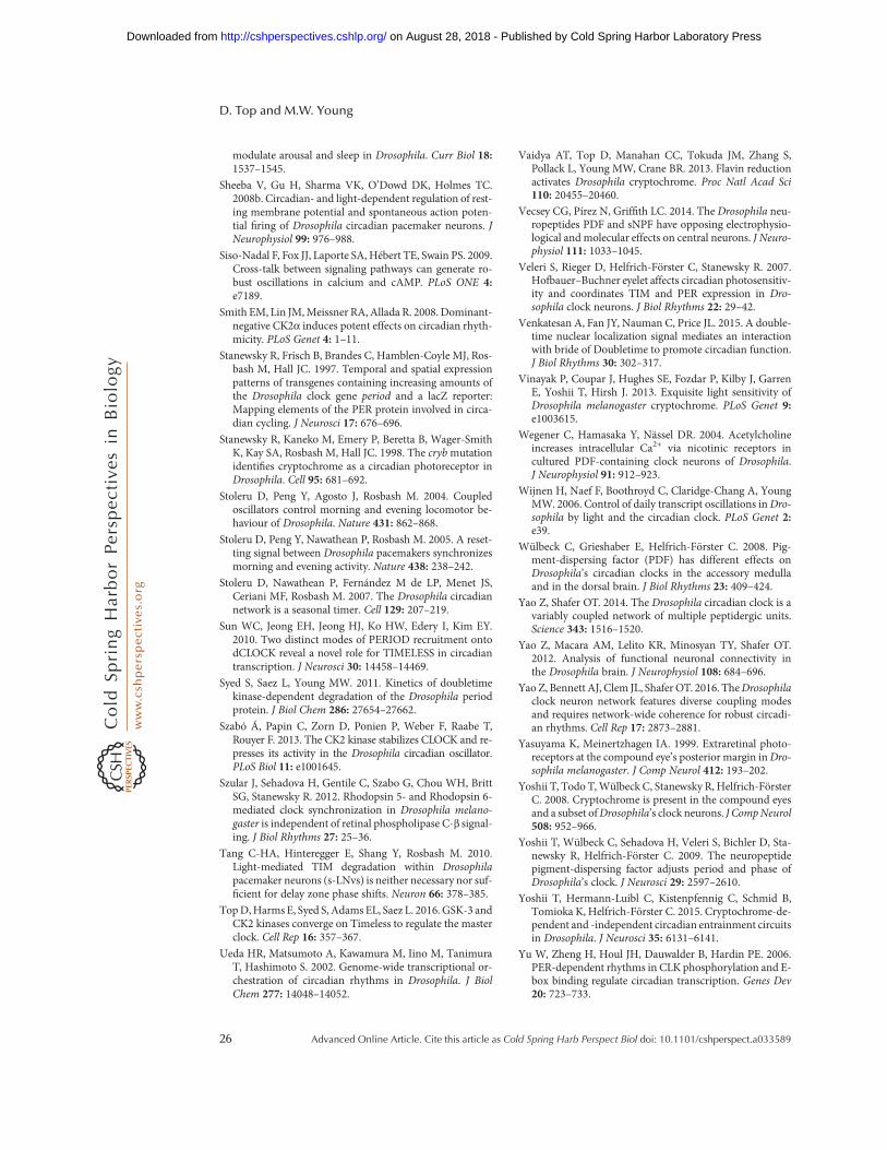
modulate arousal and sleep in Drosophila. Curr Biol 18:1537–1545.
Sheeba V, Gu H, Sharma VK, O’Dowd DK, Holmes TC.2008b. Circadian- and light-dependent regulation of rest-ing membrane potential and spontaneous action poten-tial firing of Drosophila circadian pacemaker neurons. JNeurophysiol 99: 976–988.
Siso-Nadal F, Fox JJ, Laporte SA, Hébert TE, Swain PS. 2009.Cross-talk between signaling pathways can generate ro-bust oscillations in calcium and cAMP. PLoS ONE 4:e7189.
Smith EM, Lin JM,Meissner RA, Allada R. 2008. Dominant-negative CK2α induces potent effects on circadian rhyth-micity. PLoS Genet 4: 1–11.
Stanewsky R, Frisch B, Brandes C, Hamblen-Coyle MJ, Ros-bash M, Hall JC. 1997. Temporal and spatial expressionpatterns of transgenes containing increasing amounts ofthe Drosophila clock gene period and a lacZ reporter:Mapping elements of the PER protein involved in circa-dian cycling. J Neurosci 17: 676–696.
Stanewsky R, Kaneko M, Emery P, Beretta B, Wager-SmithK, Kay SA, Rosbash M, Hall JC. 1998. The crybmutationidentifies cryptochrome as a circadian photoreceptor inDrosophila. Cell 95: 681–692.
Stoleru D, Peng Y, Agosto J, Rosbash M. 2004. Coupledoscillators control morning and evening locomotor be-haviour of Drosophila. Nature 431: 862–868.
Stoleru D, Peng Y, Nawathean P, Rosbash M. 2005. A reset-ting signal between Drosophila pacemakers synchronizesmorning and evening activity. Nature 438: 238–242.
Stoleru D, Nawathean P, Fernández M de LP, Menet JS,Ceriani MF, Rosbash M. 2007. The Drosophila circadiannetwork is a seasonal timer. Cell 129: 207–219.
Sun WC, Jeong EH, Jeong HJ, Ko HW, Edery I, Kim EY.2010. Two distinct modes of PERIOD recruitment ontodCLOCK reveal a novel role for TIMELESS in circadiantranscription. J Neurosci 30: 14458–14469.
Syed S, Saez L, Young MW. 2011. Kinetics of doubletimekinase-dependent degradation of the Drosophila periodprotein. J Biol Chem 286: 27654–27662.
Szabó Á, Papin C, Zorn D, Ponien P, Weber F, Raabe T,Rouyer F. 2013. The CK2 kinase stabilizes CLOCK and re-presses its activity in the Drosophila circadian oscillator.PLoS Biol 11: e1001645.
Szular J, Sehadova H, Gentile C, Szabo G, Chou WH, BrittSG, Stanewsky R. 2012. Rhodopsin 5- and Rhodopsin 6-mediated clock synchronization in Drosophila melano-gaster is independent of retinal phospholipase C-β signal-ing. J Biol Rhythms 27: 25–36.
Tang C-HA, Hinteregger E, Shang Y, Rosbash M. 2010.Light-mediated TIM degradation within Drosophilapacemaker neurons (s-LNvs) is neither necessary nor suf-ficient for delay zone phase shifts. Neuron 66: 378–385.
TopD,Harms E, Syed S, Adams EL, Saez L. 2016. GSK-3 andCK2 kinases converge on Timeless to regulate the masterclock. Cell Rep 16: 357–367.
Ueda HR, Matsumoto A, Kawamura M, Iino M, TanimuraT, Hashimoto S. 2002. Genome-wide transcriptional or-chestration of circadian rhythms in Drosophila. J BiolChem 277: 14048–14052.
Vaidya AT, Top D, Manahan CC, Tokuda JM, Zhang S,Pollack L, Young MW, Crane BR. 2013. Flavin reductionactivates Drosophila cryptochrome. Proc Natl Acad Sci110: 20455–20460.
Vecsey CG, Pírez N, Griffith LC. 2014. The Drosophila neu-ropeptides PDF and sNPF have opposing electrophysio-logical and molecular effects on central neurons. J Neuro-physiol 111: 1033–1045.
Veleri S, Rieger D, Helfrich-Förster C, Stanewsky R. 2007.Hofbauer–Buchner eyelet affects circadian photosensitiv-ity and coordinates TIM and PER expression in Dro-sophila clock neurons. J Biol Rhythms 22: 29–42.
Venkatesan A, Fan JY, Nauman C, Price JL. 2015. A double-time nuclear localization signal mediates an interactionwith bride of Doubletime to promote circadian function.J Biol Rhythms 30: 302–317.
Vinayak P, Coupar J, Hughes SE, Fozdar P, Kilby J, GarrenE, Yoshii T, Hirsh J. 2013. Exquisite light sensitivity ofDrosophila melanogaster cryptochrome. PLoS Genet 9:e1003615.
Wegener C, Hamasaka Y, Nässel DR. 2004. Acetylcholineincreases intracellular Ca2+ via nicotinic receptors incultured PDF-containing clock neurons of Drosophila.J Neurophysiol 91: 912–923.
Wijnen H, Naef F, Boothroyd C, Claridge-Chang A, YoungMW. 2006. Control of daily transcript oscillations inDro-sophila by light and the circadian clock. PLoS Genet 2:e39.
Wülbeck C, Grieshaber E, Helfrich-Förster C. 2008. Pig-ment-dispersing factor (PDF) has different effects onDrosophila’s circadian clocks in the accessory medullaand in the dorsal brain. J Biol Rhythms 23: 409–424.
Yao Z, Shafer OT. 2014. The Drosophila circadian clock is avariably coupled network of multiple peptidergic units.Science 343: 1516–1520.
Yao Z, Macara AM, Lelito KR, Minosyan TY, Shafer OT.2012. Analysis of functional neuronal connectivity inthe Drosophila brain. J Neurophysiol 108: 684–696.
Yao Z, Bennett AJ, Clem JL, Shafer OT. 2016. TheDrosophilaclock neuron network features diverse coupling modesand requires network-wide coherence for robust circadi-an rhythms. Cell Rep 17: 2873–2881.
Yasuyama K, Meinertzhagen IA. 1999. Extraretinal photo-receptors at the compound eye’s posterior margin inDro-sophila melanogaster. J Comp Neurol 412: 193–202.
Yoshii T, Todo T,Wülbeck C, Stanewsky R, Helfrich-FörsterC. 2008. Cryptochrome is present in the compound eyesand a subset ofDrosophila’s clock neurons. J CompNeurol508: 952–966.
Yoshii T, Wülbeck C, Sehadova H, Veleri S, Bichler D, Sta-newsky R, Helfrich-Förster C. 2009. The neuropeptidepigment-dispersing factor adjusts period and phase ofDrosophila’s clock. J Neurosci 29: 2597–2610.
Yoshii T, Hermann-Luibl C, Kistenpfennig C, Schmid B,Tomioka K, Helfrich-Förster C. 2015. Cryptochrome-de-pendent and -independent circadian entrainment circuitsin Drosophila. J Neurosci 35: 6131–6141.
Yu W, Zheng H, Houl JH, Dauwalder B, Hardin PE. 2006.PER-dependent rhythms in CLK phosphorylation and E-box binding regulate circadian transcription. Genes Dev20: 723–733.
D. Top and M.W. Young
26 Advanced Online Article. Cite this article as Cold Spring Harb Perspect Biol doi: 10.1101/cshperspect.a033589
on August 28, 2018 - Published by Cold Spring Harbor Laboratory Press http://cshperspectives.cshlp.org/Downloaded from
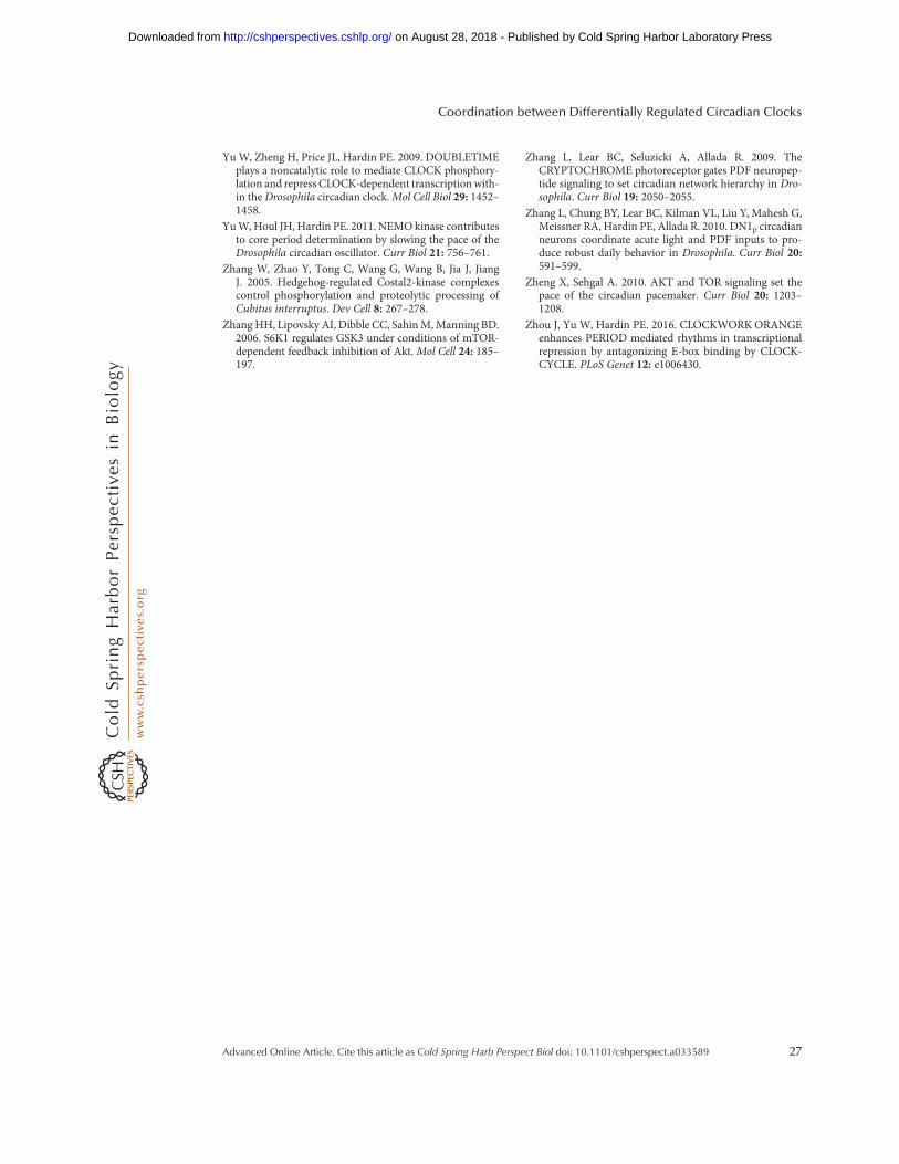
YuW, Zheng H, Price JL, Hardin PE. 2009. DOUBLETIMEplays a noncatalytic role to mediate CLOCK phosphory-lation and repress CLOCK-dependent transcription with-in the Drosophila circadian clock.Mol Cell Biol 29: 1452–1458.
YuW, Houl JH, Hardin PE. 2011. NEMOkinase contributesto core period determination by slowing the pace of theDrosophila circadian oscillator. Curr Biol 21: 756–761.
Zhang W, Zhao Y, Tong C, Wang G, Wang B, Jia J, JiangJ. 2005. Hedgehog-regulated Costal2-kinase complexescontrol phosphorylation and proteolytic processing ofCubitus interruptus. Dev Cell 8: 267–278.
Zhang HH, Lipovsky AI, Dibble CC, SahinM,Manning BD.2006. S6K1 regulates GSK3 under conditions of mTOR-dependent feedback inhibition of Akt. Mol Cell 24: 185–197.
Zhang L, Lear BC, Seluzicki A, Allada R. 2009. TheCRYPTOCHROME photoreceptor gates PDF neuropep-tide signaling to set circadian network hierarchy in Dro-sophila. Curr Biol 19: 2050–2055.
Zhang L, Chung BY, Lear BC, Kilman VL, Liu Y, Mahesh G,Meissner RA, Hardin PE, Allada R. 2010. DN1p circadianneurons coordinate acute light and PDF inputs to pro-duce robust daily behavior in Drosophila. Curr Biol 20:591–599.
Zheng X, Sehgal A. 2010. AKT and TOR signaling set thepace of the circadian pacemaker. Curr Biol 20: 1203–1208.
Zhou J, Yu W, Hardin PE. 2016. CLOCKWORKORANGEenhances PERIOD mediated rhythms in transcriptionalrepression by antagonizing E-box binding by CLOCK-CYCLE. PLoS Genet 12: e1006430.
Coordination between Differentially Regulated Circadian Clocks
Advanced Online Article. Cite this article as Cold Spring Harb Perspect Biol doi: 10.1101/cshperspect.a033589 27
on August 28, 2018 - Published by Cold Spring Harbor Laboratory Press http://cshperspectives.cshlp.org/Downloaded from
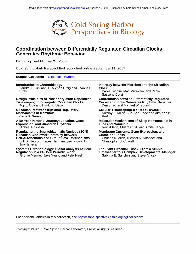
published online September 11, 2017Cold Spring Harb Perspect Biol Deniz Top and Michael W. Young Generates Rhythmic BehaviorCoordination between Differentially Regulated Circadian Clocks
Subject Collection Circadian Rhythms
Introduction to Chronobiology
DuffySandra J. Kuhlman, L. Michon Craig and Jeanne F. Clock
Interplay between Microbes and the Circadian
Sassone-CorsiPaola Tognini, Mari Murakami and Paolo
Timekeeping in Eukaryotic Circadian ClocksDesign Principles of Phosphorylation-Dependent
Koji L. Ode and Hiroki R. UedaCircadian Clocks Generates Rhythmic BehaviorCoordination between Differentially Regulated
Deniz Top and Michael W. Young
Mechanisms in MammalsCircadian Posttranscriptional Regulatory
Carla B. Green
Cellular Timekeeping: It's Redox o'Clock
ReddyNikolay B. Milev, Sue-Goo Rhee and Akhilesh B.
Expression, and Circadian RhythmsA 50-Year Personal Journey: Location, Gene
Michael RosbashFlies and MammalsMolecular Mechanisms of Sleep Homeostasis in
Ravi Allada, Chiara Cirelli and Amita Sehgal
Cell-Autonomous and Circuit-Level MechanismsCircadian Clockwork: Interplay between Regulating the Suprachiasmatic Nucleus (SCN)
Smyllie, et al.Erik D. Herzog, Tracey Hermanstyne, Nicola J.
Circadian ClocksMembrane Currents, Gene Expression, and
Christopher S. ColwellCharles N. Allen, Michael N. Nitabach and
Regulation in a 24-Hour Periodic WorldSystems Chronobiology: Global Analysis of Gene
Jérôme Mermet, Jake Yeung and Felix NaefManagerTimekeeper to a Complex Developmental
The Plant Circadian Clock: From a Simple
Sabrina E. Sanchez and Steve A. Kay
http://cshperspectives.cshlp.org/cgi/collection/ For additional articles in this collection, see
Copyright © 2017 Cold Spring Harbor Laboratory Press; all rights reserved
on August 28, 2018 - Published by Cold Spring Harbor Laboratory Press http://cshperspectives.cshlp.org/Downloaded from