Embed Size (px)
Citation preview

Coordinate Regulation of FOXO1 by miR-27a, miR-96, andmiR-182 in Breast Cancer Cells□S
Received for publication, June 8, 2009, and in revised form, June 30, 2009 Published, JBC Papers in Press, July 1, 2009, DOI 10.1074/jbc.M109.031427
Irene K. Guttilla‡ and Bruce A. White‡§1
From the Departments of §Cell Biology and ‡Molecular, Microbial and Structural Biology, University of Connecticut Health Center,Farmington, Connecticut 06030
The FOXO1 transcription factor orchestrates the regulationof genes involved in the apoptotic response, cell cycle check-points, and cellular metabolism. FOXO1 is a putative tumorsuppressor, and the expression of this gene is dysregulated insome cancers, including prostate and endometrial cancers.However, the molecular mechanism resulting in aberrantexpression of human FOXO1 in cancer cells is poorly under-stood. We show here that FOXO1 mRNA is down-regulated inbreast tumor samples as compared with normal breast tissue.Silencing of the microRNA processing enzymes, Drosha andDicer, led to an increase in FOXO1 expression. We also identi-fied functional and specific microRNA target sites in theFOXO1 3�-untranslated region for miR-27a, miR-96, and miR-182, microRNAs that have previously been linked to oncogenictransformation. The three microRNAs, miR-27a, miR-96 andmiR-182, were observed to be highly expressed inMCF-7 breastcancer cells, in which the level of FOXO1 protein is very low.Antisense inhibitors to each of these microRNAs led to a signif-icant increase in endogenous FOXO1 expression and to adecrease in cell number in amanner thatwas blockedbyFOXO1siRNA.Overexpressionof FOXO1 resulted indecreased cell via-bility because of inhibition of cell cycle traverse and induction ofcell death. We have identified a novel mechanism of FOXO1regulation, and targeting of FOXO1 by microRNAs may con-tribute to transformation or maintenance of an oncogenic statein breast cancer cells.
The Forkhead Box O subfamily of transcription factors(FOXO) regulates a variety of important cellular processesincluding metabolism, cellular differentiation, apoptosis, andcell-cycle progression (1, 2). Acting as master cellular regula-tors, FOXO transcription factors can both activate and represstarget gene expression (3).The three predominant members of the FOXO family
(FOXO1, FOXO3a, and FOXO4) were first implicated intumorigenesis based on the observation that fusion proteinsresulting from chromosomal breakpoints exist in certain typesof cancers (2). Recent evidence suggests that FOXO proteinsfunction as tumor suppressors based on their role in regulatingcell-cycle progression and inducing apoptosis (4).
One regulatory mechanism of FOXO1 activity is throughphosphorylation, primarily downstream of the insulin-stimu-lated phosphatidylinositol 3-kinase/AKT/protein kinase B sig-naling pathway, which results in nuclear exclusion (5–7).FOXO1 activity can also be regulated by acetylation (8) andubiquitination (9, 10). In addition to insulin, FOXO1 can bedown-regulated by other growth factors including estrogen (11,12) and epidermal growth factor (13). The estrogen receptor �also complexes with phosphorylated FOXO1 and mediates itsexport from the nucleus. These mitogens are important for thegrowth and survival of breast cancer cells andmay contribute tomaintaining low levels of FOXO1 in breast cancer cells.Although the activity of FOXO1 has been well characterized,
very little is known about the regulation of FOXO1 expression,particularly in breast cancer. FOXO1 is down-regulated in sev-eral other cancers including endometrial carcinoma (14) andovarian cancer (15). In addition, restoration of FOXO1 expres-sion in endometrial carcinoma cells decreases cellular prolifer-ation (14). Although the role of FOXO1 in tumorigenesis isnot entirely clear, it has been hypothesized that the down-regulation of this gene is an important step in tumor for-mation. Recently, it was shown that FOXO1 is reduced incertain endometrial carcinoma cells lines as well as endo-metriod endometrial tumors (14). Subsequent analysis sug-gested that the down-regulation of FOXO1 expression wasbecause of a post-transcriptional mechanism (14).A novel class of small RNA molecules, microRNAs, has been
implicated in the post-transcriptional regulation of thousands ofmRNA transcripts resulting in decreased protein expression oftargetgenes (16).MicroRNAsaresmall�21–25nucleotidesingle-stranded RNAmolecules that negatively regulate gene expressionby binding to the 3�-UTR2 of a targetmRNAmolecule resulting ineither degradation of the transcript or translational inhibition.Recent studies have shown that many microRNAs work in con-junction to fine tuneproteinexpressiononaglobal level (17,18). Inaddition, mice harboring knockouts inmicroRNA genes display avariety of detrimental phenotypes such as severe immune defi-ciency (19, 20) and stress-induced heart defects (21), and thesestudieshaveemphasized the important roleof thesegenes in tissuehomeostasis and disease.Here, we show that FOXO1 expression is also down-regu-
lated in breast tumor samples compared with normal breast□S The on-line version of this article (available at http://www.jbc.org) contains
supplemental Figs. S1–2 and Tables 1–3.1 To whom correspondence should be addressed: Dept. of Cell Biology, Uni-
versity of Connecticut Health Center, 263 Farmington Ave., Farmington, CT06030-3505. Tel.: 860-679-2811; Fax: 860-679-1269; E-mail: [email protected].
2 The abbreviations used are: UTR, untranslated region; siRNA, small interfer-ing RNA; GAPDH, glyceraldehyde-3-phosphate dehydrogenase; BrdUrd,bromodeoxyuridine; NGS, normal goat serum; WT, wild type; AAA, consti-tutively active FOXO1 mutant.
THE JOURNAL OF BIOLOGICAL CHEMISTRY VOL. 284, NO. 35, pp. 23204 –23216, August 28, 2009© 2009 by The American Society for Biochemistry and Molecular Biology, Inc. Printed in the U.S.A.
23204 JOURNAL OF BIOLOGICAL CHEMISTRY VOLUME 284 • NUMBER 35 • AUGUST 28, 2009
by guest on September 28, 2018
http://ww
w.jbc.org/
Dow
nloaded from

tissue. We hypothesized that the low levels of FOXO1 are aconsequence of microRNA regulation, and we subsequentlyidentified three microRNAs that directly target FOXO1 (miR-27a, miR-96, and miR-182) and regulate endogenous proteinexpression in MCF-7 breast cancer cells. Suppression of thesemicroRNAs resulted in an increase in FOXO1 protein and in adecrease in cell number that was rescued by FOXO1 siRNA. Inaddition, we show that overexpression of FOXO1 in breast can-cer cells resulted in decreased cell number both through inhi-bition of cell cycle traverse and increased cell death. This studyhas identified a novel mechanism for the down-regulation ofFOXO1 in breast cancer samples, and this may impact thetransformation of breast cells andmaintenance of an oncogenicstate.
EXPERIMENTAL PROCEDURES
Cell Culture—MCF-7, T47D, MDA-MB-231, and MDA-MB-435 cells were obtained through American Type CultureCollection. MCF-7, MDA-MB-231, and MDA-MB-435 cellswere cultured in Dulbecco’s modified Eagle’s medium/F-12media supplemented with 10% fetal bovine serum and 1% pen-icillin/streptomycin (Invitrogen). T47D cells were cultured inDulbecco’s modified Eagle’s medium/F-12 media supple-mented with 10% fetal bovine serum, 1% penicillin/streptomy-cin, and 0.2 units/ml insulin (Sigma).Analysis of FOXO1 Expression in Breast Cancer Samples—
Breast Cancer I quantitative PCR tissue arrays were purchasedfrom OriGene and used to assess the expression of FOXO1.This array consisted of cDNAs obtained from 48 samples rang-ing from stage 0 (normal) to breast tumor stage III. All theclinical information associated with each of these samples canbe found on the OriGene web site (Breast Cancer Panel 1).Luciferase Constructs—The psi-CHECKTM-2 dual luciferase
reporter (Promega) was a kind gift from Dr. Henry Furneaux(University of Connecticut Health Center, Farmington, CT).All sequences were directionally cloned into plasmids cut withXhoI and NotI restriction endonucleases. Inserts were ligatedinto the vector and transformed into DH5� competent cells.Colonieswere screened for inserts via colony PCR and standardDNA sequencing. DNA from positive clones was purified usingthe Qiagen Plasmid Midi kit and visualized on a 1% agarose gelto verify accurate concentrations. Sequences cloned into thepsi-CHECKTM-2 vector varied depending on the nature of theassay. For the tiling assay, various regions of the FOXO13�-UTR ranging from 164 to 416 bp in size were amplified usinggene-specific primers and cloned into the luciferase reporterassay. DNA oligonucleotides for the sensor assay were pur-chased from Integrated DNATechnologies (Coralville, IA) andpurified by desalting. The oligonucleotides were treated withT4 polynucleotide kinase (Invitrogen), annealed, and ligatedinto the psi-CHECKTM-2 vector. For the deletion mutantexperiments (Fig. 5D), DNA oligonucleotides matching 50 bpof endogenous FOXO1 3�-UTR sequence were annealed andinserted into the psi-CHECKTM-2 vector. Mutants were gener-ated by synthesizing oligos missing the endogenous predictedmicroRNA binding site.Luciferase Reporter Assays—The indicated cell lines were
transiently transfected with 100 ng of the indicated plasmid
using Lipofectamine 2000 Transfection reagent (Invitrogen).Cells were serum-starved for 4 h followed by adding back thefull media. Cells were harvested 24 h post-transfection by treat-ment with trypsin, lysed in 1� Passive Lysis buffer (Promega),and centrifuged at 14,000 rpm for 15min. The supernatant wasassayed for firefly and Renilla luciferase activity following thePromega protocol. Values were reported as relative Renilla tofirefly luciferase activity, and significance is reported as �S.E.Quantitative Real-time PCR—mRNA transcripts weremeas-
ured using a standard SYBR Green real-time assay. RNA wasisolated using the Trizol reagent (Invitrogen), and 1 �g of totalRNA was reverse-transcribed using the Superscript III enzyme(Invitrogen). Real-time PCR was then performed on cDNA inan iQ Sybr Green Supermix (Bio-Rad) with gene-specific prim-ers (supplemental Table S1). Amplicons were analyzed usingthe ��Ct method, and data are represented as the mean ofthree independent experiments �S.E.Semiquantitative End Point Analysis of Mature MicroRNAs—
The protocol for the amplification and detection of maturemicroRNAs using a stem-loop gene-specific reverse transcrip-tion primer was adapted from Varkonyi-Gasic et al. (40).Briefly, stem-loop primers were designed to specifically reversetranscribe the mature microRNA of interest. The cDNA wassynthesized by incubating 1 �g of total RNA, 2 pmol of gene-specific primer, and 0.5 mM dNTPs at 65 °C for 5 min. Themixture was cooled on ice, 1� first strand buffer (250mMTris-HCl (pH 8.3), 375 mMKCl, 15 mMMgCl2), 5 mM dithiothreitol,4 units of RNaseOut, and 100 units of SuperscriptTM III reversetranscriptase (Invitrogen) were added to a reaction volume of50 �l and incubated at 55 °C for 60 min, and then the enzymewas inactivated at 70 °C for 15min. To ensure noDNAcontam-ination was present, control reactions were performed asdescribed above without the addition of SuperscriptTM IIIenzyme. Subsequent PCR amplification were performed bycombining 1� PCR buffer (200 mM Tris-HCl (pH 8.4), 500 mM
KCl), 1.5 mM MgCl2, 0.5 mM dNTPs, 0.2 �M primers, 2 �l ofcDNA, 1 unit of Taq polymerase, and nuclease-free water in areaction volume of 50 �l. Primers were designed based on thesequences of interest (supplemental Table S2). The tempera-ture program was 95 °C, 5 min3 [95 °C, 30 s3 55 °C, 30 s372 °C, 30 s] � 30 cycles3 72 °C 10min. It should be noted thatmicroRNAs expressed at lower levels (such asmiR-96) required35 cycles of PCR amplification to visualize products. PCR prod-ucts were resolved on a 10% nondenaturing acrylamide gel andstained with ethidium bromide in 0.5� Tris borate-EDTA.Antisense Inhibitors—MicroRNA expression was decreased
using miRIDIANTM microRNA Hairpin Inhibitors (Dharma-con) directed against hsa-miR-27a, hsa-miR-182, hsa-miR-96,has-miR-183, hsa-miR-122, or Negative Control #1 (a non-tar-geting sequence). Inhibitors were resuspended in nuclease-freewater at a stock concentration of 20 �M and transfected intoMCF-7 cells at the indicated final concentrations using Lipo-fectamine 2000 (Invitrogen) in serum-free conditions.Western Blotting—Whole cell lysates were prepared from the
indicated cell lines. Protein concentrations were quantitatedusing a standard BCA assay, and samples were resolved oneither an 8% (Dicer,Drosha) or 10% (FOXO1,GAPDH,�-actin)SDS-PAGE gel. The protein was transferred onto a nitrocellu-
MicroRNA Regulation of FOXO1 in Breast Cancer
AUGUST 28, 2009 • VOLUME 284 • NUMBER 35 JOURNAL OF BIOLOGICAL CHEMISTRY 23205
by guest on September 28, 2018
http://ww
w.jbc.org/
Dow
nloaded from
lose membrane, then blocked in 5% milk in 0.1% Tris-bufferedsaline-Tween 20 (TBST) at room temperature for 1 h. All pri-mary antibodies were obtained from Abcam, Inc. and wereincubatedwith themembrane at 4 °C overnight at the followingconcentrations: �-FOXO1 (1:1000), �-GAPDH (1:10,000),�-Dicer (1:1000), �-Drosha (1:2000), �-�-actin (1:2500).Mem-branes were washed with 1� TBST and incubated with eitheranti-mouse or anti-rabbit IgG horseradish peroxidase-conju-gated secondary antibody, both obtained from Santa Cruz Bio-technology. Protein expression was assessed by enhancedchemiluminescence and exposure to Biomax Light film (East-man Kodak Co.). Image J software from the National Institutesof Health was used to quantify band intensities.siRNA Treatments—SMARTpool siRNA targeting Dicer,
Drosha, FOXO1, and GAPDH were obtained from Dharma-con and used at a final concentration of either 80 nM (Dicerand Drosha) or 100 nM (FOXO1 and GAPDH (supplementalFig. S5)).Trypan Blue Cell Viability Assay—MCF-7 cells were plated
at 2 � 105 cells/well in 6-well culture dishes and transfectedwith the indicated concentrations of empty vector or expres-sion constructs. In some experiments, cells were transfectedwith 40 nM antisense inhibitors to specific microRNAs alone orinhibitors plus 100 nM siRNA targeting FOXO1 or GAPDH. Atthe indicated time points, cells were trypsinized and stainedwith trypan blue, and viable cells were counted four times foreach sample. Cell viability is expressed as a percentage of theempty vector ormock-treated samples. Values are expressed asthe average percentage of viable cells �S.E. of three independ-ent experiments.BrdUrd Assay—MCF-7 cells were plated at 1� 106 cells/well
in a 6-well culture dish and transfected with 4 �g of emptyvector or expression constructs. Transfection was allowed toproceed for 30 h, then BrdUrd was added to the cells at a finalconcentration of 10 �M, and incorporation was carried out for18 h. Cells were then washed and fixed in 3.7% formaldehydeand acid-washed, and staining of BrdUrd was carried out usingthe Ultravision Detection System kit (Thermo Scientific). Fourseparate fields were counted for each sample, and percentagesrepresent the total number of BrdUrd-positive cells per treat-ment normalized to the total cell number. Error bars representthe S.D. of four independent experiments.Microscopy—MCF-7 cells were grown on coverslips and
transfected with the indicated plasmids. Transfection was car-ried out for 24 h, then cells were fixed with 4% paraformalde-hyde and permeabilized with 1% Triton X-100 in phosphate-buffered saline. Cells were blocked in 3% normal goat serum(NGS), and an �-FOXO1 antibody was used at a dilution of1:100 (Chemicon). An anti-rabbit fluorescein isothiocyanate-labeled secondary antibody was used at a dilution of 1:50. Cellswere visualized using a Zeiss Pascal microscope at 40� with aNA 1.2 water immersion objective. Images were processed andanalyzed using the LSM Image Browser.Statistical Analysis—Values reported in all analysis were
expressed as the mean � S.E. However, Western blot quantifi-cations were noted only as averages. Differences between treat-ments and/or groups were analyzed using an unpaired Stu-dent’s t test. Statistical significance was accepted at p � 0.05.
RESULTS
FOXO1 Levels Are Decreased in Human Breast TumorSamples—FOXO1, FOXO3a, and FOXO4 mRNA expressionwas measured in human breast tumor samples from a range oftumor stages aswell as in normal breast tissue samples by quan-titative real-time PCR. FOXO1mRNA levels were significantlydecreased by about 2-fold in all tumor samples as comparedwith normal breast tissue (Fig. 1A). Levels of FOXO3a andFOXO4, however, did not differ significantly between the nor-mal and tumor samples (Fig. 1, B and C).We embarked on a study of whether the lower levels of
FOXO1 in breast tumors were due, in part, to post-transcrip-tional gene silencing for two reasons. First, FOXO1 displayedapparent post-transcriptional regulation in a previous study ofuterine endometrial cancer. Second, the FOXO1 transcriptharbors a very large 3�-UTR (�3350 nucleotides), which is aregion that most often confers post-transcriptional regulationthrough several mechanisms. MicroRNAs represent a subclassof noncoding, regulatory RNAs that play a significant role inposttranscriptional gene silencing. To test the hypothesis thatmicroRNAs target FOXO1 mRNA in breast cancer, key effec-tors of the microRNA pathway were knocked down, andFOXO1 levels were assessed. Drosha is a nuclear RNase IIIenzyme and is required for the first step in microRNA process-ing, generating pre-microRNAs from primary-microRNAs.Transfection of Drosha siRNA reduced Drosha protein to non-detectable levels at 48 h, and these began to rebound at 72 h.Knockdown of Drosha yielded significantly (�2–3-fold)increased FOXO1mRNA and protein levels at 48 and 72 h (Fig.2, A and B). To corroborate the findings from the Droshaknockdown, we transfected cells with siRNA to Dicer. Thecytoplasmic RNase III, Dicer, is required for the last step inmicroRNA processing that generates a mature microRNAduplex from a precursor stem-loop form. Upon treatment ofMDA-MB-231 cells with Dicer siRNA, Dicer mRNA levels fellby 12 h and rebounded by 48 h, presumably because Dicer isneeded for its own knockdown (Fig. 2C). Protein levels of Dicerremained low after 48 h (Fig. 2D). This transient loss of Dicerwas associated with an �2-fold increase (p � 0.05) in endoge-nous FOXO1 mRNA and protein at 24 and 48 h. These datasupport the possibility that microRNAs play a role eitherdirectly or indirectly in the suppression of FOXO1 expressionin human breast cancers.MicroRNA Regulation of the FOXO1 3�-UTR—In mammals
microRNAs commonly form imperfect duplexes with targetsites within the 3�-UTR region of target mRNAs. The FOXO1transcript harbors a very large 3�-UTR (�3350 nucleotides)and, therefore, many potential microRNA binding sites. Pre-dicted microRNA binding sites were mapped in the FOXO13�-UTR using target prediction databases (miRanda, Tar-getscan, and PicTar; supplemental Fig. S2 andTable S3). Poten-tial microRNA sites formed six clusters within the FOXO1mRNA 3�-UTR. To test whether these regions contained bonafide repressive elements, small sections of the FOXO1 3�-UTRwere cloned into a dual luciferase reporter system so that theFOXO1 sequence was inserted into the 3�-UTR of the Renillaluciferase gene, and luciferase expression was compared with
MicroRNA Regulation of FOXO1 in Breast Cancer
23206 JOURNAL OF BIOLOGICAL CHEMISTRY VOLUME 284 • NUMBER 35 • AUGUST 28, 2009
by guest on September 28, 2018
http://ww
w.jbc.org/
Dow
nloaded from

an empty vector construct in two different breast cancer celllines, MCF-7 and MDA-MB-231 cells. The six sections of theFOXO1 3�-UTR (ranging in size from 164 to 413 base pairs)were analyzed, and several sections conferred up to 1.5-foldrepression (Fig. 3). Five of the microRNA candidates (miR-27a,miR-96, miR-182, miR-183, and miR-128) were predicted totarget the FOXO1 3�-UTR in two different locations (sections204–492 and 1907–2247), and consequently, these micro-RNAs were selected for further detailed analysis. Some of thesemicroRNAs are predicted to target FOXO3a (miR-27a,miR-96,miR-182, miR-128) and FOXO4 (miR-96, miR-128). However,the regulation of these isoformswas not pursued in this study asthe mRNA levels of these transcripts did not vary in normalversus tumor breast tissue.A dual luciferase reporter construct containing a target site
perfectly complementary to the microRNA of interest, termeda “sensor” construct, was used to measure the endogenousactivity of the candidate microRNAs. The binding of a
microRNA to the target sequenceprovided would result in decreasedluciferase activity because of therepressive nature of microRNAs inmost cellular contexts. Four of thefive microRNAs, as detected by thecorresponding sensor constructs,displayed significant activity inMCF-7 cells: miR-27a (�10-fold re-pression),miR-182 (�3-fold repres-sion), miR-96 (�2-fold repression),and miR-183 (�2-fold repression)(Fig. 4A). To ensure the specificityof these interactions, mutant con-structs were synthesized that con-tained a 2-nucleotide change in theseed region of the target sequence(nucleotides 3 and 4). In each casethe mutant construct abrogatedthe repressive activity. It should benoted that miR-96 and miR-182possess identical seed sequences, sooverlap in their respective activitiesmay exist. Because miR-128 didnot display activity inMCF-7 cells,it was not further studied. Addi-tionally, later studies on miR-183(Figs. 5 and supplemental Fig. S3)indicated that it neither interactedwith the predicted miR-183 sitein FOXO1 mRNA nor regulatedFOXO1 protein expression. Thus,further study focused on miR-27a,miR-96, and miR-182.In addition to measuring the ac-
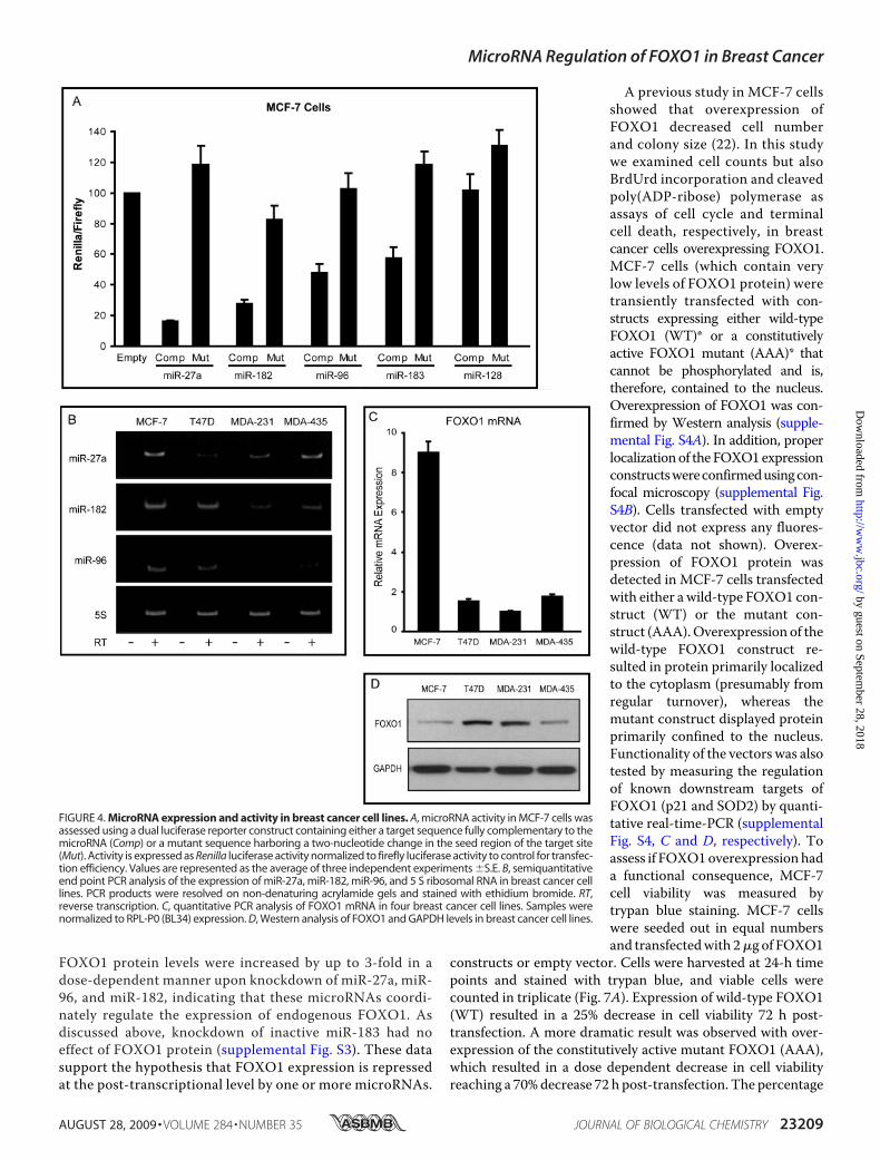
tivity of the candidate microRNAs,the expression levels of the threemost active microRNAs, miR-27a,miR-96 and miR-182, were assessedusing semiquantitative end point
PCR. Gene specific stem-loop reverse transcription primerswere designed for each mature microRNA, and PCR amplifica-tion using gene-specific primers was carried out after cDNAsynthesis. Expression of miR-27a, miR-96, and miR-182 wasmeasured in four breast cancer cell lines (Fig. 4B), and all threemicroRNAs displayed robust levels in theMCF-7 cells.We alsoexamined FOXO1 mRNA and protein levels in the four celllines. Although levels are relative, MCF-7 cells showed amarked discrepancy between FOXO1mRNA (Fig. 4C) and pro-tein levels (Fig. 4D), suggesting microRNA-mediated transla-tional repression rather than mRNA degradation as the pri-mary mechanism of microRNA regulation. Therefore, wefocused on MCF-7 cells to validate putative microRNA targetsites and the effects of suppression of endogenous microRNAlevels on FOXO1 expression.As previously stated, sections harboring base pairs 204–492
(Site 1) and 1907–2247 (Site 2) of the FOXO1 3�-UTR eachcontained one binding site for miR-27a, miR-96, and miR-182.
FIGURE 1. FOXO1 expression in human breast tumor samples. FOXO1 (A), FOXO3a (B), and FOXO4 (C) mRNAexpression was measured using gene specific primers in various breast tumor samples representing severalstages and compared with normal breast tissue. Sample categories were distributed as follows: Normal (n � 7),Stage I (n � 10), IIA (n � 13), IIB (n � 7), IIIA (n � 8). All samples were normalized to �-actin expression. *, p �0.05; **, p � 0.01; ***, p � 0.001.
MicroRNA Regulation of FOXO1 in Breast Cancer
AUGUST 28, 2009 • VOLUME 284 • NUMBER 35 JOURNAL OF BIOLOGICAL CHEMISTRY 23207
by guest on September 28, 2018
http://ww
w.jbc.org/
Dow
nloaded from

Thus, each construct (Site 1 or Site 2) was transfected intoMCF-7 cells either alone or with antisense inhibitors to miR-27a (Fig. 5A), miR-96, or miR-182 (Fig. 5B). A non-targeting
inhibitorwas used as a negative con-trol. Upon transfection of segmentsof the FOXO1 3�-UTR, a 1.5-foldrepression was observed. In thepresence of antisense inhibitors di-rected against miR-27a or miR-182,this repression was abrogated. Itshould be noted that inhibitor tomiR-96 did not relieve the suppres-sion from either site. These data indi-cate that the repression of endoge-nous FOXO1 3�-UTR sequence isspecifically because of the activity ofmicroRNAs, particularly miR-27aand miR-182.MicroRNAs often regulate gene
expression coordinately, and multi-ple microRNAs may act on a singletarget. To assess the combinatorialeffect of multiple microRNA sites inthe FOXO1 3�-UTR, a dual lucifer-ase reporter construct containing atarget site encompassing both activesections (Site 1 and Site 2) was cre-ated. When both sites were com-bined, they conferred a somewhatgreater repression (�2-fold) thaneach site alone (�1.5-fold). To test ifthe candidate microRNAs exam-ined specifically targeted thesepredicted regions, antisense inhibi-tors directed against the individualmicroRNAs (or an unrelated se-quence) were co-transfected withthe reporter constructs, and lucifer-
ase activity was measured in MCF-7 cells. Inhibitors againstmiR-27a, miR-182, and, unexpectedly, against miR-96, relievedrepression, indicating that these microRNAs are responsiblefor the microRNA-mediated repression observed (Fig. 5C).This series of experiments was followed by insertion of 50-bpregions encompassing either the endogenous miR-27a ormiR-96/182 sites into the luciferase reporter vector. Bothsites conferred �1.5–2-fold repression of luciferase activity(Fig. 5D). Excision of the endogenous site (�miR-27a or�miR-96/182) abrogated repression. As mentioned above,the endogenous miR-183 site was devoid of suppressiveactivity in MCF-7 cells.MicroRNAs Regulate Cell Proliferation and Survival Specifi-
cally through Their Suppression of Endogenous FOXO1 Protein—To assess the role of microRNA regulation on endogenousFOXO1 protein expression, antisense inhibitors directedagainst miR-27a (Fig. 6A) or miR-96 and miR-182 (Fig. 6B)were transiently transfected into MCF-7 cells and levels ofFOXO1 protein were measured by Western blot. Inhibitorsdirected against miR-122, a liver-specific microRNA, wereused as a negative control. Knockdown of the indicatedmicroRNAs was confirmed using semiquantitative end pointPCR analysis (Fig. 6, A and B, lower panels). Endogenous
FIGURE 2. Knockdown of Dicer and Drosha resulted in increased FOXO1 mRNA and protein levels. A, quanti-tative PCR analysis of FOXO1 and Drosha mRNA levels at the indicated time points post-transfection of Drosha siRNAin MDA-231 cells. Relative mRNA expression was normalized to RPL-P0 (B634). Error bars indicate the S.E. of threeindependent experiments. B, representative Western blot of Drosha and FOXO1 protein levels after treatment ofMDA-231 cells with Drosha siRNA. Numbers represent the average -fold change compared with mock for threeindependent experiments. C, quantitative PCR analysis of FOXO1 and Dicer mRNA levels at the indicated time pointspost-transfection of Dicer siRNA in MDA-231 cells. Relative mRNA expression was normalized to RPL-P0 (B634). Errorbars indicate the S.E. of three independent experiments. D, representative Western blot of Dicer, FOXO1, and GAPDHmRNA after transfection with Dicer siRNA in MDA-231 cells. Numbers represent the average -fold change comparedwith mock for three independent experiments. *, p � 0.05; **, p � 0.01. M, molecular mass standards.
FIGURE 3. Repression of predicted microRNA target sites in the FOXO13�-UTR. Short fragments of the FOXO1 3�-UTR harboring predicted microRNAsites were PCR amplified and cloned into a dual luciferase reporter plasmid. Nucle-otides corresponding to the segments of the 3�-UTR tested are indicated. Luciferaserepression was conferred with Renilla activity and normalized to firefly activity.
MicroRNA Regulation of FOXO1 in Breast Cancer
23208 JOURNAL OF BIOLOGICAL CHEMISTRY VOLUME 284 • NUMBER 35 • AUGUST 28, 2009
by guest on September 28, 2018
http://ww
w.jbc.org/
Dow
nloaded from
FOXO1 protein levels were increased by up to 3-fold in adose-dependent manner upon knockdown of miR-27a, miR-96, and miR-182, indicating that these microRNAs coordi-nately regulate the expression of endogenous FOXO1. Asdiscussed above, knockdown of inactive miR-183 had noeffect of FOXO1 protein (supplemental Fig. S3). These datasupport the hypothesis that FOXO1 expression is repressedat the post-transcriptional level by one or more microRNAs.
A previous study in MCF-7 cellsshowed that overexpression ofFOXO1 decreased cell numberand colony size (22). In this studywe examined cell counts but alsoBrdUrd incorporation and cleavedpoly(ADP-ribose) polymerase asassays of cell cycle and terminalcell death, respectively, in breastcancer cells overexpressing FOXO1.MCF-7 cells (which contain verylow levels of FOXO1 protein) weretransiently transfected with con-structs expressing either wild-typeFOXO1 (WT)* or a constitutivelyactive FOXO1 mutant (AAA)* thatcannot be phosphorylated and is,therefore, contained to the nucleus.Overexpression of FOXO1 was con-firmed by Western analysis (supple-mental Fig. S4A). In addition, properlocalizationof the FOXO1expressionconstructswereconfirmedusingcon-focal microscopy (supplemental Fig.S4B). Cells transfected with emptyvector did not express any fluores-cence (data not shown). Overex-pression of FOXO1 protein wasdetected in MCF-7 cells transfectedwith either a wild-type FOXO1 con-struct (WT) or the mutant con-struct (AAA).Overexpression of thewild-type FOXO1 construct re-sulted in protein primarily localizedto the cytoplasm (presumably fromregular turnover), whereas themutant construct displayed proteinprimarily confined to the nucleus.Functionality of the vectors was alsotested by measuring the regulationof known downstream targets ofFOXO1 (p21 and SOD2) by quanti-tative real-time-PCR (supplementalFig. S4, C and D, respectively). Toassess if FOXO1overexpression hada functional consequence, MCF-7cell viability was measured bytrypan blue staining. MCF-7 cellswere seeded out in equal numbersand transfectedwith 2�g of FOXO1
constructs or empty vector. Cells were harvested at 24-h timepoints and stained with trypan blue, and viable cells werecounted in triplicate (Fig. 7A). Expression of wild-type FOXO1(WT) resulted in a 25% decrease in cell viability 72 h post-transfection. A more dramatic result was observed with over-expression of the constitutively active mutant FOXO1 (AAA),which resulted in a dose dependent decrease in cell viabilityreaching a 70%decrease 72 h post-transfection. The percentage
FIGURE 4. MicroRNA expression and activity in breast cancer cell lines. A, microRNA activity in MCF-7 cells wasassessed using a dual luciferase reporter construct containing either a target sequence fully complementary to themicroRNA (Comp) or a mutant sequence harboring a two-nucleotide change in the seed region of the target site(Mut). Activity is expressed as Renilla luciferase activity normalized to firefly luciferase activity to control for transfec-tion efficiency. Values are represented as the average of three independent experiments �S.E. B, semiquantitativeend point PCR analysis of the expression of miR-27a, miR-182, miR-96, and 5 S ribosomal RNA in breast cancer celllines. PCR products were resolved on non-denaturing acrylamide gels and stained with ethidium bromide. RT,reverse transcription. C, quantitative PCR analysis of FOXO1 mRNA in four breast cancer cell lines. Samples werenormalized to RPL-P0 (BL34) expression. D, Western analysis of FOXO1 and GAPDH levels in breast cancer cell lines.
MicroRNA Regulation of FOXO1 in Breast Cancer
AUGUST 28, 2009 • VOLUME 284 • NUMBER 35 JOURNAL OF BIOLOGICAL CHEMISTRY 23209
by guest on September 28, 2018
http://ww
w.jbc.org/
Dow
nloaded from

MicroRNA Regulation of FOXO1 in Breast Cancer
23210 JOURNAL OF BIOLOGICAL CHEMISTRY VOLUME 284 • NUMBER 35 • AUGUST 28, 2009
by guest on September 28, 2018
http://ww
w.jbc.org/
Dow
nloaded from

of viable cells for each treatment was normalized to the numberof viable cells transfected with equivalent concentrations ofempty vector.To further expand these studies, BrdUrd staining was con-
ducted to observe the effects of FOXO1 overexpression on cellproliferation. MCF-7 cells were transfected with either emptyvector, wild-type FOXO1 (WT), ormutant FOXO1 (AAA), andthe percentage of BrdUrd-positive cells was assessed 24 h post-transfection (Fig. 7B). MCF-7 cells overexpressing wild-type
FOXO1 (WT) had �30% decrease in proliferation, whereascells overexpressing the mutant FOXO1 protein (AAA) had an�50% decrease in cell proliferation. To evaluate whether theobserved decrease in cell viability was also because of the induc-tion of apoptosis, cleaved poly(ADP-ribose) polymerase ex-pression was measured after overexpression of FOXO1 inMCF-7 cells (Fig. 7C). A significant increase in cleaved poly-(ADP-ribose) polymerase was observed after transfection ofeither wild-type FOXO1 or mutant FOXO1, and overexpres-sion of these constructs was confirmed in these samples. Levelsof GAPDH were assessed as a loading control and remainedunchanged after overexpression of FOXO1. These results showthat the restoration of FOXO1 in MCF-7 breast cancer cellsresults in reduced cell number because of a decrease in prolif-eration and induction of apoptosis. This phenotype is furtherexacerbated when a FOXO1 constitutively active mutant isoverexpressed.Next, we examined whether the down-regulation of miR-
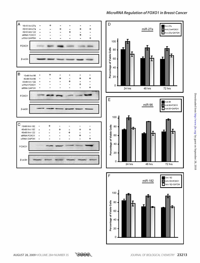
27a, miR-96, or miR-182 reduced cell number in a manner thatcould be “rescued” by siRNA to FOXO1. Again, antisenseinhibitors against either miR-27a, miR-96, or miR-182, but notmiR-183, increased endogenous FOXO1 protein levels inMCF-7 cells (Fig. 8, A–C, supplemental Fig. S3A), and this wasassociated with a �30–40% reduction in cell number by 72 hrelative to mock-transfected controls (Fig. 8, D–F). Co-trans-fection with FOXO1 siRNA, but not GAPDH siRNA, signifi-cantly blunted the effects of the antisense inhibitors againstmiR-27a, miR-96, and miR-182. These data provide direct evi-dence that the specific suppression of FOXO1 contributes tothe growth-promoting actions of these three microRNAs inMCF-7 cells.
DISCUSSION
The FOXO gene family encodes tumor-suppressive tran-scription factors that regulate multiple aspects of cell cycle tra-verse and survival. With respect to estrogen-dependent breastcancer, it should also be noted that FOXO1 heterodimerizeswith estrogen receptor � and inhibits its transcriptional activ-ity. A previous study showed that the overexpression of FOXO1in MCF-7 cells resulted in decreased cell number and colonyformation (22). In the present study we extended these findingsby showing that overexpression of FOXO1 strongly inhibitedproliferation and induced cell death in breast cancer MCF-7cells. The dysregulation of cell cycle and evasion of apoptosisplay a pivotal role in the development of cancer, and changes inFOXO1 expression and/or activity likely contribute to tumorprogression. Although numerous studies have addressed the
FIGURE 5. Repression of predicted microRNA target sites in the FOXO1 3[prime]-UTR in MCF-7 cells. Short fragments of the FOXO1 3�-UTR harboringpredicted microRNA sites were PCR-amplified and cloned into a dual luciferase reporter plasmid. Site 1 corresponds to nucleotides 204 – 492, and Site 2corresponds to nucleotides 1907–2247 of the FOXO1 3�-UTR. Luciferase repression was conferred with Renilla activity and normalized to firefly activity.A, luciferase repression of Site 1 and Site 2 followed by treatment with antisense inhibitor against miR-27a (Ant-27a) or a scrambled inhibitor (Ant-Neg). *, p �0.05 for inhibitor treatment versus Site alone. B, luciferase repression of Site 1 and Site 2 followed by treatment with antisense inhibitor against miR-96 (Ant-96),miR-182 (Ant-182), or a scrambled inhibitor (Ant-Neg). *, p � 0.05 for inhibitor treatment versus site alone. C, Site 1 and Site 2 were cloned into the 3�-UTR of theluciferase reporter plasmid. The vector containing Site 1 and Site 2 was treated with antisense inhibitors against the indicated microRNAs or a scrambledinhibitor. Error bars represent the S.E. of three independent experiments. *, p � 0.05; †, p � 0.05 for inhibitor treatment versus Site 1 � Site 2 alone. D, smallersites encompassing the predicted target site and endogenous flanking sequence (50-bp total insert size) was cloned into the dual luciferase reporter plasmid.White bars represent predicted Site 1, and black bars represent predicted Site 2 for each indicated microRNA. Luciferase repression was conferred with Renillaactivity and normalized to firefly activity for the empty vector (pMRE-Ø), the endogenous site (FOXO1), or the same site with the microRNA binding site deleted(�miR). Error bars represent the S.E. of three independent experiments. *, p � 0.05; **, p � 0.01 for endogenous site versus parent vector or site mutant.
FIGURE 6. Regulation of endogenous FOXO1 levels by miR-27a, miR-96,and miR-182. MCF-7 cells were treated with antisense inhibitors targetingmiR-27a (A) or miR-96 and miR-182 (B), or the unrelated microRNA miR-122(Ant-122) at the indicated concentrations. Cells were harvested 48 h post-transfection, and FOXO1 and GAPDH levels were measured by Western anal-ysis. Numbers represent the average -fold change compared with mock forthree independent experiments. Knockdown of endogenous microRNAs bythe appropriate inhibitors was verified using semi-quantitative reversetranscription-PCR.
MicroRNA Regulation of FOXO1 in Breast Cancer
AUGUST 28, 2009 • VOLUME 284 • NUMBER 35 JOURNAL OF BIOLOGICAL CHEMISTRY 23211
by guest on September 28, 2018
http://ww
w.jbc.org/
Dow
nloaded from

mechanisms by which FOXO1 protein activity and subcellularlocalization have been reported, little is known about the regu-lation of FOXO1 expression levels.The levels of FOXO1 proteins are depressed in a variety of
cancers, including prostate cancer, glioblastoma, and endome-trial carcinoma (14, 23–25). Goto et al. (14) found that FOXO1mRNA is �6-fold less in endometriod endometrial tumorscompared with normal cycling endometrium. This group alsomeasured the mRNA levels of the two other ubiquitouslyexpressed FOXO family members, FOXO3a and FOXO4, andfound that the levels of these messages remained unchangedbetween endometrial tumors and normal tissue. Similarly, inthis study we have shown that FOXO1 mRNA is also down-regulated2-fold in breast tumor samples comparedwith nor-mal breast tissue. In contrast, the expression of the other FOXO
family members (FOXO3a and FOXO4) that are expressed inbreast did not vary significantly between normal and tumorbreast tissue. FOXO3a and FOXO4 expression may change inresponse to specific conditions. For example, paclitaxel hasbeen shown to induce FOXO3a in taxane-sensitive MCF-7cells, resulting in the increased expression of the pro-apoptoticprotein, Bim (26).The mechanism by which FOXO1 expression is suppressed
in breast or any other cancer has not been established. Goto etal. (14) addressed this question in endometriod endometrialcarcinomausing two endometrial carcinoma cell lines,HEC-1Bcells and Ishikawa cells. Ishikawa cells express FOXO1 at barelydetectable levels, and these were not increased by proteasomeinhibition. The authors also found that the low levels of FOXO1in Ishikawa cells were not because of promotermethylation or a
FIGURE 7. Restoring FOXO1 expression in MCF-7 cells results in decreased cell proliferation and induction of apoptosis. A, MCF-7 cells were seeded atequal densities and transfected with the indicated amount of DNA. Empty vector (Empty), wild-type FOXO1 (WT), and constitutively active mutant (AAA). Viablecells were counted after staining with trypan blue at the indicated time points post-transfection. B, BrdUrd incorporation in MCF-7 cells transfected with emptyvector, wild-type FOXO1 (WT), or mutant FOXO1 (AAA). C, Western blot of cleaved poly(ADP-ribose) polymerase (c-PARP; top panel) after transfection with theindicated vectors. Middle panel, overexpression of FOXO1 was confirmed. The Western blot is a representative analysis from three independent experiments.All error bars represent the S.D. from four independent experiments. *, p � 0.05; **, p � 0.01.
MicroRNA Regulation of FOXO1 in Breast Cancer
23212 JOURNAL OF BIOLOGICAL CHEMISTRY VOLUME 284 • NUMBER 35 • AUGUST 28, 2009
by guest on September 28, 2018
http://ww
w.jbc.org/
Dow
nloaded from
MicroRNA Regulation of FOXO1 in Breast Cancer
AUGUST 28, 2009 • VOLUME 284 • NUMBER 35 JOURNAL OF BIOLOGICAL CHEMISTRY 23213
by guest on September 28, 2018
http://ww
w.jbc.org/
Dow
nloaded from

decrease in promoter activity. Subsequently, theymeasured therate of FOXO1mRNA turnover in each cell line and discoveredthat cells that expressed low levels of FOXO1 protein (Ish-ikawa) had a much faster rate of mRNA decay (half-life of �1.8h) compared with the HEC-1B cells (half-life of �4.5 h), whichexpress higher levels of FOXO1protein. Based on this evidence,the authors concluded that the down-regulation of FOXO1 inendometrial carcinoma cells was because of some form of post-transcriptional regulation.One centrally important mode of post-transcriptional regu-
lation is the repression of mRNA transcripts by microRNAs.Therefore, we hypothesized that microRNAsmay play a role inmaintaining low levels of FOXO1 in breast cancer cells. Weexamined the potential involvement of microRNAs in threegeneral ways. First, we knocked down two centrally importantenzymes in microRNA biogenesis. Drosha excises the stem-loop microRNA precursor from within the longer transcribedprimary-microRNA form. Dicer subsequently removes thehairpin from the precursor structure, yielding a microRNA:microRNA* duplex from which one strand is incorporated inthemicroRNA-induced silencing complex to repress the targetmRNAmolecule. In this study the knockdown of either Droshaor Dicer led to a significant, severalfold increase in the FOXO1mRNA and protein levels in the MDA-MB-231 breast cancercell line. This finding provides evidence that microRNAs areimportant for the suppression of endogenous FOXO1 expres-sion in some forms of breast cancer but does not indicatewhether FOXO1mRNA is, in fact, a direct target of one ormoremicroRNAs.Second, we gained experimental evidence of putative
microRNA target sites within the long FOXO1mRNA 3�-UTR.In examining the human FOXO1 3�-UTR for potentialmicroRNA target sites, we observed several clusters of targetsites. As some microRNAs display cell-specific expression andnot all predicted sites are functional, it was not surprising toobserve that only some of these fragments conferred significantrepression. However, two of these segments, termed Site 1(nucleotides 1–205) and Site 2 (nucleotides 1907–2247) con-ferred a significant degree of repression, and further analysis ofthese sites led to the finding that miR-27a, miR-96, and miR-182 were predicted to target each site. Of note, miR-96 andmiR-182 are transcribed from the same polycistronicmicroRNA cluster and have identical seed sequences. Thus,miR-96 andmiR-182 are likely to have similar levels, and wouldbe predicted to potentially share many of their targets. Addi-tionally, functional compensation of target repression mayoccur in the absence of one of the microRNAs.Analysis ofmiR-27a,miR-96, andmiR-182 levels and activity
in four breast cancer cell lines revealed that MCF-7 cells dis-played the most robust expression. Interestingly, this was cor-relatedwith discordantmRNAand protein levels. Further anal-ysis of these sites in the luciferase reporter assay showed that
antisense inhibitors against miR-27a, miR-182, or miR-96 spe-cifically blocked the repressive action of these sites. It should benoted that in this case, luciferase repression was not relieved tothe level of the empty vector, and this could be because of over-lap of coordinate microRNA regulation of these sequences.Wealso observed significantly greater repression when both siteswere combined, as opposed to that conferred by each site alone.Other studies have demonstrated the coordinate regulation of asingle mRNA transcript by multiple microRNAs. Krek et al.(27) , the group that developed PicTar in a mammalian system,tested the prediction that theMtpn gene was coordinately reg-ulated by let-7, miR-124, andmiR-375 usingWestern blot anal-ysis and a functional luciferase assay. They observed that theexogenous addition of each individual microRNA in MIN6mouse pancreatic cells was sufficient to regulate proteinexpression ofMtpn and repress luciferase activity of a constructharboring theMtpn 3�-UTR. Furthermore, the greatest amountof luciferase repression was observed after the addition of allthree microRNAs simultaneously, reinforcing the additiveeffect of combinatorial regulation bymultiple microRNAs (27).In the present studywe also observed significantlymore repres-sion from two cluster sites than by either site alone.Third, we demonstrated that the microRNAs identified
above regulate endogenous FOXO1 expression. InMCF-7 cellstreated with increasing doses of antisense inhibitors, it wasshown that knockdown of eithermiR-27a, miR-96, ormiR-182,but not the unrelated miR-122, significantly increased endoge-nous FOXO1 levels by severalfold. Although FOXO/Daf-16 hasbeen shown to repress the expression of a specific microRNA(lin-4), our findings provide the first evidence of the direct reg-ulation of endogenous FOXO1 expression by specific micro-RNAs (28).Previous to this study, miR-27a was implicated in breast can-
cer as an oncogenic microRNA. Mertens-Talcott et al. (29)found that miR-27a is highly expressed in breast cancer cells,and inhibition of this microRNA using antisense molecules inMDA-MB-231 cells decreased cell proliferation. This groupalso found that antisense RNA directed against miR-27adecreased the percentage of cells in S phase and increased thepercentage of cells in theG2-Mphase. Interestingly, the authorssuggest that miR-27a targets genes involved in regulating theG2-M phase of the cell cycle and identify one potential target(Myt-1) (29). In a separate study, Scott et al. (30) reported that apro-apoptotic dose of the inhibitor of histone deacetylases,LAQ824, rapidly decreases miR-27a levels in breast cancerSKBr3 cells. miR-27a was one of four microRNAs significantlyup-regulated in renal cell carcinoma compared with normalkidney (31) and acts as an oncogene in gastric adenocarcinoma(32). miR-27 has also been linked to hepatic stellate prolifera-tion, in which it targets the retinoid X receptor (33). Thesefindings provide supporting evidence that miR-27a can act asan oncogenic microRNA and warrant further study of miR-27a
FIGURE 8. Inhibiting miR-27a, miR-96, and miR-182 results in decreased cell viability because of increased FOXO1 expression. A–C, Western blotanalysis of FOXO1 expression after the treatment of MCF-7 cells with antisense inhibitors (Ant) to miR-27a (A), miR-96 (B), or miR-182 (C) or inhibitors plus siRNAto either FOXO1 or GAPDH. Treatments were carried out for 48 h. �-Actin was used as a loading control. D–F, MCF-7 cells were plated at 2.5 � 105 cells per welland treated with either 40 nM antisense inhibitors (Ant) to miR-27a (D), miR-96 (E), or miR-182 (F) or inhibitors plus siRNA targeting FOXO1 or GAPDH. At eachtime point wells were trypsinized, stained with trypan blue, and counted four separate times using a hemacytometer. Bars represent the average number ofviable cells �S.E. of three independent experiments.
MicroRNA Regulation of FOXO1 in Breast Cancer
23214 JOURNAL OF BIOLOGICAL CHEMISTRY VOLUME 284 • NUMBER 35 • AUGUST 28, 2009
by guest on September 28, 2018
http://ww
w.jbc.org/
Dow
nloaded from

regulation of downstream targets of FOXO1 to better charac-terize the involvement of miR-27a in maintaining a state ofuncontrolled cell growth.Although miR-96 and miR-182 have not been studied previ-
ously with respect to breast cancer, previous work has shownthat miR-96 and miR-182 are dysregulated in human disease,including a variety of cancers. Loscher et al. (34) showed thatmiR-96 and miR-182 are down-regulated in a mouse model ofretinitis pigmentosa. ThesemicroRNAs are contained in a genecluster harboringmiR-96,miR-182, andmiR-183, and this clus-ter is frequently amplified in advanced human melanoma (35)and melanoma cell lines (36). It has also been shown thatmiR-96 and miR-182 are overexpressed in colorectal cancer(37), classic Hodgkin lymphoma tumors (miR-182) and celllines (miR-96) (38), and chronic myeloid leukemia cells (39). Arecent study implicates miR-182 in the promotion of mela-nomametastasis. Overexpression of miR-182 inmelanoma celllines resulted in enhanced oncogenic properties, such asanchorage-independent growth and increased colony forma-tion on soft agar as well as invasion andmetastasis in vitro (36).Furthermore, this study identified a FOXO family member,FOXO3, as a direct target of miR-182. These studies implicatemiR-182 andmiR-96 as oncogenicmicroRNAs because of theirfrequent overexpression in cancers and the observation thattheir identified target genes are involved in the regulation of cellproliferation and apoptosis.In summary, this study demonstrated that of the three ubiq-
uitously expressed FOXO family members, FOXO1 is selec-tively down-regulated in breast cancer as compared with nor-mal tissue. We also demonstrate that FOXO1 expression isregulated by multiple microRNAs (miR-27a, miR-96, and miR-182) that have previously been implicated in oncogenesis.ThesemicroRNAs directly target various regions of the 3�-UTRto repress endogenous expression of FOXO1. Blockade of thesemicroRNAs led to restoration of FOXO1 expression. The res-toration of FOXO1 expression in MCF-7 cells resulted inreduced cell number, decreased cell cycle traverse, andincreased cell death. This effectwas further pronouncedwhen aconstitutively active mutant FOXO1 protein was overex-pressed. Additionally, suppression of FOXO1 restoration bysiRNA specifically blocked the anti-proliferative effects ofmicroRNA down-regulation, thereby functionally linkingmicroRNA expression, FOXO1 expression, and cell prolifera-tion and/or viability. These findings indicate that antisense tar-geting ofmiR-27a,miR-96, andmiR-182 alongwithmonitoringof microRNA and FOXO1 levels may be of therapeutic and/orprognostic value in breast cancer.
Acknowledgments—We greatly appreciate the gift of FOXO1 wild-type (WT) and mutant (AAA) overexpression constructs from Dr.Terry G. Unterman (Dept. of Medicine, University of Illinois, Chi-cago, IL).
REFERENCES1. Gross, D. N., van den Heuvel, A. P., and Birnbaum, M. J. (2008)Oncogene
27, 2320–23362. Fu, Z., and Tindall, D. J. (2008) Oncogene 27, 2312–23193. Obsil, T., and Obsilova, V. (2008) Oncogene 27, 2263–2275
4. Paik, J. H., Kollipara, R., Chu, G., Ji, H., Xiao, Y., Ding, Z., Miao, L., To-thova, Z., Horner, J.W., Carrasco, D. R., Jiang, S., Gilliland, D. G., Chin, L.,Wong, W. H., Castrillon, D. H., and DePinho, R. A. (2007) Cell 128,309–323
5. Rena, G., Guo, S., Cichy, S. C., Unterman, T. G., and Cohen, P. (1999) J.Biol. Chem. 274, 17179–17183
6. Guo, S., Rena, G., Cichy, S., He, X., Cohen, P., and Unterman, T. (1999)J. Biol. Chem. 274, 17184–17192
7. Zhang, X., Gan, L., Pan, H., Guo, S., He, X., Olson, S. T., Mesecar, A.,Adam, S., and Unterman, T. G. (2002) J. Biol. Chem. 277, 45276–45284
8. Perrot, V., and Rechler, M. M. (2005)Mol. Endocrinol. 19, 2283–22989. Matsuzaki, H., Daitoku, H., Hatta, M., Tanaka, K., and Fukamizu, A.
(2003) Proc. Natl. Acad. Sci. U.S.A. 100, 11285–1129010. Huang, H., Regan, K. M., Wang, F., Wang, D., Smith, D. I., van Deursen,
J.M., andTindall, D. J. (2005) Proc. Natl. Acad. Sci. U.S.A. 102, 1649–165411. Mazumdar, A., and Kumar, R. (2003) FEBS Lett. 535, 6–1012. Lengyel, F., Vertes, Z., Kovacs, K. A., Kornyei, J. L., Sumegi, B., and Vertes,
M. (2007) Steroids 72, 422–42813. Jackson, J. G., Kreisberg, J. I., Koterba, A. P., Yee, D., and Brattain, M. G.
(2000) Oncogene 19, 4574–458114. Goto, T., Takano,M., Albergaria, A., Briese, J., Pomeranz, K.M., Cloke, B.,
Fusi, L., Feroze-Zaidi, F., Maywald, N., Sajin, M., Dina, R. E., Ishihara, O.,Takeda, S., Lam, E. W., Bamberger, A. M., Ghaem-Maghami, S., andBrosens, J. J. (2008) Oncogene 27, 9–19
15. Goto, T., Takano, M., Hirata, J., and Tsuda, H. (2008) Br. J. Cancer 98,1068–1075
16. Bartel, D. P. (2004) Cell 116, 281–29717. Baek, D., Villen, J., Shin, C., Camargo, F. D., Gygi, S. P., and Bartel, D. P.
(2008) Nature 455, 64–7118. Selbach, M., Schwanhausser, B., Thierfelder, N., Fang, Z., Khanin, R., and
Rajewsky, N. (2008) Nature 455, 58–6319. Thai, T. H., Calado, D. P., Casola, S., Ansel, K. M., Xiao, C., Xue, Y.,
Murphy, A., Frendewey, D., Valenzuela, D., Kutok, J. L., Schmidt-Sup-prian,M., Rajewsky, N., Yancopoulos, G., Rao, A., and Rajewsky, K. (2007)Science 316, 604–608
20. Rodriguez, A., Vigorito, E., Clare, S., Warren, M. V., Couttet, P., Soond,D. R., van Dongen, S., Grocock, R. J., Das, P. P., Miska, E. A., Vetrie, D.,Okkenhaug, K., Enright, A. J., Dougan, G., Turner, M., and Bradley, A.(2007) Science 316, 608–611
21. van Rooij, E., Sutherland, L. B., Qi, X., Richardson, J. A., Hill, J., and Olson,E. N. (2007) Science 316, 575–579
22. Zhao, H. H., Herrera, R. E., Coronado-Heinsohn, E., Yang, M. C., Ludes-Meyers, J. H., Seybold-Tilson, K. J., Nawaz, Z., Yee, D., Barr, F. G., Diab,S. G., Brown, P. H., Fuqua, S. A., and Osborne, C. K. (2001) J. Biol. Chem.276, 27907–27912
23. Dong, X. Y., Chen, C., Sun, X., Guo, P., Vessella, R. L.,Wang, R. X., Chung,L. W., Zhou, W., and Dong, J. T. (2006) Cancer Res. 66, 6998–7006
24. Li, R., Erdamar, S., Dai, H., Wheeler, T. M., Frolov, A., Scardino, P. T.,Thompson, T. C., and Ayala, G. E. (2007) Hum. Pathol. 38, 1501–1507
25. Seoane, J., Le, H. V., Shen, L., Anderson, S. A., andMassague, J. (2004)Cell117, 211–223
26. Sunters, A., Fernandez de Mattos, S., Stahl, M., Brosens, J. J., Zoumpou-lidou, G., Saunders, C. A., Coffer, P. J.,Medema, R.H., Coombes, R. C., andLam, E. W. (2003) J. Biol. Chem. 278, 49795–49805
27. Krek, A., Grun, D., Poy, M. N., Wolf, R., Rosenberg, L., Epstein, E. J.,MacMenamin, P., da Piedade, I., Gunsalus, K. C., Stoffel, M., and Rajew-sky, N. (2005) Nat. Genet. 37, 495–500
28. Baugh, L. R., and Sternberg, P. W. (2006) Curr. Biol. 16, 780–78529. Mertens-Talcott, S. U., Chintharlapalli, S., Li, X., and Safe, S. (2007) Can-
cer Res. 67, 11001–1101130. Scott, G. K.,Mattie,M. D., Berger, C. E., Benz, S. C., and Benz, C. C. (2006)
Cancer Res. 66, 1277–128131. Gottardo, F., Liu, C. G., Ferracin, M., Calin, G. A., Fassan, M., Bassi, P.,
Sevignani, C., Byrne, D., Negrini, M., Pagano, F., Gomella, L. G., Croce,C. M., and Baffa, R. (2007) Urol. Oncol. 25, 387–392
32. Liu, T., Tang, H., Lang, Y., Liu, M., and Li, X. (2009) Cancer Lett. 273,233–242
33. Ji, J., Zhang, J., Huang, G., Qian, J., Wang, X., andMei, S. (2009) FEBS Lett.
MicroRNA Regulation of FOXO1 in Breast Cancer
AUGUST 28, 2009 • VOLUME 284 • NUMBER 35 JOURNAL OF BIOLOGICAL CHEMISTRY 23215
by guest on September 28, 2018
http://ww
w.jbc.org/
Dow
nloaded from
583, 759–76634. Loscher, C. J., Hokamp, K.,Wilson, J. H., Li, T., Humphries, P., Farrar, G. J.,
and Palfi, A. (2008) Exp. Eye Res. 87, 529–53435. Lin, W. M., Baker, A. C., Beroukhim, R., Winckler, W., Feng, W.,
Marmion, J. M., Laine, E., Greulich, H., Tseng, H., Gates, C., Hodi, F. S.,Dranoff, G., Sellers, W. R., Thomas, R. K., Meyerson, M., Golub, T. R.,Dummer, R., Herlyn, M., Getz, G., and Garraway, L. A. (2008)Cancer Res.68, 664–673
36. Segura, M. F., Hanniford, D., Menendez, S., Reavie, L., Zou, X., Alvarez-Diaz, S., Zakrzewski, J., Blochin, E., Rose, A., Bogunovic, D., Polsky, D.,Wei, J., Lee, P., Belitskaya-Levy, I., Bhardwaj, N., Osman, I., andHernando,E. (2009) Proc. Natl. Acad. Sci. U.S.A. 106, 1814–1819
37. Bandres, E., Cubedo, E., Agirre, X.,Malumbres, R., Zarate, R., Ramirez, N.,Abajo, A., Navarro, A., Moreno, I., Monzo, M., and García-Foncillas, J.(2006)Mol. Cancer 5, 29
38. Navarro, A., Gaya, A.,Martinez, A., Urbano-Ispizua, A., Pons, A., Balague,O., Gel, B., Abrisqueta, P., Lopez-Guillermo, A., Artells, R.,Montserrat, E.,and Monzo, M. (2008) Blood 111, 2825–2832
39. Agirre, X., Jimenez-Velasco, A., San Jose-Eneriz, E., Garate, L., Bandres, E.,Cordeu, L., Aparicio, O., Saez, B., Navarro, G., Vilas-Zornoza, A., Perez-Roger, I., García-Foncillas, J., Torres, A., Heiniger, A., Calasanz, M. J., Fortes,P., Roman-Gomez, J., and Prosper, F. (2008)Mol. Cancer Res. 6, 1830–1840
40. Varkonyi-Gasic, E., Wu, R., Wood, M., Walton, E. F., and Hellens, R. P.(2007) Plant Methods 3, 12
MicroRNA Regulation of FOXO1 in Breast Cancer
23216 JOURNAL OF BIOLOGICAL CHEMISTRY VOLUME 284 • NUMBER 35 • AUGUST 28, 2009
by guest on September 28, 2018
http://ww
w.jbc.org/
Dow
nloaded from

Irene K. Guttilla and Bruce A. WhiteCancer Cells
Coordinate Regulation of FOXO1 by miR-27a, miR-96, and miR-182 in Breast
doi: 10.1074/jbc.M109.031427 originally published online July 1, 20092009, 284:23204-23216.J. Biol. Chem.
10.1074/jbc.M109.031427Access the most updated version of this article at doi:
Alerts:
When a correction for this article is posted•
When this article is cited•
to choose from all of JBC's e-mail alertsClick here
Supplemental material:
http://www.jbc.org/content/suppl/2009/07/01/M109.031427.DC1
http://www.jbc.org/content/284/35/23204.full.html#ref-list-1
This article cites 40 references, 17 of which can be accessed free at
by guest on September 28, 2018
http://ww
w.jbc.org/
Dow
nloaded from