Embed Size (px)
Citation preview
TH
EJ
OU
RN
AL
OF
CE
LL
BIO
LO
GY
©
The Rockefeller University Press $8.00The Journal of Cell Biology, Vol. 171, No. 4, November 21, 2005 615–625http://www.jcb.org/cgi/doi/10.1083/jcb.200502043
JCB: ARTICLE
JCB 615
Coordinated transport of phosphorylated amyloid-
�
precursor protein and c-Jun NH
2
-terminal kinase–interacting protein-1
Zoia Muresan and Virgil Muresan
Department of Physiology and Biophysics, Case School of Medicine, Case Western Reserve University, Cleveland, OH 44106
he transmembrane protein amyloid-
�
precursorprotein (APP) and the vesicle-associated proteinc-Jun NH
2
-terminal kinase–interacting protein-1(JIP-1) are transported into axons by kinesin-1. Both pro-teins may bind to kinesin-1 directly and can be trans-ported separately. Because JIP-1 and APP can interact,kinesin-1 may recruit them as a complex, enabling theircotransport. In this study, we tested whether APP and JIP-1are transported together or separately on different vesi-cles. We found that, within the cellular context, JIP-1preferentially interacts with Thr
668
-phosphorylated APP(pAPP), compared with nonphosphorylated APP. In neu-
T
rons, JIP-1 colocalizes with vesicles containing pAPP andis excluded from those containing nonphosphorylatedAPP. The accumulation of JIP-1 and pAPP in neurites re-quires kinesin-1, and the expression of a phosphomimeticAPP mutant increases JIP-1 transport. Down-regulation ofJIP-1 by small interfering RNA specifically impairs trans-port of pAPP, with no effect on the trafficking of non-phosphorylated APP. These results indicate that thephosphorylation of APP regulates the formation of apAPP–JIP-1 complex that accumulates in neurites inde-pendent of nonphosphorylated APP.
Introduction
In neurons, membrane-bound cargoes are transported into axonsby kinesin motors such as kinesin-1 (Muresan, 2000). Becausethe rate of the delivery of proteins into axons should meet theneeds of the individual proteins at the destination, the forma-tion of the transport vesicle and recruitment of the requiredkinesin motor have to be highly regulated. This study addressesthe regulation of the axonal transport of two kinesin-1 cargoes,the amyloid-
�
precursor protein (APP) and the c-Jun NH
2
-terminal kinase (JNK)–interacting protein-1 (JIP-1).
Kinesin-1 is recruited to its vesicular cargoes by trans-membrane or cytoplasmic vesicle–associated proteins that in-teract with either the kinesin light chain (KLC) or kinesinheavy chain (KHC). Among the proposed kinesin-1 cargo link-ers are APP and JIP-1. APP, the precursor of the amyloid-
�
peptide that forms the senile plaques in Alzheimer’s disease, isa type-I transmembrane protein (Selkoe, 1998), whereas JIP-1is a scaffolding protein for the JNK signaling complex that, al-
though not an integral membrane protein, is associated with in-tracellular membranes (Whitmarsh et al., 1998). APP and JIP-1have been reported to interact with the tetratricopeptide repeat(TPR) domain of KLC (Kamal et al., 2000; Whitmarsh et al.,2001; Verhey et al., 2001), suggesting that each of them can betransported by kinesin-1 as an independent cargo. JIP-1 canalso bind to APP via its phosphotyrosine-binding domain andthe YENPTY-containing site in the cytoplasmic tail of APP(Matsuda et al., 2001; Scheinfeld et al., 2002; Taru et al.,2002). Upon exogenous expression, this complex recruits ex-pressed KLC, suggesting the possibility of the cotransport ofAPP bound to JIP-1 (Inomata et al., 2003). Whether in normalconditions APP and JIP-1 are transported as a complex or inde-pendently is not known, and is certainly determined by the typeof interactions that the endogenous proteins establish both be-tween themselves and with kinesin-1.
Starting from the premise that the in vivo interaction ofAPP with JIP-1 has to be regulated, we tested whether the phos-phorylation of residues in the cytoplasmic domain of APP playsany role in the formation of the APP–JIP-1 complex. Recentstudies have focused on the phosphorylation of Thr
668
(humanAPP695 isoform numbering), which has been implicated in neu-ronal differentiation (Ando et al., 1999), APP processing (Leeet al., 2003), and the accumulation of an APP cytoplasmic
Correspondence to Zoia Muresan: [email protected]; or Virgil Muresan:[email protected] used in this paper: APP, amyloid-
�
precursor protein; JIP-1, JNK-interacting protein-1; JNK, c-Jun NH
2
-terminal kinase; KHC, kinesin heavychain; KLC, kinesin light chain; pAPP, Thr
668
-phosphorylated APP; PP5, proteinphosphatase 5; siRNA, small interfering RNA; TPR, tetratricopeptide repeat.The online version of this article contains supplemental material.
JCB • VOLUME 171 • NUMBER 4 • 2005616
fragment in the nucleus (Muresan and Muresan, 2004). Thr
668
-phosphorylated APP (pAPP) is concentrated at neurite endings(Ando et al., 1999; Muresan and Muresan, 2005), suggesting thatphosphorylation might also target pAPP into neurites.
Using the catecholaminergic, central nervous system–derived neuronal cell line CAD, we investigated whether APPand JIP-1 are transported on the same carrier vesicle or indepen-dently, on distinct vesicles. We focused on studying the trans-port of endogenous proteins under physiological conditions, anexperimental approach intended to avoid the many pitfalls ofexogenous protein expression. We found that JIP-1 preferen-tially interacts with pAPP and is cotransported with pAPP intoneuronal processes by kinesin-1. We also discovered that non-phosphorylated APP is transported independently of pAPP andJIP-1. These findings point to a role for APP phosphorylation inthe transport of JIP-1, and possibly in regulating signaling cas-cades assembled on this scaffolding protein.
Results
JIP-1 and APP reside on distinct vesicles throughout neuronal processes
JIP-1 and APP are cargoes for kinesin-1 that could theoreti-cally be transported into neuronal processes, either indepen-dently or as a complex. To test the interdependence of transportof endogenous JIP-1 and APP, we set out to determine if thesetwo proteins localize on separate carrier vesicles along the neu-ronal processes, where most vesicles move anterogradely enroute to the neurite terminals, or on the same carrier vesicle(Fig. S1, available at http://www.jcb.org/cgi/content/full/jcb.200502043/DC1; Kaether et al., 2000; Stamer et al., 2002;unpublished data). To this end, we performed high resolution,double labeling immunocytochemistry for JIP-1 and APP inthe CAD neuronal cell line.
The specificity of the antibodies used for the immunolocal-ization of JIP-1 and APP was verified by Western blots of ratbrain lysate. Antibodies to JIP-1 detected two polypeptides(Fig. S2 A, available at http://www.jcb.org/cgi/content/full/jcb.200502043/DC1) that correspond to the previously identifiedforms of endogenous JIP-1 (Meyer et al., 1999). For APP wetested antibodies 22C11 and 4G8, which are frequently used forthe detection of total APP (Kamal et al., 2000; Lazarov et al.,2005; Lee et al., 2005). Each of them labeled a band with mobil-ity that was characteristic of APP (Fig. S2 A). However, in thiswork, we mostly used the antibody 4G8 for immunocytochemistrybecause 22C11 may cross-react with the amyloid precursor-likeprotein 2, a protein related to APP (Wasco et al., 1993).
CAD cells have proven suitable for studying the intracellu-lar traffic powered by kinesins (Verhey et al., 2001; Muresan andMuresan, 2005). When cultured in serum-free medium, CADcells differentiate and gain the biochemical and morphologicalcharacteristics of primary neurons in culture (Qi et al., 1997),with thin processes and terminals where transport vesicles andtheir associated proteins can be resolved using high power fluo-rescence microscopy. In these cells, transport of APP (see nextsection) and JIP-1 (Verhey et al., 2001) is kinesin-1 dependent,and the global localization of these proteins (Fig. S2, B and C) is
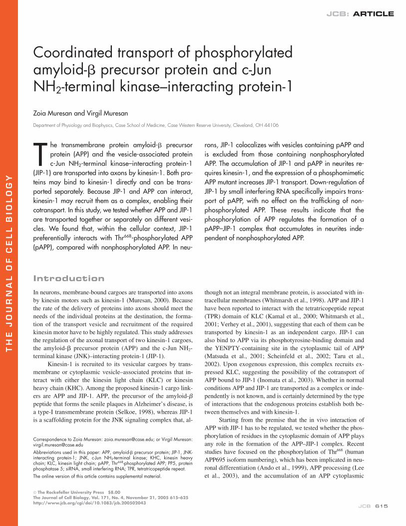
identical to that in neurons (Yamazaki et al., 1995; Yasuda et al.,1999). We found that JIP-1 immunolabeling (Fig. 1, A–F) ismostly punctuate, suggesting that endogenous JIP-1 is largelyassociated with vesicles. As previously reported for other neurons(Yasuda et al., 1999), in CAD cells JIP-1 appeared concentratedat the terminals of processes (Fig. S2 B and Fig. 1, A and D).Similar to JIP-1, APP was found in the cell body and processes,in a punctuate appearance typical of vesicle proteins (Fig. 1, A–F).Importantly, the majority of APP (
�
80%) was not localized tovesicles that contained JIP-1, a result that was consistentlyobtained using several antibodies to APP and JIP-1. This resultwas confirmed in primary cultures of cortical neurons thatclosely reproduce the distribution of these proteins in neurons insitu (Fig. 1, G–L). Based on this set of data, we conclude thatonly a small fraction of APP is cotransported with JIP-1.
JIP-1 colocalizes and interacts with pAPP rather than with nonphosphorylated APP
We focused on characterizing the fraction of APP that colocal-izes with JIP-1. We observed that the accumulation of JIP-1 at
Figure 1. Colocalization of JIP-1 with pAPP in neuronal processes. CADcells (A–F and M–Y) and cortical neurons (G–L, Z–�) were double labeledfor JIP-1 and either total APP (antibody 4G8; A–L) or pAPP (M–�). (A–L)The majority of total APP (A–F, red; G–L, green) is present in vesicles dis-tinct from those carrying JIP-1 (A–F, green; G–L, red). (B and H) Enlarge-ments of the boxed areas in A and G. (D and E) Single color enlargementsof the process terminal shown in A. Similarly, I–L represent single colorenlargements of regions from the distal and proximal neurite shown in Gand H to reveal the distinct distribution of JIP-1 and APP along the neurite.(G, H, J, and L) Arrows mark the ends of corresponding segments. (M–�)JIP-1 (M–Y and �, green; �, white) colocalizes extensively with pAPP (M–Yand �, red; Z, white) throughout the processes (P–R) and at their terminals(S–�). Bars: (A–L) 5 �m; (M–O) 20 �m; (P–�) 10 �m.
COTRANSPORT OF
P
APP AND JIP-1 • MURESAN AND MURESAN
617
the terminals (Fig. 1, A and G) is strikingly similar to that re-ported for pAPP (Ando et al., 1999). This raised the question ofwhether JIP-1 might be carried into processes along withpAPP. To test this hypothesis, we began by studying the distribu-tion of pAPP in CAD cells with an antibody to a phosphorylatedpolypeptide from a region around pThr
668
in the cytoplasmicdomain of APP. We verified the specificity of this antibodytoward pAPP in immunoblots of rat brain extract. A singlemajor band with electrophoretic mobility corresponding tomature APP was detected (Fig. S2 A). In immunocytochemistry,the antibody produced a staining (Fig. S2 D) similar to that re-ported for pAPP in neurons (Ando et al., 1999; Iijima et al.,2000). This staining was completely abolished by competitionwith the phosphorylated polypeptide from the cytoplasmic do-main of APP (Fig. S2, G and H), but was not affected by thecorresponding nonphosphorylated polypeptide (Fig. S2, Eand F). These results confirmed the specificity of this antibodyfor pAPP. Next, we used these antibodies for dual labeling ofpAPP and JIP-1 in CAD cells and cortical neurons. As shownin Fig. 1 (M–
�
), endogenous pAPP colocalized extensivelywith JIP-1, both at the terminals and throughout the processes.Quantitative measurements of the overlap indicated that
�
85%of pAPP colocalized with JIP-1. These results support the notionthat JIP-1 and pAPP mostly reside on the same vesicle popula-tion, along the neurites and at neurite terminals.
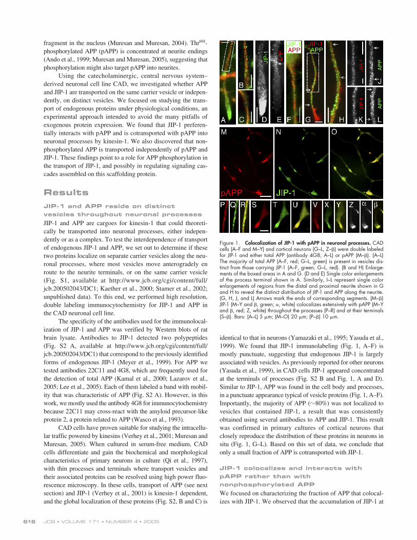
The molecular basis of this result might be the direct inter-action between pAPP and JIP-1, which is a known APP-bindingprotein (Matsuda et al., 2001). Probing for such an interaction inCAD cells proved difficult because of the small amounts of en-dogenous pAPP and JIP-1. Therefore, we prepared extractsfrom COS-1 cells transfected with FLAG-tagged JIP-1 andperformed in vitro pull-down assays on biotinylated polypep-tides comprising the entire cytoplasmic domain of APP, phos-phorylated or not, at the residue corresponding to Thr
668
(Fig.2, A and B). Polypeptides were collected on streptavidin-Sepharose and analyzed for bound JIP-1 by immunoblottingwith antibodies to FLAG. Although JIP-1 interacted with boththe phosphorylated and nonphosphorylated forms, at equalamounts of peptides (Fig. 2 B, Ponceau S) it was preferentiallycaptured by the phosphorylated peptide (Fig. 2 B). This resultwas reproduced by ELISA analysis (Fig. 2 C). In addition,ELISA showed that, at high concentration, JIP-1 binds indis-criminately to the phosphorylated and nonphosphorylatedpeptides (Fig. 2 C, left). We note that ELISA assays were con-ducted in the presence of cytosol, using low concentrations ofJIP-1–FLAG expressed in HEK 293 cells.
Unlike JIP-1, in the peptide capture assay, JIP-2 boundequally well to the peptides regardless of their state of phos-phorylation (Fig. 2 B). Using ELISA, we also confirmed thatFe65, which is an APP-binding protein unrelated to the JIPs,preferentially bound to the nonphosphorylated APP peptide (Fig.2 C, right; Ando et al., 2001), which validated our ELISA proto-col. The interaction between JIP-1 and full-length pAPP was fur-ther tested by immunoprecipitation from extracts of CAD cellsthat expressed ectopic JIP-1–FLAG and human APP695. Underthese conditions, a fraction of the expressed APP becomes phos-phorylated (Muresan and Muresan, 2005). As shown in Fig. 2 D,
only a small fraction of the total mature APP coprecipitated withJIP-1. However, a large proportion of the mature, fully glyco-sylated form of pAPP bound to JIP-1. This result suggests thatJIP-1 preferentially associates with pAPP, and not with its non-phosphorylated form. This binding preference for pAPP at lowJIP-1 concentrations, although small, may prevail under in vivoconditions and contribute to the colocalization of JIP-1 withpAPP (Fig. 1). Thus, phosphorylation of APP may facilitate therecruitment of JIP-1 to vesicles.
JIP-1 and pAPP are cotransported by kinesin-1 to neurite terminals
Based on the colocalization and preferential interaction of JIP-1with pAPP, we propose a model for the transport of these pro-
Figure 2. Biochemical analysis of JIP-1 interaction with nonphosphory-lated and phosphorylated APP. (A) SDS-PAGE separation of synthetic,biotinylated, phosphorylated (pAPPCD), and nonphosphorylated (APPCD)polypeptides encompassing the APP cytoplasmic domain (Tris-tricine gel).Peptides were detected on the same gel, but the lanes are presented inseparate boxes to indicate that the contrast was adjusted differently foreach lane. This was done to demonstrate the purity of the peptides ratherthan their relative amounts. (B) Pull-down experiment done with the polypep-tides shown in A. Equal amounts of the two polypeptides (Ponceau S) wereincubated with lysates of COS-1 cells transfected with FLAG-tagged JIP-1or JIP-2 and collected on streptavidin-Sepharose. Copurified JIPs were de-tected by immunoblotting with antibodies to the FLAG tag (marked JIP-1and JIP-2). Note that JIP-1, but not JIP-2, binds preferentially to pAPPCD.Densitometric analysis of blots showed that JIP-1 binding to APPCD is 30–40% lower than to pAPPCD. A control lane (Beads) shows residual bindingof JIP-1–FLAG to beads in the absence of added polypeptide. The anti-pAPP antibody detects the phosphorylated, but not the nonphosphory-lated, polypeptide in the pulled-down material (Anti-pAPP). (C) Differentialbinding of JIP-1 and Fe65 to phosphorylated and nonphosphorylated APPpolypeptides (ELISA). Streptavidin-coated ELISA plates, preadsorbed withbiotinylated APPCD or pAPPCD, were incubated with increasing concentra-tions of cytosol (expressed as percentages of the total incubation mixture;see Materials and methods) from HEK293 cells transfected with either JIP-1–FLAG or Fe65-myc. Bound proteins were detected colorimetrically, usingalkaline phosphatase–labeled antibodies to the tags. Error bars representSEM (n � 3). (D) Coprecipitation of phosphorylated and nonphosphory-lated APP with JIP-1. CAD cells were transfected separately with humanAPP695 and with JIP-1–FLAG; the two cell extracts were mixed and incu-bated overnight. Mixtures were immunoprecipitated with an anti-FLAG an-tibody, and immunoprecipitated material was analyzed by immunoblottingwith antibodies to total APP, pAPP, and FLAG. The three polypeptides de-tected with the anti-APP antibody correspond to immature and matureforms of APP695 (arrow points to the fully glycosylated form of APP).(right, No JIP-1) Nonspecific binding of APP and pAPP to beads, in theabsence of JIP-1–FLAG-containing cytosol, was low.
JCB • VOLUME 171 • NUMBER 4 • 2005618
teins into neuronal processes where JIP-1 mediates recruitmentof kinesin-1 to pAPP in the cell body, resulting in the transportof the pAPP–JIP-1 complex to the terminals. The model predictsthe following: (1) pAPP and JIP-1 colocalize with kinesin-1 inneurites; (2) transport of pAPP—similar to that of JIP-1—isdependent on kinesin-1; (3) phosphorylation of APP occurs inthe cell body, before kinesin-1 recruitment and initiation oftransport; (4) recruitment of kinesin-1 to pAPP and transport ofpAPP are enhanced by JIP-1; (5) transport of JIP-1 into neuritesis enhanced by APP phosphorylation; and (6) down-regulationof JIP-1 expression reduces transport of pAPP into neurites.We tested these suppositions as follows.
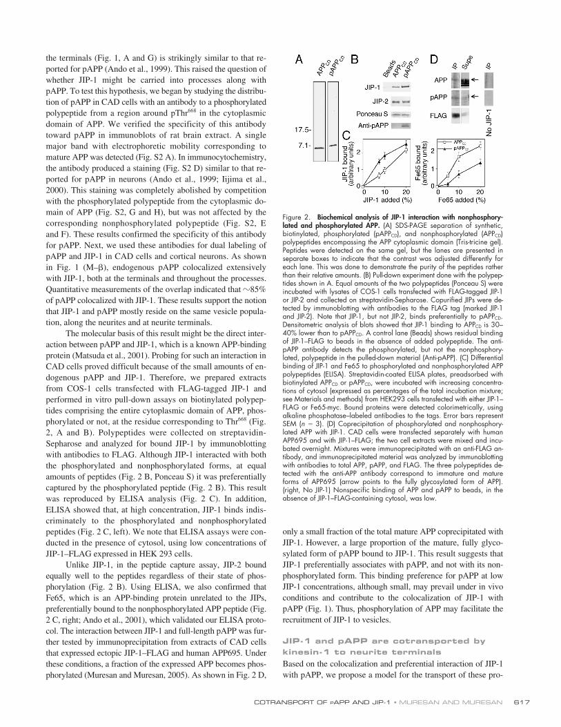
First, we tested if, consistent with its transport by a kine-sin motor, pAPP colocalizes with microtubules throughoutthe processes of CAD cells (Fig. S3, A–D, available at http://www.jcb.org/cgi/content/full/jcb.200502043/DC1). Further,pAPP colocalizes with KHC (Fig. S3, H–K) and KLC (Fig. S3,L–N) of kinesin-1, but not with another kinesin (Fig. S3, E–G).In these experiments, the KLCs were detected with the anti-body 63-90, which preferentially recognizes the nonphos-phorylated, cargo-bound form of KLCs (Pigino et al., 2003).This result is important because it suggests that pAPP is associ-ated with kinesin-1 at the neurite terminals, most likely a con-sequence of prior active transport toward this destination.
Second, to test whether transport of pAPP requires kine-sin-1, we used a dominant-negative construct that specificallyuncouples this kinesin from the cargoes that bind to the KLCs(Verhey et al., 2001). When we expressed the tagged, kinesin-1dominant-negative domain HA-KLC-TPR in CAD cells, trans-port of pAPP was significantly reduced:
�
10% of the trans-fected cells showed pAPP at their terminals (Fig. 3, A–H). Bycomparison,
�
85% of the cells that overexpressed a TPR domainunrelated to that of KLC (that of protein phosphatase 5 [PP5])showed unperturbed accumulation of pAPP at their terminals(Fig. 3, I and J). These results support our hypothesis that accu-mulation of JIP-1 and pAPP at the terminals requires kinesin-1.
Third, a closer examination of the KLC-TPR–expressingcells revealed a buildup of pAPP-containing vesicles in the cellbodies at the emergence of the processes (Fig. 3, A and D, arrow-heads). This is consistent with impaired transport of pAPP gener-ated in the cell body. Like pAPP, JIP-1 also accumulated in thecell body of cells transfected with KLC-TPRs (Fig. 3, K and L),indicating that transport of pAPP and JIP-1 are regulated by kine-sin-1 in a similar manner. Additional data in support of APP phos-phorylation in the cell body is provided by immunocytochemistryexperiments showing that pAPP associated with vesicular struc-tures in the Golgi region of CAD cells, whether they were non-transfected (Fig. 3, M and N) or transfected with APP-YFP (Fig. 3,O and P). Together, these observations suggest that an interactionwith the KLCs of kinesin-1 is required for the transport of pAPP-containing vesicles from the cell body into processes.
Using a similar experimental strategy, we also confirmedthat the delivery of total APP into processes largely relies onkinesin-1 (Yamazaki et al., 1995; Kaether et al., 2000), andprobably requires an interaction of the APP-containing cargowith the KLCs (Fig. S4, available at http://www.jcb.org/cgi/content/full/jcb.200502043/DC1).
Fourth, because our model states that JIP-1 and pAPP aretransported as a complex, we tested if increased availability ofJIP-1 facilitates transport of pAPP to neurite terminals. First,we determined biochemically whether the recruitment of ki-nesin-1 to pAPP is enhanced by increased amounts of JIP-1.We transfected CAD cells with a mutated human APP695 thatmimics pAPP, called APP-Thr668Glu (Ando et al., 1999).Lysates of these cells were mixed with extracts of CAD cellsthat were JIP-1 transfected or nontransfected (control) andincubated to allow the formation of APP-Thr668Glu–JIP-1complexes. Equal amounts of squid kinesin-1 were thenadded to the incubation mixtures. Putative complexes con-taining APP-Thr668Glu–JIP-1–kinesin-1 were immunoprecipi-tated and analyzed by immunoblotting. For immunoprecipitation,
Figure 3. Localization of pAPP at neurite terminals requires kinesin-1.(A–L) Localization of pAPP (A–J) and JIP-1 (K and L) in CAD cells overex-pressing the tagged, kinesin-1 dominant-negative construct, HA-KLC-TPR(A–H, K, and L), or the control construct, HA-PP5-TPR (I and J). Two exam-ples show the effect of HA-KLC-TPR expression on pAPP localization (A–Cand D–H). Note that pAPP and JIP-1 accumulate in the cell body of cellstransfected with HA-KLC-TPR (arrowheads) and are not detected at the ter-minals of their processes (long arrows). In contrast, pAPP and JIP-1 localizeto the neurite terminals in nontransfected cells (short arrows). Localizationof pAPP is not affected in cells transfected with HA-PP5-TPR (compare shortwith long arrows in I and J). No pAPP accumulation in the cell body of thetransfected cell is seen (I and J, arrowheads). (F–H) Enlarged images ofthe neurite shown in D and E. (M–P) pAPP can be detected in the Golgiarea in CAD cells. (M and N) Vesicular localization of endogenous pAPPin the Golgi region (arrow) of a CAD cell. (O and P) In cells transfectedwith APP695, pAPP is found throughout the processes and in the cell body(P, arrows). (O) Image depicts total APP. (C, H, and N) Phase-contrastmicrographs. Bars, 20 �m.
COTRANSPORT OF
P
APP AND JIP-1 • MURESAN AND MURESAN
619
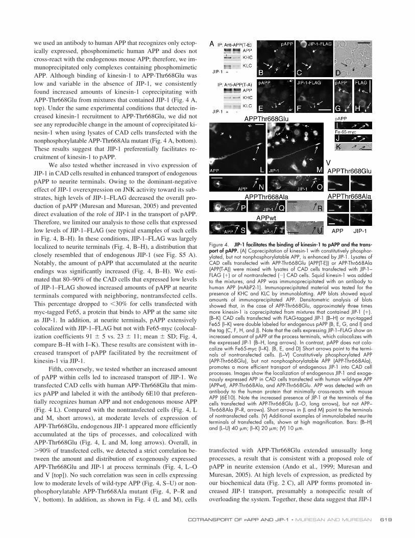
we used an antibody to human APP that recognizes only ectop-ically expressed, phosphomimetic human APP and does notcross-react with the endogenous mouse APP; therefore, we im-munoprecipitated only complexes containing phosphomimeticAPP. Although binding of kinesin-1 to APP-Thr668Glu waslow and variable in the absence of JIP-1, we consistentlyfound increased amounts of kinesin-1 coprecipitating withAPP-Thr668Glu from mixtures that contained JIP-1 (Fig. 4 A,top). Under the same experimental conditions that detected in-creased kinesin-1 recruitment to APP-Thr668Glu, we did notsee any reproducible change in the amount of coprecipitated ki-nesin-1 when using lysates of CAD cells transfected with thenonphosphorylatable APP-Thr668Ala mutant (Fig. 4 A, bottom).These results suggest that JIP-1 preferentially facilitates re-cruitment of kinesin-1 to pAPP.
We also tested whether increased in vivo expression ofJIP-1 in CAD cells resulted in enhanced transport of endogenouspAPP to neurite terminals. Owing to the dominant-negativeeffect of JIP-1 overexpression on JNK activity toward its sub-strates, high levels of JIP-1–FLAG decreased the overall pro-duction of pAPP (Muresan and Muresan, 2005) and preventeddirect evaluation of the role of JIP-1 in the transport of pAPP.Therefore, we limited our analysis to those cells that expressedlow levels of JIP-1–FLAG (see typical examples of such cellsin Fig. 4, B–H). In these conditions, JIP-1–FLAG was largelylocalized to neurite terminals (Fig. 4, B–H), a distribution thatclosely resembled that of endogenous JIP-1 (see Fig. S5 A).Notably, the amount of pAPP that accumulated at the neuriteendings was significantly increased (Fig. 4, B–H). We esti-mated that 80–90% of the CAD cells that expressed low levelsof JIP-1–FLAG showed increased amounts of pAPP at neuriteterminals compared with neighboring, nontransfected cells.This percentage dropped to
�
30% for cells transfected withmyc-tagged Fe65, a protein that binds to APP at the same siteas JIP-1. In addition, at neurite terminals, pAPP extensivelycolocalized with JIP-1–FLAG but not with Fe65-myc (colocal-ization coefficients 91
�
5 vs. 23
�
11; mean
�
SD; Fig. 4,compare B–H with I–K). These results are consistent with in-creased transport of pAPP facilitated by the recruitment ofkinesin-1 via JIP-1.
Fifth, conversely, we tested whether an increased amountof pAPP within cells led to increased transport of JIP-1. Wetransfected CAD cells with human APP-Thr668Glu that mim-ics pAPP and labeled it with the antibody 6E10 that preferen-tially recognizes human APP and not endogenous mouse APP(Fig. 4 L). Compared with the nontransfected cells (Fig. 4, Land M, short arrows), at moderate levels of expression ofAPP-Thr668Glu, endogenous JIP-1 appeared more efficientlyaccumulated at the tips of processes, and colocalized withAPP-Thr668Glu (Fig. 4, L and M, long arrows). Overall, in
�
90% of transfected cells, we detected a strict correlation be-tween the amount and distribution of exogenously expressedAPP-Thr668Glu and JIP-1 at process terminals (Fig. 4, L–Oand V [top]). No such correlation was seen in cells expressinglow to moderate levels of wild-type APP (Fig. 4, S–U) or non-phosphorylatable APP-Thr668Ala mutant (Fig. 4, P–R andV, bottom). In addition, as shown in Fig. 4 (L and M), cells
transfected with APP-Thr668Glu extended unusually longprocesses, a result that is consistent with a proposed role ofpAPP in neurite extension (Ando et al., 1999; Muresan andMuresan, 2005). At high levels of expression, as predicted byour biochemical data (Fig. 2 C), all APP forms promoted in-creased JIP-1 transport, presumably a nonspecific result ofoverloading the system. Together, these data suggest that JIP-1
Figure 4. JIP-1 facilitates the binding of kinesin-1 to pAPP and the trans-port of pAPP. (A) Coprecipitation of kinesin-1 with constitutively phosphor-ylated, but not nonphosphorylatable APP, is enhanced by JIP-1. Lysates ofCAD cells transfected with APP-Thr668Glu (APP(T-E)) or APP-Thr668Ala(APP(T-A)) were mixed with lysates of CAD cells transfected with JIP-1–FLAG (�) or of nontransfected () CAD cells. Squid kinesin-1 was addedto the mixtures, and APP was immunoprecipitated with an antibody tohuman APP (mAbP2-1). Immunoprecipitated material was tested for thepresence of KHC and KLC by immunoblotting. APP blots showed equalamounts of immunoprecipitated APP. Densitometric analysis of blotsshowed that, in the case of APP-Thr668Glu, approximately three timesmore kinesin-1 is coprecipitated from mixtures that contained JIP-1 (�).(B–K) CAD cells transfected with FLAG-tagged JIP-1 (B–H) or myc-taggedFe65 (I–K) were double labeled for endogenous pAPP (B, E, G, and I) andthe tag (C, F, H, and J). Note that the cells expressing JIP-1–FLAG show anincreased amount of pAPP at the process terminals, which colocalizes withthe expressed JIP-1 (B–H, long arrows). In contrast, pAPP does not colo-calize with Fe65-myc (I–K). (B, E, and D) Short arrows point to the termi-nals of nontransfected cells. (L–V) Constitutively phosphorylated APP(APP-Thr668Glu), but not nonphosphorylatable APP (APP-Thr668Ala),promotes a more efficient transport of endogenous JIP-1 into CAD cellprocesses. Images show the localization of endogenous JIP-1 and exoge-nously expressed APP in CAD cells transfected with human wild-type APP(APPwt), APP-Thr668Ala, and APP-Thr668Glu. APP was detected with anantibody to the human protein that minimally cross-reacts with mouseAPP (6E10). Note the increased presence of JIP-1 at the terminals of thecells transfected with APP-Thr668Glu (L–O, long arrows), but not APP–Thr668Ala (P–R, arrows). Short arrows in (L and M) point to the terminalsof nontransfected cells. (V) Additional examples of immunolabeled neuriteterminals of transfected cells, shown at high magnification. Bars: (B–H)and (L–U) 40 �m; (I–K) 20 �m; (V) 10 �m.
JCB • VOLUME 171 • NUMBER 4 • 2005620
and pAPP are cotransported by kinesin-1 as a complex. Phos-phorylation of APP facilitates the formation and the transportof this complex, as well as an overall vesicular traffic thatlikely promotes the extension of neurites.
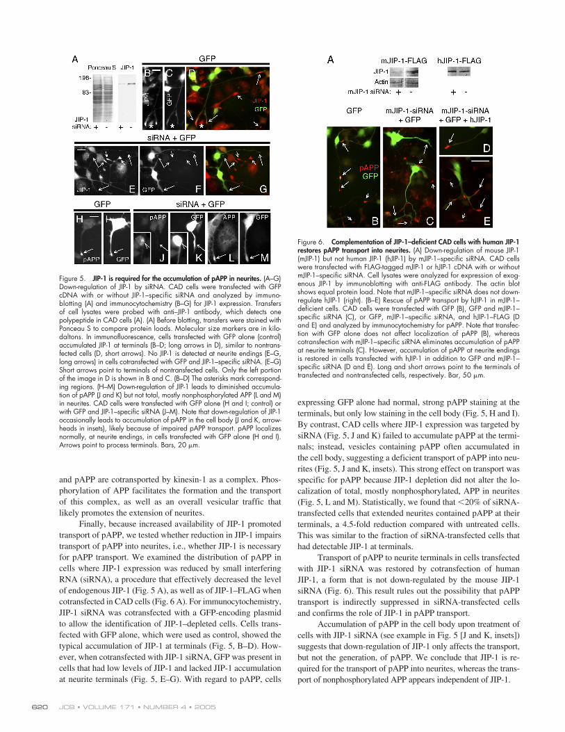
Finally, because increased availability of JIP-1 promotedtransport of pAPP, we tested whether reduction in JIP-1 impairstransport of pAPP into neurites, i.e., whether JIP-1 is necessaryfor pAPP transport. We examined the distribution of pAPP incells where JIP-1 expression was reduced by small interferingRNA (siRNA), a procedure that effectively decreased the levelof endogenous JIP-1 (Fig. 5 A), as well as of JIP-1–FLAG whencotransfected in CAD cells (Fig. 6 A). For immunocytochemistry,JIP-1 siRNA was cotransfected with a GFP-encoding plasmidto allow the identification of JIP-1–depleted cells. Cells trans-fected with GFP alone, which were used as control, showed thetypical accumulation of JIP-1 at terminals (Fig. 5, B–D). How-ever, when cotransfected with JIP-1 siRNA, GFP was present incells that had low levels of JIP-1 and lacked JIP-1 accumulationat neurite terminals (Fig. 5, E–G). With regard to pAPP, cells
expressing GFP alone had normal, strong pAPP staining at theterminals, but only low staining in the cell body (Fig. 5, H and I).By contrast, CAD cells where JIP-1 expression was targeted bysiRNA (Fig. 5, J and K) failed to accumulate pAPP at the termi-nals; instead, vesicles containing pAPP often accumulated inthe cell body, suggesting a deficient transport of pAPP into neu-rites (Fig. 5, J and K, insets). This strong effect on transport wasspecific for pAPP because JIP-1 depletion did not alter the lo-calization of total, mostly nonphosphorylated, APP in neurites(Fig. 5, L and M). Statistically, we found that
�
20% of siRNA-transfected cells that extended neurites contained pAPP at theirterminals, a 4.5-fold reduction compared with untreated cells.This was similar to the fraction of siRNA-transfected cells thathad detectable JIP-1 at terminals.
Transport of pAPP to neurite terminals in cells transfectedwith JIP-1 siRNA was restored by cotransfection of humanJIP-1, a form that is not down-regulated by the mouse JIP-1siRNA (Fig. 6). This result rules out the possibility that pAPPtransport is indirectly suppressed in siRNA-transfected cellsand confirms the role of JIP-1 in pAPP transport.
Accumulation of pAPP in the cell body upon treatment ofcells with JIP-1 siRNA (see example in Fig. 5 [J and K, insets])suggests that down-regulation of JIP-1 only affects the transport,but not the generation, of pAPP. We conclude that JIP-1 is re-quired for the transport of pAPP into neurites, whereas the trans-port of nonphosphorylated APP appears independent of JIP-1.
Figure 5. JIP-1 is required for the accumulation of pAPP in neurites. (A–G)Down-regulation of JIP-1 by siRNA. CAD cells were transfected with GFPcDNA with or without JIP-1–specific siRNA and analyzed by immuno-blotting (A) and immunocytochemistry (B–G) for JIP-1 expression. Transfersof cell lysates were probed with anti–JIP-1 antibody, which detects onepolypeptide in CAD cells (A). (A) Before blotting, transfers were stained withPonceau S to compare protein loads. Molecular size markers are in kilo-daltons. In immunofluorescence, cells transfected with GFP alone (control)accumulated JIP-1 at terminals (B–D; long arrows in D), similar to nontrans-fected cells (D, short arrows). No JIP-1 is detected at neurite endings (E–G,long arrows) in cells cotransfected with GFP and JIP-1–specific siRNA. (E–G)Short arrows point to terminals of nontransfected cells. Only the left portionof the image in D is shown in B and C. (B–D) The asterisks mark correspond-ing regions. (H–M) Down-regulation of JIP-1 leads to diminished accumula-tion of pAPP (J and K) but not total, mostly nonphosphorylated APP (L and M)in neurites. CAD cells were transfected with GFP alone (H and I; control) orwith GFP and JIP-1–specific siRNA (J–M). Note that down-regulation of JIP-1occasionally leads to accumulation of pAPP in the cell body (J and K, arrow-heads in insets), likely because of impaired pAPP transport. pAPP localizesnormally, at neurite endings, in cells transfected with GFP alone (H and I).Arrows point to process terminals. Bars, 20 �m.
Figure 6. Complementation of JIP-1–deficient CAD cells with human JIP-1restores pAPP transport into neurites. (A) Down-regulation of mouse JIP-1(mJIP-1) but not human JIP-1 (hJIP-1) by mJIP-1–specific siRNA. CAD cellswere transfected with FLAG-tagged mJIP-1 or hJIP-1 cDNA with or withoutmJIP-1–specific siRNA. Cell lysates were analyzed for expression of exog-enous JIP-1 by immunoblotting with anti-FLAG antibody. The actin blotshows equal protein load. Note that mJIP-1–specific siRNA does not down-regulate hJIP-1 (right). (B–E) Rescue of pAPP transport by hJIP-1 in mJIP-1–deficient cells. CAD cells were transfected with GFP (B), GFP and mJIP-1–specific siRNA (C), or GFP, mJIP-1–specific siRNA, and hJIP-1–FLAG (Dand E) and analyzed by immunocytochemistry for pAPP. Note that transfec-tion with GFP alone does not affect localization of pAPP (B), whereascotransfection with mJIP-1–specific siRNA eliminates accumulation of pAPPat neurite terminals (C). However, accumulation of pAPP at neurite endingsis restored in cells transfected with hJIP-1 in addition to GFP and mJIP-1–specific siRNA (D and E). Long and short arrows point to the terminals oftransfected and nontransfected cells, respectively. Bar, 50 �m.
COTRANSPORT OF
P
APP AND JIP-1 • MURESAN AND MURESAN
621
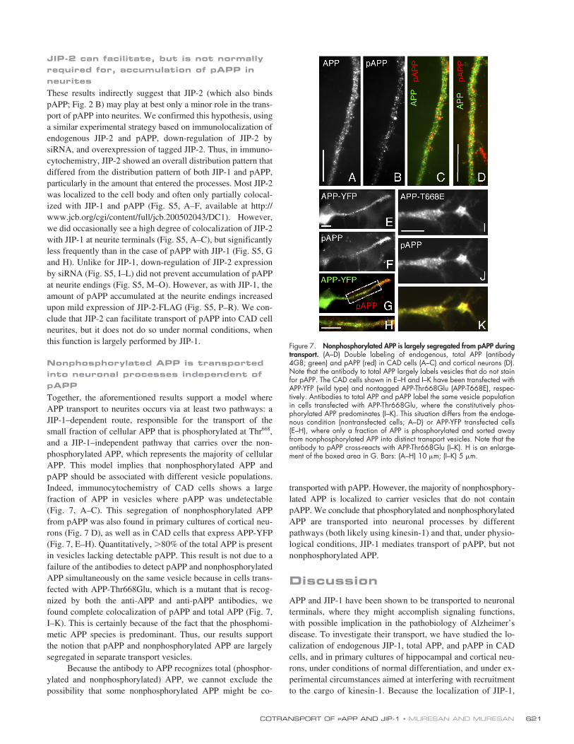
JIP-2 can facilitate, but is not normally required for, accumulation of pAPP in neurites
These results indirectly suggest that JIP-2 (which also bindspAPP; Fig. 2 B) may play at best only a minor role in the trans-port of pAPP into neurites. We confirmed this hypothesis, usinga similar experimental strategy based on immunolocalization ofendogenous JIP-2 and pAPP, down-regulation of JIP-2 bysiRNA, and overexpression of tagged JIP-2. Thus, in immuno-cytochemistry, JIP-2 showed an overall distribution pattern thatdiffered from the distribution pattern of both JIP-1 and pAPP,particularly in the amount that entered the processes. Most JIP-2was localized to the cell body and often only partially colocal-ized with JIP-1 and pAPP (Fig. S5, A–F, available at http://www.jcb.org/cgi/content/full/jcb.200502043/DC1). However,we did occasionally see a high degree of colocalization of JIP-2with JIP-1 at neurite terminals (Fig. S5, A–C), but significantlyless frequently than in the case of pAPP with JIP-1 (Fig. S5, Gand H). Unlike for JIP-1, down-regulation of JIP-2 expressionby siRNA (Fig. S5, I–L) did not prevent accumulation of pAPPat neurite endings (Fig. S5, M–O). However, as with JIP-1, theamount of pAPP accumulated at the neurite endings increasedupon mild expression of JIP-2-FLAG (Fig. S5, P–R). We con-clude that JIP-2 can facilitate transport of pAPP into CAD cellneurites, but it does not do so under normal conditions, whenthis function is largely performed by JIP-1.
Nonphosphorylated APP is transported into neuronal processes independent of pAPP
Together, the aforementioned results support a model whereAPP transport to neurites occurs via at least two pathways: aJIP-1–dependent route, responsible for the transport of thesmall fraction of cellular APP that is phosphorylated at Thr
668
,and a JIP-1–independent pathway that carries over the non-phosphorylated APP, which represents the majority of cellularAPP. This model implies that nonphosphorylated APP andpAPP should be associated with different vesicle populations.Indeed, immunocytochemistry of CAD cells shows a largefraction of APP in vesicles where pAPP was undetectable(Fig. 7, A–C). This segregation of nonphosphorylated APPfrom pAPP was also found in primary cultures of cortical neu-rons (Fig. 7 D), as well as in CAD cells that express APP-YFP(Fig. 7, E–H). Quantitatively,
�
80% of the total APP is presentin vesicles lacking detectable pAPP. This result is not due to afailure of the antibodies to detect pAPP and nonphosphorylatedAPP simultaneously on the same vesicle because in cells trans-fected with APP-Thr668Glu, which is a mutant that is recog-nized by both the anti-APP and anti-pAPP antibodies, wefound complete colocalization of pAPP and total APP (Fig. 7,I–K). This is certainly because of the fact that the phosphomi-metic APP species is predominant. Thus, our results supportthe notion that pAPP and nonphosphorylated APP are largelysegregated in separate transport vesicles.
Because the antibody to APP recognizes total (phosphor-ylated and nonphosphorylated) APP, we cannot exclude thepossibility that some nonphosphorylated APP might be co-
transported with pAPP. However, the majority of nonphosphory-lated APP is localized to carrier vesicles that do not containpAPP. We conclude that phosphorylated and nonphosphorylatedAPP are transported into neuronal processes by differentpathways (both likely using kinesin-1) and that, under physio-logical conditions, JIP-1 mediates transport of pAPP, but notnonphosphorylated APP.
Discussion
APP and JIP-1 have been shown to be transported to neuronalterminals, where they might accomplish signaling functions,with possible implication in the pathobiology of Alzheimer’sdisease. To investigate their transport, we have studied the lo-calization of endogenous JIP-1, total APP, and pAPP in CADcells, and in primary cultures of hippocampal and cortical neu-rons, under conditions of normal differentiation, and under ex-perimental circumstances aimed at interfering with recruitmentto the cargo of kinesin-1. Because the localization of JIP-1,
Figure 7. Nonphosphorylated APP is largely segregated from pAPP duringtransport. (A–D) Double labeling of endogenous, total APP (antibody4G8; green) and pAPP (red) in CAD cells (A–C) and cortical neurons (D).Note that the antibody to total APP largely labels vesicles that do not stainfor pAPP. The CAD cells shown in E–H and I–K have been transfected withAPP-YFP (wild type) and nontagged APP-Thr668Glu (APP-T668E), respec-tively. Antibodies to total APP and pAPP label the same vesicle populationin cells transfected with APP-Thr668Glu, where the constitutively phos-phorylated APP predominates (I–K). This situation differs from the endoge-nous condition (nontransfected cells; A–D) or APP-YFP transfected cells(E–H), where only a fraction of APP is phosphorylated and sorted awayfrom nonphosphorylated APP into distinct transport vesicles. Note that theantibody to pAPP cross-reacts with APP-Thr668Glu (I–K). H is an enlarge-ment of the boxed area in G. Bars: (A–H) 10 �m; (I–K) 5 �m.
JCB • VOLUME 171 • NUMBER 4 • 2005622
total APP, and pAPP at neurite terminals is largely the result ofactive anterograde transport, this experimental approach al-lowed us to gain insight into the mechanism of this process. Wefound that in situ APP transport to terminals occurs via twoseparate populations of vesicles that—as detected by immuno-cytochemistry—differ in composition and whose transport isdifferentially regulated. Thus,
�
80% of the APP-labeled ve-sicular structures along the neurites appear to carry only non-phosphorylated APP because they do not stain for pAPP;these vesicles also lack JIP-1. The transport of this vesicle pop-ulation to the neurite terminal is independent of JIP-1. The re-maining APP-containing vesicles (
�
20%) stain for pAPP andfor JIP-1, and their transport to the terminal requires JIP-1.Taking into account the increased affinity of JIP-1 for pAPPover nonphosphorylated APP, these observations suggest thatselective phosphorylation of a fraction of APP in the cell bodytriggers recruitment of JIP-1 to pAPP-containing vesicles,which then facilitates binding and/or activation of kinesin-1.The end result is the transport of pAPP–JIP-1–containing vesi-cles to the neurite terminals, which is where they accumulate.This process, unlike the seemingly constitutive transport ofnonphosphorylated APP, appears to be the subject of at leasttwo levels of regulation: the generation of pAPP and the re-cruitment of JIP-1. From a physiological point of view, thesetwo levels of regulation would provide a stringent control forthe availability of the putative pAPP–JIP-1 signaling complexat the neurite terminals.
Our immunocytochemical approach addresses the in situsituation, which is unaffected by protein overexpression.Because cells were fixed during a time of robust neurite exten-sion, it is most likely that vesicles seen along the processeswere engaged in anterograde transport. Indeed, we have shownthat in CAD cells that express low levels of APP-YFP,
�
80%of the fluorescently labeled, motile vesicles moved antero-gradely (Fig. S1) and, over extended periods of time, most ofthe fluorescently tagged vesicles were motile (
�
70%).We found that JIP-1 is vesicle associated and colocalizes
strictly with pAPP, but not with nonphosphorylated APP. It isintriguing that although JIP-1 is capable of binding to nonphos-phorylated APP in vitro or when the two proteins are overex-pressed in heterologous systems, endogenous JIP-1 apparentlydoes not do so within neurites of cultured neurons in situ, whereit colocalizes exclusively with pAPP. Our biochemical data con-firm the ability of JIP-1 to interact with nonphosphorylated APP;however, we find that this binding is enhanced upon APP phos-phorylation. Similarly, JIP-2 is capable of binding to pAPP inbiochemical assays. Yet, it does not appear to play a major rolein pAPP transport in vivo. The apparent discrepancy betweenthe results obtained by in vitro biochemical assays and thosefrom immunocytochemistry of endogenous proteins (where theinteraction of JIP-1 to the nonphosphorylated APP is undetect-able) might be accounted for by the complexity of the system invivo. For example, in vivo JIP-1 might be prevented from bind-ing to nonphosphorylated APP by other proteins (e.g., Fe65; Fig.2 C) that may selectively bind to and block the JIP-1 binding sitein nonphosphorylated APP. Technical advances will allowcollection of data that accurately reflect the in vivo processes.
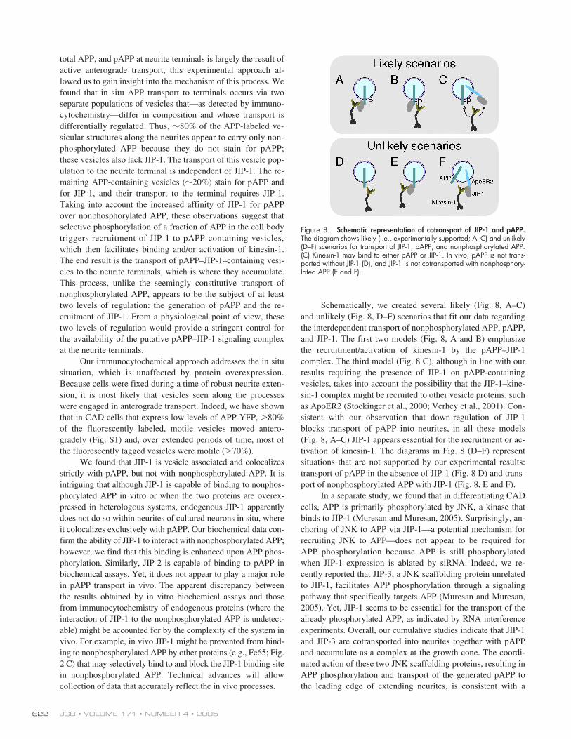
Schematically, we created several likely (Fig. 8, A–C)and unlikely (Fig. 8, D–F) scenarios that fit our data regardingthe interdependent transport of nonphosphorylated APP, pAPP,and JIP-1. The first two models (Fig. 8, A and B) emphasizethe recruitment/activation of kinesin-1 by the pAPP–JIP-1complex. The third model (Fig. 8 C), although in line with ourresults requiring the presence of JIP-1 on pAPP-containingvesicles, takes into account the possibility that the JIP-1–kine-sin-1 complex might be recruited to other vesicle proteins, suchas ApoER2 (Stockinger et al., 2000; Verhey et al., 2001). Con-sistent with our observation that down-regulation of JIP-1blocks transport of pAPP into neurites, in all these models(Fig. 8, A–C) JIP-1 appears essential for the recruitment or ac-tivation of kinesin-1. The diagrams in Fig. 8 (D–F) representsituations that are not supported by our experimental results:transport of pAPP in the absence of JIP-1 (Fig. 8 D) and trans-port of nonphosphorylated APP with JIP-1 (Fig. 8, E and F).
In a separate study, we found that in differentiating CADcells, APP is primarily phosphorylated by JNK, a kinase thatbinds to JIP-1 (Muresan and Muresan, 2005). Surprisingly, an-choring of JNK to APP via JIP-1—a potential mechanism forrecruiting JNK to APP—does not appear to be required forAPP phosphorylation because APP is still phosphorylatedwhen JIP-1 expression is ablated by siRNA. Indeed, we re-cently reported that JIP-3, a JNK scaffolding protein unrelatedto JIP-1, facilitates APP phosphorylation through a signalingpathway that specifically targets APP (Muresan and Muresan,2005). Yet, JIP-1 seems to be essential for the transport of thealready phosphorylated APP, as indicated by RNA interferenceexperiments. Overall, our cumulative studies indicate that JIP-1and JIP-3 are cotransported into neurites together with pAPPand accumulate as a complex at the growth cone. The coordi-nated action of these two JNK scaffolding proteins, resulting inAPP phosphorylation and transport of the generated pAPP tothe leading edge of extending neurites, is consistent with a
Figure 8. Schematic representation of cotransport of JIP-1 and pAPP.The diagram shows likely (i.e., experimentally supported; A–C) and unlikely(D–F) scenarios for transport of JIP-1, pAPP, and nonphosphorylated APP.(C) Kinesin-1 may bind to either pAPP or JIP-1. In vivo, pAPP is not trans-ported without JIP-1 (D), and JIP-1 is not cotransported with nonphosphory-lated APP (E and F).
COTRANSPORT OF
P
APP AND JIP-1 • MURESAN AND MURESAN
623
function of this large signaling complex in neurite growth(Ando et al., 1999; Muresan and Muresan, 2005). Further stud-ies are required to elucidate the mechanisms by which APP,JIP-3, and JIP-1 may integrate JNK signaling with vesiculartransport by kinesin-1 and neuronal differentiation.
Finally, our data suggest that a significant fraction ofAPP is transported to neurite terminals in a nonphosphorylatedform, independently of pAPP and of JIP-1. Transport of non-phosphorylated APP may occur autonomously (i.e., constitu-tive) or in conjunction with other proteins that may bind to thecytoplasmic domain of APP, such as Fe65 or X11
�
/Mint-1(i.e., regulated). Our data from experiments using a dominant-negative KLC domain that interferes with cargo binding areconsistent with a role of kinesin-1 in the transport of nonphos-phorylated APP as well. These results need to be cautiouslyinterpreted because malfunction of kinesin-1 may indirectlyimpede the transport of APP, even if it was not the motor thatcarries the APP-containing vesicle. In the case of pAPP, how-ever, our data argue strongly in favor of a JIP-1–facilitatedrecruitment of kinesin-1 to the cargo vesicle.
It is generally admitted that a deficient axonal transportmay be one of the causes of Alzheimer’s disease (Roy et al.,2005; Stokin et al., 2005). However, whether this may directlyinvolve APP as a linker for kinesin-1, as previously proposed(Kamal et al., 2000), is currently highly debated, particularlybecause pull-down and coimmunoprecipitation experimentsused to test the APP–kinesin-1 interaction are plagued by non-specific binding (Matsuda et al., 2003; Lazarov et al., 2005).Moreover, the endogenous conditions are not faithfully repro-duced after cell lysis or by overexpressing proteins that changethe equilibrium between multiple, cross-interacting partners.We believe that the current debate will only be settled by ad-dressing the transport of APP in experimental settings that areas close as possible to the endogenous conditions.
In conclusion, we found that transport of a fraction ofAPP by kinesin-1 occurs in concert with JIP-1, and this trans-port is regulated by the phosphorylation of APP. This impliesthat transport of JIP-1 is regulated by APP phosphorylation.Nonphosphorylated APP is transported on a different pathway,either through a direct interaction with the kinesin motor or bya yet to be identified mechanism. The two transport pathwaysappear to use distinct vesicle carriers and to be differently regu-lated. Further studies are required to fully understand the func-tional significance of pAPP–JIP-1 cotransport, likely relatingthese two proteins in a common signaling pathway.
Materials and methods
Antibodies
The following primary antibodies were used: mouse anti-Alzheimer precur-sor protein A4 (clone 22C11; CHEMICON International, Inc.); mouseanti–human amyloid-
�
protein (clones 4G8 and 6E10; Signet Laborato-ries); rabbit anti-APP (raised against a polypeptide from the cytoplasmicdomain of APP; Cell Signaling Technology); mouse anti–human APP(mAbP2-1; BioSource International, Inc.); and mouse anti-KHC (clone H2;CHEMICON International, Inc.). The following anti–JIP-1 and anti–JIP-2antibodies were used: goat (E-19) and mouse (B-7) anti–JIP-1; goat (C-20)and rabbit (H-95) anti–JIP-2 (all obtained from Santa Cruz Biotechnology,Inc.); and rabbit anti–JIP-1 (a gift from K. Verhey and B. Margolis, Univer-sity of Michigan, Ann Arbor, MI; Meyer et al., 1999). A mouse antibody
to KLC (63-90; Stenoien and Brady, 1997) was a gift from S. Brady(University of Illinois, Chicago, IL). A mouse antibody (K2.4) to kinesin-2(detecting the 85-kD polypeptide corresponding to mammalian KIF3A;Cole et al., 1993) was a gift from J. Scholey (University of California,Davis, Davis, CA). A rabbit antibody to pAPP (BioSource International,Inc.) was raised against a phosphopeptide derived from the region sur-rounding Thr
668
of human APP695, adsorbed against the nonphosphory-lated peptide, and affinity purified with the antigen by the manufacturer.The antibody reacted with phosphorylated, but not with nonphosphory-lated, forms of APP. Mouse antibodies to tags were used: anti–c-myc(Oncogene Research Products) and anti-FLAG (M2; Stratagene).
Cell cultures
The mouse central nervous system–derived catecholaminergic cell lineCAD (obtained from D. Chikaraishi [Duke University Medical School,Durham, NC] and J. Wang [Cogent Neuroscience, Inc., Durham, NC]; Qiet al., 1997) was grown in 1:1 F12/DME medium, with 8% FBS and pen-icillin/streptomycin. CAD cell differentiation was induced by the removalof serum from the culture medium (Qi et al., 1997). Embrynic day 16.5mouse cortical neurons (obtained from S. Cicero and K. Herrup, CaseWestern Reserve University, Cleveland, OH) were grown in neurobasalmedium with B-27 supplement,
L
-glutamine, and penicillin/streptomycin for5 d, with a change of media on day 4. Neurons were maintained ingrowth media until fixation for immunocytochemistry. COS-1 cells weregrown in high-glucose DME, containing 10% FBS and penicillin/strepto-mycin. HEK293 cells were grown in RPMI 1640 containing 8% FBS.
Transfection
CAD, HEK293, and COS-1 cells were transfected according to the man-ufacturer’s instructions, using either FuGene 6 (Roche Diagnostics) orNucleofector technology (Amaxa Inc.). The HA-tagged KLC-TPR construct(containing all six TPRs) and the HA-PP5-TPR construct were previously de-scribed (Verhey et al., 2001). Constructs in pcDNA3 of human APP695(wild-type and mutants [APP-Thr668Ala and APP-Thr668Glu]) were ob-tained from L.-H. Tsai (Harvard Medical School, Howard Hughes MedicalInstitute, Boston, MA). JIP constructs (pcMV-FLAG-JIP-1, pcDNA3-FLAG-JIP-2,and pcDNA3-FLAG-JIP-3) were obtained from R. Davis (University of Mas-sachusetts Medical School, Howard Hughes Medical Institute, Worcester,MA; Whitmarsh et al., 1998; Yasuda et al., 1999; Kelkar et al., 2000).FLAG-tagged, human JIP-1e, and mouse JIP-1b constructs were obtainedfrom L. D’Adamio (Albert Einstein College of Medicine, Bronx, NY; Schein-feld et al., 2002). Myc-tagged Fe65 was obtained from B. Margolis(University of Michigan, Howard Hughes Medical Institute, Ann Arbor, MI;Borg et al., 1996). A pcDNA3/APP-YFP (human APP695) construct wasobtained from C. Dotti (Cavalieri Ottolenghi Scientific Institute, Universitàdegli Studi di Torino, Torino, Italy) and C. Kaether (European MolecularBiology Laboratory, Heidelberg, Germany; Kaether et al., 2000).
RNA interference experiments
For immunocytochemistry, CAD cells were cotransfected with pmaxGFP(Amaxa Inc.) and either mouse JIP-1–specific (Santa Cruz Biotechnology,Inc.) or JIP-2–specific (Ambion) siRNA. In JIP-1 complementation experi-ments, cells were cotransfected with pmaxGFP, human JIP-1, and mouseJIP-1–specific siRNA. Cells were allowed to attach to the coverslip in thepresence of serum and cultured for 24–48 h in the absence of serum. Forbiochemistry experiments, cells were transfected with either pmaxGFP orFLAG-tagged JIP-1 (mouse or human) in the presence or absence of JIP-1–specific siRNA. After differentiation, cells were collected in sample bufferand analyzed by Western blotting for expressed proteins.
Pull-down and coimmunoprecipitation experiments
COS-1 cells transfected with cDNAs encoding FLAG-tagged JIPs wererinsed with phosphate-buffered saline and extracted after 30 min at 4
C inlysis buffer (10 mM Tris-HCl, pH 7.5, 50 mM NaCl, 4 mM EDTA, and 1%NP-40) containing leupeptin, pepstatin, aprotinin (10
�
g/ml each), 5 mMbenzamidin, 1 mM PMSF, and phosphatase inhibitors (1 mM Na vana-date, 5 mM NaF, and 25 mM
�
-glycerophosphate). Extracts were centri-fuged for 10 min at 16,000
g
and incubated 12–16 h at 4
C with NH
2
-terminally biotinylated polypeptides corresponding to the entire cytoplas-mic domain of APP (starting with lysine) that were chemically synthesizedwith either phosphorylated or nonphosphorylated Thr (corresponding toThr
668
in APP695). Protein complexes containing the polypeptides werecollected on streptavidin-Sepharose and analyzed by SDS-PAGE andimmunoblotting (Muresan et al., 2000) with anti-tag antibodies. Poly-peptides corresponding to the APP cytoplasmic domain were separated in16.5% Tris-tricine gels (Bio-Rad Laboratories).
JCB • VOLUME 171 • NUMBER 4 • 2005624
In immunoprecipitation experiments that tested the role of JIP-1 inthe recruitment of kinesin-1 to APP, CAD cells were separately transfectedwith human APP-Thr668Glu (or APP–Thr668Ala) and JIP-1–FLAG. Extractsof APP-transfected cells were mixed with extracts from JIP-1–transfected ornontransfected cells and incubated for 2 h at 4
C with a purified fractionof squid kinesin-1 (Muresan et al., 1996). APP was immunoprecipitatedfrom the incubation mixtures with the antibody mAbP2-1 overnight at 4C.In experiments that tested the interaction of JIP-1 with APP and pAPP, CADcells were separately transfected with human APP695 and JIP-1–FLAG.Cell extracts were mixed and incubated overnight in the presence of phos-phatase inhibitors. JIP-1 and associated proteins were immunoprecipitatedfrom the mixture with anti-FLAG antibody. In control experiments, JIP-1–FLAG–containing cytosol was replaced with cytosol from nontransfectedcells. In all cases, the collection of immunoprecipitated material on pro-tein A–Sepharose-4B and extraction of the beads in SDS-PAGE samplebuffer was done as previously described (Muresan and Arvan, 1997). Im-munoprecipitated material was analyzed by Western blotting. Image soft-ware (National Institutes of Health) was used for densitometric analysis of im-munoblots. In all immunoprecipitation experiments, extracts that containedlow levels of JIP-1–FLAG were used.
ELISABiotinylated APP polypeptides (with phosphorylated or nonphosphorylatedThr668) were adsorbed to Reacti-Bind NeutrAvidin-coated Plates that werepreblocked with SuperBlock Blocking Buffer (Pierce Chemical Co.). Cyto-solic fractions containing increasing levels of JIP-1–FLAG or Fe65-mycwere prepared by mixing decreasing volumes of cytosol from nontrans-fected HEK293 cells with increasing volumes of cytosol from transfectedcells. Cytosol concentration was thus kept constant throughout the assay.The plates were incubated with these fractions, then with alkaline phos-phatase–conjugated antibodies to FLAG or myc, and processed for colori-metric reaction, followed by an OD reading at 405 nm, according to themanufacturer’s protocol.
Preparation of brain extractsBrain tissue was collected from neonatal (P2) or 5-week-old male Sprague-Dawley rats (killed by asphyxiation with CO2; protocol approved by theInstitutional Animal Care and Use Committee at Case Western ReserveUniversity), placed in ice-cold phosphate-buffered saline, pH 7.4, and ho-mogenized in Hepes–acetate buffer (50 mM K-Hepes, 50 mM K-acetate,5 mM EGTA, 5 mM Mg acetate, 1 mM Na vanadate, 5 mM NaF, and 25mM �-glycerophosphate, pH 7.4) containing protease inhibitors. Extractswere analyzed for the presence of JIP-1, APP, and pAPP by Western blot-ting. Incubations with anti-pAPP antibodies were done in the presence orabsence of competitor polypeptides (i.e., biotinylated, phosphorylated, ornonphosphorylated polypeptides that encompassed the entire APP cyto-plasmic domain) at 100:1 molar excess over IgG.
ImmunocytochemistryTransfected or nontransfected CAD cells and primary cultures of corticalneurons were fixed for 20 min in phosphate-buffered saline containing 4%formaldehyde and 4% sucrose, permeabilized with 0.3% Triton X-100 for20 min at 20C, and processed for single or double antigen labeling aspreviously described (Muresan et al., 1998). Secondary antibodies (goatanti–mouse/rabbit IgG or donkey anti–goat/mouse/rabbit IgG) coupledto Alexa dyes (Alexa Fluor 488 and Alexa Fluor 594) were obtained fromInvitrogen. When required, double labeling experiments with two primaryantibodies raised in rabbit were done using fluorescently labeled, monova-lent Fab fragments of anti–rabbit IgG (Jackson ImmunoResearch Laborato-ries; Li et al., 2002). In control experiments, immunolabeling with anti-pAPPantibody was done in the presence of phosphorylated or nonphosphory-lated polypeptides (100:1 molar excess over IgG) that encompassed eithera 12–amino acid region centered on Thr668 or the entire APP cytoplasmicdomain. Transfected human APP was selectively detected with antibody6E10 that does not detect endogenous mouse APP. Unless otherwise spec-ified, endogenous JIP-1 and total APP were detected with goat polyclonaland mouse monoclonal (4G8) antibodies, respectively. Digital imageswere obtained with two different microscopes (Model Axiophot; CarlZeiss MicroImaging, Inc.; or model Optiphot; Nikon; 100� oil, 20�, and40� objectives) equipped with cooled charge-coupled device cameras(Olympus) and collected using either Openlab (Improvision Inc.) orMagnafire (Optronics) image analysis software. Images were processedfor contrast and brightness using Photoshop software (Adobe). In somecases, samples were analyzed by confocal microscopy; in others, imageswere collected on a microscope (DeltaVision RT) and subjected to decon-volution analysis.
In vivo motilityMotility of APP-YFP–containing vesicles was visualized in vivo, in thin pro-cesses of transiently transfected CAD cells. The experimental set-up con-sisted of an inverted microscope (model TE200U; Nikon), a camera(model CoolSnapHQ; Roper), and MetaMorph software (Molecular De-vices). Kymographs (Silverman et al., 2001) were generated from successiveimages acquired at 0.502 frames/s and used for quantifying anterogradeand retrograde motile and stationary vesicles.
Quantitative analysisThe degree of colocalization of various antigen pairs was quantified asfollows. First, the mean pixel intensity for each pair of images was deter-mined in Photoshop 7.0 (Adobe; Sabo et al., 2003). Second, imageswere thresholded at the mean intensity, to eliminate most of the diffuse,nonparticulate labeling. Finally, the thresholded images were multiplied.The resulting image showed nonzero pixel intensities only in regions ofcolocalization, i.e., regions were pixel intensity differed from zero in bothimages. Percentages of colocalization were calculated by dividing thenumber of pixels of nonzero intensities in the product image by the num-ber of pixels of nonzero intensities in the parental images.
In experiments that probed pAPP localization in cells transfectedwith JIP-1–FLAG or Fe65-myc, coefficients of colocalization of pAPP withthe tagged proteins were obtained using Image-Pro Plus software (MediaCybernetics, Inc.; Manders et al., 1993). The percentage of transfectedcells that showed increased pAPP at neurite terminals was estimated bycomparison with nontransfected cells present in the microscopic field. Foreach experiment, 20–30 microscopic fields, each containing at least onetransfected cell expressing the tagged protein at a low level, were ana-lyzed. Cells were judged as expressing low levels of JIP-1–FLAG if cellbodies contained little tagged protein compared with the processes.Experiments with JIP-1–FLAG–transfected cells were done in duplicate.
Online supplemental materialFig. S1 shows that most of the fluorescently labeled vesicles move antero-gradely within neurites of CAD cells transfected with APP-YFP. Fig. S2characterizes by Western blotting and immunocytochemistry the main an-tibodies used in this study and shows specificity of the anti-pAPP antibodystrictly for pAPP. Fig. S3 shows colocalization of pAPP with microtubulesand kinesin-1, but not kinesin-2, in CAD cell processes. Fig. S4 shows thatlocalization of total APP to neurites is kinesin-1 dependent. Fig. S5 showsthat JIP-2 is not required for the accumulation of pAPP in neurites. Onlinesupplemental material is available at http://www.jcb.org/cgi/content/full/jcb.200502043/DC1.
We thank Drs. Dona Chikaraishi and James Wang for kindly providing theCAD cell line; Samantha Cicero and Dr. Karl Herrup for kindly providing thecortical neuron cultures; Drs. Li-Huei Tsai and Ming-Sum Lee for kindly provid-ing cDNA constructs and an antibody to phosphorylated APP that was used inpreliminary experiments; and Drs. Luciano D’Adamio, Scott Brady, RogerDavis, Carlos Dotti, Christoph Kaether, Ben Margolis, and Jonathan Scholeyfor kindly providing cDNA constructs and antibodies.
This work was supported by National Institutes of Health grant5R01GM068596-02, a Mount Sinai Health Care Foundation Scholarship,and Pilot grant AG08012 from the Alzheimer’s Disease Research Center atthe University Hospitals of Cleveland and Case Western Reserve University(V. Muresan).
Submitted: 7 February 2005Accepted: 17 October 2005
ReferencesAndo, K., M. Oishi, S. Takeda, K. Iijima, T. Isohara, A.C. Nairn, Y. Kirino, P.
Greengard, and T. Suzuki. 1999. Role of phosphorylation of Alzheimer’samyloid precursor protein during neuronal differentiation. J. Neurosci.19:4421–4427.
Ando, K., K.I. Iijima, J.I. Elliott, Y. Kirino, and T. Suzuki. 2001. Phosphory-lation-dependent regulation of the interaction of amyloid precursorprotein with Fe65 affects the production of beta-amyloid. J. Biol.Chem. 276:40353–40361.
Borg, J.P., J. Ooi, E. Levy, and B. Margolis. 1996. The phosphotyrosine interac-tion domains of X11 and FE65 bind to distinct sites on the YENPTY mo-tif of amyloid precursor protein. Mol. Cell. Biol. 16:6229–6241.
Cole, D.G., S.W. Chinn, K.P. Wedaman, K. Hall, T. Vuong, and J.M. Scholey.1993. Novel heterotrimeric kinesin-related protein purified from seaurchin eggs. Nature. 366:268–270.
COTRANSPORT OF PAPP AND JIP-1 • MURESAN AND MURESAN 625
Iijima, K., K. Ando, S. Takeda, Y. Satoh, T. Seki, S. Itohara, P. Greengard, Y.Kirino, A.C. Nairn, and T. Suzuki. 2000. Neuron-specific phosphoryla-tion of Alzheimer’s beta-amyloid precursor protein by cyclin-dependentkinase 5. J. Neurochem. 75:1085–1091.
Inomata, H., Y. Nakamura, A. Hayakawa, H. Takata, T. Suzuki, K. Miyazawa,and N. Kitamura. 2003. A scaffold protein JIP-1b enhances amyloid pre-cursor protein phosphorylation by JNK and its association with kinesinlight chain 1. J. Biol. Chem. 278:22946–22955.
Kaether, C., P. Skehel, and C.G. Dotti. 2000. Axonal membrane proteins aretransported in distinct carriers: a two-color video microscopy study incultured hippocampal neurons. Mol. Biol. Cell. 11:1213–1224.
Kamal, A., G.B. Stokin, Z. Yang, C. Xia, and L.S. Goldstein. 2000. Axonaltransport of amyloid precursor protein is mediated by direct binding tothe kinesin light chain subunit of kinesin-I. Neuron. 28:449–459.
Kelkar, N., S. Gupta, M. Dickens, and R.J. Davis. 2000. Interaction of a mito-gen-activated protein kinase signaling module with the neuronal proteinJIP3. Mol. Cell. Biol. 20:1030–1043.
Lazarov, O., G.A. Morfini, E.B. Lee, M.H. Farah, A. Szodorai, S.R. DeBoer,V.E. Koliatsos, S. Kins, V.M. Lee, P.C. Wong, et al. 2005. Axonal trans-port, amyloid precursor protein, kinesin-1, and the processing apparatus:revisited. J. Neurosci. 25:2386–2395.
Lee, E.B., B. Zhang, K. Liu, E.A. Greenbaum, R.W. Doms, J.Q. Trojanowski, andV.M. Lee. 2005. BACE overexpression alters the subcellular processing ofAPP and inhibits A� deposition in vivo. J. Cell Biol. 168:291–302.
Lee, M.S., S.C. Kao, C.A. Lemere, W. Xia, H.C. Tseng, Y. Zhou, R. Neve,M.K. Ahlijanian, and L.H. Tsai. 2003. APP processing is regulated bycytoplasmic phosphorylation. J. Cell Biol. 163:83–95.
Li, X., S.H. Low, M. Miura, and T. Weimbs. 2002. SNARE expression and local-ization in renal epithelial cells suggest mechanism for variability of traf-ficking phenotypes. Am. J. Physiol. Renal Physiol. 283:F1111–F1122.
Manders, E.M.M., F.J. Verbeek, and J.A. Aten. 1993. Measurement of co-localiza-tion of objects in dual-colour confocal images. J. Microsc. 169:375–382.
Matsuda, S., T. Yasukawa, Y. Homma, Y. Ito, T. Niikura, T. Hiraki, S. Hirai, S.Ohno, Y. Kita, M. Kawasumi, et al. 2001. c-Jun N-terminal kinase(JNK)-interacting protein-1b/islet-brain-1 scaffolds Alzheimer’s amy-loid precursor protein with JNK. J. Neurosci. 21:6597–6607.
Matsuda, S., Y. Matsuda, and L. D’Adamio. 2003. Amyloid beta protein precur-sor (AbetaPP), but not AbetaPP-like protein 2, is bridged to the kinesinlight chain by the scaffold protein JNK-interacting protein 1. J. Biol.Chem. 278:38601–38606.
Meyer, D., A. Liu, and B. Margolis. 1999. Interaction of c-Jun amino-terminalkinase interacting protein-1 with p190 rhoGEF and its localization in dif-ferentiated neurons. J. Biol. Chem. 274:35113–35118.
Muresan, V. 2000. One axon, many kinesins: what’s the logic? J. Neurocytol.29:799–818.
Muresan, V., C.P. Godek, T.S. Reese, and B.J. Schnapp. 1996. Plus-end motorsoverride minus-end motors during transport of squid axon vesicles onmicrotubules. J. Cell Biol. 135:383–397.
Muresan, V., T. Abramson, A. Lyass, D. Winter, E. Porro, F. Hong, N.L. Cham-berlin, and B.J. Schnapp. 1998. KIF3C and KIF3A form a novel neu-ronal heteromeric kinesin that associates with membrane vesicles. Mol.Biol. Cell. 9:637–652.
Muresan, Z., and P. Arvan. 1997. Thyroglobulin transport along the secretorypathway. Investigation of the role of molecular chaperone, GRP94, in pro-tein export from the endoplasmic reticulum. J. Biol. Chem. 272:26095–26102 (published erratum appears in J. Biol. Chem. 1997. 272:30590)
Muresan, Z., and V. Muresan. 2004. A phosphorylated, carboxy-terminal frag-ment of {beta}-amyloid precursor protein localizes to the splicing factorcompartment. Hum. Mol. Genet. 13:475–488.
Muresan, Z., and V. Muresan. 2005. c-Jun NH2-terminal kinase-interacting pro-tein-3 facilitates phosphorylation and controls localization of amyloid-beta precursor protein. J. Neurosci. 25:3741–3751.
Muresan, Z., D.L. Paul, and D.A. Goodenough. 2000. Occludin 1B, a variant ofthe tight junction protein occludin. Mol. Biol. Cell. 11:627–634.
Pigino, G., G. Morfini, A. Pelsman, M.P. Mattson, S.T. Brady, and J. Busciglio.2003. Alzheimer’s presenilin 1 mutations impair kinesin-based axonaltransport. J. Neurosci. 23:4499–4508.
Qi, Y., J.K. Wang, M. McMillian, and D.M. Chikaraishi. 1997. Characterizationof a CNS cell line, CAD, in which morphological differentiation is initi-ated by serum deprivation. J. Neurosci. 17:1217–1225.
Roy, S., B. Zhang, V.M. Lee, and J.Q. Trojanowski. 2005. Axonal transport de-fects: a common theme in neurodegenerative diseases. Acta Neuro-pathol. (Berl.). 109:5–13.
Sabo, S.L., A.F. Ikin, J.D. Buxbaum, and P. Greengard. 2003. The amyloid pre-cursor protein and its regulatory protein, FE65, in growth cones and syn-apses in vitro and in vivo. J. Neurosci. 23:5407–5415.
Scheinfeld, M.H., R. Roncarati, P. Vito, P.A. Lopez, M. Abdallah, and L.
D’Adamio. 2002. Jun NH2-terminal kinase (JNK) interacting protein 1(JIP1) binds the cytoplasmic domain of the Alzheimer’s beta-amyloidprecursor protein (APP). J. Biol. Chem. 277:3767–3775.
Selkoe, D.J. 1998. The cell biology of beta-amyloid precursor protein and pre-senilin in Alzheimer’s disease. Trends Cell Biol. 8:447–453.
Silverman, M.A., S. Kaech, M. Jareb, M.A. Burack, L. Vogt, P. Sonderegger,and G. Banker. 2001. Sorting and directed transport of membrane pro-teins during development of hippocampal neurons in culture. Proc. Natl.Acad. Sci. USA. 98:7051–7057.
Stamer, K., R. Vogel, E. Thies, E. Mandelkow, and E.M. Mandelkow. 2002.Tau blocks traffic of organelles, neurofilaments, and APP vesicles inneurons and enhances oxidative stress. J. Cell Biol. 156:1051–1063.
Stenoien, D.L., and S.T. Brady. 1997. Immunochemical analysis of kinesin lightchain function. Mol. Biol. Cell. 8:675–689.
Stockinger, W., C. Brandes, D. Fasching, M. Hermann, M. Gotthardt, J. Herz,W.J. Schneider, and J. Nimpf. 2000. The reelin receptor ApoER2 recruitsJNK-interacting proteins-1 and -2. J. Biol. Chem. 275:25625–25632.
Stokin, G.B., C. Lillo, T.L. Falzone, R.G. Brusch, E. Rockenstein, S.L. Mount,R. Raman, P. Davies, E. Masliah, D.S. Williams, and L.S. Goldstein.2005. Axonopathy and transport deficits early in the pathogenesis ofAlzheimer’s disease. Science. 307:1282–1288.
Taru, H., K. Iijima, M. Hase, Y. Kirino, Y. Yagi, and T. Suzuki. 2002. Interac-tion of Alzheimer’s beta-amyloid precursor family proteins with scaffoldproteins of the JNK signaling cascade. J. Biol. Chem. 277:20070–20078.
Verhey, K.J., D. Meyer, R. Deehan, J. Blenis, B.J. Schnapp, T.A. Rapoport, andB. Margolis. 2001. Cargo of kinesin identified as JIP scaffolding proteinsand associated signaling molecules. J. Cell Biol. 152:959–970.
Wasco, W., S. Gurubhagavatula, M.D. Paradis, D.M. Romano, S.S. Sisodia,B.T. Hyman, R.L. Neve, and R.E. Tanzi. 1993. Isolation and character-ization of APLP2 encoding a homologue of the Alzheimer’s associatedamyloid beta protein precursor. Nat. Genet. 5:95–100.
Whitmarsh, A.J., J. Cavanagh, C. Tournier, J. Yasuda, and R.J. Davis. 1998.A mammalian scaffold complex that selectively mediates MAP kinaseactivation. Science. 281:1671–1674.
Whitmarsh, A.J., C.Y. Kuan, N.J. Kennedy, N. Kelkar, T.F. Haydar, J.P. Mordes,M. Appel, A.A. Rossini, S.N. Jones, R.A. Flavell, et al. 2001. Require-ment of the JIP1 scaffold protein for stress-induced JNK activation. GenesDev. 15:2421–2432.
Yamazaki, T., D.J. Selkoe, and E.H. Koo. 1995. Trafficking of cell surface beta-amyloid precursor protein: retrograde and transcytotic transport in cul-tured neurons. J. Cell Biol. 129:431–442.
Yasuda, J., A.J. Whitmarsh, J. Cavanagh, M. Sharma, and R.J. Davis. 1999. TheJIP group of mitogen-activated protein kinase scaffold proteins. Mol.Cell. Biol. 19:7245–7254.