Embed Size (px)
Citation preview
University of Massachusetts AmherstScholarWorks@UMass AmherstBiochemistry & Molecular Biology DepartmentFaculty Publication Series Biochemistry and Molecular Biology
2011
Converting a protein into a switch for biosensingand functional regulationMargaret M. StrattonUniversity of Massachusetts Amherst
S N. LohState University of New York Upstate Medical University
Follow this and additional works at: https://scholarworks.umass.edu/biochem_faculty_pubs
Part of the Molecular Biology Commons
This Article is brought to you for free and open access by the Biochemistry and Molecular Biology at ScholarWorks@UMass Amherst. It has beenaccepted for inclusion in Biochemistry & Molecular Biology Department Faculty Publication Series by an authorized administrator ofScholarWorks@UMass Amherst. For more information, please contact [email protected].
Recommended CitationStratton, Margaret M. and Loh, S N., "Converting a protein into a switch for biosensing and functional regulation" (2011). ProteinScience. 5.10.1002/pro.541
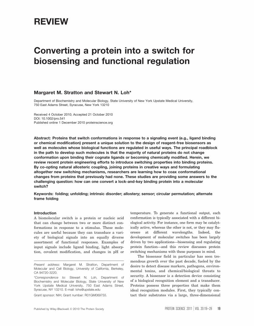
REVIEW
Converting a protein into a switch forbiosensing and functional regulation
Margaret M. Stratton and Stewart N. Loh*
Department of Biochemistry and Molecular Biology, State University of New York Upstate Medical University,
750 East Adams Street, Syracuse, New York 13210
Received 4 October 2010; Accepted 21 October 2010DOI: 10.1002/pro.541
Published online 1 December 2010 proteinscience.org
Abstract: Proteins that switch conformations in response to a signaling event (e.g., ligand bindingor chemical modification) present a unique solution to the design of reagent-free biosensors as
well as molecules whose biological functions are regulated in useful ways. The principal roadblock
in the path to develop such molecules is that the majority of natural proteins do not changeconformation upon binding their cognate ligands or becoming chemically modified. Herein, we
review recent protein engineering efforts to introduce switching properties into binding proteins.
By co-opting natural allosteric coupling, joining proteins in creative ways and formulatingaltogether new switching mechanisms, researchers are learning how to coax conformational
changes from proteins that previously had none. These studies are providing some answers to the
challenging question: how can one convert a lock-and-key binding protein into a molecularswitch?
Keywords: folding; unfolding; intrinsic disorder; allostery; sensor; circular permutation; alternate
frame folding
Introduction
A biomolecular switch is a protein or nucleic acid
that can change between two or more distinct con-
formations in response to a stimulus. These mole-
cules are useful because they can transduce a vari-
ety of biological signals into an equally diverse
assortment of functional responses. Examples of
input signals include ligand binding, light absorp-
tion, covalent modification, and changes in pH or
temperature. To generate a functional output, each
conformation is typically associated with a different bi-
ological activity. For instance, one form may be catalyt-
ically active, whereas the other is not, or they may flu-
oresce at different wavelengths. Indeed, the
development of molecular switches has been largely
driven by two applications—biosensing and regulating
protein function—and this review discusses protein
switching mechanisms with these purposes in mind.
The biosensor field in particular has seen tre-
mendous growth over the past decade, fueled by the
desire to detect disease markers, pathogens, environ-
mental toxins, and chemical/biological threats to
security. A biosensor is a detection device consisting
of a biological recognition element and a transducer.
Proteins possess three properties that make them
ideal recognition modules. First, they typically con-
tact their substrates via a large, three-dimensional
Present address: Margaret M. Stratton, Department ofMolecular and Cell Biology, University of California, Berkeley,CA 94720-3220.
*Correspondence to: Stewart N. Loh, Department ofBiochemistry and Molecular Biology, State University of NewYork Upstate Medical University, 750 East Adams Street,Syracuse, NY 13210. E-mail: [email protected]
Grant sponsor: NIH; Grant number: R01GM069755.
Published by Wiley-Blackwell. VC 2010 The Protein Society PROTEIN SCIENCE 2011 VOL 20:19—29 19
surface. This extensive interface enables binding to
be both tight and highly specific. Second, proteins
can be made to bind many targets of interest.
Nature already provides us with an abundance of
pre-existing binding proteins, as well as a means to
generate new proteins (in the form of antibodies)
that bind previously unrecognized ligands. Impor-
tantly, for small protein scaffolds, the latter process
can be carried out in vitro by directed evolution
methods.1 The basic problem, however, is that most
proteins do not change conformation upon binding
their cognate ligands (or absorbing light, changing
pH/temperature, etc.). We refer to this type of bind-
ing as lock-and-key [Fig. 1(A)]. The foremost chal-
lenge facing the researcher who wishes to build a
functional switch is therefore to transduce the
binding event or other stimulus into a measurable
output signal.
The third quality that proteins possess addresses
this need and is of primary interest to this review.
Proteins are unique in nature in that they offer mul-
tiple built-in mechanisms for reversible switching.
Binding can induce an allosteric conversion between
two or more native structures, as in the well-known
(but rare) examples of hemoglobin and calmodulin.
The folding reaction is an even more dramatic confor-
mational change that is ubiquitous to all proteins.
Because three-dimensional binding interfaces typi-
cally exist only when the protein is folded, binding
and folding are naturally coupled processes. An exam-
ple of this phenomenon occurs in nature in the form
of intrinsically disordered proteins (IDPs). It has
become clear that a significant percentage of proteins
in the cell (perhaps as high as 55%)2 contain regions
of intrinsic disorder, which may become structured
upon binding.3,4
There are two general approaches for transduc-
ing a protein’s binding event into a detectable signal.
The first is to immobilize it on an optical, electro-
chemical, or piezoelectric device. A binding-depend-
ent conformational change is not necessary and in
most cases none occurs. Rather, binding is recorded
by the difference in optical activity, mass, index of
refraction, or charge between the free receptor and
the receptor–ligand complex. The recognition mole-
cules can usually be used with minimal modification,
although it can be thorny to affix them to the
surface of the transducer without diminishing their
biological function. For a more detailed discussion of
these technologies, the reader is referred to recent
articles on the subject.5–9 The second strategy is to
use a single protein as both the recognition and
transduction element. Combining these functional-
ities reduces the need for complex and expensive
detection equipment as well as potential problems
associated with surface adsorption. Moreover, it
opens the door for the creation of hybrid proteins in
which biological function is coupled to molecular rec-
ognition in new and creative ways. The hurdle with
this approach is that much of the engineering bur-
den is transferred to the design of the biomolecule.
Switchable proteins offer a solution to this
challenge. Researchers are learning how to build
switches by co-opting existing allosteric mechanisms
(reviewed recently in Ref. 10–15). Less well under-
stood is how one can introduce switching properties
into proteins that previously had none. This issue is
crucial to the further development and widespread
availability of biological sensors. Herein, we focus
on one specific question: how can one convert an
ordinary binding protein—even one that exhibits
lock-and-key binding—into a molecular switch?
Altering Specificity of Proteins that Exhibit
Existing Conformational ChangePerhaps the most well known demonstration of how
to turn a binding protein into a biosensor is the con-
version of calmodulin into cameleon, a fluorescent cal-
cium sensor.16 The general idea behind cameleon and
related sensors is to attach environment or distance-
sensitive chromophores at positions that respond to a
conformational change between free and bound
states. This method is reliable when the confor-
mational change is large, as shown schematically in
Fig. 1(B). In the case of cameleon, the change is
extreme: calcium binding causes the calmodulin do-
main to wrap around the M13 peptide, which is fused
to its C-terminus. The N- and C-termini of the mole-
cule approach each other as a result. A donor fluores-
cent protein (FP; e.g., cyan FP) and acceptor FP (e.g.,
yellow FP) attached each terminus report on this
event by an increase in Forster resonance energy
transfer (FRET). Cameleon is therefore ratiometric as
well as genetically encoded, which are the two highly
desirable characteristics of a biological sensor. A simi-
lar approach was used to create sensors for phospho-
rylated peptides.17–20
Calmodulin is an example of a protein whose
allosteric conformational change is obvious and
dramatic. This type of highly tailored switching
mechanism is not easily transferred to other pro-
teins. What is needed is a protein that changes con-
formation upon binding its cognate ligand, but
whose binding pocket can be modified to accommo-
date alternate substrates [Fig. 1(B)]. This is not the
same as turning an arbitrary binding protein into a
switch, but it does offer the potential for converting
an existing switch to one that responds to an arbi-
trary ligand. Hellinga and coworkers created a fam-
ily of biosensors based on the bacterial periplasmic
binding protein (PBP) scaffold.21,22 PBPs contact
their substrates (mostly amino acids and small sug-
ars) in a cleft between two domains, which close by
�30� upon binding. They reported using computa-
tional methods to redesign the binding sites of vari-
ous PBPs to recognize TNT, L-lactate, serotonin, and
20 PROTEINSCIENCE.ORG Protein Switches for Biosensing and Regulation
pinacolyl methyl phosphonic acid (the hydrolytic
product of the nerve agent soman).23,24 These stud-
ies have been challenged, however, by the finding
that several of the re-engineered PBPs do not
appear to bind to their targets.25 At the present
time, design of binding sites seems to be best tackled
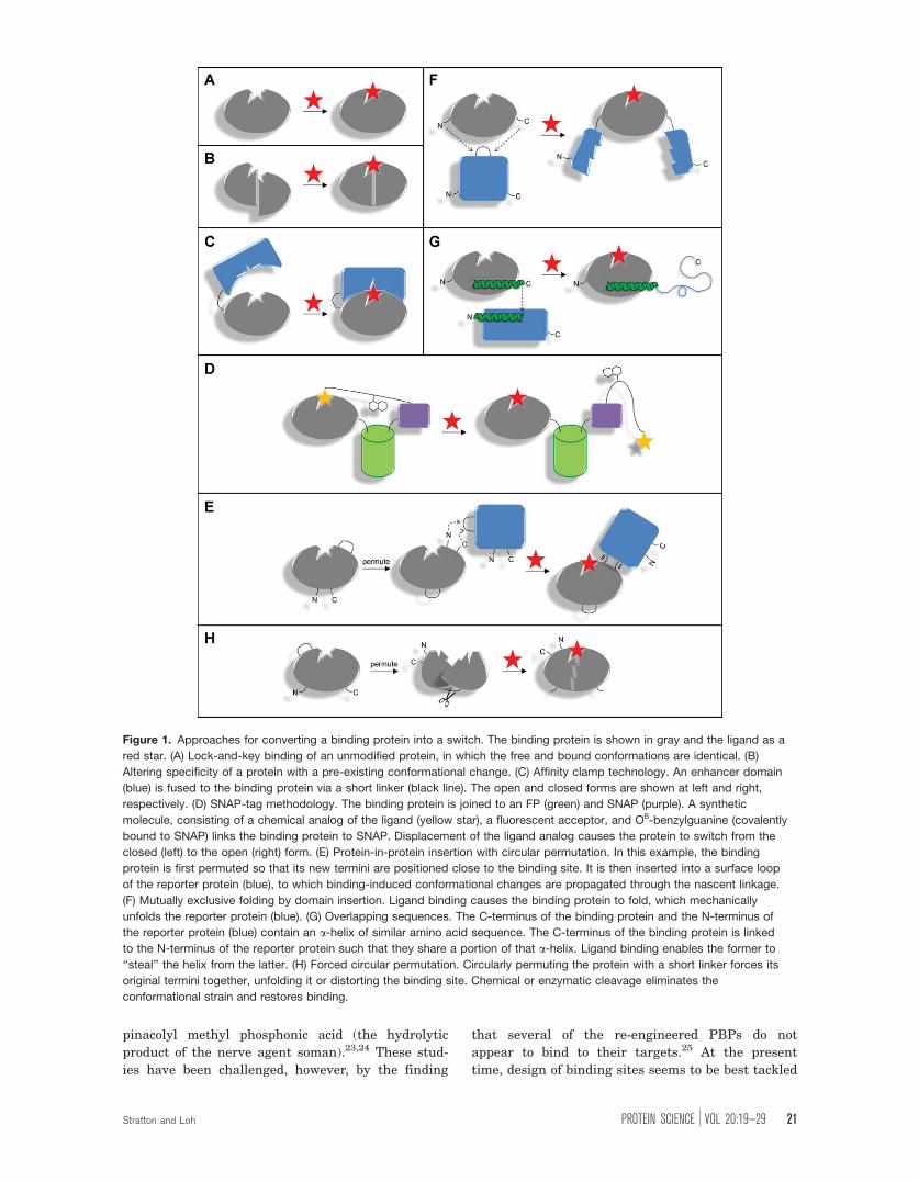
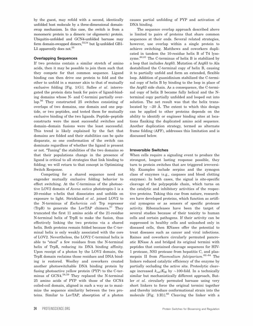
Figure 1. Approaches for converting a binding protein into a switch. The binding protein is shown in gray and the ligand as a
red star. (A) Lock-and-key binding of an unmodified protein, in which the free and bound conformations are identical. (B)
Altering specificity of a protein with a pre-existing conformational change. (C) Affinity clamp technology. An enhancer domain
(blue) is fused to the binding protein via a short linker (black line). The open and closed forms are shown at left and right,
respectively. (D) SNAP-tag methodology. The binding protein is joined to an FP (green) and SNAP (purple). A synthetic
molecule, consisting of a chemical analog of the ligand (yellow star), a fluorescent acceptor, and O6-benzylguanine (covalently
bound to SNAP) links the binding protein to SNAP. Displacement of the ligand analog causes the protein to switch from the
closed (left) to the open (right) form. (E) Protein-in-protein insertion with circular permutation. In this example, the binding
protein is first permuted so that its new termini are positioned close to the binding site. It is then inserted into a surface loop
of the reporter protein (blue), to which binding-induced conformational changes are propagated through the nascent linkage.
(F) Mutually exclusive folding by domain insertion. Ligand binding causes the binding protein to fold, which mechanically
unfolds the reporter protein (blue). (G) Overlapping sequences. The C-terminus of the binding protein and the N-terminus of
the reporter protein (blue) contain an a-helix of similar amino acid sequence. The C-terminus of the binding protein is linked
to the N-terminus of the reporter protein such that they share a portion of that a-helix. Ligand binding enables the former to
‘‘steal’’ the helix from the latter. (H) Forced circular permutation. Circularly permuting the protein with a short linker forces its
original termini together, unfolding it or distorting the binding site. Chemical or enzymatic cleavage eliminates the
conformational strain and restores binding.
Stratton and Loh PROTEIN SCIENCE VOL 20:19—29 21
by directed evolution techniques, although computa-
tional methods are improving rapidly.26
Affinity Clamps
One recent approach in which directed evolution fig-
ures prominently is the affinity clamp methodology
of Koide.27–29 An existing binding domain (capture
domain) is joined via a short linker to a small anti-
body-like protein [enhancer domain; Fig. 1(C)]. The
newly-formed domain interface is then optimized by
randomizing the antigen binding loops of the surro-
gate antibody and panning the library using phage
display. When the capture domain was PDZ, which
binds a short peptide from a p120-related catenin
with low-micromolar affinity, and the enhancer was
fibronectin type III domain, the resulting clamp
bound the peptide substrate with low nanomolar
affinity and high specificity. Importantly, binding can
be detected by attaching FPs to the N- and C-termini,
in much the same manner as cameleon. Affinity clamp
technology is significant because capture domains
can be mixed and matched with different enhancer
domains to maximize chances of success.
SNAP-Tag
Johnsson and coworkers have devised a semisyn-
thetic method for eliciting a similar closed-to-open
conformational change from a generic binding pro-
tein.30 The genetically encoded portion of their sen-
sor is comprised of three proteins joined end-to-end:
SNAP-tag (a small protein based on O6-alkylgua-
nine-DNA alkyltransferase), an FP, and the binding
protein of interest [Fig. 1(D)]. The synthetic compo-
nent is an artificial molecule consisting of O6-benzyl-
guanine on one end, the ligand to the binding pro-
tein on the other end, and a fluorescent acceptor
group in the middle. The synthetic molecule acts as
a tether; it binds covalently to the SNAP-tag at one
end and noncovalently to the binding protein at the
other. This arrangement defines the closed state
wherein the FP and the fluorescent acceptor group
are close in space. Binding of the natural ligand dis-
places the synthetic molecule from the binding pro-
tein, which causes the protein to assume the open
conformation and the donor–acceptor distance to
increase. The readout is a ratiometric change in
FRET signal. The SNAP-tag technique is powerful
because it is well-suited for sensing and imaging
in vivo as long as the artificial tether is readily syn-
thesizable and cell permeable. For metabolites and
other small ligands this is likely to be the case.
Naturally Occuring Fold SwitchesAlthough proteins generally adopt a single structure
that corresponds to the global minimum of free
energy, some can sample alternate native conforma-
tions whose energy minima are only slightly higher.
If one fold binds a ligand with higher affinity than
the others, then such a protein can exhibit switching
behavior. A striking example is provided by proteins
that can switch between unrelated folds.31 Lympho-
tactin adopts the canonical a/b chemokine fold at
low temperature and in the presence of NaCl.32 At
higher temperature and lower salt concentration,
however, it takes on a completely different all-bstructure.33 The switch is driven by binding, as the
all-b form is dimeric. The mitotic arrest deficiency
two protein (Mad2) interconverts between two func-
tionally distinct conformations (O-Mad2 and C-
Mad2).34 Binding of C-Mad2 (as part of the C-Mad2/
Mad1 complex) to a partially unfolded form of O-
Mad2 is thought to catalyze the conversion of the
latter to C-Mad2.34 Still other proteins can be
induced to switch conformations by changing a rela-
tively small number of amino acids, as demonstrated
by the classic experiments of Sauer with Cro and
Arc repressors,35,36 Regan with protein G,37 and
more recently, Cordes with Cro homologs.38
The above examples illustrate the extent to
which proteins are capable of rearranging their struc-
tures. However, it is still largely out of reach to engi-
neer this type of fold-switching mechanism into a
generic binding protein. One notable exception is the
design of metal-dependent switches.13 Here, efforts
are aided by the relative simplicity of metal binding
sites. Zinc, for example, is coordinated by four atoms
(typically supplied by side chains of His and/or Cys)
arranged in tetrahedral geometry. Several groups
have created proteins that switch to zinc finger39,40 or
helix-loop-helix41 conformations from different folded
structures, in response to zinc binding.
Internal Protein-in-Protein Fusions
When one wishes to fuse two proteins in such a way
as to minimize their potential interaction, one typi-
cally links the C-terminus of the first to the N-termi-
nus of the second using a long, structureless peptide
tether. Common examples include attaching gluta-
thione S-transferase or an FP to one of the ends of a
target protein to facilitate its purification or to visu-
alize its cellular localization, respectively. In such
applications, the goal is for each protein to be oblivi-
ous to the fact that it is covalently bonded to the
other. Conversely, joining two proteins in a more
intimate fashion—at an internal position and by
using short linkers—increases the likelihood that
changes in one domain will be communicated to the
second domain; for example, ligand binding to a
receptor domain effecting a structural or functional
change in a reporter domain [Fig. 1(E)].11,12 This
type of communication requires the presence of a
binding-induced conformational change. However,
the change can be less dramatic than in proteins
such as cameleon because even a subtle structural
change can alter optical or enzymatic activity of the
22 PROTEINSCIENCE.ORG Protein Switches for Biosensing and Regulation
attached protein,42,43 sometimes in a manner that
was not forseen.44
An effective strategy for optimizing interdomain
communication is to insert the reporter into the
receptor at a location near the receptor’s ligand
binding site, or to insert the receptor into the
reporter at a location near the reporter’s active site.
It is desirable to place the ‘‘guest’’ protein into a
surface loop of the ‘‘host’’ protein (rather than into
an a-helix or b-strand) to avoid perturbing the struc-
ture of the host. Linker length is likewise a critical
factor: a very long linker decouples interdomain
communication, whereas a very short linker can
compress the guest and stretch the host. This effect,
which occurs when the N-to-C distance of the guest
exceeds the distance between the ends of the loop in
the host, can itself be exploited as a switching mech-
anism (see Mutually Exclusive Folding). A more gen-
tle means of insertion is to first circularly permute
the guest. Circular permutation creates a new sur-
face loop connecting the original termini, and new
termini at a chosen site elsewhere in the protein
[usually at a former surface loop; Fig. 1(E)]. Thus,
the termini of a permuted protein are always close
in space. Not only does permutation help ensure
that the host–guest loop distances are compatible, it
also adds a valuable combinatorial aspect to the
process. All told, one can experiment with the follow-
ing parameters to optimize coupling: permutation
site in the guest, linker used for permutation, inser-
tion site in the host, and linkers used to join the
proteins.
Not surprisingly, many of the most successful
implementations of protein-in-protein switches have
leveraged the power of genetic screens or selections
to optimize the design. FPs, and to a lesser extent
luminescent proteins, have figured prominently in
this regard due to their compatibility with cell sort-
ing and other rapid screening methods. Tsien and
coworkers made a key advance when they dis-
covered that green FP (GFP) can tolerate insertions
of foreign sequences in the loop around Tyr145, and
that GFP can be permuted in numerous locations,
while remaining fluorescent.45 They inserted cal-
modulin and a zinc finger domain from zif268 at
position 145 and observed that metal binding
changed the GFP chromophore environment such
that fluorescence was enhanced. Reversing the host
and the guest, Nagai et al. inserted circularly per-
muted GFP in between calmodulin and the M13 pep-
tide to create a family of calcium sensors called peri-
cams.43 The spectral properties of GFP were altered
by calcium binding but the structural basis for these
changes remains unclear. To illustrate this point,
one variant became brighter upon calcium binding,
another became dimmer, and a third exhibited a
ratiometric response in which the emission wave-
length shifted. They were able to elicit these differ-
ent behaviors by mutating a few amino acids near
the chromophore and by changing the length and
sequence of the linker used to permute GFP. Fan et
al. inserted the cAMP-binding domain B from pro-
tein kinase A into a circular permutant of firefly lu-
ciferase.46 By experimenting with different permuta-
tion sites as well as linkers used to join the N- and
C-termini, they generated a sensor that increased its
luminescence by 19-fold in the presence of cAMP.
Enzymes have also drawn attention as reporter
domains, because they allow binding to be trans-
duced into a functional output. Ostermeier and co-
workers screened large libraries of mutants (�106)
in which b-lactamase was randomly permuted and
inserted into random or specific positions in maltose
binding protein.47–49 By monitoring how well cells
grew in the presence of ampicillin and maltose they
were able to identify several variants whose enzy-
matic activity was inhibited or enhanced by sugar
binding by as much as 600-fold. Edwards et al.
took a related approach to create a cytochrome b562/
b-lactamase fusion in which tolerance to antibiotic
was dependent on heme in the growth medium.50
Mutually Exclusive Folding
When constructing protein-in-protein fusions, one
can take the opposite tack and deliberately make
the N-to-C distance of the guest much larger than
the end-to-end distance of the loop in the host. This
condition results in a structural tug-of-war in which
the folding free energy of one protein is used to
mechanically unfold the other [Fig. 1(F)]. This effect
was demonstrated by inserting ubiquitin (38 A N-to-
C distance)51–53 or the GCN4 DNA binding domain
(75 A N-to-C distance)54 into a surface loop of
barnase (10 A end-to-end loop distance). When the
tethering linkers are sufficiently short, then only
one protein can be folded at any given time. Peng
and Li were able to directly observe this tug-of-war
in another chimera (the 27th Ig domain of titin
inserted into the GB1-L5 protein) using fluorescence
and atomic force microscopy.55
Thermodynamic and structural coupling between
domains provides a pathway for transducing binding
to conformational change. For example, the presence
of a cognate DNA sequence causes GCN4 to fold and
barnase to unfold and lose its ribonuclease activity.54
Barnase is thus converted to a DNA-dependent
molecular switch. With regard to generality of the
design, the extent of coupling is determined chiefly by
ratio of the guest and host distances mentioned
above. Very long linkers decouple the domains such
that they fold independently. Mutually exclusive fold-
ing appears to require a minimum guest:host distance
of �2:1, where the length of any linkers (in an
extended conformation) used to join the proteins is
added to the host distance.53 One potential caveat to
this design is that some host proteins, upon unfolding
Stratton and Loh PROTEIN SCIENCE VOL 20:19—29 23
by the guest, may refold with a second, identically
unfolded host molecule by a three-dimensional domain-
swap mechanism. In this case, the switch is from a
monomeric protein to a dimeric (or oligomeric) protein.
Ubiquitin-unfolded and GCN4-unfolded barnase may
form domain-swapped dimers,53,54 but Ig-unfolded GB1-
L5 apparently does not.55
Overlapping Sequences
If two proteins contain a similar stretch of amino
acids, then it may be possible to join them such that
they compete for that common sequence. Ligand
binding can then drive one protein to fold and the
other to unfold in a manner akin to that of mutually
exclusive folding [Fig. 1(G)]. Sallee et al. interro-
gated the protein data bank for pairs of ligand-bind-
ing domains whose N- and C-termini partially over-
lap.56 They constructed 25 switches consisting of
overlaps of two domains, one domain and one pep-
tide, or two peptides, and tested them for mutually
exclusive binding of the two ligands. Peptide–peptide
constructs were the most successful switches and
domain–domain fusions were the least successful.
This trend is likely explained by the fact that
domains are folded and their stabilities can be quite
disparate, so one conformation of the switch can
dominate regardless of whether the ligand is present
or not. ‘‘Tuning’’ the stabilities of the two domains so
that their populations change in the presence of
ligand is critical to all strategies that link binding to
folding; we will return to that concept in Optimizing
Switch Response.
Competing for a shared sequence need not
engender mutually exclusive folding behavior to
effect switching. At the C-terminus of the photoac-
tive LOV2 domain of Avena sativa phototropin-1 is a
20-residue a-helix that dissociates and unfolds on
exposure to light. Strickland et al. joined LOV2 to
the N-terminus of Eschericia coli Trp repressor
(TrpR) to generate the LovTAP chimera.57 They
truncated the first 11 amino acids of the 21-residue
N-terminal helix of TrpR to make the fusion, thus
effectively linking the two proteins via a shared
helix. Both proteins remain folded because the C-ter-
minal helix is only weakly associated with the core
of LOV2. Nevertheless, the LOV2 C-terminal helix is
able to ‘‘steal’’ a few residues from the N-terminal
helix of TrpR, reducing its DNA binding affinity.
Upon receipt of a photon by the LOV2 domain, the
TrpR domain reclaims those residues and DNA bind-
ing is restored. Woolley and coworkers created
another photoswitchable DNA binding protein by
fusing photoactive yellow protein (PYP) to the C-ter-
minus of GCN4.58,59 They replaced the N-terminal
25 amino acids of PYP with those of the GCN4
coiled-coil domain, aligned in such a way as to maxi-
mize the sequence similarity between the two pro-
teins. Similar to LovTAP, absorption of a photon
causes partial unfolding of PYP and activation of
DNA binding.
The sequence overlap approach described above
is limited to pairs of proteins that share common
sequences at their ends. Several related strategies,
however, use overlap within a single protein to
achieve switching. Matthews and coworkers dupli-
cated in tandem the 10-residue helix B of T4 lyso-
zyme.60,61 The C-terminus of helix B is stabilized by
a loop that includes Arg63. Mutation of Arg63 to Ala
destabilized the C-terminal copy of helix B, causing
it to partially unfold and form an extended, flexible
loop. Addition of guanidinium stabilized the C-termi-
nal copy of helix B by binding to the loop in place of
the Arg63 side chain. As a consequence, the C-termi-
nal copy of helix B became fully helical and the N-
terminal copy partially unfolded and looped out into
solution. The net result was that the helix trans-
located by �20 A. The extent to which this design
can be applied to other proteins depends on the
ability to identify or engineer binding sites at loca-
tions flanking the duplicated amino acid sequence.
Another duplication strategy, termed as alternate
frame folding (AFF), addresses this limitation and is
discussed below.
Irreversible Switches
When cells require a signaling event to produce the
strongest, longest lasting response possible, they
turn to protein switches that are triggered irreversi-
bly. Examples include serpins and the zymogen
class of enzymes (e.g., caspases and blood clotting
enzymes). In both cases, the signal is site-specific
cleavage of the polypeptide chain, which turns on
the catalytic and inhibitory activities of the respec-
tive proteins. Taking this cue from nature, research-
ers have developed proteins, which function as artifi-
cial zymogens or as sensors of specific protease
activity. Ribonucleases have been the target of
several studies because of their toxicity to human
cells and certain pathogens. If their activity can be
suppressed in healthy cells and unleashed only in
diseased cells, then RNases offer the potential to
treat diseases such as cancer and viral infections.
Raines and coworkers circularly permuted pancre-
atic RNase A and bridged its original termini with
peptides that contained cleavage sequences for HIV-
1 protease, NS3 protease from hepatitis C, and plas-
mepsin II from Plasmodium falciparium.62–64 The
linkers reduced catalytic efficiency of the enzyme by
partially occluding the active site. Proteolytic cleav-
age increased kcat/KM by �100-fold. In a technically
similar but mechanistically different approach, But-
ler et al. circularly permuted barnase using very
short linkers to force the original termini together
and thereby introduce conformational strain into the
molecule [Fig. 1(H)].65 Cleaving the linker with a
24 PROTEINSCIENCE.ORG Protein Switches for Biosensing and Regulation
chemical reagent relieved the strain and increased
catalytic efficiency.
Although the above strategies can be successful
for some proteins, they rely on specific properties of
the target enzyme and are therefore not general. The
linker used to circularly permute an enzyme does not
typically obscure the active site, nor is cleaving the
linker guaranteed to remove the obstruction. Simi-
larly, N- to C-terminal distances in many proteins are
too short to induce significant conformational strain
when linked. Even in the case where this distance was
compressed from 27 A to that of a single amino acid,
the protein structure was able to absorb the strain
and enzymatic activity remained high.65 Mitrea et al.
recently introduced a switching mechanism that
addresses the issue of generality.66 We defer discus-
sion of this study to the AFF section below.
Binding-Induced Folding
Intrinsically disordered proteins
In our view, binding-induced folding is the most gen-
erally applicable strategy for converting a binding
protein into a switch. The reasons are as follows. (i)
Proteins constantly cycle through all possible confor-
mations—from native to partially folded to unfolded—
according to their Boltzmann distributions. Thus,
even a lock-and-key protein is already a reversible
switch, though it spends nearly all of its time in one
state. (ii) It is straightforward to convert a folded pro-
tein into a pseudo-IDP by manipulating the relative
populations of native and unfolded forms. The latter
state can be made dominant by decreasing stability of
the former by point mutation, truncation, insertion,
and so forth. Indeed, we have already seen examples
in mutually exclusive folding and overlapping sequen-
ces. The native conformation can then be restored
by ligand binding, which, as mentioned, is thermo-
dynamically linked to folding. In principle, any pro-
tein is capable of such a reversible transformation.
(iii) The folding reaction is the most dramatic confor-
mational change that a protein undergoes. So many
of its properties change (structure, dynamics, and
charge distribution to name a few) that it is likely
that a means to detect binding can be devised.67 As a
simple example, one can attach donor and acceptor
fluorophores at the N- and C-termini. As the ends of
globular proteins are frequently proximal68,69 (or can
be made so by circular permutation) and are expected
to be more distant (on average) in the unfolded state,
binding can potentially be monitored by increase in
FRET. Parenthetically, the opposite is true for short
peptide ‘‘beacons’’.67,70–72 These unstructured peptides
are designed to bind their targets in a rigid, extended
conformation. Binding can be detected by the increase
in distance between ends of the peptide as monitored
by loss of quenching between fluorescent donor and
quencher groups.
Kohn and Plaxco elegantly demonstrated princi-
ples of binding-induced folding by progressively delet-
ing amino acids from the C-terminus of the SH3 do-
main from Fyn tyrosine kinase until it unfolded.73
Binding the cognate phosphorylated peptide induced
SH3 to fold, which was detected by fluorescence of the
intrinsic Trp residue or that of a BODIPY group pre-
viously attached to a strategically placed Cys side
chain. Impressively, their sensor worked even when
tested in the complex, contaminant-ridden environ-
ment of blood serum. Others have taken advantage of
intrinsically disordered regions in p5374 and
BRCA175 that fold upon binding to create fluorescent
sensors for their respective ligands.
The principal challenge with the binding-
induced folding paradigm is that destabilized and
unfolded proteins tend to aggregate or otherwise
misfold. Degradation is also a concern, particularly
in cellular applications. Naturally occurring IDPs
appear to have evolved the ability to remain soluble
when unfolded, likely due (at least in part) to their
unusually high content of charged groups and low
percentage of hydrophobic residues (2). Destabiliza-
tion of native proteins, on the other hand, is strongly
implicated as the root cause of numerous aggrega-
tion-related diseases (reviewed for example in76).
Truncated, unfolded SH3 (53 residues) is well-
behaved and soluble at >100 lM concentration and
was not degraded when expressed in E. coli.73 The
extent to which these attributes pertain to large or
multidomain proteins remains to be tested.
Alternate frame folding
AFF combines several of the themes discussed earlier,
including circular permutation, partial sequence over-
lap, fold switching, and binding-induced folding. The
unique feature of AFF is that it couples binding-
induced folding to unfolding of another portion of the
molecule.66,77,78 These two reactions are balanced so
that no net folding or unfolding occurs; rather, the
structure is remodeled. The conformational change is
from one native structure to another. The AFF mecha-
nism does not require a reporter protein to be fused to
the binding protein. In fact, no foreign sequence is
added to the binding protein with the exception of a
flexible peptide linker. AFF is designed to convert even
a lock-and-key binding protein into a molecular switch.
Figure 2 illustrates the basic steps of this con-
version. First, a segment from one of the termini of
the protein is duplicated and appended to the oppo-
site end. The position and length of this segment is
dictated by two considerations. First, it must contain
at least one amino acid which, when mutated,
abolishes ligand binding. Beyond that criterion, its
length is determined by the preference for the newly
generated termini of a circularly permuted protein
to reside at a former surface loop. For example, if a
critical binding residue is close to the C-terminus,
Stratton and Loh PROTEIN SCIENCE VOL 20:19—29 25
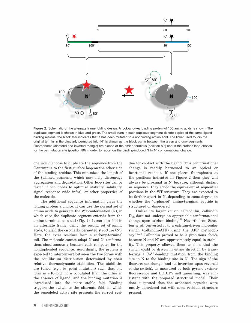
one would choose to duplicate the sequence from the
C-terminus to the first surface loop on the other side
of the binding residue. This minimizes the length of
the twinned segment, which may help discourage
aggregation and degradation. Other loop sites can be
tested if one needs to optimize stability, solubility,
signal response (vide infra), or other properties of
the molecule.
The additional sequence information gives the
folding protein a choice. It can use the normal set of
amino acids to generate the WT conformation (N), in
which case the duplicate segment extends from the
amino terminus as a tail (Fig. 2). It can also fold in
an alternate frame, using the second set of amino
acids, to yield the circularly permuted structure (N0).Here, the extra residues form a carboxy-terminal
tail. The molecule cannot adopt N and N0 conforma-
tions simultaneously because each competes for the
nonduplicated sequence. Accordingly, the protein is
expected to interconvert between the two forms with
the equilibrium distribution determined by their
relative thermodynamic stabilities. The stabilities
are tuned (e.g., by point mutation) such that one
form is �10-fold more populated than the other in
the absence of ligand, and the binding mutation is
introduced into the more stable fold. Binding
triggers the switch to the alternate fold, in which
the remodeled active site presents the correct resi-
due for contact with the ligand. This conformational
change is readily harnessed to an optical or
functional readout. If one places fluorophores at
the positions indicated in Figure 2 then they will
always be proximal in N0 because, although distant
in sequence, they adopt the equivalent of sequential
positions in the WT structure. They are expected to
be farther apart in N, depending to some degree on
whether the ‘‘orphaned’’ amino-terminal peptide is
structured or disordered.
Unlike its larger cousin calmodulin, calbindin
D9k does not undergo an appreciable conformational
change upon calcium binding.79 Nevertheless, Strat-
ton et al. converted it to a calcium-driven molecular
switch (calbindin-AFF) using the AFF methodol-
ogy.77,78 Calbindin proved to be a propitious choice
because N and N0 are approximately equal in stabil-
ity. This property allowed them to show that the
switch could be driven in either direction by trans-
ferring a Ca2þ-binding mutation from the binding
site in N to the binding site in N0. The sign of the
fluorescence change (and its inversion upon reversal
of the switch), as measured by both pyrene excimer
fluorescence and BODIPY self quenching, was con-
sistent with the proposed structural model. Their
data suggested that the orphaned peptides were
mostly disordered but with some residual structure
present.
Figure 2. Schematic of the alternate frame folding design. A lock-and-key binding protein of 100 amino acids is shown. The
duplicate segment is shown in blue and green. The small stars in each duplicate segment denote copies of the same ligand-
binding residue; the black star indicates that it has been mutated to a nonbinding amino acid. The linker used to join the
original termini in the circularly permuted fold (N0) is shown as the black bar in between the green and gray segments.
Fluorophores (diamond and inverted triangle) are placed at the amino terminus (position 800) and in the surface loop chosen
for the permutation site (position 80) in order to report on the binding-induced N to N0 conformational change.
26 PROTEINSCIENCE.ORG Protein Switches for Biosensing and Regulation
Mitrea et al. used a similar modification to con-
vert barnase into a functional switch.66 Instead
of ligand binding, the input signal was proteolytic
cleavage. A carboxy-terminal segment of barnase was
duplicated as above, and an HIV-1 protease recogni-
tion sequence was inserted into the loop that served
as the permutation site. The engineered enzyme,
barnase-AFF, was primarily in conformation N, and
this fold was rendered catalytically inert by mutating
the general acid His102 to Ala. Cleavage by the prote-
ase released the carboxy-terminal duplicated peptide,
which contained the H102A mutation. Barnase-AFF
refolded to the catalytically competent N0 conforma-
tion and RNase activity increased by 130-fold.
What are the advantages and disadvantages
of AFF compared with traditional binding-induced
folding switches? As part of the molecule is always
unfolded, AFF is subject to the same concerns of
aggregation and degradation expressed earlier. One
important distinction, however, is that AFF bypasses
populating the globally unfolded state, which can be
particularly problematic. The length of the unfolded
segment is adjustable. It can be a relatively small
portion of the molecule. For example, 30% and 7.5%
of the residues in calbindin-AFF and barnase-AFF
are involved in their respective folding/unfolding
reactions. The additional requirements of AFF are
that the protein must tolerate circular permutation
and not contain covalent linkages (e.g., disulfide
bridges) that would physically prevent the fold shift.
Optimizing switch responseThe key properties of any switch are affinity, gain
(signal change between bound and free forms), and
response time. For folding-based switches, these
properties translate to the thermodynamics and
kinetics of protein folding. Fortunately, decades of
work in this area have laid out a path for tailoring
the characteristics of the switch to suit a particular
application.78,80 In therapeutic implementations
where a toxic protein is involved, it is imperative to
rigidly enforce the off-state of the switch. Cytotoxic
RNase activity of barnase-AFF was suppressed to
nearly undetectable levels by introducing point
mutations that selectively destabilized the N0 con-
formation.66 For biosensor applications, the best
balance of robust signal change and high affinity is
generally when 50–90% of the protein is unfolded in
the absence of ligand. This condition allows for the
majority of molecules to fold upon binding. Because
binding energy is used to drive the unfavorable fold-
ing reaction, the apparent dissociation constant (Kd)
is weaker than the intrinsic Kd of the native protein
by a factor of (1 þ Kf)/Kf, where Kf is the equilibrium
constant for folding (assuming two-state unfolding).
Thus, apparent affinity is reduced by only 2–10-fold
in the example above. In other cases, it may be de-
sirable to reduce affinity further so that Kd matches
the concentration of analyte. It is straightforward to
achieve the desired Kd by tuning the stabilities of
native and unfolded forms (or the N and N0 confor-mations in the AFF mechanism) using point muta-
tions, truncations, or both. This method of affinity
tuning can be preferable to directly modifying the
binding site, because the latter process can alter
specificity as well.14 Destabilizing mutations can be
introduced at locations distant from the binding
pocket where they are unlikely to affect specificity.
Response time can be a critical parameter of a
sensor, particularly when one needs to monitor rapidly
changing levels of analyte in real time. Luckily,
small proteins tend to fold quickly. The truncated
SH3 sensor responded with a time constant of
10 ms.73 This figure is consistent with the time con-
stant reported for folding of full-length SH3. Calbin-
din-AFF took longer to respond (several seconds),
apparently because an unfolding step is at least par-
tially rate-limiting, and unfolding rates can be slow
in native conditions. Nevertheless, the switching
rate was significantly faster than the rate of global
unfolding, indicating that the entire molecule did
not need to denature in order to switch folds. It may
be possible to adjust response times by modulating
folding and unfolding rates.78
ConclusionsProtein conformational change is a natural and
powerful means for coupling an input event to an
output signal. Many of the most successful existing
designs fuse the binding protein to a reporter do-
main to propagate a conformational change through
the covalent linkage, or to engender an open-to-
closed domain movement upon binding. A more gen-
eral approach may be to link binding to folding (and
possibly to partial unfolding) within a single protein
molecule. With this strategy, even a lock-and-key
protein may be converted into a functional switch.
Acknowledgments
The authors thank Jeung-Hoi Ha, Diana Mitrea, and
Huimei Zheng for comments and discussions.
References
1. Dougherty MJ, Arnold FH (2009) Directed evolution:new parts and optimized function. Curr Opin Biotech-nol 20:486–491.
2. Uversky VN, Dunker AK (2010) Understanding proteinnon-folding. Biochim Biophys Acta 1804:1231–1264.
3. Wright PE, Dyson HJ (2009) Linking folding and bind-ing. Curr Opin Struct Biol 19:31–38.
4. Hilser VJ, Thompson EB (2007) Intrinsic disorder as amechanism to optimize allosteric coupling in proteins.Proc Natl Acad Sci USA 104:8311–8315.
5. Lubin AA, Plaxco KW (2010) Folding-based electro-chemical biosensors: the case for responsive nucleicacid architectures. Acc Chem Res 43:496–505.
Stratton and Loh PROTEIN SCIENCE VOL 20:19—29 27
6. White RJ, Plaxco KW (2010) Exploiting binding-inducedchanges in probe flexibility for the optimization of elec-trochemical biosensors. Anal Chem 82:73–76.
7. Prieto-Simin B, Campas M, Marty JL (2008) Biomole-cule immobilization in biosensor development: tailoredstrategies based on affinity interactions. Protein PeptLett 15:757–763.
8. North SH, Lock EH, Taitt CR, Walton SG (2010) Criti-cal aspects of biointerface design and their impact onbiosensor development. Anal Bioanal Chem 397:925–933.
9. Luong JH, Male KB, Glennon JD (2008) Biosensortechnology: technology push versus market pull. Bio-technol Adv 26:492–500.
10. Ambroggio XI, Kuhlman B (2006) Design of proteinconformational switches. Curr Opin Struct Biol 16:525–530.
11. Ostermeier M (2009) Designing switchable enzymes.Curr Opin Struct Biol 19:442–448.
12. Ostermeier M (2005) Engineering allosteric proteinswitches by domain insertion. Protein Eng Des Sel 18:359–364.
13. Wright CM, Heins RA, Ostermeier M (2007) As easy asflipping a switch? Curr Opin Chem Biol 11:342–346.
14. Vallee-Belisle A, Plaxco KW (2010) Structure-switchingbiosensors: inspired by Nature. Curr Opin Struct Biol20:518–526.
15. Koide S (2009) Generation of new protein functions bynonhomologous combinations and rearrangements ofdomains and modules. Curr Opin Biotech 20:398–404.
16. Miyawaki A, Llopis J, Heim R, McCaffery JM, AdamsJA, Ikura M, Tsien RY (1997) Fluorescent indicatorsfor Ca2þ based on green fluorescent proteins and cal-modulin. Nature 388:882–887.
17. Zhang J, Ma Y, Taylor SS, Tsien RY (2001) Geneticallyencoded reporters of protein kinase A activity revealimpact of substrate tethering. Proc Natl Acad Sci USA98:14997–15002.
18. Ting AY, Kain KH, Klemke RL, Tsien RY (2001) Genet-ically encoded fluorescent reporters of protein tyrosinekinase activities in living cells. Proc Natl Acad SciUSA 98:15003–15008.
19. Fosbrink M, Aye-Han NN, Cheong R, Levchenko A,Zhang J (2010) Visualization of JNK activity dynamicswith a genetically encoded fluorescent biosensor. ProcNatl Acad Sci USA 107:5459–5464.
20. Harvey CD, Ehrhardt AG, Cellurale C, Zhong H,Yasuda R, Davis RJ, Svoboda K (2008) A geneticallyencoded fluorescent sensor of ERK activity. Proc NatlAcad Sci USA 105:19264–19269.
21. Dwyer MA, Hellinga HW (2004) Periplasmic bindingproteins: a versatile superfamily for protein engineer-ing. Curr Opin Struct Biol 14:495–504.
22. De Lorimer RM, Smith JJ, Dwyer MA, Looger LL, SaliKM, Paavola CD, Rizk SS, Sadigov S, Conrad DW,Loew L, Hellinga HW (2002) Construction of a fluores-cent biosensor family. Protein Sci 11:2655–2675.
23. Allert M, Rizk SS, Looger LL, Hellinga HW (2004)Computational design of receptors for an organophos-phate surrogate of the nerve agent soman. Proc NatlAcad Sci USA 101:7907–7912.
24. Looger LL, Dwyer MA, Smith JJ, Hellinga HW (2003)Computational design of receptor and sensor proteinswith novel functions. Nature 423:185–190.
25. Schreier B, Stumpp C, Wiesner S, Hocker B (2009)Computational design of ligand binding is not a solvedproblem. Proc Natl Acad Sci USA 106:18491–18496.
26. Siegel JB, Zanghellini A, Lovick HM, Kiss G, LambertAR, St Clair JL, Gallaher JL, Hilvert D, Gelb MH,
Stoddard BL, Houk KN, Michael FE, Baker D (2010)Computational design of an enzyme catalyst for astereoselective bimolecular Diels-Alder reaction. Sci-ence 329:309–313.
27. Huang J, Koide S (2010) Rational conversion of affinityreagents into label-free sensors for peptide motifs bydesigned allostery. ACS Chem Biol 5:273–277.
28. Huang J, Koide A, Makabe K, Koide S (2008) Designof protein function leaps by directed domain interfaceevolution. Proc Natl Acad Sci USA 105:6578–6583.
29. Huang J, Makabe K, Biancalana M, Koide A, Koide S(2009) Structural basis for exquisite specificity of affin-ity clamps, synthetic binding proteins generatedthrough directed domain-interface evolution. J Mol Biol392:1221–1231.
30. Brun MA, Tan KT, Nakata E, Hinner MJ, Johnsson K(2009) Semisynthetic fluorescent sensor proteins basedon self-labeling protein tags. J Am Chem Soc 131:5873–5884.
31. Bryan PN, Orban J (2010) Proteins that switch folds.Curr Opin Struct Biol 20:482–488.
32. Kuloglu ES, McCaslin DR, Kitabwalla M, Pauza CD,Markley JL, Volkman BF (2001) Monomeric solutionstructure of the prototypical ’C’ chemokine lymphotac-tin. Biochemistry 40:12486–12496.
33. Tuinstra RL, Peterson FC, Kutlesa S, Elgin ES, KronMA, Volkman BF (2008) Interconversion between twounrelated protein folds in the lymphotactin nativestate. Proc Natl Acad Sci USA 105:5057–5062.
34. Luo X, Tang Z, Xia G, Wassmann K, Matsumoto T,Rizo J, Yu H (2004) The Mad2 spindle checkpoint pro-tein has two distinct natively folded states. Nat StructMol Biol 11:338–345.
35. Anderson TA, Cordes MH, Sauer RT (2005) Sequencedeterminants of a conformational switch in a proteinstructure. Proc Natl Acad Sci USA 102:18344–18349.
36. Mossing MC, Sauer RT (1990) Stable, monomericvariants of lambda Cro obtained by insertion of adesigned beta-hairpin sequence. Science 250:1712–1715.
37. Dalal S, Regan L (2000) Understanding the sequencedeterminants of conformational switching using proteindesign. Protein Sci 9:1651–1659.
38. Roessler CG, Hall BM, Anderson WJ, Ingram WM,Roberts SA, Montfort WR, Cordes MH (2008) Transi-tive homology-guided structural studies lead to discov-ery of Cro proteins with 40% sequence identity butdifferent folds. Proc Natl Acad Sci USA 105:2343–2348.
39. Ambroggio XI, Kuhlman B (2006) Computationaldesign of a single amino acid sequence that can switchbetween two distinct protein folds. J Am Chem Soc128:1154–1161.
40. Hori Y, Sugiura Y (2002) Conversion of antennapediahomeodomain to zinc finger-like domain: Zn(II)-inducedchange in protein conformation and DNA binding. J AmChem Soc 124:9362–9363.
41. Cerasoli E, Sharpe BK, Woolfson DN (2005) ZiCo: apeptide designed to switch folded state upon bindingzinc. J Am Chem Soc 127:15008–15009.
42. Wright CM, Majumdar A, Tolman JR, Ostermeier M(2010) NMR characterization of an engineered domainfusion between maltose binding protein and TEM1beta-lactamase provides insight into its structure andallosteric mechanism. Proteins 78:1423–1430.
43. Nagai T, Sawano A, Park ES, Miyawaki A (2001) Cir-cularly permuted green fluorescent proteins engineeredto sense Caþ2 . Proc Natl Acad Sci USA 98:3197–3202.
44. Liang J, Kim JR, Boock JT, Mansell TJ, Ostermeier M(2007) Ligand binding and allostery can emerge simul-taneously. Protein Sci 16:929–937.
28 PROTEINSCIENCE.ORG Protein Switches for Biosensing and Regulation
45. Baird GS, Zacharias DA, Tsien RY (1999) Circular per-mutation and receptor insertion within green fluores-cent protein. Proc Natl Acad Sci USA 96:11241–11246.
46. Fan F, Binkowski BF, Butler BL, Stecha PF, LewisMK, Wood KV (2008) Novel genetically encoded biosen-sors using firefly luciferase. ACS Chem Biol 3:346–351.
47. Guntas G, Mitchell SF, Ostermeier M (2004) A molecu-lar switch created by in vitro recombination of nonho-mologous genes. Chem Biol 11:1483–1487.
48. Guntas G, Ostermeier M (2004) Creation of an alloste-ric enzyme by domain insertion. J Mol Biol 336:263–273.
49. Guntas G, Mansell TJ, Kim JR, Ostermeier M (2005)Directed evolution of protein switches and their appli-cation to the creation of ligand-binding proteins. ProcNatl Acad Sci USA 102:11224–11229.
50. Edwards WR, Busse K, Allemann RK, Jones DD (2008)Linking the functions of unrelated proteins using anovel directed evolution domain insertion method.Nucleic Acids Res 36:e78.
51. Radley TL, Markowska AI, Bettinger BT, Ha J-H, LohSN (2003) Allosteric switching by mutually exclusivefolding of protein domains. J Mol Biol 332:529–536.
52. Cutler T, Loh SN (2007) Thermodynamic analysis of anantagonistic folding-unfolding equilibrium between twoprotein domains. J Mol Biol 371:308–316.
53. Cutler T, Mills BM, Lubin DJ, Chong LT, Loh SN(2009) Effect of interdomain linker length on an anta-gonistic folding-unfolding equilibrium between two pro-tein domains. J Mol Biol 386:854–868.
54. Ha J-H, Butler JS, Mitrea DM, Loh SN (2006) Modularenzyme design: regulation by mutually exclusive pro-tein folding. J Mol Biol 357:1058–1062.
55. Peng Q, Li H (2009) Direct observation of tug-of-warduring the folding of a mutually exclusive protein.J Am Chem Soc 131:13347–13354.
56. Sallee NA, Yeh BJ, Lim WA (2007) Engineering modu-lar protein interaction switches by sequence overlap.J Am Chem Soc 129:4606–4611.
57. Strickland D, Moffat K, Sosnick TR (2008) Light-acti-vated DNA binding in a designed allosteric protein.Proc Natl Acad Sci USA 105:10709–10714.
58. Morgan SA, Woolley GA (2010) A photoswitchableDNA-binding protein based on a truncated GCN4-pho-toactive yellow protein chimera. Photochem PhotobiolSci.
59. Morgan SA, Al-Abdul-Wahid S, Woolley GA (2010)Structure-based design of a photocontrolled DNA bind-ing protein. J Mol Biol 399:94–112.
60. Yousef MS, Baase WA, Matthews BW (2004) Use ofsequence duplication to engineer a ligand-triggered,long distance molecular switch in T4 lysozyme. ProcNatl Acad Sci USA 101:11583–11586.
61. Sagermann M, Baase WA, Matthews BW (1999) Struc-tural characterization of an engineered tandem repeatcontrasts the importance of context and sequence inprotein folding. Proc Natl Acad Sci USA 96:6078–6083.
62. Plainkum P, Fuchs SM, Wiyakrutta S, Raines RT(2003) Creation of a zymogen. Nat Struct Biol 10:115–119.
63. Turcotte RF, Raines RT (2008) Design and characteri-zation of an HIV-specific ribonuclease zymogen. AIDSRes Human Retro 24:1357–1363.
64. Johnson RJ, Lin SR, Raines RT (2006) A ribonucleasezymogen activated by the NS3 protease of the hepatitisC virus. FEBS J 273:5457–5465.
65. Butler JS, Mitrea DM, Mitrousis G, Cingolani G, LohSN (2009) Structural and thermodynamic analysis of aconformationally-strained circular permutant of bar-nase. Biochemistry 48:3497–3507.
66. Mitrea DM, Parsons L, Loh SN (2010) Engineering anartificial zymogen by alternate frame protein folding.Proc Natl Acad Sci USA 107:2824–2829.
67. Oh KJ, Cash KJ, Plaxco KW (2009) Beyond molecularbeacons: optical sensors based on the binding-inducedfolding of proteins and polypeptides. Chemistry 15:2244–2251.
68. Thornton JM, Sibanda BL (1983) Amino and carboxy-terminal regions in globular proteins. J Mol Biol 167:443–460.
69. Krishna MM, Englander SW (2005) The N-terminal toC-terminal motif in protein folding and function. ProcNatl Acad Sci USA 102:1053–1058.
70. Oh KJ, Cash KJ, Hugenberg V, Plaxco KW (2007) Pep-tide beacons: a new design for polypeptide-based opticalbiosensors. Bioconjug Chem 18:607–609.
71. Oh KJ, Cash KJ, Lubin AA, Plaxco KW (2007) Chimericpeptide beacons: a direct polypeptide analog of DNA mo-lecular beacons. Chem Commun (Camb) 4869–4871.
72. Oh KJ, Cash KJ, Plaxco KW (2006) Excimer-based pep-tide beacons: a convenient experimental approach formonitoring polypeptide-protein and polypeptide-oligonu-cleotide interactions. J Am Chem Soc 128:14018–14019.
73. Kohn JE, Plaxco KW (2005) Engineering a signaltransduction mechanism for protein-based biosensors.Proc Natl Acad Sci USA 102:10841–10845.
74. Geddie ML, O’Loughlin TL, Woods KK, Matsumura I(2005) Rational design of p53, an intrinsically unstruc-tured protein, for the fabrication of novel molecularsensors. J Biol Chem 280:35641–35646.
75. Cissell KA, Shrestha S, Purdie J, Kroodsma D, Deo SK(2008) Molecular biosensing system based on intrinsi-cally disordered proteins. Anal Bioanal Chem 391:1721–1729.
76. Luheshi LM, Dobson CM (2009) Bridging the gap: fromprotein misfolding to protein misfolding diseases. FEBSLett 583:2581–2586.
77. Stratton MM, Mitrea DM, Loh SN (2008) A Ca2þ-sens-ing molecular switch based on alternate frame proteinfolding ACS Chem Biol 3:723–732.
78. Stratton MM, Loh SN (2010) On the mechanism of pro-tein fold-switching by a molecular sensor. Proteins 78:3260–3269.
79. Skelton NJ, Kordel J, Chazin WJ (1995) Determinationof the solution structure of apo calbindin D9k by NMRspectroscopy. J Mol Biol 249:441–462.
80. Vallee-Belisle A, Ricci F, Plaxco KW (2009) Thermody-namic basis for the optimization of binding-induced bio-molecular switches and structure-switching biosensors.Proc Natl Acad Sci USA 106:13802–13807.
Stratton and Loh PROTEIN SCIENCE VOL 20:19—29 29