Embed Size (px)
Citation preview
DEVELO
PMENT
1691RESEARCH ARTICLE
INTRODUCTIONThe function of the petals of most animal-pollinated flowers is toattract pollinators by providing strong, recognisable visualsignals. For example, bees are attracted by the size of flowers andby the colour signals they provide. Size is particularly importantin recognising flowers from a distance, and there are numerousexamples of flower size being positively correlated withpollinator attention (Ashman, 2000; Ashman et al., 2000; Moller,1995; Spaethe et al., 2001; Stanton and Preston, 1988; Totland,2004; Young and Stanton, 1990). Indeed, in some plants the sizeof the visual signal is effectively increased by the clustering offlowers on inflorescences (Ohara and Higashi, 1994). The coloursignal provided (usually by petals) influences the distance atwhich flowers of a particular size can be recognised (Menzel etal., 1997; Spaethe et al., 2001). The colour signal is determinednot only by the composition of different pigments produced in thepetals, but also by the patterns of pigmentation, colour intensity,colour saturation and colour brightness (Lunau, 2000). In flowersthat are coloured by anthocyanin pigments (blue, purple and redpigments), colour hue, intensity and brightness are determined notonly by the chemical nature of the pigments that accumulate in thevacuoles of petal epidermal cells, but also by their environment(pH, presence of metal ions, presence of flavonoid co-pigments)and by the morphology of the epidermal cells themselves. Anestimated 80% of angiosperms have specialised petal epidermal
cells that are conical or papillate in shape, particularly on theepidermal surfaces that face prospective pollinators (Kay, 1988;Kay et al., 1981). Conical cells enhance the colour intensity andbrightness of petal surfaces by reflecting a higher proportion ofincident light into the epidermal cells, where more is absorbed bythe vacuolar pigments, relative to flat petal epidermal cells (whichappear pale and dull in comparison) (Kay, 1988; Kay et al., 1981;Noda et al., 1994). Flowers of Antirrhinum majus with conicalepidermal cells are more attractive to pollinating bees than thosewith flat petal epidermal cells (Comba et al., 2000; Glover andMartin, 1998).
The activity of the MIXTA gene in petal epidermal cells of thesnapdragon, A. majus, controls the development of conical cellshape (Noda et al., 1994). The activity of the MYB-relatedtranscription factor encoded by MIXTA appears to be bothnecessary and sufficient to drive the formation of conical epidermalcells from the default flat epidermal cells (Glover et al., 1998;Martin et al., 2002). MIXTA-like genes, encoding MYB-relatedtranscription factors that are structurally closely related to MIXTA,have been identified in a number of other plant species and includethe PhMYB1 gene from Petunia hybrida (van Houwelingen et al.,1998) and the AtMYB16 gene (also referred to as AtMIXTA)of Arabidopsis thaliana (Romero et al., 1998). However, threegenes that encode R2R3 MYB transcription factors that are veryclosely related to MIXTA (AmMYBML1, AmMYBML2 andAmMYBML3) (Perez-Rodriguez et al., 2005) are also expressedin petals of A. majus, showing that there are multiple genesencoding proteins belonging to this subclass of transcription factorsexpressed in the same tissues of one species. These genes are notfunctionally redundant, as evident by the clear phenotype of themixta mutant. Indeed, discrete functions have already beenestablished for AmMYBML1 as compared with MIXTA, the formercontrolling trichome, conical cell and mesophyll cellmorphogenesis in the ventral petal of Antirrhinum flowers (Perez-Rodriguez et al., 2005).
Control of cell and petal morphogenesis by R2R3 MYBtranscription factorsKim Baumann1, Maria Perez-Rodriguez1,2, Desmond Bradley1, Julien Venail1, Paul Bailey1, Hailing Jin1,3,Ronald Koes4, Keith Roberts1 and Cathie Martin1,*
Petals of animal-pollinated angiosperms have adapted to attract pollinators. Factors influencing pollinator attention include colourand overall size of flowers. Colour is determined by the nature of the pigments, their environment and by the morphology of thepetal epidermal cells. Most angiosperms have conical epidermal cells, which enhance the colour intensity and brightness of petalsurfaces. The MYB-related transcription factor MIXTA controls the development of conical epidermal cells in petals of Antirrhinummajus. Another gene encoding an R2R3 MYB factor very closely related to MIXTA, AmMYBML2, is also expressed in flowers of A.majus. We have analysed the roles of AmMYBML2 and two MIXTA-related genes, PhMYB1 from Petunia hybrida and AtMYB16from Arabidopsis thaliana, in petal development. The structural similarity between these genes, their comparable expressionpatterns and the similarity of the phenotypes they induce when ectopically expressed in tobacco, suggest they share homologousfunctions closely related to, but distinct from, that of MIXTA. Detailed phenotypic analysis of a phmyb1 mutant confirmed the roleof PhMYB1 in the control of cell morphogenesis in the petal epidermis. The phmyb1 mutant showed that epidermal cell shapeaffects petal presentation, a phenotypic trait also observed following re-examination of mixta mutants. This suggests that theactivity of MIXTA-like genes also contributes to petal form, another important factor influencing pollinator attraction.
KEY WORDS: Petal, Cell shape, Petunia, Antirrhinum, MYB transcription factor
Development 134, 1691-1701 (2007) doi:10.1242/dev.02836
1Department of Cell and Developmental Biology, John Innes Centre, NorwichNR4 7UH, UK. 2Departamento de Biología Molecular y Bioquímica, Universidad deMálaga, 29071 Málaga, Spain. 3University of California, Riverside, Department ofPlant Pathology, Center for Plant Cell Biology, 3447 Boyce Hall, Riverside, CA 92521,USA. 4Department of Developmental Genetics, Institute for Molecular BiologicalSciences, Vrije Universiteit, de Boelelaan 1087, 1081 HV Amsterdam, TheNetherlands.
*Author for correspondence (e-mail: [email protected])
Accepted 14 February 2007
DEVELO
PMENT
1692
Emerging data from the study of transcription factors belongingto large families of structurally related proteins suggest that verysimilar members of phylogenetically-clustered subgroups usuallyshare closely related functions, even though functions may havediverged over the entire family. Structural similarity has led toclaims of orthology and functional equivalence between MIXTA,PhMYB1 and AtMYB16 (van Houwelingen et al., 1998; Romero etal., 1998). However, to achieve a general understanding of thecontrol of morphogenesis of petal epidermal cells, the function ofnew genes needs to be assayed and compared with that of theprototype, MIXTA. In addition, the relevance of these genes to cellshaping and, specifically, to their roles in adapting petals forpollinator attraction, needs to be established in different angiospermspecies.
We have examined the function of three of the genes encodingproteins very closely related to MIXTA; PhMYB1 from Petuniahybrida, AmMYBML2 from A. majus and AtMYB16 from Arabidopsisthaliana. Structurally, these proteins are most closely related to eachother and their genes are therefore orthologous. All three proteinspromote directional cell expansion in a bioassay in tobacco. Thesimilarities between the phenotypes induced by these proteins in thisbioassay (their biochemical functions) and the similarities in theirexpression patterns in the three different species, suggest that theseproteins have equivalent effects on cellular morphology (theirdevelopmental functions). More detailed analysis of the function ofPhMYB1 in Petunia, using a transposon-induced unstable mutant,showed that PhMYB1 does indeed have a primary function indetermining the degree of extension growth of the epidermal cells ofthe petal and its activity contributes to the final shape of these cells.Thus, MIXTA and PhMYB1 have overlapping developmentalfunctions despite not being encoded by orthologous genes. The lossof PhMYB1 activity affects petal placement as well as petal cell shape.As a result of loss of PhMYB1 activity, petals are much more reflexedthan in the wild type. Re-examination of the mixta mutant phenotyperevealed it to have a similar effect on the angle of presentation of thedorsal petal lobes in A. majus. It would appear that cell shaping affectsnot only petal appearance, but also overall petal design. Both aspectsof the activity of these MIXTA-like genes in angiosperm flowers mightcontribute positively to pollinator attraction.
MATERIALS AND METHODSPhylogenetic methodsFor the phylogeny shown in Fig. 1, protein sequences were manually alignedusing MacClade 4.08 (D. R. Maddison and W. P. Maddison, SinauerAssociates). The protein sequences of the MYB domain (Kranz et al., 1998)plus 60 amino acids downstream, including the nuclear localisation signaland a conserved QWSAR motif (Kranz et al., 1998), were used to create thealignment (see Fig. S1 in the supplementary material). Phylogenetic analysiswas performed with PAUP*4.0b10 (Swofford, 2001). An optimal treeaccording to the distance criterion (minimum evolution; mean characterdifference) was obtained with a heuristic search (TBR). One thousandbootstrapped data sets were used to estimate the confidence of each tree clade.The following sequence accessions were used:AmMYBMIXTA: X79108.1 GI:485866 from Antirrhinum majus(snapdragon)AmMYBML1: CAB43399.1 GI:4886264 from A. majusAmMYBML2: AAV70655.1 GI:56069813 from A. majusAmMYBML3: AAU13905.1 GI:51895758 from A. majusOsMYB75: NT_107239.1 GI:50953764 from Orysa sativa (rice)OsMYB44: NT_079863.2 OsJNBa0072F16.11 from O. sativaAtMYB16: X99809.1 GI:1514441 from Arabidopsis thalianaAtMYB17: AF062866.1 GI:3941423 from Arabidopsis thalianaAtMYB106: NP_186763.2 GI:79386566 from Arabidopsis thalianaPhMYB1: CAA78386.1 GI:20563 from Petunia hybrida
PpMYB1: X67051.1 GI:22639 from Physcomitrella patens (moss)For the phylogeny of all R2R3 subgroup-9 members (at the time of going
to press) shown in Fig. S2 (see Fig. S2 in the supplementary material),protein sequences were aligned using the ClustalW (version 1.83) program(Thompson et al., 1994). Phylogenetic analysis was performed with Phylipprograms (version 3.63) using only the MYB domain and the adjacent MYBsubgroup-9 motif (see Fig. S2 in the supplementary material) (Kranz et al.,1998). A distance matrix method employing the Jones-Taylor-Thorntonmodel (Jones et al., 1992) was used to compare the sequences and a tree wasbuilt using the Neighbour-joining clustering method (Saitou and Nei, 1987).One thousand bootstrapped data sets were used to indicate the confidence ofeach tree clade.
Constructs for ectopic expression of PhMYB1, AmMYBML2 andAtMYB16The full-length cDNA clone of PhMYB1 was a generous gift from JavierPaz-Ares (Centro Nacional de Biotecnologia, Madrid, Spain). The isolationof the AmMYBML2 cDNA has been described previously (Perez-Rodriguezet al., 2005). AtMYB16 (At5g15310) was amplified from first-strand cDNAprepared from RNA from seedlings of Arabidopsis thaliana ecotypeColombia. The primers used for amplification were 16ats (5�-GAC -CTCTCAAAACAATGGGTAGATCAC-3�) and 16atas (5�-GAACAT -CGGTGAATCCGACGGTGAAG-3�. All three cDNAs were cloned insense orientation into pJIT60 (Guerineau and Mullineaux, 1993) forexpression driven by the double CaMV 35S promoter and terminated by theCaMV 35S terminator sequence. The expression cassettes were excised withKpnI and XhoI and cloned into the KpnI and SalI sites of pBin19.
Plant transformation and growth conditionsThe constructs in pBin19 were transferred into Agrobacterium tumefaciensstrain LBA4404, and used for transformation of tobacco (Nicotiana tabacumvar. Samsun) by the leaf disc method (Mattanovich et al., 1989; Horsch etal., 1985). Tobacco and Petunia plants were grown in a glasshouse at 22°Cwith 16 hours light. The binary vector expressing AtMYB16 under thecontrol of the CaMV 35S promoter was also used for transformation ofAntirrhinum (Colombia) as described by Jin et al. (Jin et al., 2000).
Petunia hybrida linesThe progeny of two plants of the unstable phmyb1 mutant line from Petuniahybrida (van Houwelingen et al., 1998), one showing a mutant phenotype withclear revertant sectors (line KB1/7) and the other showing a wild-type revertantphenotype (line KB1/11), were analysed. The offspring of the first plant(KB1/7) showed sectors like its parent. The wild-type revertant (KB1/11) washeterozygous for the dTph1 insertion, and progeny segregated – one mutant tothree wild-type individuals, indicating that the unstable phmyb1 allele isrecessive to the wild-type, revertant allele. The absence of the dTph1 transposoninsertion was screened in individual wild-type plants from seed of KB1/11 byPCR to identify homozygous, wild-type revertants. These plants were usedsubsequently as references for phenotypic comparison with the mutant allele.
Antirrhinum accessionsA stock laboratory line of A. majus (JI:7) was used for species comparisons.Wild-type revertant (Mixta+) and mixta mutant lines have been describedpreviously (Perez-Rodriguez et al., 2005; Noda et al., 1994). Accessions ofA. barrelieri and A. australe were obtained as vouchers from the Herbariumat Harvard University.
Localisation of dTph1 insertion in the PhMYB1 geneGenomic DNA was extracted from two young leaves or from one pair ofprophylls as described by Souer et al. (Souer et al., 1995). The entire codingregion of the PhMYB1 gene was amplified using gene-specific primersPh1-A (5�-GTTGCATTTTTCTCCAATGGG-3�) and Ph1-B (5�-AAC -TCAACACTCGATCACTAG-3�), subcloned into pGEM-T-easy (Promega)and sequenced.
RNA extraction and expression analysisTotal RNA was extracted from petals of Petunia (wild-type revertant lineKB1/11) and RNA gels were run and blotted as described by Martin et al.(Martin et al., 1985). Equivalent loading of RNA (20 �g per lane) wasconfirmed by staining the membrane with a Methylene Blue solution [0.02%
RESEARCH ARTICLE Development 134 (9)
DEVELO
PMENT
(w/v) Methylene Blue, 0.3 M Na acetate pH 5.5]. A gene-specific probe forPhMYB1 was used which consisted of a 782 bp DNA fragment from the 3�end of the gene. This was obtained by PCR amplification using the PhMYB1cDNA from plasmid pJAM1354, primers Phmyb1-Probe5 (5�-CGA -AGCCGAAGCTCGACTAG-3�) and Phmyb1-Probe3 (5�-GAATCTG -AGGGTGAAGAATTCAC-3�), and labelled by random priming with[32P]dCTP.
Total RNA was isolated from different tissues of wild-type Antirrhinum(Stock JI:7) or petals of A. majus, A. barrelieri and A. australe according toMartin et al. (Martin et al., 1985). Poly(A+) RNA was prepared usingpoly(A+) columns (Promega). Poly(A+) RNA (5 �g per lane) was separatedon denaturing gels and blotted onto nitrocellulose (Martin et al., 1985). Thelevels of RNA loaded were checked by probing the membrane with aUBIQUITIN gene fragment. Probes for MIXTA and AmMYBML2 wereprepared from full-length cDNA clones as described by Perez-Rodriguez etal. (Perez-Rodriguez et al., 2005).
RNA in situ hybridisationIn situ hybridisation with digoxigenin-labelled antisense RNA wasperformed on 7 �m sections of Petunia hybrida and A. majus flowers asdescribed previously (Perez-Rodrigues et al., 2005). RNA probes weregenerated using T7 polymerase. To produce PhMYB1 sense and antisenseRNA probes, the 3� end of the gene was amplified using primers Phmyb1-Probe5 and Phmyb1-Probe3 and subcloned into pGEM-T vector in bothorientations. The probe for AmMYBML2 was the full-length cDNA (Perez-Rodriguez et al., 2005).
Scanning electron microscopyPlant tissue was frozen in nitrogen slush at –190°C. Ice was sublimed at–95°C, and the specimens were then sputter-coated with platinum andexamined in a Philips XL 30 FEG scanning electron microscope (SEM)fitted with a cold stage. For freeze-fractures, frozen samples were warmedto –100°C prior to fracture. Cell counts and cell size measurements weremade from SEM micrographs at 400� magnification.
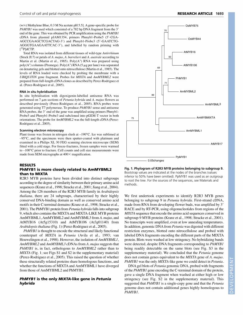
RESULTSPhMYB1 is more closely related to AmMYBML2than to MIXTAR2R3 MYB proteins have been divided into distinct subgroupsaccording to the degree of similarity between their primary amino acidsequences (Kranz et al., 1998; Stracke et al., 2001; Jiang et al., 2004).Among the 126 members of the R2R3 MYB family in Arabidopsisthaliana, there are 24 subgroups, characterised by their highlyconserved DNA-binding domain as well as conserved amino acidmotifs in their C-terminal domains (Kranz et al., 1998; Stracke et al.,2001). The PhMYB1 protein from Petunia hybrida falls into subgroup9, which also contains the MIXTA and MIXTA-LIKE MYB proteinsAmMYBML1, AmMYBML2 and AmMYBML3 from A. majus, andAtMYB16 (At5g15310) and AtMYB106 (At3g01140) fromArabidopsis thaliana (Fig. 1) (Perez-Rodriguez et al., 2005).
PhMYB1 is thought to encode the structural and likely functionalcounterpart of MIXTA in Petunia (Avila et al., 1993; vanHouwelingen et al., 1998). However, the isolation of AmMYBML1,AmMYBML2 and AmMYBML3 cDNAs from A. majus suggests thatPhMYB1 is, in fact, orthologous to AmMYBML2 rather than toMIXTA (Fig. 1; see Figs S1 and S2 in the supplementary material)(Perez-Rodriguez et al., 2005). This raised the question of whetherthese structurally related proteins share homologous functions, andwhether the functions of MIXTA and AmMYBML1 have divergedfrom those of AmMYBML2 and PhMYB1.
PhMYB1 is the only MIXTA-like gene in Petuniahybrida
We first undertook experiments to identify R2R3 MYB genesbelonging to subgroup 9 in Petunia hybrida. First-strand cDNA,made from RNA from developing flower buds, was amplified by 3�RACE and by RT-PCR, using oligonucleotides from regions of theMIXTA sequence that encode the amino acid sequences conserved insubgroup-9 MYB proteins (Kranz et al., 1998; Stracke et al., 2001).No transcripts were amplified, even at low annealing temperatures.In addition, genomic DNA from Petunia was digested with differentrestriction enzymes, blotted onto nitrocellulose and probed withlabeled DNA fragments encoding the different parts of the MIXTAprotein. Blots were washed at low stringency. No hybridizing bandswere detected, despite DNA fragments corresponding to PhMYB1being readily detectable on the same blots (see Fig. S3 in thesupplementary material). We concluded that the Petunia genomedoes not contain genes equivalent to the MIXTA gene of A. majus.PhMYB1 was the only MIXTA-like gene we could detect in Petunia.
DNA gel blots of Petunia genomic DNA, probed with fragmentsof the PhMYB1 gene encoding the C-terminal domain of the protein,gave a single DNA fragment when washed at either high or lowstringency (see Fig. S2 in the supplementary material). Thissuggested that PhMYB1 is a single-copy gene and that the Petuniagenome does not contain additional genes highly homologous toPhMYB1.
1693RESEARCH ARTICLEControl of cell and petal morphogenesis
OsMYB75
OsMYB44
AtMYB16
AtMYB106
PhMYB1
AmMYBML2
AmMYBML
AmMYBMIXTA
AmMYBML1
AtMYB17
PpMYB10.05 changes
75
93
69
71
99
75
3
Fig. 1. Phylogram of R2R3 MYB proteins belonging to subgroup 9.Bootstrap values are indicated at the nodes of the branches (valuesinferior to 50% have been omitted). PpMYB1 was used as an outgroupto root the tree. For the sources of the sequences, see Materials andmethods.

DEVELO
PMENT
1694
The Antirrhinum genome contains genes encoding threemembers of R2R3 MYB subgroup 9: AtMYB16, AtMYB17 andAtMYB106 (Kranz et al., 1998). AtMYB16 and AtMYB106 arestructurally very similar to PhMYB1, AmMYBML2 andAmMYBML3 (Fig. 1). No orthologue of MIXTA is encoded by theArabidopsis genome.
High-level expression of PhMYB1, AmMYBML2 andAtMYB16 modify the shape of tobacco epidermalcellsWe used high-level expression in tobacco plants as a bioassay todetermine whether the three most structurally similar genes insubgroup 9 from different plant species – PhMYB1 (Petuniahybrida), AmMYBML2 (A. majus) and AtMYB16 (Arabidopsisthaliana) – share similar functions, and to compare their functionswith that of MIXTA. Constructs with the cDNAs encodingAmMYBML2, PhMYB1 or AtMYB16, driven by the strongconstitutive double CaMV 35S promoter, were transformed intotobacco and the expression of each of the transgenes confirmed by
RNA blots. At least ten independent lines with comparable transcriptlevels in leaves were used for phenotypic characterisation. Tobaccoplants overexpressing AmMYBML2, PhMYB1 or AtMYB16 showedidentical phenotypes; changes induced by the activity of all threegenes were almost exclusively restricted to floral organs. Scanningelectron microscopy revealed that petal cells of transgenic tobaccoplants expressing any one of the three genes grew to a greater lengththan in wild type. In shape, modified cells of the inner petalepidermis resembled skittles rather than cones (Fig. 2A-D). Theactivity of each of the three genes also induced the formation ofoutgrowths on the cells of the carpel (Fig. 2E,F), and occasionallyoutgrowths were observed on the epidermal cells of leaves that wereborne on the inflorescence (see Fig. S4 in the supplementarymaterial). However, unlike MIXTA, which is able to induce changesin cell shape in all epidermal cells of aerial organs of tobacco(Glover et al., 1998), neither AmMYBML2, PhMYB1 nor AtMYB16induced the formation of outgrowths of epidermal cells on organsother than flowers and inflorescence leaves (see Fig. S4 in thesupplementary material). The ectopic outgrowths induced by35S:PhMYB1, 35S:AtMYB16 and 35S:AmMYBML2 were cleareston the carpel surface. The outgrowths never developed intomulticellular trichomes, unlike the response to high ectopicexpression of MIXTA or AmMYBML1 (see Fig. S5 in thesupplementary material) (Glover et al., 1998; Martin et al., 2002;Perez-Rodriguez et al., 2005). Freeze-fractured leaves and petalsalso showed that ectopic expression of these genes had no effect oncell layers other than the epidermis (not shown). These datasuggested that AmMYBML2, PhMYB1 and AtMYB16 arefunctionally homologous and are involved in the control of cellshape, but function in a manner distinct from that of MIXTA andAmMYBML1.
We also ectopically expressed AtMYB16 under the control of thedouble 35S promoter in Arabidopsis. Five independent transgeniclines were analysed. The inner epidermal cells of the petals grew toa greater length and changed from cone-shaped to bullet-shaped(Fig. 2G,H). No other changes in cell morphology in flowers wereobserved in these lines.
These data from high-level expression in tobacco demonstratedthat PhMYB1, AmMYBML2 and AtMYB16 have equivalentbiochemical functions. They can, in certain tissues, promote theformation of outgrowths, but their primary function seems to be topromote unidirectional cell expansion once an outgrowth has beeninitiated. However, developmental function, particularly that oftranscription factors, is also dependent on the cellular context inwhich proteins are active (Lee and Schiefelbein, 2001).
Expression patterns of PhMYB1, AmMYBML2 andAtMYB16The expression of the PhMYB1 gene in different organs of Petuniahas been reported by Avila et al. (Avila et al., 1993). The gene ishighly expressed in flowers, but is also expressed in sepals and at alow level in leaves. No expression was detected in roots. Weanalysed the timing of expression of the PhMYB1 gene, relative todifferent stages of flower development, by RNA gel blots. PhMYB1transcript levels peaked at stage 3 (Fig. 3A). In situ hybridisationshowed PhMYB1 to be expressed in both the inner and outerepidermal cells of the corolla limb (Fig. 3B).
For comparison, we analysed the expression of AmMYBML2 inAntirrhinum. AmMYBML2 was expressed relatively late in petaldevelopment, being maximally expressed in the corollas offlowers which had just opened. The peak of AmMYBML2expression was significantly later during petal development than
RESEARCH ARTICLE Development 134 (9)
Fig. 2. Effect of ectopic expression of AmMYBML2, AtMYB16 andPhMYB1 on epidermal cells. (A-D) SEM micrographs of conical innerepidermal cells of wild-type tobacco petal limb (A), and of tobaccopetals transformed with 2x35S::AmMYBML2 (B), 2x35S::PhMYB1 (C),or 2x35S::AtMYB16 (D). The activity of each of the three genes causesthe inner epidermal petal cells to grow to a greater length than thewild-type cells. (E,F) Micrographs of carpel epidermis of wild-typetobacco (E) and of cellular outgrowths on tobacco carpels transformedwith 2x35S::PhMYB1 (F); tobacco carpels overexpressing AmMYBML2or AtMYB16 have the same phenotype (not shown). (G,H) Micrographsof conical inner epidermal cells of wild-type Arabidopsis thaliana petals(G), and of petals transformed with 2x35S::AtMYB16 (H), where thecells grew to a greater length than in wild-type. Scale bars: 20 �m.

DEVELO
PMENT
the expression of MIXTA. AmMYBML2 was expressed in leaves,particularly leaves undergoing expansion growth and matureleaves. Expression was also detected in roots (Fig. 3C). In situhybridisation showed that AmMYBML2 was expressed at anapparently low level in both epidermal layers of the petals of A.majus (Fig. 3D).
The expression patterns of AtMYB16 and AtMYB106 wereestablished by consultation of the microarray data stored inGENEVESTIGATOR (Zimmermann et al., 2004b). Both AtMYB16and AtMYB106 are most highly expressed in flowers, particularlypetals, but expression of both genes was also detectable in leaves,particularly younger rosette leaves. These data confirmed previousfindings reported by Kranz et al. (Kranz et al., 1998) and Stracke etal. (Stracke et al., 2001). Together with the bioassay analyses thesedata showed that PhMYB1, AmMYBML2 and AtMYB16 have thesame biochemical functions and are expressed in the same tissues,implying that they probably play equivalent developmental roles.These proteins are functionally distinct from MIXTA in terms oftheir biochemical functions and expression patterns. This separationof function is also clear in Antirrhinum, where AmMYBML2 isexpressed in the petals of mixta mutants but cannot rescue theconical cell phenotype that is lost with loss of MIXTA activity (C.M.and M.P.-R., unpublished).
phmyb1 mutants have paler petals than wild typeTo determine the precise developmental functions of PhMYB1,AmMYBML2 and AtMYB16, we looked for mutations affecting thesegenes. Although we identified insertions in AtMYB16 in the ZIGGIAcollection of Antirrhinum insertion mutants, these had nodiscernable phenotype. This might have been due to the activity ofthe very closely related gene, AtMYB106, which is probablyfunctionally redundant with AtMYB16. Searches for insertionmutants of AmMYBML2 amongst the transposon-insertioncollection of A. majus were also unsuccessful. The phmyb1 mutantfrom Petunia hybrida is therefore the only mutant reported to datefor any of these orthologous MIXTA-like genes (van Houwelingenet al., 1998). To define in more detail the developmental role of
1695RESEARCH ARTICLEControl of cell and petal morphogenesis
Fig. 3. Comparison of expression patterns of PhMYB1 andAmMYBML2. (A) Expression of PhMYB1 during petal development ofPetunia hybrida. RNA gel blot analysis of total RNA from petals ofincreasing size (1, 0-1 cm; 2, 1-2 cm; 3, 2-3 cm; 4, 3-4 cm; 5, openflower). The RNA was probed with a fragment corresponding to the 3�end of the PhMYB1 gene. (B) In situ hybridisation of PhMYB1(antisense probe) in petals of Petunia between stages 2 and 3. Thecontrol shows hybridisation to the sense probe. Expression of PhMYB1was detected in both the inner and outer epidermis of the corolla lobe.(C) Expression of AmMYBML2 in A. majus. RNA gel blot analysis ofpoly(A+) RNA from petals of increasing size (1, 0-0.5 cm; 2, 0.5-1 cm;3, 1-1.5 cm; 4, 1.5-2 cm; 5, 2-2.5 cm; 6, 2.5-3 cm), leaves (y, young;me, medium; ma, mature) and roots, probed with the full-lengthAmMYBML2 cDNA. Petal RNA was also probed with MIXTA to showthat expression of AmMYBML2 peaks later than that of MIXTA. (D) Insitu hybridisation of AmMYBML2 in petals of A. majus at stage 4,showing weak expression in both the inner and outer epidermal celllayers. The control shows hybridisation to the sense probe. Scale bars:100 �m.
Fig. 4. Phenotype of the phmyb1 mutant from Petunia hybrida.(A) Petals show darker revertant sectors (white arrow) distinguishablefrom a paler background of mutant cells. (B) Micrographs of the innerepidermis of an unstable phmyb1 line showing a revertant sector (theboxed region in A). Mutant cells and wild-type revertant cells havedifferent shapes. Scale bar: 100 �m.

DEVELO
PMENT
1696
PhMYB1 in Petunia hybrida and to relate its activity to that ofMIXTA, AmMYBML1 and AmMYBML2 in A. majus, we analysed thechanges in cell shape caused by mutation of the PhMYB1 gene.
phmyb1 is a somatically unstable mutant of Petunia resultingfrom the insertion of an endogenous 284 bp transposable element,dTph1 (van Houwelingen et al., 1998), in the PhMYB1 gene. dTph1is able to undergo excision, both somatic and germinal, to generatetwo types of derivative allele; stable mutant alleles (when thetransposon leaves a footprint upon excision) and stable, wild-typerevertant alleles (where the excision event restores the wild-typesequence) (Gerats et al., 1990; Kroon et al., 1994). When derivativealleles are created in germinal cells, these events give rise to stablederivative alleles in the progeny.
The typical petal phenotype of the unstable phmyb1 mutantconsists of darker revertant sectors on a background of paler mutantcells (Fig. 4A). The size of the sectors varies depending on thetiming of excision of the dTph1 transposon. This petal phenotype isvery similar to that of the unstable mixta mutant of A. majus, wherethe difference in colour is due to a difference in shape of the cells ofthe petal epidermis (Fig. 4B) (Noda et al., 1994; van Houwelingenet al., 1998). No obvious phenotypes were seen in any tissues ofphmyb1 mutants other than in the petal epidermis.
dTph1 is inserted into the coding region ofPhMYB1The exact position of the transposon insertion in the PhMYB1 genewas determined by PCR of the mutant allele using genomic DNAfrom the phenotypically unstable line. Two PCR products wereobtained, differing in size by approximately 300 bp (Fig. 5A).Sequencing of the larger DNA fragment established that dTph1 wasinserted in the third exon of PhMYB1, a region encoding part of the
C-terminal domain that is conserved among members of R2R3MYB subgroup 9 (Kranz et al., 1998). In subgroup-9 MYB proteins,the C-terminal domain comprises two conserved motifs that are 10and 22 amino acids long; dTph1 was inserted in the region encodingthe second one, where it introduced a stop codon into the codingsequence (Fig. 5B). The smaller PCR band from genomic DNAcorresponded to the products of somatic reversion, as confirmed bysequencing.
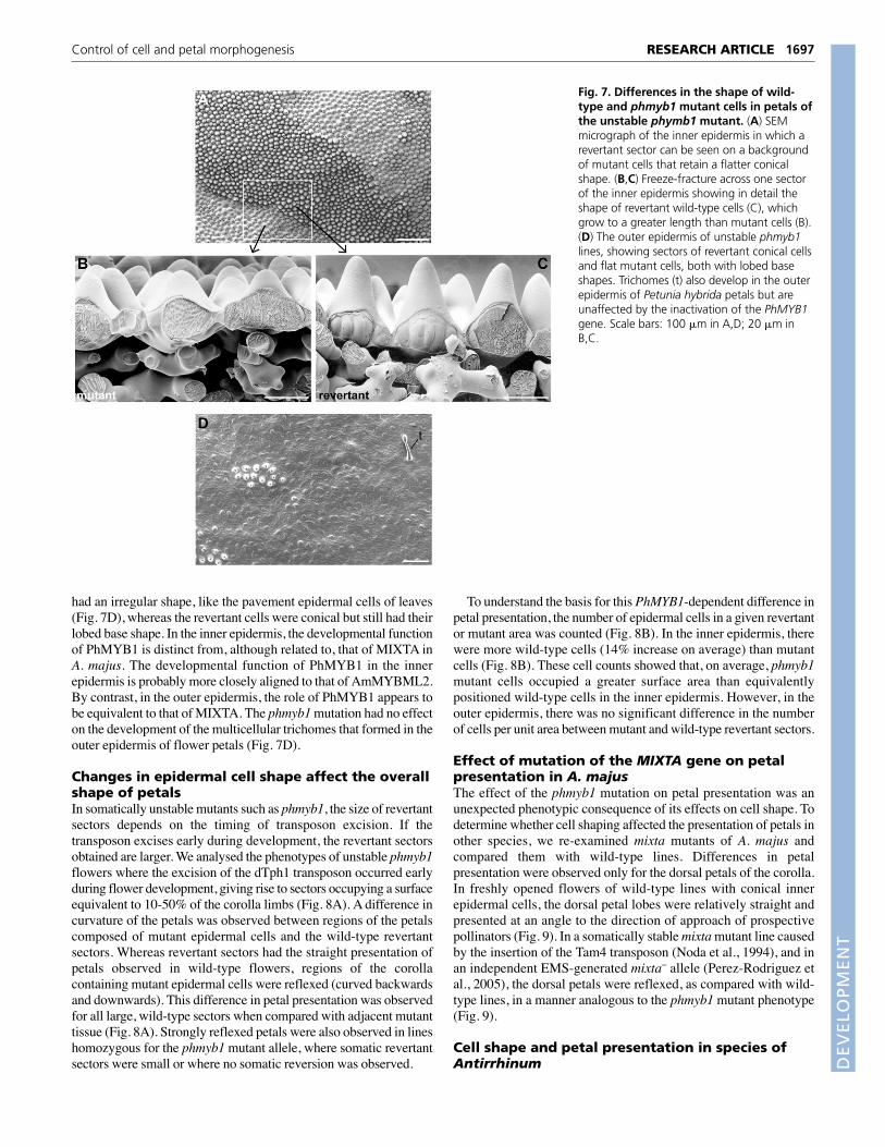
Changes in cell shape resulting from the phmyb1mutationDifferences in cell shape induced by the mutation in the PhMYB1locus were observed only in the flower petal epidermis. SEMmicrographs showed that cells of the inner epidermis of wild-typePetunia plants had a conical-papillate shape, with a pentagonal orhexagonal base, appearing very similar to those found inAntirrhinum (Fig. 6A,B). However, unlike Antirrhinum, the cells ofthe outer epidermis of the petals were conical, although they grewout less than those of the inner epidermis and were irregularlyshaped at their base (Fig. 6A,C).
The somatically unstable phmyb1 line of Petunia allowed us tocompare the cellular morphology of revertant wild-type sectors withthe background mutant cells, at exactly the same stage ofdevelopment and in a uniform genetic background. Fig. 7 showsSEM micrographs of the inner petal epidermis of the unstable line.phmyb1 mutant cells still developed as small cones, although theywere not as conical as wild-type revertant cells (Fig. 7A-C). Thiswas in contrast to the effect of MIXTA in Antirrhinum petals,because mixta mutant cells of the inner epidermis are flat (see Fig.S6 in the supplementary material) (Noda et al., 1994). In the outerepidermis of Petunia, phmyb1 mutant cells were completely flat and
RESEARCH ARTICLE Development 134 (9)
Fig. 5. The dTph1 transposon is inserted in the coding region ofthe PhMYB1 gene. (A) Agarose gel stained with ethidium bromideshowing the bands obtained by PCR amplification of the PhMYB1sequence from genomic DNA of six unstable phmyb1 lines. Thedifference in size between the lower and the upper bands correspondsto the size of dTph1. (B) Diagram of the phmyb1 allele showing theposition of the transposon insertion. Hatched boxes represent introns.The blue boxes correspond to the R2R3 MYB domain and the stippledbox corresponds to the conserved region in the C-terminal domain thatidentifies subgroup 9 within the R2R3 MYB gene family. This regioncomprises two conserved stretches of 10 and 22 amino acids; dTph1(purple box) is inserted in the second one, where it introduces a stopcodon into the phmyb1 open reading frame.
Fig. 6. Cell shapes in wild-type Petunia hybrida flower petals asseen by SEM. (A) A freeze-fracture across a wild-type petal showingthe presence of conical cells in both the inner/adaxial and outer/abaxialepidermis. ie, inner epidermis; m, mesophyll; oe, outer epidermis.(B) Inner epidermal cells of wild-type petals. (C) Cells of the outerepidermis. The base shapes of one cell in the inner and outer epidermallayers are outlined in pink. t, trichome. Scale bars: 50 �m.
DEVELO
PMENT
had an irregular shape, like the pavement epidermal cells of leaves(Fig. 7D), whereas the revertant cells were conical but still had theirlobed base shape. In the inner epidermis, the developmental functionof PhMYB1 is distinct from, although related to, that of MIXTA inA. majus. The developmental function of PhMYB1 in the innerepidermis is probably more closely aligned to that of AmMYBML2.By contrast, in the outer epidermis, the role of PhMYB1 appears tobe equivalent to that of MIXTA. The phmyb1 mutation had no effecton the development of the multicellular trichomes that formed in theouter epidermis of flower petals (Fig. 7D).
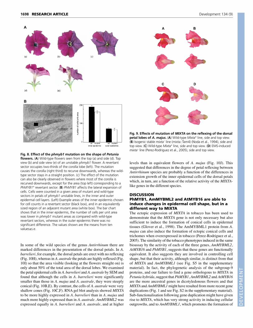
Changes in epidermal cell shape affect the overallshape of petalsIn somatically unstable mutants such as phmyb1, the size of revertantsectors depends on the timing of transposon excision. If thetransposon excises early during development, the revertant sectorsobtained are larger. We analysed the phenotypes of unstable phmyb1flowers where the excision of the dTph1 transposon occurred earlyduring flower development, giving rise to sectors occupying a surfaceequivalent to 10-50% of the corolla limbs (Fig. 8A). A difference incurvature of the petals was observed between regions of the petalscomposed of mutant epidermal cells and the wild-type revertantsectors. Whereas revertant sectors had the straight presentation ofpetals observed in wild-type flowers, regions of the corollacontaining mutant epidermal cells were reflexed (curved backwardsand downwards). This difference in petal presentation was observedfor all large, wild-type sectors when compared with adjacent mutanttissue (Fig. 8A). Strongly reflexed petals were also observed in lineshomozygous for the phmyb1 mutant allele, where somatic revertantsectors were small or where no somatic reversion was observed.
To understand the basis for this PhMYB1-dependent difference inpetal presentation, the number of epidermal cells in a given revertantor mutant area was counted (Fig. 8B). In the inner epidermis, therewere more wild-type cells (14% increase on average) than mutantcells (Fig. 8B). These cell counts showed that, on average, phmyb1mutant cells occupied a greater surface area than equivalentlypositioned wild-type cells in the inner epidermis. However, in theouter epidermis, there was no significant difference in the numberof cells per unit area between mutant and wild-type revertant sectors.
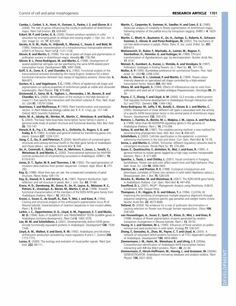
Effect of mutation of the MIXTA gene on petalpresentation in A. majusThe effect of the phmyb1 mutation on petal presentation was anunexpected phenotypic consequence of its effects on cell shape. Todetermine whether cell shaping affected the presentation of petals inother species, we re-examined mixta mutants of A. majus andcompared them with wild-type lines. Differences in petalpresentation were observed only for the dorsal petals of the corolla.In freshly opened flowers of wild-type lines with conical innerepidermal cells, the dorsal petal lobes were relatively straight andpresented at an angle to the direction of approach of prospectivepollinators (Fig. 9). In a somatically stable mixta mutant line causedby the insertion of the Tam4 transposon (Noda et al., 1994), and inan independent EMS-generated mixta– allele (Perez-Rodriguez etal., 2005), the dorsal petals were reflexed, as compared with wild-type lines, in a manner analogous to the phmyb1 mutant phenotype(Fig. 9).
Cell shape and petal presentation in species ofAntirrhinum
1697RESEARCH ARTICLEControl of cell and petal morphogenesis
Fig. 7. Differences in the shape of wild-type and phmyb1 mutant cells in petals ofthe unstable phymb1 mutant. (A) SEMmicrograph of the inner epidermis in which arevertant sector can be seen on a backgroundof mutant cells that retain a flatter conicalshape. (B,C) Freeze-fracture across one sectorof the inner epidermis showing in detail theshape of revertant wild-type cells (C), whichgrow to a greater length than mutant cells (B).(D) The outer epidermis of unstable phmyb1lines, showing sectors of revertant conical cellsand flat mutant cells, both with lobed baseshapes. Trichomes (t) also develop in the outerepidermis of Petunia hybrida petals but areunaffected by the inactivation of the PhMYB1gene. Scale bars: 100 �m in A,D; 20 �m inB,C.
DEVELO
PMENT
1698
In some of the wild species of the genus Antirrhinum there aremarked differences in the presentation of the dorsal petals. In A.barrelieri, for example, the dorsal petals are erect with no reflexing(Fig. 10H), whereas in A. australe the petals are highly reflexed (Fig.10I) so that the area visible (looking at the flowers straight on) isonly about 50% of the total area of the dorsal lobes. We examinedthe petal epidermal cells in A. barrelieri and A. australe by SEM andfound that although the cells in A. barrelieri were significantlysmaller than those in A. majus and A. australe, they were steeplyconical (Fig. 10B,E). By contrast, the cells of A. australe were veryshallow cones (Fig. 10C,F). RNA gel blot analysis showed MIXTAto be more highly expressed in A. barrelieri than in A. majus andmuch more highly expressed than in A. australe. AmMYBML2 wasexpressed equally in A. barrelieri and A. australe, and at higher
levels than in equivalent flowers of A. majus (Fig. 10J). Thissuggested that differences in the degree of petal reflexing betweenAntirrhinum species are probably a function of the differences inextension growth of the inner epidermal cells of the dorsal petalswhich, in turn, are a function of the relative activity of the MIXTA-like genes in the different species.
DISCUSSIONPhMYB1, AmMYBML2 and AtMYB16 are able toinduce changes in epidermal cell shape, but in adifferent way to MIXTAThe ectopic expression of MIXTA in tobacco has been used todemonstrate that the MIXTA gene is not only necessary but alsosufficient to induce the formation of conical cells in epidermaltissues (Glover et al., 1998). The AmMYBML1 protein from A.majus can also induce the formation of ectopic conical cells andtrichomes when overexpressed in tobacco (Perez-Rodriguez et al.,2005). The similarity of the tobacco phenotypes induced in the samebioassay by the activity of each of the three genes, AmMYBML2,AtMYB16 and PhMYB1, suggests that these genes are functionallyequivalent. It also suggests they are involved in controlling cellshape, but that their activity, although similar, is distinct from thatof MIXTA and AmMYBML1 (see Fig. S5 in the supplementarymaterial). In fact, the phylogenetic analysis of the subgroup-9proteins, and our failure to find a gene orthologous to MIXTA inPetunia hybrida, suggest that PhMYB1, AmMYBML2 and AtMYB16are the more ancestral genes in dicotyledonous flowers and thatMIXTA and AmMYBML1 might have resulted from more recent geneduplications (Fig. 1 and see Fig. S2 in the supplementary material).Sub-functionalisation following gene duplication might have givenrise to MIXTA, which has very strong activity in inducing cellularoutgrowths, and to AmMYBML1, which promotes the formation of
RESEARCH ARTICLE Development 134 (9)
Fig. 8. Effect of the phmyb1 mutation on the shape of Petuniaflowers. (A) Wild-type flowers seen from the top (a) and side (d). Topview (b) and side view (e) of an unstable phmyb1 flower. A revertantsector occupies two-thirds of the corolla lobe (left). The mutationcauses the corolla (right third) to recurve downwards, whereas the wild-type sector stays in a straight position. (c) The effect of the mutationcan also be clearly observed in flowers where most of the corolla isrecurved downwards, except for the area (top left) corresponding to aPhMYB1+ revertant sector. (B) PhMYB1 affects the lateral expansion ofcells. Cells were counted in a given area of mutant and wild-typesectors in petals of phmyb1 unstable lines, in the inner and outerepidermal cell layers. (Left) Example areas of the inner epidermis chosenfor cell counts in a revertant sector (black box), and in an equivalentlysized region of an adjacent mutant area (white box). The bar chartshows that in the inner epidermis, the number of cells per unit areawas lower in phmyb1 mutant areas as compared with wild-typerevertant sectors, whereas in the outer epidermis there was nosignificant difference. The values shown are the means from tenvalues±s.e.
Fig. 9. Effects of mutation of MIXTA on the reflexing of the dorsalpetal lobes of A. majus. (A) Wild-type Mixta+ line, side and top view.(B) Isogenic stable mixta– line (mixta::Tam4) (Noda et al., 1994), side andtop view. (C) Wild-type Mixta+ line, side and top view. (D) EMS-inducedmixta– line (Perez-Rodriguez et al., 2005), side and top view.

DEVELO
PMENT
trichomes and conical cells and tissue folding in the ventral petal ofthe corolla, a feature specific to the Scrophulariaceae (Perez-Rodriguez et al., 2005).
The spatial restriction of the ectopic expression phenotype ofAmMYBML2, AtMYB16 and PhMYB1 to floral organs and leaves ofthe inflorescence (despite the expression of the genes in all organs,driven by the double CaMV 35S promoter) suggests that, in order tobe active, these MIXTA-like proteins need different co-factors tothose requited by MIXTA, which induces outgrowths in all aerialepidermal tissues of tobacco (Glover et al., 1998) and Antirrhinum(Martin et al., 2001). Such co-factors might be present in epidermaltissues in a gradient, the maximum level being in flowers. In somecases, R2R3 MYB factors are known to interact with other proteins.For example, the C1 and Pl proteins of maize interact with bHLHproteins to induce the synthesis of anthocyanin (Goff et al., 1992;Lloyd et al., 1992), and the R2R3 MYB transcription factor GL1interacts with the bHLH proteins GL3 and EGL3 and the WD40-repeat-containing protein TTG1 to promote the formation oftrichomes in Antirrhinum leaves (Larkin et al., 1994; Payne et al.,2000; Schiefelbein, 2003; Zhang et al., 2003; Zimmermann et al.,2004a). However, the R2R3 MYB subgroup-9 proteins lack theconsensus signature motif for interaction with bHLH partners(Grotewold et al., 2000; Zimmermann et al., 2004a; Serna andMartin, 2006), so interacting proteins that contribute to thefunctional specificity of this group of proteins are unlikely to begroup IIIf bHLH co-factors (Heim et al., 2003; Zimmermann et al.,2004a). An alternative explanation for the phenotypic differencesbetween 2x35S::MIXTA and 2x35S::PhMYB1 is that, in the cellslacking a visible phenotype, other factors may negatively regulateresponses to the specific MIXTA-like genes.
PhMYB1 and MIXTA play related but distinct rolesin petal conical cell development
The phenotype of unstable phmyb1 mutant petals resembles thatof unstable mixta mutant lines: darker revertant sectors are visibleon a pale background of mutant cells. However, whereas themutation in the MIXTA gene results in flat inner epidermal cells(see Fig. S6 in the supplementary material), the inner epidermalcells in the phmyb1 mutant are still able to develop into cones (Fig.7B). The conical shape of phmyb1 mutant cells is shallower thanthat of wild-type cells in Petunia. This result reinforces the viewthat MIXTA and PhMYB1 are not functionally identical, andsuggests that the formation of fully developed conical cells in theinner epidermis of petals might require two distinct activities: one,conferred by MIXTA in Antirrhinum, might initiate the change ingrowth direction and direct the cells to grow in a polar manner,mainly along one axis; whereas the second activity, conferred byPhMYB1 in Petunia and AmMYBML2 in Antirrhinum, might beresponsible for a second phase of elongation that leads to theformation of a complete cone. In Antirrhinum, where the firstactivity is conferred by MIXTA, and the second by AmMYBML2,the relative activity of these two genes might determine the finalshape of the petal epidermal cells. In Petunia, the identity of thefirst activity is unknown; it could be another R2R3 MYB memberof subgroup 9, very similar to PhMYB1 (although we could findno molecular evidence for the existence of additional genesencoding subgroup-9 proteins in Petunia), or it might not be aMYB protein at all, but rather the result of a mechanism forpreparing cells for shape changes (A. Gouveia, PhD thesis,University of East Anglia, 2005).
Unlike the inner epidermal cells, mutant cells of the petal outerepidermis of the phmyb1 mutant of Petunia are flat. This suggeststhat the formation of cones in the outer epidermis of Petunia petalsis not controlled in exactly the same way as in the inner epidermalcell layer, as the formation of cones in the outer epidermis iscompletely dependent upon the activity of PhMYB1. Moreover, this
1699RESEARCH ARTICLEControl of cell and petal morphogenesis
Fig. 10. Comparison of petal reflexing, epidermal cell shape and the levels of MIXTA and AmMYBML2 transcripts in petals of recentlyopened buds of A. majus, A. barrelieri and A. australe. (A-C) Freeze-fracture SEM micrographs of sections across the mid-point of the leftdorsal petal of A. majus (A), A. barrelieri (B) and A. australe (C). The cells in A. barrelieri are more conical than in A. majus. The cells in A. australeare the flattest. (D-F) This is supported by surface SEM views of these cells in A. majus (D), A. barrelieri (E) and A. australe (F). Scale bars: 50 �m. (G-I) The degree of reflexing of the dorsal petals is small in A. majus (G), minimal in A. barrelieri (H) and very strong in A. australe (I). (J) RNA gel blotsof poly(A+) RNA from buds just prior to opening of A. majus (A.m), A. australe (A.a) and A. barrelieri (A.b), showing the relative expression ofMIXTA and AmMYBML2 in the different species of Antirrhinum. A cDNA fragment encoding ubiquitin was used to probe the RNA gel blots as aloading control. The ubiquitin transcript was polymorphic in A. majus (two bands). Only the larger transcript was detected in A. australe, whereas inA. barrelieri only the smaller transcript was detected.

DEVELO
PMENT
1700
shows that although PhMYB1 is not orthologous to MIXTA, itsfunction is closely related. Interestingly, the orthologous gene toPhMYB1, AmMYBML2, is unable to induce conical cell formationin the outer epidermis of the petals of Antirrhinum, even though it isexpressed in these cells. Perhaps it is not expressed at high enoughlevels to induce outgrowths of the outer epidermal cells.
The inner epidermal cell layer affects overallfloral architectureThe phmyb1 mutation not only influences the shape of single cells, butalso influences the overall design of the corolla, affecting the curvatureof the petals. This effect was seen very clearly upon comparing largesectors of revertant limb tissue with mutant tissue in the unstablephmyb1 mutant. This observation raises the important question of howcell shape might influence petal form and the degree of curvature ofpetals. Our data show that the curvature of petals is strongly influencedby the morphogenesis of the cells of the epidermis. An explanation forthe effects of mutation of PhMYB1 on the degree of petal curvature isthat when cones do not expand fully, the cells of the adaxial epidermisof the petals expand more periclinally than those on the abaxial side(Fig. 11). No phenotypic effect of loss of PhMYB1 function isobserved in cell layers other than the epidermis, suggesting that thepetal curvature results exclusively from changes in epidermal cellshape. Cell counts showed that in the inner epidermis of the phmyb1mutant, cells occupied a larger surface area than cells of wild-typepetals, but no difference was observed between wild-type and mutantcells of the outer epidermis. This suggests that the phmyb1 mutationresults in an increased lateral (periclinal) expansion of inner epidermalcells, probably as a result of loss of competing expansion in theoutward (anticlinal) direction, but these changes in cell expansionoperate only in the inner epidermis of the petal (Fig. 11). This couldresult in reflexing of the petals, such that they bend backwards anddownwards, and highlights the differential contribution of cellexpansion by each epidermal layer to overall petal design. A similareffect on petal presentation is observed in the dorsal lobes of mixtamutants of A. majus. As MIXTA is expressed only in the innerepidermal cell layer, the effects of this gene are restricted to this singlesheet of cells (Glover et al., 1998). Therefore, it is likely that the effectsof MIXTA on petal presentation result from similar effects on periclinalexpansion of the inner epidermal cells relative to the expansion of theouter epidermal cells, as suggested for PhMYB1 in Petunia.
Three species of Antirrhinum show different degrees of reflexingof their dorsal petal lobes and the shapes of their cells in the innerepidermis differ significantly. A. barrelieri has the most uprightdorsal petals and the most conical epidermal cells. A. majus has
more-reflexed dorsal petals and less-steep cones on its innerepidermis. The highly reflexed petals of A. australe have the flattestconical cells of the three species. The degree of steepness of thecones in the three species is inversely correlated with the degree ofpetal reflexing, but positively correlated with MIXTA transcriptlevels, emphasising the importance of epidermal cell shape indetermining petal form.
The effect of loss-of-function of R2R3 MYB subgroup-9 proteinson petal curvature is likely to have appreciable effects on theattractiveness of the flowers to prospective pollinators. The petalreflexing, observed in both the phmyb1 and the mixta mutants,causes an effective reduction in the diameter of the corolla, theparameter that is the principal visual signal identified by pollinatorsat a distance (Menzel et al., 1997). We estimate that the effect of thephmyb1 mutation is to reduce the apparent diameter of the corollaby at least 20%. This would affect the distance at which the floralsignal could be recognised. Extrapolation of empirical data for beessuggests the recognition distance might be reduced by as much as12 cm (Menzel et al., 1997). Reflexing of the petals also affects thedegree of colour saturation across the corolla (Eckert and Carter,2000). Varying domains of colour saturation might providepollinators with targeting information over shorter distances.Consequently, the effects of these mutations might extend tomodifying these short-range signals as well as affecting recognitionat distance through changing perceived corolla size. The shape ofthe petal epidermal cells also affects the perceived intensity of thecolour signal from the petals and its brightness as a result ofdifferences in the reflection and absorption of light by thesedifferently shaped cells (Gorton and Vogelmann, 1996; Kay, 1988;Kay et al., 1981; Noda et al., 1994). Field trials have shown thatflowers of A. majus with conical cells (Mixta+) are more attractiveto bees than flowers with flat epidermal cells (mixta–), particularlyunder conditions of low pollinator density (Comba et al., 2000;Glover and Martin, 1998). This might be due partly to thedifferences in the perceived colour intensity of the flowers, butpollinators showed preferences for flowers with conical epidermalcells even where no anthocyanin pigments were produced and theflowers were white. The explanation for this additional dimensionto the positive signal provided by conical petal cells might be theeffect of epidermal cell shaping on corolla reflexing and perceivedcorolla size. These aspects of petal design represent additionalparameters under the control of members of the MIXTA-like familyof MYB transcription factors that direct the morphogenesis of petalepidermal cells for their specialised functions in pollinator attraction.
K.B. and J.V. were supported by studentships from the John Innes Foundation.K.B. also received financial support from the Instituto Pasteur, FondazioneCenci Bolognetti, Università La Sapienza, Rome. M.P.-R. was supported by agrant from the BBSRC Cell Determination and Commitment Initiative(CAD05568) and by an award from the Spanish Ministerio de Educacion yCiencia under the programme Estancias en Centros Extranjeros de Profesoresde Universidad. D.B., P.B., K.R. and C.M. were supported by the Core StrategicGrant awarded by the BBSRC to the John Innes Centre.
Supplementary materialSupplementary material for this article is available athttp://dev.biologists.org/cgi/content/full/134/9/1691/DC1
ReferencesAshman, T. L. (2000). Pollinator selectivity and its implications for the evolution of
dioecy and sexual dimorphism. Ecology 81, 2577-2591.Ashman, T. L., Swetz, J. and Shivitz, S. (2000). Understanding the basis of
pollinator selectivity in sexually domorphic Fragaria virginiana. Oikos 90, 347-356.Avila, J., Nieto, C., Canas, L., Benito, M. J. and Paz-Ares, J. (1993). Petunia
hybrida genes related to the maize regulatory C1 gene and to animal mybproto-oncogenes. Plant J. 3, 553-562.
RESEARCH ARTICLE Development 134 (9)
Fig. 11. Model suggesting how competing directional growth ofthe inner epidermal cells of the petal might influence thepericlinal expansion of the inner epidermis and influence theoverall presentation of petals.
DEVELO
PMENT
Comba, I., Corbet, S. A., Hunt, H., Outram, S., Parker, J. S. and Glover, B. J.(2000). The role of genes influencing the corolla in pollination of Antirrhinummajus. Plant Cell Environ. 23, 639-647.
Eckert, M. P. and Carter, G. A. (2000). Flowers produce variations in colorsaturation by arranging petals at oblique and varying angles. J. Opt. Soc. Am. AOpt. Image Sci. Vis. 17, 825-830.
Gerats, A. G. M., Huits, H., Vrijlandt, E., Marana, C., Souer, E. and Beld, M.(1990). Molecular characterisation of a nonautonomous transposable element(dTph1) of Petunia. Plant Cell 2, 1121-1128.
Glover, B. and Martin, C. (1998). The role of petal cell shape and pigmentation inpollination success in Antirrhinum majus. Heredity 80, 778-784.
Glover, B. J., Perez-Rodriguez, M. and Martin, C. (1998). Development ofseveral epidermal cell types can be specified by the same MYB-related planttranscription factor. Development 125, 3497-3508.
Goff, S. A., Cone, K. C. and Chandler, V. L. (1992). Functional analysis of thetranscriptional activator encoded by the maize B gene: evidence for a directfunctional interaction between two classes of regulatory proteins. Genes Dev. 6,864-875.
Gorton, H. L. and Vogelmann, T. C. (1996). Effects of epidermal cell shape andpigmentation on optical properties of antirrhinum petals at visible and ultravioletwavelengths. Plant Physiol. 112, 879-888.
Grotewold, E., Sainz, M. B., Tagliani, L., Hernandez, J. M., Bowen, B. andChandler, V. L. (2000). Identification of the residues in the Myb domain ofmaize C1 that specify the interaction with the bHLH cofactor R. Proc. Natl. Acad.Sci. USA 97, 13579-13584.
Guerineau, F. and Mullineaux, P. (1993). Plant transformation and expressionvectors. In Plant Molecular Biology Labfax (ed. R. R. D. Croy), pp. 121-148.Oxford: BIOS Scientific Publishers.
Heim, M. A., Jakoby, M., Werber, M., Martin, C., Weisshaar, B. and Bailey, P.C. (2003). The basic helix-loop-helix transcription factor family in plants: agenome-wide study of protein structure and functional diversity. Mol. Biol. Evol.20, 735-747.
Horsch, R. B., Fry, J. E., Hoffmann, N. L., Eichholtz, D., Rogers, S. G. andFraley, R. T. (1985). A simple and general method for transferring genes intoplants. Science 227, 1229-1231.
Jiang, C. Z., Gu, X. and Peterson, T. (2004). Identification of conserved genestructures and carboxy-terminal motifs in the Myb gene family of Arabidopsisand Oryza sativa L. ssp indica. Genome Biol. 5, R46.
Jin, H., Cominelli, E., Bailey, P., Parr, A., Mehrtens, F., Jones, J., Tonelli, C.,Weisshaar, B. and Martin, C. (2000). Transcriptional repression by AtMYB4controls production of UV-protecting sunscreens in Arabidopsis. EMBO J. 19,6150-6161.
Jones, D. T., Taylor, W. R. and Thornton, J. M. (1992). The rapid generation ofmutation data matrices from protein sequences. Comput. Appl. Biosci. 8, 275-282.
Kay, Q. (1988). More than eye can see: the unexpected complexity of petalstructure. Plants Today 109-114.
Kay, Q., Daoud, H. S. and Stirton, C. H. (1981). Pigment distribution, lightreflection and cell structure in petals. Bot. J. Linn. Soc. 83, 57-84.
Kranz, H. D., Denekamp, M., Greco, R., Jin, H., Leyva, A., Meissner, R. C.,Petroni, K., Urzainqui, A., Bevan, M., Martin, C. et al. (1998). Towardsfunctional characterisation of the members of the R2R3-MYB gene family fromArabidopsis thaliana. Plant J. 16, 263-276.
Kroon, J., Souer, E., de Graaff, A., Xue, Y., Mol, J. and Koes, R. (1994).Cloning and structural analysis of the anthocyanin pigmentation locus Rt ofPetunia hybrida: characterisation of insertion sequences in two mutant alleles.Plant J. 5, 69-80.
Larkin, J. C., Oppenheimer, D. G., Lloyd, A. M., Paparozzi, E. T. and Marks,M. D. (1994). Roles of GLABROUS1 and TRANSPARENT TESTA GLABRA genes inArabidopsis trichome development. Plant Cell 6, 1065-1076.
Lee, M. M. and Schiefelbein, J. (2001). Developmentally distinct MYB genesencode functionally equivalent proteins in Arabidopsis. Development 128, 1539-1546.
Lloyd, A. M., Walbot, V. and Davis, R. W. (1992). Arabidopsis and Nicotianaanthocyanin production activated by maize regulators R and C1. Science 258,1773-1775.
Lunau, K. (2000). The ecology and evolution of visual pollen signals. Plant Syst.Evol. 222, 89-111.
Martin, C., Carpenter, R., Sommer, H., Saedler, H. and Coen, E. S. (1985).Molecular analysis of instability in flower pigmentation of Antirrhinum majus,following isolation of the pallida locus by transposon tagging. EMBO J. 4, 1625-1630.
Martin, C., Bhatt, K., Baumann, K., Jin, H., Zachgo, S., Roberts, K., Schwarz-Sommer, Z., Glover, B. and Perez-Rodrigues, M. (2002). The mechanics ofcell fate determination in petals. Philos. Trans. R. Soc. Lond. B Biol. Sci. 357,809-813.
Mattanovich, D., Ruker, F., Machado, A., Laimer, M., Regner, F.,Steinkellner, H., Himmler, G. and Katinger, H. (1989). Efficienttransformation of Agrobacterium spp. by electroporation. Nucleic Acids Res.17, 6747.
Menzel, R., Gumbart, A., Kunze, J., Shmida, A. and Vorobyev, V. (1997).Pollinators’ strategies in finding flowers. Isr. J. Plant Sci. 45, 141-156.
Moller, A. P. (1995). Bumblebee preference for symmetrical flowers. Proc. Natl.Acad. Sci. USA 92, 2288-2292.
Noda, K., Glover, B. J., Linstead, P. and Martin, C. (1994). Flower colourintensity depends on specialized cell shape controlled by a Myb-relatedtranscription factor. Nature 369, 661-664.
Ohara, M. and Higashi, S. (1994). Effects of inflorescence size on visits frompollinators and seed set of Corydalis ambigua (Papaveraceae). Oecologia 98, 25-30.
Payne, C. T., Zhang, F. and Lloyd, A. M. (2000). GL3 encodes a bHLH proteinthat regulates trichome development in Arabidopsis through interaction withGL1 and TTG1. Genetics 156, 1349-1362.
Perez-Rodriguez, M., Jaffe, F. W., Butelli, E., Glover, B. J. and Martin, C.(2005). Development of three different cell types is associated with the activityof a specific MYB transcription factor in the ventral petal of Antirrhinum majusflowers. Development 132, 359-370.
Romero, I., Fuertes, A., Benito, M. J., Malpica, J. M., Leyva, A. and Paz-Ares,J. (1998). More than 80 R2R3MYB regulatory genes in the genome ofArabidopsis thaliana. Plant J. 14, 273-284.
Saitou, N. and Nei, M. (1987). The neighbor-joining method: a new method forreconstructing phylogenetic trees. Mol. Biol. Evol. 4, 406-425.
Schiefelbein, J. (2003). Cell-fate specification in the epidermis: a commonpatterning mechanism in the root and shoot. Curr. Opin. Plant Biol. 6, 74-78.
Serna, L. and Martin, C. (2006). Trichomes: different regulatory networks lead toconvergent structures. Trends Plant Sci. 11, 274-280.
Souer, E., Quattrocchio, F., deVetten, N., Mol, J. and Koes, R. (1995). Ageneral-method to isolate genes tagged by a high copy number transposableelement. Plant J. 7, 677-685.
Spaethe, J., Tautz, J. and Chittka, L. (2001). Visual constraints in foragingbumblebees: flower size and color affect search time and flight behavior. Proc.Natl. Acad. Sci. USA 98, 3898-3903.
Stanton, M. L. and Preston, R. E. (1988). Ecological consequences andphenotypic correlates of flower size variation in wild radish Raphanus sativus L.(Brassicaceae). Am. J. Bot. 75, 528-539.
Stracke, R., Werber, M. and Weisshaar, B. (2001). The R2R3-MYB gene familyin Arabidopsis thaliana. Curr. Opin. Plant Biol. 4, 447-456.
Swofford, D. L. (2001). PAUP*: Phylogenetic Analysis using Parsimony. 4.0b10.Sunderland, MA: Sinauer Press.
Thompson, J. D., Higgins, D. G. and Gibson, T. J. (1994). CLUSTAL W:improving the sensitivity of progressive multiple sequence alignment throughsequence weighting, positions-specific gap penalties and weight matrix choice.Nucleic Acids Res. 22, 4673-4680.
Totland, O. (2004). No evidence for a role of pollinator discrimination incausing selection on flower size through female reproduction. Oikos 106,558-564.
van Houwelingen, A., Souer, E., Spelt, K., Kloos, D., Mol, J. and Koes, R.(1998). Analysis of flower pigmentation mutants generated by randomtransposon mutagenesis in Petunia hybrida. Plant J. 13, 39-50.
Young, H. J. and Stanton, M. L. (1990). Influences of floral variation on pollenremoval and seed production in wild radish. Ecology 71, 536-547.
Zhang, F., Gonzalez, A., Zhao, M., Payne, C. T. and Lloyd, A. (2003). Anetwork of redundant bHLH proteins functions in all TTG1-dependent pathwaysof Arabidopsis. Development 130, 4859-4869.
Zimmermann, I. M., Heim, M., Weisshaar, B. and Uhrig, J. F. (2004a).Comprehensive identification of Arabidopsis MYB transcription factorsinteracting with R/B-like BHLH proteins. Plant J. 40, 22-34.
Zimmermann, P., Hirsch-Hoffmann, M., Hennig, L. and Gruissem, W. (2004b).GENEVESTIGATOR. Arabidopsis microarray database and analysis toolbox. PlantPhysiol. 136, 2621-2632.
1701RESEARCH ARTICLEControl of cell and petal morphogenesis