Embed Size (px)
Citation preview

CONTRIBUTIONS OF DISTINCT TRUNK SEGMENTS TO CONTROL OF
POSTURE AND REACHING DURING TYPICAL DEVELOPMENT
by
JAYA RACHWANI PARSHOTAM
A DISSERTATION
Presented to the Department of Human Physiology
and the Graduate School of the University of Oregon
in partial fulfillment of the requirements
for the degree of
Doctor of Philosophy
December 2014

ii
DISSERTATION APPROVAL PAGE
Student: Jaya Rachwani Parshotam
Title: Contributions of Distinct Trunk Segments to Control of Posture and Reaching
During Typical Development
This dissertation has been accepted and approved in partial fulfillment of the
requirements for the Doctor of Philosophy degree in the Department of Human
Physiology by:
Dr. Andrew Karduna Chairperson
Dr. Marjorie Woollacott Core Member
Dr. Anita Christie Core Member
Dr. Dare Baldwin Institutional Representative
and
J. Andrew Berglund Dean of the Graduate School
Original approval signatures are on file with the University of Oregon Graduate School.
Degree awarded December 2014.

iii
© 2014 Jaya Rachwani Parshotam

iv
DISSERTATION ABSTRACT
Jaya Rachwani Parshotam
Doctor of Philosophy
Department of Human Physiology
December 2014
Title: Contributions of Distinct Trunk Segments to Control of Posture and Reaching
During Typical Development
The relationship between the development of sitting postural control and of
reaching during infancy has not been addressed in detail. It has recently been shown that
trunk control develops starting with the head, then the upper trunk and subsequently the
lower/pelvic regions. However, previous studies on infant reaching evaluated infants
during supported supine or reclined sitting positions, failing to address the contributions
of distinct regions of the trunk to reaching.
This dissertation explores the relationship between the progression of trunk
control and reaching performance in healthy infants. The effects of stabilizing the upper
and lower regions of the trunk were assessed by providing vertical trunk fixation at two
levels of support (thoracic and pelvic). Documentation of postural and reaching
performance reflected how control of the free regions of the trunk modulated both
behaviors. First, kinematic data were collected in infants aged 4-6 months who were
grouped according to their sitting ability and extent of trunk control. Second, a
longitudinal study was implemented in which kinematic and electromyographic
recordings were collected bi-monthly from 2.5-8 months.

v
Results from the cross-sectional study showed that postural stability and reaching
kinematics of the two groups were similar when they received support at the thoracic
level but differed when the support was limited to the pelvic level. Infants who were able
to sit independently outperformed the infants who were unable to sit without help. These
data were further expanded with the results obtained from the longitudinal study,
showing that during the months prior to independent sitting, infant reaches were
impoverished and were associated with a lack of postural stability when provided with
pelvic, in comparison to thoracic, support. In addition, infants displayed inefficient
muscle patterns in response to the instability. Differences between levels of support were
not observed once infants acquired independent sitting.
Taken together, these results offer detailed measures of the progression of trunk
control and its relation to reaching. This raises important questions regarding whether this
more specific approach may create the foundation for evaluating and improving trunk
control in atypically developing populations.
This dissertation includes previously published and unpublished co-authored
material.

vi
CURRICULUM VITAE
NAME OF AUTHOR: Jaya Rachwani Parshotam
GRADUATE AND UNDERGRADUATE SCHOOLS ATTENDED:
University of Oregon, Eugene (US)
University of Salamanca, Salamanca (Spain)
Catholic University of San Antonio, Murcia (Spain)
International University of Catalunya, Barcelona (Spain)
University of Malaga, Malaga (Spain)
DEGREES AWARDED:
Doctor of Philosophy, Human Physiology, 2014, University of Oregon
Master of Neuroscience, 2010, University of Salamanca
Master of Neuro-Rehabilitation, 2009, Catholic University San Antonio Murcia
Postgraduate Specialization in Pediatric Physical Therapy, 2008, International
University of Catalunya
Bachelor Degree in Physical Therapy, 2007, University of Malaga
AREAS OF SPECIAL INTEREST:
Typical and Atypical Motor Control
Development of Postural Control
Pediatric Neuro-Rehabilitation
Neuroscience
Movement Science
PROFESSIONAL EXPERIENCE:
Graduate Research Fellow, Department of Human Physiology, University of
Oregon, September 2011 – June 2014
Graduate Teaching Fellow, Department of Human Physiology, University of
Oregon, January 2012 – March 2014
Pediatric and Neurologic Physical Therapy, Spain, 2008 - 2011
Physical Therapy in Traumatology/Orthopedics, Spain, 2007 - 2008
English to Spanish Translator in Official Courses, Spain, 2009 – 2011

vii
Delegate of Spanish Society of Pediatric Physical Therapy (SEFIP) of Andalucia,
Spain (2007-2011).
GRANTS, AWARDS, AND HONORS:
Eugene & Clarissa Evonuk Memorial Graduate Fellowship in Environmental
Physiology, College of Arts and Sciences, June 2014
Betty Foster McCue Scholarship, University of Oregon, Graduate School, June
2014.
Accesit Award of Investigation, Ilustrious Professional College of Physical
Therapists of Andalucia, December 2010
PUBLICATIONS:
Rachwani, J., Santamaria, V., Saavedra, S., & Woollacott, M. (under review). The
development of trunk control and its relation to reaching in infancy: a longitudinal study.
Santamaria, V., Rachwani, J., Saavedra, S., & Woollacott, M. (under review). The
impact of segmental trunk support on posture and reaching in children with moderate and
severe cerebral palsy: an electromyographic study.
Santamaria, V., Rachwani, J., Saavedra, S., & Woollacott, M. (under review). The
impact of segmental trunk support on posture and reaching in children with moderate and
severe cerebral palsy: a kinematic study.
Santamaria, V., Rachwani, J., Manselle, W., Saavedra, S., & Woollacott, M.
(under review).The impact of segmental trunk support on a reaching task while sitting.
Santamaria, V., & Rachwani, J. (2014). Literature review: XL scientific
document. Development of reach-to-grasp & related sensorimotor systems in typical
developing infants. Spanish Society of Pediatric Physical Therapy. [Barcelona].
Rachwani, J., Santamaria, V., Saavedra, SL., Wood, S., Porter, F., & Woollacott,
M. (2013). Segmental trunk control acquisition and reaching in typically developing
infants. Experimental Brain Research, 228(1), 131-139.
Santamaria, V., & Rachwani, J. (2010). Physiotherapy in haemophilic artropathy
of the knee. Cuestiones Fisioterapia, 39(1), 68-77.
Rachwani Parshotam, J., Santamaria Gonzalez, V.F, & De Ru, E. (2009).
Functional effects of strengthening programmes in spastic diplegia. Developmental
Medicine & Child Neurology, 51(Suppl. 5), 23.

viii
ACKNOWLEDGMENTS
“Tell me and I forget, teach me and I may remember, involve me and I learn” --Franklin
Although I recognize and acknowledge the support of many people for
completing this dissertation, my sincerest gratitude and appreciation is extended to my
advisor, Dr. Marjorie Woollacott. She not only guided me throughout my years as a
graduate student, but she also involved me actively in every aspect of the research.
Without her mentorship, enthusiasm and dedication I would not be where I am today.
I must thank all my committee members with whom I have worked over the last
two years and who have been integral to my dissertation, each one of them in their own
unique way. Dr. Andy Karduna has always supported me with his valuable advices and
continuous guidance. I thank Dr. Anita Christie for modeling great teaching and for
furthering my thinking about motor control. My gratitude is also addressed to Dr. Dare
Baldwin for her contributions and suggestions during the analysis of this project.
The conceptual framework of this project came from the work of Dr. Sandra
Saavedra. I thank her for her willingness to collaborate and offer her expertise in this
topic. I also owe an enormous thank you to Wayne Manselle who was instrumental in
helping me resolve technical and programming issues.
The journey would have not been possible without the support of my family. To
my parents, thank you for your inspiration to follow my dreams and to never give up. I
always knew you believed in me and would support me in every stage of my life. I am
blessed to have parents like you.
To pursue a doctoral degree, it takes tremendous courage and motivation. This
motivation came from the endless love and faith of my colleague, friend and husband,
ix
Victor Santamaria. He is the person with the ability to make me smile and enlighten my
darkest moments during my years as a doctoral student.
Above all, I thank God most sincerely for everything He has done for me and for
giving me the strength that I need for facing life’s challenges.
The studies presented in this dissertation were supported by the National Institutes
of Health Grant 1R01HD062745-01, Marjorie Woollacott, principal investigator. In
addition, I received support from the Betty Foster McCue Fellowship and from the
Eugene Evonuk Memorial Graduate Fellowship.

x
TABLE OF CONTENTS
Chapter Page
I. INTRODUCTION .................................................................................................... 1
Motor Control Theories ......................................................................................... 2
Motor Development in Infancy – the Conquest Over Gravity .............................. 5
Postural Control ..................................................................................................... 9
Research Justification: Rationale ........................................................................... 22
Gaps in the Literature............................................................................................. 25
Research Aims ....................................................................................................... 27
II. THEORETICAL CONCEPT AND GENERAL METHODOLOGIES.................. 30
Principle Design Paradigm .................................................................................... 30
Informed Consent................................................................................................... 31
Trunk Stabilizing Device for the Reaching Test.................................................... 32
Laboratory Measures of Posture and Reaching ..................................................... 32
Clinical Measures on Gross/Fine Motor Skills and Trunk Control ....................... 35
III. SEGMENTAL TRUNK CONTROL ACQUISITION AND REACHING IN
TYPICALLY DEVELOPING INFANTS ................................................................... 39
Introduction ............................................................................................................ 39
Methods.................................................................................................................. 44
Results .................................................................................................................... 50
Discussion .............................................................................................................. 54
Conclusions ............................................................................................................ 57
Bridging the First and Second Study ..................................................................... 58
xi
Chapter Page
IV. THE DEVELOPMENT OF TRUNK CONTROL AND ITS RELATION TO
REACHING: A LONGITUDINAL STUDY .............................................................. 60
Introduction ............................................................................................................ 60
Methods.................................................................................................................. 65
Results .................................................................................................................... 77
Discussion .............................................................................................................. 89
Conclusions ............................................................................................................ 99
V. FINAL CONCLUSIONS ........................................................................................ 101
General Summary of Findings ............................................................................... 101
Limitations of Research ......................................................................................... 103
Clinical Implications .............................................................................................. 105
Future Directions for Research .............................................................................. 106
APPENDIX: METHODOLOGY FOR HEARTBEAT SUBTRACTION .................. 108
REFERENCES CITED ................................................................................................ 111

xii
LIST OF FIGURES
Figure Page
1.1. Example of Infant Sitting ...................................................................................... 22
2.1. Representation of Linked Mechanical System. .................................................... 31
2.2. Example of Trunk Stabilizing Device................................................................... 33
3.1. Thoracic Level of Support .................................................................................... 51
3.2. Pelvic Level of Support ........................................................................................ 52
3.3. Group Effects across Levels of Support ............................................................... 53
4.1. Representation of Infant Chair .............................................................................. 68
4.2. SATCo Scores across Age .................................................................................... 78
4.3. Visual Representation of a Reach and Photographic Images ............................... 81
4.4. Kinematic Results ................................................................................................. 82
4.5. Paraspinal Activation Frequency .......................................................................... 84
4.6. Arm Activation Frequency ................................................................................... 85
4.7. Integrated EMG .................................................................................................... 87
4.8. Onset Latency ....................................................................................................... 88
4.9. Recruitment Order ................................................................................................ 89
6.1. Example of Heartbeat Subtraction ........................................................................ 110

xiii
LIST OF TABLES
Table Page
1.1. Motor Milestones across the First Year of Life .................................................... 5
3.1. Group Characteristics............................................................................................ 46
4.1. Average Clinical Assessment Scores of All Infants across Age ........................... 66
1
CHAPTER I
INTRODUCTION
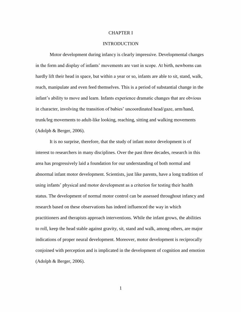
Motor development during infancy is clearly impressive. Developmental changes
in the form and display of infants’ movements are vast in scope. At birth, newborns can
hardly lift their head in space, but within a year or so, infants are able to sit, stand, walk,
reach, manipulate and even feed themselves. This is a period of substantial change in the
infant’s ability to move and learn. Infants experience dramatic changes that are obvious
in character, involving the transition of babies’ uncoordinated head/gaze, arm/hand,
trunk/leg movements to adult-like looking, reaching, sitting and walking movements
(Adolph & Berger, 2006).
It is no surprise, therefore, that the study of infant motor development is of
interest to researchers in many disciplines. Over the past three decades, research in this
area has progressively laid a foundation for our understanding of both normal and
abnormal infant motor development. Scientists, just like parents, have a long tradition of
using infants’ physical and motor development as a criterion for testing their health
status. The development of normal motor control can be assessed throughout infancy and
research based on these observations has indeed influenced the way in which
practitioners and therapists approach interventions. While the infant grows, the abilities
to roll, keep the head stable against gravity, sit, stand and walk, among others, are major
indications of proper neural development. Moreover, motor development is reciprocally
conjoined with perception and is implicated in the development of cognition and emotion
(Adolph & Berger, 2006).

2
One of the basic functional components of motor development is postural control
(Reed, 1989). In order for an infant to acquire the many motor skills that are
accomplished during the first year of life, a critical prerequisite is adequate postural
control. The infant’s ability to move in refined ways derives from their having learned
and mastered the underlying postural skills early in life. For example, it is known that
when children perform a simple voluntary task, such as reaching for a toy, they activate
postural muscles to support the body against the destabilizing effect of the movement
(Woollacott, Assaiante, & Amblard, 1996). Similarly, abnormalities in postural
development could constrain the child’s ability to perform such tasks.
Therefore, understanding normal motor development is mandatory in order to
understand the processes that are disrupted in abnormal motor development. In working
with children with abnormal motor development, it is essential to not only assess gross or
fine motor performance but also the basic postural skills, which are the foundation of
these movements. This has become a key issue in research over recent years and is the
focus of the current dissertation. The main goal of the current set of studies is to
understand the basic mechanisms of the development of trunk control and its relation to
reaching movements. The results will then be used in the clinical setting as normative
data for comparison with the trunk and reaching abilities of children who suffer from
developmental delays or neurological deficits.
MOTOR CONTROL THEORIES
In the middle of the 20th century, motor development was generally described as
the emergence of predetermined patterns of behavior, or motor milestones, which follow

3
an orderly sequence. Gesell and Amatruda (1947) noted that the general direction of
motor development follows a cranial-caudal (downward from head to feet) and proximal-
to-distal (outward from trunk to the hands and feet) sequence. Since then, several theories
of motor development have been formulated that try to relate neural structure and
behavior in developing infants. The classic theory, also known as the reflex-hierarchical
theory, places great importance on a reflex substrate for the emergence of mature human
behavior patterns. This means that in the normal child, the emergence of posture and
movement control is dependent on the appearance and subsequent integration of reflexes.
The appearance and inhibition of these reflexes reflect the increasing maturity of cortical
structures that inhibit and integrate reflexes controlled at lower levels within the central
nervous system into more functional postural and voluntary motor responses (Shumway-
Cook & Woollacott, 2012a). However, when researchers tried to examine the
development of reflexes and their association with motor development in infants, they
found that results were inconclusive and that other systems beyond the reflex circuits
contribute to the development of motor control. These conclusions then led to new
theories and concepts in motor control.
Neuronal Group Selection Theory
A widely known approach to motor development is the Neuronal Group Selection
Theory (NGST). This theory focuses on the fact that normal development is characterized
by variation. Motor development is defined as having specific phases of variability across
time. Sporns and Edelman (1993) were the pioneers of this theory and they explain that
the cortical and subcortical systems are dynamically organized into variable networks,
whose structure and function are selected by development and behavior. The NGST

4
states that development starts with a primary neuronal repertoire, which is determined by
evolution. Then, as a result of sensorimotor information elicited by behavior and
experience, development proceeds with the creation and establishment of neuronal
connections. This is known as the selection process, also known as the phase of primary
variability. When the selection process is achieved, variability of behavior is reduced. In
the secondary variability phase, variation increases as a result of continuing motor and
sensorial experiences, and continues until neuronal connectivity becomes more refined.
Thus, this theory clearly proposes that development is the result of a complex interaction
between the genetic information encoded in infants and their interaction with the
environment (Hadders-Algra, 2008).
Dynamic Systems Theory
A broader, current approach to motor development is the Dynamic Systems
Theory. This theory is based on the proposition posed by Nikolai Bernstein (Bernstein,
1967). He noted that many different solutions to a task are available due to the large
number of degrees of freedom that need to be controlled in the system. Therefore, to
simplify control, movements are activated by muscle synergies, which are functional
links of muscles that are activated as a pattern to accomplish a functional task. Dynamic
Systems Theory also states that the final outcome of a behavior is a result of (1) the
multiple neural and musculoskeletal subsystems or component parts that contribute to it,
such as muscle strength, body weight, postural support, the infant’s mood, and brain
development, and (2) the effect of environmental conditions and task requirements, that
influence the specific patterns of motor output. Thus, motor development is considered as

5
a self-organizing process where the environment plays an essential role in the maturation
of the motor system (Shumway-Cook & Woollacott, 2012b).
MOTOR DEVELOPMENT IN INFANCY – THE CONQUEST OVER GRAVITY
Infants acquire the main developmental motor milestones at specific points in
time (Table 1.1); however, the emergence of these motor abilities is characterized by
variation across infants, with contributions from the cultural context and conditions in
which they are raised (Adolph & Robinson, 2008; Piper & Darrah, 1994).
Table 1.1. Motor Milestones across the First Year of Life
Motor milestone Temporal window
Head control 3-4 months
Independent sitting 7-8 months
Pull-to-stand 9-10 months
Independent stance 10-14 months
Locomotion 12-15 months
Note: Obtained from Shumway-Cook & Woollacott, 2012a.
In addition, developmental phases may overlap as one is being refined while a
new stage emerges. For instance, infants learn to stand independently, and though this
skill continues to be refined, they move on to learn to walk, refining gait parameters as
well through further sensori-motor development and practice (Sutherland, Olshen,
Cooper, & Woo, 1980).

6
Though the acquisition of each motor milestone during the developmental
continuum has its own contribution to overall motor acquisition, among them,
independent sitting is critical. This milestone is acquired early in life and it allows
functional independence, the practice of psychosocial activities (e.g. play, work,
education and personal interactions) and the ability to perform manual skills that could
not be efficiently achieved in the lying position. Sitting posture requires the control of the
head and the trunk to offer a stable and relatively large base of support that can serve as a
secure basis when performing daily activities, such as reaching.
Head Control
Newborns have insufficient strength in the muscles of the neck to allow them to
resist gravity and hold the head upright. For instance, when they are lying in prone
position, they are able to quickly turn their heads from side to side to facilitate breathing
but cannot lift their head off the floor for a sustained period of time. But by 1-2 months of
age, they can then lift the head in prone position and by 3 months they have sufficient
control to maintain their head in midline while they use their arms for propping
themselves up. However, trunk balance is still weak, since infants at this age cannot shift
their weight from one arm to the other using their hands (Adolph & Berger, 2005).
Trunk Control
Being able to sit on a chair, unsupported and move the hands freely marks the end
of the progression toward independent sitting. Yet, infants take months to accomplish this
milestone. Due to their lack of muscle strength in the trunk and hips, infants continue to
fall forward while sitting with extended legs. There is a top-down order of progression

7
toward independent sitting which is clearly evident even to an untrained eye. As Adolph
& Berger (2005) mention:
The cephalocaudal progression seems especially striking in the development of
sitting, as if infants gain control of the sitting posture one vertebra at a time. At
first, infants’ heads flop when they are supported at the shoulders. Then, after
babies can balance their heads between their shoulders, their backs crumple when
they are supported at the hips. After babies can keep a straight back, they still
topple, chest to knees, without hip support. To sit alone, infants must have
muscular control over the entire trunk. (p. 238)
Infants at 5 months of age are able to prop sit, and are able to balance their body only
when they are supporting themselves on their arms. By 6 months, they are able to
independently sit with arms free but still cannot rotate their trunk. It is not until 7 months
of age that infants acquire sufficient lower trunk and hip control to turn while reaching
and also to transition from kneeling or crawling to sitting without falling between
postures (Adolph & Berger, 2005).
Reaching
The development of reaching depends on neurophysiological, biomechanical and
perceptual components. Infants need to lift their arm against gravity and have sufficient
strength in the trunk to maintain balance while reaching. However, they also need to
locate the object relative to the position of the hand for goal-directed reaching, which is
not required in spontaneous arm movements observed in newborns and very young
infants.

8
Goal-oriented reaching begins around the age of 3-5 months, before infants are
able to independently sit, but only if infants are placed in positions in which balance is
not a major constraint. In supported seated conditions, either with semi-reclined chairs or
with the support of a parent, infants are able to successfully reach for and contact a toy.
These reaches are less controlled, being more devious, and composed of several arm
movements, termed movement units (MUs) (von Hofsten, 1979). Each MU is composed
of an acceleration followed by a deceleration, usually accompanied by a change of
direction. Early reaches are characterized as having 4-5 MUs in contrast to 1 MU seen in
adult reaching. With practice and experience, infant reaches become straighter and
smoother, and have only 2 MUs. The first MU brings the hand close to the target,
whereas the second one prepares the hand to grasp the object (von Hofsten & Rönnqvist,
1993).
However, insufficient strength to stabilize the body is a major impediment to the
development of reaching (Konczak, Borutta, & Dichgans, 1997; Out, Van Soest,
Savelsbergh, & Hopkins, 1998). Reaching movements cause the body’s center of mass to
shift forward, and infants must compensate for such disequilibrium. Thus, goal-directed
reaching requires “whole body engagement” (Rochat & Goubet, 1995) and will have
different developmental arm trajectories depending on the initial position of the body
during the reach. For instance, infants at 4 months of age are able to successfully reach
toward a toy when in a supine or semi-reclined seated position but in prone position, they
cannot use their arms for reaching (Adolph & Berger, 2005).
Once infants achieve propped sitting, postural requirements compete with action
goals. For example, new sitters reach with only one hand and avoid leaning forward

9
because they need the supporting arm in order to not disrupt their fragile postural
equilibrium that keeps them from falling (Rochat & Goubet, 1995). Infants progress from
reaching with one hand while the other arm is used for balance support, to being able to
reach in all directions with both hands at 7 months of age. Thus, maintaining stability in
the sitting position is integral to the development of reaching. Studies have tested
reaching trajectories while experimentally mimicking the type of support infants will
eventually generate for themselves. With the extra trunk stabilization and hip support,
non-sitters’ reaching movements were as coordinated as those of sitting infants (Hopkins
& Rönnqvist, 2002; Rochat & Goubet, 1995). Therefore, additional postural control
enhances reaching performance, regardless of whether postural balance is acquired
naturally or with the use of an external device (Adolph & Berger, 2005).
POSTURAL CONTROL
Within models of motor control, actions are often subdivided, with certain
specific actions considered to be nested within other more global actions. For example,
visual tracking with the eyes is nested within visual tracking with the head, or grasping
with the hands is nested within reaching with the arms. All other motor actions - just like
looking and grasping - are embedded, in turn, within the most basic action of all: posture
(Bernstein, 1967; Gibson & Pick, 2000; Reed, 1989). Therefore, to understand the
emergence of any motor action in infancy, such as reaching while sitting upright, it is
crucial to understand the postural substrate for these skills (Shumway-Cook &
Woollacott, 2012a).

10
What Is Posture and How Is It Controlled?
Postural control is essentially defined as the ability to control the body’s position
in space for both stability (maintaining the projected center of mass within the limits of
base of support) and orientation (relative position of the body segments with respect to
one another and the environment or the task being performed); thus, postural control
emerges from the interaction of the individual, with the task and the environment. This
requires a complex interaction of musculoskeletal and neural systems (Shumway-Cook &
Woollacott, 2012c). At the level of the individual, postural control involves three
different types of tasks: 1) steady state balance, which is defined as the ability to maintain
the position of the center of mass within the base of support, 2) reactive balance, which is
defined as the ability to recover from a stable position of the center of mass following a
perturbation, and 3) proactive or anticipatory balance, which is defined as the ability to
activate the postural system in advance of a potentially destabilizing movement, to
minimize instability.
Most tasks include all three aspects of balance control, for example, reaching
while sitting. Upright sitting requires steady state, then anticipatory balance before
reaching, then reactive balance for fine balance adjustments at the end of the reach, and
then steady state balance again to maintain the limb against gravity (Shumway-Cook &
Woollacott, 2012c).
Traditionally, the primary contributors to the control of posture were considered
to be spinal reflexes and muscle tone (Peiper, 1963); however, over the last forty years,
this notion has been replaced by the understanding that postural control is an active
process involving a variety of different neural subsystems, including not only spinal

11
reflexes, but also brainstem, basal ganglia, and cerebellar pathways, as well as higher
level cortical systems and attentional resources. It is also well recognized that sensory
information from the visual, somatosensory and vestibular systems plays a critical role in
the maintenance of posture (Shumway-Cook & Woollacott, 2012c).
The Spinal Cord. Studies with humans who have spinal cord injuries have shown
that these patients have increased amounts of antigravity muscle tone but lack automatic
postural muscle responses below the level of the injured region. These results suggest that
spinal cord circuits are sufficient for maintaining antigravity support but not balance.
Balance control is thus a more complex process that requires the involvement of
supraspinal circuits (Macpherson & Fung, 1999).
The Brain Stem, Vestibular System and Cerebellum. Muscle synergies which are
necessary for automatic postural responses are organized in the brain stem, specifically in
the reticular formation. However, adaptation of postural synergies to changes in the
environment or task also requires the influence of the vestibular system and cerebellum.
The two major regions of the cerebellum that regulate orientation and balance are the
vestibulocerebellum (visual and vestibular inputs) and the spinocerebellum
(proprioceptive inputs from the body). Lesions in the brainstem and vestibulocerebellum
produce a variety of deficits in head and trunk control. Damage in the spinocerebellum
produces excessive postural sway, ataxia during walking and hypermetric postural
responses, suggesting its main role in balance reactions (Horak & Diener, 1994).
The Spinocerebellum and Basal Ganglia. Patients with spinocerebellar disorders
or basal ganglia deficits, like Parkinson disease, experience difficulties in adapting
postural responses to changing conditions. The spinocerebellum is responsible of

12
adjusting the magnitude of postural responses over the course of repeated trials but is also
able to rapidly adapt postural responses immediately after a change in condition. For
example when a healthy person balances on a platform whose movement velocity
increases with each trial, they have no problem adjusting to the changing velocities, and
remaining well balanced. However, a patient with a spinocerebellar disorder will not be
able to adapt to the perturbation velocity changes and shows muscle contractions that are
hypermetric for all velocities. On the contrary, a patient with Parkinson disease has
difficulty in changing postural responses when task conditions change; for instance, when
changing from standing upright to sitting on a stool. Postural responses to perturbations
in different conditions are inflexible and will be the same for either condition in a patient
with Parkinson disease (Horak, Nutt, & Nashner, 1992).
The Cerebral Cortex. Areas of the cerebral cortex are known to influence both
postural orientation and stability, including both anticipatory and automatic postural
reactions. The supplementary motor area is involved in anticipatory postural adjustments
that accompany voluntary movements. The temporoparietal cortex integrates sensory
information for perceiving body verticality. It is also known that the control of posture,
just like the control of any voluntary movement, requires attentional resources. In this
regard, the pre-frontal cortex is involved in the processing of visuospatial attention
(Mihara, Miyai, Hatakenaka, Kubota, & Sakoda, 2008). Research has demonstrated that
when subjects perform a cognitive task while actively maintaining posture, the
performance of either or both can degrade (Macpherson & Horak, 2013).
Sensory Information. Multiple sources of sensory information must be integrated
for an adequate response to changes in orientation and motion of the body. It is known

13
that somatosensory inputs are critical for maintaining balance during quiet stance.
Individuals with peripheral neuropathy in the legs accordingly experience ataxia and
difficulties with balance. The vestibular organs inform the nervous system about the
changes in body tilt with respect to gravity as well as body sway in all directions. In
addition, subjects with eyes closed have a substantial increase in body sway, indicating
that vision actively contributes to postural orientation (Brandt, Paulus, & Straube, 1986).
However, even though each sensory modality alone provides information about
postural orientation and body motion, their influence can change according to the task
requirements. For example, subjects on a firm, stable surface tend to rely primarily on
somatosensory information for postural orientation, but when the support is unstable,
subjects depend more on vestibular and visual information. Nevertheless, even when the
support surface is not stable, a light touch with a fingertip on a stable object is more
effective than using vision in maintaining balance. Thus, the postural control system is
able to change the relative weighting of different sensory modalities to accommodate
changes in the environment and goal of the task (Macpherson & Horak, 2013).
Development of Postural Control for Independent Sitting
The development of postural control has traditionally been associated with a
predictable sequence of motor skills, including crawling, sitting, creeping, pull-to-stand,
independent stance, and walking. However, for infants to develop trunk control, and thus
independently sit, they must learn to master the control of spontaneous background sway
of both the head and the trunk and to respond to perturbations of balance. This requires
the coordination of motor and sensory information relating the two body segments (head
and trunk) together in the control of posture. As noticed in the sections below, research

14
suggests that there are innate components of postural control, already available in the
newborn, and also emergent aspects of control, resulting from the infant interacting in a
dynamic way with the environment (Shumway-Cook & Woollacott, 2012a).
Motor Contributions to the Development of Independent Sitting
Research performed in the past on the development of sitting postural control has
focused on investigating the hypothesis that postural responses are innate, and follow two
phases of variability. In the first phase, directionally appropriate muscle responses are
noted at the age of 1 month and continue to increase until 6 months, prior to the
achievement of independent sitting. After 6 months, occurs the second phase, in which
amplitude and temporal ordering of muscle responses start to be refined and can be
attributed to the modification of neural circuitry by continuing sensory input (Hadders-
Algra, 2000). Under this theoretical viewpoint, corresponding to the neuronal group
selection theory, postural control is interpreted as being an aspect of behavior, governed
by genetic and environmental factors, which then progresses toward adult-like levels as
the nervous system matures.
Another viewpoint of the development of postural control is the one associated
with the dynamic systems theory. In this perspective, postural control derives from self-
organizing systems, and emerges as the organism interacts with the environment. Critical
features that are examined are called the “non-linear properties” of the system, in which a
behavior transforms into a new configuration when a single parameter of that behavior is
gradually altered and reaches a critical value. For example, as an animal walks faster,
there is a point at which it shifts from the walk to a trot (Shumway-Cook & Woollacott,

15
2012b). In the following section, sitting development will be discussed from both
perspectives.
Development of Sitting – Neuronal Group Selection Theory Perspective.
Hirschfeld and Forrsberg (Forssberg & Hirschfeld, 1994; Hirschfeld & Forssberg, 1994)
formulated a functional model describing the development of postural adjustments –
based on the concept that a central pattern generator (CPG) generates the basic pattern of
postural adjustments, which are then shaped by multisensory interactions from all
activated sensory systems. In general terms, CPGs involve neural networks that
coordinate the activity of the muscles, for the coordination of a variety of activities
including locomotion and respiration. These networks are controlled by reticulospinal
neurons, but segmental afferent inputs modulate and optimize the pattern (Hadders-Algra,
2005).
Therefore, similar to the concept of the two-level organization in the CPG-model
for motor activity (pattern generator plus sensory modulation), postural adjustments are
considered according to this model to have a first and second level or organization. The
first level involves the generation of direction-specific activity – this means the activation
of the muscles opposite to the direction of the body sway (for example, a perturbation
inducing a backward sway evokes responses in the ventral muscle). The second level
involves the fine-tuning of the postural response, which mainly relies on the multi-
sensorial afferent input from the visual, somatosensory and vestibular systems. Such
modulation can be accomplished in various ways, for example, by altering the degree of
activation, or by changing the recruitment order.

16
Research done in typically developing infants has shown that postural adjustments
are characterized by two phases of variability, primary and secondary, related to the first
and second level respectively, of postural organization described above (Hadders-Algra,
2000; Touwen, 1993). During the primary phase of variability, motor behavior is not
geared to external conditions, whereas in the phase of secondary variability, motor
performance is adapted to specific external situations. There are four periods of transition
that can be distinguished in the development of postural adjustments, occurring at the
ages of 3, 6, 9-10 and 13-14 months. Six months of age is probably the most important of
all, which is when infants shift from a primary to secondary phase of variability and
which coincides with the onset of independent sitting ability.
The research studies performed by Hedberg, Forssberg, and Hadders-Algra
(2004), and Hedberg, Carlberg, Forssberg, and Hadders-Algra (2005) were the first to
study postural adjustments in 1 month old infants. They used a paradigm in which
movement perturbations were generated while infants were seated on a platform. The
perturbation provoked a pelvic rotation. They found that 1 month old infants are able to
generate direction-specific postural adjustments to seated perturbations and therefore they
concluded that postural adjustments have an innate origin. These were highly variable,
especially in the number of postural muscles that were activated. The data indicated that
sensory information from the rotation of the pelvis was insufficient to trigger direction-
specific postural activity, since infants during forward perturbations often showed
direction specific postural activity in the absence of a pelvic tilt or body sway in the
opposite direction. Vestibular information did not serve as the primary trigger since head
swayed in all directions. Thus, the authors concluded that sensory information from

17
multiple sources of the pelvic region, such as proprioceptive and tactile information,
cooperate in producing postural activity. Authors found that the number of direction-
specific muscles that participated in these adjustments decreased with age, reaching its
lowest at 3 months, after which the number increased again. This observation suggests
that at 3 months of age, there is a period of developmental transition in postural control,
corresponding to the age at which goal-directed arm motility emerges.
From 3 to 6 months, infants continue to show a variable repertoire of direction-
specific adjustments and are not able to adapt postural responses to the specifics of the
situation – for example, to the degree of the perturbation or to changes in the position of
the infant (supine versus sitting) (van der Fits, Klip, van Eykern, & Hadders-Algra,
1999a). Then, once infants reach 6 months of age, directional specificity matures, and the
ability to adapt postural activity emerges. First, they develop the capacity to select a
complete pattern, in which all direction-specific muscles are activated (Hadders-Algra,
Brogren, & Forssberg, 1996; van der Fits, Otten, Klip, van Eykern, & Hadders-Algra,
1999b). Second, infants develop the capacity to adapt the selection of the complete
pattern to the degree of balance perturbation. For example, the complete pattern is more
frequently selected during sudden and vigorous perturbations of balance than by small
perturbations. Therefore, it is suggested that infants shift from the primary phase of
variability, in which postural muscles are activated without precise adaptation to the
environmental constraints, to the phase of secondary variability in which they learn to
adapt to the specifics of the situation. Six months is thus considered to be a significant
transition phase in development, which is also the age when infants generally learn to sit
independently (Piper & Darrah, 1994). This would suggest that the process of learning to

18
sit independently is not dependent on the precise ability to adapt postural muscle activity
to the specifics of the situation, as this does not begin to emerge until 6 months of age,
when sitting is already achieved. From a postural adjustment view point, the only
requirement for the development of independent sitting would be the ability to generate
direction-specific postural adjustments.
From 6 to 9-10 months of age infants continue to increase the ability to activate
the complete pattern for postural adjustments in response to perturbations of balance,
which as mentioned earlier, is especially used when the risk of losing balance is high.
This explains why the selection of the complete pattern is dominant during external
perturbations in a sitting position till the age of 30 months (van der Heide, Otten, van
Eykern, & Hadders-Algra, 2003) and during walking until 3 years (Assaiante, 1998).
From 9-10 months onwards, infants also learn to adapt postural adjustments in a more
refined way by means of 1) adapting the degree of contraction to changes in velocity of
the moving seat surface and 2) to adapt postural activity to changes in body
configuration. The emergence of the ability to fine tune postural activity to the specifics
of the situation suggest that the age of 9-10 months is regarded as the third transition
period, which also is the stage of preparation for the development of standing and
walking (Hadders-Algra, 2005).
Lastly, during the age of 13-14 months, anticipatory postural control in a sitting
position matures to adult timing characteristics (van der Fits et al., 1999b), and it is
known to be related to the development of independent walking, suggesting another
period of transition during which feed-forward mechanisms becomes embedded in the
control of posture (Hadders-Algra, 2005). However, examination of the emergence of
19
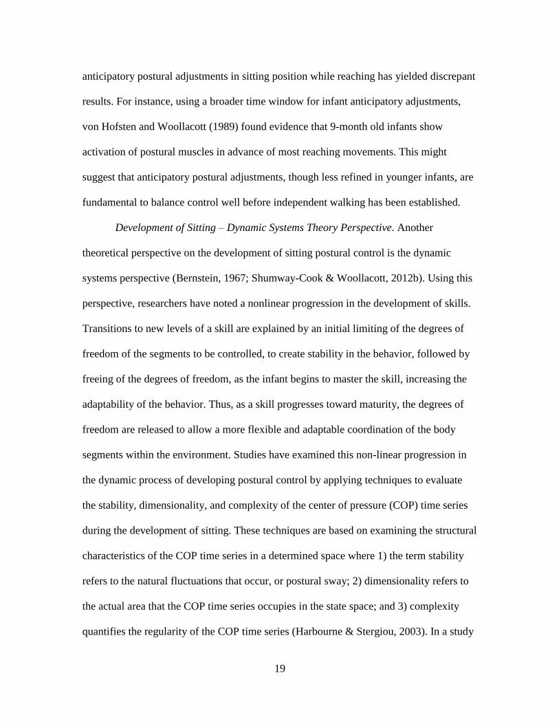
anticipatory postural adjustments in sitting position while reaching has yielded discrepant
results. For instance, using a broader time window for infant anticipatory adjustments,
von Hofsten and Woollacott (1989) found evidence that 9-month old infants show
activation of postural muscles in advance of most reaching movements. This might
suggest that anticipatory postural adjustments, though less refined in younger infants, are
fundamental to balance control well before independent walking has been established.
Development of Sitting – Dynamic Systems Theory Perspective. Another
theoretical perspective on the development of sitting postural control is the dynamic
systems perspective (Bernstein, 1967; Shumway-Cook & Woollacott, 2012b). Using this
perspective, researchers have noted a nonlinear progression in the development of skills.
Transitions to new levels of a skill are explained by an initial limiting of the degrees of
freedom of the segments to be controlled, to create stability in the behavior, followed by
freeing of the degrees of freedom, as the infant begins to master the skill, increasing the
adaptability of the behavior. Thus, as a skill progresses toward maturity, the degrees of
freedom are released to allow a more flexible and adaptable coordination of the body
segments within the environment. Studies have examined this non-linear progression in
the dynamic process of developing postural control by applying techniques to evaluate
the stability, dimensionality, and complexity of the center of pressure (COP) time series
during the development of sitting. These techniques are based on examining the structural
characteristics of the COP time series in a determined space where 1) the term stability
refers to the natural fluctuations that occur, or postural sway; 2) dimensionality refers to
the actual area that the COP time series occupies in the state space; and 3) complexity
quantifies the regularity of the COP time series (Harbourne & Stergiou, 2003). In a study

20
performed by Harbourne and Stergiou (2003), infants were tested during three
developmental stages: Stage 1, when infants could hold up their head and upper trunk,
but could not sit independently; Stage 2, when infants began to sit independently briefly;
and Stage 3, when infants could sit independently. While stability and regularity
increased over the three stages, the dimensionality followed a non-linear progression.
Information regarding the number of degrees of freedom, determined by the
dimensionality of COP time series, showed high values at Stage 1 with a significant
decrease at Stage 2, indicating a reduction in the degrees of freedom as is often seen
when attempting to learn a new skill (Woollacott et al., 1998). The significant increase
from Stage 2 to Stage 3 indicates an increment in the degrees of freedom, which provides
the infant with an increased adaptability or flexibility in controlling posture over the base
of support while sitting. With these results, it is suggested that the development of sitting
skills is softly assembled, with an initial strategy of freezing the degrees of freedom.
Infants first discover a solution to the problem of controlling the body segments while
upright sitting by stiffening the joints and reducing the degrees of freedom; then they
release the degrees of freedom to adaptively interact with the environment in a
coordinated way (Harbourne & Stergiou, 2003).
Thus, considering both perspectives, it is now evident that many factors, both
internal and external, guide the developmental process of independent sitting, in which
postural control is an essential requirement. Research has identified several variables that
influence the control of posture. One of those variables is the development of sensory
systems, in particular, the somatosensory, vestibular, and visual systems. Other variables
that have been investigated include neuromuscular development, muscle strength, body

21
mass, and the changing center of gravity through changes in body morphology (Piek,
2006).
Sensory Contributions to the Development of Independent Sitting
Research investigating the role of sensory systems during the development of
seated postural control has shown that infants appear to have a map of the relationship
between sensory inputs and muscle activity of the neck, trunk, and leg for sitting control.
Butterworth and Hicks (1977) investigated the role of vision in infants at different stages
of the development of independent sitting. Infants were given the illusion of a postural
perturbation (a moving-room paradigm, where the walls and ceiling moved, but the floor
did not). Infants with less sitting experience showed loss of balance in response to visual
stimulation, whereas infants with increased experience did not. This implies that infants
rely heavily on visual inputs for controlling sway when they are first learning to sit
independently, and this dependence decreases with age and sitting experience, as infants
rely more on somatosensory inputs. Woollacott, Debu and Mowatt (1987) used a
different protocol to study the impact of sensory inputs during the development of sitting.
They studied muscle patterns in the neck and trunk in response to platform perturbations
in seated infants with and without vision. They saw that the presence of visual stimuli did
not affect the muscle activation patterns in response to perturbations, concluding that
somatosensory and vestibular systems are capable of eliciting postural actions in isolation
of vision in infants first learning how to sit. To further study the extent to which
vestibular and visual inputs are necessary for sitting postural control, Hirschfeld and
Forrsberg (1994) performed experiments in which head orientation varied in seated
infants undergoing perturbations. They saw that coordinated muscle activity did not

22
change regardless of how the head was oriented, suggesting that postural responses to
perturbations are largely controlled by somatosensory inputs rather than vestibular or
visual stimulation.
RESEARCH JUSTIFICATION: RATIONALE
Regardless of the specific theory proposed to explain the development of postural
control in sitting, from a behavioral aspect, it is indisputable that infants gain control of
an increasing number of body segments as they develop the ability to independently sit.
Infants take approximately 3-4 months to transition from using their arm for support in
sitting (prop sitting) to independent sitting (Figure 1.1).
Figure 1.1. Example of Infant Sitting. Example of infant performing a) prop sitting at 3
months and b) independent sitting at 6 months (Obtained from Piper, Pinnell, Darrah,
Maguire, & Byrne, 1992).
This evidence suggests that infants first acquire control of the upper trunk region
(allowing prop sitting), followed by the lower trunk region (allowing independent
sitting), implying that there could be a segmental progression of control, as infants
gradually achieve full trunk control and consequently, are able to independently sit. This
a) 3 months b) 6 months

23
is further supported by the fact that the human spine is a multi-segmented structure that
requires control of the superficial and deep multifascicular trunk muscles to maintain
upright stability (Park, Tsao, Cresswella, & Hodges, 2014).
One of the main functions of the spine is to provide structural support and balance
to maintain an upright posture. The spine is a multi-segmented column with anatomically
distinct regions, cervical, thoracic, lumbar and sacral, each of which varies in structure,
movement and function. For instance, the vertebrae of the thoracic region have longer
spinous processes, which make the thoracic spine more stable than the cervical or lumbar
regions. On the other hand, the vertebrae of the lumbar region become bigger in size and
shape from L1 to L5, a design which allows them to carry most of the body’s weight
(Kapandji, 2008). Additionally, there is a transitional change in the morphology of the
spinal curvatures during development. Newborns present a complete physiological
kyphosis that evolves to a lordosis at the approximate age of 10 years. In this regard,
research has shown that the alignment of certain spinal segments, like the lumbar
segment for instance, with respect to the longitudinal axis of the vertebral column can
modulate the neuromuscular control of the spinal region (Park et al., 2014). Because of
all these variations in the segments that compose the vertebral column, assessments of
trunk function and stability should include the spinal segment to be targeted.
Panjabi (1992) was one of the first researchers to hypothesize mechanisms to
explain spinal stability. One of the basic biomechanical functions of the spine is to allow
movements between body parts. For this to happen, mechanical stability of the spine is
necessary. Panjabi proposed that the stabilizing system of the spine consists of three
subsystems: 1) the passive musculoskeletal subsystem, including vertebrae, facets,
24
articulations, intervertebral discs, spinal ligaments, joint capsules, as well as passive
components of the musculature; 2) the active musculoskeletal subsystem, consisting of
muscles and tendons surrounding the spine; and 3) the neural subsystem, including the
various forces and motion transducers, located in ligaments, tendons, muscles and neural
control centers (Panjabi, 1992). Though these three subsystems are theoretically different,
they are interdependent in function.
During normal function of the spine, the stabilizing systems work together to
control the instantaneously varying stability demands that are caused by changes in spinal
postural alignment. When there is a dysfunction of any of these three subsystems, the
neural subsystem responds to this, and consequently compensates by initiating
appropriate changes in the active subsystem. The neural subsystem has the complex task
of continuously monitoring and adjusting the forces surrounding the spinal column when
there are changes in posture, especially when this happens dynamically, since additional
considerations related to masses, inertias, and accelerations are involved (Panjabi, 1992).
Taking this into account, the coordination and balance control of the trunk
produced by the stabilizing system of the spine is absolutely crucial for upright human
tasks. While these biomechanical mechanisms for trunk postural control are evident in
healthy adults, they are not present at birth and are gradually mastered during the
development of sitting postural control.
It is proposed that the stabilizing system of the spine during development follows
a cranial-caudal progression, in accordance with the anatomically distinct regions of the
spine (cervical, thoracic, lumbar, and sacral regions). As infants master the ability to
stabilize the spine during static and dynamic changes in posture and across every region

25
of the spine, they achieve complete spinal postural control. This is the substrate for
complete trunk control and subsequent independent sitting in development.
GAPS IN THE LITERATURE
Though considerable research has been performed independently on both the
development of sitting postural control and the development of reaching, the relationship
between the maturational transition of reaching performance across early development
and its interrelation with the progressive development of postural control of the trunk has
not been thoroughly investigated.
As described earlier, postural control development appears to improve reaching
kinematics because reaching is associated with self-produced complex and internal
postural perturbations which change according to the infant's position and level of
stability (de Graaf-Peters, Bakker, Van Eykern, Otten, & Hadders-Algra, 2007; Hopkins
& Rönnqvist, 2002; Thelen & Spencer, 1998). These self-produced perturbations caused
by the reach must be compensated by preparatory postural adjustments to allow an
accurate reach to occur. Trunk control, which is the foundation of posture, is a critical
element for early reaching. Studies have demonstrated this by enabling the emergence of
reaching movements in new-born infants when given appropriate support of the entire
trunk (Amiel-Tison & Grenier, 1983; von Hofsten, 1982). This fact suggests that arm
muscular strength or control of the arm's biomechanics may be a less significant factor in
relationship to reaching efficiently once the trunk is supported (Konczak et al., 1997; Out
et al., 1998).

26
Recently, Saavedra, van Donkelaar and Woollacott (2012) examined the
segmental differences in trunk stability during the development of upright sitting and saw
that developmental changes in postural sway were unique to the region of the trunk that
was being tested. These results further refine the hypothesis regarding the progressive
development of segmental control during the development of the trunk for upright sitting.
However, it is surprising, considering that the development of reaching skills is
crucially dependent on the control of posture, that the relationship between them has not
been addressed in detail. Previous studies on sitting postural development and associated
reaching movements have considered the trunk as a single segment, and thus, failed to
address contributions of individual trunk regions to the development of postural stability
and reaching performance. Studies have dealt with the lack of trunk control in their
subjects by either using 1) supine or semi-reclined seating when infants are learning how
to sit, which alters the effect of gravity on the trunk and consequently influences the
performance of a reach (Thelen, Corbetta, & Spencer, 1996; Thelen & Spencer, 1998;
van der Fits et al., 1999a); or 2) evaluated the infant during fully supported or
unsupported conditions (de Graaf-Peters et al., 2007; Harbourne, Lobo, Karst, & Cole,
2013; van Balen, Dijkstra, & Hadders-Algra, 2012) and therefore failing to allow
observation of the progressive control of specific trunk regions during the acquisition of
upright sitting on reaching skills. Hence, the exact mechanisms by which typical infants
acquire upright sitting control and the impact on reaching development are unknown.

27
RESEARCH AIMS
Considering all the aforementioned information, the research described in this
dissertation challenges existing practices of modeling the trunk as a single segment for
evaluating sitting postural control and its relation to reaching in infancy. With the use of
an effective and practical method of securing the different segments of the trunk, the
infant’s ability to vertically align and stabilize the free segments while reaching were
investigated. These results will contribute to the first documentation of the processes
underlying the coordination of postural and reaching skills during the progressive
development of segmental trunk control.
The main goal of the studies included in this dissertation (Chapters III and IV)
was to test the contributions of the higher and lower segments of the trunk to postural and
reaching performance in typically developing infants, using an external support at
thoracic versus pelvic levels of the trunk, respectively.
Aim # 1: Determine whether or not there is an Effect of External Support on Posture
and Reaching in Typically Developing Infants Grouped According to their Extent of
Trunk Control.
A cross-sectional study was implemented to examine the effects of support
(thoracic and pelvic) on posture and reaching in 17 typically developing infants that were
grouped according to the extent of trunk control they had acquired. Group 1 infants were
unable to sit independently but demonstrated postural control in the thoracic region,
while Group 2 infants were independent sitters and demonstrated control in the thoracic
and lumbar regions. Kinematic data were used to compare postural and reaching
measures between groups, depending on the level of support provided.

28
It was hypothesized that with the use of an external support at thoracic level, all
infants would have equivalent postural and reaching skills, given that both groups
demonstrated postural control in the thoracic region. However, it was suggested that
when the external support was limited to the pelvic level, infants who had already
developed control of the lumbar region would have better performance and reaching
success. To confirm that the effect of support contributes to changes in posture and
reaching depending on the extent of trunk control infants have acquired, a follow-up,
longitudinal study outlined in the second aim was executed.
The cross-sectional study is described in Chapter III and includes previously
published, co-authored material. Victor Santamaria, Sandra L. Saavedra, Staci Wood,
Francine Porter, and Marjorie H. Woollacott are co-authors.
Aim # 2: Quantify the Effects of an External Support on Posture and Reaching across
the Progressive Development of Trunk Control in Typically Developing Infants from
2.5 to 8 Months of Age.
A longitudinal study was conducted evaluating the effect of support (thoracic and
pelvic) on posture and reaching in 10 typically developing infants from 2.5 months – 8
months. Behavioral, kinematic and electromyographic (EMG) recordings were compared
between levels of support to examine intra-individual changes and to gain a deeper
insight into the mechanisms underlying the progression of segmental trunk control
acquisition and its contributions to reaching skills. More specifically, the objectives were
to test the impact of external support across age on: reaching strategies, reaching/postural
kinematics, and EMG responses of postural and arm muscles, in terms of frequency of
activation, amplitude, latencies and recruitment order.

29
It was hypothesized that before the onset of independent sitting, infants would
demonstrate a decreased ability to reach, impoverished reaching and trunk kinematics and
inefficient postural muscle patterns when given pelvic in comparison to thoracic support.
All these observations would be explained by the challenges in remaining balanced with
pelvic support when infants have not yet acquired control of the lower trunk.
Subsequently, as infants learned to control the lower trunk and pelvic regions and thus,
acquired independent sitting, it was hypothesized that these effects would disappear and
infants would demonstrate invariable reaching and postural patterns irrespective of the
level at which they were supported. Hence, these results would confirm and further
expand previous findings showing that there is a cranio-caudal acquisition of trunk
control for independent sitting and that improvements in trunk control have direct
consequences on the development of reaching.
The longitudinal study is described in Chapter IV and includes unpublished, co-
authored material. Victor Santamaria, Sandra L. Saavedra and Marjorie H. Woollacott are
co-authors.

30
CHAPTER II
THEORETICAL CONCEPT AND GENERAL METHODOLOGIES
PRINCIPLE DESIGN PARADIGM
A new conceptual framework was used for evaluation of the development of
upright sitting and its contributions to reaching, in which the trunk is modeled as a multi-
segmented unit. The spine and head can be schematically represented as a physical
system consisting of a vertical column composed of blocks (vertebral segments) with the
top block (head) and wires (muscles) (Figure 2.1). They must exert adequate intrinsic
stiffness (steady-state) and reflexive muscle-tendon forces (reactive balance) on the
different vertebral subunits of the segments in order to program (anticipatory balance)
and carry out the optimal motor response in each situation. The physical structure
includes the vertebral segments: cervical, thoracic and lumbar vertebrae, connecting
tissues, ligaments and muscles. The number, shape and connections of the different
vertebrae determine the degrees of freedom to be controlled. Using this model, already
applied in previous investigations (Saavedra et al., 2012), we created an innovative way
to assess segmental trunk control by changing external levels of trunk support from a
high level of support (thoracic level) to a lower level of support (pelvic level), in order to
measure control of the thoracic and lumbar segments while reaching.
In this linked mechanical system, including the multi-segmented trunk, the forces
generated at any one segment during a dynamic task, like reaching, will also generate
passive forces on the other segments. It is known that a critical aspect of skilled
movement is the ability to stabilize the linked segments against motion-dependent forces
(Thelen & Spencer, 1998). In our experimental paradigm, the external support would be

31
holding the targeted trunk region stable while the infant was reaching toward an object.
Successful stabilization of the trunk segment/s over the support level requires activating
the proper muscles, at the proper time, with optimal strength and coordination in order to
resist the forces moving them away from the stability limits that are generated by the
reaching task.
Figure 2.1. Representation of Linked Mechanical System. Representation of the
linked mechanical system of the spine, including vertebral segments and connecting
structures. Black semicircles indicate external trunk supports at a) thoracic level and b)
pelvic level.
INFORMED CONSENT
The University of Oregon Institutional Review Board through Research
Compliance Services and the Committee for Protection of Human Subjects formally
approved the studies and protocols that compose this dissertation (Chapters III & IV).
Prior to all studies, all the procedures and risks were discussed with the family.
Additionally, a written informed consent was obtained from all parents prior to their
infant’s participation.
a) Thoracic support
b) Pelvic support

32
TRUNK STABILIZING DEVICE FOR THE REACHING TEST
Infants were seated on a bench with the pelvis firmly strapped in place. Two
straps were placed over the top of the thighs and one strap surrounded the posterior-
superior iliac spines so that the pelvis remained fixed in the vertical and horizontal planes
throughout the experiment. Straps were made of non-elastic, heavy bonded thread. A
rigid U-shaped, posterior support made of fiberglass that circled the trunk provided
upright stability of the trunk below the level of interest. The trunk support was raised or
lowered at specific levels of the trunk for evaluating the two main regions: 1) upper trunk
region (thoracic segment of the trunk), with the use of a support at thoracic level and 2)
upper and lower trunk region (thoracic and lumbar segment of the trunk), with the use of
a support at pelvic level. Figure 2.2 shows an infant in the trunk stabilizing device at
thoracic and pelvic levels of support. Placement accuracy and ability to limit trunk
movement below the level of support have been verified in laboratory tests. Once the
trunk was supported, the reaching test involved presenting the same colorful, circular
object at approximately the infant’s arm length in front of their sternum.
LABORATORY MEASURES OF POSTURE AND REACHING
Video Recording
Video recordings at 30 frames per second were obtained for further visualizing
and coding behavioral observations. A digital video camera was situated at the front
corner of the infant to provide a front view of the infant’s activity and to capture the body
and hand position as they reached toward the toy. The advantage of using video

33
Figure 2.2. Example of Trunk Stabilizing Device. Example of infant seated on a bench
with pelvic strapping and external trunk support device during the reaching test.
recordings for coding is to automate common analysis tasks and ensure accuracy in the
data. Doing research with infants can be very challenging in that one cannot instruct them
when to initiate the task of reaching. Therefore, with the use of video coding software,
the coder can clearly distinguish intentional reaches toward the toy. Another advantage to
the video coding software is that it temporally categorizes each event. With this, the
coder was able to visualize, frame by frame, and code what type of movement occurred at
what time and compare the sequence of events. Thus, every reach event had an onset and
offset time in milliseconds and was categorized depending on the type of reach.
Motion Tracking
Postural stability and reaching performance were measured using magnetic
tracking (miniBIRD system, Ascension Technology, Burlington, VT). The miniBIRD is a
six degrees-of-freedom measuring device that is used to measure the position and
orientation of a small sensor with respect to a transmitter, with an accuracy of 1mm and
1° (Acension Technology Coorporation, 2000). The transmitter was placed to the right
External trunk
support
Bench
Pelvic
straps

34
side of the infant’s head, at a 30 inch distance. The position and orientation of each
sensor were sampled at 84 Hz. A total of four sensors were used for providing
information regarding head, trunk and arm movements. Translational movements were
recorded with respect to the global reference axes: X (+ to the left), Y (+ pointing
forward) and Z (+ pointing downwards); as well as angular movements in the three
orthogonal planes: Azimuth, Elevation and Roll angles. There are several advantages to
using electromagnetic technology, especially when studying infant movement. First,
sensors can be embedded inside any material and still track position and orientation with
the same accuracy. This allowed us to attach the head sensor to a head-band for placing it
on the infant’s forehead, the trunk sensor with surgical tape for placing it on the infant’s
neck at the level of C7, and the arm sensors to neoprene wrist-bands for placing them on
the infant’s wrists. Second, in contrast to the use of cameras, a field of view is not
necessary for continuous tracking. This means that it has the ability to track through
people. With this advantage, the tester and if necessary, the parent were able to remain
close to the infant.
Surface Electromyography
In the longitudinal study, trunk and arm muscle activity were recorded using a 16-
channel surface electromyography system (MA300, Motion Lab Systems, Baton Rouge,
LA). Bipolar, self-adhesive surface electrodes with poles placed 2-3 cm apart were placed
bilaterally at the paraspinal muscles of the thoracic and lumbar segments of the trunk, as
well as at the belly of anterior deltoid, triceps and biceps muscles. One extra channel was
used for collection of the heart-beat for later subtraction of any heart-beat artifacts
embedded in the EMG signal (see Appendix for further information on heart-beat

35
subtraction method). Surface electrodes were attached to pre-amplifiers to increase the
magnitude of the signal.
The EMG system consisted of two units (a backpack and desktop unit) attached
with a connecting cable. Signals from the pre-amplifiers were digitized and processed
within the backpack and transmitted as digital information to the desktop unit. The
backpack had a gain setting for allowing complete control of the output signal and an
additional control to limit the maximum EMG frequency for avoiding the possibility of
recording signal aliasing errors. The EMG system also accommodated an optional
internal band-pass filter to ensure that the EMG signals produced did not exceed the
capabilities of the data collection system. Therefore, EMG signals were preamplified
(gain X 20), band-pass filtered (10-375Hz), and then further amplified at a sampling rate
of 1000Hz per channel. EMG data was timed-synched with position data with the use of a
trigger channel.
CLINICAL MEASURES ON GROSS/FINE MOTOR SKILLS AND TRUNK
CONTROL
In the study of motor development in infancy, the use of supplementary motor and
postural scales is recommended for further categorization of the motor behavior being
analyzed. These assessments assisted in the description of motor and postural progression
and most importantly in identifying the critical windows and onsets of relevant motor
abilities, such as head control and independent sitting.
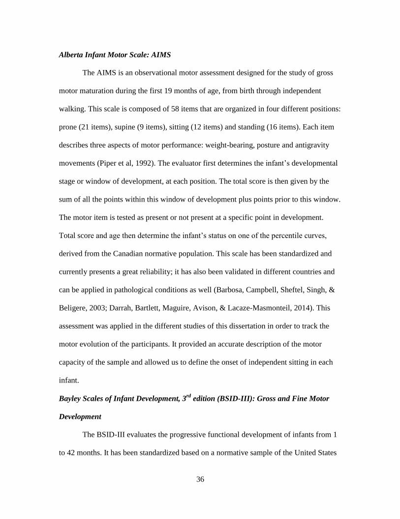
36
Alberta Infant Motor Scale: AIMS
The AIMS is an observational motor assessment designed for the study of gross
motor maturation during the first 19 months of age, from birth through independent
walking. This scale is composed of 58 items that are organized in four different positions:
prone (21 items), supine (9 items), sitting (12 items) and standing (16 items). Each item
describes three aspects of motor performance: weight-bearing, posture and antigravity
movements (Piper et al, 1992). The evaluator first determines the infant’s developmental
stage or window of development, at each position. The total score is then given by the
sum of all the points within this window of development plus points prior to this window.
The motor item is tested as present or not present at a specific point in development.
Total score and age then determine the infant’s status on one of the percentile curves,
derived from the Canadian normative population. This scale has been standardized and
currently presents a great reliability; it has also been validated in different countries and
can be applied in pathological conditions as well (Barbosa, Campbell, Sheftel, Singh, &
Beligere, 2003; Darrah, Bartlett, Maguire, Avison, & Lacaze-Masmonteil, 2014). This
assessment was applied in the different studies of this dissertation in order to track the
motor evolution of the participants. It provided an accurate description of the motor
capacity of the sample and allowed us to define the onset of independent sitting in each
infant.
Bayley Scales of Infant Development, 3rd
edition (BSID-III): Gross and Fine Motor
Development
The BSID-III evaluates the progressive functional development of infants from 1
to 42 months. It has been standardized based on a normative sample of the United States

37
and it is widely used in clinics and child healthcare research. It is divided into five major
developmental domains: cognitive, language, motor, social-emotional and adaptive
behavior. The items of the motor domain were the only ones considered in the
longitudinal study, consisting of 72 items for gross motor skills and 66 items for fine
motor skills. The overall score is given by summing all of the items for which the infant
is given credit, within the group of items that are specific to their age, and added to the
sum of the items from earlier months (Bayley, 2005).
Segmental Assessment of Trunk Control: SATCo
In infant development, assessment tools for postural control, such as the AIMS
and BSID-III, model and refer to the trunk as a non-dissociable unit, ignoring the fact that
the trunk is made up of multiple segments. These clinical tests are reliable and effective
in assessing limited aspects of functional balance while sitting and standing. For example
the AIMS evaluates simple steady state sitting (arms propped or arms free), scoring
sitting based on the amount of time the infant is able to sit securely. These scales do not
test trunk control in detail, and more specifically, the contributions of distinct trunk
regions to upright sitting balance. In addition, these assessments involve the use of other
anatomical structures, such as the upper and lower extremities in order to evaluate
dynamic trunk balance, failing to evaluate the neuromuscular coordination that must be
achieved to sit independently, including the coordination of sacral, lumbar, abdominal,
thoracic and cervical muscles used in maintaining equilibrium. In contrast, the SATCo is
a more complete analytical and specific assessment of sitting balance that increases the
accuracy of the initial assessment of trunk control.
38
The SATCo was originally designed for a population of children diagnosed with
Cerebral Palsy. The SATCo is now shown to be a reliable and valid clinical measure of
trunk control in both infants with typical development and children with neuro-motor
disability (Butler, Saavedra, Sofranac, Jarvis, & Woollacott, 2010).
The test consists in evaluating control of vertical trunk posture as the evaluator
progressively changes the manual support of the trunk from a high to a low level, with a
total of 8 different levels. This means that, as the support level is lowered, the number of
free segments increases and thus requires more intrinsic control of the trunk. For each
segmental trunk level, static, active and reactive control, are scored as present or absent.
The score is determined in relation to the specific level of the trunk (1 through 8) in
which the infant loses control in any of these three aspects (static, active or reactive).

39
CHAPTER III
SEGMENTAL TRUNK CONTROL ACQUISITION AND REACHING IN
TYPICALLY DEVELOPING INFANTS
This chapter is published in volume number 228 (1) of the journal Experimental
Brain Research in July 2013. Victor Santamaria, Sandra L. Saavedra, Staci Wood,
Francine Porter, and Marjorie H. Woollacott are co-authors. I performed the experimental
work and led the project; Marjorie H. Woollacott formulated the conceptual framework
with Sandy Saavedra, provided advice on data analysis and gave editorial assistance;
Victor Santamaria contributed to the recruitment, data acquisition, data analysis and
interpretation; Staci Wood and Francine Porter contributed to the recruitment and data
acquisition; Sandy Saavedra also helped develop the protocol and gave editorial
assistance. All co-authors formally approved this manuscript for submission.
INTRODUCTION
Postural control and reaching movements are two remarkable and complex motor
milestones that are acquired progressively during the first years of life and are
subsequently used throughout life in a variety of tasks (van der Heide et al., 2003).
Although the maturational process of these two functions is different and emerges at
various developmental stages during infancy, they are closely related to each other. It is
widely acknowledged that motor development is not only a result of neural maturation,
but is a dynamic process involving interaction between environmental constraints and
sensorimotor systems. Reaching for an object is usually accompanied by postural
adjustments prior to and during movement to provide mechanical stability and to
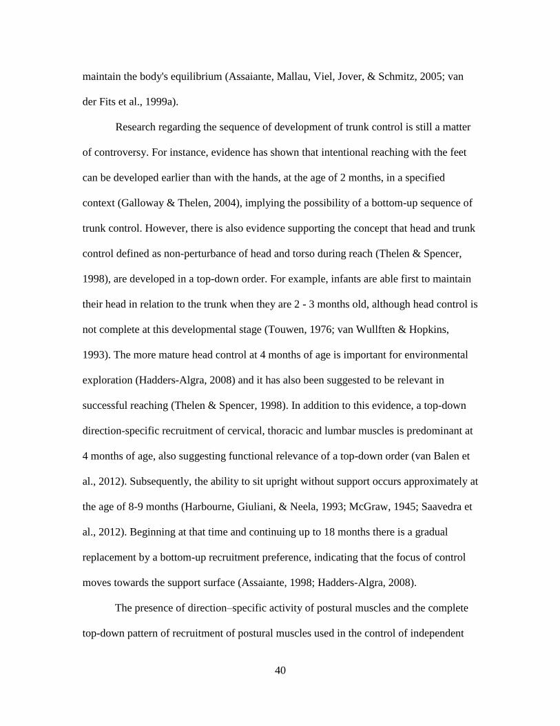
40
maintain the body's equilibrium (Assaiante, Mallau, Viel, Jover, & Schmitz, 2005; van
der Fits et al., 1999a).
Research regarding the sequence of development of trunk control is still a matter
of controversy. For instance, evidence has shown that intentional reaching with the feet
can be developed earlier than with the hands, at the age of 2 months, in a specified
context (Galloway & Thelen, 2004), implying the possibility of a bottom-up sequence of
trunk control. However, there is also evidence supporting the concept that head and trunk
control defined as non-perturbance of head and torso during reach (Thelen & Spencer,
1998), are developed in a top-down order. For example, infants are able first to maintain
their head in relation to the trunk when they are 2 - 3 months old, although head control is
not complete at this developmental stage (Touwen, 1976; van Wullften & Hopkins,
1993). The more mature head control at 4 months of age is important for environmental
exploration (Hadders-Algra, 2008) and it has also been suggested to be relevant in
successful reaching (Thelen & Spencer, 1998). In addition to this evidence, a top-down
direction-specific recruitment of cervical, thoracic and lumbar muscles is predominant at
4 months of age, also suggesting functional relevance of a top-down order (van Balen et
al., 2012). Subsequently, the ability to sit upright without support occurs approximately at
the age of 8-9 months (Harbourne, Giuliani, & Neela, 1993; McGraw, 1945; Saavedra et
al., 2012). Beginning at that time and continuing up to 18 months there is a gradual
replacement by a bottom-up recruitment preference, indicating that the focus of control
moves towards the support surface (Assaiante, 1998; Hadders-Algra, 2008).
The presence of direction–specific activity of postural muscles and the complete
top-down pattern of recruitment of postural muscles used in the control of independent

41
sitting is not a prerequisite for the emergence of reaching movements, although the
quality and success of reaching is associated with this recruitment (de Graaf-Peters et al.,
2007). Postural control development appears to improve reaching kinematics because
reaching is associated with self-produced complex and internal postural perturbations
which change according to the infant's position and level of stability (de Graaf-Peters et
al., 2007; Hopkins & Rönnqvist, 2002; Thelen & Spencer, 1998). These self-produced
perturbations caused by the reach must be compensated by preparatory postural
adjustments to allow an accurate reach to occur. Trunk control, which is the foundation of
posture, is a critical element for early reaching. Studies have demonstrated this by
enabling the emergence of reaching movements in newborn infants when given
appropriate support of the entire trunk (Grenier & Amiel-Tison, 1981; von Hofsten,
1982). This interesting fact suggests that arm muscular strength or control of the arm's
biomechanics may be a less significant factor in relationship to reaching efficiently once
the trunk is supported.
The ability to reach appears when infants are about 3 months old but reaches are
characterized by irregular trajectories and are unsuccessful in terms of grasping and
holding objects (van der Fits et al., 1999b). It is not until the age of 4-5 months that the
onset of functional reaching occurs (Gessell & Ames, 1947; von Hofsten, 1991). At this
age, full-term infants are able to grasp stationary and moving toys (Grönqvist, Strand
Brodd, & von Hofsten, 2011); infants aged 18 weeks can even grasp non-stationary toys
moving at 30cm/s (von Hofsten, 1980). At 4-5 months, successful reaches, defined as
including object contact, are characterized by large numbers of movement units (MUs)
and non-regular trajectories towards the object (Gessell & Ames, 1947). After the age of

42
6 months, the reaching sequence during which infants orient and direct their hand toward
a toy becomes straighter and shorter. Also, the movement is composed of fewer MUs (1-
2) and the first MU is differentiated by being longer in length and duration than the
second (Hopkins & Rönnqvist, 2002). At this time the kinematic parameters of a reach
start to assume an adult-like form in which straightness and smoothness are correlated;
fewer MUs are associated with a straighter trajectory of reaching, and peak velocity is
achieved at a greater percent of the reaching path (von Hofsten, 1991).
Previous studies on the development of reaching skills have been designed to test
muscular strength and control of arm mechanics of infants. These studies concluded that
insufficient muscular strength or insufficient control over the unstable arm does not
restrict early reaching and that movement becomes smoother as age increases (Konczak
et al., 1997; Out et al., 1998). Although this research has given insights into the motor
control of reaching, it has not addressed the issue of the infants’ need for trunk control as
a foundational element required for accurate reaching.
Though considerable research has been performed independently on both the
development of postural control and the development of reaching, the relationship
between the maturational transition of reaching kinematics and the progression of trunk
control acquired during early infancy has not been thoroughly investigated. Previous
studies have dealt with the lack of trunk control in 4 and 6 month old infants, by using
supine or semi-reclined seating, which alters the effect of gravity on the trunk and
subsequently influences the kinematics of reaching. In addition these studies evaluated
the trunk as a single segment, and therefore often designed protocols to observe infants
sitting in fully supported or unsupported states (de Graaf-Peters et al., 2007; Hopkins &

43
Rönnqvist, 2002; Thelen & Spencer, 1998), failing to allow observation of the
contribution of individual regions to trunk control and reaching. Addressing the
development of postural control from a multi-segment perspective is a novel technique
that has yet to be fully explored. We suggest keeping the effect of gravity constant by
using vertical alignment with higher versus lower levels of external trunk support to
allow more precise analysis of the effect of trunk control on reaching.
In summary, though previous research has given extensive insights into the
control of reaching development, it has not specifically addressed the contribution of
upper and lower regions of trunk control to reaching. This study will seek to fill this gap
through the use of two unique approaches. First we used vertical alignment with two
levels of external support (thoracic and pelvic) to test effects of regional support on
reaching in typically developing infants between the ages 4 and 6 months. Within this
temporal period sitting posture control emerges, and infants master their reaching and
grasping skills. Secondly, we classified our sample into two groups according to the
infant’s region of intrinsic trunk control as measured by the Segmental Assessment of
Trunk Control (SATCo) (Butler et al., 2010). Group 1 infants demonstrated postural
control in the thoracic region while Group 2 infants demonstrated control in the thoracic
and lumbar region. Kinematic parameters of visually guided reaches towards a toy were
examined as well as their success in grasping it. The hypothesis suggested was that with
the use of the external thoracic support all infants would have equivalent reaching
patterns and success since both groups demonstrated postural control in the thoracic
region. In addition, it suggested that when external support was provided at the pelvic

44
level, only the infants who already had developed control of the lumbar region would
have better reaching performance and success.
METHODS
Participants
Seventeen healthy infants born at term were recruited for this cross-sectional
study (9 males and 8 females). The infants were aged between 4 and 6 months. The
recruitment was carried out by using flyers in different child care centers in Eugene and
Springfield (Oregon, USA). This study was reviewed and accepted by the Institutional
Review Board for Human Subjects Research at the University of Oregon.
Materials and Procedure
Subjects were asked to come to the laboratory for one session of approximately 90
minutes. During this visit, infants were clinically tested with the SATCo to determine
their level of trunk control and the Alberta Infant Motor Scale (AIMS) to identify their
level of gross motor function. In addition, the parents were asked to respond to a health
questionnaire about their infants, were informed in detail about the experimental
procedure and signed the informed consent. All infants were video recorded during the
assessment. Table 3.1 shows the clinical characteristics for each group.
SATCo is a new clinical measure that allows a precise examination of balance
control of the trunk at various levels of support. It tests the infant’s trunk control as the
evaluator manually changes the level of trunk support from a high level of support at the
shoulder girdle to assess cervical (head) control, through support at the axillae (upper
thoracic control), inferior scapula (mid-thoracic control), lower ribs (lower thoracic

45
control), below ribs (upper lumbar control), pelvis (lower lumbar control), and finally, no
support, in order to measure full trunk control. It is designed to assess: 1. static control
(maintaining a neutral trunk posture) 2. active or anticipatory control (maintaining a
neutral posture during head movement) and 3. reactive control (maintaining or regaining
trunk control following a threat to balance, produced by a brisk nudge). The infant’s
ability to maintain or quickly regain a vertical position of the free region of the trunk in
all planes is assessed during static, active and reactive testing and scored accordingly as
present or absent. The score reflects the region where infants lose control of posture in; 1
= head, 2 = upper thoracic, 3 = mid-thoracic, 4 = lower thoracic, 5 = upper lumbar, 6 =
lower lumbar, 7 = pelvis, 8 = no loss of trunk control (Butler et al., 2010). Thus, for
example, an infant with SATCo score 4, loses control of posture in static, active or
reactive tests when the evaluator supports the lower thoracic region of the trunk (lower
ribs). However, an infant with SATCo score 6, does not lose control until the evaluator
supports the lower lumbar region of the trunk (the pelvis). In this study, we classified our
sample into two groups according to their SATCo score: Group 1 = infants with SATCo
scores 4 and 5 (demonstrating control in the thoracic region), Group 2 = infants with
SATCo scores 6 and 7 (demonstrating control in the thoracic and lumbar region). Other
tools such as the AIMS, inform us about the acquisition of infants' developmental gross
motor milestones from term age through independent walking (Piper et al., 1992). Both
of these tests follow a specific scoring criterion and have been shown to be valid, reliable
measures of developmental change in infants (Butler et al., 2010; Piper et al., 1992; van
Haastert, de Vries, Helders, & Jongmans, 2006). They thus can be used as clinical
measures of the developmental level of trunk control and motor function.

46
Table 3.1. Group Characteristics
n Sex Ratio SATCo AIMS Age Sitting
Ability
Group (Male:Female) M(min-max) M(min-max) M(min-max)
Group 1 8 (5:3) 4.50 (4 – 5) 18.25 (15 – 24) 4.50 (4 – 5) Non-sitters
Group 2 9 (3:6) 6.56 (6 – 7) 29.33 (23 – 41) 6.22 (5 – 6) Sitters
Note: M = mean.
The reaching test was conducted at pelvic and thoracic levels of support for every
infant. The support at the thoracic level was placed below the scapular girdle and the
pelvic level of support was around the pelvis, corresponding to middle thoracic level and
lower lumbar level of the SATCo, respectively. The design of the study was
counterbalanced, with half the infants first being provided with thoracic support, and half
first being provided with pelvic support, in order to eliminate fatigue or training effects as
confounding variables.
The reaching test was synchronized with the collection of kinematic data using the
Flock of Birds miniBIRD electromagnetic tracking sensors (Ascension Technology,
Burlington, VT). Four sensors were placed on the infant: one superficial to the styloid
process of the radius on each wrist, one on the posterior and prominent part of the
cervical vertebra 7 (C7), and one on a headband with the sensor centered on the forehead.
These sensors were used to track arm, trunk and head movements. Prior to starting the
reaching test, digitized position markers were taken of the left and right tragus, the
medial/lateral and anterior/posterior points of the external support (pelvic or thoracic).
This allowed us to estimate the location of the head center of mass (HCOM) using the
center of the distance between the midpoint of the two tragus markers and the head

47
sensor. The center of the base of support (BOS) was defined as the midpoint of the two
vectors created between the medial/lateral and anterior/posterior markers of the external
level of support.
The reaching test involved the infant being placed in a seated position on a bench.
The pelvis of the infant was secured to the bench with specially designed straps and
Velcro. Three straps were firmly attached to the underside of the bench: two of them
were used to wrap each hip joint and the third surrounded both posterior superior iliac
spines (Butler et al., 2010). An adjustable support device located behind the bench
provided trunk stability at one of the two levels studied. This device surrounded the
trunk, offering strong stability at the level being studied and below (Saavedra et al.,
2012). Once posture was stabilized, a colorful object was hung by the tester in front of
the infant’s sternum at approximately the arm’s length. The toy was presented 15 times
per level of support, but there were occasions in which this number had to be reduced due
to fussiness of the infant. If that was the case, the infant’s maximum number of trials was
noted and the rest was counted as missing data. In addition, the tester occasionally
presented a different toy (colored rings or blocks) in order to keep the infant engaged in
the task of reaching. The entire session was video recorded to ensure differentiation
between non-directed arm movements and reaching movements towards the toy during
the analysis.
All reaches were visually analyzed by two coders using computerized video-
coding software (www.openshapa.org) for further evaluation of the kinematic parameters.
This program allowed us to determine the onset and offset of every visually guided
intentional reach. A light emitting diode (LED), placed on the corner of the visual field,

48
was used to synchronize video and kinematic data during each reaching trial. With this,
we made sure that we were selecting reaches within the reaching test time. We defined
the onset of a reach as the moment when the infant initiated a movement of the upper
extremity towards the toy while looking at it. The offset of the reach was determined
when the infant touched the toy with the intention of grasping it. If an infant initiated a
reaching movement towards the toy and lost interest during the trajectory by stopping and
looking away, these reaches were not selected. Inter-rater reliability was validated by
having both coders evaluate 50% of the data and obtained a coefficient of agreement
above 0.85.
Reaches were coded as unimanual or bimanual. We defined bimanual reaches as
those in which we visually saw the infant touch the toy with both hands and which also
had an onset time difference between both arms of less than 1000 ms. Occasionally,
infants would begin unimanually and then switch to the other arm before reaching the
toy. In this case, for the kinematic data analysis, only one arm, considered as the
predominant arm, was selected. This selection was the same for the case of bimanual
reaches. The arm predominance was determined based on the hand that manipulated the
object once it was held. If infants used compensatory strategies like reaching with their
head or dragging the toy with their forearm, these were not considered.
Data Analysis
Data were filtered with a zero-lag fourth-order low-pass Butterworth filter with a
cut-off frequency of 6Hz, prior to calculating the kinematic variables. All unimanual or
bimanual reaches were pooled together and were analyzed at both thoracic and pelvic
levels for each group, to output kinematic data. Additionally, the total number of

49
successful and unsuccessful grasps of the toy was also counted. Kinematic data at each
level of support were analyzed using custom algorithms with the software Matlab (The
MathWorks, Inc., Boston, MA). We examined the following variables for each reach:
movement time, straightness score, MUs, reach path divided by the number of MUs,
angular head displacement, angular trunk displacement and percentage of successful
grasps.
Movement time was calculated in seconds as the time between the onset and the
end of the reach. Straightness score was calculated as the proportion between the actual
resultant trajectory of the reach and the minimum possible one, determined by a straight
line from start to the end of the reach. Using this method, values greater than one meant a
more devious arm movement (von Hofsten, 1991). A MU was defined according to
Grönqvist et al. (2011), as the portion of the arm movement between two velocity minima
with a velocity peak that should be greater than 2.3 cm/s. Also, if the difference between
the highest minima of one MU and the peak velocity of another MU was less than 8 cm/s
they were considered as one MU. Path length per MU was calculated by dividing the total
reach path by the number of MUs. In terms of postural control, the angular displacement
of the head and trunk were analyzed as two different segments to distinguish their
displacement during a reach. Head displacement and trunk displacement were calculated
as the total angular displacement during a reach in the anterior-posterior plane. For the
head displacement, the angle between the HCOM with respect to the C7 sensor was
applied and for the trunk displacement, the angle between the C7 sensor and the BOS was
used. This provided the angle of the trunk segment above the external support.

50
Statistical Analysis
The data analysis was carried out using SPSS 16.0 for Windows (SPSS Inc.,
Chicago, IL, USA). The total number of successful/unsuccessful grasps and the kinematic
parameters were computed by using the Linear Mixed procedure. This statistical
approach is more accurate when data are more unbalanced since it allows for a more
adequate modeling of the covariance structure and can deal with incomplete data.
Bayesian Criterion-type model was used to select the covariance structure with the best
fit; the structure exhibiting the smallest criteria values was considered the most desirable.
The model selected was the Scale Identity covariance for the repeated measure. Follow-
up pairwise comparisons based on the estimated marginal means were conducted to
analyze significant main or interaction effects applying the Bonferroni adjustment. For all
tests, the preset alpha level was .05.
RESULTS
Differences in Reaching Accuracy According to Level of Support
The graphs from Figures 3.1 and 3.2 are examples of a reach at the thoracic and
pelvic level of support of a Group 1 and a Group 2 infant, in addition to the photographic
image during the reach. The graphical representation of the arm trajectory shows how the
two infants showed a similar reaching position trajectory with thoracic support and both
infants seemed to look equally stable during the reach; however, the arm trajectories with
pelvic support show that the Group 1 infant had a more jerky reach than the Group 2
infant. A total of number of 293 reaches was analyzed. Figure 3.3 shows statistical results
for group effects at each level of support.
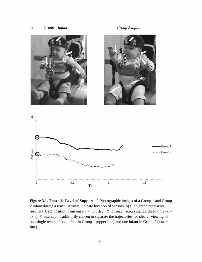
51
Group 1 infant Group 2 infant
Figure 3.1. Thoracic Level of Support. a) Photographic images of a Group 1 and Group
2 infant during a reach. Arrows indicate location of sensors. b) Line graph represents
resultant XYZ position from onset (○) to offset (X) of reach across standardized time (x –
axis). Y-intercept is arbitrarily chosen to separate the trajectories for clearer viewing of
one single reach of one infant in Group 1 (upper line) and one infant in Group 2 (lower
line).
X
X
Posi
tio
n
(cm
)
Time
(sec)
a)
b)

52
Group 1 infant Group 2 infant
Figure 3.2. Pelvic Level of Support. A) Photographic images of a Group 1 and Group 2
infant during a reach. Arrows indicate location of sensors. B) Line graph represents
resultant XYZ position from onset (○) to offset (X) of reach across standardized time (x –
axis). Y-intercept is arbitrarily chosen to separate the trajectories for clearer viewing of
one single reach of one infant in Group 1 (upper line) and one infant in Group 2 (lower
line).
X
X
Posi
tion
(cm
)
Time
(sec)
a)
b)

53
* **
**
*
*
*
Figure 3.3. Group Effects across Levels of
Support. Estimated group means for: movement
time (MT), straightness score (St.Sc.), movement
units (MU), path divided by number of MUs
(PL/MU), angular head displacement (Hd.Disp.),
angular trunk displacement (Trk.Displ.), and
percentage of successful grasps (%SG); at both
levels of support, Group 1 (dark gray bars) and
Group 2 (light gray bars).
Error bars, +/- 2 SE. * = p < .05, ** = p < .01.

54
Thoracic Support. None of the variables analyzed showed significant effects of
group for thoracic support. These results suggest that all infants were equally stable with
thoracic support and had similar kinematic parameters during the reaching sequence.
Pelvic Support. All variables except for percentage of successful grasps showed a
significant effect of group for pelvic support. Group 1 infants compared to Group 2
infants showed: greater movement time, F(1, 289) = 6.82, p < .05, a higher straightness
score, F(1, 289) = 24.90, p < .01, a higher number of MUs, F(1, 289) = 13.97, p < .01,
lesser path length per MU, F(1, 289) = 6.49, p < .05, greater head displacement, F (1,
289) = 4.42, p < .05 and greater trunk displacement, F(1, 289) = 4.07, p < .05.
Overall, these results show that when providing an external pelvic support,
stability, determined by head and trunk displacement, is better in infants who had
acquired control of their thoracic and lumbar region (SATCo scores 6 and 7); additionally
they showed straighter reaches, less MUs and covered greater distance per MU.
DISCUSSION
The primary purpose of this study was to more specifically investigate the
contribution of upper and lower regions of trunk control to reaching. We addressed this
by using vertical alignment at two levels of support (thoracic and pelvic) and by grouping
infants according to their region of intrinsic trunk control. In this manner, we confirm
previous studies by showing that the infants’ ability to control the trunk influences the
quality of reaching (Hopkins & Rönnqvist, 2002; Spencer & Thelen, 2000). In addition,
our results expand previous findings by providing evidence that depending on the
intrinsic control of the trunk region acquired, the level of external support has an impact

55
on the quality of reaching movements. In what follows, we discuss how reaching abilities
of two groups of infants who demonstrated control of different regions of the trunk
showed significant differences in their reaching patterns depending on whether they were
given thoracic vs. pelvic support.
We predicted that if infants were provided with thoracic support, the two groups
would demonstrate similar reaching behaviors and show similar patterns of control of
their stability, given that both groups had trunk control at the thoracic region, according
to their SATCo score. In contrast, we hypothesized that when provided with pelvic
support the two groups would behave differently due to the difference of the extent of
intrinsic trunk control they had developed. Our hypotheses was correct when comparing
Group 1 and 2 for movement time, straightness score, MUs, path length per MU, head
displacement and trunk displacement. However, percentage of successful grasps was the
same between Group 1 and 2 at both levels of support.
Previous research showed that 6 month old infants have quicker reaches,
straighter reaching trajectories, less MUs and increased path length per MU than 4 month
infants (de Graaf-Peters et al., 2007; Fallang, Saugstad, & Hadders-Algra, 2000; Thelen
& Spencer, 1998; von Hofsten, 1991). We expanded these findings by testing 4 to 6
month old infants in vertical alignment with external support at thoracic and pelvic levels
and demonstrated that differences in reaching kinematics depend on the infants’ region of
intrinsic control. Both groups had acquired control in the thoracic region and
demonstrated similar reaching patterns when given external support at the thoracic level.
This is consistent with other studies showing the development of head and upper torso
control as precursors for the emergence of successful reaching (Spencer & Thelen, 2000;

56
Thelen & Spencer, 1998). In contrast, Group 2 had also acquired control in the lumbar
region and demonstrated significantly better quality of reach than Group 1 when provided
external support at the pelvic level. Between Groups 1 and 2 there were many
differences, such as age, experience and gross motor development. However the fact that
differences between groups were only seen with pelvic support and not with thoracic
support indicates that region of intrinsic control achieved is the main factor contributing
to differences in quality of reaching observed in this study.
The variables related to postural stability during a reach corroborate those from
the clinical data from the SATCo and AIMS evaluation of the infants. Group 1 infants
had a mean SATCo score of 4.50 and were non-independent sitters, whereas Group 2
infants were independent sitters and had a mean SATCo score of 6.56. These results
correspond well with data from the previous literature, which suggest that the motor
strategies for independent sitting acquisition do not emerge until approximately 5 months
of age (Bayley, 1969; Gessell, 1946), and that until that point, muscle response synergies
underlying reaching movements are variable (Hadders-Algra et al., 1996; Hirschfeld &
Forssberg, 1994). In addition, the SATCo scores for each group show a trend similar to
the results found by Saavedra et al. (2012) regarding the segmental development of trunk
control, and further expand these results by showing how the region of trunk control
affects reaching parameters.
One specific item of the SATCo test is to analyze the active control of posture for
each support level, which is assessed by encouraging the infant to actively turn their head
to each side. This requires anticipatory postural adjustments to maintain the head and
trunk in the midline and this is precisely what is being challenged during a reach. Group

57
1 infants had increased displacement of the head and trunk with external pelvic support as
compared to Group 2, indicating excessive sway and thus loss of balance control. This
was expected since Group 1 infants did not have the ability to actively control their trunk
when manually supported at the lower lumbar or pelvic region during the SATCo
evaluation. On the contrary, all infants showed active trunk control with mid-thoracic
support and thus, kinematic parameters related to posture indicate that with external
thoracic support, the ability to maintain stability during a reach was the same for both
groups.
An unexpected result was the lack of difference in percentage of successful grasps
between Group 1 and 2 with pelvic support. However, our findings are consistent with
other studies showing that once onset of successful reaches appears, infants are equally
successful in a variety of testing positions. For example, the study conducted by Van der
Fits et al. (1999a) showed that 4-5 month old infants, when placed in three positions;
supine, semi-reclined and in an infant chair, were equally able to produce successful
reaches in all three testing positions. This suggests that once infants learn to successfully
reach for a toy, their further improvement of intrinsic trunk control contributes primarily
to increased quality of reaching.
CONCLUSIONS
Previous studies have shown that infants start reaching at 4 months of age before
acquiring full trunk control (Fallang, Saugstad, Grogaard, & Hadders-Algra, 2003; Out et
al., 1998; van der Fits et al., 1999a). It has also been shown that when given support both
newborns and other young infants initiate reaching movements towards objects (Grenier

58
& Amiel-Tison, 1981; von Hofsten, 1982). Studies that have investigated the effects of
trunk support in infants have not quantified either the amount of support provided or the
infant’s intrinsic level of trunk control. This study expands previous results by showing
for the first time that the specific region of trunk control influences reaching ability. More
precisely, the data suggest that reaching performance is tightly correlated with the
progressive segmental acquisition of trunk control. This raises important questions
regarding whether this correlation of reaching performance with trunk control is
comparable to that seen in infants with developmental delays and neurological disorders.
Similar information on children with neurological disorders could be used to implement
more efficient therapeutic strategies to enable reaching in daily life activities.
BRIDGING THE FIRST AND SECOND STUDY
In the cross-sectional study, we found that depending on the extent of trunk
control infants had acquired, the level of external support has an impact on postural
stability and reaching performance. With an external thoracic support, all infants had
equivalent postural and reaching patterns since both groups demonstrated control in the
thoracic region of the trunk. However, when the external support was limited to the
pelvic level, the group of infants that had already acquired control of the thoracic and
lumbar regions outperformed the infants in the group that had acquired control of the
thoracic region only, implying that they still had to learn and select those strategies that
were optimal for controlling the lumbar region. However, kinematic data do not furnish
detailed information on the strategies used by the nervous system to achieve the various
kinematic motion paths. Data on muscle recruitment characteristics provide this level of
59
information. Furthermore, cross-sectional studies have the limitation of providing
information from individual infants only at a specific point in time. Longitudinal studies
are able to detect development or changes across time and account for intra-individual
changes occurring within the developmental time frame of the study. Another advantage
of a longitudinal research design is that it eliminates cohort effects because one group of
infants is examined over time, rather than comparing different groups of different ages.
Therefore, the aim of the second study was to longitudinally examine behavioral
and kinematic changes in conjunction with electromyographic recordings, while
providing an external support at pelvic and thoracic levels in typically developing infants
from 2.5-8 months of age. Since infants first gain control of the upper trunk (thoracic)
region followed by the lower trunk (lumbar) and pelvic regions for the acquisition of
independent sitting, it was hypothesized that before the onset of independent sitting,
infants would demonstrate a decreased ability to reach, impoverished reaching and trunk
kinematics and inefficient postural muscle patterns when given pelvic in comparison to
thoracic support, as a result of the instability they experienced with pelvic support. These
differences between levels of support would then disappear once infants achieved
independent sitting.

60
CHAPTER IV
THE DEVELOPMENT OF TRUNK CONTROL AND ITS RELATION TO
REACHING: A LONGITUDINAL STUDY
This chapter is under review for publication in the journal Frontiers of Human
Neuroscience and Victor Santamaria, Sandra L. Saavedra and Marjorie H. Woollacott are
co-authors. I performed the experimental work and led the project; Marjorie H.
Woollacott formulated the conceptual framework with Sandy Saavedra, provided advice
on data analysis and gave editorial assistance; Victor Santamaria contributed to the
recruitment, data acquisition, data analysis and interpretation; Sandy Saavedra also
helped develop the protocol and gave editorial assistance. All co-authors formally
approved this manuscript for submission.
INTRODUCTION
Sitting postural control and reaching for objects are two distinguishable yet inter-
related motor milestones, which are progressively acquired during the first years of life.
When the tasks are combined, during reaching while sitting, what appears to be a simple
reach toward an object, nevertheless involves the interaction of highly specialized
neurological systems and subsystems to optimize the movement. Moreover, the
acquisition of posture and reaching skills during development is critical to subsequent
perceptual, cognitive and social development (Lobo & Galloway, 2012; Sommerville,
Woodward, & Needham, 2005; Soska, Adolph, & Johnson, 2010).
The relationship between posture and reaching is already observed in early stages
of life. Authors have shown that when newborns are fully supported, either in a reclined

61
or upright sitting position, the usual chaotic arm movements during a reach are more
coordinated and directed toward a toy placed in front of them. This fact implies the
existence of innate reaching behaviors, also known as pre-reaches that are influenced by
postural support (Amiel-Tison & Grenier, 1983; Claes von Hofsten, 1982). Thus, it is
important to include the examination of postural factors when studying reaching
development.
Previous research has shown that, starting at 3 months, general arm reflexive
movements are gradually replaced by goal directed reaches that are mainly unsuccessful
in grasping the object. Grasping is typically achieved at the age of 4 months (de Graaf-
Peters et al., 2007; van der Fits et al.,1999a); however, arm movements are jerky with
nonlinear trajectories and have high numbers of movement units (MUs), identified as the
number of accelerations and decelerations that characterize the velocity profile of the
reach (von Hofsten, 1991). From this age onwards, there is a kinematic evolution of the
arm trajectory during the reach and 6 months-old infants develop a straight path of the
arm trajectory accompanied by a smaller number of MUs (von Hofsten, 1991). During
this phase of goal-oriented reaching development, there are many factors that influence
the arm trajectory, including visual perception, neuromuscular forces, biomechanical
factors and proprioceptive information. However, the development and control of posture
for maintaining stability during the ongoing motion of the arm is indispensable to all
these other factors (Bertenthal & von Hofsten, 1998).
Research studies have shown that posture control has consequences for the
development of reaching and exploration. Studies exploring postural muscle responses
during reaching tasks in seated infants have shown that the development of postural

62
adjustments follows a top-down recruitment sequence, with infants of 4 months of age
showing the activation of neck muscles followed by trunk muscles, suggesting a
functional preference for stabilizing the head while reaching (van Balen et al., 2012).
This maturation in head control at 4 months of age has been shown to be important for
environmental exploration (Hadders-Algra, 2008) and successful reaching (Thelen &
Spencer, 1998). Postural adjustments using a top-down recruitment pattern of trunk
muscles are accompanied by reaching movements with better kinematic quality (de
Graaf-Peters et al., 2007). Subsequently, after the ability to independently sit upright is
achieved, a bottom-up muscle recruitment is preferred, starting from the lower trunk,
indicating that the focus of control moves toward the support surface (Assaiante, 1998;
Hadders-Algra, 2008).
Thus, postural control in infancy could be considered a foundational requirement
across development in that it allows the infant to explore the surrounding environment,
and develop more abstract sensorial, cognitive and behavioral experiences (Elsner &
Hommel, 2004). Authors have concluded that reaching and exploratory behaviors are
dependent upon the biomechanical and gravitational forces of posture, in which postures
such as lying supine or prone limit the reaching repertoire whereas a sitting posture
enhances them (Out et al., 1998; Soska & Adolph, 2014). Within a sitting posture, the
inability to sit independently does not limit the frequency of successfully reaching the
toy; instead it reduces the amount of time the infant invests in exploring the toy since
they often need their hands for postural maintenance (Harbourne et al., 2013).
Nevertheless, it is known that while non-sitters are supported with the use of an external
pelvic girdle, which provides the postural balance infants are lacking, reaching

63
coordination and arm kinematics are significantly improved (Hopkins & Rönnqvist,
2002; Rochat & Goubet, 1995).
In summary, previous research affirms that reaching kinematics and behavior are
influenced by the control of posture. But the question of how reaching is affected by the
progressive development of postural control still remains unanswered. Postural control
develops following a cranial-caudal progression, starting with the attainment of head
stabilization on the trunk, occurring at about 2 or 3 months of age. This provides a stable
frame of reference for reaching (Assaiante, 1998; Thelen & Spencer, 1998). Further
control of the shoulder and thoracic musculature around 4-5 months enables infants to
maintain stability and counteract the reactive forces generated by the forward extension
of the arm to successfully reach (Hopkins & Rönnqvist, 2002). As infants gain increasing
control of the head and upper trunk over time, they progress from prop sitting to sitting
without support, indicating a greater vertical control of their trunk in sitting posture
(Harbourne et al., 2013). Subsequently, the control of the lower trunk and pelvis, leg
muscles and complete extension of the trunk provides them with the ability to maintain
the center of mass within a stable base of support in upright sitting position while
reaching (Assaiante, 1998; Harbourne et al., 2013; van der Fits et al., 1999a; von Hofsten
& Woollacott, 1989). Thus, it is worth noting that there is a chronological cephalo-caudal
progression of the ability to control an increasing number of trunk segments for the
acquisition of independent sitting while reaching (Butler et al., 2010; Rachwani et al.,
2013; Saavedra et al., 2012). However, a detailed analysis across development of upright
sitting acquisition, examining the number of trunk segments involved and the relation of

64
posture to reaching skills has only been partially addressed previously (Rachwani et al.,
2013).
The current study sought to test and examine the relationship between reaching
skills and the segmental progression of trunk control for the acquisition of independent
sitting in healthy infants. Previous studies have not looked at this relationship since they
either have 1) used supine or semi-reclined seating when infants are learning how to sit,
which alters the effect of gravity on the trunk and consequently influences the
performance of a reach (Thelen et al., 1996; Thelen & Spencer, 1998; van der Fits et al.,
1999a); or 2) evaluated the infant during fully supported or unsupported conditions (de
Graaf-Peters, et al., 2007; Harbourne et al., 2013; van Balen et al., 2012) and therefore
failed to allow observation of the progressive control of specific trunk regions during the
acquisition of upright sitting on reaching skills. Comparable to recent studies in our lab,
we have kept the effect of gravity constant by creating vertical alignment with an external
trunk support at thoracic and pelvic levels to address the contributions of the progressive
development of control of the higher and lower regions of the trunk to reaching.
Hence, this is a longitudinal study examining intra-individual behavioral and
kinematic changes in conjunction with electromyographic recordings. It was
hypothesized that before the onset of independent sitting, infants would demonstrate a
decreased ability to reach, impoverished reaching and trunk kinematics and inefficient
postural muscle patterns when given pelvic in comparison to thoracic support. All these
observations would be explained by the challenges in remaining balanced with pelvic
support when infants have not yet acquired control of the lower trunk. Subsequently, as
infants learn to control the lower trunk and pelvic regions and thus, acquire independent
65
sitting, it was hypothesized that these effects would disappear and infants would
demonstrate invariable reaching and postural patterns irrespective of the level at which
they were supported.
METHODS
Participants
Ten healthy infants born at term (5 males and 5 females) with a mean age of 2.5
months (+/- SD: 0.5 months) were recruited and were tested twice a month until the age
of 8 months, with a total of 12 sessions per infant. The recruitment was carried out by
using flyers in different child care centers in Eugene and Springfield (Oregon, USA). All
procedures of this study were reviewed and accepted by the Institutional Review Board
for Human Subjects Research at the University of Oregon.
Materials and Procedures
Subjects were asked to come to the laboratory for sessions of approximately 120
minutes. At the first visit, parents were asked to respond to a health questionnaire about
their infant, they were informed in detail about the experimental procedure and were
asked to sign the informed consent. During each visit, in addition to the reaching test,
infants were clinically tested with the Segmental Assessment of Trunk Control (SATCo)
(Butler et al., 2010) to determine the level of intrinsic trunk control acquired, the Alberta
Infant Motor Scale (AIMS) (Piper & Darrah, 1994) to identify their level of gross motor
function, and the motor subscales of the Bayley Scales of Infant and Toddler
Development, III edition (Bayley, 2005) to identify their level of gross and fine motor
function. All infants were video recorded during each assessment. In addition, parents

66
were asked to do the Timed Sitting test twice per week at home to corroborate the onset
of independent sitting ability. In this test parents placed the child in sitting with legs in
front and timed how long they could stay up with both hands free. Table 4.1 shows the
clinical scores of all subjects at each month of age.
Table 4.1. Average Clinical Assessment Scores of All Infants across Age
Segmental Assessment of Trunk Control. The SATCo is a clinical measure that
examines balance control of the trunk while the evaluator manually supports the trunk at
various levels, following a top-down sequence. The evaluator starts by supporting the
trunk at a high level, at the shoulder girdle to assess cervical (head) control, through
support at the axillae (upper thoracic control), inferior scapula (mid-thoracic control),
lower ribs (lower thoracic control), below ribs (upper lumbar control), pelvis (lower
lumbar control), and finally, no support, in order to measure full trunk control. During
each level of manual support, the test is designed to assess: 1. static control (maintaining
a neutral trunk posture) 2. active or anticipatory control (maintaining a neutral posture
2
Months
3
Months
4
Months
5
Months
6
Months
7
Months
8
Months
SATCo score
(min-max)
1.43
(1-2)
2.44
(1-4) 3.77
(2-6) 4.81
(4-8) 6.55
(4-8) 7.83
(6-8) 8.00
(8-8)
AIMS
(min-max)
6.71
(3-10)
9.89
(4-20)
16.36
(7-23)
25.52
(14-33)
31.10
(22-47)
37.72
(26-50)
44.33
(35-51)
Bayleys: gross
motor (min-max)
5.57
(1-11)
12.78
(4-23)
20.67
(11-27)
26.00
(18-33)
29.00
(21-36)
31.39
(24-36)
33.33
(26-37)
Bayleys: fine
motor (min-max)
6.43
(4-9) 10.00
(7-14) 15.05
(7-24) 18.52
(11-25) 20.80
(16-25) 23.61
(19-27)
25.44
(23-28)

67
during head turning) and 3. reactive control (maintaining or regaining trunk control
following a threat to balance, produced by a brisk nudge). The infant’s ability to maintain
or quickly regain a vertical position of the free region of the trunk in all planes during the
assessment of static, active and reactive testing is scored as present or absent. The score
reflects the region where infants lose control of posture: a score of 1 = loss of control at
the head level, 2 = upper thoracic, 3 = mid-thoracic, 4 = lower thoracic, 5 = upper
lumbar, 6 = lower lumbar, 7 = pelvis, 8 = no loss of trunk control (Butler et al., 2010).
Thus, the SATCo follows a Guttman scaling, meaning that if an infant has a SATCo
score of 4, he/she loses control of posture in either static, active or reactive tests when the
evaluator supports the lower thoracic region of the trunk but does not lose control of
posture when being supported at the levels above that region. This test has been shown to
be a valid and reliable measure of the development of trunk control in infants (Butler et
al., 2010).
Reaching Test. The reaching test was conducted with an external support at pelvic
and thoracic levels for every session. The support at the thoracic level was placed below
the scapular girdle, and the pelvic level of support was surrounding the waist,
corresponding to the middle thoracic level and lower lumbar level of the SATCo,
respectively. The design of the study was counterbalanced for the first session and was
evaluated using the same order throughout the longitudinal process for each infant, with
half the infants first being provided with thoracic support, and half first being provided
with pelvic support, in order to eliminate fatigue or training effects as confounding
variables.

68
The reaching test involved the infant being placed in a seated position on a
customized infant chair. The base of the chair was covered with stiff foam in order to
create a flat surface. The hips of the infant were secured to the chair with specially
designed straps and Velcro: three straps were firmly attached to the under-side of the
chair: two of them were used to wrap each hip joint and the third surrounded both
posterior superior iliac spines (Butler et al., 2010). A rigid U-shaped posterior support,
covered with rigid foam, attached to the back of the chair circled the trunk and provided
upright stability of the trunk below the level of interest. The reclined position of the
infant chair was used as a safety device in the backwards direction, for securing the
infants if they fell backwards. The posterior support was adjusted to allow evaluation of
different trunk segments: thoracic and pelvic (Figure 4.1).
Once posture was stabilized, a colorful toy (colored ring) was presented at
approximately the infant’s arm length in front of their sternum. This was measured by the
tester prior to starting the reaching test. The presentation of the toy was consistent
Figure 4.1. Representation of Infant Chair. Schematic representation of infant chair
attached to external support device at a) thoracic and b) pelvic levels of trunk support.
a) b)
69
through every trial. This was done using a device placed over the infant’s chair that
consists of a horizontal brace made of fiberglass with an attachment for the toy. This
attachment permits the measurement of the distance from the toy to the chest
(anteroposterior axis) and calibration of the height of the toy at the sternum level (vertical
axis). Once the exact distance was measured, a toy attached to a rod was introduced in the
device and was introduced and removed by the tester from the top to the infant’s visual
field for every trial. The tester occasionally presented a different toy (blocks or squeaky
toys) in order to keep the infant engaged in the task of reaching toward the colored ring.
The toy was presented approximately 10 times per level of support, but there were
occasions in which this number had to be reduced due to fussiness of the infant. If that
was the case, the infant’s maximum number of trials was noted and the rest of the trials
were counted as missing data.
The reaching test was synchronized with the collection of kinematic data
(sampling rate = 84 Hz) using magnetic tracking (Minibird system, Ascension
Technology, Burlington, VT) and with a 16-channel electromyography (EMG) system
(MA300, Motion Lab Systems, Baton Rouge, LA), (sampling rate = 1000 Hz) and video
data (sampling rate = 60 Hz).
Kinematics. Four sensors were placed on the infant: one superficial to the styloid
process of the radius on each wrist, one on the posterior and prominent part of the
cervical vertebra 7 (c7) and one on a headband with the sensor centered on the forehead.
These sensors were used to track arm and head movements. Prior to starting the reaching
test, the position of the left and right tragus, the medial/lateral and anterior/posterior
points of the external support (pelvic or thoracic) and sternal notch were recorded. This
70
allowed us to estimate the location of the head center of mass using the center of the
distance between the midpoint of the two tragus markers and the head sensor. The center
of the trunk region being evaluated was estimated as the midpoint between the sternal
notch and C7, and the center of the external support was calculated as the midpoint of the
two vectors created by the anterior/posterior and medial/lateral markers of the external
support. Position data of all four sensors were referenced to the center of the external
support.
Electromyography. EMG was recorded via bipolar self-adhesive surface
electrodes with poles placed 2-3 cm apart. EMG signals were preamplified (gain X 20),
band-pass filtered (10–375 Hz), and then further amplified, sampled at a rate of 1000 Hz
per channel, and time-synched with position data. Two dorsal muscle groups and three
arm muscle groups were recorded bilaterally (paraspinal muscles at the thoracic spine
(T7-8) and lumbar spine (L3-4), at the belly of anterior deltoid, triceps and biceps
muscles) in addition to the heart beat (over the 7th
intercostal space, below pectoralis
major, and over the sternal angle), used during analysis to subtract any heart beat artifacts
from the EMGs.
Since collection of EMG in infants can be a difficult process, we developed a way
in that we could enclose all the preamplifiers into two strips, which were attached to both
sides of the infant chair. The infant then wore a t-shirt covering the electrodes connecting
the preamplifiers, enclosed within the sleeves to prevent grasping and dislodging of the
sensors.

71
Data Reduction and Analysis
Video analysis. The video recordings during the reaching test served three
purposes. First, the video was used to differentiate between non-directed arm movements
and visually guided intentional reaching movements towards the toy. Second, the video
was used for classification of the behavior of the movements of the arm during toy
presentation. Third, initiation and end of reach were visually analyzed using
computerized video-coding software (www.datavyu.org) for further evaluation of the
kinematic and EMG parameters. Movements were classified as either 1) pre-reaching
movements, also called “spontaneous arm movements” (i.e. oscillating movements of the
extended arms or forward directed arm movements (van der Fits et al., 1999a), 2)
reaching movements not ending in toy contact, associated with a loss of stability and/or
requiring support while reaching (unsuccessful reaches), and 3) reaching movements
which end in toy contact or grasping of the toy (successful reaches) (de Graaf-Peters et
al., 2007). The following types of reaches were not selected for coding: 1) the infant
initiated a reaching movement toward the toy and lost interest during the trajectory by
stopping and looking away; 2) the infant hit the toy; 3) the infant reached with full trunk
support, i.e. the infant leaned back against the infant seat prior to reaching; 4) the infant
used compensatory strategies like reaching with the head or dragging the toy with the
forearm.
All reaches were coded as unimanual or bimanual. We defined bimanual reaches
as those in which we visually saw the infant touch the toy with both hands and which also
had an onset time difference between both arms of less than 1000 ms. Occasionally,
infants would begin unimanually and then switch to the other arm before reaching the

72
toy. In this case, for the kinematic data analysis, only one arm, considered as the
dominant arm, was selected. This selection was the same for the case of bimanual
reaches. The arm dominance was determined based on the hand that manipulated the
object once it was held.
It is not easy to distinctly determine the start of a goal-directed reaching
movement in infants, since one cannot instruct them to start from a defined position or at
a given time. Thus, the computerized video-coding program allowed us to determine the
onset and offset of all reaches. A light emitting diode (LED), placed on the corner of the
visual field, was used to synchronize video and kinematic data during each reaching trial.
With this, we made sure that we were selecting reaches within the trial test time. We
defined the onset of a reach as the moment when the infant initiated a movement of the
upper extremity toward the toy accompanied by a visual fixation of the target. The offset
of the reach was determined when the infant intentionally touched the toy.
To evaluate inter-rater reliability, a second coder scored approximately 25% of the
video data. Coders agreed 85.9% of the time on the occurrence of a reach, its type (pre-
reach, unimanual or bimanual) (κ = 0.87), and whether it was successful or unsuccessful
(κ = 0.67). Intra-class correlation coefficient between primary and secondary coders for
reach onset and offset times was above 0.90.
After video-coding all reaches, reaching onsets was verified and adjusted, if
necessary, by using an interactive cursor display, by simultaneously plotting the XYZ
resultant of velocity and position data of the corresponding wrist sensor with the time
frame selected with the video. An increase in velocity profile immediately preceding the
initiation of the reach, identified from the video-coding software, was then verified. All

73
dependent variables were then calculated from the selected time duration of each reach
sequence. Kinematic and EMG data were digitized for off-line analysis with custom
MATLAB programs.
Kinematic Analysis. Kinematic data were filtered with a zero-lag fourth-order
low-pass Butterworth filter with a cut-off frequency of 6Hz to smooth the data and avoid
high frequency components unrelated to the movement. We examined the following
variables for each reach: movement time, MUs, straightness score, normalized jerk score,
angular head displacement, and angular trunk displacement.
Movement time was calculated in seconds between the onset and the end of the
reach. A MU was defined according to Grönqvist et al., (2011) as the portion of the arm
movement between two velocity minima with a velocity peak that should be greater than
2.3 cm/s. If the difference between the highest minima of one MU and the peak velocity
of another MU was less than 8 cm/s, they were considered as one MU. Straightness score
was calculated as the proportion between the actual resultant trajectory of the reach and
the minimum possible one, determined by a straight line from start to the end of the
reach. Using this method, values greater than one meant a more devious arm movement
(von Hofsten, 1991). The smoothness of the reach was quantified by calculating a time
and distance normalized jerk score (NJS) measured in cm/ms3. Time and amplitude were
used to normalize the jerk score to eliminate dramatic increases with movement time. The
following formula was applied to calculate NJS,
𝑁𝐽𝑆 = √1
2∙ ∫(𝑟′′′)2d𝑡 ∙ (𝑡5 𝑙2⁄ )

74
where 𝑟′′′ is the third time derivative of position data, t is movement time, and l is
movement amplitude (Chang, Wu, Wu, & Su, 2005).
In terms of postural control, the angular displacement of the head and trunk were
analyzed as two different segments to distinguish their displacement during a reach. Head
displacement and trunk displacement were calculated as the summation of the resultant
angular displacement during a reach in the anterior-posterior and medio-lateral planes.
For the head displacement, the angle between the line defined by the head center of mass
and c7 with reference to the vertical axis was applied, and for the trunk displacement, the
angle between the line defined by the trunk center and the center of external support with
reference to the vertical axis was used. This provided the angle of the trunk segment
above the external support.
EMG Analysis. A frequency domain and Welch’s power analyses on randomly
selected sessions of the raw EMG signal were used to identify the most appropriate range
of EMG signal frequency across the different muscles. Once we identified the most
common frequency range, a modified version of the protocol used by Spencer and Thelen
(2000) was applied: band-pass filter with cut-off frequencies at 20 and 160Hz, demean,
full-wave rectification and BoxCar averaging with a windows size of 7 data points in
order to remove high-frequency components. In addition to this filtering process, a
customized algorithm was applied for identifying and subtracting the cardiac QRS-
complex signal from each channel of raw EMG before rectification (Aminian, Ruffieus,
& Robert, 1988). Lastly, the right and left sides of the paraspinal muscles of the thoracic
and lumbar segments were added prior to onset identification.

75
Because this study was a within-subject design, the approach used for
normalization and identification of EMG bursts was done relative to baseline EMG. This
accounts for changes in baseline EMG magnitude and noise within-trials and across
conditions for individual participants (William & Adam, 2012). For this purpose, EMG
integrals of 10 ms bins were calculated across each muscle signal. A continuous three
second time window of EMG-baseline signal for each muscle across the entire session
was identified and the average integrated EMG of a bin was obtained during this baseline
time window (∫ 𝐸𝑀𝐺 𝐵𝑎𝑠𝑒𝑙𝑖𝑛𝑒 ). Each EMG integral (∫ 𝐸𝑀𝐺𝐼𝑛𝑡𝑒𝑔𝑟𝑎𝑙) of a bin was then
normalized relative to EMG-baseline bin,
∫ 𝐸𝑀𝐺𝑁𝑜𝑟𝑚.𝐼𝑛𝑡𝑒𝑔𝑟𝑎𝑙 =∫ 𝐸𝑀𝐺𝐼𝑛𝑡𝑒𝑔𝑟𝑎𝑙−∫ 𝐸𝑀𝐺 𝐵𝑎𝑠𝑒𝑙𝑖𝑛𝑒
∫ 𝐸𝑀𝐺 𝐵𝑎𝑠𝑒𝑙𝑖𝑛𝑒
where ∫ 𝐸𝑀𝐺𝑁𝑜𝑟𝑚.𝐼𝑛𝑡𝑒𝑔𝑟𝑎𝑙 greater than 1 would indicate an increase in EMG activity and
less than 1 would indicate inhibition of activity. Thus, for determining significant bursts
onsets and offsets, we applied an automatic onset and offset selection: 8 consecutive bins
had to have a normalized value of 1.5 or greater (for determining onsets) or smaller (for
determining offsets), prior to or during a reach. An interval of 80 ms was used since this
time has been shown to be the minimal delay in postural muscle reactions (Horak, Henry,
& Shumway-Cook, 1997; Shumway-Cook & Woollacott, 2012c).
EMG analysis was structured in two main temporal windows: anticipatory
postural adjustment stage (APA stage), the 500 ms prior to the reaching onset; and
compensatory postural adjustment stage (CPA stage), which was variable depending on
the movement time of the reach (Bigongiari et al., 2011). In comparison to previous
studies, we decided to use a larger window size for the pre-defined APA stage since
infants, especially during early stages of development, could possibly activate postural
76
muscles well in advance to the reach onset. Frequency of muscle activation during the
CPA stage was calculated as the number of times the EMG signal was active after the
reach onset (%EMGACTIVATION in CPA stage). Frequency of muscle activation during the
APA stage was calculated as the percentage of times the EMG signal initiated its
activation within the 500 ms preceding the reach onset and when its offset occurred at or
after the reach onset (%EMGACTIVATION in APA stage). To determine the amplitude of
EMG, the integrated EMG of all bins that were activated, was summed (Total iEMG)
during the CPA stage. For comparisons in onset latency of muscle activation (CPA
Latency), the time interval between the reach onset and the onset of the muscles during
the CPA stage were examined. Lastly, for those trials in which both trunk muscles were
activated, we calculated the percentage of times a top-down vs. a bottom-up recruitment
order of postural muscles had occurred.
Statistical Analysis
Mixed models, in comparison to traditional analyses that do averaging, provide
much more flexibility, by taking the full data set into account and allowing subjects to
have missing time points. Therefore, SPSS 22.0 for Windows (SPSS Inc., Chicago, IL,
USA), was used to perform a Generalized Linear Mixed Model (GLMM) analysis of the
relationship between reach outcomes across age and levels of external trunk support.
GLMM is an extension of the LMM which allows fitting binary outcomes in addition to
continuous outcomes into the model. As fixed effects, we entered age in months, level of
external support (thoracic and pelvic) and also their interaction into the model. As
random effects, we had intercepts for infants and for sessions within infants, accounting
for by-infant variability and by-session-within-infant variability in overall reach

77
outcomes. Visual inspection of residual plots did not reveal any obvious deviations from
homoscedasticity and normality. Post-Hoc comparisons using GLMM provided the
ability to obtain post-hoc pairwise comparisons of the estimated marginal means for
different levels of the fixed factors, such as level of external support across age. P-values
were obtained from post-hoc analysis after applying Bonferroni´s sequential adjustment
procedure that accounted for the multiple comparisons of the model.
RESULTS
A total of 1730 reaches met the selection criteria. Out of this number, 1587
reaches were successful and were pooled for further kinematic and EMG analysis.
Validity of the SATCo
Pearson’s product moment correlation coefficient showed high correlation of
SATCo scores with: age (r = 0.90), AIMS test (r = 0.86), and Bayley Scales of Infant and
Toddler Development test (r = 0.83). According to the SATCo test, all infants except for
two, achieved head control by the age of 3 months and upper thoracic control by 4
months. At the age of 6 months, seven out of the ten infants achieved independent sitting
and by that age, all except for one infant had achieved control of the lower trunk and
pelvic regions (Figure 4.2).
Differences in Reaching Behavior between Levels of External Support across Age
At two months of age, 6 out of the 10 infants attempted to reach toward the toy
with the higher level of support, thoracic support. The number of attempts was small (M
= 5 reaches per infant) and the majority were unsuccessful or were classified as pre-
reaches (M = 3/5 unsuccessful reaches). With the lower pelvic support, only 2 out of the

78
Figure 4.2. SATCo Scores across Age. Graph showing SATCo scores (1-8) across age
(2-8 months) for each infant. Note that discrete values of SATCo scores are interpolated
for visualizing the curve trend of each infant across development. Two main critical
motor milestones according to the AIMS test are emphasized, head control while
supported sitting and independent sitting (vertical dashed lines). The developmental time
period when infants had not yet acquired control of the head while sitting, corresponds to
SATCo scores 1 and 2. Once they had acquired head control while supported sitting but
not yet independent sitting, it corresponded to SATCo scores 3, 4, which indicated that
they are learning to control the upper trunk region. SATCo scores 5 and 6 corresponded
to the time when they were learning to control the lower trunk regions. Once they
acquired the ability to sit independently, around 6 months of age, it corresponded to
SATCo scores 7 and 8, indicating that they had control of all trunk segments.

79
10 infants attempted to reach toward the toy at two months of age, as most could not
balance with this level of support and were continuously falling backwards. One infant
was completely unsuccessful during all attempts and the other one performed 2
successful reaches from a total of 4 attempts. This suggests that when infants were two
months old, they attempted to reach more frequently only when they were provided with
more trunk support, since with pelvic support, infants tended to fall backwards. Thus, for
further analysis, 2 months old infant reaching was not included, due to the limited
number of reaching attempts that infants were able to make with the external support at
pelvic level.
Once infants were 3 months old, all of them attempted to reach with thoracic
support and 7 out of the 10 infants attempted to reach with pelvic support. At 3 months,
infants were still unsuccessful (50% of the time) but this was irrespective of the level of
external support. The success rate at three months was significantly different from that at
4, 5, 6, 7, and 8 months, F = 2.57(5, 1680), p < .05, for both levels of external support.
This suggests that 3 month old infants were able to minimally maintain their trunk against
gravity with different levels of external support; however, they were unstable, they did
not have the ability to successfully reach during all attempts and their success rate was
not related to the amount of support provided. Finally, at 4 months of age, all infants
were 100% successful when reaching at both levels of external support.
The type of reach (bimanual vs. unimanual) was variable across age and not
related to the level of support provided. However, when infants were 3 months old,
reaches that were unimanual were unsuccessful 40% of the time, while bimanual reaches
were only unsuccessful 14% of the times.

80
Differences in Reaching and Postural Kinematics between Levels of External Support
across Age
Major differences in reach outcomes between levels of external support were
observed during the months prior to the onset of independent sitting ability (M = 6
months). The graphs from Figure 4.3 are examples of a reach at the thoracic and pelvic
level of support of an infant during developmental stages prior to and after acquiring
independent sitting; in addition, a photographic image is shown of a reach of the infant
prior to the development of independent sitting. The 3-dimensional visual representation
of the arm trajectory and the image show how the infant displayed a non-linear reach and
was more unstable with pelvic support compared to thoracic support prior to the
development of independent sitting ability, and this difference was not observed once this
milestone was acquired.
These observations were further corroborated with the kinematic variables (Figure
4.4). In comparison to thoracic support, with pelvic support infants showed an increase in
angular head displacement at 4 months, F(1, 1561)= 4.90, p < .05 and 5 months, F(1,
1561)= 3.92, p < .05 and angular trunk displacement at 4 months, F(1, 1561)= 17.28, p <
.01 and 5 months, F(1, 1561)= 10.83, p < .01.
Reaching kinematics also showed differences between levels of support, being
worse with pelvic support. With pelvic support infants showed an increase in: movement
time at 3 months, F (1, 1561) = 3.95, p < .05; in straightness score at 4 months, F(1,
1561) = 7.34, p < .01 and 5 months, F(1, 1561) = 12.30, p < .01; and in normalized jerk
score at 3 months, F(1, 1561)= 6.69, p < .05 and 4 months, F(1, 1561)= 4.90, p < .05.

81
Figure 4.3. Visual Representations of a Reach and
Photographic Images. Graphs above showing 3D trajectory
of a single reach, from onset (circular shape) to offset
(diamond shape), of one infant with thoracic and pelvic
support during a) the stage prior to independent sitting (4
months) and b) after independent sitting (6 months).
Photographic images show infant reaching towards the toy
with c) thoracic and d) pelvic support at 4 months. Arrows
indicate location of kinematic sensors.
Ver
tica
l
(cm
)
Ver
tica
l
(cm
) 4 months 6 months
Thoracic support Pelvic support Thoracic support Pelvic support
c) Thoracic
support
d) Pelvic
support
a) b)

82
Movement units showed a significant effect of age, F(5, 1561)= 5.16, p < .01 irrespective
of the level of support, indicating that there was a decrease in the number of MUs from 5
months onward.
Figure 4.4. Kinematic Results. Estimated means across age for kinematic variables with
thoracic (black line) versus pelvic (green line) support. Vertical dotted line represents
average time of independent sitting onset. Error bars, ± 1 SE. * p < .05, ** p < .01.

83
Overall, these results show that when provided with external pelvic support,
maintaining stability of the trunk (determined by head and trunk displacement) was more
challenging for infants than when provided with thoracic support, only during the period
when infants had not yet acquired the ability to independently sit. Consequently, this had
an impact on their reaching performance, as indicated by their increase in time,
straightness score and normalized jerk score.
Differences in Arm and Trunk EMG between Levels of External Support across Age
Frequency of Postural and Arm Muscle Activation during the Time Period for
Compensatory Postural Adjustments (CPA). Differences between levels of external
support in frequency of activation of postural muscles were mainly observed during
months prior to independent sitting (M = 6 months). In general, thoracic and lumbar
muscles were more frequently activated when infants were supported at pelvic vs.
thoracic level and this was not observed once infants acquired independent sitting ability
(Figure 4.5).
In comparison to thoracic support, with the support at pelvic level, infants showed
an increased frequency of activation of: thoracic muscles at 4 months, F(1, 1252) = 4.38,
p < .05 and 5 months, F(1, 1252) = 10.74, p < .01; and lumbar muscles at 3 months, F(1,
1253) = 9.55, p < .01 and at 4 months, F(1, 1253) = 7.97, p < .01.
Frequency of activation for the arm muscles was characterized as being highly
variable between levels of support and across age; however, results showed a main effect
of support, indicating that the triceps muscle was more frequently activated with thoracic
support, F(1, 1165) = 8.90, p < .01 whereas anterior deltoid was more frequently

84
activated with pelvic support, F(1, 1270) = 12.76, p < .01. On the contrary, biceps was
always activated with both levels of external support (Figure 4.6).
Figure 4.5. Paraspinal Activation Frequency. Estimated means for activation
frequency, during CPA and APA stages, for postural muscles across age and with
thoracic (white bars) versus pelvic (green bars) support. Vertical dashed line represents
average time of independent sitting onset. Error bars, ± 1 SE. * p < .05, ** p < .01.
Frequency of Postural Muscle Activation during the Time Period for Anticipatory
Postural Adjustments (APA). Similar to the results obtained for CPA frequency, we found
that APAs of the thoracic muscles were also more often present with pelvic support at 4
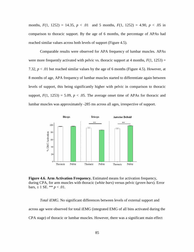
85
months, F(1, 1252) = 14.35, p < .01 and 5 months, F(1, 1252) = 4.90, p < .05 in
comparison to thoracic support. By the age of 6 months, the percentage of APAs had
reached similar values across both levels of support (Figure 4.5).
Comparable results were observed for APA frequency of lumbar muscles. APAs
were more frequently activated with pelvic vs. thoracic support at 4 months, F(1, 1253) =
7.32, p < .01 but reached similar values by the age of 6 months (Figure 4.5). However, at
8 months of age, APA frequency of lumbar muscles started to differentiate again between
levels of support, this being significantly higher with pelvic in comparison to thoracic
support, F(1, 1253) = 5.09, p < .05. The average onset time of APAs for thoracic and
lumbar muscles was approximately -285 ms across all ages, irrespective of support.
Figure 4.6. Arm Activation Frequency. Estimated means for activation frequency,
during CPA, for arm muscles with thoracic (white bars) versus pelvic (green bars). Error
bars, ± 1 SE. ** p < .01.
Total iEMG. No significant differences between levels of external support and
across age were observed for total iEMG (integrated EMG of all bins activated during the
CPA stage) of thoracic or lumbar muscles. However, there was a significant main effect

86
of age for lumbar muscles, indicating that at 6 months there was an increase in iEMG of
the lumbar muscles, F(5, 1080) = 3.55, p < .01, compared to 4 months (Figure 4.7).
Similarly, in terms of the arm muscles, iEMG of biceps and triceps displayed an
interesting age effect. At the age of 6 months, iEMG of biceps was significantly
enhanced, in contrast to 4 and 5 months, F(5, 1507) = 4.16, p < .01. A complete opposite
trend was observed for iEMG of triceps, showing a significant decrease in iEMG of
triceps at 5 months, F(1, 1350) = 3.52, p < .01 compared to 3 months (Figure 4.7). In
addition, the level of support had a main effect on the anterior deltoid muscle, showing
higher iEMG with pelvic support across all ages, F(1, 1398) = 19.96, p < .01.
CPA Latency. With regard to the postural muscles, there was a significant delay in
CPA onset with pelvic support, during the months prior to independent sitting (M = 6
months) (Figure 4.8). EMG onset latency of the thoracic muscle at 3 months was
significantly longer with pelvic support, F(1, 887) = 7.27, p < .01. After this age, EMG
onset of the thoracic muscle was invariable between levels of support. The lumbar muscle
on the contrary did not show an effect of support at any age. However, irrespective of
support, there was a general trend for lumbar muscle latency to decrease with age, but
this failed to reach significance (p = 0.07).
EMG onset latency of arm muscles once again showed high variability and results
were inconclusive between levels of support. Instead, there was a difference in
recruitment pattern, depending on the level of support. At early stages (3 or 4 months)
infants first co-activated all arm muscles. With increased age and with thoracic support,
infants tended to activate first biceps, followed by triceps and lastly anterior deltoid,
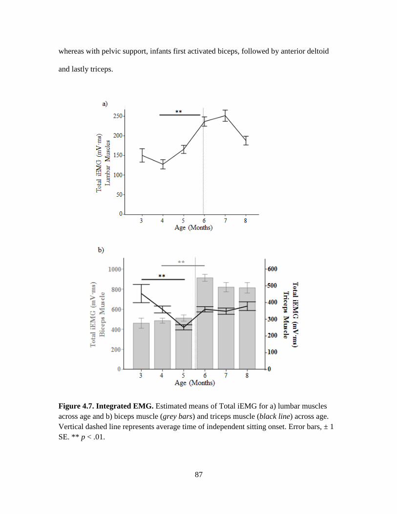
87
whereas with pelvic support, infants first activated biceps, followed by anterior deltoid
and lastly triceps.
Figure 4.7. Integrated EMG. Estimated means of Total iEMG for a) lumbar muscles
across age and b) biceps muscle (grey bars) and triceps muscle (black line) across age.
Vertical dashed line represents average time of independent sitting onset. Error bars, ± 1
SE. ** p < .01.

88
Figure 4.8. Onset Latency. Estimated means for CPA EMG onset latency for postural
muscles across age and with thoracic (black line) versus pelvic (green line) support.
Vertical dashed line represents average time of independent sitting onset. Error bars, ± 1
SE. ** p < .01.
Recruitment Order of Postural Muscles. For those trials in which both thoracic
and lumbar muscles were activated, results showed a significant difference in recruitment
pattern, depending on the age and level of support. At 4 months, infants showed a
significant preference for a top-down pattern with pelvic compared to thoracic support,
F(1, 491) = 7.39, p < .01. After the onset of independent sitting, infants showed the
opposite, demonstrating a bottom-up preference with pelvic in comparison to thoracic
support, this being significant at 7 months of age, F(1, 491) = 3.90, p < .05 (Figure 4.9).
Overall, these results, as with kinematics, indicate that when providing external
support at the pelvic level, maintaining stability of the trunk is more challenging than
when provided with thoracic support during the period when infants have not yet
acquired the ability to independently sit. This is determined by three major findings: 1)
activation frequency of postural muscles, during both APA and CPA stages, increased

89
with pelvic support to counteract the balance disturbance encountered during the reach;
2) infants at 3 months were unable to quickly activate the thoracic muscles in a feedback
mode during a reach with pelvic support, whereas this was possible with thoracic support;
3) infants at 4 months showed a higher percentage of top-down postural muscle
recruitment order with pelvic support, indicating the functional preference for stabilizing
the upper trunk in response to the instability.
Figure 4.9. Recruitment Order. Estimated means for percentage for either top-down or
bottom-up recruitment order of postural muscles with thoracic (black line) versus pelvic
(green line) support. Vertical dashed line represents average time of independent sitting
onset. Error bars, ± 1 SE. ** p < .01, * p < .05.
DISCUSSION
The purpose of this study was to investigate the relationship between reaching
skills and the segmental progression of trunk control for the acquisition of independent
sitting. This was addressed by conducting a longitudinal study on infants from 2.5-8
months of age that involved an experimental paradigm creating vertical alignment of the
trunk in sitting position with two levels of external trunk support (thoracic and pelvic)

90
during reaching. This allowed examination of the relationship between the higher and
lower regions of the trunk during the development of sitting on reaching performance.
Infants first gain control of the upper trunk region followed by the lower trunk and pelvic
regions for the acquisition of independent sitting; and thus, we hypothesized that before
the onset of independent sitting, infants would demonstrate a decreased ability to reach,
impoverished reaching and trunk kinematics and inefficient postural muscle patterns
when given pelvic in comparison to thoracic support.
In this regard, we confirm and expand previous results by showing that reaching
success and the kinematic quality of the movement was substantially affected by the
progressive development of postural control (de Graaf-Peters et al., 2007; Hopkins &
Rönnqvist, 2002; Rachwani et al., 2013; Rochat & Goubet, 1995), such that with pelvic
support, infants performed worse in comparison to thoracic support, during the months
prior to independent sitting. Additionally, the lack of trunk control with pelvic support
was further supported by EMG data. In the following paragraphs we discuss how posture
and reaching performance demonstrated significant differences between levels of external
support (thoracic versus pelvic) during the development of independent sitting.
Reaching Behavior between Levels of External Support across Age
Infants at young ages (2 months), showed a high level of difficulty in remaining
stable in the sitting position when provided with an external support at the thoracic level,
though they were able to do this minimally, with poor control. However, this ability was
absent in most infants with the lower pelvic support, in which only 2 infants were able to
maintain stability part of the time. Similar results were seen with respect to the number of
reaching attempts that the infants made with the two levels of support. Thus, even though
91
both motor milestones, upright sitting and reaching, were still immature during this
developmental time period, a better support of the trunk was indeed associated with the
ability to maintain stability and with the ability to perform more reaching attempts, as has
been observed in previous studies (Amiel-Tison & Grenier, 1983; von Hofsten, 1982).
This suggests that postural control significantly regulates the interaction of the infant with
the surrounding environment during development, facilitating new actions, like reaching,
which in turn can promote the emergence of cognitive skills and social behaviors
(Gibson, 1988).
According to the AIMS and SATCo scores, infants started to master head control
at 3 months of age, which is a critical motor milestone in infant development. With this
mastery, infants increased their ability to touch/grasp the toy, highlighting the importance
of head control for successful reaching (Thelen & Spencer, 1998). In order to lift the arm
and successfully touch the toy infants must fixate the visual target, which requires both
strength and control of the head in space as well as visual acuity. We hypothesized that
reaching abilities would be reduced when postural instability was enhanced (i.e. with
pelvic support); however, this was not supported at the age of 3 months or beyond. We
found that infants continued to successfully reach with both levels of external support
despite the challenging postural demands derived from the trunk support at the pelvic
level. Harbourne et al. (2013) showed a similar effect in that non-independent sitters
persistently and successfully reached in spite of the subsequent falls, disorganized muscle
onsets and erratic trunk movements. This suggests that once infants acquire head control,
reaching success is not perturbed by a decreased ability to control the regions of the trunk
during vertical sitting.
92
With respect to the type of reach performed, differences between bimanual and
unimanual reaches across age and level of support were not observed. Research has
shown that interlimb coordination of infants during reaching tasks follows a fluctuating
pattern and it is not object-scaled until the age of 2 years. These results agree with
previous research and suggest that infants do not adapt unimanual or bimanual reaching
with regard to the physical properties of the object or to the demands of the task in early
stages of development (2.5 to 8 months) (Corbetta & Thelen, 1996). However, infants
need to learn how to move to and cross the vertical midline with unimanual reaches in
order to reach and grasp the toy (van Hof, van der Kamp, & Savelsbergh, 2014). Even
though the progression of trunk development did not affect the type of reach performed,
at 3 months of age, the number of successful bimanual reaches was greater than the
number of successful unimanual reaches. This evidence highlights the effect of both age
and maturation on the development of unimanual reaching.
Reaching and Postural Kinematics between Levels of External Support across Age
The effect of external support on postural stability while reaching was evident in
that infants showed deterioration of postural kinematics with pelvic support when they
had not yet mastered the control of the lower trunk region. Differences in maintaining
stability of the trunk while reaching were observed in 4 and 5 month olds. Prior to this
time period (i.e. 3 months of age), this was not the case, partly due to the fact that infants
had not yet fully acquired control of both upper and lower regions of the trunk. Hence,
infants were unstable with both levels of external support, though there was a tendency
toward increased stability with thoracic support, as is depicted in Figure 4.4. A
comparable trend was observed with respect to head stability. However, head angular

93
displacement could reflect either the infant’s moving the head to better visualize the
surrounding environment or compensations for trunk movements while reaching. This
could explain why infants appeared to maintain a fairly consistent head displacement
across age, regardless of the level of trunk support. Nonetheless, it is worth noting the
significant increase in head and trunk displacement that occurred with pelvic support at 4
and 5 months of age.
The external trunk support also had an impact on reaching kinematics, in that
infants between 3-5 months of age showed a longer reach duration and a more devious
and jerky reach with pelvic compared to thoracic support, due to the increased postural
instability at pelvic level. In contrast to this, movement unit number remained invariable
across levels of support, showing its independence from postural support. Similar results
were observed by de Graaf-Peters et al. (2007) in which 4 month-old infants showed an
invariable number of movement units in supine and sitting position; however, 6 month-
old infants displayed less movement units in sitting position than at the age of 4 months.
These results suggest that the ability to reduce the number of on-line corrections, or
movement units in a reach, is a matter of maturation and might be improved with practice
and motor experience. However, during this maturation period, other qualitative reaching
parameters, like straightness and jerkiness, are significantly modulated by the progressive
development of trunk control.
Reaching and Postural EMG Patterns between Levels of External Support across Age
Research has demonstrated on numerous occasions how postural muscle activity
accompanying reaching movements increases with age (de Graaf-Peters et al., 2007;
Harbourne et al., 2013; van Balen et al., 2012). Results from the current study show that

94
postural muscle activity can be present even in early stages of sitting development, but it
is dependent on the constraints of the task. Lumbar and thoracic muscle activity was
more frequent when infants were provided with pelvic support at both 3-4 and 4-5
months. This implies that prior to acquiring independent sitting, infants were able to
recruit postural muscles while reaching, and increased recruitment frequency when the
postural task was more demanding. This means that postural muscle recruitment was
situation-specific and depended on the degree in which balance was being perturbed
(Hadders-Algra, 2008). Then, with increased age and maturation of the ability to sit
independently, the overall activation frequency of postural muscles increased and infants
showed similar values across levels of trunk support, implying that pelvic support was no
longer a condition in which balance control was being challenged. Other research has
also shown that once independent sitting ability was mastered, postural muscle activity
accompanying reaching movements while sitting was consistently present (van der Heide
et al., 2003) and thus became embedded in the task, although it could be further enhanced
if the risk of losing balance and falling over was high (Hadders-Algra, 2005; van Balen et
al., 2012).
Similarly, even though our sample demonstrated an infrequent use of APAs
during early stages of development; with pelvic support, infants at 4-5 months displayed
a higher percentage of APAs, mainly driven by thoracic muscles. This means that infants
were able to control in a feed-forward fashion the disequilibrium that the reaching arm
produced when they had not yet acquired full trunk control. With a higher support at
thoracic level, these anticipatory effects were not observed as frequently at 3-5 months of
age. However, from 6 months onward, APAs were more consistently present (50% of the

95
time) and were independent of the type of support, suggesting that at this age, infants
develop the ability to integrate feed-forward control into the task of reaching.
The study by Van der Fits et al., (1999a and 1999b) examining APAs in seated
reaching concluded that APAs were seldom present in 3-5 month infants, were present in
20% of the trials at 6 months of age and became more consistent around the age of 13-14
months. These differing results, in contrast to what we obtained, might be explained by
two major methodological differences related to the experimental paradigm and the
temporal window of APAs that was selected for analysis. First, Van der Fits et al. (1999a)
tested 3-6 month infants while placing them in an infant chair with complete support of
the trunk, failing to evaluate the destabilizing effects of gravity and motion of the arm on
the trunk per se. Second, Van der Fits et al. (1999b) used a more stringent criterion for
defining APAs and evaluated APA activity within a 200ms time window prior to prime
mover activation; whereas in the current study, we defined a 500ms time window. Feed-
forward control emerges in parallel to the development of different postural frameworks
in infancy and increase both in consistency and in temporal specificity with experience of
the motor task (Witherington et al., 2002). Thus, we propose that APAs should be
analyzed within a broader period of time before the reaching event in infants since timing
of APAs during development would appear to be less specific to the reach onset than in
adults.
It is thus concluded that during early developmental stages of upright sitting,
APAs accompanying reaching movements are present to some degree, especially when
the postural task is more demanding. These APAs are characterized by immature
temporal features. Then, with age, APAs start to play a major role in the postural

96
mechanisms for seated reaching once independent sitting has been established; at this
point their activation is not dependent on the level of postural stability, but they are
consistently activated well in advance of the reach.
Contrarily to our hypothesis, we did not expect differences in APAs to occur at
later stages of development. However, the lumbar muscles showed a significantly higher
frequency of APAs with pelvic support in comparison to thoracic support, not only at 4
months but also at 8 months, when infants were well coordinated in independent sitting.
Therefore, there was an effect of support on APA activity of lumbar muscles even when
trunk control was fully acquired, suggesting the existence of distinct feed-forward
mechanisms that are related to specific segmental regions of the trunk, in later stages of
development.
Postural muscles serve to stabilize posture and control equilibrium during a reach
in two ways. The muscles of the upper region of the trunk are used to oppose the reaction
forces generated by the arm movement, whereas the muscles of the lower trunk serve to
keep the center of mass within the stability limits (van der Fits et al., 1999a). In this
study, the activation of both thoracic and lumbar muscles was significantly enhanced
with pelvic support during the learning process of independent sitting. This outcome
implies the need to maintain stability prior to and during the ongoing motion of the arm.
However, postural muscle onset latencies reflect the ability to actively control the
goal-directed movement once it has been initiated; a slower latency would indicate less
efficient control. Results showed significant differences between levels of trunk support
for thoracic muscle onset, depending on the age of the infant. At 3 months, a slower
latency was observed with pelvic support, whereas with the help of additional support at

97
thoracic level, this latency was significantly faster. This suggests that even though infants
were able to consistently recruit postural muscles to counteract the unbalancing effects of
the self-triggered perturbation through reaching, the temporal mechanisms were
significantly delayed with the absence of full trunk control at 3 months. Nevertheless,
these mechanisms were improved by facilitating higher levels of external postural
supports, highlighting the effects of both age and support on the development of postural
muscle onset latencies.
A similar effect was observed when calculating the percentage of trials with either
a top-down or bottom-up postural muscle recruitment order. With thoracic support, the
preference for either pattern remained invariable across age. On the contrary with pelvic
support, there were a higher percentage of times that postural adjustments followed a top-
down sequence, at 4 months of age, suggesting a functional preference for stabilizing the
upper trunk in response to the instability. This strategy shifted toward a bottom-up pattern
after the onset of independent sitting. These observations have been previously observed
in numerous studies (de Graaf-Peters et al., 2007; van Balen et al., 2012; van der Fits et
al., 1999a). However, we expand the results obtained from previous literature by showing
that postural adjustments following a top-down sequence is related to increased postural
demands during the development of upright sitting, since such preference can be reduced
with higher levels of trunk support. Being able to sit independently then marks the
hallmark for shifting toward a bottom-up pattern, when stabilization of the trunk is no
longer challenging, and thus, the focus of control is at the support surface (Assaiante,
1998; Hadders-Algra, 2008).

98
Reaching and Postural EMG Patterns across Age
Unexpectedly, some of the EMG parameters were not dependent on the
progression of trunk control; instead age had a major effect. For instance, muscle
amplitudes of compensatory postural adjustments showed no changes between levels of
support, suggesting the independence to posture. This supports previous research,
indicating that changes in the degree of contraction of postural muscles to changes in
position do not occur until 9-10 months of age (Hadders-Algra, 2005).
Moreover, we observed that 6 months was the age when significant changes in
compensatory muscle amplitudes occurred and did not depend on the type of support. By
this age, infants had increased lumbar muscle amplitude during seated reaching. It could
be surmised that this general increase was related to the development of independent
sitting and may serve as a preparation for the ability to move in and out, or rotate in
sitting position. We also found that during early stages, triceps muscle amplitude (the
antagonist) was high and decreased by 6 months of age, whereas biceps muscle amplitude
(the agonist) was low during early periods and by 6 months onward, it had substantially
increased. These results, also found in other studies, suggests that during early stages of
development, reaches were associated with a more “active extension” strategy, meaning
that reaches were accompanied by increased extensor muscle amplitude (Konczak et al.,
1997). With increasing reaching proficiency and with the achievement of independent
sitting, infants shifted to a more “passive extension” strategy, which is more energy
efficient. That is, infants were able to produce more biceps activity and thus, more
flexion of the arm to accomplish the task of forward reaching while sitting.

99
Lastly, our findings related to arm muscles indicate that patterns of muscle
frequency, amplitudes and onsets in early infancy are characterized as being highly
variable and are not influenced by the progression of the development of trunk control
during the acquisition of independent sitting. At 3 and 4 months, there was a co-
activation of all arm muscles, which was associated with the onset of reaching (Thelen et
al., 1993). It is known that activation of both agonist and antagonist muscles at a joint
often occurs when the individual has lower skill levels since it stiffens or stabilizes the
entire limb. However, from 5 months onwards, infants adopted different arm muscle
activation strategies depending on the level of support they were provided. This might
suggest that the use of external devices can produce changes in the neuromuscular control
of the arm, which is unrelated to the intrinsic control of the trunk that infants had
acquired.
CONCLUSIONS
This study sought to examine the relationship between reaching performance and
the progressive development of control of specific trunk regions for the acquisition of
independent sitting. Results reinforce and further expand previous findings showing that
improvements in trunk control have direct consequences on the development of reaching.
It is concluded that there is a cranio-caudal acquisition of trunk control for independent
sitting and the regional extent of trunk control infants have acquired has an impact on the
kinematic quality of reaching movements and accompanying postural muscle patterns,
attributed to frequency of activation and timing mechanisms. However, with the help of
additional support, infants experience drastic improvements in their reaching skills and

100
subsequent muscular parameters during the development of independent sitting. This
correlation of reaching and the progressive development of trunk control should also be
examined in children with motor deficits in order to determine if they might benefit from
the use of external trunk support and consequently implement more efficient therapeutic
strategies.

101
CHAPTER V
FINAL CONCLUSIONS
GENERAL SUMMARY OF FINDINGS
Postural achievements in infancy develop in a cephalo-caudal direction, as
originally described by Gesell and Amatruda (1945). Newborns have limited postural
control, but within a few months, they develop head control followed by trunk control.
The development of trunk control will allow infants to first sit with support and
eventually to sit without any kind of support. Then, by the age of 1 year, infants achieve
independent standing which consequently leads to the ability to walk, run and skip.
Research from this dissertation provides additional evidence to support the
concept that the development of trunk control for upright sitting follows a progressive
cephalo-caudal direction, starting with the upper regions and subsequently the lower and
pelvic regions of the trunk. This progression is a fundamental prerequisite for
maintenance of functional positions of the body, such as upright sitting. The ability to
control upright sitting position is necessary for stability, balance, and orientation within
the performance of more complex skills (Shumway-Cook & Woollacott, 2012a).
Appropriate orientation ensures that the configuration of the body segments is maintained
with respect to one another during a movement or task. For example, when performing a
reaching task, the trunk needs to maintain stability with respect to the base of support,
prior to, and during the ongoing motion of the reach (Zimmermann, Meulenbroek, & de
Lange, 2012). Therefore, trunk stability is essential for effective postural maintenance
and reaching accuracy, since reaching proficiency is highly dependent on the control of
posture.

102
The results from this dissertation demonstrate that the stability of the trunk
depends on the extent of trunk control that infants have acquired. In the cross-sectional
study we found that when infants had partial trunk control, they relied on an external
trunk support that facilitated postural stability, in order to perform more efficient reaches
that were similar to the ones made by infants with complete trunk control.
Furthermore, in the longitudinal study, it was observed that measures of stability
(head and trunk angular displacement) and reaching performance (movement time,
straightness score, normalized jerk score) between levels of external trunk support (pelvic
vs. thoracic support) had non-linear changes across development. In most cases, these
reflected impoverished stability and reaching measures with pelvic support during the
development of upright sitting. These differences were at their highest when infants were
3-4 months and gradually declined to reach their lowest levels at 6 months, when infants
had acquired the ability to independently sit. From this age onward, further improvements
in posture and reaching across age were not observed and measures were insignificant
between levels of external support.
Electromyographic recordings associated with reaching and underlying trunk
stability displayed a similar, non-linear trend across development. Infant reaches with
pelvic support were characterized by high postural muscle activation frequencies in
response to the instability, at 3-5 months of age. First, onset latencies of postural muscles
were significantly delayed with pelvic support, at 3 months of age. Then, the occurrence
of APAs was more consistent with pelvic support, at 4-5 months of age, suggesting the
role of APAs for postural maintenance during reaching when stability is compromised.
From 6 months onward, APAs, postural muscle activation frequencies and onset latencies
103
accompanying the reach attained consistent values and were no longer dependent on the
level of external support. Therefore, 6 months, i.e. the age when infants learned to sit
independently, is an age of transition, when they are finally able to adapt postural activity
to the specifics of the support condition.
Additionally, there were other parameters that changed with increasing age but
were independent of the segmental progression of trunk control. For instance, lumbar
muscle amplitude increased with age, and peaked at 6 months, highlighting its association
with an increased capacity to sit independently. Also, in terms of the reaching arm, MUs
decreased with age, and obtained their lowest values from 6 months onward; similarly,
biceps muscle amplitude increased whereas triceps muscle amplitude decreased. When
taken together, these points suggest that even though the ability to reach and sit overlap in
developmental time, there are specific parameters embedded in the maturational time
course of each milestone that are not perturbed by one another.
LIMITATIONS OF RESEARCH
It is important to acknowledge that postural control is only one of the many
critical factors that must be acquired for infants to sit independently and perform skillful
reaches, since factors such as muscle strength, appropriate body proportions, sensitivity
to visual flow, the ability to detect affordances, and motivation are also influencing
factors (Thelen & Smith, 1994). Each of these factors has a different developmental time
course. From a developmental systems perspective, the goal of developmental analysis is
to understand the paths, interrelations and causal mechanisms of the time course of each
contributing factor (Adolph & Robinson, 2008). Therefore, defining complete trunk
104
control as an endpoint to the development of upright sitting and reaching skills might be a
risky proposition, since it may very well depend on rearing or testing conditions, as well
as individual and cultural variations.
However, this dissertation was motivated by the notion that the development of
sitting postural control and reaching behavior are highly interdependent functions. Full
attempts were made to tease out the causal effects of postural control on reaching. First,
having applied measures across a broad range of ages in a longitudinal design, we were
able to explore a critical window of postural development prior to independent sitting,
corresponding to ages 2-5 months. Second, with the use of experimental manipulations,
we had the means to model the type of postural support that infants progressively
generated for themselves. More specifically, we supported the thoracic and pelvic regions
of the trunk for comparing the effects of increased vs. decreased postural support on
reaching. With the higher support at thoracic level, reaching movements during pre-
sitting stages were smoother and more mature than when the support was limited to the
pelvic level. Increased postural support had a direct impact on reaching performance.
Then, as infants developed across age, they improved in many domains, including sitting
posture and thus, reaching movements were freed from balance constraints of the trunk.
Infants no longer required the additional help of a higher support for producing
coordinated reaching. With this, we can conclude that postural control is one of the main
causes contributing to reaching proficiency, regardless of whether posture is improved
naturally across age or with the help of an experimental set-up. This information creates
the basis for a wide range of future studies that can be applied in assessment and
rehabilitative protocols in children with postural dysfunctions.

105
CLINICAL IMPLICATIONS
Basic studies in animal models conjointly with research in infant behavior have
been crucial in understanding normal development and critical temporal windows at
which behaviors are acquired. In addition, knowledge about typical development is a
prerequisite for understanding abnormal development. It is during the first years of life
when major changes occur in the nervous system and motor behavioral outputs. Hence, it
is imperative to localize those critical temporal windows during development and define
how abnormal behavior deviates from typical behavior so that professionals in health
science can assess and apply therapeutic techniques in a timely manner.
Studies have shown that for children with cerebral palsy, achieving independent
sitting early in time is a key determinant of independent ambulation and future motor
skill development (Wu et al., 2004). Results of this research examine principles
underlying the development of trunk control for independent sitting and its consequences
for the performance of functional activities, like reaching. Thus, the knowledge obtained
from typically developing infants could also be compared in children with cerebral palsy
in order to test how they vary from typical development at different points in time. If a
similar correlation between the progressive achievement of trunk control and the
emergence of effective reaching performance is observed, then this might provide new
insights into specific improvements in both postural control and consequent reaching
ability in children with cerebral palsy. These insights can be used to improve current
approaches to training trunk postural control, and thus independent sitting, which has a
direct impact on the manual abilities for this vulnerable population.
106
Pediatricians often encounter children with delays of motor development in their
clinical practices. Motor delays may be the first or most obvious sign of a global
developmental disorder. It is often the case that children whose developmental
trajectories are at risk may experience challenges in meeting early motor milestones
(Noritz & Murphy, 2013). A timely diagnosis may reduce the prognostic uncertainties. If
clinicians become aware of the fact that trunk control is developed cranio-caudally and is
a prerequisite for independent sitting and subsequent reaching ability, they may refine
their clinical evaluations in a timely manner to include more precise evaluation of trunk
control. Segmental evaluation of the trunk will undoubtedly lead to new innovative ideas
concerning how to impact trunk control and reaching in the clinic as well as at home.
FUTURE DIRECTIONS FOR RESEARCH
This research has demonstrated that distinct levels of postural trunk control can be
differentiated during the development of independent sitting, and that reaching
performance is influenced by level of trunk control. This paradigm offers the foundation
for future exploration both in typical development as well as in children with
neurological deficits.
Future studies should be implemented to test whether there is a segmental
progression of postural control for the development of independent stance in typical
infants. During the process of learning to stand independently, infants must learn to
control many additional degrees of freedom compared to independent sitting, as they add
the coordination of the leg and thigh segments to those of the trunk and head. Research in
healthy adults has modeled the body during quiet stance as a multilink pendulum with

107
two coexisting modes of control for describing sway during quiet stance: ankle strategy
or a hip strategy. These two control strategies have also been described for recovery of
perturbed stance (Shumway-Cook & Woollacott, 2012c). The question of whether infants
follow a top-down progression of control across the multi-link segments at the level of
the hips, knees and ankles for acquiring independent stance, is yet to be answered.
However, of primary importance for clinical purposes would be a study to
evaluate the effects of segmental trunk support on posture and reaching in children with
cerebral palsy or even other pathological populations, such as adults with moderate to
severe stroke, for the intended goal of improving trunk control and reaching. In the severe
cases of stroke, trunk stability is compromised which subsequently impairs arm function.
However, knowledge related to the relationship between impaired trunk postural control
and loss of independent arm function following a stroke is still limited. This is a critical
barrier to progress in assessment and treatment of patients with severe to moderate stroke,
since most of their activities of daily living are performed while seated. Thus, future
studies must assess whether improvements in reaching and manipulation skills can occur
when patients with severe trunk deficits are given external trunk adapted to their level of
trunk control. In addition, the more detailed information about control of the trunk in
individual patients could help specify the clinical goals and guide the implementation of
training protocols directed at improving control in the specific area of the trunk rather
than treating the trunk as a non-dissociable, single unit (Butler et al., 2010; Butler, 1998).

108
APPENDIX
METHODOLOGY FOR HEARTBEAT SUBTRACTION
A modified version of the algorithm used by Aminian et al. (1988) was used for
subtracting the heartbeat artifact. A six stage process was used to identify and remove
EKG interference that may have been present in each channel of the EMG used in this
data analysis.
1) User is presented with a graph of the EKG signal collected by the subject being
processed. The user is asked to select an amplitude threshold to differentiate heartbeat
signal from activity originating in the surrounding musculature. The polarity of the
threshold selected determines if the particular peaks sought will be the Q (negative
polarity) or R (positive polarity) of the EKG complex.
2) The EKG signal is examined forward from the beginning, looking for points
that exceed the amplitude threshold set in the first stage. When such points are found, the
forward examination continues, counting how long the signal stays above that threshold
point. If this duration exceeds 7 ms, then that peak is kept for further consideration. The
value of 7ms was empirically determined to have the best performance when the
algorithm was first implemented by previous graduate students.
3) When a peak point is detected, the signal around it is explored for the peaks
that complete the QRS complex (mainly searching for inflection points) and the
directionality of the search is determined by the polarity of the selected threshold. Once
the complete QRS is found, ten milliseconds are added on to each end of the inflection
points to note the time location of the entire PQRST complex.

109
This process continues until the search reaches 40ms before the end of the EKG
signal. The search terminates here as any EKG signal that begins in these final 40ms is
highly likely to be an incomplete PQRST complex.
In addition to the third stage, the amplitude of each peak deemed valid above is
averaged. Any PQRST regions whose peaks exceed this average by 2 standard deviations
are discarded. The reasoning for deciding to remove these peaks was that any abnormally
large spikes in the EKG signal were likely to be due to either extraneous muscle
contractions, or other unusual sources of electric interference.
4) For each channel of EMG, the average EKG signature was generated by
averaging together the waveform found within each PQRST boundary deemed valid from
the above stages. Then, the averaged EKG signal for that EMG channel is created by
including the average EKG signature within the PQRST time boundaries on a flat signal,
with a same duration as the original EMG signals.
5) The user is presented with three graphs for each EMG channel: the unaltered
EMG signal, the averaged EKG signal for that channel, and the EMG signal with the
averaged EKG signal subtracted (Figure 6.1). For each channel, the user is asked to
choose whether the unaltered or subtracted signal is the better of the two. If the averaged
EKG signal selected is not of sufficient quality, the user has the option to return to the
first stage and pick a new amplitude threshold.
6) Finally, the algorithm removes the averaged EKG signal from those EMG
channels the user elected to remove it from, and processing on the EMG signals
continues.

110
Figure 6.1. Example of Hearbeat Subtraction. Graphs indicating a) the unaltered EMG
waveform, b) the averaged heartbeat signal for that channel, and c) the EMG signal with
the averaged heartbeat signal subtracted.
a)
b)
c)
111
REFERENCES CITED
Acension Technology Coorporation. (2000). MiniBIRD® Position and orientation
measurement system. Burlington, VT: Ascension Technology Corporation.
Adolph, K., & Berger, S. (2006). Motor development. In W. Damon & D. Lerner (Eds.),
Handbook of child psychology: Cognition, perception, and language (2nd ed., Vol.
2, pp. 161–213). New York, NY: Wiley.
Adolph, K., & Berger, S. E. (2005). Physical and motor development. In M. Bornstein &
M. Lamb (Eds.), Developmental science (pp. 223–281). Mahwah, NJ: Lawrence
Erlbaum Associates.
Adolph, K., & Robinson, S. (2008). In defense of change processes. Child Development,
79, 1648–1653.
Amiel-Tison, C., & Grenier, A. (1983). Expression of liberated motor activity (LMA)
following manual immobilization of the head. In C. Amiel-Tison & A. Grenier
(Eds.), Neurological evaluation of the newborn and the infant (pp. 87–109). New
York, NY: Masson Publishing USA.
Aminian, K., Ruffieus, C., & Robert, P. (1988). Filtering by adaptive sampling (FAS).
Medical and Biological Engineering and Computing, 26, 658–662.
Assaiante, C. (1998). Development of locomotor balance control in healthy children.
Neuroscience and Biobehavioral Reviews, 22(4), 527–532.
Assaiante, C., Mallau, S., Viel, S., Jover, M., & Schmitz, C. (2005). Development of
postural control in healthy children: a functional approach. Neural Plasticity, 12(2-
3), 109–118.
Barbosa, V., Campbell, S., Sheftel, D., Singh, J., & Beligere, N. (2003). Longitudinal
performance of infants with cerebral palsy on the Test of Infant Motor Performance
and on the Alberta Infant Motor Scale. Physical and Occupational Therapy in
Pediatrics, 23(3), 7–29.
Bayley, N. (1969). Bayley scales of infant development. New York, NY: Oxford
University Press.
Bayley, N. (2005). Bayley scales of infant and toddler development (3rd ed.). San
Antonio, TX: Pearson.
Bernstein, N. (1967). The co-ordination and regulation of movements (1st ed.). New
York, NY: Oxford, Pergamon Press.

112
Bertenthal, B., & Von Hofsten, C. (1998). Eye, head and trunk control: the foundation for
manual development. Neuroscience and Biobehavioral Reviews, 22(4), 515–520.
Bigongiari, A., de Andrade e Souza, F., Franciulli, P. M., Neto, S. E. R., Araujo, R. C., &
Mochizuki, L. (2011). Anticipatory and compensatory postural adjustments in sitting
in children with cerebral palsy. Human Movement Science, 30(3), 648–657.
Brandt, T., Paulus, W., & Straube, A. (1986). Vision and posture. In W. Bles & T. Brandt
(Eds.), Disorders of posture and gait. (pp. 157–175). Amsterdam: Elsevier Ltd.
Butler, P. (1998). A preliminary report on the effectiveness of trunk targeting in
achieving independent sitting balance in children with cerebral palsy. Clinical
Rehabilitation, 12, 281–293.
Butler, P., Saavedra, S. L., Sofranac, M., Jarvis, S., & Woollacott, M. H. (2010).
Refinement, reliability and validity of the segmental assessment of trunk control
(SATCo). Pediatric Physical Therapy, 22(3), 246–257.
Butterworth, G., & Hicks, L. (1977). Visual proprioception and postural stability in
infancy: a developmental study. Perception, 6, 255–262.
Chang, J. J., Wu, T. I., Wu, W. L., & Su, F. C. (2005). Kinematical measure for spastic
reaching in children with cerebral palsy. Clinical Biomechanics, 20(4), 381–388.
Corbetta, D., & Thelen, E. (1996). The developmental origins of bimanual coordination:
A dynamic perspective. Journal of Experimental Psychology: Human Perception
and Performance, 22(2), 502–522.
Darrah, J., Bartlett, D., Maguire, T., Avison, W., & Lacaze-Masmonteil, T. (2014). Have
infant gross motor abilities changed in 20 years? A re-evaluation of the Alberta
Infant Motor Scale normative values. Developmental Medicine and Child
Neurology, 56(9), 877–881.
De Graaf-Peters, V. B., Bakker, H., Van Eykern, L. A., Otten, B., & Hadders-Algra, M.
(2007). Postural adjustments and reaching in 4- and 6-month-old infants: An EMG
and kinematical study. Experimental Brain Research, 181, 647–656.
Elsner, B., & Hommel, B. (2004). Contiguity and contingency in action-effect learning.
Psychological Research, 68(2-3), 138–154.
Fallang, B., Saugstad, O. D., & Hadders-Algra, M. (2000). Goal directed reaching and
postural control in supine position in healthy infants. Behavioural Brain Research,
115(1), 9–18.
Fallang, B., Saugstad, O., Grogaard, J., & Hadders-Algra, M. (2003). Kinematic quality
of reaching movements in preterm infants. Pediatric Research, 53, 836–842.

113
Forssberg, H., & Hirschfeld, H. (1994). Postural adjustments in sitting humans following
external perturbations : muscle activity and kinematics. Experimental Brain
Research, 97(3), 515–527.
Galloway, J. C., & Thelen, E. (2004). Feet first: object exploration in young infants.
Infant Behavior and Development, 27(1), 107–112.
Gesell, A., & Amatruda, C. (1947). Developmental diagnosis (2nd ed.). New York, NY:
Harper and Row.
Gessell, A. (1946). The ontogenesis of infant behavior. Manual of child psychology.
Hoboken, NJ: John Wiley & Sons Inc.
Gessell, A., & Ames, L. (1947). The development of handedness. Journal of Genetic
Psychology, 70, 155–175.
Gibson, E. J. (1988). Exploratory behavior in the development of perceiving, acting, and
the acquiring of knowledge. Annual Review of Psychology, 39, 1–41.
Gibson, E. J., & Pick, A. (2000). An ecological approach to perceptual learning and
development. New York, NY: Oxford University Press.
Grenier, A., & Amiel-Tison, C. (1981). Liberated-motricity by holding the head during
the 1st weeks of life. Archives for Pediatrics, 5, 557–561.
Grönqvist, H., Strand Brodd, K., & von Hofsten, C. (2011). Reaching strategies of very
preterm infants at 8 months corrected age. Experimental Brain Research., 209(2),
225–233.
Hadders-Algra, M. (2000). The neuronal group selection theory : a framework to explain
variation in normal motor development. Developmental Medicine and Child
Neurology, 42, 566–572.
Hadders-Algra, M. (2005). Development of postural control during the first 18 months of
life. Neural Plasticity, 12(2-3), 99–108.
Hadders-Algra, M. (2008). Development of postural control. In M. Hadders-Algra & E.
Brogren Carlberg (Eds.), Postural control: A key issue in developmental disorders
(pp. 22–73). London: Mac Keith Press.
Hadders-Algra, M., Brogren, E., & Forssberg, H. (1996). Ontogeny of postural
adjustments during sitting in infancy: variation, selection and modulation. The
Journal of Physiology, 493, 273–288.
114
Harbourne, R. T., Giuliani, C., & Neela, J. (1993). A kinematic and electromyographic
analysis of the development of sitting posture in infants. Developmental
Psychobiology, 26, 51–64.
Harbourne, R. T., Lobo, M. A., Karst, G. M., & Cole, J. (2013). Sit happens : Does sitting
development perturb reaching development , or vice versa ? Infant Behavior and
Development, 36(3), 438–450.
Harbourne, R. T., & Stergiou, N. (2003). Nonlinear analysis of the development of sitting
postural control. Developmental Psychobiology, 42(4), 368–377.
Hedberg, A., Carlberg, E. B., Forssberg, H., & Hadders-Algra, M. (2005). Development
of postural adjustments in sitting position during the first half year of life.
Developmental Medicine and Child Neurology, 47(5), 312–320.
Hedberg, A., Forssberg, H., & Hadders-Algra, M. (2004). Postural adjustments due to
external perturbations during sitting in 1-month-old infants: evidence for the innate
origin of direction specificity. Experimental Brain Research, 157, 10–17.
Hirschfeld, H., & Forssberg, H. (1994). Epigenetic development of postural responses for
sitting during infancy. Experimental Brain Research, 97, 528–540.
Hopkins, B., & Rönnqvist, L. (2002). Facilitating postural control: effects on the reaching
behavior of 6-month-old infants. Developmental Psychobiology, 40(2), 168–182.
Horak, F., & Diener, H. (1994). Cerebellar control of postural scaling and central set in
stance. Journal of Neurophysiology, 72, 479–493.
Horak, F., Henry, S., & Shumway-Cook, A. (1997). Postural perturbations: new insights
for treatment of balance disorders. Physical Therapy, 77, 517–533.
Horak, F., Nutt, J., & Nashner, L. (1992). Postural inflexibility in parkinsonian subjects.
Journal of the Neurological Sciences, 111, 46–58.
Kapandji, A. (2008). The physiology of the joints: The vertebral column, pelvic girdle
and head (vol III). (6th ed.). Churchill Livingstone: Elsevier Ltd.
Konczak, J., Borutta, M., & Dichgans, J. (1997). The development of goal-directed
reaching in infants II. Learning to produce task-adequate patterns of joint torque.
Experimental Brain Research, 113(3), 465–474.
Lobo, M. A, & Galloway, J. C. (2012). Enhanced handling and positioning in early
infancy advances development throughout the first year. Child Development, 83(4),
1290–1302.

115
Macpherson, J., & Fung, J. (1999). Weight support and balance during perturbed stance
in the chronic spinal cat. Journal of Neurophysiology, 82, 3066–3081.
Macpherson, J., & Horak, F. (2013). Posture. In E. Kandel, J. Schwartz, T. Jessell, S.
Siegelbaum, & A. Hudspeth (Eds.), Principles of neural science (5th ed., pp. 935–
959). New York, NY: McGraw-Hill Companies.
McGraw, M. (1945). The neuromuscular maturation of the human infant. London: Mac
Keith Press.
Mihara, M., Miyai, I., Hatakenaka, M., Kubota, K., & Sakoda, S. (2008). Role of the
prefrontal cortex in human balance control. NeuroImage, 43, 329–336.
Motion Lab Systems. (2008). A software user guide for EMG graphing and EMG
analysis. Baton Rouge, LA: Motion Lab Systems, Inc.
Noritz, G., & Murphy, N. (2013). Motor delays: Early identification and evaluation.
Pediatrics, 131(6), 2016–2027.
Out, L., Van Soest, A. J., Savelsbergh, G. J., & Hopkins, B. (1998). The effect of posture
on early reaching movements. Journal of Motor Behavior, 30(3), 260–272.
Panjabi, M. (1992). The stabilizing system of the spine. Part I. Function, dysfunction,
adaptation, and enhancement. Journal Spinal Disorders, 5(4), 383–389.
Park, R., Tsao, H., Cresswella, A., & Hodges, P. (2014). Anticipatory postural activity of
the deep trunk muscles differs between anatomical regions based on their
mechanical advantage. Neuroscience, 261, 161–172.
Peiper, A. (1963). Cerebral functions in infancy and childhood. New York, NY:
Consultants Bureau.
Piek, J. (2006). Infant motor development. Champaign, IL: Human Kinetics.
Piper, M. C., & Darrah, J. (1994). Alberta Infant Motor Scale. Canada: Elsevier - Health
Sciences Division.
Piper, M., & Darrah, J. (1994). Motor assessment of the developing infant. Philadelphia,
PA: WB Saunders Company.
Piper, M., Pinnell, L., Darrah, J., Maguire, T., & Byrne, P. (1992). Construction and
validation of the Alberta Infant Motor Scale (AIMS). Canadian Journal of Public
Health, 83, 46–50.

116
Rachwani, J., Santamaria, V., Saavedra, S. L., Wood, S., Porter, F., & Woollacott, M. H.
(2013). Segmental trunk control acquisition and reaching in typically developing
infants. Experimental Brain Research, 228(1), 131–139.
Reed, E. S. (1989). Changing theories of postural development. In M. H. Woollacott &
A. Shumway-Cook (Eds.), Development of posture and gait across the lifespan (pp.
3–24). Columbia, SC: University of South Carolina.
Rochat, P., & Goubet, N. (1995). Development of sitting and reaching in 5- to 6-month-
old infants. Infant Behavior and Development, 18(1), 53–68.
Saavedra, S. L., van Donkelaar, P., & Woollacott, M. H. (2012). Learning about gravity:
segmental assessment of upright control as infants develop independent sitting.
Journal of Neurophysiology, 108(8), 2215–2229.
Shumway-Cook, A., & Woollacott, M. H. (2012a). Development of postural control. In
A. Shumway-Cook & M. H. Woollacott (Eds.), Motor control: Translating research
into clinical practice (4th ed., pp. 195–222). Baltimore, MD: Lippincott Williams &
Wilkins.
Shumway-Cook, A., & Woollacott, M. H. (2012b). Motor control: Issues and theories. In
A. Shumway-Cook & M. H. Woollacott (Eds.), Motor control: Translating research
into clinical practice (4th ed., pp. 3–20). Baltimore, MD: Lippincott Williams &
Wilkins.
Shumway-Cook, A., & Woollacott, M. H. (2012c). Normal postural control. In A.
Shumway-Cook & M. H. Woollacott (Eds.), Motor control: Translating research
into clinical practice (4th ed., pp. 161–193). Baltimore, MD: Lippincott Williams &
Wilkins.
Sommerville, J. A., Woodward, A. L., & Needham, A. (2005). Action experience alters
3-month-old infants’ perception of others' actions. Cognition, 96(1), B1–11.
Soska, K. C., & Adolph, K. E. (2014). Postural position constrains multimodal object
exploration in infants. Infancy, 19(2), 138–161.
Soska, K. C., Adolph, K. E., & Johnson, S. P. (2010). Systems in development: motor
skill acquisition facilitates 3D object completion. Developmental Psychology, 46(1),
129–138.
Spencer, J. P., & Thelen, E. (2000). Spatially specific changes in infants’ muscle
coactivity as they learn to reach. Infancy, 1(3), 275–302.
Sporns, O., & Edelman, G. (1993). Solving Bernstein’s problem: A proposal for the
development of coordinated movement by selection. Child Development, 62, 336–
353.

117
Sutherland, D., Olshen, R., Cooper, L., & Woo, S. (1980). The development of mature
gait. Journal of Bone and Joint Surgery, 62, 336–353.
Thelen, E., Corbetta, D., Kamm, K., Spencer, J. P., Schneider, K., & Zernicke, R. F.
(1993). The transition to reaching: mapping intention and intrinsic dynamics. Child
Development, 64(4), 1058–1098.
Thelen, E., Corbetta, D., & Spencer, J. P. (1996). Development of reaching during the
first year: role of movement speed. Journal of Experimental Psychology: Human
Perception and Performance, 22(5), 1059–1076.
Thelen, E., & Smith, L. (1994). A dynamic systems approach to the development of
cognition and action. Cambridge, MA: MIT Press.
Thelen, E., & Spencer, J. P. (1998). Postural control during reaching in young infants: a
dynamic systems approach. Neuroscience and Biobehavioral Reviews, 22(4), 507–
514.
Touwen, B. (1976). Neurological development in infancy. Philadelphia, PA: JB Lipincott.
Touwen, B. (1993). How normal is variable, or how variable is normal? Early Human
Development, 34, 1–12.
Van Balen, L. C., Dijkstra, L. J., & Hadders-Algra, M. (2012). Development of postural
adjustments during reaching in typically developing infants from 4 to 18 months.
Experimental Brain Research, 220(2), 109–119.
Van der Fits, I. B., Klip, A. W., Van Eykern, L. A., & Hadders-Algra, M. (1999a).
Postural adjustments during spontaneous and goal-directed arm movements in the
first half year of life. Behavioural Brain Research, 106(1-2), 75–90.
Van der Fits, I. B., Otten, E., Klip, A. W., Van Eykern, L. A, & Hadders-Algra, M.
(1999b). The development of postural adjustments during reaching in 6- to 18-
month-old infants. Evidence for two transitions. Experimental Brain Research,
126(4), 517–528.
Van der Heide, J. C., Otten, B., Van Eykern, L. A, & Hadders-Algra, M. (2003).
Development of postural adjustments during reaching in sitting children.
Experimental Brain Research, 151(1), 32–45.
Van Haastert, I., de Vries, L., Helders, P., & Jongmans, M. (2006). Early gross motor
development of preterm infants according to the Alberta Infant Motor Scale. Journal
of Pediatrics, 149, 617–622.

118
Van Hof, R., van der Kamp, J., & Savelsbergh, G. J. R. (2014). The relation of unimanual
and bimanual reaching to crossing the midline. Child Development, 73(5), 1353–
1362.
Van Wullften, P., & Hopkins, B. (1993). A longitudinal study of neural maturation and
early mother-infant interaction. Journal of Child Psychology and Psychiatry, 34,
1031–1041.
Von Hofsten, C. (1979). Development of visually directed reaching: The appraoch phase.
Journal of Human Movement Studies, 5, 160–178.
Von Hofsten, C. (1980). Predictive reaching for moving objects by human infants.
Journal of Experimental Child Psychology, 30(3), 369–382.
Von Hofsten, C. (1982). Eye-hand coordination in the newborn. Developmental
Psychology, 18(3), 450–461.
Von Hofsten, C. (1991). Structuring of early reaching movements: a longitudinal study.
Journal of Motor Behavior, 23(4), 280–292.
Von Hofsten, C., & Rönnqvist, L. (1993). The structuring of neonatal arm movements.
Child Development, 64(4), 1046–1057.
Von Hofsten, C., & Woollacott, M. H. (1989). Postural preparations for reaching in 9-
months-old infants. Neuroscience Abstracts, 15, 1199.
William, P., & Adam, J. (2012). The role of electromyography (EMG) in the study of
anticipatory postural adjustments. In C. Steele (Ed.), Applications of EMG in
clinical and sports medicine. InTech.
Witherington, D. C., Von Hofsten, C., Rosander, K., Robinette, A., Woollacott, M. H., &
Bertenthal, B. I. (2002). The development of anticipatory postural adjustments in
Infancy. Infancy, 3(4), 495–517.
Woollacott, M., Debu, B., & Mowatt, M. (1987). Neuromuscular control of posture in the
infant and child: is vision dominant? Journal of Motor Behavior, 19, 167–186.
Woollacott, M. H., Assaiante, C., & Amblard, B. (1996). Development of balance and
gait control. In A. M. Bronstein, T. Brandt, & M. H. Woollacott (Eds.), Clinical
disorders of balance, posture and gait (pp. 41–63). London: Oxford University
Press.
Woollacott, M. H., Burtner, P., Jensen, J., Jasiewicz, J., Roncesvalles, N., & Sveistrup, H.
(1998). Development of postural responses during standing in healthy children and
in children with spastic diplegia. Neuroscience and Biobehavioral Reviews, 22, 583–
589.

119
Zimmermann, M., Meulenbroek, R. G. J., & de Lange, F. P. (2012). Motor planning is
facilitated by adopting an action’s goal posture: an fMRI study. Cerebral Cortex,
22(1), 122–131.


![Investor Presentation · 2020. 9. 8. · Sealant Systems. Industrial Tapes Production Facility [1] [1] Our manufacturing facilities are distinct to their respective segments with](https://img.pdfslide.us/doc/110x75/6048e7274d3ee1754f1df511/investor-presentation-2020-9-8-sealant-systems-industrial-tapes-production.jpg)