Embed Size (px)
Citation preview

Contributions of Aryl Hydrocarbon Receptor Genetic Variants to the Risk of Glioma and PAH-DNA Adducts
Aihua Gu,*,†,1 Guixiang Ji,‡,1 Tao Jiang,§,1 Ailin Lu,* Yongping You,* Ning Liu,* Chengzhang Luo,*,1 Wei Yan,§ and Peng Zhao*,2
*Department of Neurosurgery, the First Affiliated Hospital of Nanjing Medical University, Nanjing 210029, China; †State Key Laboratory of Reproductive Medicine, Institute of Toxicology, School of Public Health, Nanjing Medical University, Nanjing 210029, China; ‡Nanjing Institute of Environmental Sciences/Key Laboratory of Pesticide Environmental Assessment and Pollution Control, Ministry of Environmental Protection, Nanjing 210042, China; and §Department
of Neurosurgery, Tiantan Hospital, Capital Medical University, Beijing 100050, China
1These authors contributed equally to this work and should be regarded as joint first authors. 2To whom correspondence should be addressed at Department of Neurosurgery, the First Affiliated Hospital of Nanjing Medical University,
300 Guangzhou Road, Nanjing, Jiangsu, China. Fax: + 86-25-83716602. E-mail: [email protected].
Received January 24, 2012; accepted April 19, 2012
occur in the United States during 2010. Glioma, the most com-mon and deadly brain tumor in adults, derives from glia cells in the brain (van Meir et al., 2010) and has a poor prognosis, with an overall median survival of only 10–15 months (Linnebank et al., 2008; Sanson et al., 2009; Weller et al., 2009). Gliomas are thought to develop from interactions between genetic and environmental risk factors. To our knowledge, the only well-documented environmental risk factor for gliomas is ionizing radiation, and several genetic variants linked to an increase in the risk of glioma have been explored by recent scientific re-search (Kyritsis et al., 2010; Shete et al., 2009). However, in spite of the years of intensive research, the etiology of glioma has not been fully elucidated (Ohgaki and Kleihues, 2009).
Polycyclic aromatic hydrocarbons (PAHs) are a family of compounds released during incomplete combustion or pyrolysis of organic matter. The extensive sources of human exposure include tobacco smoke, air pollution, and occupational settings. Previous studies have indicated that PAH exposure increases the risks of developing lung cancer, bladder cancer, prostate cancer, and breast cancer (Armstrong and Gibbs, 2009; Rybicki et al., 2004; Santella et al., 2000; Zeegers et al., 2001). In addition, recent experiments demonstrate that paternal or maternal exposure to PAHs increases the risk of childhood cancer in offspring, including brain tumors (Cordier et al., 2004; Perera et al., 2005). In spite of this strong evidence of carcinogenicity, few studies have reported an association between exposure to PAHs and glioma.
The aryl hydrocarbon receptor (AHR), a ligand-activated transcription factor and the regulator for cytochrome P450 enzymes (Nebert et al., 2004), mediates the toxic effects of a variety of environmental chemicals including PAHs (Xue and Warshawsky, 2005) and plays an important role in carcinogen-esis (Shimizu et al., 2000). The activation of AHR by PAHs results in a wide range of cell cycle perturbations, a diminished
The aryl hydrocarbon receptor (AHR) gene is involved in the response to polycyclic aromatic hydrocarbon (PAH) exposure. To investigate the hypothesis that the genetic variants in the AHR gene might be a causal genetic susceptibility to PAH-DNA adduct for-mation and glioma risk, we conducted a case-control study of 384 glioma cases and 384 cancer-free controls to explore the association between six common single-nucleotide polymorphisms of the AHR gene and glioma risk. Using PAH-DNA adducts as biomarkers, we then evaluated the association between PAH-DNA adduct levels and glioma risk based on a tissue microarray including 11 con-trols and 77 glioma patients. We further explored the contributions of the glioma risk–associated AHR polymorphisms to the levels of PAH-DNA adducts in glioma tissues based on 77 glioma patients. We found that PAH-DNA adduct staining existed in normal brain tissues and grades I–IV gliomas, and the staining intensity was significantly associated with the glioma grade. Two AHR polymor-phisms (rs2066853 and rs2158041) demonstrated significant asso-ciation with glioma risk. Intriguingly, we also found statistically significant associations between these two variants and PAH-DNA adduct levels in glioma tissue. These data suggest the contributions of AHR rs2066853 and rs2158041 to glioma risk and the PAH-DNA adduct levels, which shed new light on gene-environment interac-tions in the etiology of glioma. Further studies with a larger sample size and ethnically diverse populations are required to elucidate the potential biological mechanism for, as well as the impact of, the susceptibility to glioma due to genetic variants of AHR.
Key Words: aryl hydrocarbon receptor (AHR); polymorphism; glioma; PAH-DNA adducts.
Approximately 22,000 new cases of primary malignant brain tumors and 13,140 deaths from these cancers were projected to
Disclaimer: The authors certify that all research involving human subjects was done under full compliance with all government policies and the Helsinki Declaration.
toxicological sciences 128(2), 357–364 (2012)doi:10.1093/toxsci/kfs158Advance Access publication April 26, 2012
© The Author 2012. Published by Oxford University Press on behalf of the Society of Toxicology. All rights reserved. For permissions, please email: [email protected]
capacity for DNA replication and an inhibition of cell prolifera-tion (Dietrich and Kaina, 2010; Puga et al., 2002). Recently, an accumulating body of evidence has indicated that genetic variants in AHR not only lead to substantial differences in sen-sitivity to biochemical and toxic effects in laboratory animals (Celius and Matthews, 2010; Nebert, 1989) but are also in-volved in an increased cancer risk in humans, including breast cancer, lung cancer, and non-Hodgkin’s lymphoma (Long et al., 2006; Ng et al., 2010; Zhang et al., 2009). However, a di-rect demonstration of the contribution of AHR polymorphisms to the toxic effects of PAHs and brain tumor risk is still lacking.
Because the AHR gene is involved in the response to PAH exposure, we hypothesized that the genetic variants in the AHR gene may increase the risk of glioma upon exposure to PAHs. The purpose of this study is to investigate the possible contribu-tions of AHR polymorphisms to glioma risk and PAH-DNA ad-duct formation in glioma tissues and to provide more information about gene-environment interactions in the etiology of glioma.
MATERIALS AND METHODS
Subjects and sample collection. A total of 447 patients with diagnosed glioma at the Department of Neurosurgery of Jiangsu Province Hospital (the First Affiliated Hospital of Nanjing Medical University) and Beijing Tiantan Hospital Neurosurgery Center from 2005 to 2010 were recruited, and 408 pa-tients (87%) agreed. Control subjects were recruited from cancer-free traumatic brain injury patients and healthy people (from the health examination centers) in the same two hospitals during the same time period of case recruitment and were frequency matched to cases based on age and sex. Four hundred healthy controls and 11 traumatic brain injury patients were enrolled. Each participant read and signed the informed consent in accordance with the requirements of the institutional review board of each participating institution. Detailed in-formation on diet, weight, height, smoking habits, and drinking status were collected by trained interviewers using a structured questionnaire. After the interview, an approximate 5 ml of venous blood sample was collected from each participant and stored at −70°C until used for DNA extraction. Finally, 384 glioma patients and 384 cancer-free controls, whose DNA samples were available and adequate, were included in our work (Table 1).
Among the subjects, the brain tissues of grade I–IV glioma patients (n = 90) and traumatic brain injury controls (n = 11) were obtained after preliminary screening during surgical resection of the lesion for constructing a tissue micro-array (TMA). Histological diagnosis and grading of tumors were performed in compliance with the WHO criteria (World Health Organization, 2007).
The study was approved by the Ethics Review Board of the Nanjing Medi-cal University. All of the studies involving human subjects were conducted in full compliance with the government policies and the Declaration of Helsinki.
TMA and immunohistochemistry. Two separate cores taken from paraf-fin-embedded specimens of 90 glioma cases and 11 normal brain tissues were used to construct TMAs (collaborating with Shanghai Biochip, Shanghai, China). Excluding some severely hemorrhaged, necrotic, and poorly stained areas in glioma tissues, the brain tissues of grade I–IV glioma patients (n = 77, n
Grade I = 15, n
Grade II = 32, n
Grade III = 21, n
Grade IV = 9) and traumatic brain injury
controls (cancer-free normal brain tissue, n = 11) in a TMA were adequate for immunohistochemistry (IHC) analysis.
The immunohistochemical assay for PAH-DNA adducts was performed as described previously (Rundle et al., 2000). PAH-DNA adducts of TMA sec-tions were reacted with a benzo(a)pyrene diol epoxide (BPDE)-DNA (5D11) monoclonal antibody (sc-52625; Santa Cruz, 1:100 dilution) and a horserad-ish peroxidase–conjugated secondary antibody. Before performing the IHC experiment of the glioma TMA, we have validated the antibody specificity in
preliminary experiments by incubating the antibody with DNA extracted from BPDE-treated cells. The antibody with or without the blocking DNA adducts is then used to stain glioma tissue (Supplementary fig. 1). By this way, we confirmed that the 5D11 monoclonal antibody is specific and could be used in the TMA IHC tests. TMA sections were deparaffinized and washed with PBS, treated with 100 µg/ml RNase at 37°C for 1 h, washed with PBS and treated with proteinase K (10 µg/ml at room temperature) for 7 min and then washed. Samples were incubated with 4 N HCl for 10 min to denature the DNA followed by 50mM Tris-base for 5 min at room temperature. After blocking with 5% normal goat serum in Tris buffer (45 min at 37°C), samples were then incubated with mouse monoclonal antibody raised against BPDE-I-G (dilution 1:50; overnight at 4°C). After washing with PBS, samples were incubated with biotinylated anti-mouse antibody (dilution 1:200, at 37°C for 45 min). Endog-enous peroxidase was blocked by treating the slides with 0.3% H
2O
2 in metha-
nol for 15 min at room temperature. Staining was detected with a EnVision DAB kit (Dako), and sections were counterstained with hematoxylin (Sigma).
TMA digital images were managed and analyzed by the Aperio ScanScope and the Aperio TMA Lab software (Aperio Technologies). The Aperio analy-sis package includes algorithms for nuclear, membrane, micrometastasis, and staining intensity quantification, and the ability to run ImagePro Plus macros on entire digital slides. Staining intensity was independently scored by two ob-servers. An average staining score was determined for tissue cores of each case. Histopathologic diagnosis was made independently of immunohistochemical analysis to eliminate bias in scoring staining intensity. Staining intensity from the nuclei of cancer cells was graded on a 0–3 scale, in which 0 indicated no staining, 1 indicated weak, 2 indicated moderate, and 3 indicated intense. The staining index was computed as follows: staining index = staining intensity × positive area (Han et al., 2008).
AHR polymorphisms selection and genotyping. Six single-nucleotide polymorphisms (SNPs) of the AHR gene were selected according to the HapMap data (HapMap Data Rel 24/Phase II Nov08, on NCBI B36 assembly, dbSNP b126) and the pair-wise option of the Haploview 4.0 software. All six SNPs passed testing for the Hardy-Weinberg equilibrium (HWE) on the basis of pair-wise linkage disequilibrium (LD) with the r2 threshold of 0.8 and minor allele frequency ≥ 0.05 to capture all of the common SNPs (Table 2). Among the six variants, rs2066853 G > A, located within an exon, is a nonsynonymous polymorphism, which leads to an amino acid change from an arginine to a ly-sine. The remaining five tag SNPs were located within introns. Supplementary figure 2 presents an LD map for the six SNPs within the AHR gene region in CHB HapMap samples in the current study. The colors of the diamonds represent the r2 values. Blocks connecting pairs of SNPs are shaded according to the strength of the LD between the SNPs, from 0.0 (white) to 1.0 (red), as measured by the squared correlation statistic (r2 within the diagonal boxes).
Genotyping of AHR polymorphisms was performed using the OpenArray Real-Time PCR System (Applied Biosystems, Foster City, CA) according to
TABLE 1Distribution of Selected Characteristics Between Glioma
Cases and Control Subjects
Variables Case (n = 384) Control (n = 384) p*
Age, y (mean ± SD) 62.4 ± 10.8 61.5 ± 12.1 0.277Gender, no. (%) Male 222 (57.8) 217 (56.5) 0.715 Female 162 (42.2) 167 (43.5)Smoking status, no. (%) Never 228 (59.4) 218 (56.8) 0.738 Former 70 (18.2) 72 (18.7) Current 86 (22.4) 94 (24.5)Pack-years (mean ± SD) 32.7 ± 25.1 30.3 ± 27.7 0.209
*p values were derived from the χ2 test for categorical variables (gender and smoking status) and t-test for continuous variables (age and pack-years).
358 GU ET AL.
the manufacturer’s protocols, and laboratory personnel were blinded to case-control status (Gu et al., 2011). Meanwhile, a random 5% of the cases and con-trols were genotyped repeatedly, and the final concordance rate was 100%. To confirm genotyping results, we then examined several selected PCR-amplified DNA samples (n = 2, for each genotype) with DNA sequencing, and these results were also consistent.
Statistical analyses. All statistical analyses were performed with STATA version 10.0 (Stata Corporation, College Station, TX). The Pearson chi-squared test was used to assess the differences between cases and controls with re-gard to categorical variables, such as gender and smoking status, and compared observed SNP genotype frequencies with those expected under conditions of HWE. Student’s t-test was used to test for continuous variables, including age and pack-years.
Unconditional multivariate logistic regression analysis was used to exam-ine associations between genetic polymorphisms and glioma risk by estimat-ing odds ratios (ORs) and 95% confidence intervals (95% CI). A score test of linear trend was conducted for each SNP using a two-sided likelihood ratio test. To correct for multiple comparison testing, we applied the false discovery
rate (FDR) method to the p values to reduce the potential for spurious findings. Because the PAH-DNA adduct staining index was not normally distributed, the Kruskal-Wallis one-way ANOVA by ranks was used to compare the PAH-DNA adduct staining index of the three genotypes of AHR. A two-tailed p value of less than 0.05 was considered statistically significant.
RESULTS
PAH-DNA Adduct Levels in Normal Brain and Glioma Tissues
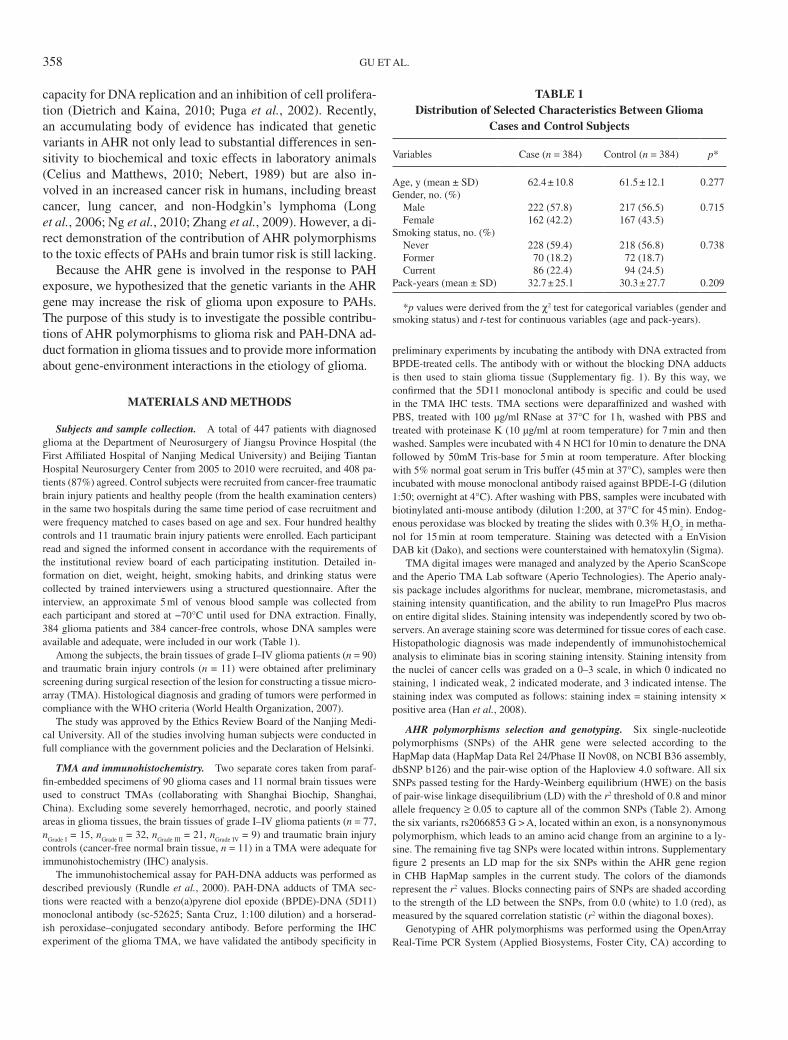
By detecting the distribution and staining intensity of PAH-DNA adducts in the TMA analysis using IHC, we found that PAH-DNA adduct staining existed in normal brain and gliomas of grades I–IV, and the staining intensity was significantly asso-ciated with the glioma grade (as shown in Fig. 1A). In the TMA IHC analysis, PAH-DNA adduct staining was observed in 15 of 23 (65.2%) pilocytic astrocytomas (Grade I), 32 of 35 (91.4%)
Fig. 1. The distribution and staining intensity of PAH-DNA adducts in the TMA analysis using IHC and the association with the glioma grade. (A) Repre-sentative immunoperoxidase staining using 5D11 monoclonal antibody for PAH-DNA adducts of paraffin-embedded TMA; the adducts were detected in normal brain and gliomas of grades I–IV, and the staining intensity was significantly associated with the glioma grade. (B) The PAH-DNA adducts staining intensity index increased with increasing glioma grades (with a mean staining index value of 31.363 for normal, 66.146 for Grade I, 85.647 for Grade II, 156.597 for Grade III, and 190.714 for Grade IV). The Avg. indicates the mean values.
A
B
AHR SNPS AND THE GLIOMA/PAH-DNA ADDUCTS RISK 359
diffuse astrocytomas (Grade II), 21 of 21 (100%) anaplastic as-trocytomas (Grade III), and 9 of 11 (81.8%) glioblastoma multi-forme (GBM) (Grade IV). PAH-DNA adducts were detected in gliomas with substantial (over 10-fold) interindividual variability and primarily exhibited an endonuclear localization. The stain-ing intensity index increased with increasing glioma grades (with a mean staining index value of 31.363 for normal, 66.146 for Grade I, 85.647 for Grade II, 156.597 for Grade III, and 190.714 for Grade IV), which indicated the PAH-DNA adduct levels were elevated with an accompanied increase in tumor grade (Fig. 1B).
AHR Polymorphisms and Glioma Risk
Distribution of data on age, gender, and smoking status for the cases and controls are shown in Table 1. Three hundred eighty-four healthy controls frequency matched to the 384 gli-oma cases by age, gender, and smoking status (p = 0.277 for age, p = 0.715 for gender, and p = 0.738 for smoking status) are included. Table 2 shows the information on six genotyped SNPs of AHR, including the location, the minor allelic frequen-cies (MAF), HWE test, and genotyped rates.
The genotype and allele of AHR polymorphisms in the glioma cases and controls and their associations with the risk of adult glioma are shown in Table 3. All of the individuals of missing genotypes were excluded with a genotype failure rate of no more than 0.034. Two AHR polymorphisms (rs2066853 and rs2158041) demonstrated significant association with the risk of glioma as analyzed by the dominant model (variant-containing genotypes vs. common homozygote). Compared with rs2066853 GG homozygotes, carriers with the A allele exhibited more than a 1.39-fold increased risk of glioma occurrence (adjusted OR = 1.39; 95% CI = 1.04–1.85), and the risk increased significantly with an increasing number of variant alleles (AA homozygotes, adjusted OR = 1.81; 95% CI = 1.16–2.84; P-trend = 0.001). Similarly, individuals with the rs2158041 A allele showed a significant association with the risk of glioma compared with the GG homozygotes (adjusted OR = 1.71; 95% CI = 1.06–2.76; P-trend = 0.023), which indicated that the two polymorphisms contributed to glioma
risk. As to the other polymorphisms, no significant differences were found between the cases and the controls.
AHR Polymorphisms and PAH-DNA Adduct Levels in Glioma
Among the glioma patients, 77 brain tumor samples were available to evaluate the PAH-DNA adduct levels in glioma among different genotype groups of patients. The elevated PAH-DNA adduct levels in glioma correlated with rs2066853 risk genotypes (Fig. 2) and rs2158041 risk genotypes (Fig. 3). Associations between three patient groups with different genotypes of rs2066853 and the values of brain tissue PAH-DNA adduct levels based on 31 patients with GG genotype, 36 patients with GA genotype, and 10 patients with AA genotype were shown in Figure 2. The elevated PAH-DNA adduct levels were observed in the patients carrying
TABLE 3Genotype and Allele of AHR Polymorphisms in Cases and
Controls and Their Associations With Risk of Adult Glioma
Genotypes
Cases Controls
p valuea OR (95% CI)bn (%) n (%)
rs2066853 GG 154 (40.8) 188 (49.2) 0.019 1.00 (reference) GA 162 (43.0) 153 (40.1) 1.27 (0.93–1.73) AA 62 (16.4) 41 (10.7) 1.81 (1.16–2.84) GA/AA 224 (59.4) 194 (50.8) 1.39 (1.04–1.85) P for trend 0.001rs1476080 AA 118 (31.2) 125 (33.2) 0.423 1.00 (reference) AC 172 (45.5) 179 (47.5) 0.99 (0.72–1.38) CC 89 (23.5) 74 (19.6) 1.24 (0.83–1.85) AC/CC 261 (69.0) 253 (67.1) 1.07 (0.79–1.45) P for trend 0.268rs2158041 GG 182 (48.9) 211 (55.7) 0.063 1.00 (reference) GA 138 (37.1) 134 (35.4) 1.22 (0.89–1.66) AA 51 (13.7) 34 (9.0) 1.78 (1.10–2.86) GA/AA 189 (50.8) 168 (44.3) 1.33 (1.00–1.77) P for trend 0.023rs2106728 AA 225 (60.5) 221 (58.8) 0.787 1.00 (reference) AG 130 (34.9) 133 (35.4) 0.94 (0.70–1.28) GG 17 (4.6) 21 (5.6) 0.78 (0.40–1.52) AG/GG 147 (39.5) 154 (41.0) 0.92 (0.69–1.23) P for trend 0.551rs713150 CC 162 (42.7) 167 (43.8) 0.437 1.00 (reference) CG 157 (41.4) 166 (43.6) 0.96 (0.70–1.30) GG 60 (15.8) 48 (12.6) 1.27 (0.82–1.96) CG/GG 217 (57.3) 214 (56.2) 1.03 (0.77–1.37) P for trend 0.395rs6960165 AA 199 (52.2) 207 (54.3) 0.634 1.00 (reference) AG 154 (40.4) 142 (37.3) 1.15 (0.85–1.55) GG 28 (7.3) 32 (8.4) 0.92 (0.54–1.59) AG/GG 182 (47.8) 174 (45.7) 1.10 (0.83–1.47) P for trend 0.820
Note. Data in boldface represent p < 0.05.aFDR corrected p value.bAdjusted for age, smoking, and drinking status.
TABLE 2Information on Genotyped SNPs of AHR Gene
Evaluated in This Study
SNP IDNucleotide
change Function MAF*p value for HWE test
% Genotyped rate
rs2066853 G > A Exon 0.367 0.243 98.3rs1476080 A > C Intron 0.400 0.492 98.6rs2158041 G > A Intron 0.256 0.063 98.7rs2106728 A > G Intron 0.167 0.866 97.9rs713150 C > G Intron 0.381 0.502 97.9rs6960165 A > G Intron 0.233 0.280 97.7
Note. MAF, minor allelic frequency; HWE, Hardy-Weinberg equilibrium.*Minor allele frequency in the Chinese (CHB, Han Chinese in Beijing,
China) population, as reported in dbSNP database.
360 GU ET AL.

either one copy (mean ± SD, 110.49 ± 89.72 for GA carriers; p = 0.022) or two copies (mean ± SD, 202.62 ± 70.14 for AA carriers; p < 0.001) of the risk-associated rs2066853 A allele. In contrast, in patients not carrying the risk-associated allele (with the GG genotype), the adduct levels were significantly decreased (mean ± SD, 57.18 ± 59.82). In addition, associations between three patient groups with different genotypes of rs2158041 and the values of brain tissue with PAH-DNA adduct levels based on 38 patients with GG genotype, 29 patients with GA genotype, and 10 patients with AA genotype were also evaluated and are shown in Figure 3. Similarly, a gradual increase in PAH-DNA adduct levels in glioma was found in the three rs2158041 subgroups, and the increase was statistically significant. Compared with the rs2158041 GG-common homozygotes, rs2158041 AA-genotype carriers showed a significantly higher PAH-DNA adduct level (mean ± SD; 201.38 ± 63.48 for AA homozygotes vs. 78.62 ± 70.69 for GG homozygotes; p ≤ 0.001).
DISCUSSION
PAH-DNA Adducts Are Associated With Glioma Development Risk
An estimated two thirds of all cancer cases are linked to en-vironmental causes. PAHs are very widespread environmental
contaminants, and a number of PAHs and their derivatives have been recognized as major culprits causing an increased inci-dence of cancer (lung, skin, breast, and urinary cancers). How-ever, until this study, the association of PAHs exposure and the risk of glioma was still unclear.
Given the carcinogenicity of these PAHs and their presence in ambient air from a range of sources, a toxicological endpoint is required to investigate the potential health impact of PAHs in cancer risk. However, finding the appropriate toxicological endpoint to determine the exact PAHs exposure level and its effects in vivo is difficult. Accordingly, in spite of hundreds of PAHs that have been identified in atmospheric particles, only 33 PAHs are available for toxicological endpoint information, which possibly depends on the relatively rapid and complex me-tabolism and bodily distribution process of PAHs in mammals.
Herein, PAH-DNA adducts were measured as an endpoint biomarker to evaluate the effects of internal exposure to PAHs on glioma risk due to some great advantages to traditional ex-posure assessment (Godschalk et al., 2003; Ken-Dror, 2005). (1) PAH-DNA adducts smooth the extreme variability in ex-posure, which is typical for environmental toxicants, and may integrate exposure over longer periods of time. (2) More im-portant, PAH-DNA adducts not only account for all exposure routes but also account for interindividual differences in up-take, elimination, distribution, metabolism, and repair among
Fig. 2. Association between three patient groups with different genotypes of rs2066853 and the values of brain tissue PAH-DNA adducts level based on 31 patients with GG genotype, 36 patients with GA genotype, and 10 patients with AA genotype. The Avg. indicates the mean values. Significant difference measured by Kruskal-Wallis tests. *p < 0.05 compared with the GG groups, **p < 0.01 compared with the GG groups.
Fig. 3. Association between three patient groups with different genotypes of rs2158041 and the values of brain tissue PAH-DNA adducts level based on 38 patients with GG genotype, 29 patients with GA genotype, and 10 patients with AA genotype. The Avg. indicates the mean values. Significant difference measured by Kruskal-Wallis tests. *p < 0.05 compared with the GG groups.
AHR SNPS AND THE GLIOMA/PAH-DNA ADDUCTS RISK 361

exposed individuals. (3) Many scientific studies have justified the application of DNA adduct measurements as biomarkers in exposure assessment (Castano-Vinyals et al., 2004; Gyorffy et al., 2008). In this study, although the 5D11 antibody was produced in response to the adducts of the carcinogenic inter-mediate of BPDE-DNA, it cross-reacts with various affinities to other structurally related PAHs; hence, the terminology “PAH-DNA” is commonly used (Rybicki et al., 2004).
In our case-control study, cigarette consumption had no ef-fect on glioma development (Table 1), whereas PAH-DNA ad-duct levels were significantly associated with glioma grade. This inconsistency indicates that smoking is not the primary factor in PAH-DNA adduct formation and that environmental exposure (likely including ambient pollution, other inhaled products of organic combustion, and diet) may have a larger ef-fect on PAH-DNA adduct levels than smoking (Jia et al., 2011). Our results agree with a previous study that found no correla-tion between PAH-DNA adduct formation and smoking status (Pratt et al., 2011).
By detecting the staining intensity of PAH-DNA adducts in TMA, we found a significant association between DNA adduct formation and the risk of glioma. Furthermore, the PAH-DNA adduct staining intensity correlated with the tumor grade, sug-gesting PAH-DNA adduct formation may play a role in glioma carcinogenesis. The same findings were also reported in other cancers (Rybicki et al., 2006; Santella et al., 2000). Nonetheless, how PAHs and their derivatives cross the blood-brain barrier and the causal associations between PAH exposures, PAH-DNA adduct formation, and glioma risk remain to be established.
AHR Polymorphisms and Their Contributions to Cancer Susceptibility and DNA Adduct Formation
Since the discovery of the AHR, studies have determined that it can be activated by a wide range of classes of compounds including PAHs (Denison and Nagy, 2003; Ramadoss et al., 2005). AHR polymorphisms exist in the human population that will result in either forms of AHRs with higher binding affini-ties or forms that are more functionally efficient, thereby mak-ing their carriers more sensitive to environment compounds exposure (Maier et al., 1998).
Because of the wide distribution of AHR pathway proteins in human tissue and its potential role in carcinogenesis, we specu-lated that abnormalities in AHR action could contribute to glio-ma carcinogenesis. In the current study, we asked two questions: (1) whether AHR gene polymorphisms are associated with an increased risk of glioma and (2) whether polymorphisms in AHR result in altered PAH-DNA adduct formation, thereby affecting the rate of glioma carcinogenesis. In this study, six tag SNPs of the AHR gene, rs2066853, rs1476080, rs2158041, rs2106728, rs713150, and rs6960165, were evaluated. Only two of them, the rs2066853 and rs2158041 variants, were as-sociated with glioma risk.
The rs2066853 polymorphism is located in the transactivation domain and leads to a nonsynonymous amino acid substitution
in the AHR protein, which has been demonstrated to influence its function. Previous study observes that the AHR-R554K vari-ant displays an increased ability to induce the transcription of CYP1A1 activity in lymphocytes compared with common genotype after induction with 3-methylcholanthrene (Smart and Daly, 2000). However, conflicting evidence also exists. Some studies show that the R554K change does not alter the ability of AHR to regulate CYP1A1- or CYP1B1-driven transcrip-tion (Celius and Matthews, 2010). Previous results showed no associations of this variant and human breast cancer risk (Le Marchand et al., 2005; Long et al., 2006; Sangrajrang et al., 2009; Zhang et al., 2011). The lack of associations between this variant and non-Hodgkin’s lymphoma (De Roos et al., 2006; Ng et al., 2010) and lung cancer risk (Cauchi et al., 2001; Kawajiri et al., 1995) has also been reported. However, the Lys/Lys geno-type of rs2066853 (Arg554Lys) conferred a statistically signifi-cant higher risk of lung cancer in heavy smokers (Chen et al., 2009). In this study, we found that the variant genotype of the rs2066853 positively correlated to glioma risk (p = 0.019).
Additionally, AHR rs2158041, an intronic polymorphism in the AHR gene, was also associated with an increased glioma risk. This finding is consistent with recent results in lung can-cer research that demonstrate that the heterozygous genotype of rs2158041 was significantly associated with an increased risk of lung cancer compared with the common genotype (Chen et al., 2009). The molecular mechanism remains unclear. Perhaps this intronic polymorphism potentially influences the alternative spicing of the gene products (Hirose et al., 2008), or it might be in LD with other causal loci or genes. To identify additional SNPs that could be associated with glioma risk that may be in high LD with this SNP, we screened all of the common variants (with MAF > 0.05) within an approximately 50-kb-long region surrounding this site (~25 kb upstream and ~25 kb downstream of these loci) based on the CHB HapMap data resource. We found that rs2158041 is in LD with SNP rs6461312 (r2 = 0.825) which is located ~5.0 kb downstream of stop codon of AHR. Therefore, it is likely that the rs6461312 SNP near the 3' re-gion of the AHR gene may be the causal variant. However, the exact location and biological functions of the real causal SNPs in AHR are of great interest and warrant further investigation.
Intriguingly, we also found statistically significant associa-tions between these two variants (rs2066853 and rs2158041) and PAH-DNA adduct levels in glioma tissue. Together, these findings show that AHR is an important gene known to regu-late enzymes that metabolize PAHs and that AHR gene poly-morphisms may increase susceptibility to PAH-DNA adduct formation and glioma risk. Notwithstanding, multiple studies in experimental models and a number of epidemiological in-vestigations have demonstrated an association between DNA adduct formation and an increased cancer risk; our study pro-vides the first direct epidemiological evidence for the contri-bution of PAH-DNA adducts to glioma risk and also proposes the point for the first time that AHR genetic variants influence PAH-DNA adduct levels in glioma.
362 GU ET AL.

Further studies are needed to elucidate two mechanisms: (1) the mechanism by which retrospective parental exposure to PAHs affects the development of brain tumors in offspring and (2) the manner in which PAHs and their derivatives cross the blood-brain barrier and form PAH-DNA adducts. These ques-tions are of great interest and warrant further investigation.
In conclusion, in this case-control study in a Chinese popu-lation, we identified possible contributions of AHR rs2066853 and rs2158041 to glioma risk and the PAH-DNA adduct levels in glioma tissue. Although the etiology of glioma remains elu-sive, our findings are undoubtedly helpful to shed further light on gene-environment interactions conferring susceptibility to glioma and to better understand other cancers caused by PAHs. However, ongoing and future research with a large sample size and ethnically diverse populations is required to elucidate the impact of genetic variants of AHR on the susceptibility to glio-ma and their potential biological mechanism.
SUPPLEMENTARY DATA
Supplementary data are available online at http:// toxsci.oxfordjournals.org/.
FUNDING
National Natural Science Foundation of China (Grant No. 30901534, Grant No. 81172694, and Grant No.30901210); the Jiangsu Province’s Natural Science Foundation (Grant No. BK2009444); the Grant for the 135 Key Medical Project of Ji-angsu Province (No. XK201117); the National High Technology Research and Development Program 863 (No. 2012AA02A508), and Jiangsu Province’s Medical Major Talent program (No. RC2011051).
ACKNOWLEDGMENTS
We thank Prof. Binghua Jiang (Department of Pathology, Thomas Jefferson University, PA) for proofreading and editing the manuscript. No potential conflicts of interest were disclosed.
REFERENCES
Armstrong, B. G., and Gibbs, G. (2009). Exposure-response relationship be-tween lung cancer and polycyclic aromatic hydrocarbons (PAHs). Occup. Environ. Med. 66, 740–746.
Castano-Vinyals, G., D’Errico, A., Malats, N., and Kogevinas, M. (2004). Bio-markers of exposure to polycyclic aromatic hydrocarbons from environmen-tal air pollution. Occup. Environ. Med. 61, e12.
Cauchi, S., Stucker, I., Solas, C., Laurent-Puig, P., Cenee, S., Hemon, D., Jac-quet, M., Kremers, P., Beaune, P., and Massaad-Massade, L. (2001). Poly-morphisms of human aryl hydrocarbon receptor (AhR) gene in a French population: Relationship with CYP1A1 inducibility and lung cancer. Car-cinogenesis 22, 1819–1824.
Celius, T., and Matthews, J. (2010). Functional analysis of six human aryl hy-drocarbon receptor variants in human breast cancer and mouse hepatoma cell lines. Toxicology 277, 59–65.
Chen, D., Tian, T., Wang, H., Liu, H., Hu, Z., Wang, Y., Liu, Y., Ma, H., Fan, W., Miao, R., et al. (2009). Association of human aryl hydrocarbon receptor gene polymorphisms with risk of lung cancer among cigarette smokers in a Chinese population. Pharmacogenet. Genomics 19, 25–34.
Cordier, S., Monfort, C., Filippini, G., Preston-Martin, S., Lubin, F., Mueller, B. A., Holly, E. A., Peris-Bonet, R., McCredie, M., Choi, W., et al. (2004). Parental exposure to polycyclic aromatic hydrocarbons and the risk of child-hood brain tumors: The SEARCH International Childhood Brain Tumor Study. Am. J. Epidemiol. 159, 1109–1116.
De Roos, A. J., Gold, L. S., Wang, S., Hartge, P., Cerhan, J. R., Cozen, W., Yeager, M., Chanock, S., Rothman, N., and Severson, R. K. (2006). Meta-bolic gene variants and risk of non-Hodgkin’s lymphoma. Cancer Epide-miol. Biomarkers Prev. 15, 1647–1653.
Denison, M. S., and Nagy, S. R. (2003). Activation of the aryl hydrocarbon receptor by structurally diverse exogenous and endogenous chemicals. Ann. Rev. Pharmacol. Toxicol. 43, 309–334.
Dietrich, C., and Kaina, B. (2010). The aryl hydrocarbon receptor (AhR) in the regulation of cell-cell contact and tumor growth. Carcinogenesis 31, 1319–1328.
Godschalk, R. W., van Schooten, F. J., and Bartsch, H. (2003). A critical evalua-tion of DNA adducts as biological markers for human exposure to polycyclic aromatic compounds. J. Biochem. Mol. Biol. 36, 1–11.
Gu, A., Ji, G., Long, Y., Zhou, Y., Shi, X., Song, L., and Wang, X. (2011). Assessment of an association between an aryl hydrocarbon receptor gene (AHR) polymorphism and risk of male infertility. Toxicol. Sci. 122, 415–421.
Gyorffy, E., Anna, L., Kovacs, K., Rudnai, P., and Schoket, B. (2008). Correla-tion between biomarkers of human exposure to genotoxins with focus on carcinogen-DNA adducts. Mutagenesis 23, 1–18.
Han, C. P., Lee, M. Y., Tzeng, S. L., Yao, C. C., Wang, P. H., Cheng, Y. W., Chen, S. L., Wu, T. S., Tyan, Y. S., and Kok, L. F. (2008). Nuclear Receptor Interaction Protein (NRIP) expression assay using human tissue microar-ray and immunohistochemistry technology confirming nuclear localization. J. Exp. Clin. Cancer Res. 27, 25.
Hirose, Y., Chiba, K., Karasugi, T., Nakajima, M., Kawaguchi, Y., Mikami, Y., Furuichi, T., Mio, F., Miyake, A., Miyamoto, T., et al. (2008). A func-tional polymorphism in THBS2 that affects alternative splicing and MMP binding is associated with lumbar-disc herniation. Am. J. Hum. Genet. 82, 1122–1129.
Jia, Y., Stone, D., Wang, W., Schrlau, J., Tao, S., and Simonich, S. L. (2011). Estimated reduction in cancer risk due to PAH exposures if source control measures during the 2008 Beijing Olympics were sustained. Environ. Health Perspect. 119, 815–820.
Kawajiri, K., Watanabe, J., Eguchi, H., Nakachi, K., Kiyohara, C., and Hayashi, S. (1995). Polymorphisms of human Ah receptor gene are not involved in lung cancer. Pharmacogenetics 5, 151–158.
Ken-Dror, G. (2005). [DNA adducts as biological markers for human exposure to polycyclic aromatic compounds]. Harefuah 144, 583–587, 596.
Kyritsis, A. P., Bondy, M. L., Rao, J. S., and Sioka, C. (2010). Inherited predis-position to glioma. Neuro Oncol. 12, 104–113.
Le Marchand, L., Donlon, T., Kolonel, L. N., Henderson, B. E., and Wilkens, L. R. (2005). Estrogen metabolism-related genes and breast cancer risk: The multiethnic cohort study. Cancer Epidemiol. Biomarkers Prev. 14, 1998–2003.
Linnebank, M., Semmler, A., Moskau, S., Smulders, Y., Blom, H., and Simon, M. (2008). The methylenetetrahydrofolate reductase (MTHFR) variant c.677C>T (A222V) influences overall survival of patients with glioblastoma multiforme. Neuro Oncol. 10, 548–552.
Long, J. R., Egan, K. M., Dunning, L., Shu, X. O., Cai, Q., Cai, H., Dai, Q., Holtzman, J., Gao, Y. T., and Zheng, W. (2006). Population-based case-con-trol study of AhR (aryl hydrocarbon receptor) and CYP1A2 polymorphisms and breast cancer risk. Pharmacogenet. Genomics 16, 237–243.
AHR SNPS AND THE GLIOMA/PAH-DNA ADDUCTS RISK 363
Maier, A., Micka, J., Miller, K., Denko, T., Chang, C. Y., Nebert, D. W., and Al-varo, P. (1998). Aromatic hydrocarbon receptor polymorphism: Development of new methods to correlate genotype with phenotype. Environ. Health Per-spect. 106, 421–426.
Nebert, D. W. (1989). The Ah locus: Genetic differences in toxicity, cancer, mutation, and birth defects. Crit. Rev. Toxicol. 20, 153–174.
Nebert, D. W., Dalton, T. P., Okey, A. B., and Gonzalez, F. J. (2004). Role of aryl hydrocarbon receptor-mediated induction of the CYP1 enzymes in envi-ronmental toxicity and cancer. J. Biol. Chem. 279, 23847–23850.
Ng, C. H., Janoo-Gilani, R., Sipahimalani, P., Gallagher, R. P., Gascoyne, R. D., Connors, J. M., Weber, J. P., Lai, A. S., Leach, S., Le, N. D., et al. (2010). Interaction between organochlorines and the AHR gene, and risk of non-Hodgkin lymphoma. Cancer Causes Control 21, 11–22.
Ohgaki, H., and Kleihues, P. (2009). Genetic alterations and signaling path-ways in the evolution of gliomas. Cancer Sci. 100, 2235–2241.
Perera, F., Tang, D., Whyatt, R., Lederman, S. A., and Jedrychowski, W. (2005). DNA damage from polycyclic aromatic hydrocarbons measured by benzo[a]-pyrene-DNA adducts in mothers and newborns from Northern Manhattan, the World Trade Center Area, Poland, and China. Cancer Epidemiol. Biomarkers Prev. 14, 709–714.
Pratt, M. M., John, K., MacLean, A. B., Afework, S., Phillips, D. H., and Poirier, M. C. (2011). Polycyclic aromatic hydrocarbon (PAH) exposure and DNA adduct semi-quantitation in archived human tissues. Int. J. Environ. Res. Public Health 8, 2675–2691.
Puga, A., Marlowe, J., Barnes, S., Chang, C. Y., Maier, A., Tan, Z., Kerzee, J. K., Chang, X., Strobeck, M., and Knudsen, E. S. (2002). Role of the aryl hydrocarbon receptor in cell cycle regulation. Toxicology 181–182, 171–177.
Ramadoss, P., Marcus, C., and Perdew, G. H. (2005). Role of the aryl hydro-carbon receptor in drug metabolism. Expert Opin. Drug Metab. Toxicol. 1, 9–21.
Rundle, A., Tang, D., Hibshoosh, H., Estabrook, A., Schnabel, F., Cao, W., Gru-met, S., and Perera, F. P. (2000). The relationship between genetic damage from polycyclic aromatic hydrocarbons in breast tissue and breast cancer. Carcinogenesis 21, 1281–1289.
Rybicki, B. A., Nock, N. L., Savera, A. T., Tang, D., and Rundle, A. (2006). Polycyclic aromatic hydrocarbon-DNA adduct formation in prostate car-cinogenesis. Cancer Lett. 239, 157–167.
Rybicki, B. A., Rundle, A., Savera, A. T., Sankey, S. S., and Tang, D. (2004). Polycyclic aromatic hydrocarbon-DNA adducts in prostate cancer. Cancer Res. 64, 8854–8859.
Sangrajrang, S., Sato, Y., Sakamoto, H., Ohnami, S., Laird, N. M., Khuhaprema, T., Brennan, P., Boffetta, P., and Yoshida, T. (2009). Genetic polymorphisms
of estrogen metabolizing enzyme and breast cancer risk in Thai women. Int. J. Cancer 125, 837–843.
Sanson, M., Marie, Y., Paris, S., Idbaih, A., Laffaire, J., Ducray, F., El Hallani, S., Boisselier, B., Mokhtari, K., Hoang-Xuan, K., et al. (2009). Isocitrate de-hydrogenase 1 codon 132 mutation is an important prognostic biomarker in gliomas. J. Clin. Oncol. 27, 4150–4154.
Santella, R. M., Gammon, M. D., Zhang, Y. J., Motykiewicz, G., Young, T. L., Hayes, S. C., Terry, M. B., Schoenberg, J. B., Brinton, L. A., Bose, S., et al. (2000). Immunohistochemical analysis of polycyclic aromatic hydrocarbon-DNA adducts in breast tumor tissue. Cancer Lett. 154, 143–149.
Shete, S., Hosking, F. J., Robertson, L. B., Dobbins, S. E., Sanson, M., Malmer, B., Simon, M., Marie, Y., Boisselier, B., Delattre, J. Y., et al. (2009). Genome-wide association study identifies five susceptibility loci for glioma. Nat. Genet. 41, 899–904.
Shimizu, Y., Nakatsuru, Y., Ichinose, M., Takahashi, Y., Kume, H., Mimura, J., Fujii-Kuriyama, Y., and Ishikawa, T. (2000). Benzo[a]pyrene carcinogen-icity is lost in mice lacking the aryl hydrocarbon receptor. Proc. Natl. Acad. Sci. U.S.A. 97, 779–782.
Smart, J., and Daly, A. K. (2000). Variation in induced CYP1A1 levels: Rela-tionship to CYP1A1, Ah receptor and GSTM1 polymorphisms. Pharmaco-genetics 10, 11–24.
Van Meir, E. G., Hadjipanayis, C. G., Norden, A. D., Shu, H. K., Wen, P. Y., and Olson, J. J. (2010). Exciting new advances in neuro-oncology: The avenue to a cure for malignant glioma. CA Cancer J. Clin. 60, 166–193.
Weller, M., Felsberg, J., Hartmann, C., Berger, H., Steinbach, J. P., Schramm, J., Westphal, M., Schackert, G., Simon, M., Tonn, J. C., et al. (2009). Mo-lecular predictors of progression-free and overall survival in patients with newly diagnosed glioblastoma: A prospective translational study of the Ger-man Glioma Network. J. Clin. Oncol. 27, 5743–5750.
Xue, W., and Warshawsky, D. (2005). Metabolic activation of polycyclic and heterocyclic aromatic hydrocarbons and DNA damage: A review. Toxicol. Appl. Pharmacol. 206, 73–93.
Zeegers, M. P., Swaen, G. M., Kant, I., Goldbohm, R. A., and van den Brandt, P. A. (2001). Occupational risk factors for male bladder cancer: Results from a population based case cohort study in The Netherlands. Occup. Environ. Med. 58, 590–596.
Zhang, B., Beeghly-Fadiel, A., Lu, W., Cai, Q., Xiang, Y. B., Zheng, Y., Long, J., Ye, C., Gu, K., Shu, X. O., et al. (2011). Evaluation of functional genet-ic variants for breast cancer risk: Results from the Shanghai breast cancer study. Am. J. Epidemiol. 173, 1159–1170.
Zhang, Y., Tao, S., Shen, H., and Ma, J. (2009). Inhalation exposure to ambient polycyclic aromatic hydrocarbons and lung cancer risk of Chinese popula-tion. Proc. Natl. Acad. Sci. U.S.A. 106, 21063–21067.
364 GU ET AL.

(CO)(PPh 3) 2 (Aryl = Ph, o-tolyl) are resistant to C-Si reductive elimination](https://img.pdfslide.us/doc/110x75/611abf595f158e49e46c8377/bioscience-engineering-2020-9-30-ed-silyl-aryl-osmiumii-complexes-ossioet.jpg)