Embed Size (px)
Citation preview
1
Contribution of topology determinants of a viral movement protein to its membrane 1
association, intracellular traffic and viral cell-to-cell movement 2
3
A. Genovés1, V. Pallás*, and J. A. Navarro 4
Instituto de Biología Molecular y Celular de Plantas, IBMCP (Universitat Politècnica de 5
València-Consejo Superior de Investigaciones Científicas) Avenida Ingeniero Fausto Elio, s/n, 6
46022, Valencia, Spain. 7
1Present address: Centro de Investigación Príncipe Felipe, Avenida Autopista del Saler, 16, 8
E-46013, Valencia, Spain. 9
10
11
12
13
Abstract word count: 242 14
Text word count: 6722 15
Running title: MNSV p7B topology determinants-function relationship 16
Corresponding author address: 17
Dr. Vicente Pallás 18
Instituto de Biología Molecular y Celular de Plantas (IBMCP). UPV-CSIC, Avenida Ingeniero 19
Fausto Elio, s/n, 46022, Valencia, Spain. 20
Telephone: 34 963877877, FAX: 34 963877859, e-mail: [email protected] 21
2
ABSTRACT 22
The p7B movement protein (MP) of Melon necrotic spot virus (MNSV) is a single-pass 23
membrane protein associated with the endoplasmic reticulum (ER), the Golgi apparatus (GA) 24
and plasmodesmata (Pd). Experimental data presented here revealed that the p7B 25
transmembrane domain (TMD) was sufficient to target the green fluorescent protein (GFP) to 26
ER membranes. In addition, the short extramembrane regions of p7B were essential for 27
subsequent ER export and transport to GA and Pd. Microsomal partitioning and bimolecular 28
fluorescence assays supported a type II topology of p7B in planta. Mutations affecting 29
conventional determinants of p7B membrane topology such as TMD secondary structure, 30
overall hydrophobicity profile, the so-called “aromatic belt” and the net charge distribution on 31
either side of the TMD were engineered into infectious RNAs to investigate the relationship 32
between the MP structure and MNSV cell-to-cell movement. Results revealed that: i) the 33
overall hydrophobic profile and the α-helix integrity of the TMD were relevant for virus 34
movement; ii) modification of the net charge balance of the regions flanking both TMD sides 35
drastically reduced cell-to-cell movement; iii) localization of p7B to GA was necessary but not 36
sufficient for virus movement and iv) membrane insertion was essential for p7B function in 37
virus movement. Our results therefore indicate that MNSV cell-to-cell movement requires a 38
sequential transport of p7B from the ER via the GA to Pd, which is modulated by a 39
combination of several signals with different strengths in the extramembrane regions and 40
TMD of the MP. 41
3
INTRODUCTION 42
The association of plant positive-strand RNA viruses with plant cell endomembranes is a 43
critical event that occurs during the virus life cycle. Many viral factors are proteins containing 44
hydrophobic regions that, generally, induce drastic modifications in endoplasmic reticulum 45
(ER) morphology, nuclear envelope and others membrane-bound organelles. These 46
cytopathic rearrangements either result in the formation of membrane-associated 47
invaginations, free cytoplasmic vesicles or multivesicular bodies that provide protective 48
environments and enlarged surfaces for not only genome replication, translation and particle 49
assembly but also for intracellular and cell-to-cell movement (1, 33, 39, 53). 50
How plant viruses take advantage of the intracellular endomembrane system to move 51
appears related to the structural characteristics of one or more virus-encoded movement 52
proteins (MPs) (37, 52, 55). For example, Tobacco mosaic virus (TMV) MP is a tightly 53
associated membrane protein that was proposed to contain two helical hydrophobic domains 54
spanning the ER membrane (8, 16). TMV MP together with TMV 126-kDa protein, which 55
respectively have movement and replication functions, cause a transient aggregation of the 56
ER resulting in the formation of cytoplasmic membrane bodies where TMV replicates (viral 57
replication complexes, VRCs) (23, 31). These viral factories travel toward the cell host 58
periphery in a cytoskeleton-dependent manner, resulting in entry into neighboring cells by 59
traversing the cell wall through the plasmodesmata (Pd) (24, 48). MP association with ER 60
membranes has also been described for viruses whose MPs induce tubule formation in Pd. 61
For example, Alfalfa mosaic virus (AMV) MP behaves as an integral membrane protein and 62
localizes to the ER (29). In addition, Prunus necrotic ringspot virus (PNRSV) MP has one 63
hydrophobic region that mediates its association with biological membranes. However, this 64
region is not thought to traverse the entire lipid bilayer, but rather is embedded in the 65
4
membrane interface with the N and C termini oriented toward the cytoplasm (41). The 66
intracellular transport and cell-to-cell movement of some flexible rod viruses belonging to nine 67
different genera are driven by the triple gene block of movement proteins (TGBp1, TGBp2 68
and TGBp3). TGBp1s are multifunctional RNA-binding proteins. All TGBp2s contain two 69
transmembrane domains (TMD) whereas TGBp3s form two main groups having either one 70
(potex-like group) or two (hordei-like group) TMDs. Several groups have reported that the 71
interactions among the three components of the TGB module might differ depending on virus 72
genera, with four models recently proposed to summarize the collective findings (57). 73
Regardless of the interaction model and virus, TGB2 and TGB3 proteins are always 74
associated with ER membranes and, occasionally, with ER-derived vesicles which move 75
along the actin network to reach the Pd (57). 76
Association of viral MPs with plant endomembranes has also been reported for p6 of Beet 77
yellows virus (BYV) and for p7B of Melon necrotic spot virus (MNSV) (20, 44). Both proteins 78
contain a very hydrophobic TMD that inserts into the ER membrane. Interestingly, the most 79
important difference between these MPs is that p7B moves from ER to Golgi apparatus (GA), 80
followed by targeting to Pd (20) while BYV p6 remains in the ER membrane (44). The ER-GA-81
Pd pathway described for p7B migration has also been observed for cellular proteins such as 82
the class 1 reversibly glycosylated polypeptides and the family of plasmodesmata-located 83
proteins (PDLP1) (49, 56). 84
The MNSV p7B is one of the two small MPs encoded by carmoviruses that are essential 85
for virus cell-to-cell movement (18). By using in vitro translation assays in the presence of 86
canine pancreas rough microsomes, it was recently demonstrated that membrane association 87
of p7B requires its TMD (42). Moreover, as occurs for most integral membrane proteins, the 88
p7B TMD adopts an α-helix conformation that is co-translationally inserted into the lipidic 89
5
bilayer through the ER translocon (40). The translocation machineries play a central role in 90
helix-bundle membrane protein topology controlling TMD insertion and orientation into lipid 91
bilayers (25). Total hydrophobicity and helix arrangement of the TMD together with the 92
distribution of aromatic and charged amino acids on either side of the hydrophobic region are 93
critical features that affect TMD-lipid bilayer interaction and, consequently, TMD recognition in 94
the ribosome-translocon channel (27, 34, 59, 60). 95
While viral MPs have been shown to interact with biological membranes, our knowledge 96
about the role of MP membrane topology in MP transport to Pd and in viral cell-to-cell 97
movement has been largely restricted to TMV-like MPs and the TGB system (28, 57). 98
Therefore, extending these studies to other viruses with different types of MPs should allow a 99
better understanding of virus-plant interactions. For this report, we investigated whether 100
membrane association, intracellular transport of p7B and MNSV cell-to-cell movement were 101
influenced by altering topological determinants of the MP. 102
MATERIALS AND METHODS 103
Construction of binary vectors for p7B membrane topology studies and site-directed 104
mutagenesis. MNSV p7B ORF was PCR-amplified from plasmid pMNSV(Al) (accession 105
number DQ339157) (18). Amino-terminal and carboxy-terminal fusions of MNSV p7B to the 106
N-terminal fragment (residues 1-155) of the yellow fluorescent protein (p7B-Nt[YFP] and 107
Nt[YFP]-p7B, respectively) were cloned into binary vector pMOG 800 between the CaMV 35S 108
promoter and the potato proteinase inhibitor II terminator (PoPit) (32). CaMV 35S expression 109
cassettes corresponding either to the ER-targeted Nt[YFP] (ER-Nt[YFP]) or to the ER-targeted 110
C-terminal fragment (residues 156-238) of the YFP (ER-Ct[YFP]) were obtained by HindIII-111
digestion of pRT-YN-ER and pRT-YC-ER, respectively (62), and then cloned into HindIII-112
linearized pMOG800. pRT-YN-ER and pRT-YC-ER plasmids were kindly provided by Dr Jari 113

6
P. T. Valkonen. Binary vectors expressing either Nt[YFP] or Ct[YFP] cytosolic fragments were 114
previously described (3). 115
Site-direct mutations were introduced into the p7B open reading frame (ORF) of the 116
pMNSV(Al)-∆cp-GFP clone from which infectious RNAs expressing the green fluorescent 117
protein (GFP) can be obtained (18, 19). For subcellular localization in N. benthamiana, 118
mutations were incorporated into the previously described binary vector expressing GFP-p7B 119
(20). For membrane orientation studies in N. benthamiana, several mutations were introduced 120
into binary vectors expressing either p7B-Nt[YFP] or Nt[YFP]-p7B. QuickChangeR XL-Site 121
Direct Mutagenesis Kit (Stratagene, La Jolla, California) and the primer pairs enumerated in 122
supplemental Table S1 were used. 123
In vitro transcription and plant inoculation. Viral cell-to-cell movement was assayed as 124
previously described (18, 19). Briefly, infectious RNAs were obtained from pMNSV(Al)-∆cp-125
GFP clones containing either the wild-type p7B or each mutant variant ORF (see 126
supplemental Table S1). Transcripts were inoculated in Cucumis melo (cv. Galia) cotyledons 127
transiently expressing MNSV coat protein (p42) (18, 19). At least, three independent 128
bioassays with five plants per each mutant RNA were performed. At three days post-129
inoculation (dpi), 20-30 images of individual fluorescent infection foci were taken with a 130
confocal microscope (Leica TCS SL, Leica Microsystems GmbH, Wetzlar, Germany). 131
Fluorescent infection areas were measured using ImageJ 1.41o software and data analyzed 132
with MS Excel. 133
Agrobacterium tumefaciens-mediated transient expression and bimolecular 134
fluorescence complementation assays. Transient expression assays on N. benthamiana 135
plants were performed as previously described (18). Briefly, the binary constructs were 136
7
introduced into the Agrobacterium tumefaciens strain C58C1 by electroporation (GenePulser 137
XcellTM electroporation system, Bio-Rad Laboratories, Hercules, California). Transformed 138
bacteria were grown overnight in a shaking incubator at 28ºC in Luria-Bertani (LB) medium 139
supplemented with the appropriate antibiotic mixture. Cultures were collected by slow-speed 140
centrifugation and adjusted to the required final OD600 value (0.2) with 10 mM MgCl2, 10 mM 141
MES pH 5.6 and 150 µM acetosyringone. These suspensions were infiltrated into two-week-142
old N. benthamiana plants by gentle pressure infiltration into the lower side of the leaves. For 143
co-localization and bimolecular fluorescence complementation experiments requiring the 144
simultaneous expression of two or more different proteins, individual bacterial cultures 145
containing the corresponding binary vectors were adjusted to a final OD600 of 0.2 and mixed 146
before leaf infiltration. Moreover, a gene silencing suppressor (HC-Pro) was added in BiFC 147
assays as reported (62). Plants were kept in growth chambers in 16h light at 25ºC and 8h 148
dark at 22ºC. 149
Membrane partitioning and immunoblotting analysis. Approximately 2 g of N. 150
benthamiana leaves transiently expressing either GFP-p7B or each mutant protein were 151
homogenized in lysis buffer containing 20 mM HEPES, pH 6.8; 150 mM potassium acetate; 152
250 mM mannitol and 1 mM MgCl2. Large cellular debris was removed by gentle 153
centrifugation at 3000 X g for 10 min, and the resulting supernatant was ultracentrifuged at 154
30000 X g for 1 h to generate the soluble (S30) and the microsomal (P30) fractions. For 155
chemical treatments, microsomal pellets were resuspended in 10 vol of 100 mM Na2CO3 (pH 156
11), 4 M urea, lysis buffer containing 1% Triton X-100 or original lysis buffer. After incubation 157
for 30 min on ice the samples were centrifuged at 30000 X g for 1 h. The resulting pellet was 158
resuspended in lysis buffer. All fractions were analyzed by SDS-PAGE in 12% polyacrylamide 159
gels, and subsequently transferred to polyvinylidene fluoride membranes (PVDF) (GE 160
8
Healthcare, Buckinghamshire, England) for immunoblotting with anti-GFP mouse polyclonal 161
antibody (Roche Diagnostics GmbH, Mannheim, Germany). 162
Confocal laser scanning microscopy. N. benthamiana leaf tissue was mounted in water 163
under a coverslip between 36 and 48 h following infiltration with Agrobacterium containing the 164
required constructs. All imaging was conducted with a Leica TCS SL confocal laser-scanning 165
microscope using an HCX APO 40x/0.90 w water dipping lens to study the subcellular 166
localization of the fluorescent tagged proteins. eGFP and YFP fluorescence was visualized by 167
488 nm excitation with a Kr/Ar laser and their emission was examined with a bandpass filter 168
for 500-530 nm. For imaging of ChFP and mRFP fluorescence, excitation at 514 nm (He/Ne 169
laser) was used whereas the emission was observed at 600-620 (ChFP) and 550-590 nm 170
(mRFP). Sequential scanning was used to suppress optical cross-talk between the 171
fluorophores in stationary structures colocalization assays. However, for dynamic structures, 172
simultaneous scanning was employed to avoid green and red color misalignment. 173
Computer-assisted analysis of secondary structure and transmembrane helices. 174
Prediction of transmembrane helices of MNSV p7B and mutant sequences was performed 175
with Membrane Protein Explorer (MPEx) software version 3.0 176
(http://blanco.biomol.uci.edu/mpex/). Free energy profile for membrane insertion of 177
hydrophobic transmembrane proteins was estimated with ∆G Prediction Server v1.0 178
(http://www.cbr.su.se/DGpred/). Secondary structure prediction of proteins was performed 179
with SSPro 2.01 software (SYMPRED server, http://www.ibi.vu.nl/programs/sympredwww/). 180
RESULTS 181
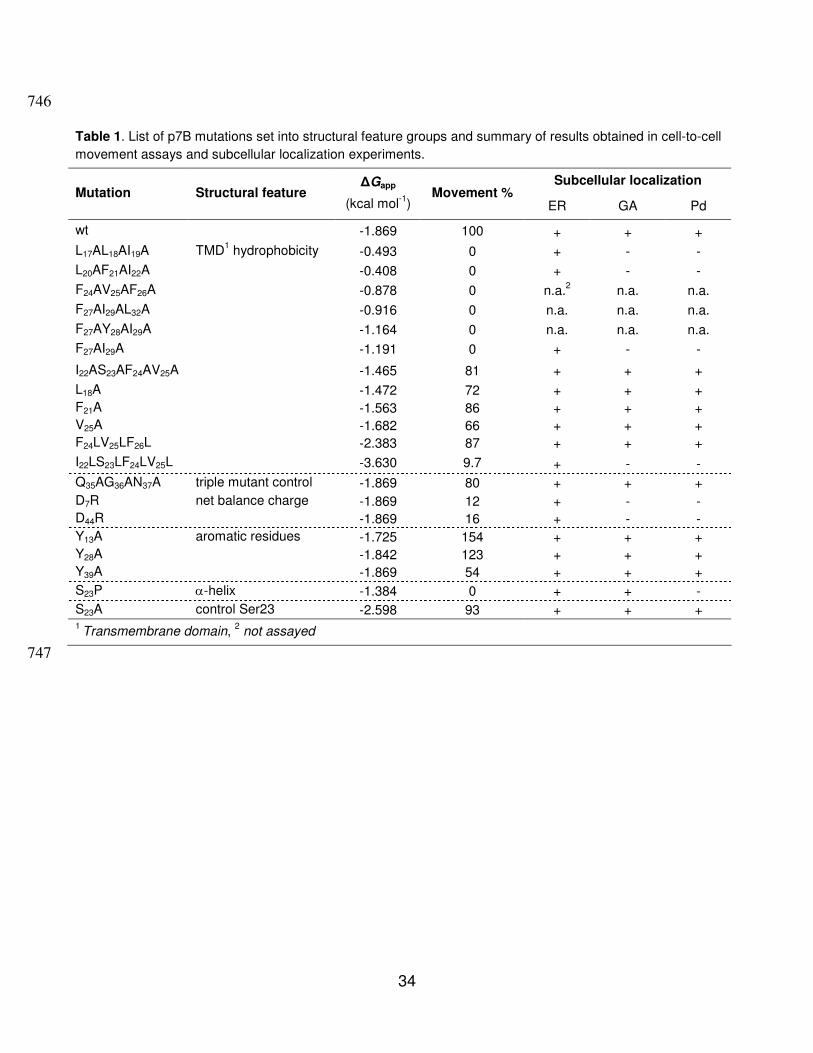
Structural characteristics of the MNSV p7B hydrophobic domain. 182
MNSV p7B is a single-pass membrane protein that inserts co-translationally into the 183
membrane of canine ER microsomes following a translocon-mediated pathway (40, 42). 184
9
Depending on the computer-based method used, the p7B TMD length oscillates from 18 to 23 185
residues (data not shown) although all predictions share a consensus hydrophobic core from 186
Y13 to L32 that mainly contains hydrophobic residues (A, I, V, L and F) (Fig. 1A). The 187
theoretical value calculated for the apparent free energy of the p7B TMD was negative 188
enough (∆Gapp = -1.869 kcal mol-1) to be efficiently integrated into the ER membrane (25, 27) 189
as previously reported (40). 190
Residues distribution and conformational preferences of the lateral chains determine the 191
precise positioning of transmembrane helices in cellular membranes. Residues such as 192
lysine, arginine, tryptophan and tyrosine in membrane-buried helices often extend their side 193
chain perpendicularly to the membrane bilayer and point away from the hydrophobic core 194
generating a snorkeling effect (11). According to these observations, the side chain of the Y28 195
residue is expected to snorkel out of the p7B TMD hydrophobic core. In addition, analyses of 196
TMDs from numerous membrane proteins have revealed that aromatic residues, in particular 197
tyrosine, have a propensity to mainly locate at or near the lipid-water interface of the 198
membrane (22, 35, 43). Therefore, the Y13 residue may reside at the p7B TMD helix end 199
where it favorably interacts with cellular environment. Aromatic residues in this position have 200
been referred to as “aromatic belts”. Their functional significance is not known, although it has 201
been speculated that aromatic belts perform an important function in membrane anchoring 202
and stabilization of the corresponding protein (25). Finally, turn-promoting proline residues are 203
excluded from the p7B TMD (26, 58). 204
Assessment of the MNSV p7B topology in plant ER membranes. 205
To study the integration of p7B in plant cell ER membranes, the p7B TMD (Y13-L32) was 206
cloned as a carboxy-terminal fusion to the green fluorescent protein (GFP-tmd(p7B)) (Fig. 1A) 207
and transiently expressed by agroinfiltration in N. benthamiana leaves. At 48 h post infiltration, 208
10
GFP-tmd(p7B) fluorescence appeared as an elaborate polygonal network (Fig. 1B) very 209
reminiscent to that observed with mGFP5-KDEL, a luminal ER marker (Fig. 1C). Remarkably, 210
GFP-tmd(p7B) was retained in the ER while GFP-p7B was fully exported from the ER and 211
located at cytoplasmic granules (Fig. 1D) which colocalized with Golgi stacks (Fig. 1E) (20). 212
STtmdChFP, a Golgi-specific reporter consisting in the transmembrane domain of the rat α-213
2,6-sialyltransferase fused to the cherry fluorescent protein was used for this co-localization 214
assay. Images here and elsewhere in the report, are representative of those observed in at 215
least three independent experiments. 216
We also analyzed the association of GFP-tmd(p7B) with cellular membranes by subcellular 217
fractionation. N. benthamiana tissue expressing GFP-tmd(p7B) was lysed and extracts were 218
separated by high-speed centrifugation into pellet (P30) and supernatant (S30) fractions. The 219
P30 fraction was resuspended and divided into several aliquots which were extracted with 220
either 0.1M Na2CO3 (pH 11) which would dislodge proteins entrapped within membrane 221
structures, 4M urea which would dislodge proteins peripherically bound to the membrane or 222
extracted with Triton X-100 which would release integral membrane proteins. Control 223
experiments were performed including GFP-p7B fusion and Beet yellows virus p6 fused to 224
GFP (GFP-p6) (20, 44). GFP-tmd(p7B), as well as GFP-p7B and GFP-p6 control proteins 225
behaved like typical integral membrane proteins because all of them were extracted from the 226
membrane fraction only after incubation with Triton X-100 (Fig. 1F). 227
In vitro and in vivo assays using canine microsomal membranes and prokaryotic cells 228
(Escherichia coli), respectively, revealed that p7B integrates into the ER membrane with an 229
Ncyt-Clum orientation (40). Since it has been shown that topological determinants of membrane 230
proteins are affected by lipid composition of host membranes, which can differ among animal 231
microsomes, the prokaryotic inner-membrane, and the ER membrane of plant cells (14), we 232
11
studied in vivo the p7B membrane orientation in the plant ER. We employed a novel 233
bimolecular fluorescence complementation (BiFC) based assay previously described for 234
topology studies of both BYV p6 and Potato mop-top virus-encoded integral membrane 235
TGBp2 (62). This technique relies on the formation of a fluorescent complex between a 236
fragment of the yellow fluorescent protein (YFP) targeted either to the cytoplasm or to the ER 237
luminal space and a counterpart fragment fused to the integral membrane protein N- or C-238
terminus. When overexpressed, the two YFP halves interact and therefore yield a fluorescent 239
YFP, if they are located in the same subcellular compartment (62). 240
Amino-terminal and carboxy-terminal fusions of p7B to the N-terminal YFP fragment (p7B-241
Nt[YFP] and Nt[YFP]-p7B, respectively) were transiently coexpressed with the complementary 242
C-terminal half of YFP, targeted either to the cytosol (Ct[YFP]) or to the ER lumen (ER-243
Ct[YFP]). Confocal microscopy analysis revealed that p7B-Nt[YFP] expression in the presence 244
of either Ct[YFP] or ER-Ct[YFP] only restored fluorescence, which clearly highlighted the ER 245
network, in the latter combination (Fig. 1G and 1H, respectively). According to single-pass 246
membrane status of p7B, the fluorescence was also observed when Nt[YFP]-p7B and Ct[YFP] 247
were expressed together (Fig. 1I). No fluorescence was visible for the Nt[YFP]-p7B and ER-248
Ct[YFP] combination (Fig. 1J). After the coexpression of Nt[YFP] and Ct[YFP], fluorescence 249
was observed in the cytoplasm and nuclei (Fig. 1K) indicating that expression level of each 250
split YFP-fragment was sufficient to allow fluorescence complementation (62). Alternatively, 251
fluorescence was confined to the ER network after the coexpression of ER-Nt[YFP] and ER-252
Ct[YFP] (Fig. 1L) indicating that both proteins were properly targeted to the ER lumen (62). 253
The coexpression of either Nt[YFP] and ER-Ct[YFP] or the ER-Nt[YFP] and Ct[YFP] 254
combination resulted in no fluorescence detection indicating that each YFP-split fragment was 255
fully located in the appropriate subcellular compartment (Fig. 1M and 1N, respectively). Taken 256
12
together our observations indicate that in plant cells, as occurs in heterologous systems, the 257
N-terminal region of p7B (residues 1-12) is exposed to the cytoplasm whereas the C-terminal 258
domain (residues 33-60) faces the ER luminal side (type II topology). 259
Contribution of the p7B topology determinants to MNSV cell-to-cell movement. 260
To assess the relevance of p7B topology determinants in MNSV cell-to-cell movement, a 261
series of site-directed mutations was introduced into the p7B ORF of the pMNSV(Al)-∆cp-GFP 262
clone (Fig. 2A) from which infectious transcripts expressing free GFP can be obtained (18). 263
Wild-type (Fig. 2B) and mutant (Fig. 2C to 2U and Table 1) MNSV RNAs were inoculated in 264
cotyledons of melon plants and local spread of infection was measured by quantifying the 265
fluorescent area. The results are presented according to the p7B topological determinant 266
affected. 267
Mutations affecting p7B TMD hydrophobicity. Since the most prominent p7B topological 268
determinant is the Y13-L32 hydrophobic region (30, 61), different mutations were directed to 269
modify TMD hydrophobicity. Amino acid replacement strategy was used instead of amino acid 270
deletion/insertion to preserve the p7B TMD length (Table S2) because the distribution of 271
single-pass membrane proteins along the organelles of the secretory pathway depends on 272
their TMD length (7). Hydrophobicity was reduced by engineering different alanine 273
replacements (A-replacement) of hydrophobic V, L and I as well as of aromatic F and Y 274
residues which were positioned between residues S14 and L32 (5). A-replacements, which 275
were initially made in groups of three, led to a 0.6-4 fold reduction of wild-type TMD 276
hydrophobicity (Table 1). TMD hydrophobicity of each mutant was measured as free energy 277
for membrane insertion using the “biological” scale determined by Hessa et al. (25). Upon 278
inoculation, all the triple mutants were completely arrested to single, initially infected cells (Fig 279
2C to 2F and Table 1). 280
13
Considering that a hydrophobicity threshold is required for accurate integration of TMDs, 281
we evaluated the relationship of this phenomenon with MNSV cell-to-cell movement. For this 282
purpose, p7B TMD hydrophobicity was progressively reduced by introducing double or single 283
A-replacements into the S14-L32 hydrophobic region. All mutants showing TMD free energies 284
higher than -1.191 kcal mol-1 (obtained with the F27AI29A mutant, Fig. 2G and Table 1) were 285
competent in cell-to-cell movement although the infected area slightly reduced (Fig. 2H to 2J 286
and Table 1) compared with wild-type RNA cell-to-cell movement (Fig 2B). To segregate the 287
putative adverse effect in the cell-to-cell movement generated by introducing multiple 288
mutations into p7B TMD from that produced by the TMD hydrophobicity reduction itself, 289
another p7B mutant was obtained in which positions 22 through to 25 were replaced with 290
alanines (the I22AS23AF24AV25A mutant). The resulting ∆Gapp was similar to that obtained with 291
the L18A mutant (-1.465 kcal mol-1 vs -1.472 kcal mol-1, respectively). Interestingly, this mutant 292
was functional in viral cell-to-cell movement (81% of the local movement in relation to the 293
wild-type RNA, Fig. 2K and Table 1). 294
In addition, the effect of increasing p7B TMD hydrophobicity in cell-to-cell movement was 295
also assessed. For this purpose, positions 24 through to 26 were altered to leucines instead 296
of to alanines (the F24LV25LF26L mutant) to slightly increment TMD hydrophobicity 297
(∆Gapp (3L) = -2.383 vs ∆Gapp(wt) = -1.869 kcal mol-1) (Table 1). In contrast to the 298
F24AV25AF26A mutant, this modification apparently had no effect on cell-to-cell movement (Fig. 299
2L and Table 1). However, a 2-fold increment of TMD hydrophobicity, which was obtained by 300
replacing positions 22 through to 25 with leucines (∆Gapp (4L) = -3.630 vs 301
∆Gapp(wt) = -1.869 kcal mol-1), drastically reduced viral movement (9.7%) (Fig. 2M and Table 302
1). As expected given the fact that residues I, L, V, F and A, in decreasing order, display the 303
14
highest α-helix propensities in a non polar environment (36), computer predictions obtained 304
using the SSPro 2.01 software (SYMPRED server) and Membrane Protein Explorer (MPEx) 305
software version 3.0, respectively show that the α-helix conformation and the TM length of 306
p7B TMD are not disturbed by mutations (Table S2). A feasible conclusion that could be 307
drawn from these results is that an appropriate p7B TMD hydrophobicity value is required to 308
MNSV cell-to-cell movement. 309
Mutation affecting p7B TMD secondary structure. To test whether the p7B TMD secondary 310
structure was relevant in MNSV cell-to-cell movement, the S23P mutant was constructed. 311
Statistical studies have revealed that α-helical membrane-buried regions of non channel 312
proteins are largely devoid of intramembrane proline residues (13). The cyclic structure of this 313
amino acid strongly restricts the conformational space resulting in a redirection of the peptide 314
chain that can distort α-helix structures (13). According to this observation, the S23P mutation 315
abolished MNSV cell-to-cell movement (Fig. 2N and Table 1) whereas only a slight reduction 316
of local spread was observed when the same serine was changed to alanine (Fig. 2O and 317
Table 1). Thus, structural requirements are also critical for efficient MNSV cell-to-cell 318
movement. 319
Mutations affecting the net charge distribution on either side of the p7B TMD. The 320
membrane orientation of transmembrane proteins is primarily determined by the net charge 321
balance in the 13 to 25 residues flanking both sides of the TMD, whereby the more positive 322
end is predominantly positioned to the cytoplasmic side of the membrane. This phenomenon 323
is known as the “positive-inside rule” (34). Since p7B shows an identical net charge balance (-324
1) on either side of the TMD, we decided to obtain the D7R and D44R mutants, which resulted 325
in a negative-to-positive inversion of the net charge balance (-1 to +1) in the Nt and Ct 326
15
regions, respectively, to study the contribution of the positive-inside rule in MNSV cell-to-cell 327
movement. Despite the D7R and D44R mutations possibly favoring wild-type and opposite p7B 328
membrane orientation, respectively, cell-to-cell movement was drastically reduced in both 329
cases (10-15%) (Fig. 2P and 2Q, respectively and Table 1). 330
Mutations affecting p7B aromatic residues. To study the implication of residues Y13 331
(putative “aromatic belt”) and Y28 (putative snorkeling effector) in MNSV cell-to-cell 332
movement, we constructed the Y13A and the Y28A mutants. Unexpectedly, infected 333
fluorescent area of both mutants was greater than that obtained with wild-type RNA (Fig. 2R 334
and 2S, respectively and Table 1) whereas half-reduction of the local movement relative to 335
the wild-type was observed in the Y39A mutant. The location of the residue Y39 in a 336
conserved β-sheet conformation (42), which might be involved in alternative functions that are 337
also relevant in cell-to-cell movement, could explain the discrepancies observed (Fig. 2T and 338
Table 1). In this sense, the Q35AG36AN37A mutation located downstream of the TMD but not 339
affecting the conserved region produced only a 20% reduction in local movement relative to 340
the wild-type (Fig. 2U and Table 1). 341
Distortion of the TMD secondary structure is the unique topological determinant 342
affecting p7B membrane association. 343
In order to study whether the different topological determinants analyzed above affect the 344
p7B membrane association, four representative mutants were analyzed. The selected 345
mutants included: i) either the L20AF21AI22A or I22LS23LF24LV25L mutation, which brought 346
about the greatest reduction or increase of TMD hydrophobicity, respectively; ii) the D44R 347
mutation, which generates a positive net charge balance in the p7B C-terminal domain; and 348
finally iii) the S23P mutation, which most likely distorts the membrane-spanning α-helix. All 349
16
four mutations were individually introduced into the GFP-p7B binary construct and transiently 350
expressed in N. benthamiana leaves. Membrane association status of mutant MPs was 351
characterized by subcellular fractionation followed by different treatments as described 352
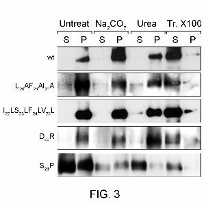
before. Neither the L20AF21AI22A, the I22LS23LF24LV25L or the D44R mutations had an effect on 353
membrane association given that the segregation between S and P fractions of mutant MPs 354
was like wild-type p7B (Fig. 3). With the S23P mutant, the subcellular fractionation analysis 355
revealed that a significant fraction of the protein (more than 50% of the signal) was recovered 356
in the soluble fraction with no solubilization treatment (Fig. 3). Moreover, the S23P mutant 357
fraction that remained associated with the membrane behaved like peripheral membrane 358
proteins because it was partially and fully extracted with alkaline and 4M urea solutions, 359
respectively (Fig. 3). 360
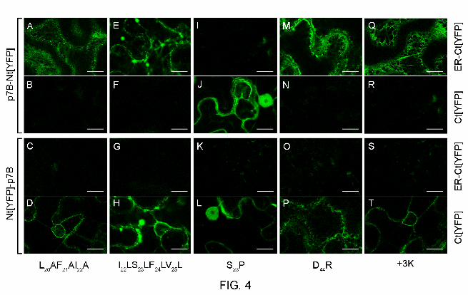
Next, we introduced the above mutations into the binary constructs expressing either 361
Nt[YFP]-p7B or p7B-Nt[YFP] to study membrane orientation. It has been previously 362
demonstrated that the N-terminus of a membrane protein is inserted first into the translocon 363
(21). Thus, very hydrophobic TMDs leave no time for N-terminal re-orientation resulting in 364
Nlum-Ccyt orientation whereas less hydrophobic TMDs delay their translocon exit leaving open 365
the possibility for re-orientation of the N-terminus (21). In this scenario, the L20AF21AI22A and 366
I22LS23LF24LV25L mutants favor wild-type and opposite orientation, respectively. However, 367
both mutants adopted wild-type orientation since ER fluorescence was observed when the 368
corresponding p7B-Nt[YFP] and Nt[YFP]-p7B mutants were co-expressed with ER-Ct[YFP] 369
and Ct[YFP], respectively, (Fig. 4A and 4D for the L20AF21AI22A mutant, Fig. 4E and 4H for the 370
I22LS23LF24LV25L mutant, respectively). Fluorescence was not observed in the opposite 371
combinations (Fig. 4B and 4C for the L20AF21AI22A mutant, Fig. 4F and 4G for the 372
I22LS23LF24LV25L mutant, respectively). On the other hand, either p7B(S23P)-Nt[YFP] or 373
17
Nt[YFP]-p7B(S23P) together with Ct[YFP] resulted in both nuclear and cytoplasmic 374
fluorescence (Fig. 4J and 4L, respectively) most likely as a consequence of the presence of a 375
soluble fraction of the S23P mutant supporting the results previously observed in the 376
microsomal partitioning assay (Fig. 3). No fluorescence was visible with the p7B(S23P)-377
Nt[YFP] and ER-Ct[YFP] combination indicating that the C-terminal region of p7B(S23P)-378
Nt[YFP] is not translocated into the ER lumen (Fig. 4I). 379
The contribution of the positive-inside rule in p7B membrane orientation was assessed in 380
both the D44R mutant and the +3K insertion mutant that contains three extra lysine residues 381
after the S32 position. Despite both mutant proteins should adopt a wild-type opposite 382
orientation according to the positive-inside rule, no topology inversion was observed (Fig. 4M 383
to 4P for the D44R mutant and Fig. 4Q to 4T for the +3K mutant). These results contrast with 384
those previously reported in a prokaryotic system where topology inversion of a MNSV p7B-385
GFP recombinant protein harboring the +3K insertion was achieved (40). This discrepancy 386
may be due to several factors: i) differences in the composition of the anionic phospholipids 387
between the E. coli inner membrane and the ER membrane of plant cells; ii) the absence of 388
electrochemical potential in plant ER membranes; and iii) the possibility that different solutions 389
to membrane insertion would operate in different organisms. 390
According to that, only the disruption of the TMD secondary structure producing an 391
alteration of the p7B membrane topology might explain the previously observed cell-to-cell 392
movement inhibition. However, since the topology of p7B was not substantially affected in the 393
remaining mutants, no direct correlation between non-competent cell-to-cell movement 394
phenotype and appropriate p7B insertion in the ER membrane can be drawn. These results 395
suggest that other factors influencing the functionality of p7B in viral movement were affected 396
in these mutants. 397
18
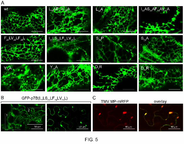
Trafficking of p7B to the cell periphery is affected at different stages in non-398
competent movement mutants. 399
Several studies have identified aromatic, charged and hydrophobic residues in addition to 400
TMD hydrophobicity as structural determinants controlling the intracellular trafficking of 401
proteins (4, 10). Thus, those mutations which were initially engineered to modify p7B topology 402
may have also affected the ER-GA-Pd pathway followed by p7B to reach the Pd (20). To test 403
this possibility, we introduced both competent and deficient movement mutations into the 404
GFP-p7B recombinant protein. Agrobacterium-mediated transient expression was used to 405
examine the subcellular localization of GFP-p7B mutants in N. benthamiana leaves. Until 36 h 406
post agroinfiltration, expression of all mutant proteins resulted in a reticular staining pattern 407
characteristic of ER localization (Fig. 5A and Table 1). Strong ER labeling was observed 408
except for GFP-p7B(S23P), which faintly labeled this cellular compartment (scan of this mutant 409
used a higher laser intensity than the rest). 410
At 48 h post agroinfiltration, ER labeling was scarcely observed except for GFP-411
p7B(I22LS23LF24LV25L). In this case, the ER staining underwent morphological changes 412
leading to the conversion of the tubular ER pattern into small cytoplasmic bodies that 413
collapsed into large aggregates (Fig. 5B and 6G). Similar behavior was previously reported for 414
TMV MP (45). When transiently expressed together in N. benthamiana leaves, TMV MP-415
mRFP and GFP-p7B(I22LS23LF24LV25L) colocalized in the same ER aggregates (Fig. 5C), 416
indicating that GFP-p7B(I22LS23LF24LV25L) induces rearrangements of the plant ER 417
membranes in contrast to GFP-p7B(wt). One possibility is that the I22LS23LF24LV25L mutation 418
retained the protein in the ER membrane, and that ER aggregates were an experimental 419
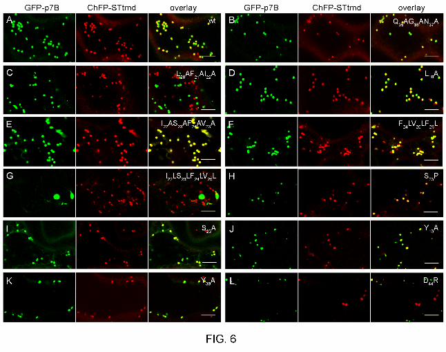
artifact caused by its overexpression. In marked contrast, the remaining mutants were mostly 420
localized in motile cytoplasmic vesicles (Fig. 6). These structures were visualized in 421
19
colocalization with the GA marker STtmdChFP but only when mutants were competent for 422
virus movement (Fig. 6A-B, 6D-F, 6I, 6J-K and Table 1). One exception was the S23P mutant, 423
which associated with the Golgi marker but the corresponding viral RNA did not move (Fig. 424
6H and Table 1). 425
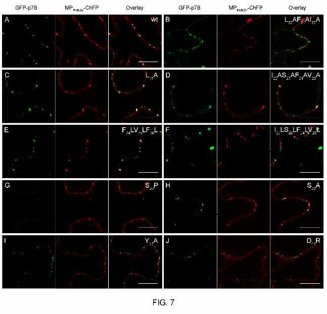
Localization of MP to GA appears to be important for virus movement but it is not sufficient 426
for this activity. To further analyze whether the p7B post-Golgi transport to Pds was affected, 427
we transiently co-expressed GFP-p7B (Fig. 7A) and the different mutants (Fig 7B-J) with the 428
PNRSV MP, a Pd marker (2). GFP-p7B holding competent movement mutations (Fig. 7C-D, 429
7E, 7H-I and Table 1) generated fluorescent peripheral punctuate structures which 430
colocalized with the Pd marker. By contrast, GFP-p7B holding non-competent movement 431
mutations including GFP-p7B(S23P) were not localized to Pds (Fig. 7B, 7F-G, 7J and Table 1). 432
Overall, these data indicate that the mutations inhibiting virus cell-to-cell movement affect the 433
intracellular pathway of p7B to the cell periphery at different stages including ER exit, GA 434
targeting and post-Golgi transport. 435
DISCUSSION 436
The final topology of membrane proteins is determined by a set of intricate interactions 437
between the structural determinants of the protein, which are recognized by the translocon 438
complex, and the particular properties of the destination membrane. These properties include 439
lipid composition, cholesterol concentration, membrane thickness and electrochemical 440
potential (6, 14). Host-specific chaperones such as TRAM may also be required for 441
membrane insertion of viral MPs (50). The high variability of these properties among different 442
organisms emphasizes the importance of studying membrane protein structure/function 443
relationships in the native lipid environment context. In this work, live-cell fluorescence 444
imaging was performed to investigate the topology of MNSV p7B, a plant virus MP, in the ER 445

20
membrane of plant cells. MNSV p7B has been previously shown to integrate co-translationally 446
into both canine pancreas microsomes and the E. coli inner membrane with an Ncyt-Clum 447
orientation (40, 42). We also demonstrated that GFP-p7B fusion was able to integrate into the 448
plant ER membrane. Nevertheless, information about the contribution of the hydrophobic 449
region to the process and about the orientation of the viral MP in the membrane is lacking 450
(20). 451
The results presented herein reveal that the p7B TMD can promote efficient ER targeting 452
and strong association of GFP with ER membranes, thus acting as an ER-targeting domain in 453
plant cells. Although the plant GA has been reported to be the possible default destination for 454
single-pass membrane proteins containing TMDs of about 19-20 amino acid in length (7), the 455
p7B TMD by itself was unable to promote GFP transport from the ER to GA, resulting in ER 456
retainment. Thus, MNSV p7B export from the ER appears to be controlled by specific export 457
motifs located in the cytoplasmic and/or luminal domains. In the absence of these dominant 458
motifs, ER retention may result from a better match between the lipid composition of the ER 459
membrane and p7B TMD length/hydrophobicity. The p7B TMD clearly differs from the TMD of 460
the plasmodesmata-located protein PLPd1a since this latter domain contains all the 461
information required for the intracellular transport of heterologous proteins to Pd via the 462
secretory pathway (56). Characterization of putative p7B ER export signals will be the subject 463
of further investigation since p7B ORF does not contain any of the classical ER export 464
sequences identified in single-spanning membrane proteins (4). A similar situation has been 465
recently reported for the rice secretory carrier membrane protein 1 (SCAMP 1) for which the 466
ER export signal is apparently unique (10). 467
On the other hand, there are many p7B structural properties that could participate in 468
establishing its final topology. Among them, the more prominent are the length, hydrophobicity 469
21
and α-helix configuration of the TMD as well as the net balance charge of the extra-470
membrane TMD proximal regions. For an efficient insertion into the lipid bilayer, protein 471
transmembrane segments require a minimum hydrophobicity threshold which must be 472
markedly high for ER-targeting sequences like the p7B TMD (38). Accordingly to this 473
assumption, a two-fold reduction of p7B TMD hydrophobicity was sufficient to eliminate cell-474
to-cell movement. Similar results were also obtained for the I22LS23LF24LV25L mutant, whose 475
thermodynamic properties for membrane insertion proved more favourable than that of the 476
wild-type sequence, strongly suggesting the need of an appropriate hydrophobicity value for 477
the p7B function in cell-to-cell movement. Intriguingly, both the insertion and membrane 478
orientation of the L20AF21AI22A and I22LS23LF24LV25L mutants, with the most reduced and 479
increased TMD hydropathy, respectively, were apparently not affected. Moreover, non-480
competent mutations which either lower or increase TMD hydrophobicity strongly correlate 481
with either incorrect intracellular targeting to unidentified cytoplasmic vesicles or a drastic 482
rearrangement of ER membranes, respectively, which impeded the ability of p7B to access 483
the Pd. It is well-known that plant viruses need to exploit Pd to move from cell to cell in a 484
process where virus-encoded movement proteins play a central role. Therefore, the ability of 485
p7B to function in viral movement most likely depends on Pd localization which is defective in 486
non competent cell-to-cell movement phenotypes. We can conclude that the p7B TMD 487
controls the insertion of the MP into the ER membrane and can also modulated the next 488
cellular destination of the MP in the presence of the ER export motifs located at the 489
cytoplasmic and/or luminal domains. 490
There are three different TMD determinants controlling the subcellular localization of a 491
membrane protein: the length of the TMD, the presence of specific sequence elements and 492
the overall hydrophobic profile (7, 9, 56). We found that the length of the predicted p7B TMD 493
22
was unaffected by mutations and that the p7B TMD itself was unable to transport GFP out of 494
the ER. Therefore, other biophysical features of the TMD, such as the overall hydrophobicity 495
profile, modulate the proper targeting of the MP. Accordingly, changes in the hydropathy 496
profile of the TMD present in ER-resident cytocrome P450 2C1 also cause mislocation to 497
different subcellular compartments in COS1 cells (54). Moreover, the sarcolemmal 498
membrane-associated protein (SLMAP) possesses two alternatively spliced transmembrane 499
regions (tail anchor TA1 or TA2); TA2 is less hydrophobic than TA1 and determines the 500
subcellular localization of the SLMAP in the mitochondria, while SLMAP-TA1 is targeted to the 501
ER. However, the TA2-4L mutant has a transmembrane region that is only slightly less 502
hydrophobic than the wild type TA1 and it shares similar TA1 targeting properties (9). It is 503
tempting to speculate that large variations below or above wild-type p7B TMD hydrophobicity 504
affect the balance between TMD-membrane lipids interaction and signal-mediated export 505
resulting in different outcomes depending on TMD strength. For example, p7B ER-retention 506
dictated by the strong TMD hydrophobicity of the I22LS23LF24LV25L mutant predominated over 507
the p7B ER-export signals located at the extramembrane domains. Addition of a diacidic 508
export motif suppresses the ER retention of the Yellow fever protein (YFV) E envelope protein 509
that is mediated by a TMD, but the same tagging did not override the ER retention signal of 510
the TMD from the YFV prM envelope protein (12). 511
Although many transmembrane α-helices contain bends and other distortions that have 512
important implications for protein function (47, 58), the proline-induced kink resulting from the 513
S23P mutation substantially affected the topology and intracellular transport of p7B as well as 514
viral cell-to-cell movement. The effect of the S23P mutation on MP membrane insertion was 515
most likely due to the central position of the proline within the TMD (25). The distorted helix 516
was likely too short to be well-accommodated into the hydrophobic core of ER membrane 517
23
generating a negative hydrophobic mismatch. In this situation, transmembrane helices can 518
adopt a surface orientation rather than a membrane-inserted state as occurred in S23P mutant 519
(46). Unexpectedly, this weak membrane association was sufficient to promote ER to GA 520
transport but not post-Golgi targeting to Pd. This indicates that membrane insertion is 521
essential for the p7B function in virus movement. 522
It is largely accepted that charged residues flanking the hydrophobic stretch strongly 523
modulate protein orientation relative to the membrane. A positive-inside rule seems to apply 524
universally to all the integral membrane proteins by which positively charged residues are 525
predominantly found in the flanking sequence that remains exposed to the cytosol. Our 526
results showed that p7B always adopts a Ncyt-Clum orientation in planta although no biased 527
distribution of positively charged residues between cytoplasmic and ER luminal segments was 528
observed. Mutations that violate the positive-inside rule can prevent insertion of a TMD. 529
However, p7B was efficiently inserted into the ER membrane in the correct membrane 530
orientation when a negative to positive inversion of the Ct domain net charge was obtained in 531
both the D44R and the +3K mutants. A similar override of the positive-inside rule has been 532
described for the polytopic channel that constitutes the ductin protein and mitochondrial inner 533
membrane proteins (15, 17). Critical contribution of charged residues to intracellular transport 534
of p7B and MNSV cell-to-cell movement most likely relates to its involvement in interactions 535
with host and/or viral factors. Interestingly, point mutations affecting charged residues 536
introduced into ER luminal or cytosolic segments of BYV p6 resulted in a drastic reduction of 537
cell-to-cell movement (44). However, neither the topology nor the subcellular localization of 538
these BYV p6 mutants was studied. 539
Orientation of proteins in membranes can also be controlled by both TMD length and 540
hydrophobicity. Long and very hydrophobic TMDs favor translocation of the N-terminus 541
24
across ER membranes (Nlum-Ccyt orientation) whereas short and less hydrophobic segments 542
adopt an Ncyt-Clum topology. However, a two-fold increase in TMD hydrophobicity of wild-type 543
p7B (I22LS23LF24LV25L mutant) did not result in the opposite membrane orientation. BYV p6 is 544
structurally very similar to MNSV p7B but it shows an Nlum-Ccyt orientation in plant ER 545
membranes. BYV p6 exhibits a positive charge bias (+2) for the cytoplasmic Ct domain and 546
its TMD is longer (23 residues) and more hydrophobic (∆Gapp(p6) = -6.094 kcal mol-1) than wild-547
type p7B TMD. Therefore, a simultaneous and/or a higher modification of the putative 548
topological determinants might be necessary to invert the p7B membrane orientation, 549
suggesting a strong stability of the MP in the ER membrane. Similar results have also been 550
reported for Carnation mottle virus p9 MP using a heterologous system (51). 551
Finally, according to the observation that movement-competent p7B mutations do not affect 552
p7B subcellular localization, both the Y13A and the Y28A mutants were properly targeted to 553
ER, GA and Pd but unexpectedly, local spread of the corresponding mutant RNAs was 554
significantly higher than that of wild-type RNA. The Y13 residue is located at the amino end of 555
the TMD. After membrane insertion, it is most likely to be positioned at the membrane-556
cytoplasm interface. Interfacial aromatic residues, commonly found among membrane 557
proteins, are important for the positioning and anchoring of the TMD in the lipid bilayer. In this 558
scenario, the precise positioning of p7B relative to the membrane does not appear essential 559
for its function but; nonetheless, the lack of anchoring restrictions can favor lateral mobility of 560
the p7B between different ER subdomains (for example, ER export sites or ERES) resulting in 561
an increased intracellular transport efficiency which, consequently, may facilitate MNSV local 562
infection progress. A similar effect can be produced when the snorkelling effect of Y28 was 563
removed. 564
25
In summary, the results presented herein demonstrate that the p7B Golgi-mediated traffic 565
to Pd is essential for efficient MNSV cell-to-cell movement. Moreover, the multiple structural 566
requirements of the MP appear to be involved in controlling Pd targeting. This probably 567
reflects the multiple interactions that must occur between cellular factors and this small MP to 568
guarantee functionality in the appropriate destination, the Pd. Similarly, multiple cytosolic and 569
transmembrane determinants are also required for SCAMP 1 trafficking via an ER/GA/trans-570
Golgi network/plasma membrane pathway (10). Therefore, the reduced size and simple 571
topology of p7B make this a useful system to gain a better understanding of not only the 572
trafficking mechanism of single transmembrane proteins but of the interactions produced 573
taking place between the viral protein and the cellular environment during the viral life cycle. 574
ACKNOWLEDGEMENTS 575
The work in our laboratory has been supported by grant BIO08-03528 from the Spanish 576
granting agency DGICYT and by grant ACOMP 2011-074 from the Generalitat Valenciana. J. 577
A. N. and A. G. are recipients of a postdoctoral contract and a PhD fellowship from the 578
Spanish Ministerio de Ciencia e Innovación. We thank Dra. M.C. Herranz for critical reading of 579
the manuscript and Dr V.V. Dolja (Oregon State University, Corvallis) for kindly providing the 580
STtmdChFP and MP(TMV)-mRFP binary vectors. PNRSV MP and mGFP5 binary vectors 581
were a generous gift of Dr. J. A. Sánchez (IBMCP, Valencia) and Dr. C. Torres (IBMCP, 582
Valencia), respectively. We thank L. Corachán and L. Latorre for their technical assistance. 583
26
REFERENCES 584
1. Adams, M.J. and J.F. Antoniw. 2005. Membrane proteins from plant viruses in Viral 585
membrane proteins: structure, function, and drug design, Wolfgang B. Fischer ed. Kluwer 586
Academic/Plenum Publishers, New York. pp 3-20. 587
2. Aparicio, F., V. Pallás and J.A. Sánchez-Navarro. 2010. Implication of the C terminus of the 588
Prunus necrotic ringspot virus movement protein in cell-to-cell transport and in its 589
interaction with the coat protein. J. Gen. Virol. 91:1865-1870. 590
3. Aparicio, F., J.A. Sánchez-Navarro and V. Pallás. 2006. In vitro and in vivo mapping of the 591
Prunus necrotic ringspot virus coat protein C-terminal dimerization domain by bimolecular 592
fluorescence complementation. J. Gen. Virol. 87:1745-50. 593
4. Barlowe, C. 2003. Signals for COPII-dependent export from the ER: what’s the ticket out?. 594
Trends Cell Biol. 13:295-300. 595
5. Bechinger, B. 2001. Membrane insertion and orientation of polyalanine peptides: A 15N 596
solid-state NMR spectroscopy investigation. Biophys. J. 81:2251-2256. 597
6. Brambillasca, S., ;M. Yabal, P. Soffientini, S. Stefanovic, M. Makarow, R.S. Hedge and N. 598
Borgesse. 2005. Transmembrane topogenesis of a tail-anchored protein is modulated by 599
membrane lipid composition. EMBO J. 24:2533-2542. 600
7. Brandizzi, F., N. Frangne, S. Marc-Martin, C. Hawes, J.M. Neuhaus and N. Paris. 2002. 601
The destination for single-pass membrane proteins is influenced markedly by the length of 602
the hydrophobic domain. Plant Cell 14:1077-1092. 603
8. Brill, L.M., R.S. Nunn, T.W. Kahn, M. Yeager and R.N. Beachy. 2000. Recombinant 604
Tobacco mosaic virus movement protein is an RNA-binding, alpha-helical membrane 605
protein. Proc. Natl. Acad. Sci. USA 97:7112-7117. 606
27
9. Byers, J.T., R.M. Guzzo, M. Salih and B.S. Tuana. 2009. Hydrophobic profiles of the tail 607
anchors in SLMAP dictate subcellular targeting. BMC Cell Biol. 10:48. 608
10. Cai, Y., T. Jia, S.K. Lam, Y. Ding, C. Gao, M.W. San, P. Pimpl and L. Jiang. 2011. 609
Multiple cytosolic and transmembrane determinants are required for the trafficking of 610
SCAMP1 via an ER-Golgi-TGN-PM pathway. Plant J. no. doi: 10.1111/j.1365-611
313X.2010.04469.x. 612
11. Chamberlain, A.K., Y. Lee, S. Kim and J.U. Bowie. 2004. Snorkeling preferences foster 613
an amino acid composition bias in transmembrane helices. J Mol Biol. 339:471-479. 614
12. Ciczora, Y., N. Callens, K. Séron, Y. Rouillé and J. Dubuisson. 2010. Identification of a 615
dominant endoplasmic reticulum-retention signal in yellow fever virus pre-membrane 616
protein. J. Gen. Virol. 91:404-414. 617
13. Deber, C.M. and A.G. Therien. 2002. Putting the β-breaks on membrane protein 618
misfolding. Nat. Struct. Biol. 9:318-319. 619
14. Dowhan, W. and M. Bogdanov. 2009. Lipid-dependent membrane protein topogenesis. 620
Annu. Rev. Biochem. 78:515-540. 621
15. Dunlop, J., P.C Jones and M.E. Finbow. 1995. Membrane insertion and assembly of 622
ductin: a polytopic channel with dual orientation. EMBO Journal 14:3609-3616. 623
16. Fujiki, M., S. Kawakami, R.W. Kim and R.N. Beachy. 2006. Domains of tobacco mosaic 624
virus movement protein essential for its membrane association. J. Gen. Virol. 9:2699-2707. 625
17. Gavel, Y. and G. von Heijne. 1992. The distribution of charged amino acids in 626
mitochondrial inner-membrane proteins suggests different modes of membrane integration 627
for nuclearly and mitochondrially encoded proteins. Eur. J. Biochem. 205:1207-1215. 628
18. Genovés, A., J.A. Navarro and V. Pallás. 2006. Functional analysis of the five Melon 629
necrotic spot virus genome-encoded proteins. J. Gen. Virol. 87:2371-2380. 630
28
19. Genovés, A., J.A. Navarro and V. Pallás. 2009. A self-interacting carmovirus 631
movement protein plays a role in binding of viral RNA during the cell-to-cell movement and 632
shows an actin cytoskeleton dependent location in cell periphery. Virology 395:133-142. 633
20. Genovés, A., J.A. Navarro and V. Pallás. 2010. The intra- and intercellular movement 634
of Melon necrotic spot virus (MNSV) depends on an active secretory pathway. Mol. Plant 635
Microbe Interact. 23:263-272. 636
21. Goder, V. and M. Spiess. 2003. Molecular mechanism of signal sequence orientation in 637
the endoplasmic reticulum. EMBO J. 22:3645-3653. 638
22. Granseth, E., G. von Heijne and A. Elofsson. 2005. A study of the membrane-water 639
interface region of membrane proteins. J. Mol. Biol. 346:377-385. 640
23. Guenoune-Gelbart, D., M. Elbaum, G. Sagi, A. Levy and B.L. Epel. 2008. Tobacco 641
mosaic virus (TMV) replicase and movement protein function synergistically in facilitating 642
TMV spread by lateral diffusion in the plasmodesmal desmotubule of Nicotiana 643
benthamiana. Mol. Plant Microbe Interact. 21:335-345. 644
24. Harries, P.A., J.W. Park, N. Sasaki, K.D. Ballard, A.J. Maule and R.S. Nelson. 2009. 645
Differing requirements for actin and myosin by plant viruses for sustained intercellular 646
movement. Proc. Natl. Acad. Sci. USA. 106:17594-17599. 647
25. Hessa, T., H. Kim, K. Bihlmaier, C. Lundin, J. Boekel, H. Andersson, I. Nilsson, S.H. 648
White and G. von Heijne 2005. Recognition of transmembrane helices by the endoplasmic 649
reticulum translocon. Nature 433:377-381. 650
26. Hessa, T., S.H. White, and G. von Heijne. 2005. Membrane insertion of a potassium 651
channel voltage sensor. Science 307:1427. 652
29
27. Hessa, T., N.M. Meindl-Beinker, A. Bernsel, H. Kim, Y. Sato, M. Lerch-Bader, I. 653
Nilsson, S.H. White and G. von Heijne. 2007. Molecular code for transmembrane-helix 654
recognition by the Sec61 translocon. Nature 450:1026-1030. 655
28. Huang, M., L. Jongejan, H. Zheng, L. Zhang and J.F. Bol. 2001. Intracellular 656
localization and movement phenotypes of Alfalfa mosaic virus movement protein mutants. 657
Mol. Plant Microbe Interact. 14:1063-1074. 658
29. Huang, M. and L. Zhang. 1999. Association of the movement protein of Alfalfa mosaic 659
virus with the endoplasmic reticulum and its trafficking in epidermal cells of onion bulb 660
scales. Mol. Plant-Microbe Interact. 12:680-690. 661
30. Higy, M., T. Junne and M. Spiess. 2004. Topogenesis of membrane proteins at the 662
endoplasmic reticulum. Biochemistry 43:12716-12722. 663
31. Kawakami, S., Y. Watanabe and R.N. Beachy. 2004. Tobacco mosaic virus infection 664
spreads cell to cell as intact replication complexes. Proc. Natl. Acad. Sci. USA 101:6291-665
6296. 666
32. Knoester, M., L.C. van Loon, J. van den Heuvel, J. Hennig, J.F. Bol and H.J.M. 667
Linthorst. 1998. Ethylene-insensitive tobacco lacks non-host resistance against soil-borne 668
fungi. Proc. Natl. Acad. Sci. USA 95:1933-1937. 669
33. Laliberté, J.F. and H. Sanfaçon. 2010. Cellular remodeling during plant virus infection. 670
Annu. Rev. Phytopathol. 48:69-91. 671
34. Lerch-Bader, M., C. Lundin, H. Kim, I. Nilsson and G. von Heijne. 2008. Contribution of 672
positively charged flanking residues to the insertion of transmembrane helices into the 673
endoplasmic reticulum. Proc. Natl. Acad. Sci. U S A 105:4127-4132. 674
30
35. Liang, J., L. Adamian and R. Jr. Jackups. 2005. The membrane–water interface region 675
of membrane proteins: structural bias and the anti-snorkeling effect. Trends Biochem. Sci. 676
30:355-357. 677
36. Liu, L.P. and C.M. Deber. 1999. Combining hydrophobicity and helicity: a novel 678
approach to membrane protein structure prediction. Bioorg. med. Chem. 7:1-7. 679
37. Lucas, W.J. 2006. Plant viral movement proteins: agents for cell-to-cell trafficking of 680
viral genomes. Virology 344:169-184. 681
38. Lundin, C., H. Kim, I. Nilsson, S. White and G. von Heijne. 2008. The molecular code 682
for protein insertion in the ER membrane is similar for Nin–Cout and Nout–Cin transmembrane 683
helices. Proc. Natl. Acad. Sci. USA 105:15702-15707. 684
39. Mandahar, C.L. 2006. Multiplication of RNA plant virus. Springer-Verlag ed. Dordrecht, 685
The Netherlands. 686
40. Martínez-Gil, L., A. Saurí, M. Vilar, V. Pallás and I. Mingarro. 2007. Membrane insertion 687
of the p7B movement protein of Melon necrotic spot virus (MNSV). Virology 367:348-357. 688
41. Martínez-Gil, L., J.A. Sánchez-Navarro, A. Cruz, V. Pallás, J. Pérez-Gil and I. Mingarro. 689
2009. Plant virus cell-to-cell movement is not dependent on the transmembrane disposition 690
of its movement protein. J. Virol. 83:5535-5543. 691
42. Navarro, J.A., A. Genovés, J. Climent, A. Saurí, L. Martínez-Gil, I. Mingarro, and V. 692
Pallás. 2006. RNA-binding properties and membrane insertion of Melon necrotic spot virus 693
(MNSV) double gene block movement proteins. Virology 356:57-67. 694
43. Nilsson, J., B. Persson and G. von Heijne. 2005. Comparative analysis of amino acid 695
distributions in integral membrane proteins from 107 genomes. Proteins 60:606-616. 696
31
44. Peremyslov, V.V., Y.W. Pan and V.V. Dolja. 2004. Movement protein of a closterovirus 697
is a type III integral transmembrane protein localized to the endoplasmic reticulum. J. Virol. 698
78:3704-3709. 699
45. Reichel, C. and R.N. Beachy. 1998. Tobacco mosaic virus infection induces severe 700
morphological changes of the endoplasmic reticulum. Proc. Natl. Acad. Sci. USA 701
19:11169-11174. 702
46. Ren, J., S. Lew, Z. Wang and E. London. 1997. Transmembrane orientation of 703
hydrophobic α-helices is regulated both by the relationships of helix length to bilayer 704
thickness and by cholesterol concentration. Biochemestry 36:10213-10220. 705
47. Riek, R.P., I. Rigoutsos, J. Novotny and R.M. Graham. 2001. Non-alpha-helical 706
elements modulate modulate polytopic membrane protein architecture. J. Mol. Biol. 707
306:349-362. 708
48. Sambade, A. and M. Heinlein. 2009. Approaching the cellular mechanism that supports 709
the intercellular spread of Tobacco mosaic virus. Plant Signal Behav. 4:35-38. 710
49. Sagi, G., A. Katz, D. Guenoune-Gelbart and B.L. Epel. 2005. Class 1 reversibly 711
glycosylated polypeptides are plasmodesmal-associated proteins delivered to 712
plasmodesmata via the golgi apparatus. Plant Cell 17:1788-800. 713
50. Saurí, A., P.J. McCormick, A.E. Johnson and I. Mingarro. 2007. Sec61alpha and TRAM 714
are sequentially adjacent to a nascent viral membrane protein during its ER integration. J. 715
Mol. Biol. 366:366-374. 716
51. Saurí, A., S. Tamborero, L. Martínez-Gil, A.E. Johnson and I. Mingarro. 2009. Viral 717
membrane topology is dictated by multiple determinants in its sequence. J. Mol. Biol. 718
387:113-128. 719
32
52. Scholthof, H.B. 2005. Plant virus transport: motions of functional equivalence. Trends 720
Plant Sci. 10:376-382. 721
53. Schwartz, M., J. Chen, W.-M. Lee, M. Janda and P. Ahlquist. 2004. Alternate, virus-722
induced membrane rearrangements support positive-strand RNA virus genome replication. 723
Proc. Natl. Acad. Sci. USA. 101:11263-11268. 724
54. Szczesna-Skorupa, E. and B. Kemper. 2000. Endoplasmic reticulum retention 725
determinants in the transmembrane and linker domains of cytochrome P450 2C1. J. Biol. 726
Chem. 275:19409-19415. 727
55. Taliansky, M., L. Torrance and N.O. Kalinina. 2008. Role of plant virus movement 728
proteins. Methods Mol. Biol. 451:33-54. 729
56. Thomas, C., E.M. Bayer, C. Ritzenthaler, L. Fernandez-Calvino, and A.J. Maule. 2008. 730
Specific targeting of a plasmodesmal protein affecting cell-to-cell communication. PLOS 731
Biology 6:1-11. 732
57. Verchot-Lubicz, J., L. Torrance, A.G. Solovyev, S.Y. Morozov, A.O. Jackson and D. 733
Gilmer. 2010. Varied movement strategies employed by triple gene block-encoding viruses. 734
Mol. Plant Microbe Interact. 23:1231-1247. 735
58. von Heijne, G. 1991. Proline kinks in transmembrane α-helices. J. Mol. Biol. 218:499-736
503. 737
59. von Heijne, G. 2006. Membrane-protein topology. Nat. Rev. Mol. Cell Biol. 7:909-918. 738
60. von Heijne, G. 2007. Formation of transmembrane helices in vivo -is hydrophobicity all 739
that matters? J. Gen. Physiol. 129:353-356. 740
61. White, S.H. and G. von Heijne. 2005. Transmembrane helices before, during and after 741
insertion. Curr. Opin. Struct. Biol. 15:378-386. 742
33
62. Zamyatnin, A.A.Jr., A.G. Solovyev, P.V. Bozhkov, J.P. Valkonen, S.Y. Morozov and 743
E.I. Savenkov. 2006. Assessment of the integral membrane protein topology in living cells. 744
Plant J. 46:145-154. 745
34
746
Table 1. List of p7B mutations set into structural feature groups and summary of results obtained in cell-to-cell
movement assays and subcellular localization experiments.
Subcellular localization Mutation Structural feature
∆Gapp
(kcal mol-1
) Movement %
ER GA Pd
wt -1.869 100 + + +
L17AL18AI19A TMD1 hydrophobicity -0.493 0 + - -
L20AF21AI22A -0.408 0 + - -
F24AV25AF26A -0.878 0 n.a.2 n.a. n.a.
F27AI29AL32A -0.916 0 n.a. n.a. n.a.
F27AY28AI29A -1.164 0 n.a. n.a. n.a.
F27AI29A -1.191 0 + - -
I22AS23AF24AV25A -1.465 81 + + +
L18A -1.472 72 + + +
F21A -1.563 86 + + +
V25A -1.682 66 + + +
F24LV25LF26L -2.383 87 + + +
I22LS23LF24LV25L -3.630 9.7 + - -
Q35AG36AN37A triple mutant control -1.869 80 + + +
D7R net balance charge -1.869 12 + - -
D44R -1.869 16 + - -
Y13A aromatic residues -1.725 154 + + +
Y28A -1.842 123 + + +
Y39A -1.869 54 + + +
S23P α-helix -1.384 0 + + -
S23A control Ser23 -2.598 93 + + + 1 Transmembrane domain,
2 not assayed
747
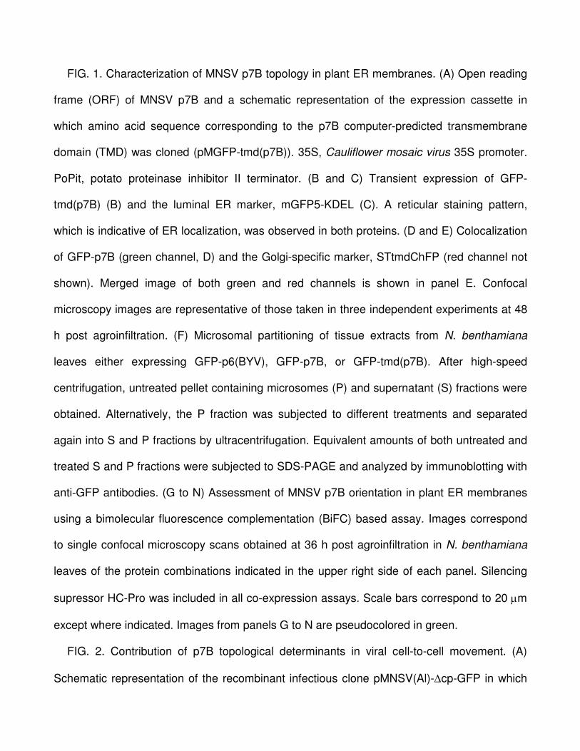
FIG. 1. Characterization of MNSV p7B topology in plant ER membranes. (A) Open reading
frame (ORF) of MNSV p7B and a schematic representation of the expression cassette in
which amino acid sequence corresponding to the p7B computer-predicted transmembrane
domain (TMD) was cloned (pMGFP-tmd(p7B)). 35S, Cauliflower mosaic virus 35S promoter.
PoPit, potato proteinase inhibitor II terminator. (B and C) Transient expression of GFP-
tmd(p7B) (B) and the luminal ER marker, mGFP5-KDEL (C). A reticular staining pattern,
which is indicative of ER localization, was observed in both proteins. (D and E) Colocalization
of GFP-p7B (green channel, D) and the Golgi-specific marker, STtmdChFP (red channel not
shown). Merged image of both green and red channels is shown in panel E. Confocal
microscopy images are representative of those taken in three independent experiments at 48
h post agroinfiltration. (F) Microsomal partitioning of tissue extracts from N. benthamiana
leaves either expressing GFP-p6(BYV), GFP-p7B, or GFP-tmd(p7B). After high-speed
centrifugation, untreated pellet containing microsomes (P) and supernatant (S) fractions were
obtained. Alternatively, the P fraction was subjected to different treatments and separated
again into S and P fractions by ultracentrifugation. Equivalent amounts of both untreated and
treated S and P fractions were subjected to SDS-PAGE and analyzed by immunoblotting with
anti-GFP antibodies. (G to N) Assessment of MNSV p7B orientation in plant ER membranes
using a bimolecular fluorescence complementation (BiFC) based assay. Images correspond
to single confocal microscopy scans obtained at 36 h post agroinfiltration in N. benthamiana
leaves of the protein combinations indicated in the upper right side of each panel. Silencing
supressor HC-Pro was included in all co-expression assays. Scale bars correspond to 20 µm
except where indicated. Images from panels G to N are pseudocolored in green.
FIG. 2. Contribution of p7B topological determinants in viral cell-to-cell movement. (A)
Schematic representation of the recombinant infectious clone pMNSV(Al)-∆cp-GFP in which
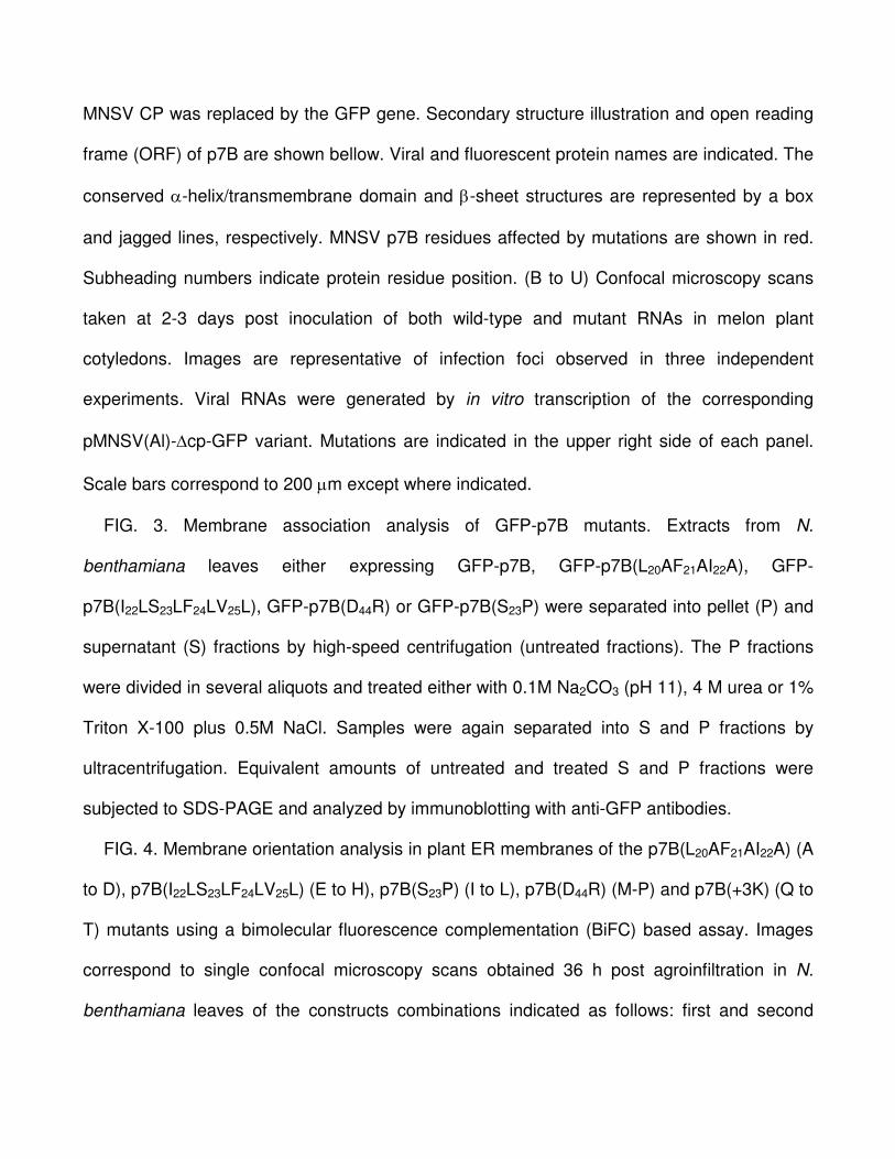
MNSV CP was replaced by the GFP gene. Secondary structure illustration and open reading
frame (ORF) of p7B are shown bellow. Viral and fluorescent protein names are indicated. The
conserved α-helix/transmembrane domain and β-sheet structures are represented by a box
and jagged lines, respectively. MNSV p7B residues affected by mutations are shown in red.
Subheading numbers indicate protein residue position. (B to U) Confocal microscopy scans
taken at 2-3 days post inoculation of both wild-type and mutant RNAs in melon plant
cotyledons. Images are representative of infection foci observed in three independent
experiments. Viral RNAs were generated by in vitro transcription of the corresponding
pMNSV(Al)-∆cp-GFP variant. Mutations are indicated in the upper right side of each panel.
Scale bars correspond to 200 µm except where indicated.
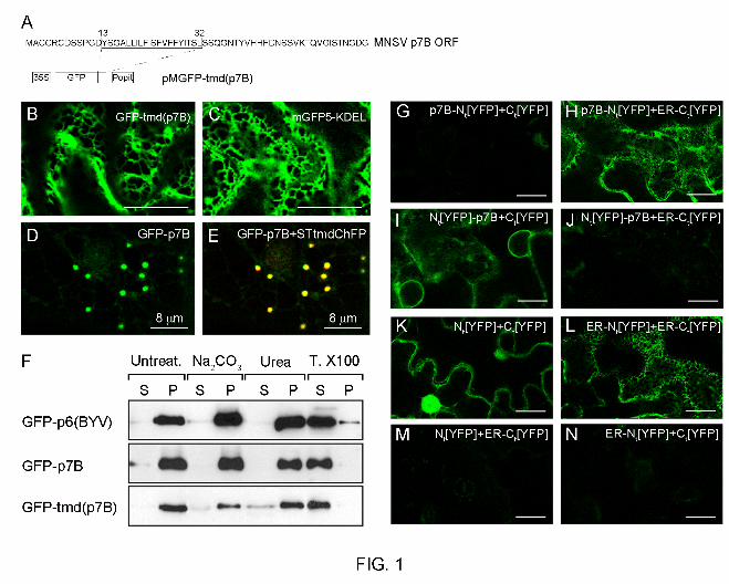
FIG. 3. Membrane association analysis of GFP-p7B mutants. Extracts from N.
benthamiana leaves either expressing GFP-p7B, GFP-p7B(L20AF21AI22A), GFP-
p7B(I22LS23LF24LV25L), GFP-p7B(D44R) or GFP-p7B(S23P) were separated into pellet (P) and
supernatant (S) fractions by high-speed centrifugation (untreated fractions). The P fractions
were divided in several aliquots and treated either with 0.1M Na2CO3 (pH 11), 4 M urea or 1%
Triton X-100 plus 0.5M NaCl. Samples were again separated into S and P fractions by
ultracentrifugation. Equivalent amounts of untreated and treated S and P fractions were
subjected to SDS-PAGE and analyzed by immunoblotting with anti-GFP antibodies.
FIG. 4. Membrane orientation analysis in plant ER membranes of the p7B(L20AF21AI22A) (A
to D), p7B(I22LS23LF24LV25L) (E to H), p7B(S23P) (I to L), p7B(D44R) (M-P) and p7B(+3K) (Q to
T) mutants using a bimolecular fluorescence complementation (BiFC) based assay. Images
correspond to single confocal microscopy scans obtained 36 h post agroinfiltration in N.
benthamiana leaves of the constructs combinations indicated as follows: first and second

panels rows correspond to coexpression of p7B-Nt[YFP] mutants with either ER-Ct[YFP] or
Ct[YFP], respectively; third and fourth panels rows correspond to coexpression of Nt[YFP]-
p7B mutants with either ER-Ct[YFP] or Ct[YFP], respectively. Mutations are indicated under
each panel column. Silencing supressor HC-Pro was included in all coexpression assays.
Scale bars correspond to 20 µm. Images are pseudocolored in green.
FIG. 5. (A) Subcellular localization of wild type and mutants of GFP-p7B in the endoplasmic
reticulum (ER). Confocal microscopy scans were taken at 36 h post agroinfiltration in N.
benthamiana leaves. Scale bar corresponds to 15 µm. A complete review of the results can
be found in the Table 1. Mutations are indicated in the upper left side of each panel. (B)
Confocal images of Nicotiana benthamiana epidermical cells expressing GFP-
p7B(I22LS23LF24LV25L) taken at 48 h post agroinfiltration. Images show how ER staining
obtained after GFP-p7B(I22LS23LF24LV25L) expression underwent morphological changes
leading to the conversion of the tubular ER pattern (region enclosed in the square 1 on the left
panel) into small cytoplasmic bodies (region enclosed in the square 2 on the left panel) that
collapsed into large aggregates (panel on the right). (C) Colocalization of TMV MP-mRFP
(panel on the left) and GFP-p7B(I22LS23LF24LV25L) (image not shown) into ER aggregates at
48 h post agroinfiltration in N. benthamiana leaves. The merged image of green and red
channels is shown (right panel).
FIG. 6. Coexpression of wild-type and mutants of GFP-p7B (“GFP-p7B” panel columns)
with the Golgi apparatus (GA) marker STtmdChFP (“STtmdChFP” panel columns). Merged
images of green and red channels are shown in the “overlay” columns. Confocal images
correspond to coexpression of the GA marker with either the wild type GFP-p7B (A), the
Q35AG36AN37A mutant (B), the L20AF21AI22A, L18A, I22AS23AF24AV25A, F24LV25LF26L and

I22LS23LF24LV25L mutants affecting p7B transmembrane domain hydrophobicity (TMD) (C to
G, respectively), the S23P mutant affecting the α-helix structure of the p7B TMD (H), the S23A
mutant used as control of the S23P mutant (I), the Y13A mutant lacking the Y13 putative
aromatic belt (J), the Y39A mutant affecting the conserved β-sheet structure at the p7B C-
terminal domain (K) or the D44R mutant displaying a C-terminal bias of positively charged
amino acids (L). Mutations are indicated in the overlay panels. Confocal microscopy scans
were taken at 48 h after agroinfiltration. Scale bars correspond to 8 µm. A complete review of
the results can be found in the Table 1.
FIG. 7. Coexpression of wild type and mutants of GFP-p7B (“GFP-p7B” panel columns)
with the plasmodesmata marker, PNRSV MP-ChFP (“PNRSV MP-ChFP” panel columns).
Merged images of green and red channels are shown in the “overlay” columns. Confocal
images correspond to coexpression of the Pd marker with either the wild-type GFP-p7B (A),
the L20AF21AI22A, L18A, I22AS23AF24AV25A, F24LV25LF26L or I22LS23LF24LV25L mutants affecting
p7B transmembrane domain (TMD) hydrophobicity (B to F, respectively), the S23P mutant
affecting the α-helix structure of the p7B TMD (G), the S23A mutant used as control of the
S23P mutant (H), the Y13A mutant lacking the Y13 putative aromatic belt (I) or the D44R mutant
displaying a C-terminal bias of positively charged amino acids (J). Mutations are indicated in
the overlay panels. Scale bars correspond to 8 µm. Confocal microscopy scans were taken at
48 h after agro-infiltration. A complete review of the results can be found in the Table 1.


![Viral and Epidemiological Determinants of the Invasion Dynamics … · 2017. 3. 23. · by the Aedes aegypti and Aedes albopictus mosquitoes vector [4]. Ae. aegypti is extremely well](https://img.pdfslide.us/doc/110x75/6140dc3483382e045471b81f/viral-and-epidemiological-determinants-of-the-invasion-dynamics-2017-3-23-by.jpg)