Embed Size (px)
Citation preview

doi:10.1152/jn.00101.2011 106:538-553, 2011. First published 11 May 2011;J NeurophysiolTerrence M. Wright, Jr. and Ronald L. Calabresefictive motor pattern generationContribution of motoneuron intrinsic properties to
You might find this additional info useful...
39 articles, 25 of which can be accessed free at:This article cites http://jn.physiology.org/content/106/2/538.full.html#ref-list-1
2 other HighWire hosted articlesThis article has been cited by
[PDF] [Full Text] [Abstract]
, November 30, 2011; 31 (48): 17555-17571.J. Neurosci.Terrence Michael Wright, Jr and Ronald L. CalabresePatterns of Presynaptic Activity and Synaptic Strength Interact to Produce Motor Output
[PDF] [Full Text] [Abstract], March 15, 2012; 107 (6): 1681-1693.J Neurophysiol
Rebecca C. Roffman, Brian J. Norris and Ronald L. CalabresegeneratorAnimal-to-animal variability of connection strength in the leech heartbeat central pattern
including high resolution figures, can be found at:Updated information and services http://jn.physiology.org/content/106/2/538.full.html
can be found at:Journal of Neurophysiologyabout Additional material and information http://www.the-aps.org/publications/jn
This information is current as of April 30, 2012.
American Physiological Society. ISSN: 0022-3077, ESSN: 1522-1598. Visit our website at http://www.the-aps.org/.(monthly) by the American Physiological Society, 9650 Rockville Pike, Bethesda MD 20814-3991. Copyright © 2011 by the
publishes original articles on the function of the nervous system. It is published 12 times a yearJournal of Neurophysiology
on April 30, 2012
jn.physiology.orgD
ownloaded from

Contribution of motoneuron intrinsic properties to fictive motorpattern generation
Terrence M. Wright, Jr. and Ronald L. CalabreseDepartment of Biology, Emory University, Atlanta, Georgia
Submitted 4 February 2011; accepted in final form 10 May 2011
Wright TM Jr, Calabrese RL. Contribution of motoneuron intrinsicproperties to fictive motor pattern generation. J Neurophysiol 106: 538–553,2011. First published May 11, 2011; doi:10.1152/jn.00101.2011.—Previ-ously, we reported a canonical ensemble model of the heart motoneurons thatunderlie heartbeat in the medicinal leech. The model motoneurons containeda minimal set of electrical intrinsic properties and received a synaptic inputpattern based on measurements performed in the living system. Although themodel captured the synchronous and peristaltic motor patterns observed inthe living system, it did not match quantitatively the motor output observed.Because the model motoneurons had minimal intrinsic electrical properties,the mismatch between model and living system suggests a role for additionalintrinsic properties in generating the motor pattern. We used the dynamicclamp to test this hypothesis. We introduced the same segmental input patternused in the model to motoneurons isolated pharmacologically from theirendogenous input in the living system. We show that, although the segmentalinput pattern determines the segmental phasing differences observed inmotoneurons, the intrinsic properties of the motoneurons play an importantrole in determining their phasing, particularly when receiving the synchro-nous input pattern. We then used trapezoidal input waveforms to show thatthe intrinsic properties present in the living system promote phase advancescompared with our model motoneurons. Electrical coupling between heartmotoneurons also plays a role in shaping motoneuron output by synchroniz-ing the activity of the motoneurons within a segment. These experimentsprovide a direct assessment of how motoneuron intrinsic properties interactwith their premotor pattern of synaptic drive to produce rhythmic output.
central pattern generator; dynamic clamp
RHYTHMIC MOTOR BEHAVIORS, such as swimming and walking,are generated by networks of rhythmically active neuronscalled central pattern generators (CPGs; Marder and Calabrese1996). A defining characteristic of these networks is theirability to generate a rhythmic pattern in in vitro preparations inwhich sensory feedback has been removed (i.e., the fictivepattern). The majority of CPGs provide a pattern of rhythmicsynaptic activation to the motoneurons that underlie the behav-ior. While much work has focused on identifying and analyz-ing elements of these rhythmically active networks, less isknown about how motoneurons themselves contribute to thegeneration of the motor pattern.
Some studies have shown that motoneurons possess intrinsicproperties that may affect motor pattern generation. For exam-ple, motoneurons in cats (Lee and Heckman 1998) and turtles(Hounsgaard and Kiehn 1989) exhibit bistable membrane prop-erties (i.e., a stable membrane potential at rest and at depolar-ized levels) that may confer upon them the ability to sustainmotor output in the absence of synaptic input. Kiehn et al.(2000) characterized a hyperpolarization-activated current, Ih,
present in motoneurons in the neonatal rodent spinal cord.They showed that this current advanced the transition of amotoneuron from its inhibited phase to its firing phase, sug-gesting that this current could confer upon a motoneuron anability to “escape” from ongoing inhibitory synaptic input andthus influence motor output.
Electrical coupling between motoneurons can also affectmotor pattern generation. For example, studies have shown thatthere is electrical coupling between hypoglossal motoneuronsassociated with breathing (Rekling et al. 2000) and tonguemovements (Sharifullina et al. 2005) as well as among mo-toneurons in Xenopus embryos (Perrins and Roberts 1995).Furthermore, when gap junctions were blocked in Xenopusembryos, Zhang et al. (2009) noted that the burst durations ofmotoneurons increased, resulting in alteration of rostro-caudaldelays during fictive swimming. These results indicate thatelectrical coupling in these preparations may contribute to themotor output observed in vitro.
Elucidating the functional significance of neuronal intrinsicproperties has remained elusive, however, owing in part to alack of detailed information about the activity patterns andsynaptic connections of premotor interneuronal networks thatprovide the synaptic information to the motoneurons in thesesystems. For example, in the absence of synaptic input, themotoneurons involved in fictive scratch in turtles exhibit large-amplitude voltage fluctuations in response to current injections,indicating that they possess complex intrinsic properties (Ala-burda et al. 2005). When the premotor network driving fictivescratch is activated, however, the voltage fluctuations observedare diminished because of the synaptic conductance impingingupon the motoneurons. This result implies that the intrinsicproperties responsible for the voltage oscillations make a minorcontribution to the motor pattern observed in vitro. Theseresults suggest that a detailed description of the premotorpattern of synaptic drive onto these motoneurons is required inorder to determine the role of motoneuron intrinsic propertiesin the generation of the fictive scratch.
Invertebrates, with their simple and accessible nervous sys-tems, have long been useful for elucidating synaptic connec-tivity within central pattern generating networks (Nusbaum andBeenhakker 2002) and how motor patterns are modulated andselected (DeLong et al. 2009). Here we use the leech heartbeatCPG to assess how motoneuron intrinsic properties and elec-trical coupling contribute to rhythmic motor output.
The leech heartbeat system has been described in detail(Kristan et al. 2005; Thompson and Stent 1976a, 1976b,1976c), so we provide a brief summary here. Blood flow in theleech circulatory system is accomplished by the rhythmicconstriction of a pair of longitudinal vessels, the lateral heart
Address for reprint requests and other correspondence: T. M. Wright, Jr.,Dept. of Biology, Emory Univ., 1510 Clifton Rd, NE, Atlanta, GA 30322(e-mail: [email protected]).
J Neurophysiol 106: 538–553, 2011.First published May 11, 2011; doi:10.1152/jn.00101.2011.
538 0022-3077/11 Copyright © 2011 the American Physiological Society www.jn.org
on April 30, 2012
jn.physiology.orgD
ownloaded from

tubes (referred to as “hearts”). The hearts are coordinated suchthat one heart constricts with a rear-to-front progression (i.e.,peristaltically), while the other heart constricts nearly synchro-nously along its length. The asymmetry in the constrictionpatterns is not permanent; rather, there are regular switches inthe constriction patterns roughly every 20–40 cycles. Theconstriction patterns of the hearts are the result of excitatorydrive arising ipsilaterally from segmental heart (HE) motoneu-rons (Thompson and Stent 1976a). Heart motoneurons occur aselectrically coupled bilateral pairs in midbody segmental gan-glia 3 through 18 of the 21 midbody ganglia in the ventralnerve cord. The heart motoneurons receive rhythmic inhibitoryinput from ipsilateral premotor heart (HN) interneurons that arecomponents of the heartbeat CPG. The firing pattern of theheart motoneurons (i.e., the fictive motor pattern) is alsobilaterally asymmetric: motoneurons on one side fire with arear-to-front progression of activity while the heart motoneu-rons on the other side fire nearly synchronously with theappropriate side-to-side coordination (Wenning et al. 2004a,2004b). The firing pattern of the premotor heart (HN) interneu-rons (i.e., the temporal pattern) is bilaterally asymmetric, withheart interneurons on one side firing with a rear-to-front pro-gression, while the interneurons on the other side fire nearlysynchronously.
Previously, Norris and colleagues (2006, 2007a, 2007b)quantified the individual components of presynaptic input tothe heart motoneurons as well as their output. In the first of aseries of studies, they quantified the temporal patterns (peri-staltic and synchronous) of the heart interneurons. They thencharacterized the pattern of synaptic strengths arising fromeach of the premotor interneurons onto their appropriate heartmotoneuron targets (i.e., a heart motoneuron’s synapticstrength profile). Finally, they quantified the phasing of theheart motoneurons with respect to the premotor interneurons.These studies led to the development of a first-generationmodel of the ensemble of heart motoneurons (Garcia et al.2008). The model motoneurons were single-compartment, con-ductance-based models and were given a minimal set of volt-age-gated conductances. The synaptic input pattern introducedto the model motoneurons was based on the Norris et al. (2006,2007a, 2007b) experiments just described. Although this first-generation model exhibited general trends in activity (i.e., aperistaltic and synchronous pattern of activity) as those ob-served in the living system, a quantitative comparison revealedsubstantive differences in phase between the model and theliving system (Fig. 9, Garcia et al. 2008). For example, inthe peristaltic mode (pink lines, Fig. 9, Garcia et al. 2008), themodel motoneurons do not capture the amount of peristalticphase progression observed in the living system; in anteriorsegments (e.g., 8), the phase of the model motoneurons leadsthe average of that in the living system, whereas in moreposterior segments (e.g., 14), the phase of the model motoneu-ron lags the average of that in the living system. In thesynchronous mode (blue lines, Fig. 9, Garcia et al. 2008),model motoneuron activity occurs nearly synchronously, butthe phase of the model motoneurons lags the average of that inthe living system in midbody segments 7–15, as illustrated inFig. 9 of Garcia et al. (2008). These results raise the possibilitythat the intrinsic properties of the heart motoneurons in theliving system, which were not accounted for in our modelmotoneurons, may be critical for their appropriate phasing.
To test this hypothesis, we used the dynamic clamp tech-nique (Prinz et al. 2004; Sharp et al. 1993) to construct hybridnetworks in which heart motoneurons in the living systemreceived the same synaptic conductance waveform introducedinto our model motoneurons (Fig. 1). We show that, althoughthe segmental input pattern determines the phasing differencesobserved in motoneurons in segments 8 through 14, the intrin-sic properties of the motoneurons also influence their phasingin the hybrid system. Indeed, appropriate phasing can beachieved when motoneurons receive the synchronous segmen-tal input patterns. We also explore how the heart motoneuronsin the living system integrate their inputs differently from ourcanonical model motoneurons. Finally, we show that electricalcoupling between heart motoneurons can influence their phas-ing. Taken together, these results show that motoneurons canbe active participants in motor pattern generation.
METHODS
Terminology. Heart (HE) motoneurons and heart (HN) interneuronsare indexed according to midbody ganglion number [e.g., HE(8),HN(4)]. In all experiments, we used bilateral pairs of heart motoneu-rons. We introduced the peristaltic input pattern to one heart motoneu-ron and the synchronous input pattern to the other heart motoneuron.Therefore, we omitted body-side indexing and labeled heart motoneu-rons as receiving the peristaltic or the synchronous input pattern.
Animals and solutions. Leeches (Hirudo sp.) were purchased froma commercial supplier (Leeches USA, Westbury, NY) and maintainedin artificial pond water at 15°C. Animals were anesthetized in ice andthen dissected in chilled saline. Individual ganglia from segments 8,10, 12, and 14 were dissected and pinned out, ventral surface up, in35-mm petri dishes lined with Sylgard (184, Dow Corning, Midland,MI). The ventral sheath of the ganglion was removed in all experi-ments. We superfused the preparation with leech saline containing (inmM) 115 NaCl, 4 KCl, 1.8 glucose, 10 HEPES buffer, and 1.8 CaCl2adjusted to a pH of 7.4 with NaOH at 1–2 ml/min in a bath volume of0.5–1 ml. All experiments were performed at room temperature(20–25°C). In most of the experiments included in this study, 10�4 Mbicuculline methiodide (Sigma-Aldrich, Allentown, PA) was added tothe leech saline to block inhibitory synaptic input to heart motoneu-rons (Cymbalyuk et al. 2002). In other experiments, CaCl2 wasreplaced with an equimolar amount of MnCl2 (Sigma-Aldrich) toblock the premotor inputs.
Intracellular recording techniques and data acquisition. Heartmotoneurons were identified based on soma location within theganglion, by soma size, and finally by their characteristic activity ofbouts of firing interrupted by barrages of inhibitory postsynapticpotentials (IPSPs). Intracellular voltage recordings from heart mo-toneurons were made with sharp intracellular microelectrodes(�25–40 M� filled with 2 M KAc, 20 mM KCl) made fromborosilicate glass (1.0-mm outer diameter, 0.75-mm inner diameter;AM Systems, Sequim, WA). Intracellular recordings and currentinjections were performed with an Axoclamp-2A amplifier (Molecu-lar Devices, Sunnyvale, CA) in discontinuous current clamp (DCC)mode with a sampling rate of 2.5–3.0 kHz. To ensure electrodesettling, the electrode potential was monitored with an oscilloscope.Output bandwidth of the amplifier was 0.3 kHz. Data were digitized(10-kHz sampling rate) with a digitizing board (Digi-data 1200 SeriesInterface, Molecular Devices) and acquired with pCLAMP software(Molecular Devices) on a personal computer (Dell, Round Rock, TX).
In all experiments, both heart motoneurons in a given ganglionwere impaled and recorded simultaneously. After penetration, theinput resistance of both cells was measured with �0.3-nA pulses. Wedid not proceed with experiments unless the input resistance of bothmotoneurons was �30 M� and the difference in input resistancebetween the two motoneurons was �15%. Upon termination of the
539MOTONEURON INTRINSIC PROPERTIES
J Neurophysiol • VOL 106 • AUGUST 2011 • www.jn.org
on April 30, 2012
jn.physiology.orgD
ownloaded from

experiment, the microelectrode was withdrawn from the cell and theelectrode potential was recorded. Only experiments in which theelectrode potential was within �5 mV of 0 mV were accepted in thisstudy. Therefore, membrane potentials are accurate to �5 mV.
Standard heart motoneuron ensemble model. We compared datafrom our physiological experiments with a model of the entire en-semble of heart motoneurons previously developed by Garcia et al.(2008). Briefly, the motoneurons in this model were single-compart-ment, conductance-based models whose membrane potential (V) isgiven by the following current-balance equation:
CdV
dt� ��INa � Ip � IKA � IK1 � IK2 � Ileak � Icoup � ISyn � Iinject�
where t is time, C is the total membrane capacitance, Ileak is the leakcurrent, Icoup is the current due to electrical coupling between themotoneurons, ISyn is the sum of the inhibitory synaptic currentsarising from each of the premotor inputs, and Iinject is any injectedcurrent. The model motoneurons contained five voltage-dependentionic currents: 1) a fast Na� current (INa), 2) a persistent Na� current(Ip), 3) a fast transient K� current (IKA), 4) an inactivating delayedrectifier K� current (IK1), and 5) a noninactivating delayed rectifierK� current (IK2). The Hodgkin-Huxley equations (Hodgkin andHuxley 1952) describing these voltage-gated currents were the sameas those used in a model of an oscillator heart interneuron (Hill et al.2001). Each motoneuron was modeled as an isopotential cylinderwhose length and diameter were both 60 �m with a specific mem-brane resistance of 1.1 �m2 and a specific membrane capacitance of0.05 Fm�2. With these parameters, the input resistance of a model
motoneuron was 97 M�. The maximal conductances of the individualionic currents as well as electrical coupling were set empirically sothat the activity of the model motoneurons mimicked that observedduring intracellular recordings of heart motoneurons in the absence ofsynaptic input (Garcia et al. 2008).
The model motoneurons received an inhibitory synaptic inputpattern that consisted of both timing information and a pattern ofsynaptic strengths; both components were determined from physio-logical experiments of the type performed by Norris and colleagues(2006, 2007a, 2007b) as described below.
For the model motoneurons, the firing pattern of the premotorinterneurons (referred to here as the temporal pattern) was taken from60 s of simultaneous extracellular recordings of the ipsilateral HN(3),HN(4), HN(6), and HN(7) premotor interneurons in both the peristal-tic and synchronous coordination modes, as in Norris et al. (2006).The peristaltic and synchronous input patterns were aligned to eachother to create a bilateral input pattern—left synchronous-right peri-staltic—by assigning a phase of 0.0 to the middle spike of the firstperistaltic HN(4) premotor interneuron burst [therefore, the peristalticHN(4) premotor interneuron is our absolute phase reference] and aphase of 0.506 to the middle spike of the first burst of the synchronousHN(4) premotor interneuron. These phase values match the averagephase difference between the two HN(4) interneurons as measured inthe living system (Norris et al. 2006). Each segmental pair of modelmotoneurons received the same temporal pattern (1 peristaltic, 1synchronous) offset by an intersegmental conduction delay of 20 msper segment. Therefore, the model heart motoneurons in segment 12receive the same temporal pattern as the model heart motoneurons in
Fig. 1. Hybrid system design and implementation. A: simul-taneous computed synaptic conductances arising fromeach of the premotor heart interneurons [gHN(i); colortraces] as well as the sum of these synaptic conductances(gSyn; black trace) for both the peristaltic and synchro-nous coordination modes. gSyn is the time-varying con-ductance [gSyn(t); see METHODS] introduced into theHE(10) motoneuron. The 2 gSyn traces illustrate thedifference between the peristaltic and synchronous inputpatterns. In the peristaltic mode gSyn rises and declinesgradually, as the firing of the premotor inputs are spreadout over time, while in the synchronous mode gSyn risesand falls rapidly, as the firing of the premotor inputsoccurs at nearly the same time. B: hybrid system setup.We recorded simultaneously from a pair of heart mo-toneurons [VHE(L,i) and VHE(R,i)] from a given segment andpharmacologically isolated the motoneurons from their pre-motor heart interneuron inputs (“X”; see METHODS). Thedynamic clamp computes and injects in real time theartificial equivalent of the appropriate synaptic current(Idc) into the heart motoneurons. In some experiments, thedynamic clamp was also used to compute Icoup. C: exemplardynamic clamp experiment and calculation of phase.Simultaneous intracellular recordings from a pair ofHE(10) motoneurons are shown. At the beginning of thevoltage recording, the heart motoneurons were firingtonically; gSyn is 0 nS. Once the dynamic clamp synapsewas activated (vertical line), the dynamic clamp injects atime-varying current proportional to the synaptic conduc-tance. In some figures, the dynamic clamp current (Idc) isomitted and only the synaptic conductance is shown. Thevertical green lines on the traces show the middle spikeof the peristaltic HN(4) interneuron (0/1—our phasereference) and of the synchronous HN(4) interneuron(0.5). The interval between the 2 green lines of our phasereference is the cycle period. The average phase for anindividual experiment here and in subsequent figures isindicated next to a filled diamond.
540 MOTONEURON INTRINSIC PROPERTIES
J Neurophysiol • VOL 106 • AUGUST 2011 • www.jn.org
on April 30, 2012
jn.physiology.orgD
ownloaded from

segment 8, offset by 80 ms. The period of the input pattern was 4.3 s[the range of periods measured in the living system is 4–8.5 s;average period � 5.3 s (Norris et al. 2006)]. Because the timinginformation used in our temporal pattern came from a living prepa-ration, the temporal pattern is not precisely regular, and therefore theaverage phases presented for the ensemble model display a variance.
The distribution of synaptic conductances elicited by each of thepremotor heart interneurons in a postsynaptic heart motoneuron (re-ferred to here as a heart motoneuron’s synaptic strength profile) wasalso derived from experiments in the living system, described in Fig.1B of Norris et al. (2007b). They recorded from each of the premotorheart interneurons, as described above, and then voltage clamped aseries of ipsilateral heart motoneurons. They recorded spontaneousinhibitory postsynaptic currents (IPSCs) in the heart motoneuronsarising from activity in the premotor interneurons. From these record-ings, they generated spike-triggered averages of the IPSCs for eachpresynaptic heart interneuron to each heart motoneuron. They selectedthe peak of the spike-triggered average trace as their measure of anindividual premotor heart interneuron’s synaptic input. These peakIPSCs were then converted to conductances [ESyn � �62.5 mV(Angstadt and Calabrese 1991)]. They then computed the averagesynaptic conductance across animals and expressed these averages aspeak synaptic conductances [gSynHN(i)]. There is no difference in thesynaptic strength profile between the synchronous and peristalticcoordination modes (Norris et al. 2007b). The set of four maximalconductances [gSynHN(i)] is unique to each segmental motoneuronpair; thus each motoneuron pair has a unique synaptic strength profile.Each model motoneuron received its segment-specific synapticstrength profile as in Fig. 3 of Garcia et al. (2008). Each presynapticheart interneuron spike elicited a unitary conductance that followed adouble exponential function scaled by the synaptic weight for thatinput in that motoneuron [gSynHN(i)]. The model computes gSyn(t)from the sum of the four individual inhibitory synaptic conductances[gSynHN(i)] associated with a particular presynaptic input HNi. Theheart motoneuron ensemble model with standard parameters (Garciaet al. 2008) is referred to as the canonical ensemble model.
The heart motoneuron ensemble model was implemented inGENESIS (GEneral NEural Simulator System), with each modelmotoneuron receiving its segment-appropriate temporal pattern andsynaptic strength profile. We ran the model for 60 s of model time.The model used the Euler integration method with a time step of0.0001 s. The 13 bouts of inhibitory synaptic input sculpted 12 burstsof activity of the model motoneurons. We used these 12 bursts toassess the phase of the model fictive motor pattern (see below). Werecorded and saved the computed synaptic conductance waveformsarising from each premotor HN interneuron as well as their sum(gSynTotal; Fig. 1) in each motoneuron for subsequent use in thedynamic clamp (see below).
Hybrid system design and implementation. We used the dynamicclamp technique (Prinz et al. 2004) to produce a virtual version of theheart interneuron-to-heart motoneuron synapse. The dynamic clampboth computes and injects, in real time (time step: 0.0001 s), a modelof the synaptic current (ISyn) based on the recorded intracellularmembrane potential (Vm), a conductance [gSyn(t)], and a reversalpotential (ESyn) according to Ohm’s law. Because we are linking amodel of this synapse with heart motoneurons in the living system, werefer to these preparations as hybrid systems. The virtual synapse wasimplemented according to the following equation:
ISyn � �gSyn�t��Vm � ESyn�where ISyn is the synaptic current, gSyn(t) is the time-varying synapticconductance waveform representing the sum of all the individualsynaptic inputs to a model motoneuron, � is an integer value used toscale gSyn(t), Vm is the membrane potential of the motoneuron, andESyn is the synaptic reversal potential (Angstadt and Calabrese 1991).To generate the synaptic conductance waveforms introduced in ourhybrid system experiments, we extracted gSyn(t) from canonical en-
semble model simulations (see above). For simplicity, we label gSyn(t)as gSyn in figures and text. Figure 1A shows how gSyn wasassembled by summing the individual time-varying synaptic con-ductance waveforms from each input to an HE(10) motoneuronpair. Figure 1A also illustrates the difference in the total synapticconductance trajectory (gSyn, black trace) between the peristaltic(Fig. 1A, top) and the synchronous (Fig. 1A, bottom) modes. In theperistaltic mode, the synaptic conductance trajectory rises and fallsslowly because the firing of the premotor interneurons is spread out,with the HN(7) interneuron leading the HN(3) interneuron, resultingin a gradual rise and decay of the synaptic conductance. In thesynchronous mode, however, the synaptic conductance trajectory risesand falls more precipitously because the firing of the premotorinterneurons occurs at nearly the same time, resulting in a much morerapid rise and decay of the synaptic conductance envelope. Thesynaptic conductance waveforms used in our hybrid system experi-ments were the same as in the ensemble model except they werescaled by �. The scaling factor allowed us to increase the overallsynaptic conductance while preserving the relative synaptic strengthof the individual premotor synaptic conductances. Unless indicatedotherwise, the canonical segmental input pattern was used both in thedynamic clamp and in the ensemble model.
In some experiments, we used the dynamic clamp to add to thenatural electrical coupling between heart motoneurons. Electricalcoupling was implemented according to:
Icoup � gcoup�Va � Vb�where Va and Vb each represent the membrane potentials of onemember of the pair of coupled heart motoneurons, Icoup is the couplingcurrent from cell b to cell a (and �Icoup is the coupling current fromcell a to cell b), and gcoup is the junctional conductance. Therefore, thetotal dynamic clamp current, Idc, introduced to a pair of heart mo-toneurons (Fig. 1B) is defined as:
Idc � ISyn � Icoup
where, unless otherwise noted, Icoup � 0 nA (i.e., gcoup � 0 nS).In another set of experiments, we varied the structure of the
premotor synaptic conductance waveform in order to study how thecoherence of the synaptic input pattern influences HE motoneuronphase. The temporal pattern of this synaptic input was based on thefiring pattern of the HN(4) interneurons (both peristaltic and synchro-nous, each with their appropriate phasing) only; in this way, we couldcompute phase using the same phase reference as in our otherexperiments. The synaptic strength profile associated with this tem-poral pattern was the sum of the individual premotor heart interneuronsynaptic conductances [i.e., gSyn � �gSynHN(i)]. The synaptic conduc-tance waveform was trapezoidal in shape. The onset of the conduc-tance envelope was triggered by the first spike of the HN(4) interneu-ron burst. The conductance envelope increased linearly from 0 nS togSyn during the first 500 ms of the HN(4) interneuron burst and thenreturned (termed “offset ramp conductance” in this study) to 0 nS1) abruptly at the last spike of the HN(4) interneuron burst, 2) duringthe last 250 ms of the HN(4) interneuron burst, or 3) during the last500 ms of the HN(4) interneuron burst. One heart motoneuronreceived this series of waveforms in order of increasing offset rampconductance while its contralateral partner was receiving the same setof waveforms in order of decreasing offset ramp conductance.
In hybrid system experiments, we blocked the endogenous synapticinput with 10�4 M bicuculline methiodide unless otherwise noted. Alldynamic clamp calculations were performed with a real-time dedi-cated processing board (DS1104; dSPACE, Detroit, MI). We acti-vated the dynamic clamp synapses and electrical coupling only whenthe motoneurons were spiking tonically and had no discernible IPSPsin the voltage recording (Fig. 1C). In our hybrid system experiments,we 1) bilaterally varied the synaptic scaling factor (�) introduced to apair of heart motoneurons, 2) varied the origin of the segmental input
541MOTONEURON INTRINSIC PROPERTIES
J Neurophysiol • VOL 106 • AUGUST 2011 • www.jn.org
on April 30, 2012
jn.physiology.orgD
ownloaded from

pattern introduced into the same heart motoneuron pair, 3) varied thecoupling conductance, gcoup, between heart motoneurons within asegment, or 4) introduced modified synaptic conductance patterns toheart motoneurons (see above). In experiments 1–3, we introduced 13cycles of inhibitory synaptic conductance into a heart motoneuronpair, yielding 12 bursts of activity over 60 s. In experiment 4, weintroduced 10 cycles of inhibitory synaptic conductance over 45 s,yielding 9 bursts of heart motoneuron activity.
Data analysis. Electrophysiological data were analyzed off-linewith a combination of pCLAMP 9.2 (Molecular Devices) and customscripts written in Matlab (The Mathworks, Natick, MA) and Spike2(CED Systems, Cambridge, UK). First, the raw voltage recordingswere high-pass filtered (cutoff frequency �1 Hz). These data werethen used for spike detection. Spike detection was carried out bymethods reported previously (Norris et al. 2006).
After detection, spikes were grouped into bursts as follows: Afteran interburst interval of 500 ms, the next spike was deemed the firstspike of that burst. Each subsequent spike was included in that burstuntil the interspike interval became � 500 ms (interburst interval). Aminimum of four spikes were required in order to qualify as a burst.In some experiments, the dynamic clamp-mediated inhibitory synapticcurrent injected into a heart motoneuron did not inhibit it sufficiently,so that the heart motoneuron continued firing, but at a low frequency,during the inhibited phase of its oscillations (i.e., during the peakdynamic clamp injected current). In those recordings, we removed thissmall number (usually � 4) of spikes during the trough of theinhibited phase to create a sufficient interburst interval for burstdetection.
We defined period as the interval between successive middle spikesof the peristaltic HN(4) interneuron [THN(4)]. We then computed thephase of the heart motoneurons with respect to the synaptic inputpattern that they received. We defined phase as the difference in timefor a spike of interest of a heart motoneuron and the time of the middlespike of the phase reference, the peristaltic HN(4) interneuron[tHE(i)f,m,l�HN(4)]. This difference is then normalized to the period ofthe phase reference. Thus phase is given by:
�f,m,l � ��tHE�i�f,m,l�HN�4� ⁄ THN�4��We calculated the average first (f), middle (m), and last (f) spikephases, burst period (T), and duty cycle (D) for each heart motoneuronrecorded. In the text and figures, the generic term phase and symbol� are applied to the middle spike phase as defined above. In figures,we indicate the middle spike phase within each heart motoneuronburst by a filled diamond above that burst. All phase values areexpressed modulo one. Duty cycle is defined as the difference be-tween the average last spike phase and the average first spike phase:
D � �last � �first
Because the duty cycle is the difference between two averages,standard deviation is not reported.
Statistics. Data were compiled and analyzed with Microsoft Excel(2010, Microsoft, Redmond, WA), SigmaPlot 11 (Systat Software,San Jose, CA), Minitab (v14, Minitab, State College, PA), or Matlab(The Mathworks). We generated an average phase and duty cycle foreach preparation, and the average (�SD, n � either 6 or 7 prepara-tions) across animals was used for all statistical analyses. In theexperiments in which the synaptic conductance was scaled (by vary-ing �; see Hybrid system design and implementation, above), thecoupling conductance (gcoup) was varied, or the synaptic conductance(gSyn) was modified, all phases and duty cycles were analyzed with aone-way repeated-measures ANOVA with follow-up Bonferroni post-tests. For comparisons between the hybrid and living systems, weused a two-sample t-test to compare the appropriate phases. Forcomparisons between either the living or hybrid system and themodel, we used a one-sample t-test. Finally, in experiments in whichthe segmental input pattern was varied, a two-way (Cell Pattern)
repeated-measures ANOVA was used, with Bonferroni posttests.Statistical significance was set at P � 0.05 for all statistical tests. Allfigures were generated with Adobe Illustrator 15.0 (Adobe Systems,San Jose, CA).
RESULTS
Scaling the synaptic input conductance by a constant factoraffects hybrid system heart motoneuron duty cycles but nottheir phasing. In our previous modeling efforts (Garcia et al.2008), we hand-tuned the output of the model motoneurons byscaling the synaptic conductance waveform by a constantfactor. This manipulation allowed us to match total synapticconductance to the model neuron intrinsic properties whilepreserving the relative contribution of each of the premotorheart interneurons. Adjusting the scaling factor effectivelytuned the duty cycle of the model motoneurons, withoutaffecting their middle spike phasing. To determine the appro-priate scaling factor for our experiments and to determine itseffects on firing phase and duty cycle, we varied the scalingfactor of the synaptic conductance in the hybrid system. Werecorded simultaneously from the pair of heart motoneurons insegment 10 (n � 6) and played in the segmental input patternfor the HE(10) motoneurons from our canonical ensemblemodel. We then scaled this input by three different constantvalues: 3, 5, and 8 (�; see METHODS) (Fig. 2, A–C). We chosethese values because values � 3 resulted in bursts that werepoorly defined at their beginning and ends, with duty cyclestypically � 0.9, while values � 8 did not further decrease theduty cycle because at these scaling values the heart motoneu-rons were silenced for the duration of the input waveform eachcycle. We then compared the average first spike, middle spike,and last spike phase, as well as the duty cycles observed amongthe three different scaling factors. Figure 2D shows summa-rized results for experiments performed in segments 8, 10, 12,and 14 as a bilateral phase diagram. There was a significantdecrease in duty cycle with increasing scaling factor in allsegments tested in the peristaltic mode and in segments 10, 12,and 14 in the synchronous mode (1-way repeated-measuresANOVA, P � 0.05). In those segments where there was asignificant difference in duty cycle, there were also significantdifferences in the average first and last spike phase among thethree scaling factors (1-way repeated-measures ANOVA, P �0.05). As the scaling factor is increased, burst onsets (asmeasured by the average first spike phase) are delayed, whileburst offsets (as measured by last spike phase) are advanced.The middle spike phase, therefore, is not affected because thescaling factor decreases the incidence of spikes nearly equallyfrom both the beginning and the end of each heart motoneuronburst.
The middle spike phase, however, was significantly differentacross the three scaling factors in some of the segments tested,specifically the HE(10) motoneuron receiving the peristalticinput pattern and the HE(10) and HE(12) motoneurons receiv-ing the synchronous input pattern. To assess which of thescaling factors were different, we conducted Bonferroni post-tests. In each segment, the scaling factor of 8 was significantlydifferent from both scaling factors 3 and 5 (P � 0.05); scalingfactors 3 and 5 were not significantly different from each other.Although there are significant differences in middle spikephase among the three scaling factors in our experiments, it is
542 MOTONEURON INTRINSIC PROPERTIES
J Neurophysiol • VOL 106 • AUGUST 2011 • www.jn.org
on April 30, 2012
jn.physiology.orgD
ownloaded from

important to note that each of the values observed falls withinthe range of middle spike phase measured in the living system(Norris et al. 2007a). In subsequent experiments, however, wefocused only on segments where the scaling factor did notshow this effect (i.e., segments 8 and 14).
Taken together, these results suggest that, as in our canonicalensemble model (Garcia et al. 2008), scaling the synapticconductance waveform by a constant factor over a moderaterange allowed us to tune the output of the heart motoneuronswithout affecting the middle spike phase. We settled on thescaling factor of 3 for comparison of hybrid system phasing toliving system phasing, because the duty cycle associated with
this scaling factor was the most similar to that observed in theliving system (Table 1).
The segmental input pattern determines segmental phasedifferences in motoneuron phasing in the hybrid system. Be-fore assessing whether heart motoneuron intrinsic propertiescontribute to their appropriate segmental phasing, we askedwhether the intrinsic properties of the motoneurons weresimilar across segments. Because the canonical ensemblemodel motoneurons are identical in their intrinsic proper-ties, they naturally assume different output phasing depend-ing on the segmental input pattern introduced into them. Totest whether heart motoneurons in midbody segments 8
Fig. 2. Tuning the output of heart motoneurons using the synaptic scaling factor in the hybrid system. A–C: simultaneous intracellular recordings from a pairof HE(10) motoneurons receiving the same HE(10) segmental input pattern as that used in our canonical ensemble model. A–C are from the same experiment.The synaptic conductance was scaled by a constant value (�; see METHODS) of 3 (A), 5 (B), or 8 (C). D: bilateral phase diagram for HE motoneurons in segments8, 10, 12, and 14 for each of the 3 scaling factors. Symbols show the average (n � at least 6 preparations per segment) middle spike phase for segment 8 (�),10 (Œ), 12 (�), and 14 (�); vertical bars connected to the filled symbols show the average first (left vertical lines) and last (right vertical lines) spike phase.Lines and asterisks indicate significant differences in the appropriate phasing or duty cycle among the 3 scaling factors (1-way repeated-measuresANOVA, P � 0.05).
543MOTONEURON INTRINSIC PROPERTIES
J Neurophysiol • VOL 106 • AUGUST 2011 • www.jn.org
on April 30, 2012
jn.physiology.orgD
ownloaded from

through 14 show any systematic variation in their intrinsicproperties, we isolated segmental ganglia 8 and 14 (n � 6each) in each experiment and synaptic strength profiles forsegments 8 and 14 were introduced into the heart motoneu-ron in both ganglia. Figure 3A compares the response of theHE(8) motoneurons to the HE(8) and HE(14) synapticstrength profiles; Fig. 3B compares the response of theHE(14) motoneurons to the HE(8) and the HE(14) synapticstrength profiles. Figure 3C shows the average (�SD) phaseof the HE(8) motoneuron (Fig. 3C, left) and the HE(14)motoneuron (Fig. 3C, right) when receiving both synapticstrength profiles. The lines connecting the phase symbolsshow the effect of the synaptic strength profile on phase.There was a significant effect of the synaptic strength profileon phase in both coordination modes (2-way repeated-measures ANOVA; peristaltic mode F � 410.87, df � 1,P � 0.01; synchronous mode F � 147.47, df � 1, P � 0.01).Comparison of the average phase of the HE(8) and HE(14)motoneurons when receiving the same synaptic strength profile[e.g., compare the HE(8) and the HE(14) motoneuron phasewhen both receive the HE(8) synaptic strength profile in Fig.3C] indicates the effect of the segmental origin of the heartmotoneuron on phase. There was no significant effect of thesegmental origin of the heart motoneuron on phase in eithermode (2-way repeated-measures ANOVA; peristaltic modeF � 0.04, df � 1, P � 0.85; synchronous mode F � 0.41,df � 1, P � 0.55). These results suggest that there are nosystematic differences in heart motoneuron intrinsic propertiesbetween midbody segments 8 and 14 that cause them to responddifferently to similar input patterns. In addition, they suggest thatthe segmental input pattern independently determines the phasingdifferences observed in motoneurons in segments 8 through 14,regardless of any role heart motoneuron intrinsic properties mayplay in the final phasing assumed.
Comparison of motoneuron phasing in the hybrid and livingsystems. We now compared the phasing of heart motoneuronsin the hybrid system to the phasing observed in the living
system and the model. Figure 4, A–D, show typical recordingsfrom heart motoneurons from segments 8 (Fig. 4A), 10 (Fig.4B), 12 (Fig. 4C), and 14 (Fig. 4D) (n � at least 6 persegment). Each of these motoneurons received the same seg-ment-appropriate input pattern as those in our canonical en-semble model. Figure 4E shows a bilateral phase diagram forthe summarized data for each of these segments. Asterisks inFig. 4E show comparisons of the hybrid system to the livingsystem, and pound signs show comparisons of the canonicalensemble model to the living system; Table 1 includes allpossible comparisons. We present the segment-specific resultsfor the peristaltic mode first, followed by the synchronousmode.
In the hybrid system for HE(8) motoneurons receiving theperistaltic input pattern, first spike phase was similar to thatobserved in the living system (2-sample t-test, P � 0.41),but their middle and last spike phases were delayed (2-sample t-test, P � 0.05 for both comparisons) (Fig. 4E).Comparing canonical ensemble model phasing to the livingsystem, model first spike phase was delayed and middle andlast spike phases were advanced (1-sample t-test, P � 0.05for all comparisons) (Fig. 4E). Compared with the canonicalensemble model, the hybrid system first spike phase wasadvanced (1-sample t-test, P � 0.05), but middle and lastspike phases were similar (1-sample t-test, middle spike P �0.50; last spike P � 0.27) (Table 1). Considering middlespike phase only, HE(8) bursts in both the model and thehybrid system are phase advanced compared with the livingsystem.
In the hybrid system for HE(10) motoneurons receivingthe peristaltic input pattern, first, middle, and last spikephases were similar to those observed in the living system(2-sample t-test, first P � 0.86, middle P � 0.69, last P �0.66) (Fig. 4E). Comparing canonical ensemble model phas-ing to the living system, model first spike phase was delayed(1-sample t-test, P � 0.05), middle spike phase was similar(1-sample t-test, P � 0.67), and last spike phase was
Table 1. Statistical comparison of phasing observed in the living system, hybrid system, and ensemble model for heart motoneurons insegments 8, 10, 12, and 14
Peristaltic Coordination Mode Synchronous Coordination Mode
SegmentFirstspike SD
Middlespike SD
Lastspike SD
Firstspike SD
Middlespike SD
Lastspike SD
8Living 0.20 0.07 0.47 0.04 0.77 0.07 0.73 0.06 0.01 0.04 0.32 0.05Hybrid 0.18† 0.03 0.43* 0.02 0.68* 0.05 0.73 0.04 0.02† 0.04 0.32 0.04Model 0.26‡ 0.01 0.43‡ 0.02 0.67‡ 0.04 0.82‡ 0.04 0.12‡ 0.03 0.35 0.04
10Living 0.03 0.09 0.37 0.07 0.71 0.08 0.70 0.10 0.02 0.07 0.36 0.05Hybrid 0.02† 0.02 0.36 0.02 0.69† 0.04 0.66† 0.04 0.01† 0.03 0.36† 0.04Model 0.16‡ 0.03 0.38 0.01 0.62‡ 0.01 0.83‡ 0.04 0.15‡ 0.03 0.39‡ 0.03
12Living 0.05 0.08 0.35 0.05 0.69 0.07 0.73 0.13 0.03 0.08 0.35 0.07Hybrid 0.04† 0.02 0.36 0.02 0.67† 0.04 0.67† 0.04 0.03† 0.03 0.38† 0.04Model 0.10 0.02 0.36 0.01 0.63‡ 0.04 0.83‡ 0.04 0.15‡ 0.03 0.41‡ 0.04
14Living 0.94 0.11 0.27 0.10 0.69 0.10 0.73 0.13 0.04 0.10 0.44 0.09Hybrid 0.02 0.03 0.35 0.02 0.70 0.04 0.72† 0.05 0.05† 0.03 0.39† 0.04Model 0.07‡ 0.02 0.36‡ 0.02 0.71 0.04 0.82 0.04 0.16‡ 0.03 0.43 0.04
Comparison of first, middle, and last spike phasing among the living system, hybrid system and canonical ensemble model is shown. *Significant differencesbetween hybrid and living systems (2-sample t-test); †significant differences between hybrid system and model (1-sample t-test); ‡significant differences betweenmodel and living system (1-sample t-test). See METHODS.
544 MOTONEURON INTRINSIC PROPERTIES
J Neurophysiol • VOL 106 • AUGUST 2011 • www.jn.org
on April 30, 2012
jn.physiology.orgD
ownloaded from

advanced (1-sample t-test, P � 0.05) (Fig. 4E). Comparedwith the canonical ensemble model, the hybrid system firstspike phase was advanced (1-sample t-test, P � 0.05),middle spike phase was similar (1-sample t-test, P � 0.11),and last spike phase was delayed (1-sample t-test, P � 0.05)(Table 1). Considering middle spike phase only, HE(10)bursts in both the model and the hybrid system are phasedsimilarly to the living system.
In the hybrid system for HE(12) motoneurons receivingthe peristaltic input pattern, first, middle, and last spikephases were similar to those observed in the living system
(2-sample t-test, first P � 0.61, middle P � 0.54, last P �0.55) (Fig. 4E). Comparing canonical ensemble model phas-ing to the living system, model first and middle spike phaseswere similar (1-sample t-test, first spike P � 0.06, middlespike P � 0.67) and last spike phase was advanced (1-sample t-test, P � 0.05) (Fig. 4E). Compared with thecanonical ensemble model, the hybrid system first spikephase was advanced (1-sample t-test, P � 0.05), middlespike phase was similar (1-sample t-test, P � 0.57), and lastspike phase was delayed (1-sample t-test, P � 0.05) (Table1). Considering middle spike phase only, HE(12) bursts both
Fig. 3. Heart motoneurons respond differently to different synaptic strength profiles. A: simultaneous intracellular recordings from a pair of HE(8) motoneuronsreceiving the segment 8 (left) followed by the segment 14 (right) input patterns. Both panels are from the same experiment. The synaptic strength profiles aredifferent between the 2 segmental patterns (note the different scale bars). B: same as in A, but for the HE(14) motoneurons. Both preparations came from thesame animal. C: the average phase (�SD, n � 6) of heart motoneurons in segments 8 (left) and 14 (right) each receiving the synaptic strength profiles forsegments 8 and 14 are shown. Asterisks on each dashed line connecting data points indicate a significant effect of synaptic strength profile on motoneuron phase(2-way repeated-measures ANOVA, P � 0.05).
545MOTONEURON INTRINSIC PROPERTIES
J Neurophysiol • VOL 106 • AUGUST 2011 • www.jn.org
on April 30, 2012
jn.physiology.orgD
ownloaded from

the in the model and in the hybrid system are phasedsimilarly to the living system.
In the hybrid system for HE(14) motoneurons receiving theperistaltic input pattern, first and middle spike phases tend tobe delayed compared with those observed in the living system,although this delay is not statistically significant (2-samplet-test, first spike P � 0.11, middle spike P � 0.06, last spike P� 0.67) (Fig. 4E). Comparing canonical ensemble modelphasing to the living system, model first and middle spikephases were delayed (1-sample t-test, P � 0.05 for bothcomparisons), whereas last spike phase was similar (1-samplet-test, P � 0.97) (Fig. 4E). Compared with the canonicalensemble model, the hybrid system first, middle, and last spikephases were similar (1-sample t-test, first spike P � 0.12,
middle spike P � 0.99, last spike P � 0.77) (Table 1).Considering middle spike phase only, HE(14) bursts in thehybrid system are phased similarly, but in the model they aredelayed compared with the living system.
In the hybrid system for HE(8), HE(10), HE(12), andHE(14) motoneurons receiving the synchronous input pattern,the results were similar, so we treat them together. In each ofthese motoneurons, the hybrid system first, middle, and lastspike phases were similar to the living system (2-sample t-test,P � � 0.05 for all comparisons) (Fig. 4E). Comparing canon-ical ensemble model phasing to the living system, in the HE(8)motoneuron first and middle spike phases were delayed (1-sample t-test, P � 0.05 for both comparisons) and last spikephase was similar (1-sample t-test, P � 0.14) (Fig. 4E). In the
Fig. 4. Heart motoneuron intrinsic properties contribute totheir segment-appropriate phasing in the hybrid system.A–D: simultaneous intracellular recordings and synapticinput conductances from heart motoneurons in segments 8(A), 10 (B), 12 (C), and 14 (D). E: bilateral phase diagramof the average first, middle, and last spike phases measuredin the living system (n � at least 8 preparations persegment), the hybrid system (n � at least 6 preparations persegment), and the canonical ensemble model for segments8, 10, 12, and 14. *Significant difference between the hybridand living systems; #significant difference between theensemble model and the living system (t-test, P � 0.05).Statistical comparisons between the hybrid system and thecanonical ensemble model can be seen in Table 1.
546 MOTONEURON INTRINSIC PROPERTIES
J Neurophysiol • VOL 106 • AUGUST 2011 • www.jn.org
on April 30, 2012
jn.physiology.orgD
ownloaded from

HE(10) and HE(12) motoneurons first, middle, and last spikephases were delayed (1-sample t-test, P � 0.05 for all com-parisons) (Fig. 4E). In the HE(14) motoneuron only the middlespike phase was delayed (1-sample t-test, P � 0.05); the firstand last spike phases were similar (1-sample t-test, first spikeP � 0.06, last spike P � 0.95) (Fig. 4E). Compared with thecanonical ensemble model, the hybrid system first, middle, andlast spike phases were all advanced (1-sample t-test, P � 0.05for all comparisons) (Table 1). Considering middle spike phaseonly for all these motoneurons, bursts in the hybrid system arephased similarly but bursts in the model are delayed comparedwith the living system.
Because the motoneurons in the hybrid system receivedthe same segmental input pattern as the model motoneurons,we infer that any differences in phasing (first, middle, or lastspike phase) between the hybrid system and the canonicalensemble model indicate that the motoneurons in the livingsystem possess additional intrinsic properties, not present inthe model motoneurons. Similarity in the phasing of themotoneurons in the hybrid and living systems further corrob-orates this inference and further suggests that these additionalintrinsic properties contribute to appropriate motoneuron phas-ing in the living system. Heart motoneurons in the hybridsystem receiving the synchronous segmental input patternshowed phasing similar to that observed in the living systemand different from the model. We conclude that living mo-toneurons possess intrinsic properties that contribute to properphasing when receiving the synchronous input. When receiv-ing peristaltic input in the hybrid system, in those cases [i.e.,HE(10) and HE(12) motoneurons] where ensemble modelphasing is similar to the living system, the hybrid system alsoshowed phasing similar to the living system. However, theHE(8) motoneurons in the hybrid system, like their modelcounterparts, are phase advanced compared with the livingsystem, and the HE(14) motoneurons in the hybrid systemshow a tendency to be phase delayed compared with the livingsystem, although this delay is not statistically significant, asthis delay is in their model counterparts.
To assess whether the discrepancies observed between thehybrid system and the living system phasing (in the peristalticmode) were due to nonspecific effects of the bicucullinemethiodide block of the premotor inputs, we performed exper-iments in which we blocked the premotor inputs with a mod-ified leech saline in which the Ca2� was replaced with anequimolar amount of Mn2�. In these experiments, the HE(8)motoneurons received the HE(8) segmental input pattern dur-ing exposure to this modified saline. We then compared thephasing observed with the modified saline to that observed inbicuculline methiodide. Although the average (n � 6) middlespike phase was delayed in both the peristaltic (0.01) andsynchronous (0.02) modes compared with the bicuculline me-thiodide block, there was not a significant difference in middlespike phase between the two forms of presynaptic block(2-sample t-test; data not shown). Furthermore, the delay inphase did not change the correspondence between the hybridsystem and the living system in either mode. This resultindicates that bicuculline methiodide did not strongly affect thephasing observed in the hybrid system.
Heart motoneurons in the hybrid system receiving the syn-chronous segmental input pattern, however, show phasingconsistent with that observed in the living system.
Contribution of heart motoneuron intrinsic properties tophasing observed in the hybrid system. Next, we explored howthe intrinsic properties of the heart motoneurons in the livingsystem may allow them to integrate a segmental input patterndifferently from the model motoneurons in the canonical en-semble model. In the canonical ensemble model, the currentprimarily responsible for depolarization and burst formation isa persistent Na� current, which was characterized by Opdykeand Calabrese (1994) in heart interneurons and termed Ip. TheIp in the model was hand-tuned so the model motoneurons firedtonically at an appropriate spike frequency in the absence ofsynaptic input (Schmidt and Calabrese 1992). The modelmotoneurons did not include currents such as Ih, which couldproduce postinhibitory rebound in response to the synapticinput pattern they received. The synchronous premotor synap-tic conductance declines precipitously because of near syn-chrony in the termination of the premotor inputs. Therefore,model motoneurons receiving the synchronous input patternare only able to initiate their bursts once the synaptic conduc-tance has nearly ended. When a model motoneuron initiates itsfiring, its spike frequency increases throughout a burst. Heartmotoneuron pairs share inhibitory synaptic current via theirelectrical coupling. As inhibition in the opposite motoneuronwanes, spike frequency in uninhibited motoneurons increasesbecause of less shared inhibitory current. This sharing ofinhibitory current delays the depolarization of a model mo-toneuron during its burst (cf. Fig. 5, Garcia et al. 2008).Therefore, the lack of postinhibitory rebound, combined withthe delaying effect of the electrical coupling, contributes to adelay in model phase compared with the living system in thesynchronous mode. In the hybrid system, heart motoneuronsreceiving the synchronous input pattern show modest reboundspiking and, on average, initiate their firing at an earlier phasethan model motoneurons (Fig. 4). Although there were somesignificant differences in last spike phase between the modeland the hybrid system in the synchronous mode (Table 1),these differences were not as substantial as those observedin the first and middle spike phases, so we focused on therole of intrinsic properties in the initiation of firing ratherthan on the termination of firing. We hypothesized that heartmotoneurons, owing to their intrinsic properties, are able toinitiate their bursts earlier in the hybrid system than themotoneurons in the canonical ensemble model. Therefore,motoneurons in the hybrid system are able to match thephasing observed in the living system, unlike the modelmotoneurons.
Opdyke and Calabrese (1995) characterized multiple out-ward currents in the heart motoneurons. The inward currentsthey found were small when measured at the level of the heartmotoneuron soma and hence not analyzed. Therefore, it wasnot feasible to test, via pharmacological blockade, which in-ward currents might account for the difference in synapticinput integration between the model and living heart motoneu-rons. As a compromise, we tested our hypothesis that heartmotoneurons initiate their firing earlier by replacing the gSynextracted from the canonical ensemble model simulations withmodified inhibitory synaptic conductance waveforms (Fig. 5).These modified waveforms were designed to engage the com-plement of intrinsic properties present in the heart motoneuronsin different ways. The waveforms were timed to the inputsfrom the HN(4) premotor interneuron (to preserve our absolute
547MOTONEURON INTRINSIC PROPERTIES
J Neurophysiol • VOL 106 • AUGUST 2011 • www.jn.org
on April 30, 2012
jn.physiology.orgD
ownloaded from

phase reference) and were trapezoidal in shape. The modifiedconductance waveforms had the same onset kinetics (a rampconductance rising from 0 nS to gSyn over 500 ms) but variedin their offset kinetics (i.e., the offset ramp conductance re-turning from gSyn to 0 nS) 1) instantaneously (offset rampconductance � instantaneous), 2) over the last 250 ms of theHN(4) interneuron burst (offset ramp conductance � 80 nS/s)or 3) over the last 500 ms of the HN(4) interneuron burst(offset ramp conductance � 40 nS/s). To determine how thesewaveforms were integrated by the heart motoneurons, weperformed bilateral recordings of heart motoneurons in seg-ment 8. One of the HE(8) motoneurons received three wave-forms in which the offset ramp conductance increased from 40nS/s to the instantaneous offset (increasing offset ramp; Fig. 5,top), while the other HE(8) motoneuron received the reversesequence (decreasing offset ramp; Fig. 5, bottom). We thencompared the average (n � 7) first, middle, and last spikephases among the three waveforms. There was a significant
difference in the first and middle spike phases among the threeinhibitory synaptic conductance waveforms (Fig. 5D; 1-wayrepeated-measures ANOVA, P � 0.05), with heart motoneu-rons receiving the instantaneous offset ramp conductancewaveform firing later in phase than the 40 nS/s ramp offsetwaveforms. As expected, there was not a significant differencein last spike phase, as the conductance waveform onset rampwas the same for each of the trapezoidal waveforms used(1-way repeated-measures ANOVA, P � 0.05). The waveformwith the instantaneous offset should most closely replicate thesituation observed in the canonical ensemble model; by leavinggSyn at its maximal value until the end of the HN(4) interneuronburst, this waveform overrides the expression of those heartmotoneuron intrinsic properties that would initiate firing at anearlier phase. In contrast, the waveform with the slowest offsetramp allows the heart motoneurons to initiate their firing earlierin phase owing to their intrinsic properties. Interestingly, thephasing observed in Fig. 5A, top, would be well matched to the
Fig. 5. Replacing the standard synaptic con-ductance waveform with a modified conduc-tance waveform affects heart motoneuronphase. Simultaneous intracellular record-ings from the HE(8) motoneurons (top) re-ceiving a trapezoidal conductance wave-form in which the offset rate increased from40 nS/s (A) to 80 nS/s (B) to instantaneous(C) while the other heart motoneuron (bot-tom) received a trapezoidal conductancewaveform in which the offset rate decreasedfrom instantaneous (A) to 80 nS/s (B) to 40nS/s (C) are shown. Because we played inthe series of waveforms to the 2 heart mo-toneurons in opposite directions, A and Ccorrespond. All panels are from the sameexperiment. D: summary phasing of theHE(8) motoneurons receiving the series ofmodified waveforms described in A–C. As-terisks on each line indicate significant dif-ferences in phase among the 3 waveforms(1-way repeated-measures ANOVA, P �0.05).
548 MOTONEURON INTRINSIC PROPERTIES
J Neurophysiol • VOL 106 • AUGUST 2011 • www.jn.org
on April 30, 2012
jn.physiology.orgD
ownloaded from
peristaltic mode phasing while that in Fig. 5A, bottom, wouldbe well matched to the synchronous mode phasing observed inthe living system (Norris et al. 2007b), suggesting that thesetwo waveforms correspond roughly to our “peristaltic” and“synchronous” coordination mode segmental input patterns.That the hybrid system phasing with the “peristaltic” waveformresults in a better match to the living system than the biolog-ically derived peristaltic input waveform could be due to thefact that this waveform provides a greater amount of overallinhibition to the heart motoneurons and thus forces the heartmotoneurons to assume a nearly antiphase (i.e., 0.5) outputcompared with the HN(4) interneuron. Alternatively, thesewaveforms may access heart motoneuron intrinsic properties ina manner not replicated completely by the dynamics of thebiological segmental input pattern. Taken together, these re-sults suggest that heart motoneuron intrinsic properties con-tribute to their appropriate phasing, particularly when receivingthe synchronous input pattern, by promoting phase advances.
Adding and subtracting electrical coupling alters the middlespike phasing of the heart motoneurons in the hybrid system. Inaddition to their intrinsic properties, another potential contri-bution to the phasing of heart motoneurons observed in theliving system is the electrical coupling between segmentalpairs (Peterson 1983). Our canonical ensemble model (Garciaet al. 2008) suggested that this coupling may be important inestablishing motoneuron phasing. We wanted to explore, then,the extent to which electrical coupling could modify the phas-ing of motoneurons observed in the hybrid system and whetheror not this influenced the difference in phase between thehybrid system and the living system in the peristaltic mode. Weexplored the effect of electrical coupling in the hybrid systemby adding virtual electrical coupling between the pair of heartmotoneurons in segment 8.
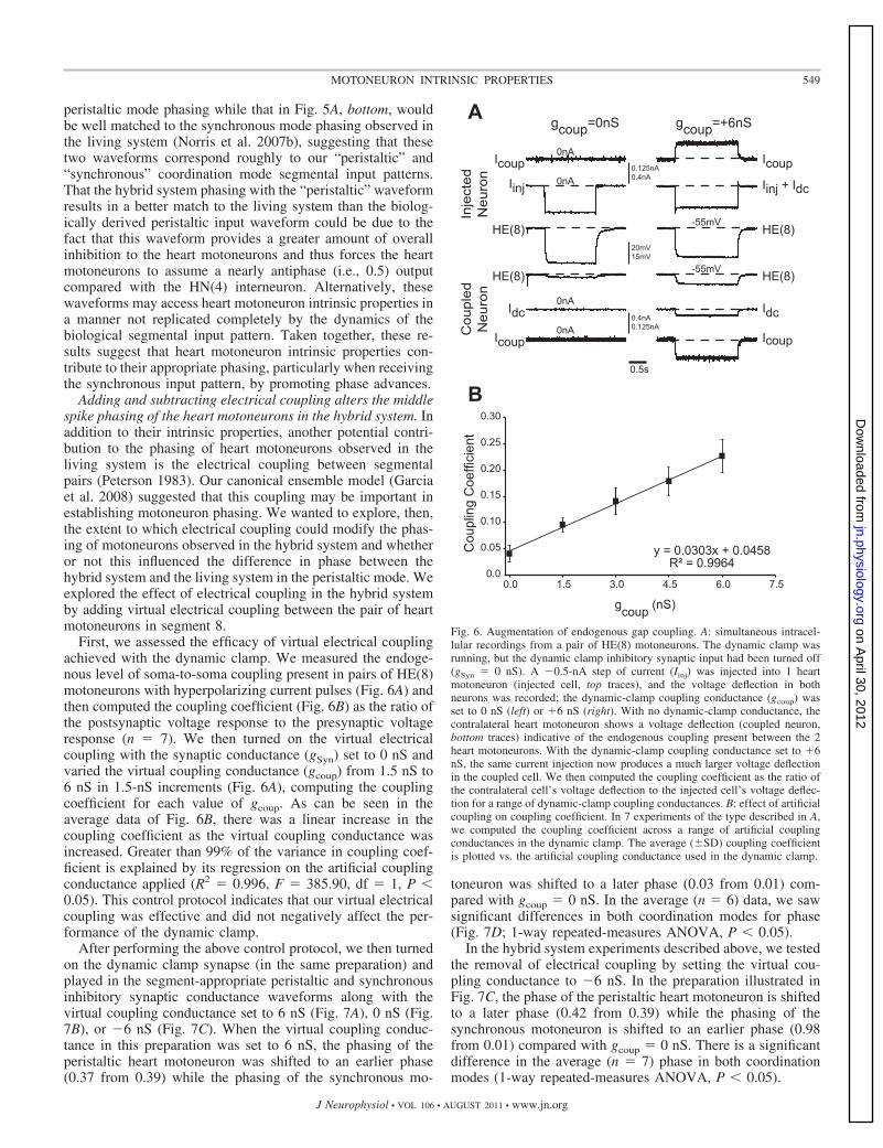
First, we assessed the efficacy of virtual electrical couplingachieved with the dynamic clamp. We measured the endoge-nous level of soma-to-soma coupling present in pairs of HE(8)motoneurons with hyperpolarizing current pulses (Fig. 6A) andthen computed the coupling coefficient (Fig. 6B) as the ratio ofthe postsynaptic voltage response to the presynaptic voltageresponse (n � 7). We then turned on the virtual electricalcoupling with the synaptic conductance (gSyn) set to 0 nS andvaried the virtual coupling conductance (gcoup) from 1.5 nS to6 nS in 1.5-nS increments (Fig. 6A), computing the couplingcoefficient for each value of gcoup. As can be seen in theaverage data of Fig. 6B, there was a linear increase in thecoupling coefficient as the virtual coupling conductance wasincreased. Greater than 99% of the variance in coupling coef-ficient is explained by its regression on the artificial couplingconductance applied (R2 � 0.996, F � 385.90, df � 1, P �0.05). This control protocol indicates that our virtual electricalcoupling was effective and did not negatively affect the per-formance of the dynamic clamp.
After performing the above control protocol, we then turnedon the dynamic clamp synapse (in the same preparation) andplayed in the segment-appropriate peristaltic and synchronousinhibitory synaptic conductance waveforms along with thevirtual coupling conductance set to 6 nS (Fig. 7A), 0 nS (Fig.7B), or �6 nS (Fig. 7C). When the virtual coupling conduc-tance in this preparation was set to 6 nS, the phasing of theperistaltic heart motoneuron was shifted to an earlier phase(0.37 from 0.39) while the phasing of the synchronous mo-
toneuron was shifted to a later phase (0.03 from 0.01) com-pared with gcoup � 0 nS. In the average (n � 6) data, we sawsignificant differences in both coordination modes for phase(Fig. 7D; 1-way repeated-measures ANOVA, P � 0.05).
In the hybrid system experiments described above, we testedthe removal of electrical coupling by setting the virtual cou-pling conductance to �6 nS. In the preparation illustrated inFig. 7C, the phase of the peristaltic heart motoneuron is shiftedto a later phase (0.42 from 0.39) while the phasing of thesynchronous motoneuron is shifted to an earlier phase (0.98from 0.01) compared with gcoup � 0 nS. There is a significantdifference in the average (n � 7) phase in both coordinationmodes (1-way repeated-measures ANOVA, P � 0.05).
Fig. 6. Augmentation of endogenous gap coupling. A: simultaneous intracel-lular recordings from a pair of HE(8) motoneurons. The dynamic clamp wasrunning, but the dynamic clamp inhibitory synaptic input had been turned off(gSyn � 0 nS). A �0.5-nA step of current (Iinj) was injected into 1 heartmotoneuron (injected cell, top traces), and the voltage deflection in bothneurons was recorded; the dynamic-clamp coupling conductance (gcoup) wasset to 0 nS (left) or �6 nS (right). With no dynamic-clamp conductance, thecontralateral heart motoneuron shows a voltage deflection (coupled neuron,bottom traces) indicative of the endogenous coupling present between the 2heart motoneurons. With the dynamic-clamp coupling conductance set to �6nS, the same current injection now produces a much larger voltage deflectionin the coupled cell. We then computed the coupling coefficient as the ratio ofthe contralateral cell’s voltage deflection to the injected cell’s voltage deflec-tion for a range of dynamic-clamp coupling conductances. B: effect of artificialcoupling on coupling coefficient. In 7 experiments of the type described in A,we computed the coupling coefficient across a range of artificial couplingconductances in the dynamic clamp. The average (�SD) coupling coefficientis plotted vs. the artificial coupling conductance used in the dynamic clamp.
549MOTONEURON INTRINSIC PROPERTIES
J Neurophysiol • VOL 106 • AUGUST 2011 • www.jn.org
on April 30, 2012
jn.physiology.orgD
ownloaded from

Increasing electrical coupling in the ensemble model (Garciaet al. 2008) produces a synchronizing effect on the activity ofthe heart motoneurons (i.e., the side-to-side phase differencewithin a segment decreased). Here we explored the physiolog-ical significance of the electrical coupling between the heartmotoneurons in the hybrid system. In the case of adding to theendogenous coupling between heart motoneurons, when oneheart motoneuron is hyperpolarized because of the inhibitoryinput pattern, that motoneuron passes hyperpolarizing currentto its contralateral homolog (arrow on top Icoup trace, Fig. 7A);at the same time, the contralateral motoneuron, which isspiking, passes depolarizing current back to the hyperpolarizedcell (arrow on bottom Icoup trace, Fig. 7A). The net effect ofenhancing endogenous electrical coupling is twofold: The firsteffect is to decrease the impact of a given heart motoneuron’sISyn because some portion of that motoneuron’s synaptic cur-rent is passed to its contralateral homolog via the electricalcoupling. Second, hyperpolarizing current passing from aninhibited motoneuron to its contralateral homolog during its
spiking phase attenuates spiking activity during its burst. Inthe peristaltic mode, these two effects combine to promotea phase advance, as passage of hyperpolarizing current tothe peristaltic motoneuron from its inhibited contralateralhomolog slows its burst, shifting a greater incidence ofspikes to the beginning of the burst. In the synchronousmode, these two effects promote a phase delay, as passage ofhyperpolarizing current attenuates the onset of the burst. Thesetwo effects result in a significantly smaller within-segmentphase difference compared with gcoup � 0 nS (Fig. 7E; 1-wayrepeated-measures ANOVA, P � 0.05), suggesting that elec-trical coupling synchronizes (i.e., brings their phasing closertogether) activity between heart motoneurons within a seg-ment. In our ensemble model, Garcia et al. (2008) suggestedthat this synchronizing effect was not uniform across segmentsbecause of the side-to-side differences in the phasing of theinput among segments; therefore, in addition to affectingsegmental phasing, electrical coupling could also influenceintersegmental coordination. Alternatively, in setting the cou-
Fig. 7. Effect of artificial coupling on HE(8) motoneuron phase: simultaneous intracellular recordings from the HE(8) motoneurons with the artificial couplingconductance gcoup set to �6 nS (A) , 0 nS (B), and �6 nS (C). Each panel is from the same experiment. D: summary phase (average � SD, n � 6) for the HE(8)motoneurons receiving the coupling conductances as described in A–C. Asterisks on each line indicate a significant difference in phase among the 3 artificialcoupling values (1-way repeated-measures ANOVA, P � 0.05). E: summary (average � SD, n � 6) within-segment phase difference between the 2 HE(8)motoneurons. Asterisks indicate a significant difference in the within-segment phase difference across the 3 artificial coupling values (1-way repeated-measuresANOVA, P � 0.05).
550 MOTONEURON INTRINSIC PROPERTIES
J Neurophysiol • VOL 106 • AUGUST 2011 • www.jn.org
on April 30, 2012
jn.physiology.orgD
ownloaded from

pling conductance to a negative value, we reverse the sign ofthe coupling current (i.e., Icoup), thus effectively canceling partor all of the endogenous coupling, potentially even adding netnegative coupling. By removing endogenous coupling, weenhance the net effect of a given motoneuron’s own ISyn andfunctionally uncouple the two motoneurons. Note that �gcoupenhances the inhibition of one motoneuron during its inhibitedphase (top arrow on Icoup, Fig. 7C) as well as enhancing thedepolarization of the contralateral motoneuron (bottom arrowon Icoup, Fig. 7C). The net effect is a phase delay of theperistaltic motoneuron and a phase advance of the synchronousmotoneuron, thus promoting a significant increase in side-to-side phase difference (Fig. 7E, 1-way repeated-measuresANOVA, P � 0.05).
Taken together, these results affirmed our modeling predic-tion that the electrical coupling between the heart motoneuronsserves to synchronize their ongoing bursting activity. Althoughthese results show that electrical coupling affected the phase ofthe heart motoneurons in the hybrid system, the synchronizingeffect did not improve the correspondence between the hybridand living systems in the peristaltic mode, nor did it change thegood correspondence between the hybrid and living systems inthe synchronous mode.
DISCUSSION
The goal of the present investigation was to assess howmotoneuron intrinsic properties contribute to rhythmic motoroutput. We used the leech heartbeat CPG, a system in whichthe pattern of premotor synaptic drive has been quantitativelydefined, to address this question. Previously, we introduced abiologically derived input pattern into a canonical model of theensemble of heart motoneurons in which their intrinsic elec-trical properties were kept to a minimum (Garcia et al. 2008).While the model motoneurons captured the bilateral asymme-try observed in the living system, model output phasing wassignificantly different from that observed in the living system(Fig. 4, Table 1), suggesting a role for heart motoneuronintrinsic properties in producing their appropriate output phas-ing. Here we constructed hybrid systems using segmental inputpatterns which were derived from the living system and whichwere used in the canonical ensemble model. We show directlyin living motoneurons that receive these segmentally appropri-ate input patterns that motoneuron intrinsic properties do con-tribute to appropriate motor output phasing.
The segmental input pattern determines the segmental phasedifferences in heart motoneuron phasing. The same heartmotoneuron [HE(8) and HE(14) motoneurons] responds dif-ferently to different synaptic strength profiles (i.e., the segment8 and 14 synaptic strength profiles), while different motoneu-rons (segments 8 and 14) respond similarly to the same inputpattern (segment 8 input pattern, Fig. 3). This result impliesthat the segmental input pattern determines the phasing differ-ences observed in motoneurons in segments 8 through 14 andthat the intrinsic properties present in the heart motoneurons inthese segments show no systematic segmental variation. Inprevious work with the heart motoneurons, Opdyke and Cala-brese (1995) showed that passive properties (input resistanceand capacitance) were not significantly different, particularlyamong heart motoneurons between segments 7 and 12. It wasalso shown that there were no significant differences in the
maximal current measured for two of the outward currents,which are termed IK1 and IK2, between these segments. Al-though measuring heart motoneuron inward currents at thelevel of the soma has proved difficult, these results suggest weshould not expect to find significant differences among theinward currents in these segments.
The finding that the input pattern determines the segmentalphasing differences observed in motoneurons is perhaps notsurprising. In the absence of premotor synaptic input, the heartmotoneurons fire tonically and do not show intrinsic burstingoscillations (Schmidt and Calabrese 1992). Furthermore, heartmotoneurons do not appear to exhibit voltage sags indicative ofthe hyperpolarization-activated Ih (A. Wenning and R. L.Calabrese, personal communication), a current that has beenshown in neonatal rodent motoneurons to promote the transi-tion from their inhibited phase to their firing phase (Kiehn et al.2000). Therefore, in the absence of intrinsic oscillations andrebound-promoting currents, the output of the motoneuronsshould be largely determined by the input pattern they receive.Although heart motoneurons receiving the synchronous patternof input show modest rebound spiking, this rebound spikingdoes not cause them to assume a phasing inconsistent with thesegmental input pattern they receive.
If the segmental input pattern determines the segmentalphasing differences observed in motoneurons, we might expectto find correlations between the premotor synaptic conductanceand the output phasing observed in the living system. In thepyloric network of the stomatogastric system, for example, theonset of the LP motoneuron is correlated to the synapticconductance of each of its inputs from the pacemaker kernel,the AB/PD complex (Goaillard et al. 2009). We do not see suchcorrelations between single inputs and output phase in oursystem (Norris et al. 2011), suggesting that there may be amore complex interaction between elements of the premotorsynaptic conductance pattern and the heart motoneuron intrin-sic properties.
In our experiments, within a coordination mode, the onlydifference between the segmental input patterns introducedinto the heart motoneurons was the synaptic strength profile.The temporal pattern was identical; therefore, we cannot de-termine, on the basis of these results, what the relative contri-butions of the temporal pattern and the synaptic strength profileof a segmental input pattern are to motoneuron output phase orto intersegmental coordination. Hybrid experiments in whichseveral temporal patterns and synaptic strength profiles aremixed and matched could help to address this question.
Heart motoneuron intrinsic properties are important forappropriate phasing. Despite the importance of the inputpattern, we show that the intrinsic properties of heart motoneu-rons play a substantive role in determining their output. Whileit is well established that motoneuron intrinsic properties con-tribute to motor pattern generation in the pyloric (Marder andBucher 2007) and gastric mill (Nusbaum and Beenhakker2002) networks of the crustacean stomatogastric system, al-most all of the neurons within those CPGs are themselvesmotoneurons, and the stomatogastric system is not segmentallydistributed like the leech heartbeat system. Because we usedthe identical segmental input patterns used in our canonicalensemble model, any differences between the hybrid systemand model phasing should be attributable to the additionalintrinsic properties of the living heart motoneurons not present
551MOTONEURON INTRINSIC PROPERTIES
J Neurophysiol • VOL 106 • AUGUST 2011 • www.jn.org
on April 30, 2012
jn.physiology.orgD
ownloaded from

in the model motoneurons. Specifically, for motoneurons re-ceiving the synchronous pattern of synaptic input, regardless ofthe segmental strength profile, the intrinsic properties of themotoneurons promote a phase advance (compared with thecanonical ensemble model) sufficient to match the phasingobserved in the living system. In many cases, this phaseadvance is substantial: In each of the segments tested, theaverage phase advance per segment (compared with the en-semble model) is at least 0.09. While we are unable to deter-mine what set of intrinsic properties are present in the heartmotoneurons, a likely candidate could be a low-threshold Ca2�
current. In the experiments where endogenous inputs wereblocked with 0 Ca2�- high Mn2� instead of bicuculline me-thiodide, hybrid system synchronous phasing was delayed(although not significantly so) by �0.02 compared with bicu-culline methiodide block. This shift in phase may serve as anestimate of the contribution of such a current to the outputphasing of these motoneurons. Addition of a low-thresholdCa2� current to the model motoneurons, therefore, could pro-vide a phase advance compared with the canonical ensemblemodel.
Our inability to replicate, in the hybrid system, a similarcorrespondence between the hybrid system and the livingsystem in the peristaltic mode is surprising. Across segments,heart motoneurons receiving the peristaltic pattern of synapticinput show phasing similar to the ensemble model. We do notattribute these discrepancies to experimental or proceduralerrors; furthermore, all of the appropriate inputs are accountedfor in the segmental input patterns that were used in our modeland dynamic clamp experiments (Norris et al. 2011). A mod-ulatory extrinsic input, acting on a cycle-by-cycle basis, maycontribute to peristaltic phasing, but as of yet no such inputshave been identified in the leech. One final possibility is that,because we are using the identical inputs in every experimentin the presence of the normal intrinsic properties of the heartmotoneurons, we are not accounting for natural animal-to-animal variability in both the temporal pattern and synapticstrength profiles. We are currently exploring how naturalvariability of both the temporal pattern and synaptic strengthprofiles affect output phasing.
Heart motoneurons escape rhythmic inhibition, while modelmotoneurons are released from inhibition. We used trapezoidalwaveforms designed to control when a motoneuron initiated itsfiring (Fig. 5). By constraining the onset of heart motoneuronfiring until at or very near the offset of the premotor synapticconductance, we were able to delay middle spike phase in amanner similar to that observed in the model motoneurons.Alternatively, waveforms that allowed for an earlier onset ofmotoneuron firing during the premotor synaptic conductanceshow phasing consistent with that measured in our hybridsystem experiments with natural synaptic input patterns. Thisresult suggests a fundamental difference between how mo-toneurons in the living system and the ensemble model mayintegrate their inputs: Heart motoneurons in the living systemseem able to escape inhibition by beginning to fire at a pointwhere the synaptic conductance is still ongoing (note thedifference in first spike phase between the hybrid system andmodel, Fig. 4E). Model motoneurons, on the other hand,initiate firing only when the synaptic conductance has fallen toa lower conductance value; i.e., model motoneurons fire whenthey are nearly fully released from inhibition (Sharp et al.
1996). These results also suggest potential improvements to themodel motoneurons; we are currently revising our canonicalmodel to account for the spatial extent of the motoneurons inthe living system as well as implementing inward currents thatmay affect their response to segment-specific patterns of syn-aptic input (Lamb et al. 2010).
Electrical coupling synchronizes ongoing heart motoneuronactivity within a segment. Adding artificial electrical couplingvia dynamic clamp promotes synchronization of phase betweenheart motoneurons in the same segment. This observation isconsistent with our previous modeling efforts (Garcia et al.2008), as well as observations in Xenopus embryos (Zhang etal. 2009). This result suggests that electrical coupling alsocontributes to the output phasing observed in the living system.The synchronizing effect of the artificial coupling did notimprove the correspondence in peristaltic phase between thehybrid system and the living system. Because the artificialcoupling was added at the soma, distant from the potentialendogenous coupling sites on fine neurites (Tolbert and Cala-brese 1985), the synchronizing effect shown here may repre-sent the lower physiological limit of the synchronizing effectobserved in the living system.
Electrical coupling may also influence intersegmental coor-dination. When similar (although using different values ofgcoup) hybrid experiments were done in segment 14 receivingthe segment 14 input pattern, the reduction in side-to-sidephase difference in segment 14 was smaller than that measuredin segment 8. At present, we cannot distinguish whether thissegment-specific effect is due to side-to-side phasing in theinput pattern, as suggested in out ensemble model (Garcia et al.2008), or possibly due to segmental variation in the couplingbetween heart motoneurons. A systematic survey of the cou-pling coefficients between heart motoneurons in the midbodysegments could address this question.
Larger implications. The results presented here show thatmotoneuron intrinsic properties can make a functional contri-bution to motor output in the context of an ongoing rhythmicinput. Work in the cat spinal cord (Lee and Heckman 1998),neonatal rodent spinal cord (Kiehn et al. 2000), hypoglossalmotoneurons (Purvis and Butera 2005), and turtle fictivescratch (Hounsgaard and Kiehn 1989) have all shown thatmotoneurons in these preparations possess intrinsic properties(and electrical coupling) that appear ideally suited to influencethe phase and intensity of motor output. Our work suggests thata prerequisite for assessing how motoneuron intrinsic proper-ties in these preparations contribute to motor output will be aquantitative description of the premotor pattern of synapticinput. For example, recent studies in which the patterns ofpremotor synaptic drive to motoneurons in the neonatal rodentspinal cord (Endo and Kiehn 2008) and turtle fictive scratch(Berg et al. 2007) were decomposed into their inhibitory andexcitatory components were used to make inferences about thecomposition of the premotor interneuronal networks that drivethese motor patterns. The neurons that make up these networksand their connections are still being identified. The strength ofthe present study is that we have identified the constitutivepremotor elements and their relative contributions to the in-hibitory synaptic drive impinging upon the motoneurons in thissystem. By focusing on those segments in which the premotorinputs have been clearly defined, we have provided the relevant
552 MOTONEURON INTRINSIC PROPERTIES
J Neurophysiol • VOL 106 • AUGUST 2011 • www.jn.org
on April 30, 2012
jn.physiology.orgD
ownloaded from
context to ascertain how motoneuron intrinsic properties maycontribute to motor output.
GRANTS
This work was supported by a Ruth L. Kirschstein National ResearchService Award, funded by National Institute of Neurological Disorders andStroke (NINDS) Grant NS-8472431, a National Science Foundation IGERTProgram Grant (DGE-0333411), and American Psychological Association/Diversity Program in Neuroscience Predoctoral Fellowship 5 T06 MH-1882,funded by the National Institute of Mental Health, to T. M. Wright, Jr. and byNINDS Grant NS-24072 to R. L. Calabrese.
DISCLOSURES
No conflicts of interest, financial or otherwise, are declared by the author(s).
REFERENCES
Alaburda A, Russo R, MacAulay N, Hounsgaard J. Periodic high-conduc-tance states in spinal neurons during scratch-like network activity in adultturtles. J Neurosci 25: 6316–6321, 2005.
Angstadt JD, Calabrese RL. Calcium currents and graded synaptic transmis-sion between heart interneurons of the leech. J Neurosci 11: 746–759, 1991.
Berg RW, Alaburda A, Hounsgaard J. Balanced inhibition and excitationdrive spike activity in spinal half-centers. Science 315: 390–393, 2007.
Cymbalyuk GS, Gaudry Q, Masino MA, Calabrese RL. Bursting in leechheart interneurons: cell-autonomous and network-based mechanisms. J Neu-rosci 22: 10580–10592, 2002.
DeLong ND, Kirby MS, Blitz DM, Nusbaum MP. Parallel regulation of amodulator-activated current via distinct dynamics underlies comodulation ofmotor circuit output. J Neurosci 29: 12355–12367, 2009.
Endo T, Kiehn O. Asymmetric operation of the locomotor central patterngenerator in the neonatal mouse spinal cord. J Neurophysiol 100: 3043–3054, 2008.
Garcia PS, Wright TM, Cunningham IR, Calabrese RL. Using a model toassess the role of the spatiotemporal pattern of inhibitory input and intraseg-mental electrical coupling in the intersegmental and side-to-side coordina-tion of motor neurons by the leech heartbeat central pattern generator. JNeurophysiol 100: 1354–1371, 2008.
Goaillard JM, Taylor AL, Schulz DJ, Marder E. Functional consequencesof animal-to-animal variation in circuit parameters. Nat Neurosci 12: 1424–1430, 2009.
Hill AAV, Lu J, Masino MA, Olsen OH, Calabrese RL. A model of asegmental oscillator in the leech heartbeat neuronal network. J ComputNeurosci 10: 281–302, 2001.
Hodgkin AL, Huxley AF. A quantitative description of membrane current andits application to conduction and excitation in nerve. J Physiol 117: 500–544, 1952.
Hounsgaard J, Kiehn O. Serotonin-induced bistability of turtle motoneuronescaused by a nifedipine-sensitive calcium plateau potential. J Physiol 414:265–282, 1989.
Kiehn O, Kjaerulff O, Tresch MC, Harris-Warrick RM. Contributions ofintrinsic motor neuron properties to the production of rhythmic motor outputin the mammalian spinal cord. Brain Res Bull 53: 649–659, 2000.
Kristan WB, Calabrese RL, Friesen WO. Neuronal control of leech behav-ior. Prog Neurobiol 76: 279–327, 2005.
Lamb DG, Wright TM, Wenning A, Norris BJ, Calabrese RL. Contribu-tion of intrinsic properties to the input-output relationship of leech heartmotor neurons evaluated with a multi-compartmental model (Abstract).Society for Neuroscience Annual Meeting, San Diego, CA, 2010.
Lee RH, Heckman CJ. Bistability in spinal motoneurons in vivo: systematicvariations in persistent inward currents. J Neurophysiol 80: 583–593, 1998.
Marder E, Bucher D. Understanding circuit dynamics using the stomatogas-tric nervous system of lobsters and crabs. Annu Rev Physiol 69: 291–316,2007.
Marder E, Calabrese RL. Principles of rhythmic motor pattern generation.Physiol Rev 76: 687–717, 1996.
Norris BJ, Weaver AL, Morris LG, Wenning A, Garcia PA, CalabreseRL. A central pattern generator producing alternative outputs: temporalpattern of premotor activity. J Neurophysiol 96: 309–326, 2006.
Norris BJ, Weaver AL, Wenning A, Garcia PS, Calabrese RL. A centralpattern generator producing alternative outputs: pattern, strength, and dy-namics of premotor synaptic input to leech heart motor neurons. J Neuro-physiol 98: 2992–3005, 2007a.
Norris BJ, Weaver AL, Wenning A, Garcia PS, Calabrese RL. A centralpattern generator producing alternative outputs: phase relations of leechheart motor neurons with respect to premotor synaptic input. J Neurophysiol98: 2983–2991, 2007b.
Norris BJ, Wenning A, Wright TM, Calabrese RL. Constancy and vari-ability in the output of a central pattern generator. J Neurosci 31: 4663–4674, 2011.
Nusbaum MP, Beenhakker MP. A small-systems approach to motor patterngeneration. Nature 417: 343–350, 2002.
Opdyke CA, Calabrese RL. Outward currents in heart motor neurons of themedicinal leech. J Neurophysiol 74: 2524–2537, 1995.
Opdyke CA, Calabrese RL. A persistent sodium current contributes tooscillatory activity in heart interneurons of the medicinal leech. J CompPhysiol A 175: 781–789, 1994.
Perrins R, Roberts A. Cholinergic and electrical synapses between synergis-tic spinal motoneurons in the Xenopus laevis embryo. J Physiol 485:135–144, 1995.
Peterson E. Frequency-dependent coupling between rhythmically active neu-rons in the leech. Biophys J 43: 53–61, 1983.
Prinz AA, Abbott LF, Marder E. The dynamic clamp comes of age. TrendsNeurosci 27: 218–224, 2004.
Purvis LK, Butera RJ. Ionic current model of a hypoglossal motoneuron. JNeurophysiol 93: 723–733, 2005.
Rekling JC, Shao XM, Feldman JL. Electrical coupling and excitatorysynaptic transmission between rhythmogenic respiratory neurons in thepreBotzinger complex. J Neurosci 20: RC113, 2000.
Schmidt J, Calabrese RL. Evidence that acetylcholine is an inhibitorytransmitter of heart interneurons in the leech. J Exp Biol 171: 329–347,1992.
Sharifullina E, Ostroumov K, Nistri A. Metabotropic glutamate receptoractivity induces a novel oscillatory pattern in neonatal rat hypoglossalmotoneurones. J Physiol 563: 139–159, 2005.
Sharp AA, O’Neil MB, Abbott LF, Marder E. The dynamic clamp: artificialconductances in biological neurons. Trends Neurosci 16: 389–394, 1993.
Sharp AA, Skinner FK, Marder E. Mechanisms of oscillation in dynamicclamp constructed two-cell half-center circuits. J Neurophysiol 76: 867–883, 1996.
Thompson WJ, Stent GS. Neuronal control of heartbeat in medicinal leech.1. Generation of vascular constriction rhythm by heart motor neurons. JComp Physiol 111: 261–279, 1976a.
Thompson WJ, Stent GS. Neuronal control of heartbeat in medicinal leech.2. Intersegmental coordination of heart motor neuron activity by heartinterneurons. J Comp Physiol 111: 281–307, 1976b.
Thompson WJ, Stent GS. Neuronal control of heartbeat in medicinal leech.3. Synaptic relations of heart interneurons. J Comp Physiol 111: 309–333,1976c.
Tolbert LP, Calabrese RL. Anatomical analysis of contacts between identi-fied neurons that control heartbeat in the leech Hirudo medicinalis. CellTissue Res 242: 257–267, 1985.
Wenning A, Cymbalyuk GS, Calabrese RL. Heartbeat control in leeches. I.Constriction pattern and neural modulation of blood pressure in intactanimals. J Neurophysiol 91: 382–396, 2004a.
Wenning A, Hill AAV, Calabrese RL. Heartbeat control in leeches. II.Fictive motor pattern. J Neurophysiol 91: 397–409, 2004b.
Zhang HY, Li WC, Heitler WJ, Sillar KT. Electrical coupling synchronisesspinal motoneuron activity during swimming in hatchling Xenopus tadpoles.J Physiol 587: 4455–4466, 2009.
553MOTONEURON INTRINSIC PROPERTIES
J Neurophysiol • VOL 106 • AUGUST 2011 • www.jn.org
on April 30, 2012
jn.physiology.orgD
ownloaded from