Embed Size (px)
Citation preview
Spontaneous intracerebral hemorrhage (ICH) is one ofthe most devastating forms of stroke (1). SpontaneousICH is a type of stroke that arises in the brain parenchy-
ma in the absence of trauma or surgery. Common caus-es include hypertension, amyloid angiopathy, coagu-lopathy, vascular anomalies, tumors, and treatmentwith various drugs. However, hypertension is the singlemost important risk factor for spontaneous ICH.Spontaneous ICH accounts for 10-15% of all strokesand is associated with a higher mortality rate than eitherischemic stroke or subarachnoid hemorrhage (SAH) (2).
J Korean Radiol Soc 2007;56:413-422
─ 413 ─
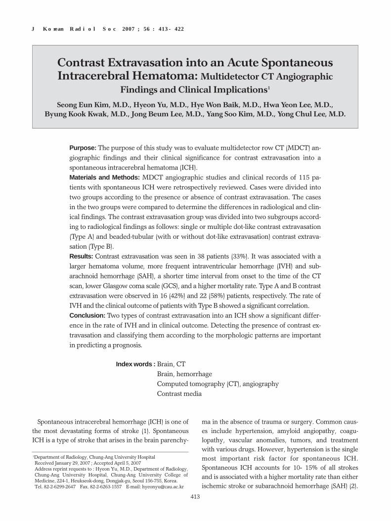
Contrast Extravasation into an Acute SpontaneousIntracerebral Hematoma: Multidetector CT Angiographic
Findings and Clinical Implications1
Seong Eun Kim, M.D., Hyeon Yu, M.D., Hye Won Baik, M.D., Hwa Yeon Lee, M.D., Byung Kook Kwak, M.D., Jong Beum Lee, M.D., Yang Soo Kim, M.D., Yong Chul Lee, M.D.
1Department of Radiology, Chung-Ang University HospitalReceived January 29, 2007 ; Accepted April 5, 2007Address reprint requests to : Hyeon Yu, M.D., Department of Radiology,Chung-Ang University Hospital, Chung-Ang University College ofMedicine, 224-1, Heukseok-dong, Dongjak-gu, Seoul 156-755, Korea.Tel. 82-2-6299-2647 Fax. 82-2-6263-1557 E-mail: [email protected]
Purpose: The purpose of this study was to evaluate multidetector row CT (MDCT) an-giographic findings and their clinical significance for contrast extravasation into aspontaneous intracerebral hematoma (ICH). Materials and Methods: MDCT angiographic studies and clinical records of 115 pa-tients with spontaneous ICH were retrospectively reviewed. Cases were divided intotwo groups according to the presence or absence of contrast extravasation. The casesin the two groups were compared to determine the differences in radiological and clin-ical findings. The contrast extravasation group was divided into two subgroups accord-ing to radiological findings as follows: single or multiple dot-like contrast extravasation(Type A) and beaded-tubular (with or without dot-like extravasation) contrast extrava-sation (Type B).Results: Contrast extravasation was seen in 38 patients (33%). It was associated with alarger hematoma volume, more frequent intraventricular hemorrhage (IVH) and sub-arachnoid hemorrhage (SAH), a shorter time interval from onset to the time of the CTscan, lower Glasgow coma scale (GCS), and a higher mortality rate. Type A and B contrastextravasation were observed in 16 (42%) and 22 (58%) patients, respectively. The rate ofIVH and the clinical outcome of patients with Type B showed a significant correlation.Conclusion: Two types of contrast extravasation into an ICH show a significant differ-ence in the rate of IVH and in clinical outcome. Detecting the presence of contrast ex-travasation and classifying them according to the morphologic patterns are importantin predicting a prognosis.
Index words : Brain, CT Brain, hemorrhage Computed tomography (CT), angiography Contrast media
The mortality rate in spontaneous ICH is increased inpatients with a large hematoma, low GCS score, and in-traventricular hemorrhage (IVH) (3-8). Although spon-taneous ICH is usually a monophasic event, sometimesbleeding can persist up to several hours postictus.Continued bleeding associated with ICH may lead to en-largement of an existing hematoma. A hematoma that isenlarging is associated with poor outcome (9-11). Theincidence of hematoma enlargement is about 14-20%by computed tomography (CT) (12, 13). A previousstudy showed that active extravasation of contrast intospontaneous ICH is a marker for continued bleedingthat is associated with hematoma enlargement, and it isthe most significant radiological variable associated withfatality in a multivariate model (3). This study reportedthat the mortality rate in the group with contrast ex-travasation is significantly higher than the control groupwithout contrast extravasation. However, to date, nostudy has documented radiological findings of contrastextravasation into a spontaneous ICH by multidetectorrow CT (MDCT) angiography and their relationshipwith on the clinical outcome of patients. The purpose ofthis study was to evaluate the MDCT angiographic find-ings of contrast extravasation into a spontaneous ICH,and to correlate those radiological findings with clinicaloutcome.
Materials and Methods
Between November 2003 and December 2005, 115consecutive patients who presented with acute sponta-neous ICH were included in this study. Patients withhemorrhage due to aneurysmal rupture, arteriovenousmalformation, moyamoya disease, or infective endo-carditis, as well as those receiving anticoagulants or an-tiplatelet agents, were excluded. The age range of these
patients was 36-88 years, and the mean age of thestudy patients was 59.9 years. The study group wascomposed of 67 males and 48 females.
All CT examinations were performed with a 16-MD-CT scanner (LightSpeed Pro 16, GE Healthcare,Milwaukee, WI U.S.A.). In all patients, a non-enhancedscan was obtained according to the following protocol:5-mm slice thickness, maximum of 200 mAs at 120-kVtube voltage. For MDCT angiography, the followingscanning protocol was used: 1.25-mm slice thickness,0.6 sec of rotation speed, 4 mm per rotation of tablefeed, pitch of 0.938:1, 210 mAs of tube current, and 120kVp. A power injector was used to administer 120 mL of300 mg/mL iohexol (Omnipaque 300, Cork, Ireland), anonionic contrast medium, into the right antecubitalvein at a flow rate of 4 mL/sec. There was no differencein the injection rate between patients. The individualcirculation time was determined in the lumen of thecommon carotid artery using a test bolus of 4 mL of IV-administered contrast medium at a flow rate 4 mL/sec.A scan commenced 3 seconds after the common carotidartery reached 100 Hounsfield unit (HU). After MDCTangiography, a post-enhanced scan was performed withthe same protocol as the pre-contrast scan. The acquiredimages were transferred from the CT scanner to a work-station for image reconstruction. Maximum intensityprojections (MIP) in axial, coronal, and sagittal planesand volume-rendered images from the source datasetswere generated by using the software (AdvantageWindows; GE Medical Systems) installed on the work-station.
Two neuroradiologists who were blinded to the clini-cal data evaluated the CT and CT angiographic images.The hematoma volume was estimated on the pre-con-trast CT images obtained before CT angiography withthe ABC/2 method, where A, B, and C represent the re-
Seong Eun Kim, et al : Contrast Extravasation into an Acute Spontaneous Intracerebral Hematoma
─ 414 ─
Table 1. Comparison of the Clinical and Radiological Data in Patients with and Without Contrast Extravasation
Patient Data With Contrast Extravasation (n = 38) Without Contrast Extravasation (n = 77) p-value
Age 57.71±12.88 60.68±12.26 0.264GCS 8.82±3.95 11.49±3.820 <0.001Time interval 3.13±4.26 15.59±26.02 <0.001ICH volume 59.57±44.60 27.98±32.32 <0.001IVH 28/38 (73.7%) 33/77 (42.9%) 0.004SAH 10/38 (26.3%) 6/77 (7.8%) 0.016End result
Improvement 20/38 (52.6%) 65/77 (84.4%) <0.001No change 3/38 (7.9%) 2/77 (2.6%) 0.410Aggravation 04/38 (10.5%) 4/77 (5.2%) 0.504Death 11/38 (28.9%) 6/77 (7.8%) 0.006
CTA = CT angiography
spective radii of the hematoma in three dimensions. Thehematoma volume was calculated as the average of bothvalues estimated from two image readers. The presenceor absence of contrast extravasation was determined byvisual detection of the presence of hyperdense contrastmedia inside the hematoma without evidence of feedingor draining vessels on post-contrast CT images obtainedafter CT angiography. The number and morphology ofcontrast extravasation, hematoma location, the presenceor absence of IVH, and the presence or absence of SAHwere recorded.
Clinical results were obtained by patient medical chartreview by an investigator blinded to the radiological da-ta. The following data were recorded: the time intervalbetween symptom onset and CT angiography, systolicand diastolic blood pressures at the time of CT angiogra-phy, GCS, initial level of consciousness, and clinical out-come. The presence or absence of a surgical procedurewas also recorded. The initial level of consciousness wasscored on a six-point scale, with scores defined as fol-lows: coma (grade 1), semicoma (grade 2), stupor (grade3), drowsiness (grade 4), near alert (grade 5), and alert(grade 6). Clinical outcome was compared with the ini-tial clinical status of the patient and classified as follows:improvement, no interval change, aggravation, anddeath.
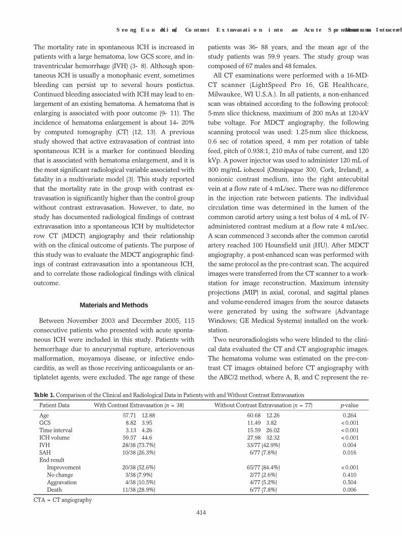
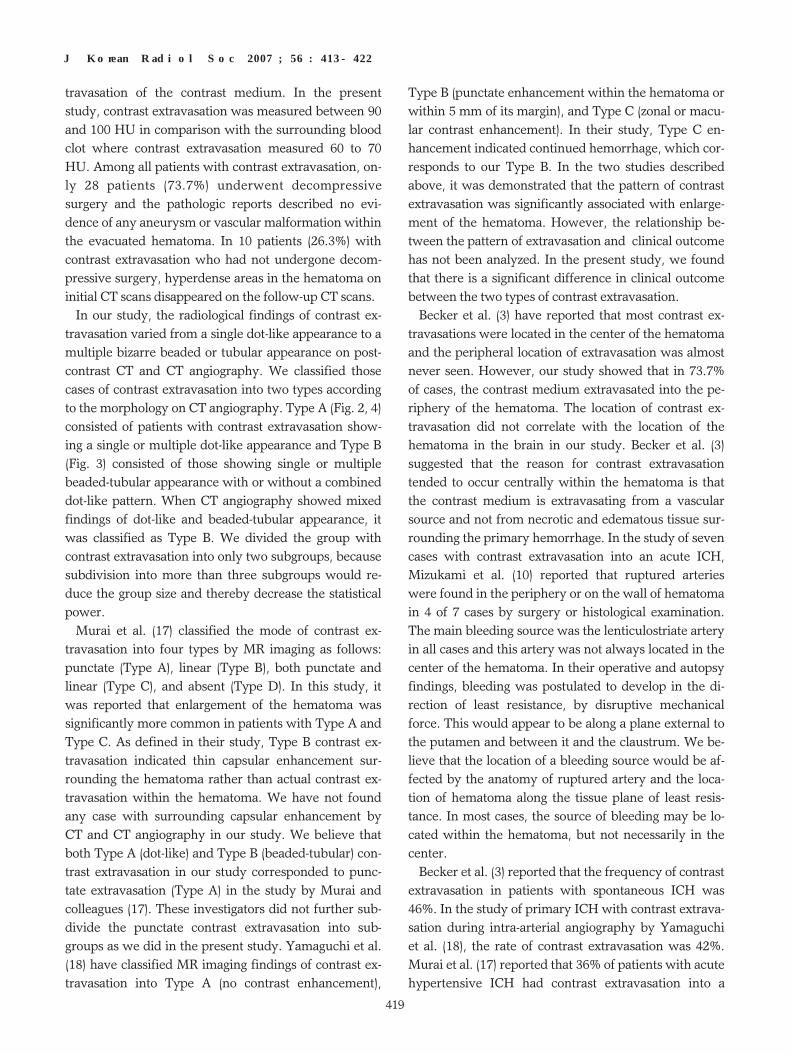
Patient cases were divided into two groups (with orwithout contrast extravasation), and the contrast ex-travasation group was subdivided into two groups ac-cording to the radiological findings as follows: single ormultiple dot-like contrast extravasation (Type A) and
beaded-tubular (with or without dot-like extravasation)contrast extravasation (Type B) (Fig. 1).
Statistical analysis was performed with a commercial-ly available statistical software program (SPSS 11,Chicago, IL U.S.A.). To determine a statistically signifi-cant difference in the time interval between symptomonset and CT angiography, GCS, initial level of con-sciousness, systolic and diastolic blood pressures, andhematoma volume between the two groups with andwithout contrast extravasation and between the twosubgroups with contrast extravasation, the Mann-Whitney U test was employed. A difference of clinicaloutcome was determined with the χ2 test or Fisher’s ex-act test. The level of statistical significance was set at p≤0.05.
Results
Contrast extravasation into a spontaneous ICH wasobserved in 38 of 115 patients (33%).
Contrast extravasation was seen as a localized hyper-
J Korean Radiol Soc 2007;56:413-422
─ 415 ─
A B
Fig. 1. The classification of contrast ex-travasation patterns on brain CT andCT angiography imaging are illustrat-ed on the schematic drawing.A. Type A: single or multiple and cen-tral or peripheral dot-like areas of highattenuation within the hematoma.B. Type B: single or multiple and cen-tral or peripheral beaded-tubular areasof high attenuation within thehematoma.
Table 2. Morphology and the Location of the ContrastExtravasation
LocationMorphology of Contrast Extravasation
TotalDot-like Beaded tubular Mixed
Periphery 12 14 2 28 (73.7%)Center 4 3 - 07 (18.4%)Mixed - 2 1 3 (7.9%)
Total 16 (42.1%) 19 (50%) 3 (7.9%) 38
Dot-like = Type ABeaded tubular and mixed = Type B
dense zone that was well demarcated from a surround-ing blood clot. The density of the hematoma was be-tween 60 and 70 HU in comparison with 90-100 HU ofcontrast extravasation that was easily detected on a post-contrast CT scan. In most cases, extravasation of con-trast media was located in the periphery of thehematoma (73.7%).
Table 1 shows the comparative clinical and radiologi-cal findings of the patients with contrast extravasation (n= 38) and without contrast extravasation (n=77). In pa-tients with contrast extravasation, there was a trend to-ward a larger hematoma volume, a shorter time intervalfrom symptom onset to CT angiography, a lower GCS, ahigher frequency of IVH, a lower score of initial con-
Seong Eun Kim, et al : Contrast Extravasation into an Acute Spontaneous Intracerebral Hematoma
─ 416 ─
A
B
C
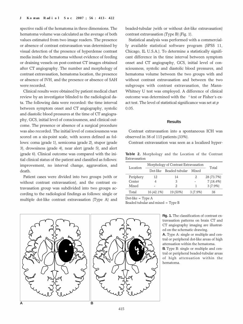
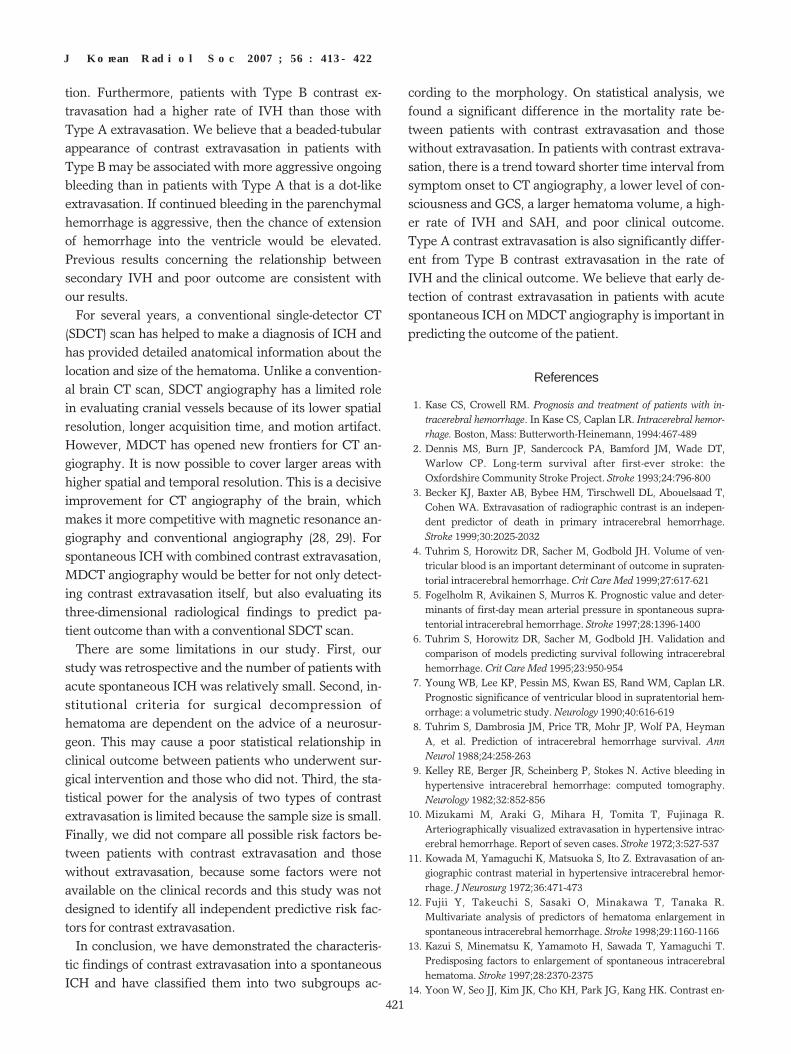
Fig. 2. Brain CT angiography with Type A contrast extravasationin a 51-year-old man with an acute spontaneous intracerebralhematoma (ICH). A. A source image of CT angiography shows a small hemorrhageinvolving the left basal ganglia, internal capsule, and thalamuswith dot-like contrast extravasation into the peripheral portion ofthe hematoma (arrow). Coronal (B) and sagittal (C) maximum intensity projection (MIP)images show dot-like contrast extravasation into the hematoma(arrow).
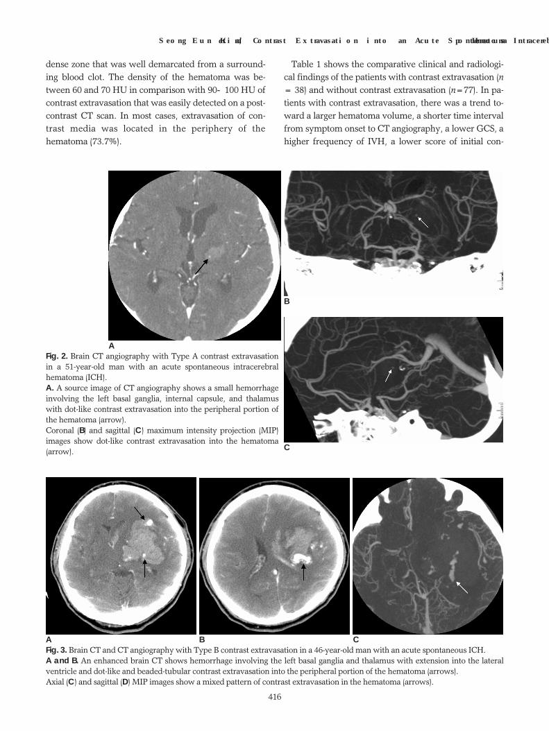
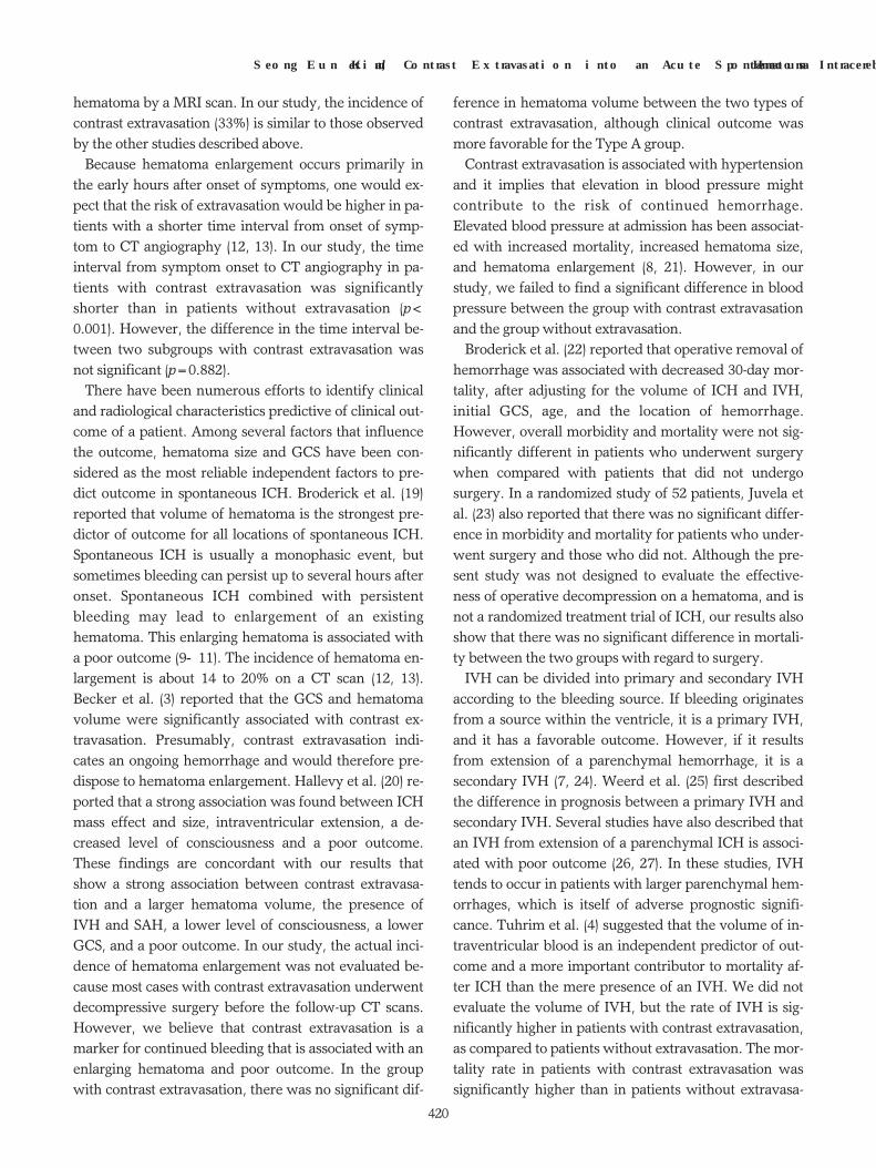
A B CFig. 3. Brain CT and CT angiography with Type B contrast extravasation in a 46-year-old man with an acute spontaneous ICH. A and B. An enhanced brain CT shows hemorrhage involving the left basal ganglia and thalamus with extension into the lateralventricle and dot-like and beaded-tubular contrast extravasation into the peripheral portion of the hematoma (arrows). Axial (C) and sagittal (D) MIP images show a mixed pattern of contrast extravasation in the hematoma (arrows).
sciousness, a lower improvement rate, and a highermortality rate than in patients without extravasation.The overall mortality rate was 17.8% (17 of 115). Themortality rate for patients with contrast extravasation(11/38;28.9%), was significantly higher than in patientswithout extravasation (6/77;7.8%) (p=0.006). Neitherthe systolic blood pressure nor the diastolic blood pres-sure was associated with the presence of contrast ex-travasation. No significant differences were observedfor other factors between two groups. The two groupswere not significantly different in the location ofhematoma except for the posterior fossa where thenumber of patients without extravasation was signifi-
cantly larger than those with extravasation (p≤0.001).The mortality rate of patients who underwent decom-pressive surgery was not significantly different fromthose patients that did not undertake surgery in bothgroups with or without contrast extravasation.
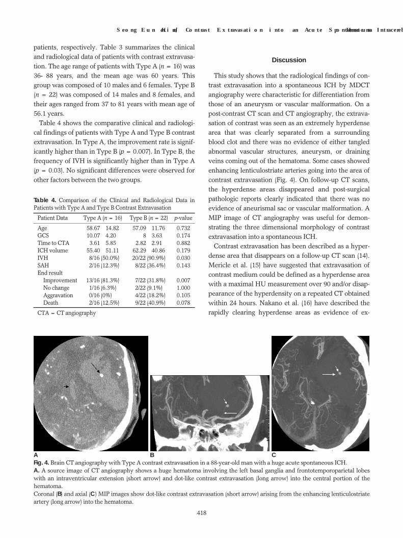
The morphology and location of contrast extravasationinto an ICH is shown in Table 2. On brain CT and CTangiography, there was no evidence of tangled abnor-mal vascular structures, aneurysm, or draining veinscoming out of the hematoma. However, some casesshowed lenticulostriate arteries going into the area ofthe contrast extravasation (Fig. 4).
Type A and B were observed in 16 (42%) and 22 (58%)
J Korean Radiol Soc 2007;56:413-422
─ 417 ─
Table 3. Clinical and CT Angiographic Findings in Patients with Contrast Extravasation
No. Sex/Age TI (hrs) MS GCS Type ICH location ICH Volume (cm3) IVH SAH
01 M/36 1.5 near alert 13 A Rt. BG 8.98 - -02 M/46 1 near alert 15 A BG 17.6 - -03 M/49 3 coma 04 A Rt. BG & IC 83.0 + -04 F/50 1.5 semicoma 04 A Rt. TH 36.4 + -05 F/51 1 drowsy 12 A BG 11.3 - -06 M/51 2 alert 12 A Lt. BG & IC 3.94 - -07 M/53 0.67 drowsy 12 A Rt. BG & CR 59.5 + -08 F/57 2 drowsy 13 A Lt. TH 9.90 + -09 M/58 5 drowsy 13 A Rt. F-P lobe, Rt. BG 84.0 + -10 F/61 4 stupor 11 A Lt. F-P lobe 36.0 - -11 M/66 0.5 stupor 10 A Lt. TH 9.84 - -12 F/66 4 semicoma 06 A Lt. BG & CR 113 + -13 M/67 3 drowsy 13 A Rt. P lobe 81.8 - +14 M/79 1 stupor 09 A Rt. F-T & BG 161 + -15 F/82 2 drowsy 14 A Lt. BG 28.7 - -16 M/88 24 semicoma 04 A Lt. BG 149 + -17 F/37 4 stupor 07 B Lt. T lobe 35.0 + +18 M/42 1.67 drowsy 13 B Rt. TH & IC 19.4 + -19 M/45 1 drowsy 13 B Rt. Insula, EC, & BG 52.6 - -20 M/46 1 semicoma 05 B Lt. BG & TH 93.0 + -21 M/47 3.5 drowsy 13 B Rt. P lobe 31.0 + -22 M/47 6 coma 03 B Rt. T lobe 85.7 + +23 F/48 1 stupor 07 B Lt. TH 19.8 + -24 M/50 2 semicoma 04 B Lt. BG & IC 105 + +25 M/50 1 semicoma 04 B Lt. BG & TH 93.0 + -26 M/50 0.5 semicoma 04 B Rt. BG & TH 128 + +27 F/51 5 stupor 05 B Lt. BG & F-T lobe 59.6 + +28 F/53 0.5 stupor 05 B Lt. BG & Rt. TH 83.5 + -29 M/55 9 stupor 06 B Rt. F lobe 128 + +30 M/56 1 alert 12 B Rt. BG 8.91 - -31 M/58 9 drowsy 13 B Rt. BG 65.3 + -32 M/63 10 stupor 07 B Both F-T lobe 107 + +33 M/66 0.5 drowsy 13 B Rt. F-P lobe 23.1 + -34 F/67 2 semicoma 05 B Rt. BG 2 + -35 F/70 0.67 semicoma 4 B Lt. TH & BG 68.3 + -36 M/72 2 drowsy 12 B Lt. P-O lobe 99.0 + +37 F/79 1 stupor 10 B Lt. TH 22.5 + -38 F/81 0.5 semicoma 05 B Lt. BG 106 + +
TI = Time interval from onset of symptoms to CT angiography, MS = Initial mental status, GCS = Glasgow coma scale, BG = basal gan-glia, TH = thalamus, IC = internal capsule, EC = external capsule, CR = corona radiate, F = frontal, T = temporal, P = parietal, O =occipital, + = presence, - = absence
patients, respectively. Table 3 summarizes the clinicaland radiological data of patients with contrast extravasa-tion. The age range of patients with Type A (n = 16) was36-88 years, and the mean age was 60 years. Thisgroup was composed of 10 males and 6 females. Type B(n = 22) was composed of 14 males and 8 females, andtheir ages ranged from 37 to 81 years with mean age of56.1 years.
Table 4 shows the comparative clinical and radiologi-cal findings of patients with Type A and Type B contrastextravasation. In Type A, the improvement rate is signif-icantly higher than in Type B (p = 0.007). In Type B, thefrequency of IVH is significantly higher than in Type A(p = 0.03). No significant differences were observed forother factors between the two groups.
Discussion
This study shows that the radiological findings of con-trast extravasation into a spontaneous ICH by MDCTangiography were characteristic for differentiation fromthose of an aneurysm or vascular malformation. On apost-contrast CT scan and CT angiography, the extrava-sation of contrast was seen as an extremely hyperdensearea that was clearly separated from a surroundingblood clot and there was no evidence of either tangledabnormal vascular structures, aneurysm, or drainingveins coming out of the hematoma. Some cases showedenhancing lenticulostriate arteries going into the area ofcontrast extravasation (Fig. 4). On follow-up CT scans,the hyperdense areas disappeared and post-surgicalpathologic reports clearly indicated that there was noevidence of aneurismal sac or vascular malformation. AMIP image of CT angiography was useful for demon-strating the three dimensional morphology of contrastextravasation into a spontaneous ICH.
Contrast extravasation has been described as a hyper-dense area that disappears on a follow-up CT scan (14).Mericle et al. (15) have suggested that extravasation ofcontrast medium could be defined as a hyperdense areawith a maximal HU measurement over 90 and/or disap-pearance of the hyperdensity on a repeated CT obtainedwithin 24 hours. Nakano et al. (16) have described therapidly clearing hyperdense areas as evidence of ex-
Seong Eun Kim, et al : Contrast Extravasation into an Acute Spontaneous Intracerebral Hematoma
─ 418 ─
Table 4. Comparison of the Clinical and Radiological Data inPatients with Type A and Type B Contrast Extravasation
Patient Data Type A (n = 16) Type B (n = 22) p-value
Age 58.67±14.82 57.09±11.76 0.732GCS 10.07±4.200 0.08±3.63 0.174Time to CTA 3.61±5.85 2.82±2.91 0.882ICH volume 55.40±51.11 62.29±40.86 0.179IVH 08/16 (50.0%) 20/22 (90.9%) 0.030SAH 02/16 (12.3%) 08/22 (36.4%) 0.143End result
Improvement 13/16 (81.3%) 07/22 (31.8%) 0.007No change 1/16 (6.3%) 2/22 (9.1%) 1.000Aggravation 0/16 (0%)0. 04/22 (18.2%) 0.105Death 02/16 (12.5%) 09/22 (40.9%) 0.078
CTA = CT angiography
A B CFig. 4. Brain CT angiography with Type A contrast extravasation in a 88-year-old man with a huge acute spontaneous ICH. A. A source image of CT angiography shows a huge hematoma involving the left basal ganglia and frontotemporoparietal lobeswith an intraventricular extension (short arrow) and dot-like contrast extravasation (long arrow) into the central portion of thehematoma. Coronal (B) and axial (C) MIP images show dot-like contrast extravasation (short arrow) arising from the enhancing lenticulostriateartery (long arrow) into the hematoma.
travasation of the contrast medium. In the presentstudy, contrast extravasation was measured between 90and 100 HU in comparison with the surrounding bloodclot where contrast extravasation measured 60 to 70HU. Among all patients with contrast extravasation, on-ly 28 patients (73.7%) underwent decompressivesurgery and the pathologic reports described no evi-dence of any aneurysm or vascular malformation withinthe evacuated hematoma. In 10 patients (26.3%) withcontrast extravasation who had not undergone decom-pressive surgery, hyperdense areas in the hematoma oninitial CT scans disappeared on the follow-up CT scans.
In our study, the radiological findings of contrast ex-travasation varied from a single dot-like appearance to amultiple bizarre beaded or tubular appearance on post-contrast CT and CT angiography. We classified thosecases of contrast extravasation into two types accordingto the morphology on CT angiography. Type A (Fig. 2, 4)consisted of patients with contrast extravasation show-ing a single or multiple dot-like appearance and Type B(Fig. 3) consisted of those showing single or multiplebeaded-tubular appearance with or without a combineddot-like pattern. When CT angiography showed mixedfindings of dot-like and beaded-tubular appearance, itwas classified as Type B. We divided the group withcontrast extravasation into only two subgroups, becausesubdivision into more than three subgroups would re-duce the group size and thereby decrease the statisticalpower.
Murai et al. (17) classified the mode of contrast ex-travasation into four types by MR imaging as follows:punctate (Type A), linear (Type B), both punctate andlinear (Type C), and absent (Type D). In this study, itwas reported that enlargement of the hematoma wassignificantly more common in patients with Type A andType C. As defined in their study, Type B contrast ex-travasation indicated thin capsular enhancement sur-rounding the hematoma rather than actual contrast ex-travasation within the hematoma. We have not foundany case with surrounding capsular enhancement byCT and CT angiography in our study. We believe thatboth Type A (dot-like) and Type B (beaded-tubular) con-trast extravasation in our study corresponded to punc-tate extravasation (Type A) in the study by Murai andcolleagues (17). These investigators did not further sub-divide the punctate contrast extravasation into sub-groups as we did in the present study. Yamaguchi et al.(18) have classified MR imaging findings of contrast ex-travasation into Type A (no contrast enhancement),
Type B (punctate enhancement within the hematoma orwithin 5 mm of its margin), and Type C (zonal or macu-lar contrast enhancement). In their study, Type C en-hancement indicated continued hemorrhage, which cor-responds to our Type B. In the two studies describedabove, it was demonstrated that the pattern of contrastextravasation was significantly associated with enlarge-ment of the hematoma. However, the relationship be-tween the pattern of extravasation and clinical outcomehas not been analyzed. In the present study, we foundthat there is a significant difference in clinical outcomebetween the two types of contrast extravasation.
Becker et al. (3) have reported that most contrast ex-travasations were located in the center of the hematomaand the peripheral location of extravasation was almostnever seen. However, our study showed that in 73.7%of cases, the contrast medium extravasated into the pe-riphery of the hematoma. The location of contrast ex-travasation did not correlate with the location of thehematoma in the brain in our study. Becker et al. (3)suggested that the reason for contrast extravasationtended to occur centrally within the hematoma is thatthe contrast medium is extravasating from a vascularsource and not from necrotic and edematous tissue sur-rounding the primary hemorrhage. In the study of sevencases with contrast extravasation into an acute ICH,Mizukami et al. (10) reported that ruptured arterieswere found in the periphery or on the wall of hematomain 4 of 7 cases by surgery or histological examination.The main bleeding source was the lenticulostriate arteryin all cases and this artery was not always located in thecenter of the hematoma. In their operative and autopsyfindings, bleeding was postulated to develop in the di-rection of least resistance, by disruptive mechanicalforce. This would appear to be along a plane external tothe putamen and between it and the claustrum. We be-lieve that the location of a bleeding source would be af-fected by the anatomy of ruptured artery and the loca-tion of hematoma along the tissue plane of least resis-tance. In most cases, the source of bleeding may be lo-cated within the hematoma, but not necessarily in thecenter.
Becker et al. (3) reported that the frequency of contrastextravasation in patients with spontaneous ICH was46%. In the study of primary ICH with contrast extrava-sation during intra-arterial angiography by Yamaguchiet al. (18), the rate of contrast extravasation was 42%.Murai et al. (17) reported that 36% of patients with acutehypertensive ICH had contrast extravasation into a
J Korean Radiol Soc 2007;56:413-422
─ 419 ─
hematoma by a MRI scan. In our study, the incidence ofcontrast extravasation (33%) is similar to those observedby the other studies described above.
Because hematoma enlargement occurs primarily inthe early hours after onset of symptoms, one would ex-pect that the risk of extravasation would be higher in pa-tients with a shorter time interval from onset of symp-tom to CT angiography (12, 13). In our study, the timeinterval from symptom onset to CT angiography in pa-tients with contrast extravasation was significantlyshorter than in patients without extravasation (p<0.001). However, the difference in the time interval be-tween two subgroups with contrast extravasation wasnot significant (p=0.882).
There have been numerous efforts to identify clinicaland radiological characteristics predictive of clinical out-come of a patient. Among several factors that influencethe outcome, hematoma size and GCS have been con-sidered as the most reliable independent factors to pre-dict outcome in spontaneous ICH. Broderick et al. (19)reported that volume of hematoma is the strongest pre-dictor of outcome for all locations of spontaneous ICH.Spontaneous ICH is usually a monophasic event, butsometimes bleeding can persist up to several hours afteronset. Spontaneous ICH combined with persistentbleeding may lead to enlargement of an existinghematoma. This enlarging hematoma is associated witha poor outcome (9-11). The incidence of hematoma en-largement is about 14 to 20% on a CT scan (12, 13).Becker et al. (3) reported that the GCS and hematomavolume were significantly associated with contrast ex-travasation. Presumably, contrast extravasation indi-cates an ongoing hemorrhage and would therefore pre-dispose to hematoma enlargement. Hallevy et al. (20) re-ported that a strong association was found between ICHmass effect and size, intraventricular extension, a de-creased level of consciousness and a poor outcome.These findings are concordant with our results thatshow a strong association between contrast extravasa-tion and a larger hematoma volume, the presence ofIVH and SAH, a lower level of consciousness, a lowerGCS, and a poor outcome. In our study, the actual inci-dence of hematoma enlargement was not evaluated be-cause most cases with contrast extravasation underwentdecompressive surgery before the follow-up CT scans.However, we believe that contrast extravasation is amarker for continued bleeding that is associated with anenlarging hematoma and poor outcome. In the groupwith contrast extravasation, there was no significant dif-
ference in hematoma volume between the two types ofcontrast extravasation, although clinical outcome wasmore favorable for the Type A group.
Contrast extravasation is associated with hypertensionand it implies that elevation in blood pressure mightcontribute to the risk of continued hemorrhage.Elevated blood pressure at admission has been associat-ed with increased mortality, increased hematoma size,and hematoma enlargement (8, 21). However, in ourstudy, we failed to find a significant difference in bloodpressure between the group with contrast extravasationand the group without extravasation.
Broderick et al. (22) reported that operative removal ofhemorrhage was associated with decreased 30-day mor-tality, after adjusting for the volume of ICH and IVH,initial GCS, age, and the location of hemorrhage.However, overall morbidity and mortality were not sig-nificantly different in patients who underwent surgerywhen compared with patients that did not undergosurgery. In a randomized study of 52 patients, Juvela etal. (23) also reported that there was no significant differ-ence in morbidity and mortality for patients who under-went surgery and those who did not. Although the pre-sent study was not designed to evaluate the effective-ness of operative decompression on a hematoma, and isnot a randomized treatment trial of ICH, our results alsoshow that there was no significant difference in mortali-ty between the two groups with regard to surgery.
IVH can be divided into primary and secondary IVHaccording to the bleeding source. If bleeding originatesfrom a source within the ventricle, it is a primary IVH,and it has a favorable outcome. However, if it resultsfrom extension of a parenchymal hemorrhage, it is asecondary IVH (7, 24). Weerd et al. (25) first describedthe difference in prognosis between a primary IVH andsecondary IVH. Several studies have also described thatan IVH from extension of a parenchymal ICH is associ-ated with poor outcome (26, 27). In these studies, IVHtends to occur in patients with larger parenchymal hem-orrhages, which is itself of adverse prognostic signifi-cance. Tuhrim et al. (4) suggested that the volume of in-traventricular blood is an independent predictor of out-come and a more important contributor to mortality af-ter ICH than the mere presence of an IVH. We did notevaluate the volume of IVH, but the rate of IVH is sig-nificantly higher in patients with contrast extravasation,as compared to patients without extravasation. The mor-tality rate in patients with contrast extravasation wassignificantly higher than in patients without extravasa-
Seong Eun Kim, et al : Contrast Extravasation into an Acute Spontaneous Intracerebral Hematoma
─ 420 ─
tion. Furthermore, patients with Type B contrast ex-travasation had a higher rate of IVH than those withType A extravasation. We believe that a beaded-tubularappearance of contrast extravasation in patients withType B may be associated with more aggressive ongoingbleeding than in patients with Type A that is a dot-likeextravasation. If continued bleeding in the parenchymalhemorrhage is aggressive, then the chance of extensionof hemorrhage into the ventricle would be elevated.Previous results concerning the relationship betweensecondary IVH and poor outcome are consistent withour results.
For several years, a conventional single-detector CT(SDCT) scan has helped to make a diagnosis of ICH andhas provided detailed anatomical information about thelocation and size of the hematoma. Unlike a convention-al brain CT scan, SDCT angiography has a limited rolein evaluating cranial vessels because of its lower spatialresolution, longer acquisition time, and motion artifact.However, MDCT has opened new frontiers for CT an-giography. It is now possible to cover larger areas withhigher spatial and temporal resolution. This is a decisiveimprovement for CT angiography of the brain, whichmakes it more competitive with magnetic resonance an-giography and conventional angiography (28, 29). Forspontaneous ICH with combined contrast extravasation,MDCT angiography would be better for not only detect-ing contrast extravasation itself, but also evaluating itsthree-dimensional radiological findings to predict pa-tient outcome than with a conventional SDCT scan.
There are some limitations in our study. First, ourstudy was retrospective and the number of patients withacute spontaneous ICH was relatively small. Second, in-stitutional criteria for surgical decompression ofhematoma are dependent on the advice of a neurosur-geon. This may cause a poor statistical relationship inclinical outcome between patients who underwent sur-gical intervention and those who did not. Third, the sta-tistical power for the analysis of two types of contrastextravasation is limited because the sample size is small.Finally, we did not compare all possible risk factors be-tween patients with contrast extravasation and thosewithout extravasation, because some factors were notavailable on the clinical records and this study was notdesigned to identify all independent predictive risk fac-tors for contrast extravasation.
In conclusion, we have demonstrated the characteris-tic findings of contrast extravasation into a spontaneousICH and have classified them into two subgroups ac-
cording to the morphology. On statistical analysis, wefound a significant difference in the mortality rate be-tween patients with contrast extravasation and thosewithout extravasation. In patients with contrast extrava-sation, there is a trend toward shorter time interval fromsymptom onset to CT angiography, a lower level of con-sciousness and GCS, a larger hematoma volume, a high-er rate of IVH and SAH, and poor clinical outcome.Type A contrast extravasation is also significantly differ-ent from Type B contrast extravasation in the rate ofIVH and the clinical outcome. We believe that early de-tection of contrast extravasation in patients with acutespontaneous ICH on MDCT angiography is important inpredicting the outcome of the patient.
References
1. Kase CS, Crowell RM. Prognosis and treatment of patients with in-tracerebral hemorrhage. In Kase CS, Caplan LR. Intracerebral hemor-rhage. Boston, Mass: Butterworth-Heinemann, 1994:467-489
2. Dennis MS, Burn JP, Sandercock PA, Bamford JM, Wade DT,Warlow CP. Long-term survival after first-ever stroke: theOxfordshire Community Stroke Project. Stroke 1993;24:796-800
3. Becker KJ, Baxter AB, Bybee HM, Tirschwell DL, Abouelsaad T,Cohen WA. Extravasation of radiographic contrast is an indepen-dent predictor of death in primary intracerebral hemorrhage.Stroke 1999;30:2025-2032
4. Tuhrim S, Horowitz DR, Sacher M, Godbold JH. Volume of ven-tricular blood is an important determinant of outcome in supraten-torial intracerebral hemorrhage. Crit Care Med 1999;27:617-621
5. Fogelholm R, Avikainen S, Murros K. Prognostic value and deter-minants of first-day mean arterial pressure in spontaneous supra-tentorial intracerebral hemorrhage. Stroke 1997;28:1396-1400
6. Tuhrim S, Horowitz DR, Sacher M, Godbold JH. Validation andcomparison of models predicting survival following intracerebralhemorrhage. Crit Care Med 1995;23:950-954
7. Young WB, Lee KP, Pessin MS, Kwan ES, Rand WM, Caplan LR.Prognostic significance of ventricular blood in supratentorial hem-orrhage: a volumetric study. Neurology 1990;40:616-619
8. Tuhrim S, Dambrosia JM, Price TR, Mohr JP, Wolf PA, HeymanA, et al. Prediction of intracerebral hemorrhage survival. AnnNeurol 1988;24:258-263
9. Kelley RE, Berger JR, Scheinberg P, Stokes N. Active bleeding inhypertensive intracerebral hemorrhage: computed tomography.Neurology 1982;32:852-856
10. Mizukami M, Araki G, Mihara H, Tomita T, Fujinaga R.Arteriographically visualized extravasation in hypertensive intrac-erebral hemorrhage. Report of seven cases. Stroke 1972;3:527-537
11. Kowada M, Yamaguchi K, Matsuoka S, Ito Z. Extravasation of an-giographic contrast material in hypertensive intracerebral hemor-rhage. J Neurosurg 1972;36:471-473
12. Fujii Y, Takeuchi S, Sasaki O, Minakawa T, Tanaka R.Multivariate analysis of predictors of hematoma enlargement inspontaneous intracerebral hemorrhage. Stroke 1998;29:1160-1166
13. Kazui S, Minematsu K, Yamamoto H, Sawada T, Yamaguchi T.Predisposing factors to enlargement of spontaneous intracerebralhematoma. Stroke 1997;28:2370-2375
14. Yoon W, Seo JJ, Kim JK, Cho KH, Park JG, Kang HK. Contrast en-
J Korean Radiol Soc 2007;56:413-422
─ 421 ─

대한영상의학회지 2007;56:413-422
급성 자발성 뇌내혈종 내로의 조영제 유출에 대한 다중검출 전산화단층촬영혈관조영술의 영상의학적 소견 및 임상적 의의1
1중앙대학교병원 영상의학과
김성은·유 현·백혜원·이화연·곽병국·이종범·김양수·이용철
목적: 다중검출 전산화단층촬영(MDCT) 혈관조영술에서 발견된 급성 자발성 뇌내혈종 내로의 조영제 유출에 관한
영상소견과 임상적 의의를 알아보고자 하였다.
대상과 방법: 급성 자발성 뇌내혈종으로 MDCT 혈관조영술을 시행한 115명을 대상으로 조영제 유출 여부에 따라
두 집단으로 나누어 조영제 유출과 관련된 위험인자와 임상소견 및 예후에서 두 집단 간에 유의한 차이가 있는지
알아보았다. 조영제 유출이 있었던 집단을 그 모양에 따라 다시 두 집단으로 나누어 임상소견 및 예후에 유의한 차
이가 있는지 분석하였다. 점 모양의 조영제 유출이 있으면 유형 A로, 관 모양 및 혼합형의 유출이 있으면 유형 B
로 분류하였다.
결과: MDCT 혈관조영술에서 혈종 내로의 조영제 유출은 33%에서 관찰되었다. 조영제 유출이 있는 집단에서 혈종
의 크기가 더 컸고, 뇌실 및 지주막하출혈의 빈도가 높았으며, 증상발현 후 검사 시행까지의 시간 간격은 더 짧았
고, 글라스고우혼수척도가 더 낮았고, 사망률은 더 높았다. 조영제 유출이 있었던 집단에서는 유형 A가 42%, 유형
B가 58%에서 관찰되었다. 유형 B에서는 뇌실 내 출혈의 빈도가 높았고 예후는 유형 A에서 유의하게 좋았다.
결론: 유형 A와 B 사이에는 뇌실 내 출혈의 빈도 및 예후에서 유의한 차이가 있다. MDCT 혈관조영술을 이용하여
초기에 조영제 유출을 발견하고 특징적인 영상소견에 따라 유형을 분류해 봄으로써 환자의 예후를 예측하는 데에
도움을 얻을 수 있다.
hancement and contrast extravasation on computed tomographyafter intra-arterial thrombolysis in patients with acute ischemicstroke. Stroke 2004;35:876-881
15. Mericle RA, Lopes DK, Fronckowiak MD, Wakhloo AK,Guterman LR, Hopkins LN. A grading scale to predict outcomesafter intra-arterial thrombolysis for stroke complicated by contrastextravasation. Neurosurgery 2000;46:1307-1314
16. Nakano S, Iseda T, Kawano H, Yoneyama T, Ikeda T, Wakisaka S.Parenchymal hyperdensity on computed tomography after intra-arterial reperfusion therapy for acute middle cerebral artery occlu-sion: incidence and clinical significance. Stroke 2001;32:2042-2048
17. Murai Y, Ikeda Y, Teramoto A, Tsuji Y. Magnetic resonance imag-ing-documented extravasation as an indicator of acute hyperten-sive intracerebral hemorrhage. J Neurosurg 1998;88:650-655
18. Yamaguchi K, Uemura K, Takahashi H, Kowada M, Kutsuzawa T.Intracerebral leakage of contrast medium in apoplexy. Br J Radiol1971;44:689-691
19. Broderick JP, Brott TG, Grotta JC. Intracerebral hemorrhage vol-ume measurement. Stroke 1994;25:1081
20. Hallevy C, Ifergane G, Kordysh E, Herishanu Y. Spontaneoussupratentorial intracerebral hemorrhage. Criteria for short-termfunctional outcome prediction. J Neurol 2002;249:1704-1709
21. Terayama Y, Tanahashi N, Fukuuchi Y, Gotoh F. Prognostic valueof admission blood pressure in patients with intracerebral hemor-rhage. Keio Cooperative Stroke Study. Stroke 1997;28:1185-1188
22. Broderick JP, Brott TG, Duldner JE, Tomsick T, Huster G. Volume
of intracerebral hemorrhage. A powerful and easy-to-use predictorof 30-day mortality. Stroke 1993;24:987-993
23. Juvela S, Heiskanen O, Poranen A, Valtonen S, Kuurne T, KasteM, et al. The treatment of spontaneous intracerebral hemorrhage.A prospective randomized trial of surgical and conservative treat-ment. J Neurosurg 1989;70:755-758
24. Weisberg LA, Elliott D, Shamsnia M. Intraventricular hemorrhagein adults: clinical-computed tomographic correlations. ComputMed Imaging Graph 1991;15:43-51
25. de Weerd AW. The prognosis of intraventricular hemorrhage. JNeurol 1979;222:46-51
26. Hayashi M, Handa Y, Kobayashi H, Kawano H, Nozaki J, HiroseS. Prognosis of intraventricular hemorrhage due to hypertensivehemorrhagic cerebrovascular disease. Zentralbl Neurochir1988;49:101-108
27. Steiner I, Gomori JM, Melamed E. The prognostic value of the CTscan in conservatively treated patients with intracerebralhematoma. Stroke 1984;15:279-282
28. Klingebiel R, Zimmer C, Rogalla P, Kivelitz D, Bohner G, Gotze R,et al. Assessment of the arteriovenous cerebrovascular system bymulti-slice CT. A single-bolus, monophasic protocol. Acta Radiol2001;42:560-562
29. Jones TR, Kaplan RT, Lane B, Atlas SW, Rubin GD. Single- versusmulti-detector row CT of the brain: quality assessment. Radiology2001;219:750-755
Seong Eun Kim, et al : Contrast Extravasation into an Acute Spontaneous Intracerebral Hematoma
─ 422 ─