Embed Size (px)
Citation preview
341
Contrast-Enhanced CT of Acute lsodense Subdural Hematoma Orest B. Boyko, 1 Daniel F. Cooper,2 and C. Barrie Grossman3
Originally, all acute subdural hematomas were thought to be hyperdense with respect to brain on CT (1 ). But over the last 1 0-15 years, cases of acute isodense subdural and epidural hematomas have been reported [2-6). Although contrast-enhanced CT has aided in the visualization of chronic isodense subdural hematomas [7), the use of contrast material was not efficacious in two of the acute isodense hematoma cases reported (4, 5) , and was not performed in four others [2-4, 6].
This report illustrates the usefulness of contrast-enhanced CT in the evaluation of a case of acute head trauma with unexplained midline shift. In our case, an acute isodense subdural hematoma enhanced, as did the CSF cisterns and the isodense cerebral contusions. The recognition of this constellation of radiologic signs on unenhanced and enhanced CT in the setting of acute head trauma has an important hematologic implication and suggests the underlying presence of disseminated intravascular coagulation (DIC).
Case Report
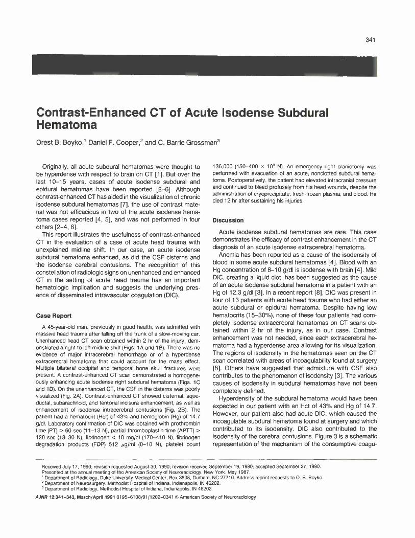
A 45-year-old man, previously in good health, was admitted with massive head trauma after falling off the trunk of a slow-moving car. Unenhanced head CT scan obtained within 2 hr of the injury, demonstrated a right to left midline shift (Figs. 1 A and 1 8). There was no evidence of major intracerebral hemorrhage or of a hyperdense extracerebral hematoma that could account for the mass effect. Multiple bilateral occipital and temporal bone skull fractures were present. A contrast-enhanced CT scan demonstrated a homogeneously enhancing acute isodense right subdural hematoma (Figs. 1 C and 1 D). On the unenhanced CT, the CSF in the cisterns was poorly visualized (Fig. 2A). Contrast-enhanced CT showed cisternal, aqueductal, subarachnoid, and tentorial incisura enhancement, as well as enhancement of isodense intracerebral contusions (Fig. 28). The patient had a hematocrit (Hct) of 43% and hemoglobin (Hg) of 14.7 gjdl. Laboratory confirmation of DIG was obtained with prothrombin time (PT) > 60 sec (11-13 N), partial thromboplastin time (APTT) > 120 sec (18-30 N), fibrinogen < 10 mg/dl (170-41 0 N), fibrinogen degradation products (FOP) 512 11gjml (0-10 N), platelet count
136,000 (150-400 x 1 03 N). An emergency right craniotomy was performed with evacuation of an acute, nonclotted subdural hematoma. Postoperatively, the patient had elevated intracranial pressure and continued to bleed profusely from his head wounds, despite the administration of cryoprecipitate, fresh-frozen plasma, and blood. He died 12 hr after sustaining his injuries.
Discussion
Acute isodense subdural hematomas are rare. This case demonstrates the efficacy of contrast enhancement in the CT diagnosis of an acute isodense extracerebral hematoma.
Anemia has been reported as a cause of the isodensity of blood in some acute subdural hematomas [4) . Blood with an Hg concentration of 8-1 0 gfdl is isodense with brain [ 4). Mild DIC, creating a liquid clot, has been suggested as the cause of an acute isodense subdural hematoma in a patient with an Hg of 12.3 g/dl [3). In a recent report [8], DIC was present in four of 13 patients with acute head trauma who had either an acute subdural or epidural hematoma. Despite having low hematocrits (15-30%), none of these four patients had completely isodense extracerebral hematomas on CT scans obtained within 2 hr of the injury, as in our case. Contrast enhancement was not needed, since each extracerebral hematoma had a hyperdense area allowing for its visualization . The regions of isodensity in the hematomas seen on the CT scan correlated with areas of incoagulability found at surgery [8). Others have suggested that admixture with CSF also contributes to the phenomenon of isodensity (3). The various causes of isodensity in subdural hematomas have not been completely defined.
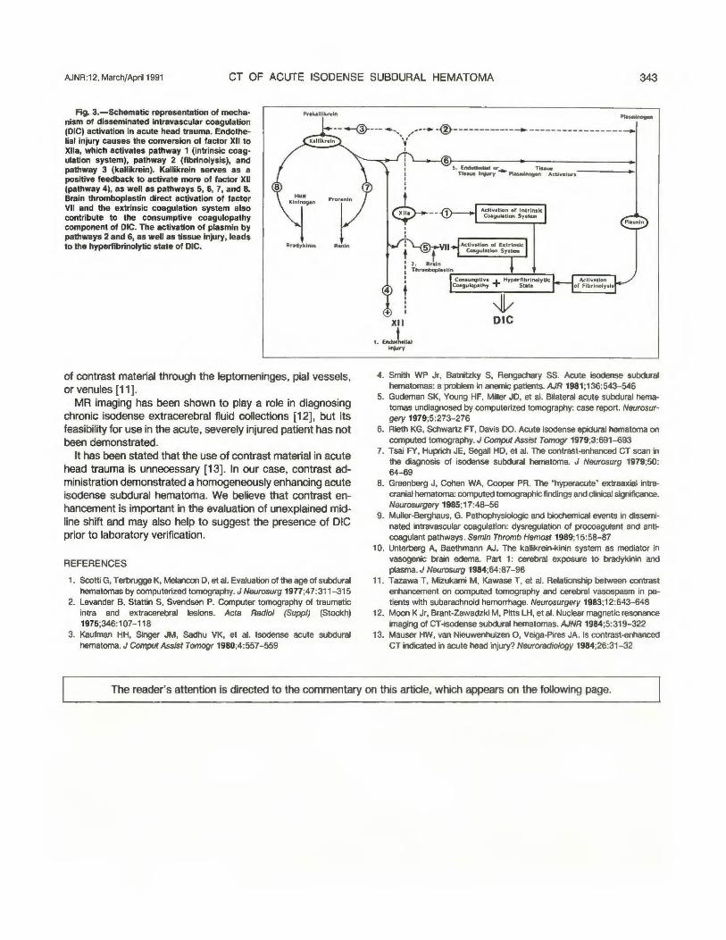
Hyperdensity of the subdural hematoma would have been expected in our patient with an Hct of 43% and Hg of 14.7. However, our patient also had acute DIC, which caused the incoagulable subdural hematoma found at surgery and which contributed to its isodensity. DIC also contributed to the isodensity of the cerebral contusions. Figure 3 is a schematic representation of the mechanism of the consumptive coagu-
Received July 17, 1990; revision requested August 30, 1990; revision received September 19, 1990: accepted September 27 . 1990. Presented at the annual meeting of the American Society of Neuroradiology. New York. May 1987. 'Department of Radiology. Duke University Medical Center. Box 3808. Durham. NC 27710. Address reprint requests to 0 . B. Boyko. 7 Department of Neurosurgery, Methodist Hospital of Indiana. Indianapolis, IN 46202. 3 Department of Radiology, Methodist Hospital of Indiana, Indianapolis. IN 46202.
AJNR 12:341-343, March/April1991 0195-6108/91/1202-0341 © American Society of Neuroradiology
342 BOYKO ET AL. AJNR:12, March{Apri11991
A B
A 8
lopathy and the hyperfibrinolytic state of head trauma-induced DIG (refer to [9] for more detailed information).
The homogeneous contrast enhancement of our patient's isodense subdural hematoma and isodense cerebral contusions most likely was related to increased blood brain barrier
Fig. 1.-A and 8, Noncontrast CT scans show right to left midline shift without an identifiable extracerebral fluid collection.
C and 0, Contrast-enhanced CT scans show homogeneous enhancement of an isodense acute right subdural hematoma (arrows). There is diffuse subarachnoid enhancement (black ar· rowhead in 0) and enhancement of an isodense left cerebral contusion (white arrowhead in 0).
Fig. 2.-A, Noncontrast CT scan shows poor visualization of CSF in perimesencephalic cis· tern.
B, Contrast-enhanced CT scan shows cisternal (closed arrows) and aqueductal (open arrow) enhancement with marked enhancement of tentorial incisura (arrowheads).
permeability, partly induced by the activation of the kallikrein system and generation of bradykinin (Fig. 3) [1 0]. Explanations for cisternal enhancement include active bleeding, meningeal hyperemia, chemical irritation of the meninges by blood, and increased blood brain barrier permeability, with leakage
AJNR:12, March/April1991 CT OF ACUTE ISODENSE SUBDURAL HEMATOMA 343
1'"19. 3.-Schematic representation of mechanism of disseminated intravascular coagulation (DIC) activation in acute head trauma. Endothelial injury causes the conversion of factor XII to Xlla, which activates pathway 1 (intrinsic coagulation system), pathway 2 (fibrinolysis), and pathway 3 (kallikrein). Kallikrein serves as a positive feedback to activate more of factor XII (pathway 4), as well as pathways 5, 6, 7, and 8. Brain thromboplastin direct activation of factor VII and the extrinsic coagulation system also contribute to the consumptive coagulopathy component of DIC. The activation of plasmin by pathways 2 and 6, as well as tissue injury, leads to the hyperfibrinolytic state of DIC. Br.tyklnin Renin
of contrast material through the leptomeninges, pial vessels, or venules [11).
MR imaging has been shown to play a role in diagnosing chronic isodense extracerebral fluid collections [12], but its feasibility for use in the acute, severely injured patient has not been demonstrated.
It has been stated that the use of contrast material in acute head trauma is unnecessary [13]. In our case, contrast administration demonstrated a homogeneously enhancing acute isodense subdural hematoma. We believe that contrast enhancement is important in the evaluation of unexplained midline shift and may also help to suggest the presence of DIC prior to laboratory verification.
REFERENCES 1. Scotti G, Terbrugge K, Melancon D, et al. Evaluation of the age of subdural
hematomas by computerized tomography. J Neurosurg 19n;47:311 - 315 2. Levander B, Stattin S, Svendsen P. Computer tomography of traumatic
intra and extracerebral lesions. Acta Radio/ (Suppl) (Stockh) 1975;346:107-118
3. Kaufman HH, Singer JM, Sadhu VK, et al. lsodense acute subdural hematoma. J Comput Assist Tomogr 1980;4:557-559
--. ,,,--• -®--------------. -----------------... I
'\'--.-..-1 : I I I
' I I
<$>---' ' '
5 t VII
Actlv•tlon or Intrinsic Coegui•Uon System
1 2. Br•ln i Th romboplutli'-n -------l._---l _ _, ' +
+ l ~ XII DIC
•.• -.l ,lol Injur-y
4. Smith WP Jr, Batnitzky S, Rengachary SS. Acute isodense subdural hematomas: a problem in anemic patients. AJR 1981;136:543-546
5. Gudeman SK, Young HF, Miller JD, et al. Bilateral acute subdural hematomas undiagnosed by computerized tomography: case report. Neurosurgery 1979;5:273-276
6. Rieth KG, Schwartz FT, Davis DO. Acute isodense epidural hematoma on computed tomography. J Comput Assist Tomogr 1979;3:691~93
7. Tsai FY, Huprich JE, Segall HD, et al. The contrast-enhanced CT scan in the diagnosis of isodense subdural hematoma. J Neurosurg 1979;50: 64-69
8. Greenberg J, Cohen WA, Cooper PR. The "hyperacute" extraaxiai intracranial hematoma: computed tomographic findings and clinical significance. Neurosurgery 1985;17:43-56
9. Muiier-Berghaus, G. Pathophysiologic and biochemical events in disseminated intravascular coagulation: dysregulation of procoagulant and anticoagulant pathways. Semin Thromb Hemost 1989;15:58-87
10. Unterberg A, Baethmann AJ. The kallikrein-kinin system as mediator in vasogenic brain edema. Part 1 : cerebral exposure to bradykinin and plasma. J Neurosurg 1984;64:87-96
11. Tazawa T, Mizukami M, Kawase T, et al. Relationship between contrast enhancement on computed tomography and cerebral vasospasm in patients with subarachnoid hemorrhage. Neurosurgery 1983;12:643-648
12. Moon K Jr, Brant-Zawadzki M, Pitts LH, et al. Nuclear magnetic resonance imaging of CT -isodense subdural hematomas. AJNR 1984;5:319-322
13. Mauser HW, van Nieuwenhuizen 0, Veiga-Pires JA. Is contrast-enhanced CT indicated in acute head injury? Neuroradiology 1984;26:31 - 32
The reader's attention is directed to the commentary on this article, which appears on the following page.