1
first manifest. This study evaluates how a mutation impairing mitochondrial metabolism affects the development and metabolism of mouse embryos and assesses how the phenotype is influenced by maternal protein. DESIGN: Animal model experimental study. MATERIALS AND METHODS: The dihydrolipoamide dehydrogenase gene (Dld) encodes an oxidoreductase required in two pathways of mito- chondrial energy metabolism. Previous studies showed that Dld-deficient embryos arrest in development several days after implantation. In this study, embryos produced by mating Dld heterozygotes were collected at 21 hrs post hCG, cultured individually for 100 hours and then genotyped. Development was assessed by morphology and differential cell counts. Metabolic activity was evaluated by determining rates of glucose consumption and lactate pro- duction. DLD enzymatic activity was determined by a newly developed mi- crofluorometric assay. RESULTS: Dld-deficient embryos had similar development and cell counts to other genotypic classes (total cell count: wild type (þ/þ) ¼5915, hetero- zygous (þ/) ¼5714 and homozygous mutant (-/-) ¼5216; inner cell mass: þ/þ¼10.62.6, þ/¼11.14.7 and -/-¼9.73.6). Late blastocysts from all genotypic classes had similar rates of glucose consumption and lactate production, 4.9-5.0 and 0.97-1.0 pmoles/embryo/hour, respectively. DLD activity was found to decline slowly over the preimplantation period with a half-life of 106 hours. Enzymatic activity in blastocysts correlated with gene dosage; however, differences were not significant: þ/þ¼25.815.1, þ/¼22.110.8 and -/-¼19.77.2 pmoles/embryo/hr. CONCLUSIONS: These studies demonstrate that Dld-deficient mouse embryos have no phenotypic manifestations during the preimplantation pe- riod due to the relatively slow maternal-to-embryonic transition for this en- zyme. For proteins that undergo such gradual transitions, phenotype- or protein-based-approaches to identify associated genetic disorders are un- likely to be successful during the preimplantation period. Supported by: NIH K08-HD047431. P-702 CONTINUOUS MICROSCOPIC TIME-LAPSE EVALUATION OF EARLY EMBRYO DEVELOPMENT PROVIDES NEW CLUES FOR OPTIMAL EMBRYO SELECTION. N. Noyes, Y. Kramer, M. E. Fino, F. Piano, C. McCaffrey, K. C. Gunsalus. Dept. of Gynecology; Reproductive Endocrinology, New York University School of Medicine, New York, NY; New York University School of Medicine, New York, NY; New York Univer- sity Center for Functional Genomics, New York, NY. OBJECTIVE: IVF embryos are currently selected following periodic mi- croscopic laboratory assessment. However, transient developmental varia- tions exist and may elude current conventional ‘‘snapshot-in-time’’ surveillance protocols. In fact, no complete, continuous systematic library of early embryonic development in either human or mouse exists. Newly available time-lapse microscopy (TLM) affords reproducible high-resolution continuous cellular imaging. DESIGN: Analysis of repeated time-lapse recordings of early mouse em- bryo development from sperm insemination (SI) to blastocyst (BL). MATERIALS AND METHODS: CB6F1 mice were superovulated using PMSG, then hCG. Oocytes were retrieved 14 h later by ovarian and ampullae puncture. CB6F1 epididymal sperm were allowed 1 h for capacitation before oocyte SI. Resultant zygotes were cultured continuously for 5 d in KSOM- gAA media under oil in an equilibrated microscope stage-top incubator (Tokai Hit). TLM images were captured every 240-420 seconds. Sequential images were converted to videos which could then be analyzed. RESULTS: 154 MII oocytes were retrieved and inseminated; 70 (45%) achieved 2-PN fertilization; all advanced to 2-cells, 97% to 4-cells, 93% to 8-cells, 91% to morula and 84% to BL. Timing of these advancements is in the table. TABLE 1. Mean interval from SI to advance through stage and individual stage duration Embryo Stage 2-cell 4-cell 8-cell Morula BL SI to Stage (h) 20.32 43.54 59.22 59.75 85.311 Stage Duration (h) 3.62 7.81 6.93 7.34 10.55 The most predictable timing occurred from SI to the 2-cell stage. The greatest variation (11 h) was observed for the transformation from morula to BL; initiation of cavitation ranged 8.9 h. From initial cavitation to expan- sion with a prominent inner cell mass took the longest (10.5 h). Several more slowly developing embryos in the early stages caught up in later stages, ad- vancing even to full, intact BL. The course of observed cellular fragmenta- tion was sometimes surprising and unpredictable. CONCLUSIONS: TLM videos allow systematic analysis of early embry- onic events. Representative clips will be shown. Our preliminary observa- tions suggest current lab protocols are limited and may miss critical clues of developmental advantage. Computer software allows precise retrospective appraisal of parameters such as daughter cell symmetry and cellular fragmen- tation, currently being measured. This information could suggest alterations to standard IVF embryo assessment leading to improved embryo selection and transfer of fewer embryos. Supported by: None. P-703 INNER CELL MASS (ICM) AND TROPHECTODERM (TE) CELL GENE EXPRESSION OF MURINE IVF PREIMPLANTATION EM- BRYOS IS DIFFERENT FROM IN VIVO EMBRYOS. G. Giritharan, L. Delle Piane, A. Donjacour, F. J. Esteban, J. A. Horcajadas, P. F. Rinaudo. Obstetrics, Gynecology and Reproductive Sciences, Univer- sity of California San Francisco, San Francisco, CA; Fundacion IVI, Valen- cia, Spain. OBJECTIVE: Preimplantation embryos produced in vitro show delayed development, reduced cell number and abnormal gene expression compared to in vivo embryos. In particular, the TE cells of IVF embryos are signifi- cantly reduced in number compared to in vivo embryos. This finding is im- portant because could indicate suboptimal placentation. This study was conducted to determine if the gene expression pattern of TE and ICM cells originated from embryo produced in vivo or in vitro is significantly different. DESIGN: Gene expression of ICM, TE generated in vivo (ICM-v; TE-v), or by IVF (ICM-IVF; TE-IVF) was compared using microarray technology. MATERIALS AND METHODS: IVF was performed using CF1 eggs and B6D2F1/J sperm. Fertilized eggs were cultured in Whitten’s medium under 5% CO 2 to blastocyst stage (IVF). Control in vivo embryos were collected 4 days after natural breeding from superovulated CF1 mice (In vivo). ICM was collected after immunosurgery and destruction of TE cells. TE cells were col- lected by manual dissection using a 40m glass pipette. Total RNA from ICM and TE samples (triplicates) was extracted and used for reverse transcription, amplification, fragmentation, labeling and hybridization onto Affymetrix mouse 430 2.0 GeneChip. Statistical analysis was performed using R soft- ware and the appropriate Bioconductor packages run under R. Gene ontology and KEGG pathway were used to analyze the data. RESULTS: Unsupervised hierarchical clustering revealed that in vivo and IVF embryos as well as ICM and TE samples clustered separately. More genes were different when comparing TE-IVF and TE-v (748 genes > than 2 fold) than when comparing ICM-IVF and ICM-v (492 genes), indicat- ing a more significant effect on future placenta formation. TE-IVF cells showed dys-regulation of differentiation, retinoic acid metabolism and mor- phogenesis related genes. CONCLUSIONS: These data enhance our understanding of the first step in murine cellular differentiation. IVF affects specific genes involved in early differentiation of the embryos. TE cells are particularly affected by the method of conception. These results have also important implication because indicate that embryonic and trophoblastic stem cells derived from in vivo or IVF embryos could be different. Supported by: WRHR and U54. P-704 EPIGENETIC MODIFICATIONS ALTER TELOMERE LENGTH DURING PREIMPLANTATION EMBRYO DEVELOPMENT. L. G. Abreu, J. Huang, R. M. Reis, L. Liu, A. Seyfang, D. L. Keefe. Ob/Gyn, University of South Florida, Tampa, FL; University of South Florida, Tampa; University of South Florida, Tampa, FL. OBJECTIVE: To study epigenetic effects on telomere length during pre- implantation development and in embryonic stem cells (ESC’s). DESIGN: Laboratory study of embryos and ESC’s. MATERIALS AND METHODS: Telomere length affects the develop- ment, differentiation and longevity of cells. In oocytes telomeres shorten with age and reset during early embryo development (NatureCell Biol S344 Abstracts Vol. 90, Suppl 1, September 2008

Continuous microscopic time-lapse evaluation of early embryo development provides new clues for optimal embryo selection

  • Upload
    kc

  • View
    213

  • Download
    0

Embed Size (px)

Citation preview

first manifest. This study evaluates how a mutation impairing mitochondrialmetabolism affects the development and metabolism of mouse embryos andassesses how the phenotype is influenced by maternal protein.

DESIGN: Animal model experimental study.MATERIALS AND METHODS: The dihydrolipoamide dehydrogenase

gene (Dld) encodes an oxidoreductase required in two pathways of mito-chondrial energy metabolism. Previous studies showed that Dld-deficientembryos arrest in development several days after implantation. In this study,embryos produced by mating Dld heterozygotes were collected at 21 hrs posthCG, cultured individually for 100 hours and then genotyped. Developmentwas assessed by morphology and differential cell counts. Metabolic activitywas evaluated by determining rates of glucose consumption and lactate pro-duction. DLD enzymatic activity was determined by a newly developed mi-crofluorometric assay.

RESULTS: Dld-deficient embryos had similar development and cell countsto other genotypic classes (total cell count: wild type (þ/þ) ¼59�15, hetero-zygous (þ/�) ¼57�14 and homozygous mutant (-/-) ¼52�16; inner cellmass: þ/þ¼10.6�2.6, þ/�¼11.1�4.7 and -/-¼9.7�3.6). Late blastocystsfrom all genotypic classes had similar rates of glucose consumption and lactateproduction, 4.9-5.0 and 0.97-1.0 pmoles/embryo/hour, respectively. DLDactivity was found to decline slowly over the preimplantation period witha half-life of 106 hours. Enzymatic activity in blastocysts correlated withgene dosage; however, differences were not significant: þ/þ¼25.8�15.1,þ/�¼22.1�10.8 and -/-¼19.7�7.2 pmoles/embryo/hr.

CONCLUSIONS: These studies demonstrate that Dld-deficient mouseembryos have no phenotypic manifestations during the preimplantation pe-riod due to the relatively slow maternal-to-embryonic transition for this en-zyme. For proteins that undergo such gradual transitions, phenotype- orprotein-based-approaches to identify associated genetic disorders are un-likely to be successful during the preimplantation period.

Supported by: NIH K08-HD047431.

P-702

CONTINUOUS MICROSCOPIC TIME-LAPSE EVALUATION OFEARLY EMBRYO DEVELOPMENT PROVIDES NEW CLUES FOROPTIMAL EMBRYO SELECTION. N. Noyes, Y. Kramer, M. E. Fino,F. Piano, C. McCaffrey, K. C. Gunsalus. Dept. of Gynecology; ReproductiveEndocrinology, New York University School of Medicine, New York, NY;New York University School of Medicine, New York, NY; New York Univer-sity Center for Functional Genomics, New York, NY.

OBJECTIVE: IVF embryos are currently selected following periodic mi-croscopic laboratory assessment. However, transient developmental varia-tions exist and may elude current conventional ‘‘snapshot-in-time’’surveillance protocols. In fact, no complete, continuous systematic libraryof early embryonic development in either human or mouse exists. Newlyavailable time-lapse microscopy (TLM) affords reproducible high-resolutioncontinuous cellular imaging.

DESIGN: Analysis of repeated time-lapse recordings of early mouse em-bryo development from sperm insemination (SI) to blastocyst (BL).

MATERIALS AND METHODS: CB6F1 mice were superovulated usingPMSG, then hCG. Oocytes were retrieved 14 h later by ovarian and ampullaepuncture. CB6F1 epididymal sperm were allowed 1 h for capacitation beforeoocyte SI. Resultant zygotes were cultured continuously for 5 d in KSOM-gAA media under oil in an equilibrated microscope stage-top incubator(Tokai Hit). TLM images were captured every 240-420 seconds. Sequentialimages were converted to videos which could then be analyzed.

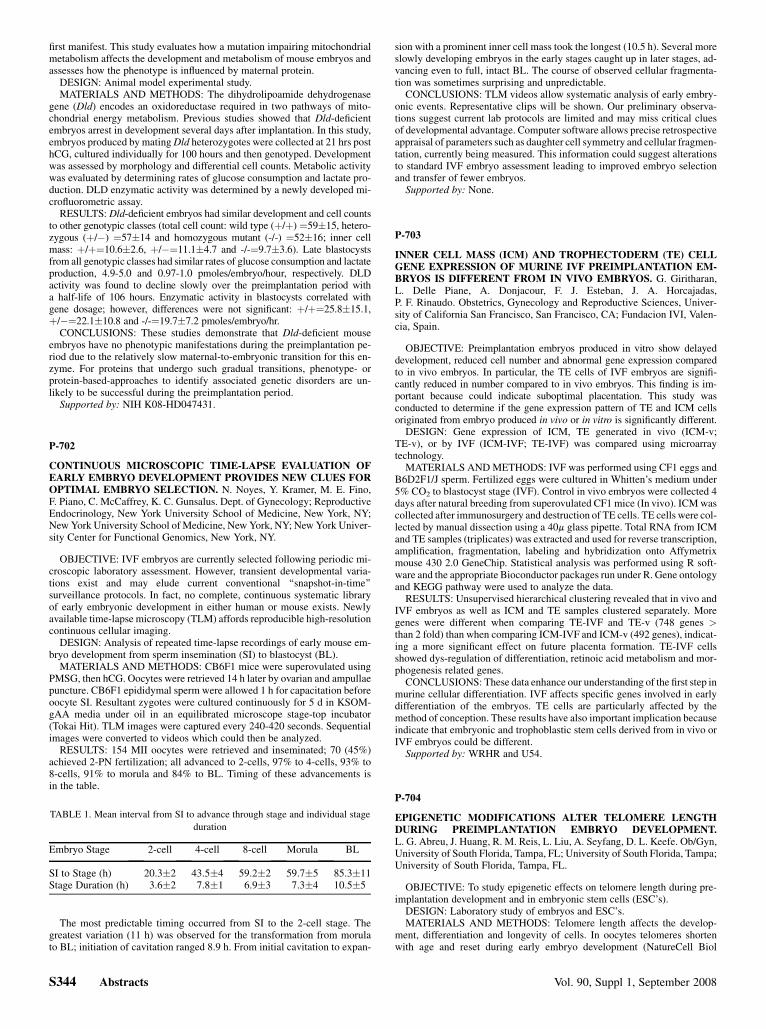
RESULTS: 154 MII oocytes were retrieved and inseminated; 70 (45%)achieved 2-PN fertilization; all advanced to 2-cells, 97% to 4-cells, 93% to8-cells, 91% to morula and 84% to BL. Timing of these advancements isin the table.

TABLE 1. Mean interval from SI to advance through stage and individual stage

duration

Embryo Stage

S344 Abstrac

2-cell

ts

4-cell

8-cell Morula BL

SI to Stage (h)

20.3�2 43.5�4 59.2�2 59.7�5 85.3�11 Stage Duration (h) 3.6�2 7.8�1 6.9�3 7.3�4 10.5�5

The most predictable timing occurred from SI to the 2-cell stage. Thegreatest variation (11 h) was observed for the transformation from morulato BL; initiation of cavitation ranged 8.9 h. From initial cavitation to expan-

sion with a prominent inner cell mass took the longest (10.5 h). Several moreslowly developing embryos in the early stages caught up in later stages, ad-vancing even to full, intact BL. The course of observed cellular fragmenta-tion was sometimes surprising and unpredictable.

CONCLUSIONS: TLM videos allow systematic analysis of early embry-onic events. Representative clips will be shown. Our preliminary observa-tions suggest current lab protocols are limited and may miss critical cluesof developmental advantage. Computer software allows precise retrospectiveappraisal of parameters such as daughter cell symmetry and cellular fragmen-tation, currently being measured. This information could suggest alterationsto standard IVF embryo assessment leading to improved embryo selectionand transfer of fewer embryos.

Supported by: None.

P-703

INNER CELL MASS (ICM) AND TROPHECTODERM (TE) CELLGENE EXPRESSION OF MURINE IVF PREIMPLANTATION EM-BRYOS IS DIFFERENT FROM IN VIVO EMBRYOS. G. Giritharan,L. Delle Piane, A. Donjacour, F. J. Esteban, J. A. Horcajadas,P. F. Rinaudo. Obstetrics, Gynecology and Reproductive Sciences, Univer-sity of California San Francisco, San Francisco, CA; Fundacion IVI, Valen-cia, Spain.

OBJECTIVE: Preimplantation embryos produced in vitro show delayeddevelopment, reduced cell number and abnormal gene expression comparedto in vivo embryos. In particular, the TE cells of IVF embryos are signifi-cantly reduced in number compared to in vivo embryos. This finding is im-portant because could indicate suboptimal placentation. This study wasconducted to determine if the gene expression pattern of TE and ICM cellsoriginated from embryo produced in vivo or in vitro is significantly different.

DESIGN: Gene expression of ICM, TE generated in vivo (ICM-v;TE-v), or by IVF (ICM-IVF; TE-IVF) was compared using microarraytechnology.

MATERIALS AND METHODS: IVF was performed using CF1 eggs andB6D2F1/J sperm. Fertilized eggs were cultured in Whitten’s medium under5% CO2 to blastocyst stage (IVF). Control in vivo embryos were collected 4days after natural breeding from superovulated CF1 mice (In vivo). ICM wascollected after immunosurgery and destruction of TE cells. TE cells were col-lected by manual dissection using a 40m glass pipette. Total RNA from ICMand TE samples (triplicates) was extracted and used for reverse transcription,amplification, fragmentation, labeling and hybridization onto Affymetrixmouse 430 2.0 GeneChip. Statistical analysis was performed using R soft-ware and the appropriate Bioconductor packages run under R. Gene ontologyand KEGG pathway were used to analyze the data.

RESULTS: Unsupervised hierarchical clustering revealed that in vivo andIVF embryos as well as ICM and TE samples clustered separately. Moregenes were different when comparing TE-IVF and TE-v (748 genes >than 2 fold) than when comparing ICM-IVF and ICM-v (492 genes), indicat-ing a more significant effect on future placenta formation. TE-IVF cellsshowed dys-regulation of differentiation, retinoic acid metabolism and mor-phogenesis related genes.

CONCLUSIONS: These data enhance our understanding of the first step inmurine cellular differentiation. IVF affects specific genes involved in earlydifferentiation of the embryos. TE cells are particularly affected by themethod of conception. These results have also important implication becauseindicate that embryonic and trophoblastic stem cells derived from in vivo orIVF embryos could be different.

Supported by: WRHR and U54.

P-704

EPIGENETIC MODIFICATIONS ALTER TELOMERE LENGTHDURING PREIMPLANTATION EMBRYO DEVELOPMENT.L. G. Abreu, J. Huang, R. M. Reis, L. Liu, A. Seyfang, D. L. Keefe. Ob/Gyn,University of South Florida, Tampa, FL; University of South Florida, Tampa;University of South Florida, Tampa, FL.

OBJECTIVE: To study epigenetic effects on telomere length during pre-implantation development and in embryonic stem cells (ESC’s).

DESIGN: Laboratory study of embryos and ESC’s.MATERIALS AND METHODS: Telomere length affects the develop-

ment, differentiation and longevity of cells. In oocytes telomeres shortenwith age and reset during early embryo development (NatureCell Biol

Vol. 90, Suppl 1, September 2008