Embed Size (px)
Citation preview
Continuing Education
Making Implants Part of Your Everyday Practice: A Necessary Alternative to Traditional ToothReplacement
by Robert F. Faulkner, DDS
Abstract:Treatment with dental implants has become a popular treatment option for manydentists and their patients. Unfortunately, not all treatment outcomes prove to besuccessful or always meet the patient’s expectations. Failure of this treatmentmodality often leads to lost income, unproductive chairtime, and mostimportantly, the patient questioning the treating dentist’s competency. Ifpractitioners are to be successful with this type of treatment, they must masterthe fundamentals of successful diagnosis and treatment planning and be fullyinformed of the many facets of this particular treatment alternative. This articlewill review the overall principles and necessary information for considering dentalimplants as a treatment option for patients.
Learning Objectives:After reading this article, the reader should be able to:
understand the need for proper diagnosis and treatment planning prior toinitiating treatment with dental implants.describe the specific elements of implant selection.recognize the biomechanical parameters necessary for occlusal andfunctional loading of implants.discuss the requirements for an esthetic outcome for a dental implantrestoration.
Tooth replacement has long been an established treatment objective to provideproper function and oral health. This is evident in the educational process that
dental students receive early in their predoctoral training1-5 (Figure 1 ViewFigure). However, much debate remains as to the best treatment alternative. Mostdentists have been taught that a fixed prosthodontic option is the first choice andis preferable to a removable partial denture (RPD). This viewpoint can be readilyobserved from the disproportionate number of fixed prosthodontic restorations,compared with RPDs, being fabricated by the dental laboratory technicians. Yetthe long-term prognosis for these treatment modalities should be questioned,especially because both types are dependent on abutment teeth for retention and
support of the prostheses.2,4 Recurrent decay remains the primary cause of failurefor both fixed and removable prostheses, followed by loss of one or moreabutment teeth. This may make future restorations using the same type ofprostheses difficult, if not impossible. Today, dental implants provide dentists withan alternative for tooth replacement.
Dental implants were introduced into ancient civilizations with the Mayan Indiansin approximately 600 A.D. Modern society saw the advent of dental implants in
1905 by Scholl.6 In the next several decades, many implant designs, materials, and
theories were introduced. Then in 1969 Brånemark et al7 published their researchdescribing the process of dental implant integration with bone. Brånemark is
credited with the concept of osseointegration, and this discovery has significantlyimpacted how modern dentists consider tooth replacement. This article reviewsmany of the necessary parameters for determining the viability of using dentalimplants for replacing missing teeth.
Diagnosis and Treatment Planning: the Basis for Success
Many factors must be considered for dental implants;8 however, one of the mostcrucial is an accurate diagnosis. There may be several treatment options forpartially or completely edentulous patients, but for each patient there is only onediagnosis. Care should be taken to adequately evaluate the patient beforeweighing treatment options (Table 1). Several important considerations are the
completion of growth in a young patient,9 health status in a more mature
patient,10 the patient’s financial concerns, and time constraints in completing theproposed treatment. In addition, patient expectations should be fully reviewedprior to initiating treatment: patients must have realistic expectations andunderstand the type of prostheses that can be fabricated for their particulartreatment needs (Figure 2 View Figure and Figure 3 View Figure). When theevaluation is complete, multidisciplinary treatment planning that includes thelaboratory technician, implant surgeon, orthodontist, and the restorative dentistshould be considered. A comprehensive case presentation should then becompleted with the patient to discuss all options, including no treatment. A
written summation of the treatment plan11 and informed consent are thenprovided to the patient to review and accept before treatment commences. Therestorative dentist must initiate the process in determining the prosthesis designand coordinate the treatment sequence with the other dental professionals. Whiletraditional treatment planning has been the standard, the advent of cone beamcomputed tomography (CBCT) has allowed 3-dimensional planning, which offersmore comprehensive information to be supplied to the patient prior to anysurgical procedure and which can greatly enhance the outcome.
Types of Implant ProsthesesWhen dental implants were introduced by Brånemark, they were intendedprimarily for the completely edentulous patient, specifically the edentulousmandible. The initial prosthesis was a fixed restoration commonly referred to as afixed bone anchored bridge. That particular prosthetic design is now the fixed-detachable hybrid prosthesis and has been highly successful for both the implants
and prostheses.12,13 Implants have also been used for overdenture restorations14
and can be implant-assisted or implant-supported. With implant-assistedprostheses, the implants and mucoperiosteum share the forces of occlusion. Asimple two-implant overdenture, either a Hader Bar® (Sterngold™, Attleboro, MA)or Locator® abutment (Zest Anchors, Escondido, CA), are examples of implant-assisted overlay prostheses and are always a removable restoration (Figure 4 ViewFigure and Figure 5 View Figure). With implant-supported prostheses, the forces ofocclusion are borne solely by the implants. This prosthesis can be an overlayprosthesis or a fixed restoration. Milled bar restorations, overdentures with barsubstructures, and metal ceramic restorations attached to implant abutments byeither screws or cement are examples of implant-supported restorations. One ofthe primary benefits of using dental implants in edentulous patients is thepreservation of the residual bone, which will provide a better opportunity forfuture successful prosthetic restorations.
Restoration of partially edentulous patients with dental implants has not onlyadded a new opportunity for replacement of missing teeth but also presented adifferent set of challenges for providing successful, long-term dental prostheses.Initially, many treatment protocols that were followed for the completelyedentulous patient proved to be less than ideal for the partially edentulous
patient.15 Combining natural teeth and dental implants in the same arch,sometimes in the same quadrant, creates a biomechanical difference betweenteeth and implant(s) during function. Traditional three-unit fixed partial denturesplaced on two dental implants can be successful. Yet when forces of occlusion arehigh, the need to place one implant per tooth or three implants to support four
units in the posterior quadrants is often recommended.8,15 Moreover, blendingnatural teeth and bony/gingival contours with the edentulous spaces beingrestored with implants can be difficult in meeting the patient’s expectations andcan potentially create difficulty with oral hygiene access.
Single-tooth replacement with dental implants has proven to be very beneficialyet challenging for both the patient and treating dentist. Long-term success has
now been achieved in the anterior and posterior areas,16 although specificcharacteristics can affect the prognosis, such as the quality and quantity of bone,occlusal loads, and parafunctional habits. While these restorations have beenshown to be highly successful, there remains the critical challenge of completelyrestoring the esthetic and functional needs for the patient and making theserestorations undetectable from the other natural teeth. A complete understandingof the edentulous space is necessary for an accurate diagnosis, which then allowsfor the development of a treatment sequence for the best possible outcome.Frequently, preprosthetic procedures are required prior to implant placement if acompromise is to be avoided. Consideration should be given not only to theesthetic value of the single-implant tooth replacement but also should account forthe patient’s oral hygiene needs.
Implant Selection: How Important Is it?Endosseous root form dental implants have undergone numerous changes sincethey were introduced by Brånemark. Early implants were of relatively narrowdiameter (3.5 mm) and externally hexagonal. These implants were turned duringthe fabrication process, leaving a somewhat smooth, machined surface to thebody of the implant. Eventually, implants were increased to a 3.75-mm diameter.This was the standard for several years, although these implants were usedprimarily for edentulous patients and were commonly splinted together.Subsequent research revealed that implant-to-bone contact was important in the
long-term function of the implant.17 The implant-to-bone contact in the initialmachined-surface implants was between 30% and 40% and provided acceptable
stability in most edentulous patients.18 When implants became more popular,especially in partially edentulous situations, it became apparent that other factorswere necessary for long-term functional and esthetic needs. By roughening thesurface of the implant, bone apposition to the implant can now reach 70% to 80%
or higher and provide better support for the function of the prostheses.19 Otherdesign changes have significantly improved the long-term prognosis of the implant:increasing the diameter of the implant body, changing the thread pitch of theimplant, providing microthreads in the coronal portion of the implant, andchanging the implant/abutment connection from an external flat-top clearance-fit
connection to a conical connection. These improvements not only improvedfunctionality of the implant but also significantly advanced the esthetic
possibilities for long-term success of the restorations.20
Implant size should be considered based on the tooth to be replaced. In theedentulous patient, when an overlay or hybrid prosthesis is being planned, a 4-mmdiameter implant body size is recommended for all of the implants used in the
prosthesis.15 When placing implants in partially edentulous patients, size selectioncan be as small as 3 mm for the mandibular central and lateral incisors and smallmaxillary lateral incisors. However, implant size should match tooth dimensions. Inthe posterior regions of the maxilla and mandible, implant recommendationsrange from 4-mm diameter in the premolar regions to 6-mm diameter in the molar
areas.21,22 Increasing the implant diameter can provide acceptable support forboth the patient’s functional and esthetic needs. In the maxillary anterior area,the diameter of the implant must be carefully considered, especially if the patienthas a thin, scalloped periodontal biotype. Implants should have 1.5 mm of bonesurrounding the implant, especially on the facial surface of the implant, forlong-term maintenance of the bone. If an implant diameter is too wide, boneremodeling can occur, followed by gingival recession, possibly resulting in aless-than-acceptable esthetic result. Without preprosthetic augmentation (soft- orhard-tissue grafts), the clinician should proceed with a slightly smaller-diameterimplant in the anterior area if there is a concern that bone maintenance could bean issue. Careful assessment of the tooth or teeth diagnostically waxed to fullcontour on the diagnostic cast will provide guidance in determining implantdiameter.
Most implants used today are screw-shaped designs as opposed to cylinderpress-fits. As such, many designs have emerged, in respect to the threads of theimplant and their role in the initial stability of the implant and to the long-term
maintenance of the bone surrounding the implant.23 Thread designs increase thesurface area of the implant body, thereby allowing a greater potential forincreasing the quantity of bone attached to the implant. This also helps with theinitial stability of the implant at the time of placement. In addition, someimplants have smaller microthreads at the coronal portion of the implant to helpdivert the stresses from the top of the implant where the critical area of crestalbone needs to be maintained. If crestal bone is preserved, the potential increasesfor providing patients with more predictable esthetic results and improvedhygiene access to the implant restoration.
Finally, the implant/abutment connection is important not only in the stability ofthe prosthesis but also in the periodontal health of the implant–restorationcomplex. Conical connections have been shown to provide a bacterial seal far
superior than the original flat-top implant/abutment connections.20 By eliminatingthe bacterial contamination of the internal portion of the implant, the crestalbone is not subjected to the same inflammatory processes, which may promotecrestal bone maintenance and reduce the possibility of peri-implantitis.
Biomechanics and Dental Implants—The Occlusal FactorWhen implants were introduced for tooth replacement, little consideration wasgiven to occlusal loads placed on implants. Most implant prostheses were placed inthe mandibular arch and were opposed by a complete denture in an edentulous
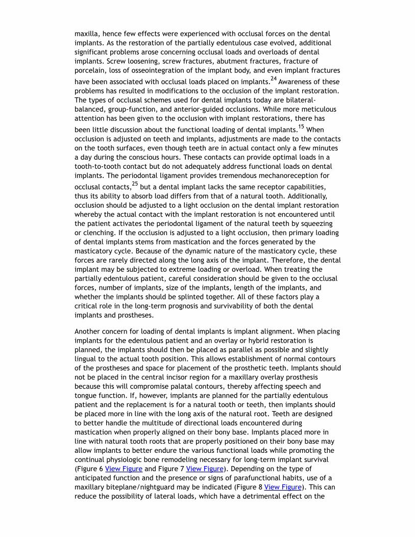
maxilla, hence few effects were experienced with occlusal forces on the dentalimplants. As the restoration of the partially edentulous case evolved, additionalsignificant problems arose concerning occlusal loads and overloads of dentalimplants. Screw loosening, screw fractures, abutment fractures, fracture ofporcelain, loss of osseointegration of the implant body, and even implant fractures
have been associated with occlusal loads placed on implants.24 Awareness of theseproblems has resulted in modifications to the occlusion of the implant restoration.The types of occlusal schemes used for dental implants today are bilateral-balanced, group-function, and anterior-guided occlusions. While more meticulousattention has been given to the occlusion with implant restorations, there has
been little discussion about the functional loading of dental implants.15 Whenocclusion is adjusted on teeth and implants, adjustments are made to the contactson the tooth surfaces, even though teeth are in actual contact only a few minutesa day during the conscious hours. These contacts can provide optimal loads in atooth-to-tooth contact but do not adequately address functional loads on dentalimplants. The periodontal ligament provides tremendous mechanoreception for
occlusal contacts,25 but a dental implant lacks the same receptor capabilities,thus its ability to absorb load differs from that of a natural tooth. Additionally,occlusion should be adjusted to a light occlusion on the dental implant restorationwhereby the actual contact with the implant restoration is not encountered untilthe patient activates the periodontal ligament of the natural teeth by squeezingor clenching. If the occlusion is adjusted to a light occlusion, then primary loadingof dental implants stems from mastication and the forces generated by themasticatory cycle. Because of the dynamic nature of the masticatory cycle, theseforces are rarely directed along the long axis of the implant. Therefore, the dentalimplant may be subjected to extreme loading or overload. When treating thepartially edentulous patient, careful consideration should be given to the occlusalforces, number of implants, size of the implants, length of the implants, andwhether the implants should be splinted together. All of these factors play acritical role in the long-term prognosis and survivability of both the dentalimplants and prostheses.
Another concern for loading of dental implants is implant alignment. When placingimplants for the edentulous patient and an overlay or hybrid restoration isplanned, the implants should then be placed as parallel as possible and slightlylingual to the actual tooth position. This allows establishment of normal contoursof the prostheses and space for placement of the prosthetic teeth. Implants shouldnot be placed in the central incisor region for a maxillary overlay prosthesisbecause this will compromise palatal contours, thereby affecting speech andtongue function. If, however, implants are planned for the partially edentulouspatient and the replacement is for a natural tooth or teeth, then implants shouldbe placed more in line with the long axis of the natural root. Teeth are designedto better handle the multitude of directional loads encountered duringmastication when properly aligned on their bony base. Implants placed more inline with natural tooth roots that are properly positioned on their bony base mayallow implants to better endure the various functional loads while promoting thecontinual physiologic bone remodeling necessary for long-term implant survival(Figure 6 View Figure and Figure 7 View Figure). Depending on the type ofanticipated function and the presence or signs of parafunctional habits, use of amaxillary biteplane/nightguard may be indicated (Figure 8 View Figure). This canreduce the possibility of lateral loads, which have a detrimental effect on the
dental implant(s). The biteplane is adjusted in centric relation with simultaneousocclusal contacts and shallow anterior guidance.
Esthetics and Dental Implants—One of the Final ChallengesWhen dental implants became a predictable solution for tooth replacement, thefocus became making implants appear more like natural teeth and undetectablefrom the surrounding dentition. Greater emphasis on diagnosing and classifying theedentulous area(s) has become necessary to properly plan the treatment withdental implants. When tooth loss occurs, there is also a corresponding loss of boneand soft tissue. The extent of the defect depends on the cause of tooth loss, themethod used for tooth removal, and the length of time that the tooth has beenmissing. If the tooth loss was from periodontal disease and bone loss has occurred,then replacement with a dental implant and achievement of an esthetic resultmay be extremely challenging. Teeth that have been treated endodontically anddeveloped vertical root fractures necessitating removal may have significant lossof bone associated with the site. Tooth extraction itself may also compromise thesurrounding bone, especially the facial plate of bone, and a buccal bone defectmay occur. Determining the type of defect is important in the diagnosis and
treatment planning stage to facilitate the outcome for the patient.26
Patient expectations remain extremely critical. The treatment options must beable to meet these expectations, otherwise tooth replacement of the edentulousarea with dental implants should not be attempted. A set of mounted casts incentric relation with a full contour diagnostic wax-up, including the hard- andsoft-tissue deficits, is mandatory prior to initiating treatment. This is beneficial inaccessing the edentulous space and communicating the information concerningthe edentulous defect with the laboratory technician, the implant surgeon, andmost importantly, the patient. In addition, CBCT can also be extremely helpful indiagnosing the edentulous site 3-dimensionally and providing vital information tothe surgeon prior to implant placement (Figure 9 View Figure). Only after thediagnosis and treatment plan is fully understood by all parties should thetreatment be initiated. In many situations, hard- and soft-tissue grafts arenecessary in order to reconstruct the edentulous site as near to its originalcontours as possible (Figure 10 View Figure, Figure 11 View Figure and Figure 12View Figure). This will significantly enhance proper positioning of the implant for
prosthetic needs and help to increase the prognosis for an esthetic result.27 At thetime of implant placement, a surgical template should be used to help ensure thatthe implant is placed in the proper position. The surgical template can befabricated from the diagnostic wax-up or generated from planning software usingthe Digital Imaging and Communications in Medicine data obtained from the CBCT.The esthetic result is generally dictated at the time the implant is placed, hencethe need for such a detailed assessment.
Other factors may play a role in the final esthetic outcome. Indexing the positionof the implant at the time of placement, use of provisional restorations, andcustomized impression copings to record gingival contours for laboratoryfabrication of the abutment and crown are examples of techniques to enhance theoverall esthetic result (Figure 13 View Figure, Figure 14 View Figure, Figure 15View Figure and Figure 16 View Figure). In addition, newer materials have beenintroduced to aid the fabrication of esthetic abutments and restorations placed ondental implants. Zirconium, both as an abutment and restoration, has been shownto have a beneficial esthetic effect and is well tolerated by the surrounding
tissue.28 Caution, though, must be emphasized in the use of this material becauseit is not fully known how it will withstand functional loads long term, especially inthe posterior areas. Titanium abutments remain the material of choice in the
posterior areas.29 Waxing and casting of premachined UCLA-type abutments alsooffers an option for abutment design, enabling precise angle corrections whennecessary, although the rise in the cost of precious metals may limit the use ofthis material in the future. The significant improvement of abutments generatedwith computer-aided design and computer-aided manufacturing (CAD/CAM) hasgreatly enhanced the abutment design and fabrication. Metal ceramic restorationsprovide suitable restorations in the posterior region and are used because of their
proven long-term success,1-3 although proper metal coping design is necessary forstrength of the veneered porcelain. Gold occlusal surfaces, especially in the molarregion, may be the best choice if the patient has high occlusal forces orparafunctional habits that may make material fracture a concern.
ConclusionMany factors play a role in the treatment outcome when using dental implants.Care must be taken to properly assess and select the patient for this type oftreatment and to ensure not only that the treatment will be successful but thatthe patient’s expectations are met as well. Diagnosis and treatment planning arekey elements for positive outcomes, yet other variables such as the biology ofimplant selection, biomechanics and functional loading of dental implants, andesthetic parameters are all necessary for successful results. In addition, thetreatment should provide for re-treatment in the future, as the patient will likelyoutlive the restorations placed on the dental implants. Failure to adhere to thebasic principles of diagnosis and treatment planning may lead to complicationsthat might otherwise be avoided, as well as unnecessary treatment in the future.Finally, dentists are afforded an opportunity for a treatment alternative that willnot only replace missing teeth but, in turn, preserve remaining structures vital tothe health, form, and function of patients.
AcknowledgementsThe author wishes to express his sincere appreciation to Wayne H. Gordner, DMD,MS, FACP; Larry J. Brandau, DDS; and Erin Shearer for their assistance in compilingthis article. Without your help, this project would not have been possible.
References1. Johnston JF, Phillips RW, Dykema RW. Modern Practice in Crown and BridgeProsthodontics. 3rd ed. Philadelphia, PA: W.B. Saunders Company; 1971.
2. Shillingburg HT, Hobo S, Whitsett LD, et al. Fundamentals of FixedProsthodontics. 3rd ed. Chicago, IL: Quintessence Publishing Co; 1997.
3. Rosenteil SF, Land MF, Fujimoto J. Contemporary Fixed Prosthodontics. 2nd ed.St. Louis, MS: Mosby Year Book, Inc.; 1995.
4. McGivney GP, Castleberry DJ. Removable Partial Prosthodontics. 9th ed. St.Louis, MS: Mosby-Year Book, Inc.; 1995.
5. Boucher CO, Hickey JC, Zarb GA. Prosthodontic Treatment for EdentulousPatients. 7th ed. St. Louis, MS: C.V. Mosby Company; 1975.
6. Scholl CR. Implantation of a porcelain tooth. Dental Cosmos. 1905;47(1):
157-158.
7. Brånemark PI, Adell R, Breine U, et al. Intra-osseous anchorage of dentalprostheses. I. Experimental studies. Scand J Plast Reconstr Surg. 1969;3(2):81-100.
8. Wood MR, Vermilyea SG; Committee on Research in Fixed Prosthodontics of theAcademy of Fixed Prosthodontics. A review of selected dental literature onevidence-based treatment planning for dental implants: report of the Committeeon Research in Fixed Prosthodontics of the Academy of Fixed Prosthodontics. JProsthet Dent. 2004;92(5): 447-462.
9. Westwood RM, Duncan JM. Implants in adolescents: a literature review and casereports. Int J Oral Maxillofac Implants. 1996;11(6): 750-755.
10. Bryant SR, Zarb GA. Osseointegration of oral implants in older and youngeradults. Int J Oral Maxillofac Implants. 1998;13(4):492-499.
11. Preston JD, Sheppard GA. The confirmation letter: information and protection.Int J Prosthodont. 1988;1(2): 143-148.
12. Zarb GA, Schmitt A. The edentulous predicament. I: a prospective study of theeffectiveness of implant-supported fixed prostheses. J Am Dent Assoc.1996;127(1):59-65.
13. Mericske-Stern RD, Taylor TD, Belser U. Management of the edentulouspatient. Clin Oral Implants Res. 2000;11(suppl 1):108-125.
14. Mericske-Stern R. Overdentures with roots or implants for elderly patients: acomparison. J Prosthet Dent. 1994;72(5):543-550.
15. Rangert BR, Sullivan RM, Jemt TM. Load factor control for implants in theposterior partially edentulous segment. Int J Oral Maxillofac Implants. 1997;12(3):360-370.
16. Priest G. Single-tooth implants and their role in preserving remaining teeth: a10 year survival study. Int J Oral Maxillofac Implants. 1999;14(2): 181-188.
17. Ericsson I, Johansson CB, Bystedt H, et al. A histomorphometric evaluation ofbone-to-implant contact on machine-prepared and roughened titanium dentalimplants: a pilot study in the dog. Clin Oral Implants Res. 1994;5(4): 202-206.
18. Trisi P, Lazzara R, Rao W, et al. Bone-implant contact and bone quality:evaluation of expected and actual bone contact on machined and osseotiteimplant surfaces. Int J Periodontics Restorative Dent. 2002;22(6): 535-545.
19. Ellingsen JE, Johansson CB, Wennerberg A, et al. Improved retention andbone-to-implant contact with fluoride-modified titanium implants. Int JOraMaxillofac Implants. 2004;19(5):659-666.
20. Zipprich H, Weigl P, Lange B, et al. Micromovements at the implant-abutmentinterface: measurement, causes, and consequences. Implantologie.2007;15(1):31-46.
21. Davarpanah M, Martinez H, Kebir M, et al. Wide-diameter implants: newconcepts. Int J Periodontics Restorative Dent. 2001;21(2):149-159.
22. Langer B, Langer L, Herrmann I, et al. The wide fixture: a solution for specialbone situations and a rescue for the compromised implant. Part 1. Int J OralMaxillofac Implants. 1993;8(4):400-408.
23. Hansson S. The dental implant meets bone—a clash of two paradigms. AppOsseointegration Res. 2006;5:5-17.
24. Goodacre CJ, Bernal G, Rungcharassaeng K, et al. Clinical complications withimplants and implant prostheses. J Prosthet Dent. 2003;90(2): 121-132.
25. Dawson PE. Evaluation, Diagnosis, and Treatment of Occlusal Problems. 2nded. St. Louis, MS: The C.V. Mosby Company; 1989.
26. Salama H, Garber DA, Salama MA, et al. Fifty years of interdisciplinary sitedevelopment: lessons and guidelines from periodontal prosthesis. J Esthet Dent.1998;10(3):149-156.
27. Buser D, Martin W, Belser UC. Optimizing esthetics for implant restorations inthanterior maxilla: anatomic and surgical considerations. Int J Oral MaxillofacImplants. 2004;92(7):43-61.
28. Raigrodski AJ. Contemporary materials and technologies for all-ceramic fixedpartial dentures: a review of the literature. J Prosthet Dent. 2004;92(6):557-562.
29. Butz F, Heydecke G, Okutan M, et al. Survival rate, fracture strength andfailure mode of ceramic implant abutments after chewing simulation. J OralRehabil. 2005;32(11):838-843.
To take the quiz and receive credit for this course, click here and you will beredirected to the Dawson Center CE Portal.Thank you.
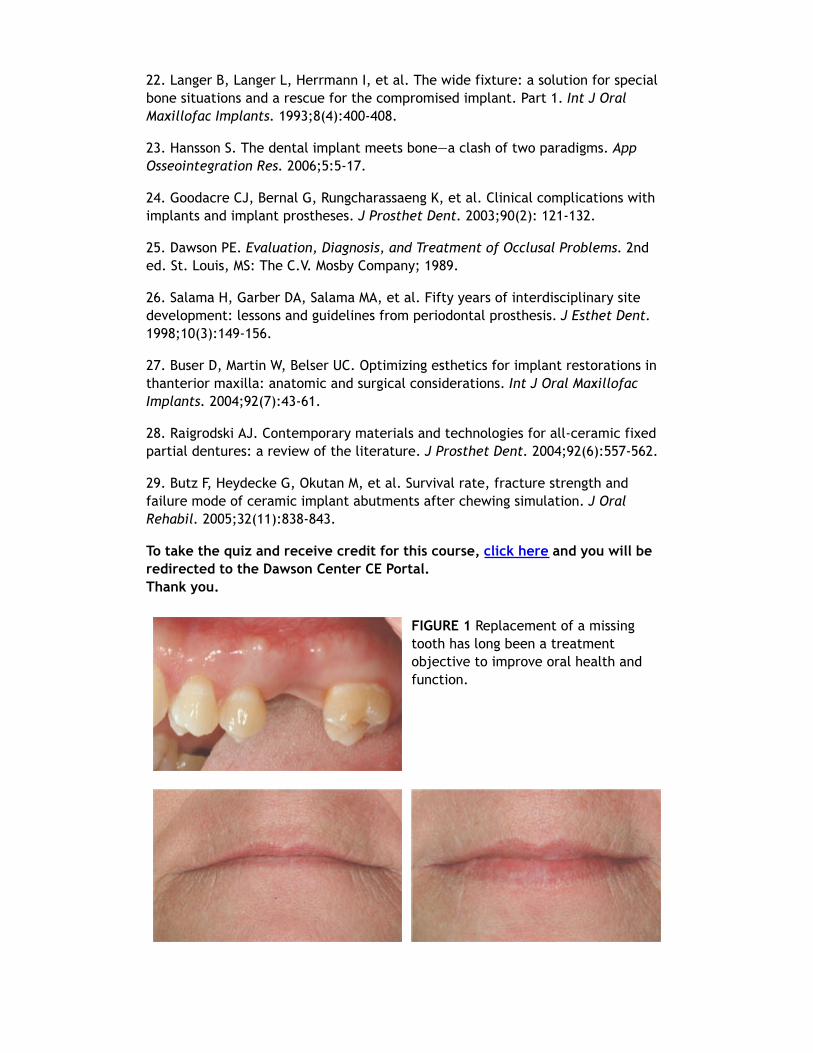
FIGURE 1 Replacement of a missingtooth has long been a treatmentobjective to improve oral health andfunction.

FIGURE 2 AND FIGURE 3 Removable prostheses can provide proper support andenhance overall facial esthetics for the patient.
FIGURE 4 An example of a two-implanttissue bar for an implant-assistedprosthesis.
FIGURE 5 Locator abutments placed onimplants can also be used for animplant-assisted prosthesis.
FIGURE 6 Tomograms showing naturalteeth alignments on their bony bases(Wheeler R. Dental Anatomy,Physiology and Occlusion. 5th ed.Philadelphia, PA: WB Saunders;1974:364).
FIGURE 7 Implants placed more inalignment with natural tooth roots.
FIGURE 8 A maxillarybiteplane/nightguard adjusted tocentric relation.
FIGURE 9A computed tomography scancan provide additional information toaid the implant team for properplacement of the implant(s).
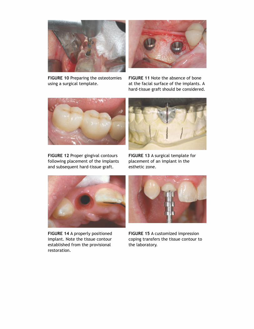
FIGURE 10 Preparing the osteotomiesusing a surgical template.
FIGURE 11 Note the absence of boneat the facial surface of the implants. Ahard-tissue graft should be considered.
FIGURE 12 Proper gingival contoursfollowing placement of the implantsand subsequent hard-tissue graft.
FIGURE 13 A surgical template forplacement of an implant in theesthetic zone.
FIGURE 14 A properly positionedimplant. Note the tissue contourestablished from the provisionalrestoration.
FIGURE 15 A customized impressioncoping transfers the tissue contour tothe laboratory.
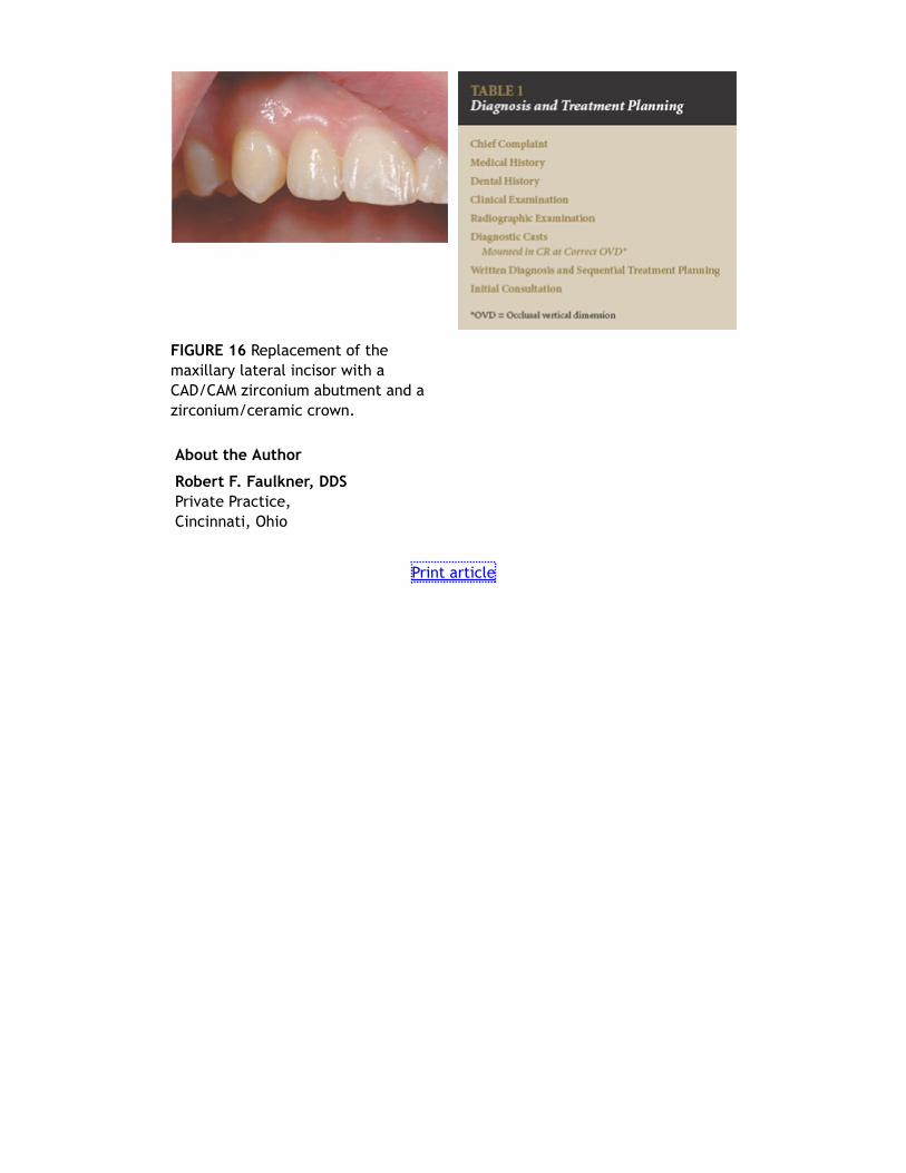
FIGURE 16 Replacement of themaxillary lateral incisor with aCAD/CAM zirconium abutment and azirconium/ceramic crown.
About the Author
Robert F. Faulkner, DDSPrivate Practice,Cincinnati, Ohio
Print article