Embed Size (px)
Citation preview

TABLE OF CONTENTS
Contents Page No.
UNIVERSAL EYE SPECULUM 3 WIRE SPECULUM [CLOSED LOOP] 4 THERMOCAUTERY 5 GLOBE FIXATION FORCEPS 6 CONJUNCTIVAL/ PLAIN SCISSORS 8 CORNEAL SPRING SCISSORS [UNIVERSAL] 9 IRIS FORCEPS 11 DE-WECKER’S IRIS SCISSORS 13 IRIS REPOSITOR 14 BARRAQUER’S NEEDLE HOLDER 15 SUPERIOR RECTUS HOLDING FORCEPS 16 SUTURE TYING FORCEPS 16 IRRIGATING WIRE VECTIS 17 SIMCOE’S IRRIGATION-ASPIRATION BI WAY CANNULA 18 IOL DIALLER/ SINSKY’S HOOK 19 LENS EXPRESSOR [LENS HOOK] 20 ST. MARTIN’S FORCEPS AND COLIBRI FORCEPS 21 CRESCENT KNIFE, SIDE PORT BLADE AND ANGULAR KERATOME 22
STRABISMUS HOOK/ MUSCLE HOOK/ SQUINT HOOK 24 DESMARRE’S LID RETRACTOR 24

CHALAZION FORCEPS [CLAMP] 25 CHALAZION SCOOP [CURETTE] 26
MULLER’S SELF RETAINING HEMOSTATIC LACRIMAL WOUND RETRACTOR 27 BONE PUNCH 29 LACRIMAL SAC DISSECTOR WITH CURETTE/ SCOOP 30 EVISCERATION SCOOP/ SPOON 31 ENUCLEATION SCISSORS 32 OTHER INSTRUMENTS:
POSTERIOR CHAMBER IOL [PCIOL] ANTERIOR CHAMBER IOL [ACIOL] PIN HOLE LENSE DISPOSABLE SYRINGE WITH 20G NEEDLE FLUORESCEIN STRIP
33
Pictures are taken from Ophthalmology Department, R.G.KAR.MEDICAL COLLEGE.
Text are taken from the following sources:
1. Bassak’s Oral and Practical Ophthalmology.
2. Yanoff’s Ophthalmology.
3. Parson’s Disease Of The Eye.
4. Wikipedia.
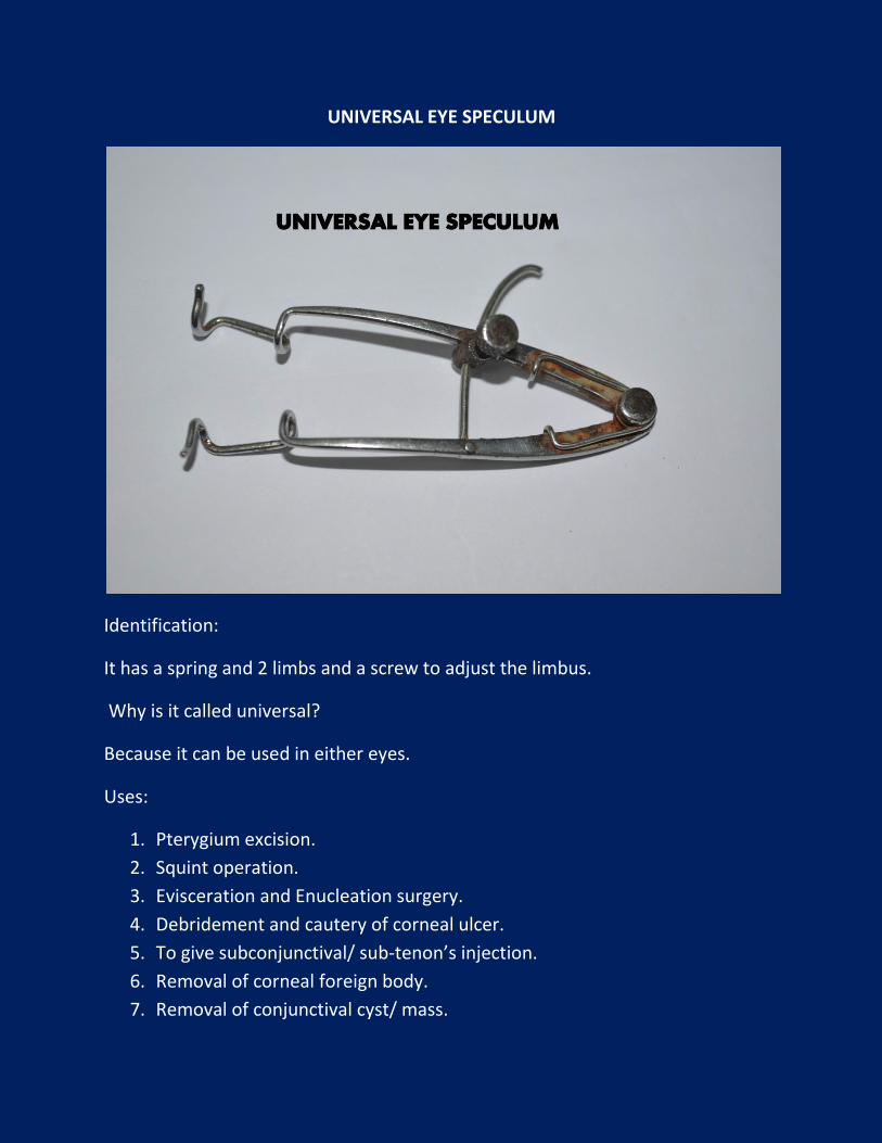
UNIVERSAL EYE SPECULUM
Identification:
It has a spring and 2 limbs and a screw to adjust the limbus.
Why is it called universal?
Because it can be used in either eyes.
Uses:
1. Pterygium excision.
2. Squint operation.
3. Evisceration and Enucleation surgery.
4. Debridement and cautery of corneal ulcer.
5. To give subconjunctival/ sub-tenon’s injection.
6. Removal of corneal foreign body.
7. Removal of conjunctival cyst/ mass.
8. Removal of suture.
Why it is not used routinely in intraocular operations?
Because it gives pressure over the globe, which elevates the intraocular pressure
[IOP] during operation. To avoid this, one can put a cotton pellet during the
operation.
Disadvantages:
1. Since it has no guard, eyelashes/ upper lid come in the field of operation.
2. It has a distinct disadvantage of giving more vitreous thrust and
subsequently, more vitreous loss can occur.
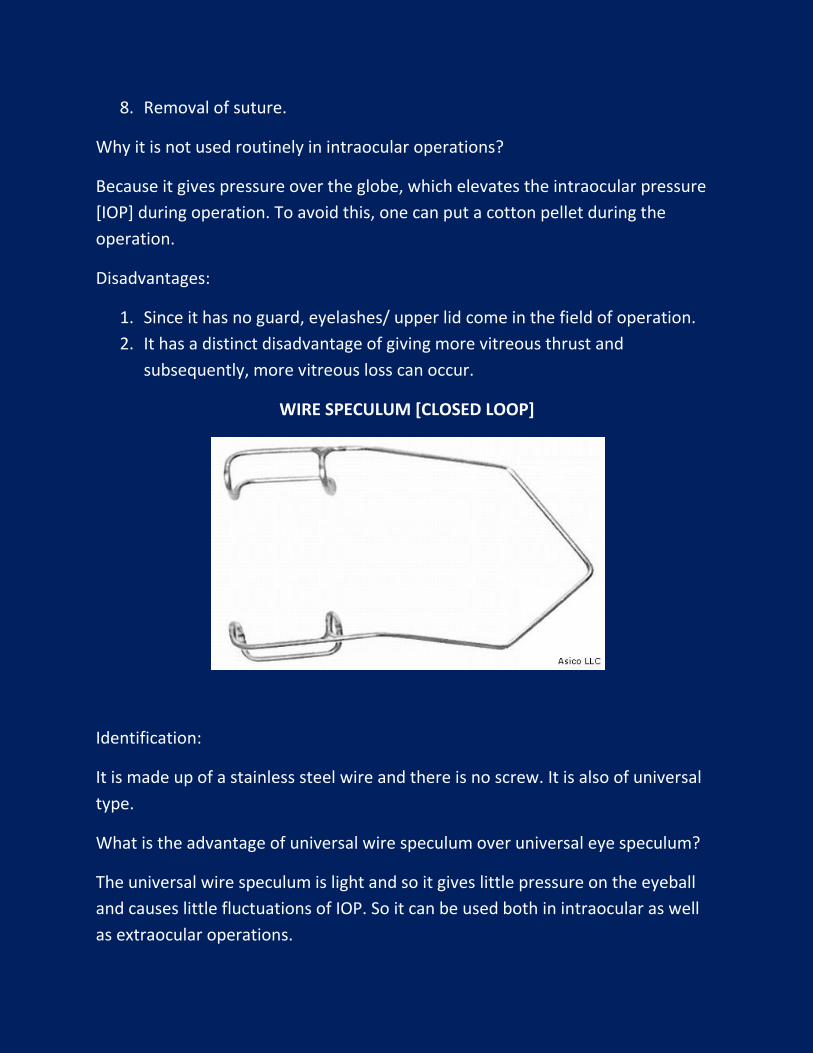
WIRE SPECULUM [CLOSED LOOP]
Identification:
It is made up of a stainless steel wire and there is no screw. It is also of universal
type.
What is the advantage of universal wire speculum over universal eye speculum?
The universal wire speculum is light and so it gives little pressure on the eyeball
and causes little fluctuations of IOP. So it can be used both in intraocular as well
as extraocular operations.
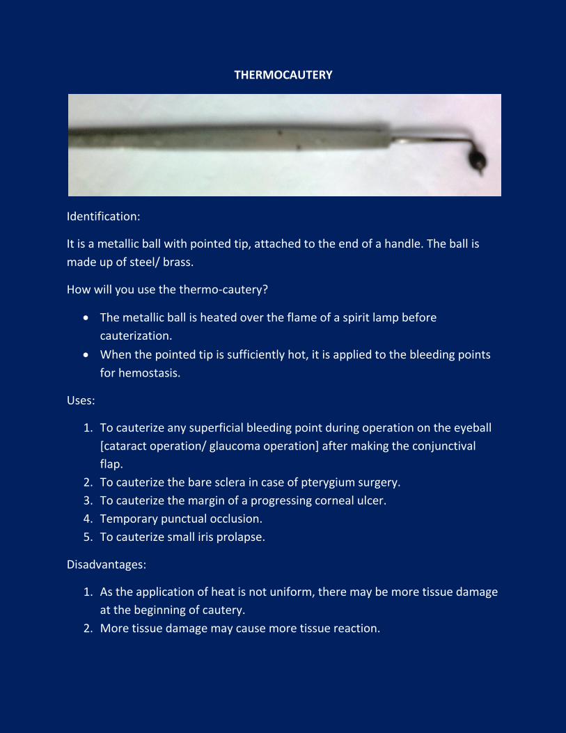
THERMOCAUTERY
Identification:
It is a metallic ball with pointed tip, attached to the end of a handle. The ball is
made up of steel/ brass.
How will you use the thermo-cautery?
The metallic ball is heated over the flame of a spirit lamp before
cauterization.
When the pointed tip is sufficiently hot, it is applied to the bleeding points
for hemostasis.
Uses:
1. To cauterize any superficial bleeding point during operation on the eyeball
[cataract operation/ glaucoma operation] after making the conjunctival
flap.
2. To cauterize the bare sclera in case of pterygium surgery.
3. To cauterize the margin of a progressing corneal ulcer.
4. Temporary punctual occlusion.
5. To cauterize small iris prolapse.
Disadvantages:
1. As the application of heat is not uniform, there may be more tissue damage
at the beginning of cautery.
2. More tissue damage may cause more tissue reaction.
Which is the best method of hemostasis?
1. Wet field cautery.
2. Diathermy.
Why you called the above two methods best for achieving hemostasis?
Because the application of heat is uniform and tissue damage and tissue reaction
are minimal.
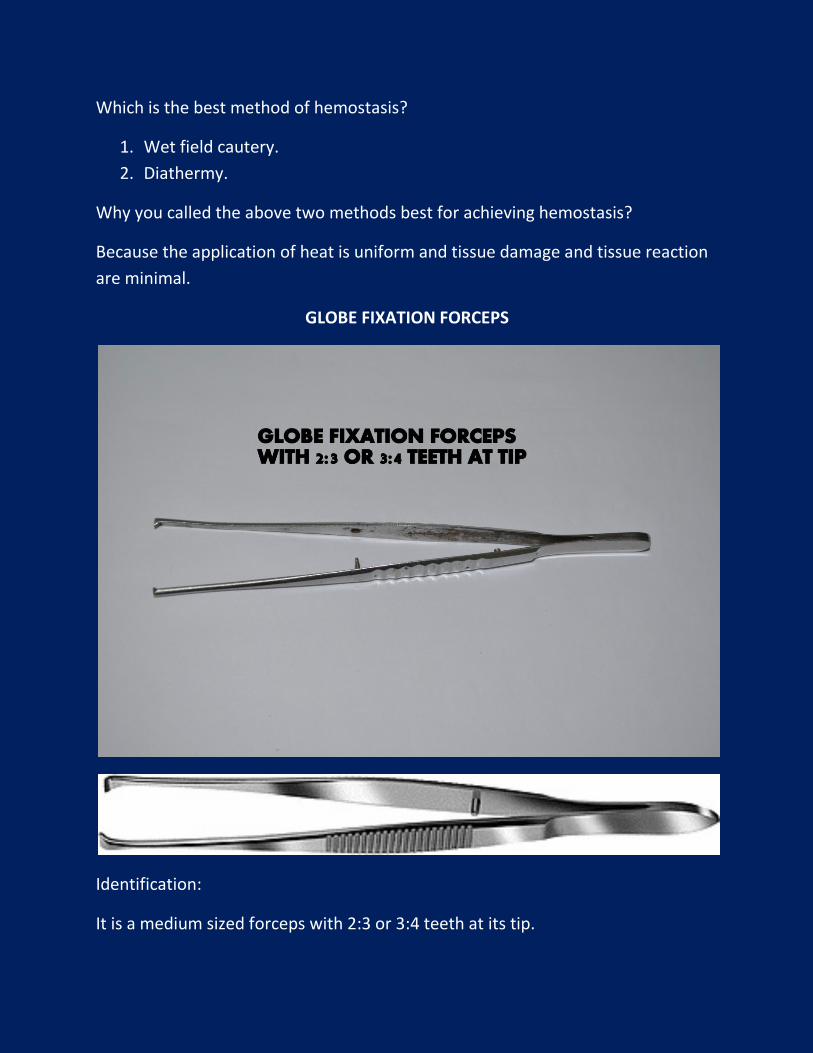
GLOBE FIXATION FORCEPS
Identification:
It is a medium sized forceps with 2:3 or 3:4 teeth at its tip.
How will you fix the globe?
The conjunctiva and episcleral tissue are firmly held at 6 o’clock position within 3-
4 mm of limbus with left hand.
Why this position is preferred for globe fixation?
Because conjunctiva is firmly adherent to the episcleral tissue and sclera around
the limbus.
Uses:
1. To fix the eyeball during operative procedures like:
a. Cataract surgery.
b. Pterygium surgery etc.
2. To catch superior rectus muscle/ Tenon’s capsule to pass bridle suture.
3. To lift bulbar conjunctiva during sub-conjunctival/ sun-Tenon injection.
4. Used in Forced Duction Test [FDT]* in case of squint to know any
mechanical restriction.
5. To hold the sponge piece/ gauze piece/ cotton ball for swabbing.
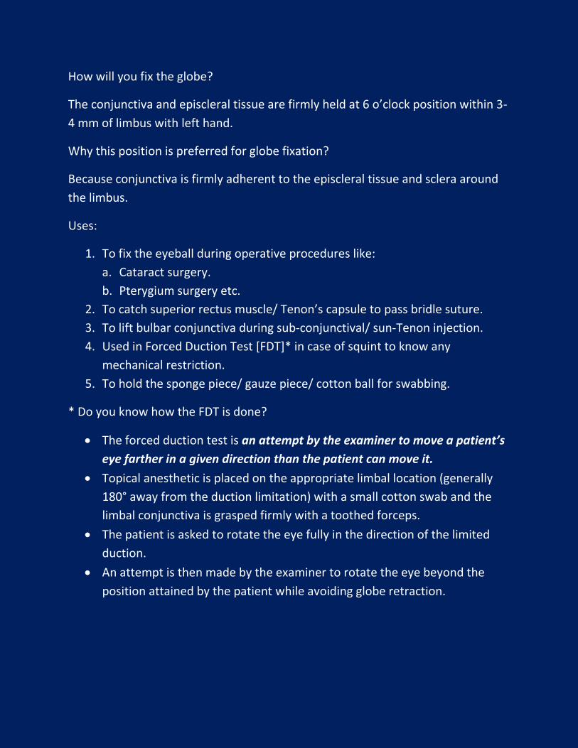
* Do you know how the FDT is done?
The forced duction test is an attempt by the examiner to move a patient’s
eye farther in a given direction than the patient can move it.
Topical anesthetic is placed on the appropriate limbal location (generally
180° away from the duction limitation) with a small cotton swab and the
limbal conjunctiva is grasped firmly with a toothed forceps.
The patient is asked to rotate the eye fully in the direction of the limited
duction.
An attempt is then made by the examiner to rotate the eye beyond the
position attained by the patient while avoiding globe retraction.
Care must be taken not to abrade the cornea.
Patients who have pure nerve palsy exhibit no restriction to full movement
by the examiner.
Patients who have pure restriction (Ex.: Entrapment of ocular contents
after blowout fracture) exhibit restricted movements (A positive forced
duction test).
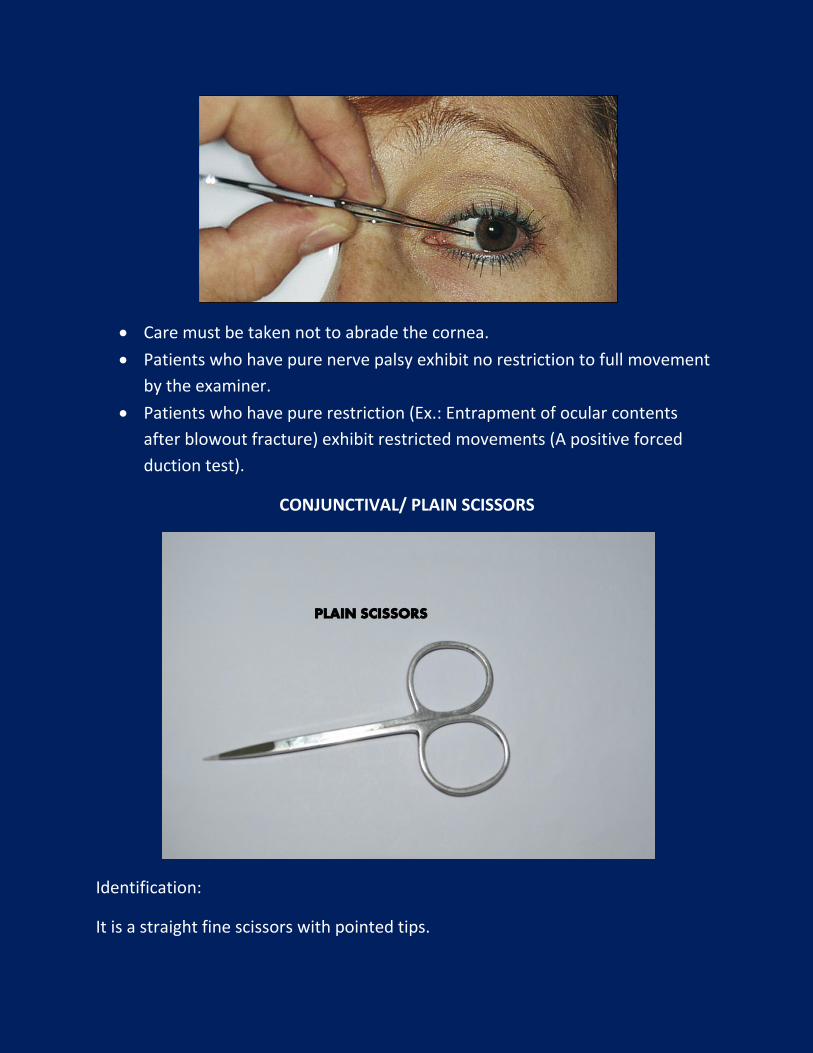
CONJUNCTIVAL/ PLAIN SCISSORS
Identification:
It is a straight fine scissors with pointed tips.
Uses:
1. It is used to dissect the bulbar conjunctiva in the following operations:
a. Conjunctival flap in cataract operation.
b. Conjunctival flap in trabeculectomy operation.
c. Enucleation operation/ Squint surgery/ Surgical correction of retinal
detachment etc.
2. To cut the suture ends.
3. To cut other tissues like Tenon’s capsules/ Pterygium etc.
What are the different types of conjunctival flaps made in cataract surgery?
The conjunctival flap may be of 2 types:
1. Fornix based flap:
Conjunctiva is cut at the limbus and retracted towards fornix.
2. Limbal based flap:
Conjunctiva is cut few mm away from the limbus and then it is reflected
over the cornea.
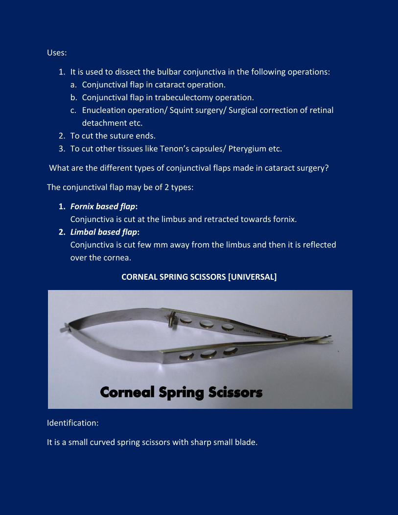
CORNEAL SPRING SCISSORS [UNIVERSAL]
Identification:
It is a small curved spring scissors with sharp small blade.
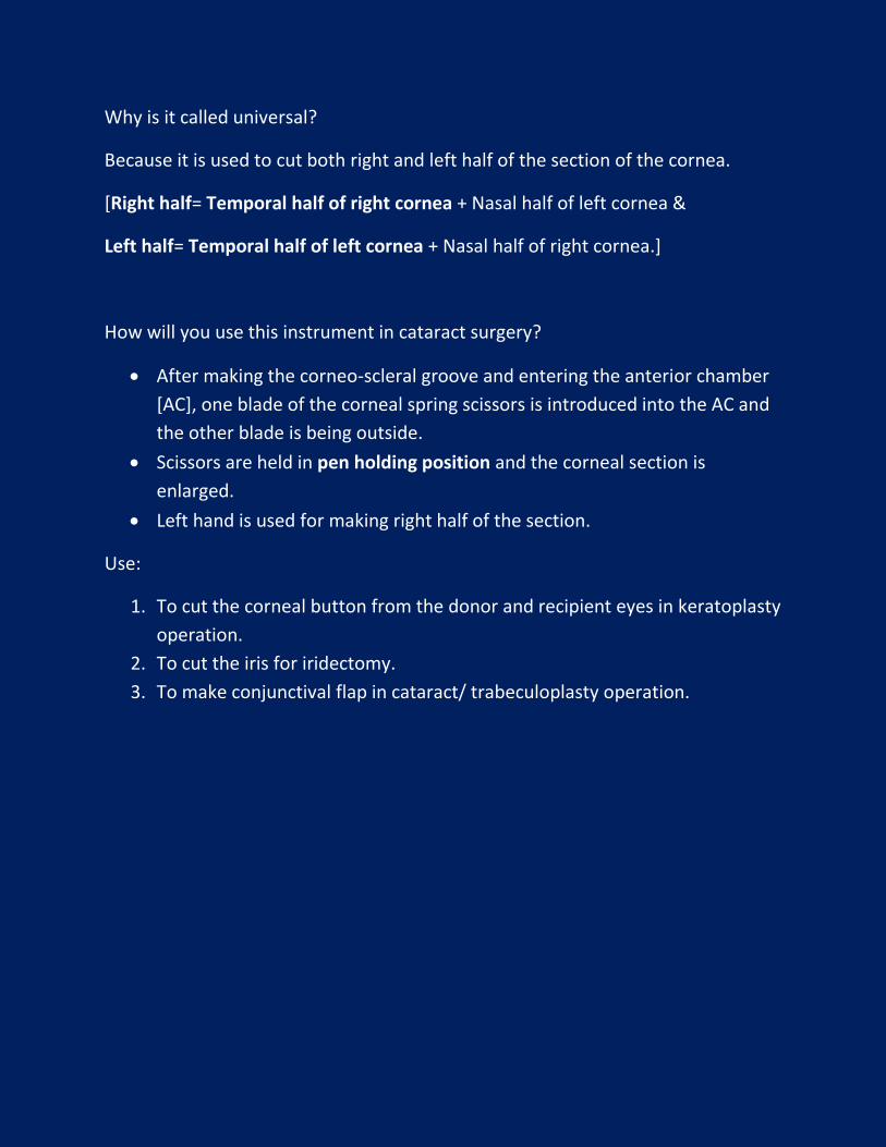
Why is it called universal?
Because it is used to cut both right and left half of the section of the cornea.
[Right half= Temporal half of right cornea + Nasal half of left cornea &
Left half= Temporal half of left cornea + Nasal half of right cornea.]
How will you use this instrument in cataract surgery?
After making the corneo-scleral groove and entering the anterior chamber
[AC], one blade of the corneal spring scissors is introduced into the AC and
the other blade is being outside.
Scissors are held in pen holding position and the corneal section is
enlarged.
Left hand is used for making right half of the section.
Use:
1. To cut the corneal button from the donor and recipient eyes in keratoplasty
operation.
2. To cut the iris for iridectomy.
3. To make conjunctival flap in cataract/ trabeculoplasty operation.
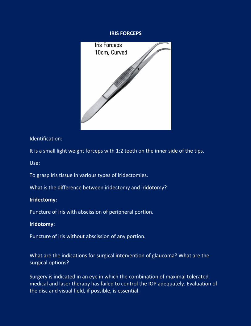
IRIS FORCEPS
Identification:
It is a small light weight forceps with 1:2 teeth on the inner side of the tips.
Use:
To grasp iris tissue in various types of iridectomies.
What is the difference between iridectomy and iridotomy?
Iridectomy:
Puncture of iris with abscission of peripheral portion.
Iridotomy:
Puncture of iris without abscission of any portion.
What are the indications for surgical intervention of glaucoma? What are the surgical options? Surgery is indicated in an eye in which the combination of maximal tolerated medical and laser therapy has failed to control the IOP adequately. Evaluation of the disc and visual field, if possible, is essential.
Surgical options are: 1. Clear corneal peripheral iridectomy: In an eye with an ongoing or recent
acute attack and if unable to successfully perform a laser iridotomy. 2. Clear corneal peripheral iridectomy combined with goniosynechialysis: If
in the preceding scenario, peripheral anterior synechiae have already formed. This procedure involves breaking the synechiae in the angle to allow it to reopen.
3. Guarded filtering procedure (GFP)/ Trabeculectomy: In situations in which the preceding approach has failed or a patent laser PI has failed to resolve the attack.
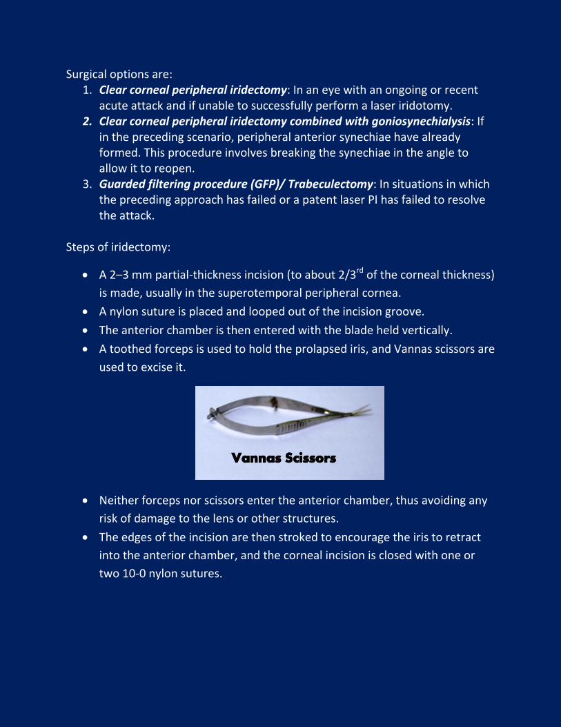
Steps of iridectomy:
A 2–3 mm partial-thickness incision (to about 2/3rd of the corneal thickness)
is made, usually in the superotemporal peripheral cornea.
A nylon suture is placed and looped out of the incision groove.
The anterior chamber is then entered with the blade held vertically.
A toothed forceps is used to hold the prolapsed iris, and Vannas scissors are
used to excise it.
Neither forceps nor scissors enter the anterior chamber, thus avoiding any
risk of damage to the lens or other structures.
The edges of the incision are then stroked to encourage the iris to retract
into the anterior chamber, and the corneal incision is closed with one or
two 10-0 nylon sutures.
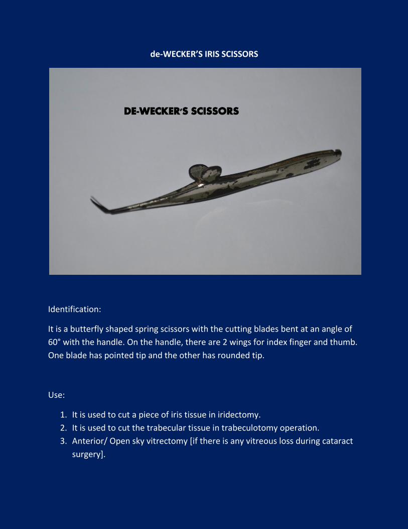
de-WECKER’S IRIS SCISSORS
Identification:
It is a butterfly shaped spring scissors with the cutting blades bent at an angle of
60° with the handle. On the handle, there are 2 wings for index finger and thumb.
One blade has pointed tip and the other has rounded tip.
Use:
1. It is used to cut a piece of iris tissue in iridectomy.
2. It is used to cut the trabecular tissue in trabeculotomy operation.
3. Anterior/ Open sky vitrectomy [if there is any vitreous loss during cataract
surgery].
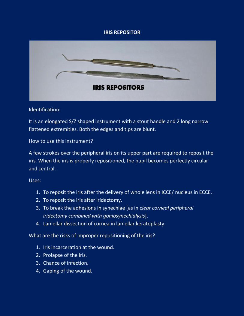
IRIS REPOSITOR
Identification:
It is an elongated S/Z shaped instrument with a stout handle and 2 long narrow
flattened extremities. Both the edges and tips are blunt.
How to use this instrument?
A few strokes over the peripheral iris on its upper part are required to reposit the
iris. When the iris is properly repositioned, the pupil becomes perfectly circular
and central.
Uses:
1. To reposit the iris after the delivery of whole lens in ICCE/ nucleus in ECCE.
2. To reposit the iris after iridectomy.
3. To break the adhesions in synechiae [as in clear corneal peripheral
iridectomy combined with goniosynechialysis].
4. Lamellar dissection of cornea in lamellar keratoplasty.
What are the risks of improper repositioning of the iris?
1. Iris incarceration at the wound.
2. Prolapse of the iris.
3. Chance of infection.
4. Gaping of the wound.

BARRAQUER’S NEEDLE HOLDER
Identification:
It is a medium sized spring needle holder with 2 narrow & fine curved jaws.
Uses:
It is used to hold thin needles in the following cases:
1. Corneo-scleral suturing after cataract operation.
2. Scleral suturing in squint/ detachment/ trabeculotomy operation.
3. Corneal suturing in keratoplasty operation.
4. Suturing the mucosal flaps in DCR operation.
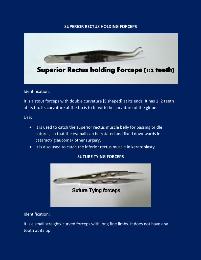
SUPERIOR RECTUS HOLDING FORCEPS
Identification:
It is a stout forceps with double curvature [S shaped] at its ends. It has 1: 2 teeth
at its tip. Its curvature at the tip is to fit with the curvature of the globe.
Use:
It is used to catch the superior rectus muscle belly for passing bridle
sutures, so that the eyeball can be rotated and fixed downwards in
cataract/ glaucoma/ other surgery.
It is also used to catch the inferior rectus muscle in keratoplasty.
SUTURE TYING FORCEPS
Identification:
It is a small straight/ curved forceps with long fine limbs. It does not have any
tooth at its tip.
Use:
1. To hold the suture ends during tying the sutures after proper tightening.
2. To hold the cut ends of the suture during its removal.
3. To catch the margin of incised conjunctiva during suturing.
CATARACT SURGERY
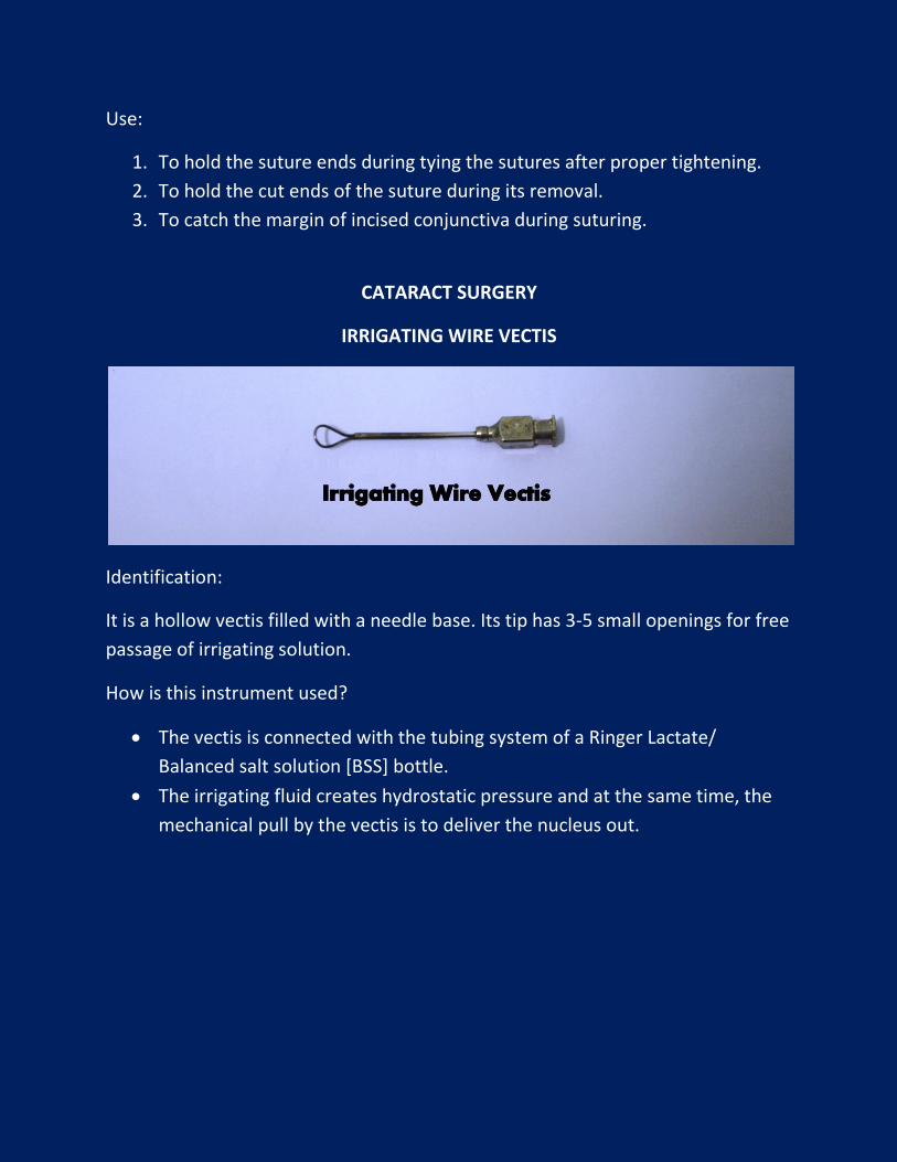
IRRIGATING WIRE VECTIS
Identification:
It is a hollow vectis filled with a needle base. Its tip has 3-5 small openings for free
passage of irrigating solution.
How is this instrument used?
The vectis is connected with the tubing system of a Ringer Lactate/
Balanced salt solution [BSS] bottle.
The irrigating fluid creates hydrostatic pressure and at the same time, the
mechanical pull by the vectis is to deliver the nucleus out.
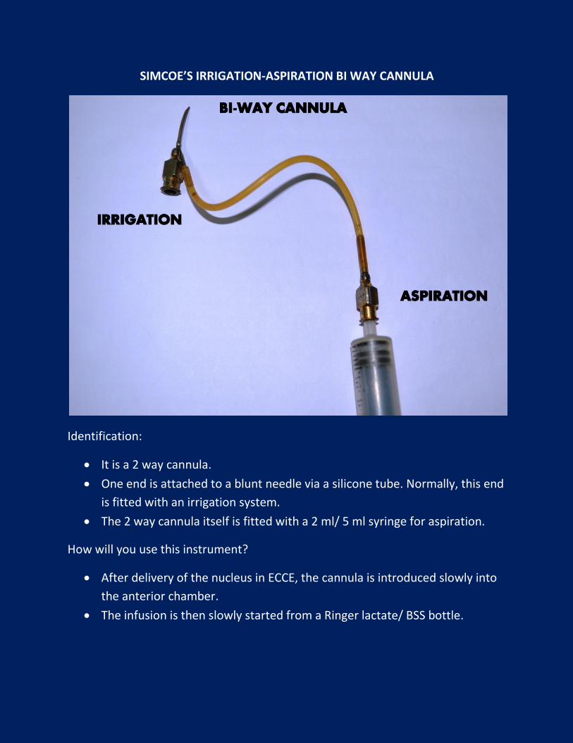
SIMCOE’S IRRIGATION-ASPIRATION BI WAY CANNULA
Identification:
It is a 2 way cannula.
One end is attached to a blunt needle via a silicone tube. Normally, this end
is fitted with an irrigation system.
The 2 way cannula itself is fitted with a 2 ml/ 5 ml syringe for aspiration.
How will you use this instrument?
After delivery of the nucleus in ECCE, the cannula is introduced slowly into
the anterior chamber.
The infusion is then slowly started from a Ringer lactate/ BSS bottle.
As the AC is formed, the surgeon starts aspirating cortical slowly till the
posterior capsule is cleaned [a brilliant fundal glow will appear the axial
illumination of operating microscope].
Uses:
1. Aspiration of cortical material in ECCE.
2. To remove viscoelastic material after insertion of IOL.
3. To remove blood in hyphaema.
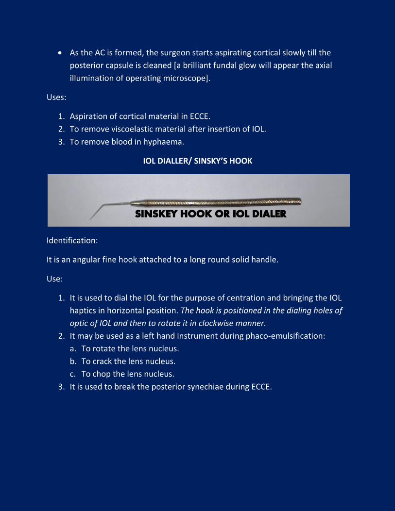
IOL DIALLER/ SINSKY’S HOOK
Identification:
It is an angular fine hook attached to a long round solid handle.
Use:
1. It is used to dial the IOL for the purpose of centration and bringing the IOL
haptics in horizontal position. The hook is positioned in the dialing holes of
optic of IOL and then to rotate it in clockwise manner.
2. It may be used as a left hand instrument during phaco-emulsification:
a. To rotate the lens nucleus.
b. To crack the lens nucleus.
c. To chop the lens nucleus.
3. It is used to break the posterior synechiae during ECCE.
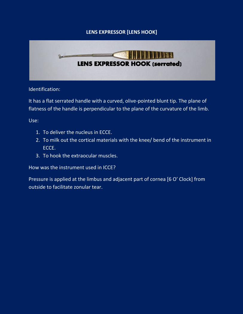
LENS EXPRESSOR [LENS HOOK]
Identification:
It has a flat serrated handle with a curved, olive-pointed blunt tip. The plane of
flatness of the handle is perpendicular to the plane of the curvature of the limb.
Use:
1. To deliver the nucleus in ECCE.
2. To milk out the cortical materials with the knee/ bend of the instrument in
ECCE.
3. To hook the extraocular muscles.
How was the instrument used in ICCE?
Pressure is applied at the limbus and adjacent part of cornea [6 O’ Clock] from
outside to facilitate zonular tear.
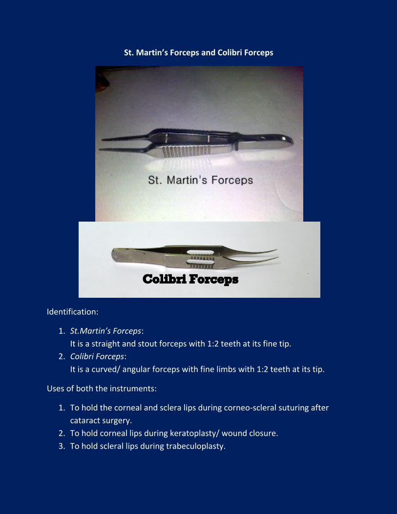
St. Martin’s Forceps and Colibri Forceps
Identification:
1. St.Martin’s Forceps:
It is a straight and stout forceps with 1:2 teeth at its fine tip.
2. Colibri Forceps:
It is a curved/ angular forceps with fine limbs with 1:2 teeth at its tip.
Uses of both the instruments:
1. To hold the corneal and sclera lips during corneo-scleral suturing after
cataract surgery.
2. To hold corneal lips during keratoplasty/ wound closure.
3. To hold scleral lips during trabeculoplasty.
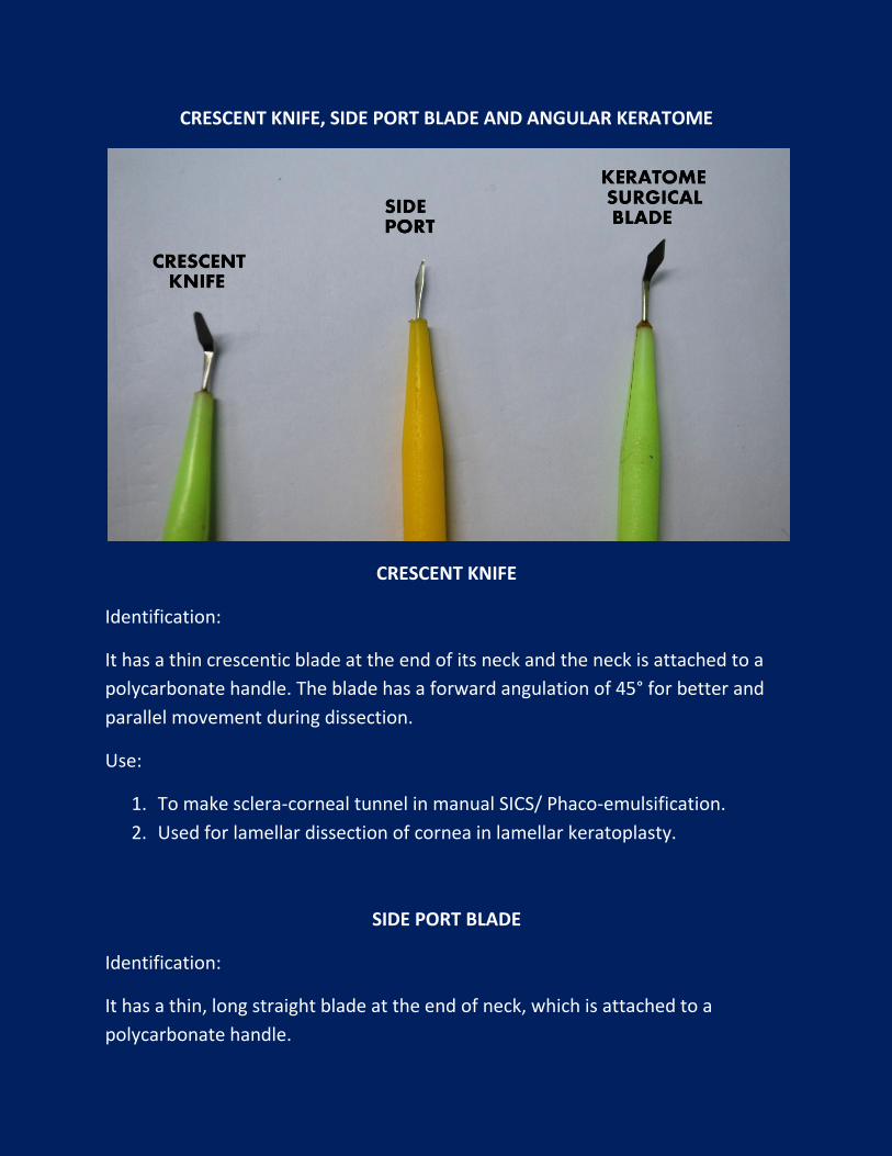
CRESCENT KNIFE, SIDE PORT BLADE AND ANGULAR KERATOME
CRESCENT KNIFE
Identification:
It has a thin crescentic blade at the end of its neck and the neck is attached to a
polycarbonate handle. The blade has a forward angulation of 45° for better and
parallel movement during dissection.
Use:
1. To make sclera-corneal tunnel in manual SICS/ Phaco-emulsification.
2. Used for lamellar dissection of cornea in lamellar keratoplasty.
SIDE PORT BLADE
Identification:
It has a thin, long straight blade at the end of neck, which is attached to a
polycarbonate handle.
Use:
1. To make the side port in cataract surgery.
2. To drain blood in hyphaema.
3. To remove small intracameral foreign body.
Why side port is important in case of cataract surgery?
1. For paracentesis.
2. To inject visco-elastic substance.
3. To inject Trypan Blue dye.
4. To clear the lens cortex by irrigation-aspiration cannula.
5. To manipulate second instrument [like Phaco-chopper/ Sinsky hook etc.] in
phaco-emulsification operation.
6. To insert anterior chamber maintainer.
ANGULAR KERATOME BLADE
Identification:
It has a thin triangular blade at the end of its neck, which is attached to a
polycarbonate handle. The blade has a forward angulation of 45° for better and
parallel movement during dissection.
Use:
1. Used to enter the AC after making sclera-corneal tunnel in manual SICS/
Phaco-emulsification.
2. To enlarge the tunnel.
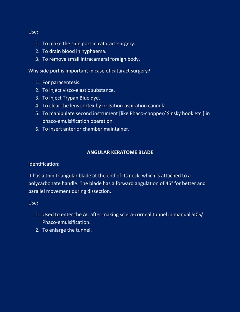
STRABISMUS HOOK/ MUSCLE HOOK/ SQUINT HOOK
Identification:
It has a solid handle with a long narrow limb with 90° bent at its tip. The plane of
curvature is as same as the plane of handle.
Use:
It is used to hook the extraocular muscles during:
a. Squint operation,
b. Retinal detachment operation,
c. Enucleation operation.
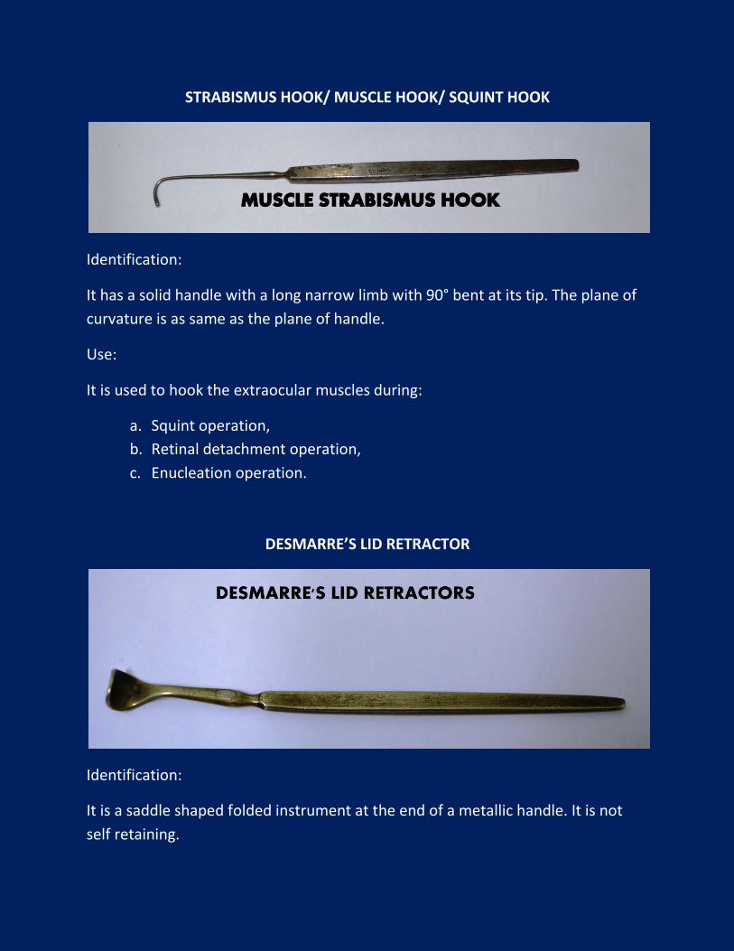
DESMARRE’S LID RETRACTOR
Identification:
It is a saddle shaped folded instrument at the end of a metallic handle. It is not
self retaining.
How will you use this instrument?
Patient is made to lie on the table and an assistant fixes his head.
Lingocaine [4%] eye drop is instilled.
Lid retractor is introduced in the eye from the temporal side sliding against
the eyeball.
Upper lid is retracted and the eye is examined.
Uses:
1. Examination of conjunctiva and anterior segment of the eye:
a. In children and non-co-operative patients.
b. In case of severe blepharospasm and photophobia.
c. In case of lid edema.
2. To examine upper fornix by double eversion of lid.
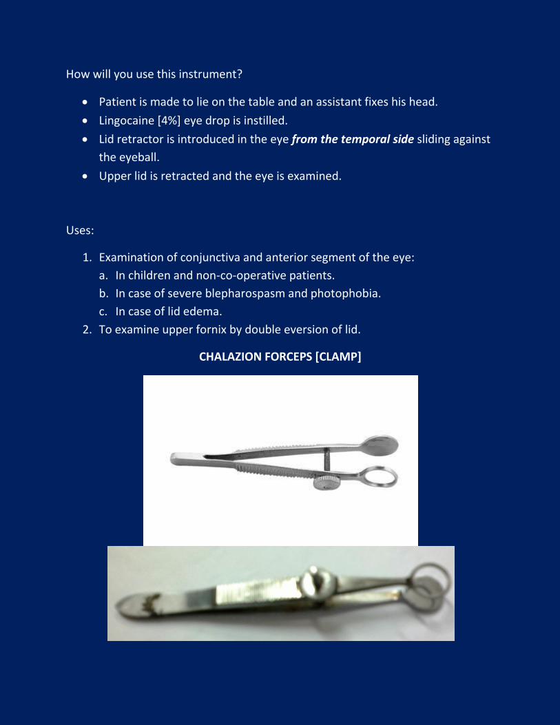
CHALAZION FORCEPS [CLAMP]
Identification:
It is a forceps with a large screw for fixing/ tightening the limbs like a clamp.
One limb has a solid disc shaped plate.
Another limb has a ring at its end.
It is hemostatic and self retaining.
How will you use this instrument?
The solid plate is applied on the skin surface of the lid and the ring side is
applied on the tarsal conjunctiva, encircling the chalazion.
Then screw is tightened and lid is everted, exposing chalazion for incision.
What are the functions of the screw in the chalazion clamp?
1. Fixation of the lid.
2. Haemostasis by means of pressure.
Uses:
1. To fix the chalazion for surgery and to ensure hemostasis.
2. To give intralesional injection of steroids in chalazion after fixation.
3. Excision of a small granuloma/ papilloma of the lid.
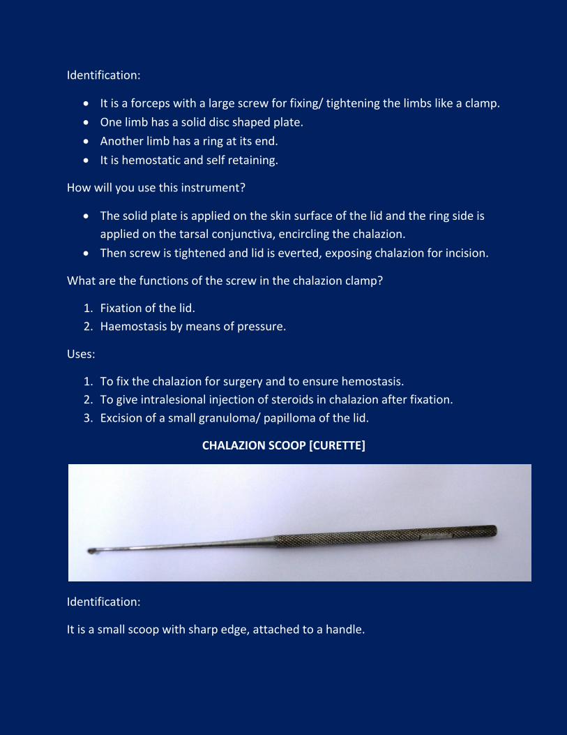
CHALAZION SCOOP [CURETTE]
Identification:
It is a small scoop with sharp edge, attached to a handle.
Uses:
To scoop out the granulation tissue after giving incision on chalazion.
Scooping the chalazion must be complete, otherwise recurrence rate
increases significantly.
After removal of the chalazion forceps, bleeding is controlled by pressure
over the lid, against the incised area.
A pressure bandage is applied for few hours with antibiotic ointment.
LACRIMAL SURGERY
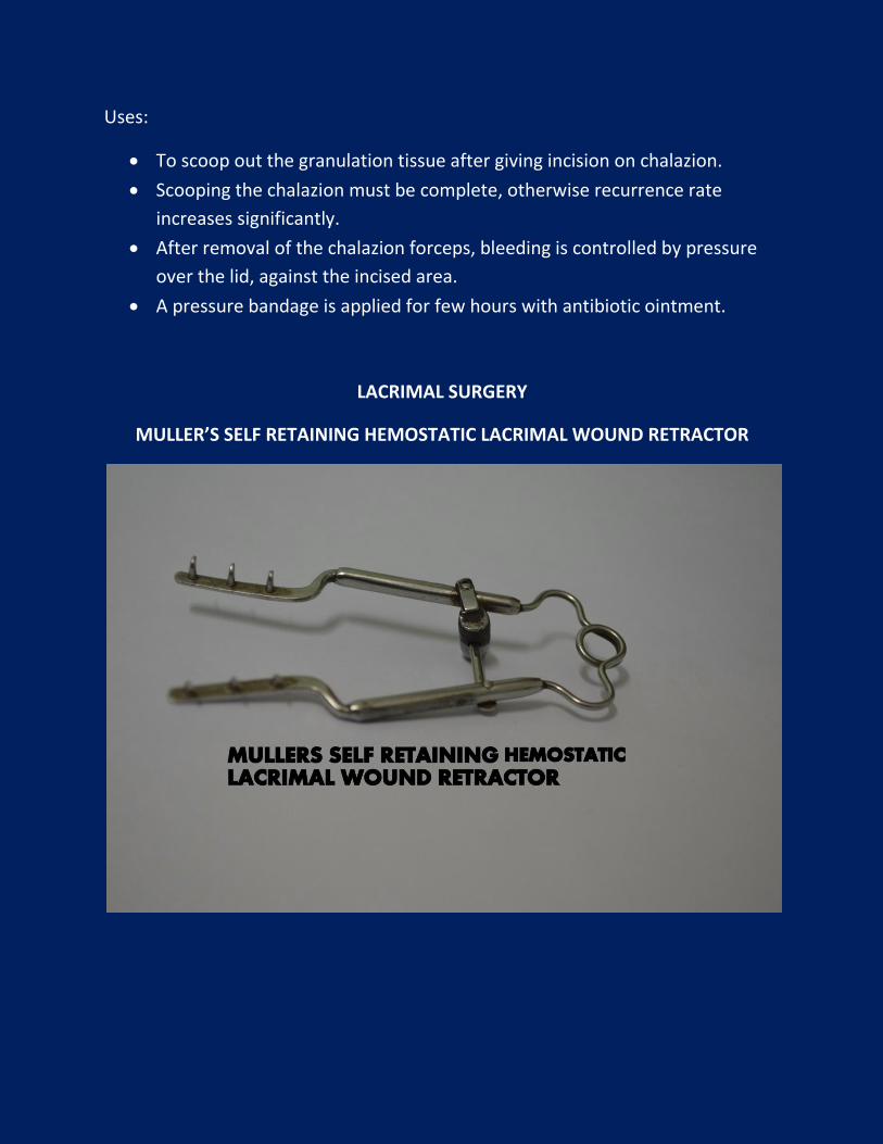
MULLER’S SELF RETAINING HEMOSTATIC LACRIMAL WOUND RETRACTOR
Identification:
It is made up of 2 limbs with a screw to fix the limbus in a retracted position. Each
limb has 2/3 right angled curved pointed hooks for engaging the incised edges of
the skin and deeper tissues.
Uses:
To retract the incised edges of the skin and deeper tissues during DCR and
DCT operations.
What is the mechanism of the hemostatic effect of this instrument?
Its hemostatic effect is due to angled hooks causing compression of the blood
vessels by the spring action of the retractor.
Disadvantages:
This instrument has the disadvantage of reducing field of operation.
Name some complications that may occur during giving incision to the skin?
1. Injury to the angular vein,
2. Cutting the medial palpebral ligament,
3. Injury to the lacrimal sac.
Can you name the structures which have to be cut to reach the lacrimal sac
proper in DCR operation?
1. Skin.
2. Subcutaneous tissue.
3. Fibres of orbicularis oculi.
4. Medial palpebral ligament.
5. Anterior lacrimal fossa.
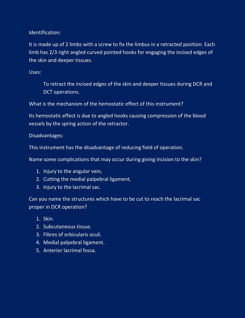
BONE PUNCH
Identification:
It is a large instrument which consists of a spring handle and 2 long blades. The
upper blade has a hole with sharp cutting edge and the lower blade has a cup like
depression with sharp edge.
Use:
It is used to punch/ break the bones in DCR operation to create a bone ostium.
Which bones are punched by Bone punch?
1. Lacrimal bone.
2. Nasal bone.
3. Frontal process of maxilla.
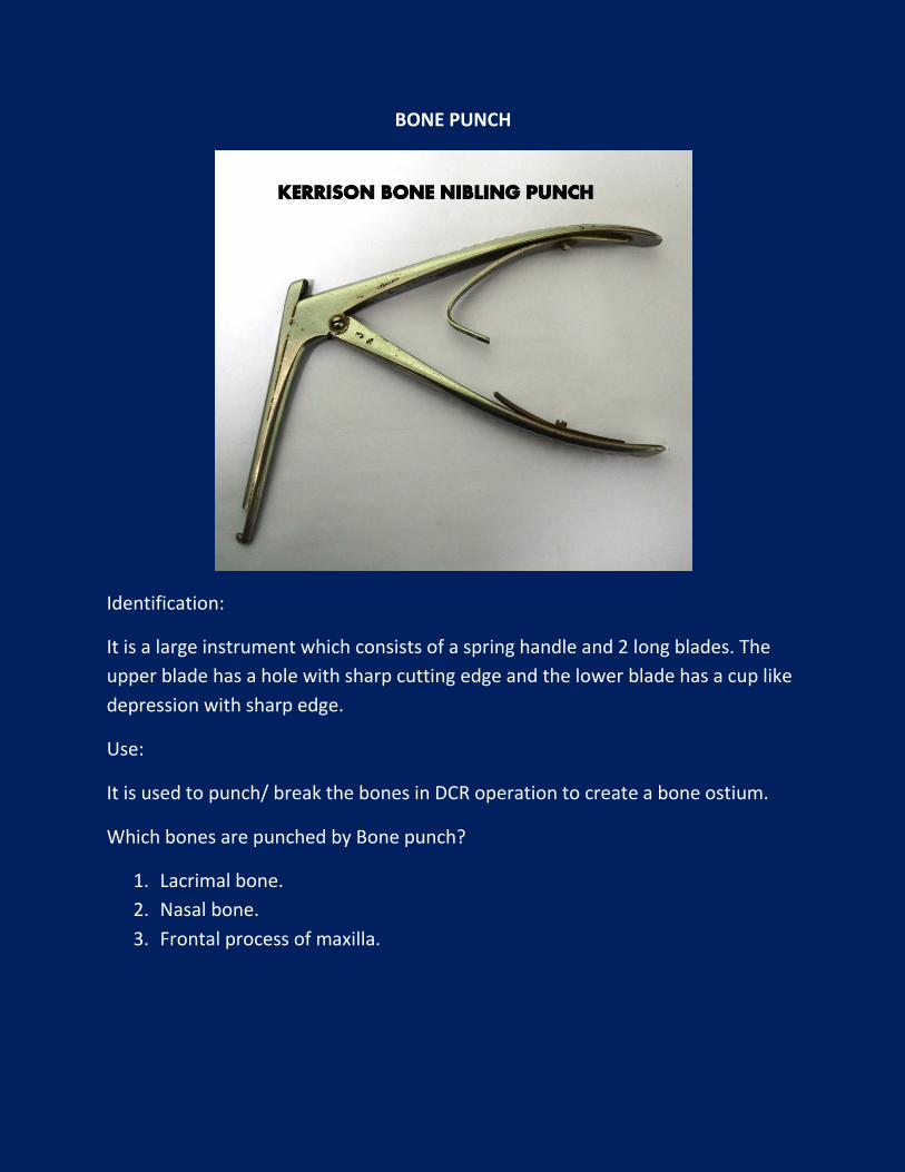
LACRIMAL SAC DISSECTOR WITH CURETTE/ SCOOP
Identification:
It is a narrow long instrument with a stout pointed dissector at one end and an
elongated scoop at the other end.
Uses:
1. Dissector end:
a. It is used for blunt dissection of the lacrimal sac in DCR/ DCT operation.
b. It is used to clean bone pieces from the punch, while punching/ cutting
the bone in DCR.
2. Scoop end:
To scoop out the tissue remnants after excision of the lacrimal sac.
EVISCERATION AND ENUCLEATION
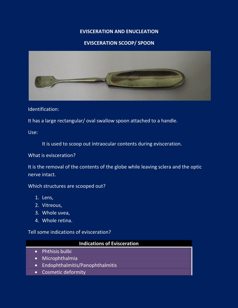
EVISCERATION SCOOP/ SPOON
Identification:
It has a large rectangular/ oval swallow spoon attached to a handle.
Use:
It is used to scoop out intraocular contents during evisceration.
What is evisceration?
It is the removal of the contents of the globe while leaving sclera and the optic
nerve intact.
Which structures are scooped out?
1. Lens,
2. Vitreous,
3. Whole uvea,
4. Whole retina.
Tell some indications of evisceration?
Indications of Evisceration
Phthisis bulbi
Microphthalmia
Endophthalmitis/Panophthalmitis
Cosmetic deformity
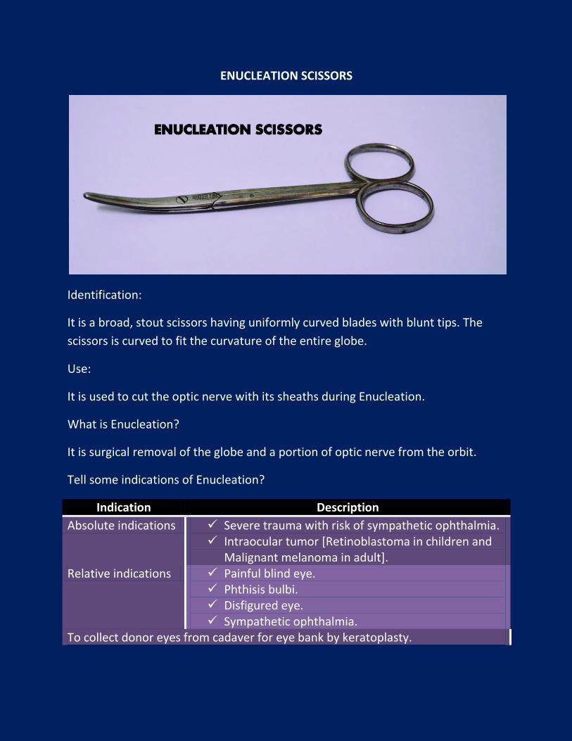
ENUCLEATION SCISSORS
Identification:
It is a broad, stout scissors having uniformly curved blades with blunt tips. The
scissors is curved to fit the curvature of the entire globe.
Use:
It is used to cut the optic nerve with its sheaths during Enucleation.
What is Enucleation?
It is surgical removal of the globe and a portion of optic nerve from the orbit.
Tell some indications of Enucleation?
Indication Description
Absolute indications Severe trauma with risk of sympathetic ophthalmia. Intraocular tumor [Retinoblastoma in children and
Malignant melanoma in adult]. Relative indications Painful blind eye.
Phthisis bulbi. Disfigured eye. Sympathetic ophthalmia.
To collect donor eyes from cadaver for eye bank by keratoplasty.
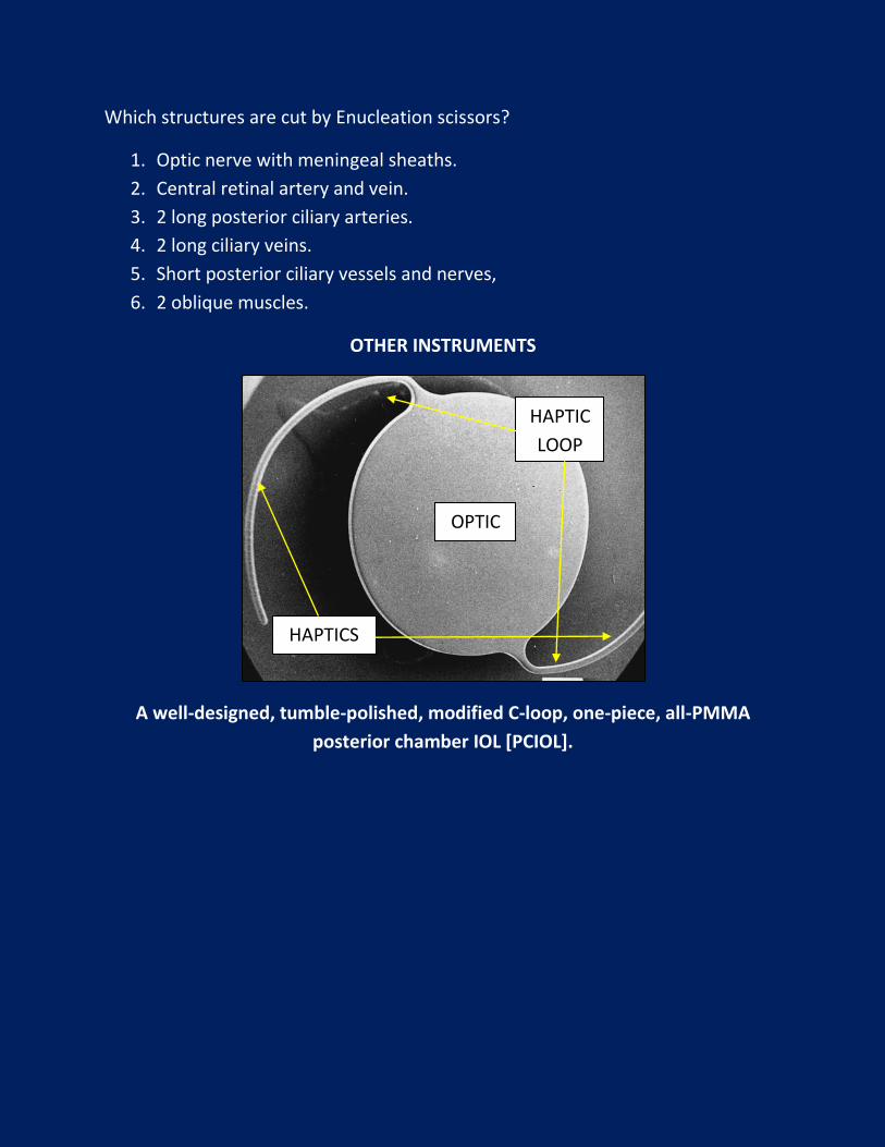
Which structures are cut by Enucleation scissors?
1. Optic nerve with meningeal sheaths.
2. Central retinal artery and vein.
3. 2 long posterior ciliary arteries.
4. 2 long ciliary veins.
5. Short posterior ciliary vessels and nerves,
6. 2 oblique muscles.
OTHER INSTRUMENTS
A well-designed, tumble-polished, modified C-loop, one-piece, all-PMMA
posterior chamber IOL [PCIOL].
OPTIC
HAPTIC
LOOP
HAPTICS
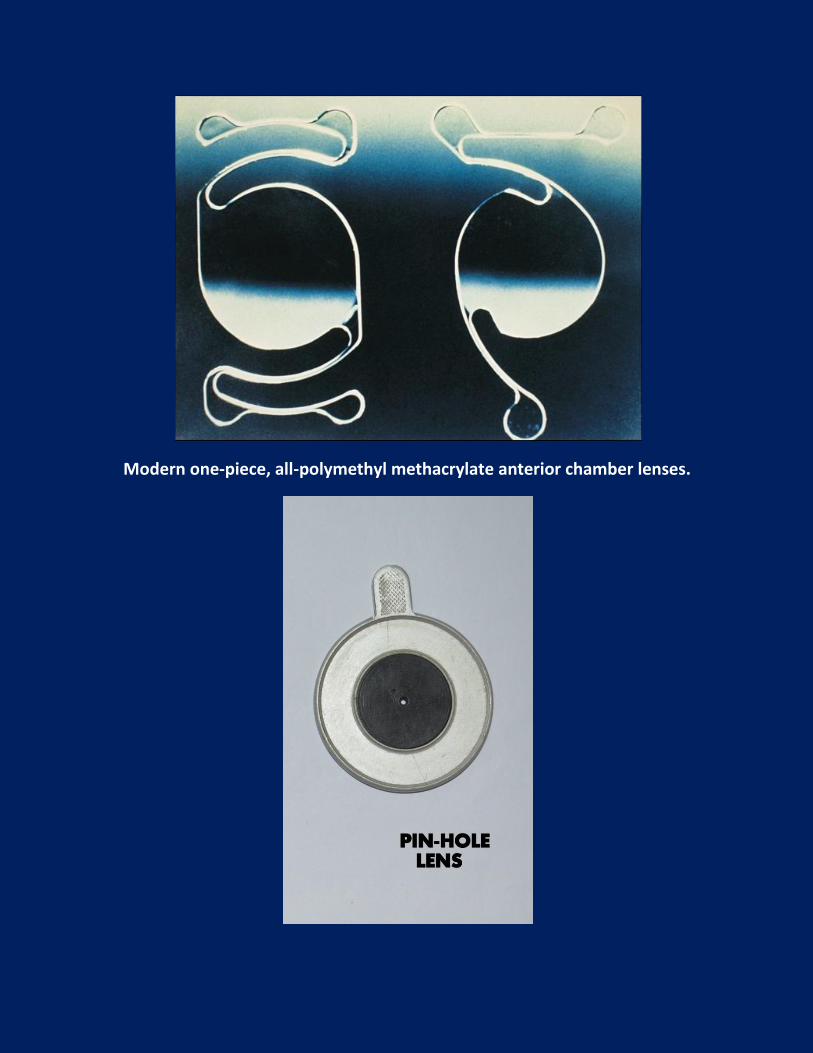
Modern one-piece, all-polymethyl methacrylate anterior chamber lenses.
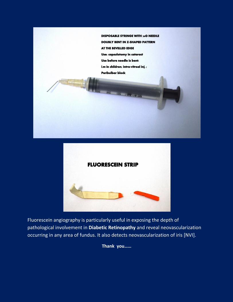
Fluorescein angiography is particularly useful in exposing the depth of
pathological involvement in Diabetic Retinopathy and reveal neovascularization
occurring in any area of fundus. It also detects neovascularization of iris [NVI].
Thank you……