Embed Size (px)
Citation preview

Contact Transscleral Nd:YAGLaser CyclophotocoagulationMidterm Results
Joel S.'Schuman, MD,I,2A. Robert Bellows, MD,3,4 Bradford]. Shingleton, MD,3.4Mark A. Latina , MD,5 R. Rand Allingham, MD,2C. Davis Belcher, MD,3.4Carmen A. Puliafito, MDl ,2
Background: Early reports of both contact and noncontact transscleral Nd:YAGlaser cyclophotocoagulation have been encouraging; however, recent evidence indicatesa significant incidence of hypotony, visual loss, and phthisis with the noncontact techniquewith more than 6 months of follow-up. The authors sought to determine the intermediateterm effects of contact transscleral Nd:YAG laser cyclophotocoagulation (CYC).
Methods: The authors followed 116 eyes of 114 patients for a minimum of 1 yearafter treatment of advanced glaucoma with CYC.
Results: The mean preoperative intraocular pressure (lOP) of 35 .0 ± 1.0 mmHgdecreased to 18.6 ± 1.1 mmHg (P < 0.0001) during the average follow-up of 19.0 ± 0.6months (range , 12 to 36 months). Intraocular pressure control of 3 to 25 mmHg wasachieved in 72%, 3 to 22 mmHg in 65%, and 3 to 19 mmHg in 56% of eyes . Retreatmentwas required in 31 of the 116 eyes (27%). Intraocular pressure decreased to less than3 mmHg in 9 eyes and to 0 mmHg in 6 of these 9 eyes . Nineteen eyes, all with initialvisual acuity of counting fingers or worse, progressed to no light perception; 17 of 36eyes (47%) with visual acuity of 20/200 or better lost 2 or more Snellen lines.
Conclusion: Midterm results of CYC continue to be encouraging but are temperedby a nearly 10% incidence of hypotony or phth isis and the progression of visual loss .Ophthalmology 1992;99;1089-1095
Originally received: October 15, 199 1.Revision accepted: February 17, 1992.
I New England Eye Center, New England Medical Center Hospitals,Tufts University School of Medicine. Boston .
2 Howe Laboratory of Opht halmo logy, Massachusetts Eye and Ear Infirmary, Harvard Medical School, Boston .
3 Center for Eye Research, Boston .
4 Ophthalmic Consultants of Boston, Boston.
5 Wellman Laboratories, Massachusetts General Hospital , HarvardMedical School. Boston.
Dr. Allingham is currently affiliated with the University of Texas, Southwestern Medical Center , Dallas.
Presented in part at the American Academy of Ophthalmology AnnualMeeting. Anaheim, October , 1991.
The authors have no proprietary interest in the development or marketingof the instrument used in this study.
Reprint requests to Joe) S. Schuman, MD, New England Eye Center.Tufts University School of Medicine, 750 Washington St. Box 450, Boston, MA 02 111.
Early results of contact transscleral Nd:YAG laser cyclophotocoagulation (CYC) were encouraging, with reportedsuccess rates of 49% to 71%.,,2 Few complications oftransscleral Nd:YAG laser cyclophotocoagulation werereported, although Trope and Ma,3 and more recentlyHampton et al" indicated a significant incidence of hypotony, phthisis, and visual loss with noncontact transscleral Nd:YAG laser cyclophotocoagulation (NCYC) onfollow-upoflonger than 6 months , We describe the resultsof CYC in 116 eyes of 114 patients followed for a minimum of 1 year after treatment.
Materials and Methods
Laser and Delivery System
A continuous-wave Nd:YAG laser with a 600 JLm quartzfiberoptic contact probe was used (Surgical Laser Tech-
1089

Ophthalmology Volume 99, Number 7, July 1992
Table 1. Retreatments*
nologies , Inc, Malvern, PA). The probe tip was 2.2 mmin diameter and was constructed of synthetic sapphire.
Glaucoma Type and Previous Treatments
Six types ofglaucoma were represented in the population(Table 2): neovascular glaucoma (NVG), primary openangle glaucoma (POAG), chronic angle closure glaucoma
Retreatment
Twenty-five patients had I eye retreated with CYC onceeach, four patients had one eye retreated twice, one patienthad three retreatments, and one patient was retreated 5times (Table I). Retreated eyes were analyzed with theremainder of the study population and were consideredas continuations of the first treatment for the purpose ofstatistical analysis. Two patients had two eyes treated.
23 eyes (20%)
1 eye (1%)7 eyes (6%)
44 eyes (39%)72 eyes (65%)14 eyes (12%)17 eyes (15%)
48/116 (41%)26/116 (22%)17/116 (15%)4/116 (3%)
10/116 (9%)11/116 (9%)
Primary open-angle glaucomaNeovascular glaucomaChronic angle closure glaucomaMixed mechanism glaucomaSecondary open-angle glaucomaAngle recession or other
(CACG), mixed mechanism, secondary open-angle glaucoma, and angle recession. Differences between thesegroups were studied, as well as differences between neovascular and non-neovascular, and POAG and nonPOAG.
A variety of laser and surgical treatments had beenperformed before CYC in many patients (Table 3).
Of the 116 eyes, there were 38 phakic eyes and 26 eyeswith NVG.
Table 2. Glaucoma Subtypes"
• Subtype s of glaucoma present in patients treated with contact transscleralNd :YAG laser cyclophotocoagulation.
Race
Additional Interventions
Age
Nineteen patients were 40 years ofage or younger (mean,27 years). The mean age for patients greater than 40 yearsold was 69 years.
Table 3. Prior Surgery"
Fifteen of the patients were nonwhite (black, Hispanic,or Asian); the remainder were white. There was no significant correlation between race and treatment groups.
Nineteen patients required further surgical intervention.Eleven were treated with cyclocryotherapy to achieve lOPcontrol. Four patients underwent enucleation, three hadsurgery with Molteno valve implantation, and one received a revision of his existing filter. All patients wereincluded in the data analysis until the time of failure (additional intervention) (Table 4).
Cyclocryotherapy
CyclophotocoagulationContactNoncontact
Filtration surgeryCataract extractionScleral bucklingVitrectomy
85 eyes (73%)25 eyes (22%)4 eyes (3%)1 eye (1%)1 eye (1%)
1 treatment2 treatments3 treatments4 treatments6 treatments
Patient Selection and Protocol
Transscleral photocoagulation of the ciliary body wasperformed in 157 treatments of 116 eyes in 114 patients(average age, 61.7 ± 1.9 years; range, 4 to 90 years). Alltreatments were performed by the authors and 17 additional surgeons in accordance with the protocol below;patients were followed prospectively. Patients undergoingCYC had glaucoma refractory to conventional treatmentand were using maximum tolerated medical therapy.There were no entry criteria regarding absolute intraocularpressure (lOP) or visual acuity. The treatment protocolwas reviewed and approved by the Human Studies Committee of the Institutional Review Board of the Massachusetts Eye and Ear Infirmary.
Treatments were performed with the anterior edge ofthe probe positioned 0.5 to 1.5 mm posterior to the limbus , and energy was delivered in up to 40 applications of7 to 9 watts for 0.7 seconds each. The 3-0'clock and 9o'clock meridians were spared treatment so as to avoidinjury to the long posterior ciliary arteries. The protocolwas such that surgeons were permitted to vary the treatment power, the number of applications, or the distanceof the probe from the limbus at their discretion. Patientswere seen in follow-up at I hour, I day, I week, I month,3 months, 6 months, 12 months, and as needed thereafter.Treatment technique and data collected at each visit wereas reported previously (7 watts , 0.7 seconds, 32 applications, anterior edge of probe 0.5 to 1.0 mm posterior tolimbus).'
• Eyes retreated after initial contact transscleral Nd:YAG laser cvclophotocoagulation .
• Eyes undergoing surgery and operations performed before contacttransscleral Nd:YAG laser cyclophotocoagulation.
1090

Schuman et al . Cyc1ophotocoagulation Midterm Results
12
II
369Time After Treatment (months)
ooL-...l.- ,L-__----:!L--__-----:!- ---:-'::----'
10
30
DoQ 20
40 ----------------------,
Figure 1. Intraocular pressure response to contact transsc1eral continuouswave Nd:YAG laser cyclophotocoagulation, lOP = intraocular pressure.
CONTACT CYCLOPHOTOCOAGULATIONMEAN lOP
Statistical Analysis
All data were coded and entered into a commercialspreadsheet (Lotus 1-2-3, Release 3.0, 1990, Lotus Development Corporation, Cambridge, MA) and analyzedusing a commercial statistical software package (CSS:Statistica, 1991, StatSoft, Tulsa, OK). Tests for significanceincluded analysis of variance (ANOVA), multiple regression, two-tailed t test, and chi-square, and are listed alongwith P values. Mean values are listed with standard errorsof the mean; P values are for two-tailed t tests unless otherwise indicated.
Retreatments were considered as continuations of initial treatments; that is, each eye was considered a singlecase whether it had one treatment or several.
Results
Table 4. Further Intervention"
Intraocular Pressure
The mean pretreatment lOP level was 35.0 ± 1.04mmHg(range, 18 to 75 mmHg), and 12 months or more aftertreatment the mean lOP level was 18.6 ± 1.06 mmHg(range, 0 to 53 mmHg), a decrease of 43.1 ± 3.4% (P< 0.0001) (Fig 1). The mean follow-up was 19.0± 0.6 months (range, 12 to 36 months). The procedurereduced lOP to 3 to 25 mmHg in 72% of patients, to 3to 22 mmHg in 65% of patients, and to 3 to 19 mmHgin 56% of patients (Fig 2) (Table 5). These data comparefavorably with previously published work on both CYCand NCyc. 1
-7 As we reported in our previous article, lOP
appears to reach its nadir at 1 week to 1 month and remains at that level through at least the 12-month followup (Fig 1). A rise in the lOP level after treatment wasuncommon, with an 8 mmHg or greater lOP elevationat 1 hour postoperatively in 8 of 86 eyes (9%), as we reported previously.1
Unlike our previous report with short-term follow-up,there was no correlation found between preoperative andfinal lOP levels by regression analysis «(:3 = 0.077;P = 0.43).
Once again, NVG patients had a greater absolute decrease in lOP than non-NVG patients, but the percentdecrease in lOP was not significantly different betweenthe two groups (decrease in lOP = 25.8 mmHg NVG,14.0 mmHg non-NVG, P = 0.001; percent decrease inlOP = 54.8% NVG, 39.8% non-NVG, P = 0.068). Meanpretreatment lOP was higher in the NVG than the nonNVG group, but there was no significant difference be-
3 6 9 12 15 18 21 24Time After Treatment (months)
1OOr--~~~ --,::J 90~ 80Ql
~ 70~ 60g 50:e 40og. 30
c: 2010
o 0
FAILURE AFTER CYClOP >22 mmHg, <3 mmHg, CCT or Surgery
tween the groups for lOP at the last follow-up visit (meanpretreatment lOP = 43.5 mmHg NVG, 32.3 mmHg nonNVG, P < 0.001; mean final lOP = 18.6 mmHg NVG,18.5 mmHg non-NVG, P = 0.972). Patients with NVGhad a lower success rate for lOP of 3 to 25 mmHg or 3to 22 mmHg than non-NVG patients, but the differencebetween these groups for final lOP levelsof 3 to 19 mmHgdid not achieve statistical significance (3 to 25 mmHg,P = 0.01; 3 to 22 mmHg, P = 0.03; 3 to 19 mmHg,P = 0.64).
Also similar to our previous study, nonprimary openangle glaucoma patients had a significantly greater magnitude of reduction and percent decrease in lOP thanPOAG patients (mean reduction in lOP = 11.7 mmHgPOAG, 20.2 mmHg non-POAG, P = 0.005; percentdecrease in lOP = 34.2% POAG, 49.7% non-POAG,P = 0.024).
While there was no significant difference in lOP response to CYC comparing phakic and aphakic or pseudophakic eyes, phakic eyes had significantly higher preoperative but not postoperative, lOP levels (preoperativephakic lOP = 38.7 mmHg, pseudo/aphakic lOP = 32.6mmHg, P = 0.005; postoperative phakic lOP = 19.7mmHg, pseudo/aphakic lOP = 17.2 mmHg, P = 0.275).
11 eyes4 eyes3 eyes1 eye
CyclocryotherapyEnucleationMolteno implantationFilter revision
• Eyes requiring further intervention and surgery performed after contacttranssc1eral Nd:YAG laser cyc1ophotocoagulation.
Figure 2. Kaplan-Meier survival curve for contact transsc1eral continuouswave Nd:YAG laser cyc1ophotocoagulation; CCT = cyc1ocryotherapy;lOP = intracular pressure.
1091

Ophthalmology Volume 99, Number 7, July 1992
Table 5. Success Rates by Glaucoma Subtypes
Range (mmHg)
3-25 3-22 3-19
Primary open-angle glaucoma 78%(35/45) 68%(32/47) 60%(28/47)Neovascular glaucoma 43% (10/23) 39%(9/23) 39%(9/23)Glaucoma in aphakia or
pseudophakia 76%(52/69) 69% (49/71) 63% (45/71)After penetrating keratoplasty 70%(7/10) 60%(6/10) 50% (5/10)
• Success rates in four subtypes of glaucoma present in patients treatedwith contact transscleral Nd:YAG laser cyclophotocoagulation.
The differences in success rates for NVG, POAG, glaucoma after cataract extraction, and glaucoma after penetrating keratoplasty are shown in Table 5. Not unexpectedly, eyes with NVG had the lowest percentage ofsuccessfully treated eyes. The difference in the reductionofIOP was not statistically significant between the following groups: white versus nonwhite patients, eyes after filtration surgery versus no surgery, cyclocryotherapy versusno cyclocryotherapy, cyclodialysis versus no cyclodialysis,NCYC versus no NCYC, vitrectomy versus no vitrectomy,or scleral buckling versus no retinal detachment repair.There was no significant difference in effect for patientsolder or younger than 40 years of age. Varying the distanceofthe probe from the limbus or the number of applicationsofthe laser had no statistically significant effecton pressurereduction. There was no significant difference in lOP reduction for patients treated with 7 watts of power as opposed to those treated with 9 watts. Covariate analysis byANOV A and multiple regression failed to show any effecton lOP of changes in power, distance of the probe fromthe limbus or number ofapplications; however, there wasless success (lOP, 3 to 25 mmHg, 3 to 22 mmHg, or 3 to19 mmHg) in eyes treated with 9 watts power comparedwith those treated with 7 watts.
There was no significant correlation between pain aftertreatment and reduction of lOP or postoperative inflammation and lOP reduction. There was no significant difference between the treatment results of the various surgeons.
Hypotony
Eyes that progressed to lOP levels below 3 mmHg wereconsidered hypotonus or phthisical; 9 eyes were includedin this group. Six eyes had an lOP of 0 mmHg, and 1each had final lOP levels of 1,2, and 2.5 mmHg. Therewas no significant difference between the mean preoperative pressures of those eyes that became hypotonus andthose that did not (hypotony: preoperative lOP = 39.1mmHg; nonhypotony: lOP = 34.5 mmHg; P = 0.235);however, as might be expected, there was a significantlygreater change in lOP in eyes that became hypotonus(change in lOP, hypotony lOP = 38.5 mmHg, nonhypotony lOP = 14.5 mmHg, P < 0.001). Eyes that developed hypotony were more likely than others to havesustained an elevated lOP more than 8 mmHg greaterthan baseline at 1 to 24 hours after treatment (P < 0.001).
1092
The initial and final visual acuities were poorer in thehypotony bound eyes, and the decrease in vision was moreprofound in this group as well (P < 0.020). There wasmore initial flare, cell, and conjunctival redness in eyesthat progressed to hypotony (P < 0.020); however, whilethere was more increase in cell postoperatively in theseeyes (P < 0.050), there was no significant difference inflare between the hypotony group and other eyes.
The proportion of eyes developing hypotony containedsignificantly more NVG (5 of 9, P = 0.006), and lessPOAG (1 of9, P = 0.050), than treated eyes as a whole.More eyes with hypotony than other eyes progressed tono light perception (6 of9, P < 0.001).
There were no significant differences for probe positionor number of applications in comparing eyes progressingto hypotony to others. There was no significant differencein total energy used in eyes that developed hypotonycompared with those that did not; however, there was atrend toward those developing hypotony having beentreated with higher powers (9 watts, 6 of 9; 7 watts, 3 of9; P = 0.094).
Visual Acuity
The median pretreatment visual acuity was counting fingers; the median change in acuity was loss of 1 Snellenline ofvision. Seventeen of 36 eyes (47%)with visual acuity of 20/200 or better lost 2 or more lines of Snellenacuity (final vision two or more lines worse than pretreatment). Overall, 48 of 116 eyes (41%) lost 2 or more linesof vision. No statistically significant differenceswere foundfor race, history ofNVG, POAG, prior cataract, filtration,cyclodestructive or retinal surgery or vitrectomy, amountof treatment power or total energy used, number of applications, or distance of the probe from the limbus.
Eyes that progressed to lOP levels below 3 mmHg hadpoorer preoperative and postoperative visual acuities anda greater change in vision than other eyes (P -s::: 0.04).
No Light Perception
Nineteen eyes lost light perception, all with preoperativevisual acuities of counting fingers or worse; this pretreatment vision was significantly worse than those eyes thatretained vision (P < 0.001). All but five eyes had reducedlOP levels (2 of these eyes were unchanged), and threeeyes had pressure spikes of 8 mmHg or more 1 hour aftertreatment (P = 0.001). Significantly more eyes that progressed to no light perception (NLP) were eyes with NVG(14 of 19,74%, P < 0.0001), and significantly fewer hadPOAG (2 of 19, 11%, P = 0.002).
Eyes losing light perception had higher mean pretreatment lOP levels (NLP lOP = 41.7 mmHg, non-NLP= 33.5, P = 0.003), although this difference in lOP waslost by 6 months after treatment. Eyes that progressed toNLP had a greater change in lOP than other eyes, butthere was no difference in percent lOP decrease (changein lOP NLP = 24.4 mmHg, non-NLP = 14.9, P = 0.016).Eyes that lost light perception were less likely to have a"successful" result of CYC (P = 0.01, 3 to 25 mmHg, 2to 22 mmHg, or 3 to 19 mmHg).

Schuman et al . Cyclophotocoagulation Midterm Results
Eyes that lost light perception had more initial cell andflare anterior chamber reaction than other eyes (P < 0.01),but there was no difference between NLP bound eyes andothers by 1 week for cell and 1 month for flare, and therewere no differences between these groups for change ininflammation.
Patients who lost light perception experienced mild tomoderate pain after treatment, while those who retainedtheir vision experienced minimal to mild pain (P = 0.029).
Eyes losing light perception were more likely to bephakic (P = 0.007), but no differences were found forrates of prior vitrectomy, scleral buckling, or filtrationsurgery. Patients progressing to NLP were more likely tohave had prior cyclecryotherapy (8 of 19 eyes, 42%, P= 0.040).
Inflammation
Inflammation (cell and flare) was graded on a scale of 0(no cell or no flare) to 4 (cell too numerous to count orfibrinoid anterior chamber reaction). Cell and flare measurements were graded independently. Twenty-eight percent of patients developed mild (1+) postoperative anterior chamber inflammation, 19%manifest 1 to 2+ flareand cell and 10% experienced 3 to 4+ inflammation at1 to 7 d~Ys. Inflammation generally returned to baselineby 1 week to 1 month. Eighteen percent of patients hadtrace to 1+ cell preoperatively. Mild to moderate flarewas apparent in 53%of patients after treatment, and mildto moderate conjunctival injection was present in 44% ofpatients postoperatively. There was no statistically significant difference between cellular reaction, aqueous flare,or conjunctival injection before treatment and that at 3,6, or 12 months after treatment or at the final visit; however, cellular reaction, flare, and conjunctival injectioneach were significantlygreater from 1 hour after treatmentthrough 1 month after treatment than before CYC.
Nonwhite patients had a significantly greater increasein cellular reaction and flare at 1 hour to 1 week thanwhite patients (P < 0.040), but there was no significantdifference between the groups in terms of inflammationafter that time. Nonwhite patients also had a greater increase than white patients in conjunctival injection at 1hour and 1 week (P = 0.040).
There were no differences found between groups regarding postoperative inflammation, including the following: NVG compared with non-NVG, POAG versusnon-POAG, phakic versus pseudo/aphakic, age of patient,history of filtration, cataract, vitreoretinal or cyclodestructive surgery and those with no history of such surgeryrespectively, probe placement, number of applications,and 7 or 9 watts power.
Cataract
Cataract was rated on a scale of 0 to 4+. Patients withNVG had a greater increase in lens changes than nonNVG patients at 1week, 3 months, and 1year (P < 0.024).There were no significantdifferencesin cataract formationbetween any of the other groups tested.
Pain after Treatment
Average pain after treatment was "minimal" to "mild"(none = 0 to severe = 4). Patients reported significantlyless pain from repeated treatments than on the first (firsttreatment mean minimal to mild, retreatment mean noneto minimal, P = 0.008). There was no significantdifferencein postoperative pain experienced by white and nonwhitepatients, nor were there significant differences in pain forphakic versus aphakic or pseudophakic eyes, neovascularversus non-neovascular glaucoma, POAG versus nonPOAG, patient age, prior cyclodestructive or vitreoretinalsurgery versus no history of such surgery, probe placement, number of applications or power used in treatment.
Discussion
When our group and others reported initial results ofCYC, the treatment seemed to offer a viable option forthe management of glaucoma resistant to other forms oftherapy, with little postoperative pain and inflammationand only a 7% incidence of visual loss."Our longer-termresults resonate with those described for other forms ofcyclodestruction, with a nearly 50% incidence of visualloss of 2 or more lines in eyes with preoperative visualacuities of 20/200 or better, progression to no light perception in 19 of 116 eyes, and an almost 10% incidenceof phthisis. The decision to perform CYC, still a usefultool for the management of glaucoma refractory to conventional treatment, must be tempered by interpretationof these data.
The cause of visual loss in our patients remains unclear.Earlier reports of decreased vision associated with cyclodestruction found rates of visual loss of 0% to 67% andgenerally laid the blame on macular edema or progressionof the glaucoma.Y'":" Fluorescein angiography was notperformed in this or the aforementioned studies, however,and the cause of visual loss, therefore, can only be inferred.Eyes with a final lOP below 3 mmHg were at higher riskoflosing vision; this was related to the higher progressionto no light progression in this group. Our investigationdid not show an increase in cataract or persistent inflammation that would explain the loss of vision.
The loss of light perception is an especially troublingfinding, although it is consistent with published work onother forms of cyclodestruction.v!"" Although only eyeswith visual acuities of counting fingers or worse lost lightperception, 41% of eyes overall lost vision. Progression tono light perception may be due to the same factors asvisual loss in general; however, hypotony appears to playa role in this problem as well, with a significantly higherincidence of loss of light perception in those eyes withfinal lOP levels below 3 mmHg than in treated eyesoverall.
The development of hypotony or phthisis is commonto cyclodestructive procedures. Reported phthisis ratesrange up to 12% or higher for cyclocryotherapy andNCYC.3,4,8-14 While only less than 2% of eyes had an lOPbelow 3 mmHg in our earlier report with a 3-month average follow-up,' 8% (9 of 116 eyes) developed hypotony
1093

Ophthalmology Volume 99, Number 7, July 1992
with a mean observation period of 19 months. As expected, hypotony was more common in eyes with NVG.There was more postoperative inflammation and a higherincidence of immediate postoperative lOP elevations ineyes developing hypotony.
In keeping with other forms of cyclodestructive treatment, early reports on CYC were encouraging, with lowincidences of visual loss and hypotony.l-' Our currentstudy illustrates the continued effectiveness of this procedure for lOP control, while demonstrating some of thelonger-term complications of this technique as well. Thefact that these (and other) sequelae may require time toelapse before becoming evident is not surprising. This wasthe case with initial and subsequent reports on diathermy,cyclocryotherapy, and NCYC. Walton and Granes demonstrated the low success rates of penetrating diathermymany years after its initial description. Bellows andGrant.v" Feibel and Bigger," Krupin and co-workers, II
and Brindley and Shields" described the complications ofcyclocryotherapy more than 20 years after the techniquewas first described," and nearly 10 or more years afterthe technique was first used.18, 19 Trope and Ma3 andHampton and colleagues" demonstrated the rates ofphthisis and visual loss in NCYC 5 years after the firstreport of the treatment of patients with NCYC (Cyrlin,unpublished data; presented at 1985 ARVO annualmeeting).
Our results are consistent with the reported complication rates for NCYC, with a 10.7% phthisis rate and a50%incidence of visual loss in that procedure.Y":" whilewe have found an 8% rate of hypotony or phthisis and a41% rate of visual loss overall for Cyc. In addition, itappears that NCYC results initially in more inflammationand pain than we report for CYC.3
-7 Our retreatment rate
of 25% is nearly identical to that reported by Hamptonand co-workers" for NCYC, and slightly less than that ofTrope and Ma3 for NCYC as well. We did not observeany instances of scleral thinning." malignant glaucoma,2
I
or sympathetic ophthalmia.Fr" conditions reported to beassociated with NCYC, in our patients.
In summary, CYC remains an effective therapeuticmodality for the treatment of glaucoma refractory to othermedical and surgical treatments. This technique causesless pain and is less likely to result in hypotony and visualloss than other forms of cyclodestruction, includingNCYC, diathermy, and cyclocryotherapy, based on reports in the literature. 3,4,8- 16 The definitive study is yet tobe done comparing CYC, NCYC, and cyclocryotherapyin a controlled, randomized, prospective fashion. Untilthat time, CYC appears to be an excellent alternative intreating these difficultglaucomas, with enthusiasm for thistechnique tempered by recognition of the risks of hypotony and visual loss.
References
I. Schuman JS, Puliafito CA, Allingham RR, et al. Contacttransscleral continuous wave neodymium:YAG laser cyclophotocoagulation. Ophthalmology 1990;97:571-80.
1094
2. Brancato R, Giovanni L, Trabucchi G, Pietroni C. Contacttransscleral cyclophotocoagulation with Nd:YAG laser inuncontrolled glaucoma. Ophthalmic Surg 1989;20:547-51.
3. Trope GE, Ma S. Mid-term effects of neodymium:YAGtransscleral cyclocoagulation in glaucoma. Ophthalmology1990;97:73-5.
4. Hampton C, Shields MB, Miller KN, BIasini M. Evaluationof a protocol for transscleral neodymium:YAG cyclophotocoagulation in one hundred patients. Ophthalmology1990;97:910-17.
5. Devenyi RG, Trope GE, Hunter WH, Badeeb O. Neodymium:YAG transscleral cyclocoagulation in human eyes.Ophthalmology 1987;94:1519-22.
6. Schwartz LW, Moster MR. Neodymium:YAG laser transscleral cyclodiathermy. Ophthalmic Laser Ther 1986;I:13541.
7. Klapper RM, Wandel T, Donnefeld E, Perry HD. Transscleral neodymium:YAG thermal cyclophotocoagulation inrefractory glaucoma. A preliminary report. Ophthalmology1988;95:719-22.
8. BellowsAR, Grant WM. Cyclocryotherapy of chronic openangle glaucoma in aphakic eyes. Am J Ophthalmol 1978;85:615-21.
9. Brindley G, Shields MB. Values and limitations of cyclocryotherapy. Graefes Arch Clin Exp OphthalmoI1986;224:545-8.
10. Feibel RM, BiggerJF. Rubeosis iridis and neovascular glaucoma. Evaluation of cyclocryotherapy. Am J Ophthalmol1972;74:862-7.
II. Krupin T, Mitchell KB, Becker B. Cyclocryotherapy inneovascular glaucoma. Am J Ophthalmol 1978;86:24-6.
12. Benson MT, Nelson ME. Cyclocryotherapy: a reviewof casesover a IO-year period. Br J Ophthalmol 1990;74:103-5.
13. Gross RL, Feldman RM, Spaeth GL, et al. Surgical therapyof chronic glaucoma in aphakia and pseudophakia. Ophthalmology 1988;95:1195-1201.
14. Caprioli J, Strang SL, Spaeth GL, Poryzees EH. Cyclocryotherapy in the treatment of advanced glaucoma. Ophthalmology 1985;92:947-54.
15. Walton DS, Grant WM. Penetrating cyclodiathermy for filtration. Arch Ophthalmol 1970;83:47-8.
16. Bellows AR, Grant WM. Cyclocryotherapy in advanced inadequately controlled glaucoma. Am J OphthalmoI1973;75:679-84.
17. Bietti G. Surgical intervention on the ciliary body. Newtrends for the relief of glaucoma. JAMA 1950;142:889-97.
18. Polack FM, de Roetth A Jr. Effect of freezing on the ciliarybody (cyclocryotherapy). Invest Ophthalmol 1964;3:16470.
19. McLean JM, Lincoff HA. Cryosurgery of the ciliary body.Trans Am Ophthalmol Soc 1964;62:385-407.
20. Fiore PM, Melamed S, Krug JH)r. Focal scleral thinningafter transsc1eral Nd:YAG cyc1ophotocoagulation.Ophthalmic Surg 1989;20:215-16.
21. Hardten DR, Brown JD. Malignant glaucoma after Nd:YAGcyclophotocoagulation [letter]. Am J Ophthalmol 1991;III:245-7.
22. Edward DP, Brown SVL, Higginbotham E, et al. Sympathetic ophthalmia followingneodymium:YAG cyclotherapy.Ophthalmic Surg 1989;20:544-6.
23. Minckler DS. Does Nd:YAG cyclotherapy cause sympathetic ophthalmia [editorial]? Ophthalmic Surg 1989;20:543.
Schuman et al . Cyclophotocoagulation Midterm Results
Discussionby
Louis W. Schwartz, MD
CYC = contact YAG cyclophotocoagulation; NLP = no light perception.
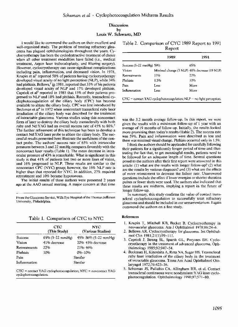
Table 2. Comparison of CYC 1989 Report to 1991Report
Success (3-22 mmHg) 59% 65%
Vision Minimal change (3 NLP) 41% decrease (19 NLP)
Retreatments ll% 27%
Phthisis 1.5% 10%
was the 3.2 month average follow-up. In this report, we weregiven the results with a minimum follow-up of I year with anaverage of 19 months of follow-up. Initially, the results lookedmore promising than today's results (Table 2). The success ratewas 59%. Pain and inflammation were described as less andthere was minimal visual change. Phthisis occurred only in 1.5%.
I think the authors should beapplauded for carefully followingtheir patients for a significantly longer period of time and illustrating the fact that, to get meaningful r~sults, patients nee? tobe followed for an adequate length of time. Several questionsposed to the authors after their first report were answered in thisarticle: (I) what are the results with longer follow-up? (2) whatare the results by various diagnosis? and (3) what are the effectsof more retreatment to decrease the failure rate. Unansweredquestions include the effect iflower energies or shorter durationpulses or fewer shots were used . The authors also indicated thatthese results are midterm, implying a report in the future oflonger follow-up.
In summary, this study confirms the value of contact transscleral cyclophotocoagulation to successfully trea~ refracto~
glaucoma and should be included in our armamentanum. I againcommend the authors on a fine study.
1991
More
More
1989
Less
Less
Pain
Inflammation
From the Glaucoma Service, Wills Eye Hospital of the Thomas JeffersonUniversity, Philadelphia.
I would like to commend the authors on their excellent andwell-organized study. The problem of treating refractory glaucoma has plagued ophthalmologists throughout the years. Cyclocryotherapy has been the cyclodestructive treatment of choicewhen all other treatment modalities have failed (i.e., medicaltreatment, Argon laser trabeculoplasty, and filtering surgery).However, cyclocryotherapy can cause significant complicationsincluding pain, inflammation, and decreased vision. In 1978,Krupin et al l reported 58% of patients having cyclocryotherapydeveloped visual acuity ofno light perception (NLP), ~hile.34%had phthisis. Bellows," j,l) 1981, reported that 33%ofhISpatI~ll:ts
developed visual acuity of NLP and 17% deve~oped. phthisis.Caprioli et aJ3 reported in 1985 that 15% of their patIents progressed to NLP and 18% had phthisis. Recently, transscleral cyclophotocoagulation of the ciliary body (CPC). has becomeavailable to ablate the ciliary body. CPC was first mtroduced byBeckman et al" in 1972 when noncontact transscleral ruby laserirradiation of the ciliary body was described for the treatmentof intractable glaucoma. Various studies using this noncontactform oflaser to destroy the ciliary body transsclerally with bothruby and Nd :YAG had an overall success rate of 45% to 86%.5The further refinement of this technique has been to develop acontact Nd :YAG laser probe to ablate the ciliary body. The successful results presented here illustrate the usefulness ofthe contact probe. The authors' success rate of 65% with intraocularpressures between 3 and 22 mmHg compares favorably ~i~h thenoncontact laser results (Table I). The mean decrease m mtraocular pressure of 43% is remarkable. What is significant in thisstudy is that 41% of patients lost two or more lines of vision,and 16% progressed to NLP. These results are similar to thenoncontact CPC (NYC) group; however, the 16% NLP rate ishigher than that reported for NYC. In addition, 27% requiredretreatment and 10% became hypotonous.
The initial results of these authors were presented 2 yearsago at the AAO annual meeting. A major concern at that time
Table 1. Comparison of CYC to NYC
CYC = contact YAG cyclophotocoagulation; NYC = noncontact YAGcyclophotocoagulation.
SuccessVis ionRetreatmentsPhthisisPainInflammation
CYC(This Study)
65% (3-22 mmHg)41% decrease27%10%
NYC(Various Studies)
45%-86% (5-22 mmHg)20%-43% decrease21%-46%0%-10%
SimilarSimilar
References
I . Krupin T, Mitchell KB, Becker B. Cyclocryotherapy inneovascular glaucoma. Am J Ophthalmol 1978;86:24-6.
2. Bellows AR. Cyclocryotherapy for glaucoma. Int Ophthalmol Clin 1981;21(I ):99-111.
3. Caprioli J, Strang SL, Spaeth GL, Poryzees EH. Cyclocryotherapy in the treatment of advanced glaucoma. Ophthalmology 1985;92:947-54.
4. Beckman H, Kinoshita A, Rota NA, Sugar HS. Transscleralruby laser irradiation of the ciliary body in the treatmentof intractable glaucoma. Trans Am Acad Ophthalmol Otolaryngol 1972;76:423-36.
5. Schuman JS, Puliafito CA, Allingham RR, et al. Contacttransscleral continuous wave neodymium:YAG laser cyclophotocoagulation. Ophthalmology 1990;97:571-80.
1095