Embed Size (px)
Citation preview

REVIEW
Constitutive heterochromatin: a surprising varietyof expressed sequences
Patrizio Dimitri & Ruggiero Caizzi & Ennio Giordano &
Maria Carmela Accardo & Giovanna Lattanzi &Giuseppe Biamonti
Received: 9 September 2008 /Revised: 30 March 2009 /Accepted: 1 April 2009 /Published online: 2 May 2009# Springer-Verlag 2009
Abstract The organization of chromosomes into euchro-matin and heterochromatin is amongst the most importantand enigmatic aspects of genome evolution. Constitutiveheterochromatin is a basic yet still poorly understoodcomponent of eukaryotic chromosomes, and its molecularcharacterization by means of standard genomic approachesis intrinsically difficult. Although recent evidence indicates
that the presence of transcribed genes in constitutiveheterochromatin is a conserved trait that accompanies theevolution of eukaryotic genomes, the term heterochromatinis still considered by many as synonymous of genesilencing. In this paper, we comprehensively review datathat provide a clearer picture of transcribed sequenceswithin constitutive heterochromatin, with a special empha-sis on Drosophila and humans.
Historical features
Heterochromatin was originally defined cytologically as theset of chromosomal regions that stain deeply at prophaseand maintain a compact organization throughout all stagesof the mitotic cell cycle (Heitz 1928). In a wide variety ofeukaryotes, large chromosomal portions, or even entirechromosomes, are made up of heterochromatin. Hete-rochromatin was further classified into facultative orconstitutive (Brown 1966). Facultative heterochromatincorresponds to euchromatic regions (chromosome portions,entire chromosomes, or whole genomes), the structure andactivity of which are subjected to control in that they can bealternatively functional or silenced during specific phasesof development (Plath et al. 2002). In contrast, constitutiveheterochromatin is commonly found in large blocks nearcentromeres and telomeres; it consists mostly of repetitiveDNA sequences and maintains its characteristic organiza-tion on both homologous chromosomes.
Constitutive heterochromatin is a basic component ofeukaryotic genomes in that it forms about 5% of thegenome in Arabidopsis thaliana, 30% in Drosophila andhumans, and up to 70–90% in certain nematodes and plants(Moritz and Roth 1976; Peterson et al. 1998; Arabidopsisgenome initiative 2000; Dimitri et al. 2005a, b), yet the
Chromosoma (2009) 118:419–435DOI 10.1007/s00412-009-0211-y
Communicated by S. Pimpinelli
P. Dimitri (*) :M. Carmela AccardoLaboratorio di Genomica Funzionale e Proteomica di Sistemimodello and Istituto Pasteur-Fondazione Bolognetti,Dipartimento di Genetica e Biologia Molecolare “Charles Darwin“,Università “La Sapienza“,Via dei sardi 70,Rome 00185, Italye-mail: [email protected]
R. CaizziDipartimento di Genetica e Microbiologia, Università di Bari,Via Amendola 165/A,Bari 70126, Italy
E. GiordanoDipartimento di Genetica, Biologia generale e Molecolare,Università di Napoli Federico II,Via Mezzocannone 8,Naples 80100, Italy
G. LattanziIstituto di Genetica Molecolare IGM unit of Bologna c/o IstitutoOrtopedico Rizzoli (IOR),-via di Barbiano 1/10,Bologna I40136, Italy
G. BiamontiIstituto di Genetica Molecolare,Via Abbiategrasso, 207,Pavia 27100, Italy
reasons for its widespread occurrence are still unclear. A setof distinctive properties, antagonistic compared to the restof the genome, have historically been recognized forconstitutive heterochromatin in virtually all animal andplant species: (1) strongly reduced level of meioticrecombination; (2) low gene density; (3) mosaic inactiva-tion of the expression of euchromatic genes when movednearby, a phenomenon termed position effect variegation;(4) late replication during S phase; (5) transcriptionalinactivity; and (6) enrichment in highly repetitive satelliteDNAs and transposable element remnants.
Together, these properties led to the view of constitutiveheterochromatin as a “desert” or a “graveyard” of geneticfunctions (reviewed by John 1988). In the last three decades,however, studies primarily conducted in Drosophilamelanogaster have shown that constitutive heterochromatindoes in fact play roles in important cellular functions, suchas chromosome organization and inheritance, and containsgenes essential for viability and fertility (Gatti andPimpinelli 1992; Williams and Robbins 1992; Weiler andWakimoto 1995; Dernburg et al. 1996; Elgin 1996; Karpenet al. 1996; Dimitri and Junakovic, 1999; Eissenberg andHilliker 2000; Henikoff et al. 2001; Coulthard et al. 2003;Dimitri et al. 2005a; Fitzpatrick et al. 2005; Villasante et al.2007). Thus, the idea that constitutive heterochromatin ismerely a genomic wasteland has become obsolete.
Drosophila heterochromatin genes
Essential genes early defined by genetic and cytologicalanalysis
D. melanogaster is the model organism in which thegreatest progress in the study of heterochromatin functionshas been made due to the ability to combine genetic,cytological, and genomic approaches. Using chromosomebanding techniques, the mitotic heterochromatin of D.melanogaster has been subdivided into 62 regions withdistinctive cytological properties (Gatti and Pimpinelli1983; Pimpinelli and Dimitri 1989; Dimitri 1991). Genesessential for viability and fertility were initially identified inD. melanogaster by recessive mutations genetically linkedto regions of constitutive heterochromatin (Brosseau 1960;Hilliker 1976; Marchant and Holm 1988). The identifica-tion of such mutations was followed by complementationanalysis using chromosome rearrangements with cytologi-cally determined breakpoints that mapped to mitoticheterochromatin, which yielded significant insight into thelocation and structural organization of the genetic locilocated in autosomal and sex heterochromatin of D.melanogaster (Gatti and Pimpinelli 1983; Pimpinelli et al.1985; Dimitri 1991; Koryakov et al. 2002).
Sex chromosome heterochromatin genes
Some loci located in the sex chromosome heterochromatin arephysically very large and mainly consist of high- and middle-repetitive DNAs. The kl-5, kl-3, and kl-1 fertility factors onthe Y-chromosome are estimated to contain about 4 Mb ofDNA; they require structural integrity for function and formgiant loops that are actively transcribed in primary spermato-cytes (reviewed by Gatti and Pimpinelli 1992). These giganticloci, which were originally suggested to perform structuralfunctions, have in fact turned out to harbor protein-codinggenes. For example, kl-5 encodes an axonemal–dynein heavychain that is expressed in the testis (Gepner and Hays 1993).These genes are made up of small unique exons andtransposable element-rich mega-introns that can account for1 or 2 Mb of DNA (Kurek et al. 2000; Carvalho et al. 2001).
Other loci found on the Y and X heterochromatin, suchas bobbed, encoding the ribosomal genes (Ritossa andSpiegelman 1965) are not inactivated by breakpoints oftranslocations or inversions, like the Y-fertility factors, butonly by deletions; they consist of an array of middle-repetitive sequences whose number is critical for theiractivity. A similar organization is exhibited by Suppressorof Stellate [Su(Ste)] locus (Litvak 1984; Bozzetti et al.1995) which is involved in a natural case of RNA silencing-mediated regulation. Su(Ste) repeats produce shortsense and antisense RNAs that cause the repression oftestis-expressed homologous Stellate genes on the X-chromosome (Aravin et al. 2004). In addition, the X-chromosome heterochromatin carries a group of still poorlycharacterized genetic loci that are thought to be all composedof repeated elements (reviewed by Gatti and Pimpinelli 1992):compensatory response (cr), ABO, collochore (col), andRibosomal exchange (Rex) with its suppressor. Thecollochore locus mediates proper sex chromosome pairingand disjunction during the first meiotic division; cr controlsrDNA gene dosage compensation; the ABO elements rescuethe maternal defects caused by a recessive maternal effectmutation called abnormal oocyte (abo). Finally, mutations inRex cause a high frequency of exchanges and deletions in therDNA (Rasoly and Robbins 1991).
It is worth noting that both ABO and Su(Ste) loci are partof genetic systems that involve specific interactions betweenheterochromatin and euchromatin genetic elements. Suchheterochromatin elements were defined “criptic” in that theyescape genetic analysis and their effect can be detected onlyin the presence of mutations in the euchromatic counterpart(Palumbo et al. 1994).
Autosomal heterochromatin essential genes
Thus far, at least 32 genes essential for viability have beenmapped to mitotic heterochromatin of chromosomes 2 and
420 Chromosoma (2009) 118:419–435

3 (Dimitri 1991; Koryakov et al. 2002), but only a few ofthose are clearly defined at the molecular level: RpL5, light,concertina, rolled, RpL38, Nipped-B, Nipped-A, Parp andRpL15 (Hilliker 1976; Devlin et al. 1990a, b; Parks andWieschaus 1991; Biggs et al. 1994; Rollins et al. 1999;Tulin et al. 2002; Myster et al. 2004; Marygold et al. 2005;Schulze et al. 2005). Most of the genes detected thus farwere mapped cytologically to heterochromatin regions ofmoderate fluorescence after staining with 4,6-diamino-2-phenylindole-dihydrochloride (DAPI); as example, see themapping of essential gene shown in Fig. 1. These regionsharbor clusters of transposable elements and are devoid ofhighly repetitive satellite DNAs (Pimpinelli et al. 1995;Lohe et al. 1993).
Putative genes defined by heterochromatin sequenceannotation
In the last decade, the completion of genome sequencingprojects has yielded a great amount of information on DNAsequences in several organisms. The release of the sequenceof D. melanogaster heterochromatin by the BerkeleyDrosophila Genome Project (http://www.fruitfly.org/) andDrosophila Heterochromatin Genome Project (DHGP;http://www.dhgp.org/index_release_notes.html) has greatlyfacilitated studies of mapping, molecular organization, andfunction of genes located in pericentromeric heterochroma-tin. Initially, 3.8 Mb of about 120 Mb of the D.melanogaster euchromatic genome sequence included in
Fig. 1 Mapping of essential andputative genes to the hetero-chromatin of mitotic chromo-some 2. Cytogenetic mapping ofboth essential genes defined bymutational analyses (below) andof putative genes (above) de-fined by computational analyses.Shades of blue correspond to theintensity of DAPI staining, withthe darkest blue blocks repre-senting regions with strongfluorescence intensity and openblocks representing non-fluorescent regions. The differ-ent cytological regions arenumbered in red. Scaffold des-ignation is shown at the top ofeach gene model list. Additional2Rh lethal genes defined byMyster et al. (2004) are notincluded in the map, as it ispresently unclear whether theselethals correspond to new vitalgenes
Chromosoma (2009) 118:419–435 421

release1 were found to correspond to sequences originatedfrom distal heterochromatin regions (Adams et al. 2000).More recently, an improved whole genome shotgunassembly (heterochomatic-WGS3; Hoskins et al. 2002)has been produced, which includes 20.7 Mb of draft-quality heterochromatin sequence. In the last year, 15 Mbof this sequence has been further improved or completed(Hoskins et al. 2007), and a BAC-based physical map of13 Mb of pericentric heterochromatin, together with thecytogenetic map that locates some 11 Mb to specificheterochromatin regions, has been constructed (Hoskins etal. 2007). About 450 predicted genes were initiallyidentified by the annotation of the heterochromatinsequence (Hoskins et al. 2002). More recently, about 250protein-coding genes were defined in the release 5.1annotation of the currently sequenced heterochromatinDNA (Smith et al. 2007). According to these results, thenumber of active genes in constitutive heterochromatin ofD. melanogaster appears to be higher than that defined bygenetic analysis.
Several studies have concentrated on an effort to mappredicted genes to the mitotic heterochromatin of D.melanogaster using fluorescent in situ hybridization (FISH)with BACs, cDNAs, and P-elements (Hoskins et al. 2002;Corradini et al. 2003; Yasuhara et al. 2003; Rossi et al.2007). For example, about 161 predicted genes have beenassigned to specific regions of the mitotic heterochromatinof chromosome 2 (Rossi et al. 2007; Hoskins et al. 2007;Figs. 1 and 2; Table 1) in which genetic analyses defined 17essential genes. Essential and putative genes are groupedwithin regions h35 and h46 in the constitutive heterochro-matin of chromosome 2, which represent the most distalportions of mitotic heterochromatin. Most of these genesare located in weakly DAPI-fluorescent chromosomalregions, which harbor clusters of transposable element-homologous sequences and lack highly repetitive satelliteDNAs. The high number of predicted genes found inheterochromatin can be explained by assuming that theseregions contain an excess of non-essential coding genes thatescape mutational analysis. For example, CG40293, p120,and CG17486 of 2Rh were found to be non-essential(Myster et al. 2004). It may be also possible that singlecoding sequences are fragmented due to assembly artifacts,thus giving rise to multiple shorter CGs.
PiRNAs and esiRNA clusters: a special classof heterochromatin genes
The flamenco/COM locus, involved in the regulation ofgypsy, Idefix, and ZAM retrotransposons, has beenmapped to the distal portions of the X heterochromatin(Prud'homme et al. 1995; Desset et al. 2003). Molecularly,flamenco was located proximally to the DIP-1 gene and
proposed to span a region that corresponds to a Piwi-interacting RNA (piRNA) cluster recently identified byBrennecke et al. (2007). This piRNA cluster mainlyconsists of nested transposable elements (TEs) spanning atotal length of 179 kb and includes numerous fragments ofgypsy, Idefix, and ZAM retrotransposons. Brennecke et al.(2007) found about 130 piRNA loci in pericentromeric andtelomeric heterochromatin, which display a high content ofdefective and nested TEs. Those piRNAs are restricted togonads and at least a set of them arise through Piwi-mediated cleavage of single-stranded RNAs (Brennecke etal. 2007). Maternally transmitted I-element piRNAs origi-nated from 42AB polytene chromosome region areinvolved in the control of I element transposition(Brennecke et al. (2008). Endogenous small interferingRNA (esiRNA) originated from heterochromatin TE clus-ters, and dependent on Dicer-2 and Argonaute-2, wererecently detected in somatic and gonad cells (Ghildiyal etal. 2008; Czech et al. 2008). Both piRNA and esiRNAsequence clusters present in heterochromatin are found to beinvolved in TE silencing. It is tempting to speculate, however,that piRNA and esiRNA clusters, and possibly other non-protein-coding RNAs originating from high TE density
Fig. 2 FISH mapping of cDNA to mitotic heterochromatin. FISHmapping of two different cDNA clones from predicted genes onheterochromatin of the right arm of chromosome 2
422 Chromosoma (2009) 118:419–435
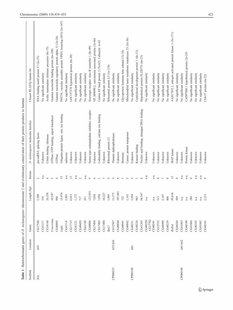
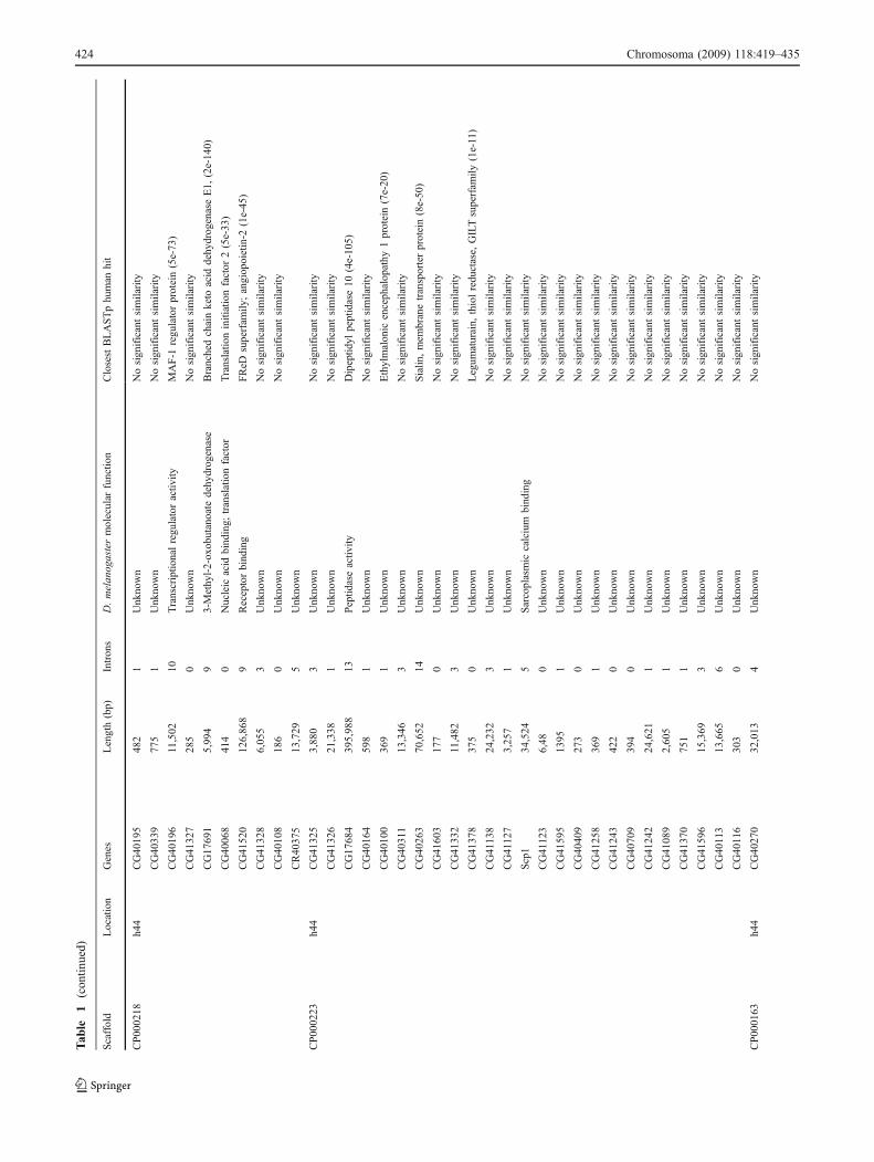
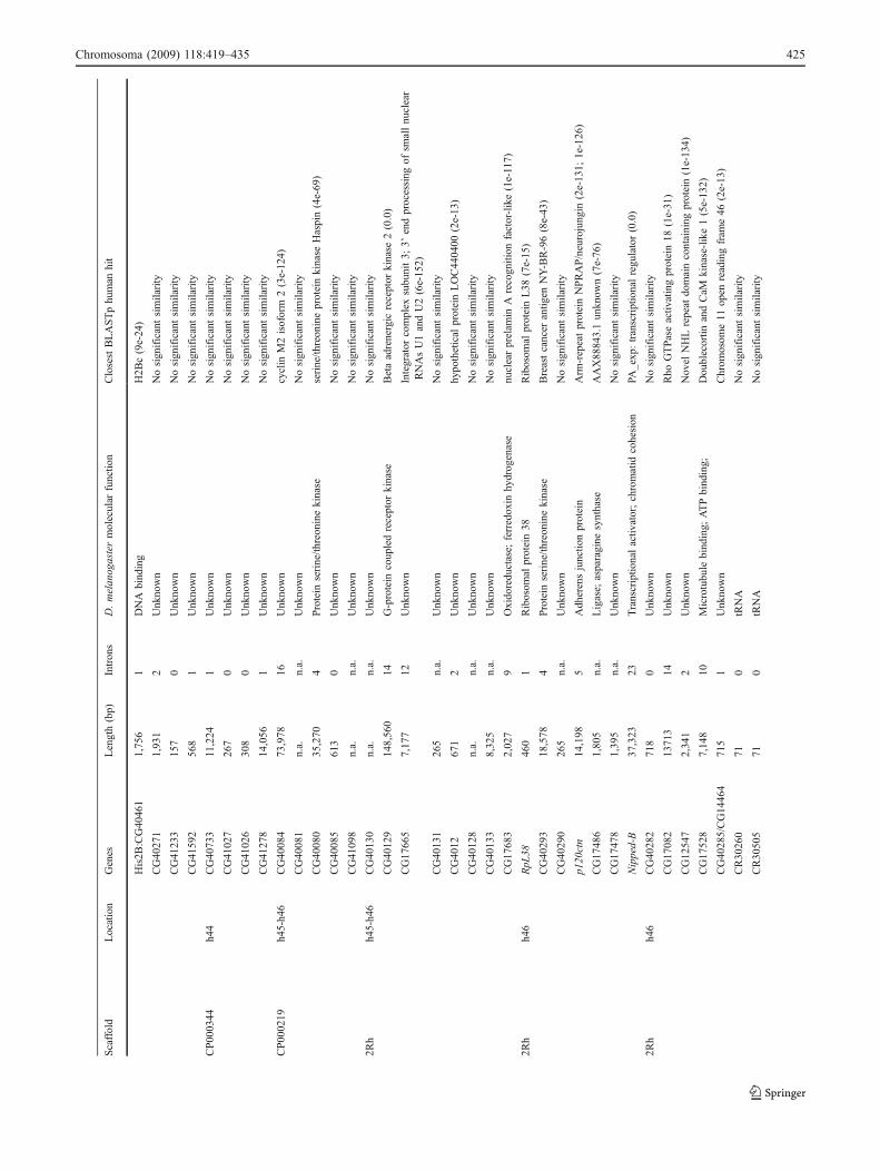
Tab
le1
Heterochrom
atin
genesof
D.melan
ogasterchromosom
e2andevolutionary
conservatio
nof
theirproteinprod
uctsin
humans
Scaffold
Location
Genes
Length(bp)
Introns
D.melanogastermolecular
functio
nClosestBLASTphuman
hit
2Lh
h35
CG17540
1,580
2pre-mRNA
splicingfactor
RNA
bindingmotifprotein17
(2e-53)
CG41117
310
n.a.
Unknown
Nosignificantsimilarity
CG18140
26,358
8Chitin
binding;
chitinase
Acidicmam
malianchitinase
precursor(8e-75)
Concertina
10,547
5GTPase;
GTPbinding;
signal
transducer
Guanine
nucleotid
ebindingprotein(6e-108)
CG40005
988
4GTPase
Guanine
nucleotid
eregulatory
proteinalpha13
(3e-26)
Light
15,974
15Ubiquitin-proteinlig
ase;
zinc
ionbinding
P49754,
vacuolar
assemblyproteinVPS41
homolog
(S53)(3e-147)
CG41118
2,901
n.a.
unknow
nNosignificantsimilarity
CG17715
6,015
13Unknown
LOC157378
hypotetical
protein(8e-30)
CG41121
1,172
n.a.
Unknown
Nosignificantsimilarity
CG40439
717
2Unknown
Nosignificantsimilarity
CG41120
451
n.a.
Unknown
Nosignificantsimilarity
CG40006
133,933
9Serine-type
endopeptidaseinhibitor;receptor
Scavenger
receptor
classB
mem
eber
1(8e-49)
CG17494
7,056
6Unknown
NP_009090.2,
sarcolem
maassociated
protein(3e-66)
CG17493
1,056
1Calmodulin
binding;
calcium
ionbinding
Centrin
EFhand
protein2(7e-62);Caltractin
1e-62
CG17490
10,227
7Unknown
Nosignificantsimilarity
RpL
51,905
9Ribosom
alproteinL5
Ribosom
alproteinL5(1e-119)
CP000215
h35-h36
CG12567
13,373
4Thiam
indiphosphokinase
Nosignificantsimilarity
CG40040
107,081
8Unknown
Nosignificantsimilarity
CG40041
732
1Hormone
Glycoproteinhorm
onebeta
subunit(7e-10)
CG40042
1,141
5Carrier;proteintransporter
Mito
chondrialinnermem
branetranslocase23
(5e-36)
CP000188
h41
CG40211
3,954
4Unknown
Nosignificantsimilarity
CG40218
963
1Kinesin
binding
Craniofacialdevelopm
entprotein1(4e-13)
CG41265
94,547
8Nucleic
acid
binding;
damaged
DNA
binding
Hypothetical
proteinFLJ20753
(6e-25)
CG40220/
CG17702
n.a.
n.a.
Unknown
Nosignificantsimilarity
CG40241
n.a.
n.a.
Unknown
Nosignificantsimilarity
CG12552
513
0Unknown
Nosignificantsimilarity
CG40498
2,107
7Unknown
Nosignificantsimilarity
CG40239
408
0Unknown
Nosignificantsimilarity
Rolled
49,634
15MAPkinase
AAH17832.1,
mito
genactiv
ated
proteinkinase
1(3e-171)
CG40244
499
0Unknown
Nosignificantsimilarity
CP000188
h41-h42
CG41063
n.a.
n.a.
Unknown
Nosignificantsimilarity
CG40190
n.a.
n.a.
Protein
kinase
CAD97888.1hypothetical
protein(2e-25)
CG41066
288
0Unknown
Nosignificantsimilarity
CG40192
n.a.
n.a.
Unknown
Nosignificantsimilarity
CG41065
n.a.
n.a.
Unknown
Nosignificantsimilarity
CG40191
2,213
9Unknown
CGI-57
protein(6e-22)
Chromosoma (2009) 118:419–435 423
Tab
le1
(con
tinued)
Scaffold
Location
Genes
Length(bp)
Introns
D.melanogastermolecular
functio
nClosestBLASTphuman
hit
CP000218
h44
CG40195
482
1Unknown
Nosignificantsimilarity
CG40339
775
1Unknown
Nosignificantsimilarity
CG40196
11,502
10Transcriptio
nalregulatoractiv
ityMAF-1
regulatorprotein(5e-73)
CG41327
285
0Unknown
Nosignificantsimilarity
CG17691
5,994
93-Methyl-2-oxobutanoate
dehydrogenase
Branchedchainketo
acid
dehydrogenaseE1,
(2e-140)
CG40068
414
0Nucleic
acid
binding;
translationfactor
Translatio
ninitiationfactor
2(5e-33)
CG41520
126,868
9Receptorbinding
FReD
superfam
ily;angiopoietin-2
(1e-45)
CG41328
6,055
3Unknown
Nosignificantsimilarity
CG40108
186
0Unknown
Nosignificantsimilarity
CR40375
13,729
5Unknown
CP000223
h44
CG41325
3,880
3Unknown
Nosignificantsimilarity
CG41326
21,338
1Unknown
Nosignificantsimilarity
CG17684
395,988
13Peptid
aseactiv
ityDipeptid
ylpeptidase10
(4e-105)
CG40164
598
1Unknown
Nosignificantsimilarity
CG40100
369
1Unknown
Ethylmalonic
encephalopathy
1protein(7e-20)
CG40311
13,346
3Unknown
Nosignificantsimilarity
CG40263
70,652
14Unknown
Sialin
,mem
branetransporterprotein(8e-50)
CG41603
177
0Unknown
Nosignificantsimilarity
CG41332
11,482
3Unknown
Nosignificantsimilarity
CG41378
375
0Unknown
Legum
aturain,
thiolreductase,
GILTsuperfam
ily(1e-11)
CG41138
24,232
3Unknown
Nosignificantsimilarity
CG41127
3,257
1Unknown
Nosignificantsimilarity
Scp1
34,524
5Sarcoplasmic
calcium
binding
Nosignificantsimilarity
CG41123
6,48
0Unknown
Nosignificantsimilarity
CG41595
1395
1Unknown
Nosignificantsimilarity
CG40409
273
0Unknown
Nosignificantsimilarity
CG41258
369
1Unknown
Nosignificantsimilarity
CG41243
422
0Unknown
Nosignificantsimilarity
CG40709
394
0Unknown
Nosignificantsimilarity
CG41242
24,621
1Unknown
Nosignificantsimilarity
CG41089
2,605
1Unknown
Nosignificantsimilarity
CG41370
751
1Unknown
Nosignificantsimilarity
CG41596
15,369
3Unknown
Nosignificantsimilarity
CG40113
13,665
6Unknown
Nosignificantsimilarity
CG40116
303
0Unknown
Nosignificantsimilarity
CP000163
h44
CG40270
32,013
4Unknown
Nosignificantsimilarity
424 Chromosoma (2009) 118:419–435
Tab
le1
(con
tinued)
Scaffold
Location
Genes
Length(bp)
Introns
D.melanogastermolecular
functio
nClosestBLASTphuman
hit
His2B
:CG40461
1,756
1DNA
binding
H2B
c(9e-24)
CG40271
1,931
2Unknown
Nosignificantsimilarity
CG41233
157
0Unknown
Nosignificantsimilarity
CG41592
568
1Unknown
Nosignificantsimilarity
CP000344
h44
CG40733
11,224
1Unknown
Nosignificantsimilarity
CG41027
267
0Unknown
Nosignificantsimilarity
CG41026
308
0Unknown
Nosignificantsimilarity
CG41278
14,056
1Unknown
Nosignificantsimilarity
CP000219
h45-h46
CG40084
73,978
16Unknown
cyclin
M2isoform
2(3e-124)
CG40081
n.a.
n.a.
Unknown
Nosignificantsimilarity
CG40080
35,270
4Protein
serine/th
reoninekinase
serine/th
reonineproteinkinase
Haspin(4e-69)
CG40085
613
0Unknown
Nosignificantsimilarity
CG41098
n.a.
n.a.
Unknown
Nosignificantsimilarity
2Rh
h45-h46
CG40130
n.a.
n.a.
Unknown
Nosignificantsimilarity
CG40129
148,560
14G-protein
coupledreceptor
kinase
Betaadrenergic
receptor
kinase
2(0.0)
CG17665
7,177
12Unknown
Integrator
complex
subunit3;
3’endprocessing
ofsm
allnuclear
RNAsU1andU2(6e-152)
CG40131
265
n.a.
Unknown
Nosignificantsimilarity
CG4012
671
2Unknown
hypothetical
proteinLOC440400
(2e-13)
CG40128
n.a.
n.a.
Unknown
Nosignificantsimilarity
CG40133
8,325
n.a.
Unknown
Nosignificantsimilarity
CG17683
2,027
9Oxidoreductase;
ferredoxin
hydrogenase
nuclearprelam
inA
recognition
factor-like(1e-117)
2Rh
h46
RpL
38460
1Ribosom
alprotein38
Ribosom
alproteinL38
(7e-15)
CG40293
18,578
4Protein
serine/th
reoninekinase
Breastcancer
antig
enNY-BR-96(8e-43)
CG40290
265
n.a.
Unknown
Nosignificantsimilarity
p120ctn
14,198
5Adherensjunctio
nprotein
Arm
-repeatproteinNPRAP/neurojungin
(2e-131;
1e-126)
CG17486
1,805
n.a.
Ligase;
asparagine
synthase
AAX88843.1unknow
n(7e-76)
CG17478
1,395
n.a.
Unknown
Nosignificantsimilarity
Nipped-B
37,323
23Transcriptio
nalactiv
ator;chromatid
cohesion
PA_exp:transcriptionalregulator(0.0)
2Rh
h46
CG40282
718
0Unknown
Nosignificantsimilarity
CG17082
13713
14Unknown
Rho
GTPaseactiv
atingprotein18
(1e-31)
CG12547
2,341
2Unknown
Novel
NHLrepeat
domaincontaining
protein(1e-134)
CG17528
7,148
10Microtubule
binding;
ATPbinding;
Doublecortin
andCaM
kinase-like1(5e-132)
CG40285/CG14464
715
1Unknown
Chrom
osom
e11
open
readingfram
e46
(2e-13)
CR30260
710
tRNA
Nosignificantsimilarity
CR30505
710
tRNA
Nosignificantsimilarity
Chromosoma (2009) 118:419–435 425

Tab
le1
(con
tinued)
Scaffold
Location
Genes
Length(bp)
Introns
D.melanogastermolecular
functio
nClosestBLASTphuman
hit
2Rh
h46
CG33492
72,289
4Ionotropic
glutam
atereceptor
Glutamatereceptor,ionotropic,delta
1(2e-07)
TpnC41C
3,920
3Calcium
ionbinding
Calmodulin
2(phosphorylase
kinase,delta)(4e-25)
CG3107
4,504
4Metalloendopeptidase
Metalloprotease
1(0.0)
CG2944
11,103
11Oocyteanterior/posterior
axisdeterm
ination
SPRYdomain-containing
SOCSboxproteinSSB-1
variant
(7e-117)
CG3136
10065bp
5DNA
binding;protein
homodim
erization
cAMPresponse
elem
entbinding
protein-related(3e-13)
Nipped-A
73,048
29Transcriptio
nregulator;cytokinesis
Transform
ation/transcriptiondomain-associated
proteinvariant(0.0)
CG2682
35,992
8Transcriptio
nfactor;ubiquitin
-protein
ligase
D4,
zinc
anddouble
PHD
fingersfamily
2(5e-49)
hetero-euchrom
atin
CG10392
22,231
12Transferringglycosyl
groups
O-linkedGlcNActransferaseisoform
2(0.0)
transitio
nregion
CG10465
1,290
1Voltage-gated
potassium
channel;p
r.binding
Unnam
edproteinproduct(9e-100)
CG10395
1,480
1HIT
Zn-finger
proteindomain
Highmobility
groupAT-hook
1-lik
e4(5e-14)
CG30441
409
n.a
Unknown
Intraflagellartransportprotein20-likeprotein(9e-12)
CG10396
733
1Cytochrom
e-coxidase
Cytochrom
ecoxidasesubunitIV
isoform
1(1e-23)
CG10417
2,778
6Protein
serine/th
reoninephosphatase
Protein
phosphatase1G
variant(1e-61)
CG30440
27,523
7Guanyl-nucleotid
eexchange
factor
MCF.2celllin
ederivedtransformingsequence
(3e-77)
CG30438
52,876
9Transferringglycosyl
groups
Ceram
ideUDPgalactosyltransferase(5e-68)
TpnC4
4,431
4Calcium
ionbinding
Calmodulin
2(7e-25)
CG17510
1,243
9Unknown
Tetratricopeptiderepeat
domain11
(5e-12)
CG17508
2,966
3Unknown
C20orf108
(7e-24)
CG11665
11,129
4Monocarboxylic
acid
transporter
Solutecarrierfamily
16(3e-33)
CG32838/CG42345
41,278
11Laccase;copper
ionbinding
Nosignificantsimilarity
Locationrefersto
themapping
ofscaffoldsandgeneson
mito
ticheterochromatin
map;C
Gindicatetheanno
tatedgenes;leng
thmeans
theph
ysicalsize
ofthegeno
micregion
ofagivengene.2
Lh
correspo
ndsto
release5assemblyof
2Larm
thatincorporates
release3heterochromatin
scaffolds(A
ABU10
0263
7andAABU10
0276
8);the
genesmapping
toh3
5belong
toAABU10
0276
8;2R
hcorrespo
ndsto
release5assemblyof
2Rarm
that
incorporates
release3heterochromatin
scaffolds(A
ABU01
0027
11,AABU01
0027
52and2R
.wgs3_
extension).In
release5sequ
ence,release3
scaffoldswerealso
assembled
inlarger
scaffoldsdesign
ated
with
theCP
acrony
m(H
oskins
etal
2007
).On2L
heterochromatin,thescaffold
CP00
0215
contains
therelease3scaffold
AABU01
0027
56.On2R
heterochromatin,CP00
0188
contains
therelease3scaffoldsAABU01
0019
47,AABU01
0021
99andAABU01
0025
49,while
CP00
0218
andCP00
0219
contain
AABU01
0027
50andAABU01
0027
48,respectiv
ely.
The
cytologicalbo
rder
ofhetero-euchrom
atin
transitio
nregion
was
establishedby
FISH
mapping
ofBACs(Corradini
etal.20
03)andis
approx
imate;someof
thegenesassign
edto
thisregion
may
beactually
locatedin
heterochromatin.G
eneanno
tatio
nswereaccordingto
release5sequ
ence
(http
://flyb
ase.org/;www.dhg
p.org)
and
toSmith
etal(200
7).O
nlyBLASTphitswith
e<
15wereselected.InFlybase,the
anno
tatio
nof
geno
micregion
with
exon
–intronstructurewas
notavailableforanu
mberof
genes;weindicated
thesecaseswith
n.a.=no
tavailable
426 Chromosoma (2009) 118:419–435
heterochromatin regions, can be in fact endowed with stillunidentified genetic functions.
Gene density in D. melanogaster heterochromatinvs euchromatin
It has been previously estimated that the density of single-copy genes in heterochromatin is some100-fold lower thanthat found in euchromatin (Hilliker et al. 1980). In light ofthe recent annotation of release 5.1 Drosophila heterochro-matin sequence, ten to 11 genes per Megabase have beenfound in sequenced heterochromatin that correspond totransposon-rich regions compared with 127 genes perMegabase in euchromatin; in other words, gene densitywould appear to be only one order of magnitude lowercompared to euchromatin. This estimate, however, does notinclude the satellite DNA-rich regions, within which thegene density is likely to be still very low. In this context,the Y-chromosome heterochromatin represents an interest-ing exception because combined cytogenetic and molecularanalyses suggested it to be an almost continuous array ofphysically large functional genetic elements (Pimpinelliet al. 1985).
Functional and structural aspects of single-copyheterochromatin genes in Drosophila
The single-copy heterochromatin genes of D. mela-nogaster encode proteins involved in important cellularprocesses. The light gene product is required for cellularprotein trafficking (Warner et al. 1998), while concertinaencodes a maternal α-like subunit of a G-protein essentialfor gastrulation (Parks and Wieschaus 1991). The rolledgene was shown to be essential for imaginal discdevelopment and suggested to be involved in cellproliferation (Hilliker 1976; Dimitri 1991); indeed, itsencoded product is a mitogen-activated protein kinaserequired in the signal transduction pathway of thesevenless gene (Biggs et al. 1994) and may also beimplicated in the spindle integrity checkpoint (Inoue andGlover 1998). The Nipped-A product facilitates assemblyof the Notch activator complex and targets gene transcrip-tion (Gause et al. 2006), while the Nipped-B protein isrequired for both transcriptional regulation and sisterchromatid cohesion (Misulovin et al. 2008). The l(2)41Afgene corresponds to the predicted gene CG18001 whichencodes the RpL38 ribosomal protein (Marygold et al.2005). Two more ribosomal protein genes, RpL5 andRpL15, are also found on 2L and 3L heterochromatin,respectively (Marygold et al. 2005; Schulze et al. 2005).The Parp gene on 3Rh encodes a poly(ADP-ribose)polymerase, a major NAD-dependent modifying enzyme
that mediates important steps in DNA repair, transcription,and apoptosis (Tulin et al. 2002). A significant number ofessential genes located in chromosome 2 heterochromatin(Fig. 1) still need to be identified molecularly. Among thosegenes, l(2)41Aa and l(2)41Ad in the heterochromatin of theright arm of chromosome 2 (2Rh) are thought to be requiredfor chromosome condensation (Cenci et al. 2003) and forproper leg and wing morphogenesis (Dimitri et al. 2005a),respectively. In particular, l(2)41Ad is the only known vitalgene mapping to region h44 (Dimitri 1991) that contains 44predicted genes (Fig. 1 and Table 1). Interestingly, l(2)41Adis a highly mutable gene in the I-R dysgenesis system(Dimitri et al. 1997) and most of its I-R induced lethal alleleswere found to be associated with cytologically visibledeletions of regions of h44 spanning roughly up to 1 Mbof DNA (Dimitri et al. 2005b). In light of these genetic andcytological features, l(2)41Ad was suggested to be a largegene (Rossi et al. 2007). A good putative gene candidate tobe l(2)41Ad is CG17684, the largest found in h44 and in allautosomal heterochromatin; CG17684 is about 400 kb andencodes a putative protein sharing high identity with thehuman dipeptidyl peptidase enzyme. Although this corre-spondence is suggestive, additional genetic and functionalgenomic studies are needed to establish the molecularidentity and function of l(2)41Ad.
In general, therefore, based on molecular and bioinfor-matic analyses, both predicted and known genes resident inheterochromatin do not apparently have molecular func-tions that would distinguish them from genes located ineuchromatin (Hoskins et al. 2002; reviewed by Dimitri etal. 2005a; Flybase 2009). In other words, the heterochro-matin genome does not seem to encode a distinctiveproteome. However, according to Smith et al. (2007), someclasses of genes appear to be overrepresented in hetero-chromatin, relative to euchromatin. This is the case ofputative membrane cation transporter domains and ofDNA- and protein-binding domains.
A difference between heterochromatin and euchromatingenes lies in their size and molecular structure. Theexample of the “giant” Y-chromosome fertility factors ofD. melanogaster mentioned above is paradigmatic in thisrespect (Gatti and Pimpinelli 1992). Some of the hetero-chromatin essential genes of chromosomes 2 and 3 are alsolarge due to the presence of long introns made up of TEremnants (Devlin et al. 1990a, b; Tulin et al. 2002; Dimitriet al. 2003). On average, heterochromatin gene introns arefive times longer than those present in euchromatin genes(Smith et al. 2007). There are, however, some exceptions:For example, RpL38, RpL5, and RpL15, three essentialprotein-coding genes on chromosome 2 and 3, are all ofshort size (Marygold et al. 2005; Schulze et al. 2005). Howmight these observations be explained? One may imaginethat during evolution, older genes in heterochromatin have
Chromosoma (2009) 118:419–435 427
increased their size by becoming targets for reiteratedtransposable-element insertions in the intronic regions. Ifthat is true, short genes in heterochromatin shouldrepresent a set of genes recently “moved” to hetero-chromatin. Alternatively, genes in heterochromatin mightbe differentially targeted by transposable elements, withsome genes being more refractory than others. Finally,there might be selective pressure to maintain somegenes of short size in heterochromatin owing toparticular functional properties. Interestingly, in thatrespect, highly expressed genes have been shown toharbor substantially shorter introns than genes expressedat low levels (Castillo-Davis et al. 2002). This is the caseof RpL38, RpL5, and RpL15 genes are that highlyexpressed and are all indeed short genes carrying shortintrons (Marygold et al. 2005; Schulze et al. 2005).
The paradox of active heterochromatin genes
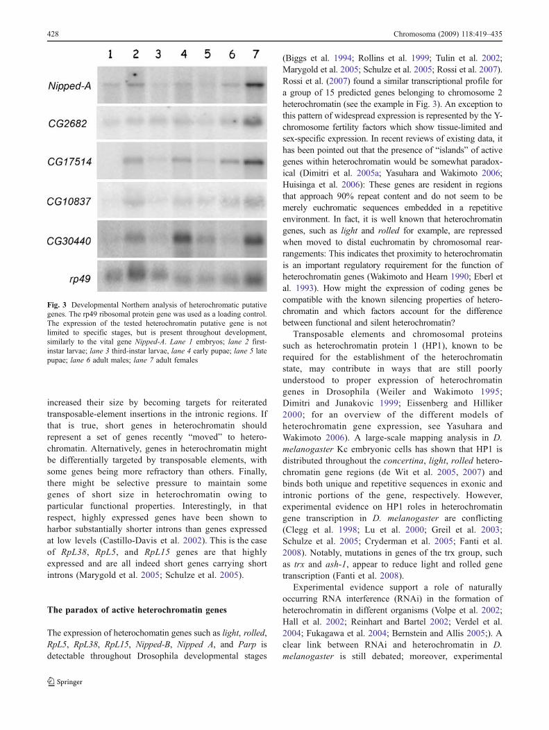
The expression of heterochomatin genes such as light, rolled,RpL5, RpL38, RpL15, Nipped-B, Nipped A, and Parp isdetectable throughout Drosophila developmental stages
(Biggs et al. 1994; Rollins et al. 1999; Tulin et al. 2002;Marygold et al. 2005; Schulze et al. 2005; Rossi et al. 2007).Rossi et al. (2007) found a similar transcriptional profile fora group of 15 predicted genes belonging to chromosome 2heterochromatin (see the example in Fig. 3). An exception tothis pattern of widespread expression is represented by the Y-chromosome fertility factors which show tissue-limited andsex-specific expression. In recent reviews of existing data, ithas been pointed out that the presence of “islands” of activegenes within heterochromatin would be somewhat paradox-ical (Dimitri et al. 2005a; Yasuhara and Wakimoto 2006;Huisinga et al. 2006): These genes are resident in regionsthat approach 90% repeat content and do not seem to bemerely euchromatic sequences embedded in a repetitiveenvironment. In fact, it is well known that heterochromatingenes, such as light and rolled for example, are repressedwhen moved to distal euchromatin by chromosomal rear-rangements: This indicates thet proximity to heterochromatinis an important regulatory requirement for the function ofheterochromatin genes (Wakimoto and Hearn 1990; Eberl etal. 1993). How might the expression of coding genes becompatible with the known silencing properties of hetero-chromatin and which factors account for the differencebetween functional and silent heterochromatin?
Transposable elements and chromosomal proteinssuch as heterochromatin protein 1 (HP1), known to berequired for the establishment of the heterochromatinstate, may contribute in ways that are still poorlyunderstood to proper expression of heterochromatingenes in Drosophila (Weiler and Wakimoto 1995;Dimitri and Junakovic 1999; Eissenberg and Hilliker2000; for an overview of the different models ofheterochromatin gene expression, see Yasuhara andWakimoto 2006). A large-scale mapping analysis in D.melanogaster Kc embryonic cells has shown that HP1 isdistributed throughout the concertina, light, rolled hetero-chromatin gene regions (de Wit et al. 2005, 2007) andbinds both unique and repetitive sequences in exonic andintronic portions of the gene, respectively. However,experimental evidence on HP1 roles in heterochromatingene transcription in D. melanogaster are conflicting(Clegg et al. 1998; Lu et al. 2000; Greil et al. 2003;Schulze et al. 2005; Cryderman et al. 2005; Fanti et al.2008). Notably, mutations in genes of the trx group, suchas trx and ash-1, appear to reduce light and rolled genetranscription (Fanti et al. 2008).
Experimental evidence support a role of naturallyoccurring RNA interference (RNAi) in the formation ofheterochromatin in different organisms (Volpe et al. 2002;Hall et al. 2002; Reinhart and Bartel 2002; Verdel et al.2004; Fukagawa et al. 2004; Bernstein and Allis 2005;). Aclear link between RNAi and heterochromatin in D.melanogaster is still debated; moreover, experimental
Fig. 3 Developmental Northern analysis of heterochromatic putativegenes. The rp49 ribosomal protein gene was used as a loading control.The expression of the tested heterochromatin putative gene is notlimited to specific stages, but is present throughout development,similarly to the vital gene Nipped-A. Lane 1 embryos; lane 2 first-instar larvae; lane 3 third-instar larvae, lane 4 early pupae; lane 5 latepupae; lane 6 adult males; lane 7 adult females
428 Chromosoma (2009) 118:419–435

evidence on rasiRNA pathway involvement in heterochroma-tin formation in somatic tissues are conflicting (reviewed byHuisinga and Elgin 2009). Interestingly, piwi was found to berequired for the expression of subtelomeric TAS repeats inboth soma and germ line of D. melanogaster (Yin and Lin2007). In light of this result, it may be interesting to test theeffects of piwi mutations on transcription of D. melanogastersingle-copy genes located in pericentromeric heterochromatin.
Histone modifications are also likely to play roles inthe control of heterochromatin gene expression. Thedistribution of modified histones in heterochromatingenes has recently been studied by Yasuhara andWakimoto (2008). They found that H3-di-methylated-at-lysine 9 (H3K9me2) is depleted at the 5′ end, but enrichedthroughout the transcribed portion, of heterochromatingenes, a profile different from that found in euchromaticgenes. The authors suggest that heterochromatin genes areintegrated into, rather than insulated from, the H3K9me2-enriched domain.
The presence of coding genes in heterochromatinis a conserved trait in the evolution of eukaryoticgenomes
Recent studies have investigated the origin of D. mela-nogaster heterochromatin genes by comparing putativeorthologous genes in different species of the Drosophilalineage. The first study analyzed a cluster of genesspanning 594 kb of DNA around the light gene, whichmaps to heterochromatin in D. melanogaster but has aeuchromatic location in both Drosophila pseudobscura andDrosophila virilis (Yasuhara et al. 2005). In another study,the entire heterochomatic chromosome 4 ofD. melanogaster(4–5 Mb of DNA) was compared to the homologousD. virilis “dot” chromosome, which is instead euchromatic(Slawson et al. 2006). Together, the results of these studiesindicate that promoter regions of euchromatin and hetero-chromatin genes are per se essentially similar and thattransposable elements play a fundamental role in theformation of heterochromatin domains.
An interesting approach designed to understand whethergenes have moved into, or out of, heterochromatin regionsin other species has been developed by Smith et al. (2005)and is based on the analysis of repeat content oforthologous introns and scaffolds. The location wasconfirmed for over 80% of the predicted orthologous genesby FISH mapping analysis on polytene chromosomes indifferent Drosophila species. The results indicate that asignificant portion of D. melanogaster heterochromatingenes are likely to descend from euchromatin progenitors(C. Smith, F. Rossi, S. Celniker, P. Dimitri and Gary H.Karpen, unpublished). Thus, it would appear that during
evolution, some genes have “jumped” between the twogenomic compartments.
The presence of transcribed sequences in heterochroma-tin, far from being a peculiarity of Drosophila species,appears to be a conserved trait in the evolution ofeukaryotic genomes. Single-copy protein coding genes areindeed found in Schizosaccharomyces pombe, rice,A. thaliana, and humans (reviewed by Dimitri et al.2005a; Yasuhara and Wakimoto 2006). In particular,mapping and sequencing of the human genome indicatesthat pericentromeric heterochromatin is characterized byseveral blocks of duplicated sequences, probably generatedby transposition (Eichler et al. 1996; Horvath et al. 2000;Brun et al. 2003). Fragments of genes, complete genes, andrepeats are duplicated in pericentromeric regions. General-ly, the pericentromeric duplications are non-functionalpseudogenes, but some mRNAs and expressed sequencetags from pericentromeric sequences have been identified.Genes coding for growth factors, immunoglobins K, l andD, plasminogen, and others have been found in theseparalogous sequences (listed in Horvath et al. 2000).Moreover, many pericentromeric paralogous sequences aretranscribed in germ line, fetal, or cancerous tissues(Horvath et al. 2000; Brun et al. 2003), suggesting thatthey are involved in fundamental biological processes. Inmouse, pericentric heterochromatin is not transcriptionallyinert and can give rise to transcripts spanning the majorsatellite repeats (Lehnertz et al. 2003).
Drosophila heterochromatin genes related to humandisease genes
Developmental defects, diseases, and mechanisms underly-ing the onset of tumorigenesis can be investigated usingDrosophila as a model system. Systematic searches forhuman disease-causing genes in Drosophila have shown thatabout 75% of human disease genes match unique Drosophilasequences (Reiter et al. 2001). Orthologs of essential andputative heterochromatin genes of D. melanogaster (e.g.,rolled, Parp, Nipped-A, Nipped-B, RpL38, and others) havebeen found in several organisms, including yeast, mouse,and humans, and are all located in euchromatin. Table 1shows the evolutionary conservation of D. melanogasterheterochromatin gene protein products in humans. Inparticular, among 161 predicted genes mapped to hetero-chromatin of chromosome 2, 47 (30%) encode proteinproducts sharing significant conservation. Notably, thehuman orthologs of some of these genes are involved inhuman genetic diseases. For example, mutations in NIPBL,the human ortholog of the Drosophila Nipped-B gene, areresponsible for the Cornelia de Lange syndrome, a multiplemalformation disorder (Krantz et al. 2004; Tonkin et al.
Chromosoma (2009) 118:419–435 429
2004). Another interesting example is given by CG17528, aputative Drosophila heterochromatin gene that encodes anevolutionarily conserved microtubule-binding protein. Thehuman orthologs of CG17528, DCX, DCKL1, and DCKL2are implicated in lissencephaly, a genetic disorder character-ized by severe mental retardation. Moreover, the DrosophilaCG40218 gene encodes a protein belonging to the evolu-tionarily conserved family of BCNT found in severalanimals and plants (A. thaliana, Oryza sativa, Neurospora,Saccharomyces cerevisiae, Caenorhabditis elegans, mosqui-to, flies, mouse, and humans). Little is known about thefunction of BCNT-like family. Craniofacial developmentprotein 1, the human ortholog of CG40218, encodes aprotein phosphorylated by casein kinase II, the function ofwhich is still unknown. Intriguingly, it maps to chromosome16 in 16q22.2-q22.3, in proximity to several loci associatedwith inherited craniofacial diseases, such as Fanconi anemiatype A (Diekwisch et al. 1999).
Our preliminary data using RNAi provide some hintsabout the functions of CG40218 and CG17528. RNAi-treated cells revealed that chromosome condensation washighly defective upon depletion of the CG40218 geneproduct compared to non-treated control cells. This resultsupports the view that the CG40218 protein plays a keyrole in chromosome organization. After inactivation ofCG17528, several defects were found to occur with higherfrequency in RNAi-treated compared to control cells: (1)aberrant anaphases, (2) binucleate cells, and (3) abnormallyshaped cells (F. Rossi,P. Dimitri and G. Karpen, unpub-lished). These data are compatible with a role of CG17528in spindle and cytoskeleton organization. In vivo depletionof CG17528 product by RNAi also causes the loss of wingmargins and severe wing-to-notum transformation, suggest-ing that the CG17528 protein may be a new component ofthe wingless (wg) pathway (E. Giordano and P. Dimitri,unpublished). These observations suggest an intriguing linkbetween the cytoskeleton dynamics and wg-mediatedmorphogenesis during development (Ciani et al. 2004;Shimada et al. 2006).
Heterochromatin in humans
Centromeres
Previous studies have highlighted a conserved organiza-tion of centromeric heterochromatin in Drosophila andhumans (Blower et al. 2002). Constitutive heterochro-matin in centromeric regions is typically associated with(1) specific histone methylation patterns, (2) high levels ofDNA methylation, (3) low recombination frequency, and(4) repression of transcription. Human centromeres aregenomically defined by tandem arrays of 171-bp mono-
meric α-satellite repeats. They are flanked by pericentro-meric heterochromatin domains with a complex structurein which arrays of different repetitive elements, includingsatellite II and III, are interspersed with unique sequenceelements. Although the size and repetitive nature of theseregions have hampered the assembly of molecular mapsand limited comprehensive functional analyses, it appearsthat the general organization of centromeric regions ishighly conserved in mammals (Partridge et al. 2000). BothCEN chromatin and flanking heterochromatin are requiredfor chromosome segregation and de novo chromosomeassembly. CEN chromatin and constitutive pericentro-meric heterochromatin in humans are distinct epigeneticentities (Sullivan and Karpen 2004). CEN chromatin iscontinuous and contains the histone variant CENP-A aswell as histone H3 dimethylated on lysine 4 (H3K4me2).The flanking heterochromatin is defined by H3-K9dimethylation and trimethylation (H3K9me2 andH3K9me3) and, contrarily to the CEN domain, exerts arepressive effect on gene transcription. This inhibitoryeffect appears to be relevant for the activity of thecentromere (Lam et al. 2006).
Duplications, genes, and pseudogenes
In the course of evolution, most human pericentromericregions have been subjected to a complex series ofduplications, which account for at least 5% of thegenome. A total of 8,343 pericentromeric duplicationshave been identified in the human genome, which arelikely to derive from the duplication of 271 ancestralsegmental duplications to 43 pericentromeric regions(She et al. 2004). This biased distribution of genomeduplications within juxtacentromeric heterochromatin mayreflect a higher tolerance for new insertions into theseregions, as both ectopic recombination between duplicatedblocks and transcription of genes in the new copy wouldbe repressed. These duplications may have played apivotal role in the evolution of the architecture of thehuman genome, in the emergence of new genes, and in theadaptation to the environment. Moreover, they contributeto large-scale structural polymorphisms and to genomicdiseases (Stankiewicz and Lupski 2002). Notably, only afew juxtapositions of ancestral cassettes have created newtranscripts. It has been estimated that a novel or mosaictranscript may have emerged through pericentromericduplication once every million years. The fate andfunction of such evolutionary novelties remain to bedetermined. An example of segmental duplication hasbeen elucidated in analyzing the pericentromeric hetero-chromatin region of human chromosome 9. This region ishighly polymorphic in both size and orientation andcontains several duplicons in which genes and pseudo-
430 Chromosoma (2009) 118:419–435
genes are embedded. One of them, the CNTNAP3 gene, isthe first documented example of amplification for a genein the euchromatin region bordering a pericentric hetero-chromatin block (Boyadjiev et al. 2005). Similar pericen-tromeric heterochromatin regions exist in chromosomes 1and 16 and may also be implicated in the amplification ofneighboring genes (Neglia et al. 2003).
Detailed transcriptional maps of duplication-rich regionsare still rare; some features, however, emerge, indicatingthat genes in duplication-rich regions generally havemethylated promoters (Grunau et al. 2005). Remarkably,these genes are usually silent in normal cells, yet becomeexpressed in some tumors and in the testis (Brun et al.2003). Microarray data on the transcription profiles ofpericentromeric sequences of all human chromosomes indifferent tissues have been inspected in silico (Mudge andJackson 2005). This analysis has revealed an approximatefivefold excess of transcripts specific to cancer and/or testisin pericentromeric duplications compared to the surround-ing single-copy sequences, with the expression of >50% ofall transcripts in duplications being restricted to thesetissues. This transcriptional activation probably reflects thephysiological reprogramming of the epigenome that takesplace in cancer and/or testis, which is characterized bydemethylation of CpG islands.
Activation of SatIII DNA transcription
As mentioned above, the ability to repress transcription ofgenes embedded in pericentromeric heterochromatinappears to be critical both for the centromeric functionand for the evolution of novel genes. On the other hand,“euchromatinization” of these regions, which occurs underparticular circumstances, offers the opportunity to test theactivity of genes embedded in heterochromatin regions. It isstill unknown whether the reorganization of heterochroma-tin domains is part of a physiological gene expressionprogram or whether it is an undesirable product inpathological situations. In this light, it is noteworthy thatthe “euchromatinization” of specific blocks of pericentro-meric heterochromatin is elicited by heat shock and otherstress treatments and can be part of a general stressresponse program activated in human cells to cope withharmful conditions (Valgardsdottir et al. 2008). The criticalsequence in this process is satellite III DNA, a human-specific repetitive element that forms long tandem arrays ina few pericentromeric heterochromatin bands, among whichis 9q12. Heat shock and other stress treatments induce“euchromatinization” and transcription of SatIII DNAwithout affecting the organization of other centromericrepetitive sequences, such as α-satellite DNA. Thisphenomenon depends on a few transcription factorsinvolved in stress response (e.g., heat-shock factor 1
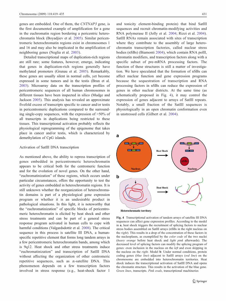
and tonicity element-binding protein) that bind SatIIIsequences and recruit chromatin-modifying activities andRNA polymerase II (Jolly et al. 2004; Rizzi et al. 2004).SatIII RNAs remain associated with sites of transcriptionwhere they contribute to the assembly of large hetero-chromatin transcription factories, called nuclear stressbodies (nSBs) (Biamonti 2004), which contain RNA polII,chromatin modifiers, and transcription factors along with aspecific subset of pre-mRNA processing factors. Thefunction of these structures is still a matter of investiga-tion. We have speculated that the formation of nSBs canaffect nuclear function and gene expression programsbecause the sequestration of transcription and RNAprocessing factors in nSBs can reduce the expression ofgenes in other nuclear districts. At the same time (asschematically proposed in Fig. 4), it may control theexpression of genes adjacent to arrays of SatIII repeats.Notably, a small fraction of the SatIII sequences isphysiologically in an open chromatin conformation evenin unstressed cells (Gilbert et al. 2004).
Fig. 4 Transcriptional activation of tandem arrays of satellite III DNAsequences can affect gene expression profiles. According to the modelin a, heat shock triggers the recruitment of splicing factors to nuclearstress bodies assembled on SatIII arrays (nSBs in the right nucleus onthe right). This results in a drop of the concentration of these factors inthe nucleoplasm, as exemplified by the color code of the two nuclei(heavy orange before heat shock and light pink afterwards). Thedecreased level of splicing factors can modify the splicing program ofgenes: exon inclusion in the nucleus on the left and exon skipping inthe nucleus on the right. Model b: Under normal conditions, proteincoding genes (blue line) adjacent to SatIII arrays (red line) on thechromosome are embedded into heterochromatin territories. Heatshock induces the transcriptional activation of SatIII DNA and opensthe chromatin structure. This results in the activation of the blue gene.Green lines, transcripts. Pink ovals, transcriptional machineries
Chromosoma (2009) 118:419–435 431

Intriguingly, the expression of SatIII RNAs increases inprogeroid laminopathies (Shumaker et al. 2006). Lamin Aand B are structural components of a protein meshwork, thenuclear lamina, which underlies the inner nuclear mem-brane. More than 12 human diseases arise from mutationsin the lamin A/C genes, among which the premature agingdisorders Hutchinson–Gilford progeria syndrome (HGPS).A distinctive feature of progeroid laminopathies is the lossof peripheral heterochromatin, which is accompanied byloss of heterochromatin markers such as H3K9me3 and analtered transcription profile (Scaffidi and Misteli 2005;Columbaro et al. 2005).
Lamins are implicated in the structural integrity of thenucleus; nuclei from mouse LmnA-null cells are mechan-ically weak (Lammerding et al. 2004), and cells that lackA-type lamins have mechanotransduction defects that leadto misregulation of mechanosensitive genes (Stewart et al.2007). This is probably linked to increased sensitivity tostress of HGPS cells (Caron et al. 2007). In this light, theactivation of SatIII arrays and adjacent genes in pericen-tromeric domains may be relevant for the clinical manifes-tation of laminopathies.
Conclusions
In this paper, we draw the attention to recent evidence ongenes found in constitutive heterochromatin in Drosophilaand other organisms. Constitutive heterochromatin forms asignificant fraction of metazoan genomes, which suggestsan evolutionary conserved function of this distinctivegenomic component. Despite persisting fragmentary knowl-edge, accumulating data, summarized in this review,confirm that idea and begin to unveil novel aspects ofeukaryotic genome organization with relevant implicationsfor function and evolution of constitutive heterochromatin.It is now clear that this peculiar genomic compartmentcontains a large variety of genetics elements. Essential andputative single-copy genes were identified in constitutiveheterochromatin of D. melanogaster, yeast, Arabidopsis,rice, and human genomes. Rather than mere euchromatinsequences embedded in a “junk DNA”, genes activelytranscribed in heterochromatin may turn out to be an aspectof a relevant evolutionary process where a given sequencemight have established positive interactions with hetero-chromatin environment. TE remnants, heterochromatinproteins, and specific histone modifications may haveplayed important roles in this phenomenon. In addition tosingle-copy genes, Drosophila heterochromatin is knownfor a long time to contain repetitive loci-like ribosomalgenes and a special class of “criptic” genetic elements suchas Su (Ste) and ABO. More recently, a novel class ofunconventional loci is given by the multiple PiRNAs and
esiRNA heterochromatin clusters, some of which areinvolved in transposable element silencing and heterochro-matin formation. These data, together with the notion thatthe size of heterochromatin genes can be very large,converge to conclude that the density of genetic functionsin constitutive heterochromatin is not as low as previouslyclaimed. The more we shed light on heterochromatin inDrosophila and in higher eukaryotes, the more we will besurprised by its peculiar functions and positive role on theevolution of the eukaryotic genomes. The next years willundoubtedly witness progress in this highly intriguinggenomic component.
Acknowledgements We are grateful to Patrizia Lavia for criticalreading of the manuscript and to Roger Hoskins and DHGP for sharinginformations on heterochromatin sequence and gene annotation. Wealso wish to thank three anonymous referees for helpful comments andsuggestions. The P. Dimitri laboratory was supported by grants fromIstituto Pasteur-Fondazione Cenci Bolognetti and National Institute ofHealth. G.Biamonti was supported by grants from AIRC and Cariplo.
References
Adams MD, Celniker SE, Holt RA, Evans CA, Gocayne JD,Amanatides PG et al (2000) The genome sequence of Drosophilamelanogaster. Science 287:2185–2195
Arabidopsis genome initiative (2000) Analysis of the genomesequence of the flowering plant Arabidopsis thaliana. Nature408:796–781
Aravin AA, Klenov MS, Vagin VV, Bantignies F, Cavalli G, GvozdevVA (2004) Dissection of a natural RNA silencing process in theDrosophila melanogaster germ line. Mol Cell Biol 24:6742–6750
Bernstein E, Allis CD (2005) RNA meets chromatin. Genes Dev19:1635–1655
Biamonti G (2004) Nuclear stress bodies: a heterochromatin affair?Nat Rev Mol Cell Biol 5:493–498
Biggs HW, Zavitz HK, Dikinson B, Van Der Straten A, Brunner D,Hafen E et al (1994) The Drosophila rolled locus encodes a MAPkinase required in the sevenless signal transduction pathway.EMBO J 13:1628–1635
Blower MD, Sullivan BA, Karpen GH (2002) Conserved organization ofcentromeric chromatin in flies and humans. Dev Cell 2:319–330
Boyadjiev SA, South ST, Radford CL, Patel A, Zhang G, Hur DJ,Thomas GH, Gearhart JP, Stetten G (2005) A reciprocaltranslocation 46, XY, t(8;9)(p11.2;q13) in a bladder exstrophypatient disrupts CNTNAP3 and presents evidence of a pericen-tromeric duplication on chromosome 9. Genomics 85:622–629
Bozzetti MP, Massari S, Finelli P, Meggio F, Pinna LA, Boldyreff B etal (1995) The Ste locus, a component of the parasitic cry-Stesystem of Drosophila melanogaster, encodes a protein that formscrystals in primary spermatocytes and mimics properties of thebeta subunit of casein kinase 2. PNAS 92:6067–6071
Brennecke J, Aravin AA, Stark A, Dus M, Kellis M, SachidanandamR, Hannon GJ (2007) Discrete small RNA-generating loci asmaster regulators of transposon activity in Drosophila. Cell128:1089–1103
Brennecke J, Malone CD, Aravin AA, Sachidanandam R, Stark A,Hannon GJ (2008) An epigenetic role for maternally inheritedpiRNAs in transposon silencing. Science 322:1387–1392
432 Chromosoma (2009) 118:419–435
Brosseau GE (1960) Genetic analysis of male fertility factors on the Ychromosomes of Drosophila melanogaster. Genetics 45:257–274
Brown SW (1966) Heterochromatin. Science 151:417–425Brun ME, Ruault M, Ventura M, Roizes G, De Sario A (2003)
Juxtacentromeric region of human chromosome 21: a boundarybetween centromeric heterochromatin and euchromatic chromo-some arms. Gene 312:41–50
Caron M, Auclair M, Donadille B, Bereziat V, Guerci B, Laville M,Narbonne H, Bodemer C, Lascols O, Capeau J, Vigouroux C(2007) Human lipodystrophies linked to mutations in A-typelamins and to HIV protease inhibitor therapy are both associatedwith prelamin A accumulation, oxidative stress and prematurecellular senescence. Cell Death Differ 14:1759–1767
Carvalho AB, Dobo BA, Vibranovski MD, Clark AG (2001) Identifica-tion of five new genes on the Y chromosome of Drosophilamelanogaster. Proc Natl Acad Sci U S A 98:13225–13230
Castillo-Davis CI, Mekhedov SL, Hartl DL, Koonin EV, KondrashovFA (2002) Selection for short introns in highly expressed genes.Nat Genet 31:415–418
Cenci G, Belloni G, Dimitri P (2003) 1(2) 41Aa, a heterochromaticgene of Drosophila melanogaster, is required for mitotic andmeiotic chromosome condensation. Genet Res 81:15–24
Ciani L, Krylova O, Smalley MJ, Dale TC, Salinas PC (2004) Adivergent canonical WNT-signaling pathway regulates microtu-bule dynamics: dishevelled signals locally to stabilize micro-tubules. J Cell Biol 164:243–253
Clegg NJ, Honda BM, Whitehead IP, Grigliatti TA, Wakimoto B,Brock HW et al (1998) Suppressors of position-effect variegationin Drosophila melanogaster affect expression of the heterochro-matic gene light in the absence of a chromosome rearrangement.Genome 41:495–503
Columbaro M, Capanni C, Mattioli E, Novelli G, Parnaik VK,Squarzoni S, Maraldi NM, Lattanzi G (2005) Rescue ofheterochromatin organization in Hutchinson–Gilford progeriaby drug treatment. Cell Mol Life Sci 62:2669–2678
Corradini N, Rossi F, Vernì F, Dimitri P (2003) FISH analysis ofDrosophila heterochromatin using BACs and P-elements. Chro-mosoma 112:26–37
Coulthard AB, Eberl DF, Sharp CB, Hilliker AJ (2003) Geneticanalysis of the second chromosome centromeric heterochromatinof Drosophila melanogaster. Genome 46:343–352
Cryderman DE, Grade SK, Li Y, Fanti L, Pimpinelli S, Wallrath LL(2005) Role of Drosophila HP1 in euchromatic gene expression.Dev Dyn 232:767–774
Czech B, Malone CD, Zhou R, Stark A, Schlingeheyde C, Dus M,Perrimon N, Kellis M, Wohlschlegel JA, Sachidanandam R,Hannon GJ, Brennecke J (2008) An endogenous small interferingRNA pathway in Drosophila. Nature 453:798–802
Desset S, Meignin C, Dastugue B, Vaury C (2003) COM, aheterochromatic locus governing the control of independentendogenous retroviruses from Drosophila melanogaster. Genetics164:501–509
De Wit E, Greil F, van Steensel B (2005) Genome-wide HP1 bindingin Drosophila: developmental plasticity and genomic targetingsignals. Genome Res 15:1265–1273
De Wit E, Greil F, van Steensel B (2007) High-resolution mappingreveals links of HP1 with active and inactive chromatincomponents. PLoS Genet 2007:346–357
Dernburg AF, Sedat JW, Hawley RS (1996) Direct evidence of a rolefor heterochromatin in meiotic chromosome segregation. Cell86:135–146
Devlin RH, Bingham B, Wakimoto BT (1990a) The organization andexpression of the light gene, a heterochromatic gene ofDrosophila melanogaster. Genetics 125:129–140
Devlin RH, Holm DG, Morin KR, Honda BM (1990b) Identifyingsingle-copy DNA sequence associated with the expression of a
heterochromatic gene, the light locus of Drosophila melanogaster.Genome 33:405–415
Diekwisch TG, Marches F, Williams A, Luan X (1999) Cloning, geneexpression, and characterization of CP27, a novel gene in mouseembryogenesis. Gene 235:19–30
Dimitri P (1991) Cytogenetic analysis of the second chromosomeheterochromatin of Drosophila melanogaster. Genetics 127:553–564
Dimitri P, Junakovic N (1999) Revising the selfish DNA hypothesis:new evidence on accumulation of transposable elements inheterochromatin. Trends Genet 15:123–124
Dimitri P, Arcà B, Berghella L, Mei E (1997) High genetic instabilityof heterochromatin after transposition of the LINE-like I factor inDrosophila melanogaster. Proc Natl Acad Sci U S A 94:8052–8057
Dimitri P, Junakovic N, Arcà B (2003) Colonization of heterochro-matic genes by transposable elements in Drosophila. Mol BiolEvol 20:503–512
Dimitri P, Corradini N, Rossi F, Vernì F (2005a) The paradox offunctional heterochromatin. Bioessays 27:29–41
Dimitri P, Vernì F, Mei E, Rossi F, Corradini N (2005b) Transposableelements as artisans of the heterochromatic genome. CytogenetGenome Res 110:165–172
Eberl D, Duyf BJ, Hilliker AH (1993) The role of heterochromatin inthe expression of a heterochromatic gene, the rolled gene ofDrosophila melanogaster. Genetics 134:277–292
Eichler EE, Lu F, Shen Y, Antonacci R, Jurecic V, Doggett NA,Moyzis RK, Baldini A, Gibbs RA, Nelson DL (1996) Duplica-tion of a gene-rich cluster between 16p11.1 and Xq28: a novelpericentromeric-directed mechanism for paralogous genomeevolution. Hum Mol Genet 5:899–912
Eissenberg JC, Hilliker AJ (2000) Versatility of conviction: hetero-chromatin as both repressor and an activator of transcription.Genetica 109:19–24
Elgin SCR (1996) Heterochromatin and gene regulation in Drosoph-ila. Curr Opin Genet Dev 6:193–200
Fanti L, Perrini B, Piacentini L, Berloco M, Marchetti E, Palumbo G,Pimpinelli S (2008) The trithorax group and Pc group proteinsare differentially involved in heterochromatin formation inDrosophila. Chromosoma 117:25–39
Fitzpatrick KA, Sinclair DA, Schulze SR, Syrzycka M, Honda BM(2005) A genetic and molecular profile of third chromosome centricheterochromatin in Drosophila melanogaster. Genome 48:571–584
Fly Base 2009 (http://flybase.org/)Fukagawa T, Nogami M, Yoshikawa M, Ikeno M, Okazaki TY et al
(2004) Dicer is essential for formation of the heterochromatinstructure in vertebrate cells. Nat Cell Biol 6:784–781
Gatti M, Pimpinelli S (1983) Cytological and genetical analysis of theY chromosome of Drosophila melanogaster. Chromosoma88:349–373
Gatti M, Pimpinelli S (1992) Functional elements in Drosophilamelanogaster heterochromatin. Annu Rev Genet 26:239–275
Gause M, Eissenberg JC, Macrae AF, Dorsett M, Misulovin Z, DorsettD (2006) Nipped-A, the Tra1/TRRAP subunit of the DrosophilaSAGA and Tip60 complexes, has multiple roles in Notch signalingduring wing development. Mol Cell Biol 26:2347–2359
Gepner J, Hays TS (1993) A fertility region on the Y chromosome ofDrosophila melanogaster encodes a dynein microtubule motor.Proc Natl Acad Sci U S A 90:11132–11136
Ghildiyal M, Seitz H, Horwich MD, Li C, Du T, Lee S, Xu J, KittlerEL, Zapp ML, Weng Z, Zamore PD (2008) Endogenous siRNAsderived from transposons and mRNAs in Drosophila somaticcells. Science 320:1077–1081
Gilbert N, Boyle S, Fiegler H, Woodfine K, Carter N, Bickmore WA(2004) Chromatin architecture of the human genome: gene-richdomains are enriched in open chromatin fibers. Cell 118:555–566
Chromosoma (2009) 118:419–435 433
Greil F, van der Kraan I, Delrow J, Smothers JF, de Wit E,Bussemaker HJ et al (2003) Distinct HP1 and Su(var) 3-9complexes bind to sets of developmentally coexpressed genesdepending on chromosomal location. Genes Dev 17:2825–2838
Grunau C, Sanchez C, Ehrlich M, van der Bruggen P, Hindermann W,Rodriguez C, Krieger S, Dubeau L, Fiala E, De Sario A (2005)Frequent DNA hypomethylation of human juxtacentromericBAGE loci in cancer. Genes Chromosomes Cancer 43:11–24
Hall IM, Shankaranarayana GD, Noma K, Ayoub N, Cohen A, GrewalSI (2002) Establishment and maintenance of a heterochromatindomain. Science 297:2232–2237
Heitz E (1928) Das heterochromatin der Moose. Jb Wiss Bot 69:762–818
Henikoff S, Ahmad K, Malik HS (2001) The centromere paradox:stable inheritance with rapidly evolving DNA. Science293:1098–1102
Hilliker AJ (1976) Genetic analysis of the centromeric heterochroma-tin of chromosome 2 of Drosophila melanogaster: deficiencymapping of EMS-induced lethal complementation groups. Ge-netics 83:765–782
Hilliker AJ, Appels R, Schalet A (1980) The genetic analysis of D.melanogaster heterochromatin. Cell 21:607–619
Horvath JE, Schwartz S, Eichler EE (2000) The mosaic structureof human pericentromeric DNA: a strategy for characterizingcomplex regions of the human genome. Genome Res 10:839–852
Hoskins RA, Smith CD, Carlson JW, Carvalho AB, Halpern A,kaminker JS et al (2002) Heterochromatic sequences in aDrosophila whole-genome shotgun assembly. Genome Biology3:research0085.1–0085.16
Hoskins RA, Carlson JW, Kennedy C, Acevedo D, Evans-Holm M,Frise E, Wan KH, Park S, Mendez-Lago M, Rossi F, VillasanteA, Dimitri P, Karpen GH, Celniker SE (2007) Sequence finishingand mapping of Drosophila melanogaster heterochromatin.Science 316:1625–1628
Huisinga KL, Elgin SC (2009) Small RNA- directed heterochromatinformation in the context of development: what flies might learnfrom fission yeast. Biochim Biophys 1789:3–16
Huisinga KL, Brower-Toland B, Elgin SC (2006) The contradictorydefinitions of heterochromatin: transcription and silencing.Chromosoma 115:110–122
Inoue YH, Glover DM (1998) Involvement of the rolled/MAP kinasegene in Drosophila mitosis: interaction between genes for theMAP kinase cascade and abnormal spindle. Mol Gen Genet258:334–341
John B (1988) The biology of heterochromatin. In: Verma RS (ed)Heterochromatin: molecular and structural aspects. CambridgeUniversity Press, Cambridge, pp 1–128
Jolly C, Metz A, Govin J, Vigneron M, Turner BM, Khochbin S,Vourc’h C (2004) Stress-induced transcription of satellite IIIrepeats. J Cell Biol 164:25–33
Karpen GH, Le MG, Le H (1996) Centric heterochromatin and theefficiency of achiasmate disjunction in Drosophila femalemeiosis. Science 273:118–122
Koryakov DE, Zhimulev IF, Dimitri P (2002) Cytogenetic analysis ofthe third chromosome heterochromatin of Drosophila mela-nogaster. Genetics 160:509–517
Krantz ID, McCallum J, De Scipio C, Kaur M, Gillis LA, Yaeger D etal (2004) Cornelia de Lange syndrome is caused by mutations inNIPBL, the human homolog of Drosophila melanogaster Nipped-B. Nat Genet 36:631–635
Kurek RA, Reugels M, Lammermann U, Buenemann H (2000)Molecular aspects of intron evolution in dynein encoding mega-genes on the heterochromatic Y chromosome of Drosophila sp.Genetica 109:113–123
Lam AL, Boivin CD, Bonney CF, Rudd MK, Sullivan BA (2006)Human centromeric chromatin is a dynamic chromosomaldomain that can spread over noncentromeric DNA. Proc NatlAcad Sci U S A 103:4186–4191
Lammerding J, Schulze PC, Takahashi T, Kozlov S, Sullivan T,Kamm RD, Stewart CL, Lee RT (2004) Lamin A/C deficiencycauses defective nuclear mechanics and mechanotransduction. JClin Invest 113:370–378
Lehnertz B, Ueda Y, Derijck AHA, Braunschweig U, Perez-Burgos L,Kubicek S et al (2003) Suv39h-mediated histone H3 lysine 9methylation directs DNA methylation to major satellite repeats atpericentric heterochromatin. Curr Biol 13:1192–1200
Litvak KJ (1984) Organization and mapping of a sequence on theDrosophila melanogaster X and Y chromosomes that is tran-scribed during spermatogenesis. Genetics 107:611–634
Lohe AR, Hilliker AJ, Roberts PA (1993) Mapping simple repeatedDNA sequences in heterochromatin of Drosophila melanogaster.Genetics 134:1149–1174
Lu BY, Emtage PC, Duyf BJ, Hilliker AJ, Eissenberg JC (2000)Heterochromatin protein 1 is required for the normal expression oftwo heterochromatin genes in Drosophila. Genetics 155:699–708
Marchant GE, Holm DG (1988) Genetic analysis of the heterochro-matin of chromosome 3 in Drosophila melanogaster. II. Vital lociidentified through EMS mutagenesis. Genetics 120:519–532
Marygold SJ, Coelho CM, Leevers SJ (2005) Genetic analysis ofRpL38 and RpL5, two minute genes located in the centricheterochromatin of chromosome 2 of Drosophila melanogaster.Genetics 169:683–695
Misulovin Z, Schwartz YB, Li XY, Kahn TG, Gause M, MacArthur S,Fay JC, Eisen MB, Pirrotta V, Biggin MD, Dorsett D (2008)Association of cohesin and Nipped-B with transcriptionallyactive regions of the Drosophila melanogaster genome. Chromo-soma 117:89–102
Moritz KB, Roth GE (1976) Complexity of germline and somaticDNA in Ascaris. Nature 259:55–57
Mudge JM, Jackson MS (2005) Evolutionary implications ofpericentromeric gene expression in humans. Cytogenet GenomeRes 108:47–57
Myster SH, Wang F, Cavallo R, Christian W, Bhotika S, Anderson CT,Peifer M (2004) Genetic and bioinformatic analysis of 41C andthe 2R heterochromatin of Drosophila melanogaster: a windowon the heterochromatin–euchromatin junction. Genetics166:807–822
Neglia M, Bertoni L, Zoli W, Giulotto E (2003) Amplification ofthe pericentromeric region of chromosome 1 in a newlyestablished colon carcinoma cell line. Cancer Genet Cytogenet142:99–106
Palumbo G, Berloco M, Fanti L, Bozzetti MP, Massari S, Caizzi R,Caggese C, Spinelli L, Pimpinelli S (1994) Interaction systemsbetween heterochromatin and euchromatin in Drosophila mela-nogaster. Genetica 94:267–74
Parks S, Wieschaus E (1991) The Drosophila gastrulation geneconcertina encodes a Ga-like protein. Cell 64:447–458
Partridge JF, Borgstrom B, Allshire RC (2000) Distinct proteininteraction domains and protein spreading in a complex centro-mere. Genes Dev 14:783–791
Peterson DG, Pearson WR, Stack SM (1998) Characterization of thetomato (Lycopersicon esculentum) genome using in vitro and insitu DNA reassociation. Genome 41:346–356
Pimpinelli S, Dimitri P (1989) Cytogenetic analysis of segregationdistortion in drosophila melanogaster: the cytological organiza-tion of the responder (Rsp) locus. Genetics 121:765–772
Pimpinelli S, Bonaccorsi S, Gatti M, Sandler L (1985) The peculiargenetic organization of Drosophila heterochromatin. TrendsGenet 2:17–20
434 Chromosoma (2009) 118:419–435
Pimpinelli S, Berloco M, Fanti L, Dimitri P, Bonaccorsi S, MarchettiE et al (1995) Transposable elements are stable structuralcomponents of Drosophila melanogaster heterochromatin. ProcNatl Acad Sci U S A 92:3804–3808
Plath K, Mlynarczyk-Evans S, Nusinov DA, Panning B (2002) XistRNA and the mechanism of X chromosome inactivation. AnnuRev Genet 36:233–278
Prud’homme N, Gans M, Masson M, Terzian C, Bucheton A (1995)Flamenco, a gene controlling the gypsy retrovirus of Drosophilamelanogaster. Genetics 139:697–711
Rasoly RS, Robbins LG (1991) Rex and suppressor of Rexarerepeated neomorphic loci in the Drosophila melanogasterribosomal DNA. Genetics 129:119–132
Reinhart BJ, Bartel DP (2002) Small RNAs correspond to centromereheterochromatic repeats. Science 297:1831
Reiter LT, Potocki L, Chien S, Gribskov M, Bier E (2001) Asystematic analysis of human disease-associated gene sequencesin Drosophila melanogaster. Genome Res 11:1114–11125
Ritossa FM, Spiegelman S (1965) Localization of DNA complemen-tary to ribosomal RNA in the nucleolus organizer region ofDrosophila melanogaster. PNAS 53:737–745
Rizzi N, Denegri M, Chiodi I, Corioni M, Valgardsdottir R, CobianchiF, Riva S, Biamonti G (2004) Transcriptional activation of aconstitutive heterochromatic domain of the human genome inresponse to heat shock. Mol Biol Cell 15:543–551
Rollins RA, Morcillo P, Dorsett D (1999) Nipped-B, a Drosophilahomologue of chromosomal adherins, participates in activationby remote enhancers in the cut and Ultrabithorax genes. Genetics152:577–593
Rossi F, Moschetti R, Caizzi R, Corradini N, Dimitri P (2007)Cytogenetic and molecular characterization of heterochromatingene models in Drosophila melanogaster. Genetics 175:595–607
Scaffidi P, Misteli T (2005) Reversal of the cellular phenotype in thepremature aging disease Hutchinson–Gilford progeria syndrome.Nat Med 11:440–445
She X, Horvath JE, Jiang Z, Liu G, Furey TS, Christ L, Clark R,Graves T, Gulden CL, Alkan C et al (2004) The structure andevolution of centromeric transition regions within the humangenome. Nature 430:857–864
Schulze SR, Sinclair DA, Fitzpatrick KA, Honda BM (2005) Agenetic and molecular characterization of two proximal hetero-chromatic genes on chromosome 3 of Drosophila melanogaster.Genetics 169:2165–2177
Shimada Y, Yonemura S, Ohkura H, Strutt D, Uemura T (2006)Polarized transport of Frizzled along the planar microtubulearrays in Drosophila wing epithelium. Dev Cell 10:209–222
Shumaker DK, Dechat T, Kohlmaier A, Adam SA, Bozovsky MR,Erdos MR, Eriksson M, Goldman AE, Khuon S, Collins FS et al(2006) Mutant nuclear lamin A leads to progressive alterations ofepigenetic control in premature aging. Proc Natl Acad Sci U S A103:8703–8708
Slawson EE, Shaffer CD, Malone CD, Leung W, Kellmann E,Shevchek RB, Craig CA, Bloom SM, 2nd Bogenpohl J, Dee J,Morimoto ET, Myoung J, Nett AS, Ozsolak F, Tittiger ME, ZeugA, Pardue ML, Buhler J, Mardis ER, Elgin SC (2006)Comparison of dot chromosome sequences from D. melanogasterand D. virilis reveals an enrichment of DNA transposonsequences in heterochromatic domains. Genome Biol 7:R15
Smith CD, Yandell M, Edgar RC, Kennedy C, Carlson J et al (2005)The Drosophila Heterochromatin Genome Project (DHGP):genes and repeat annotation. Seventh International Conferenceon Drosophila Heterochromatin. Gubbio, Italy
Smith CD, Shu S, Mungall CJ, Karpen GH (2007) The Release 5.1annotation of Drosophila melanogaster heterochromatin. Science316:1586–1591
Stankiewicz P, Lupski JR (2002) Genome architecture, rearrangementsand genomic disorders. Trends Genet 18:74–82
Stewart CL, Roux KJ, Burke B (2007) Blurring the boundary: thenuclear envelope extends its reach. Science 318:1408–1412
Sullivan BA, Karpen GH (2004) Centromeric chromatin exhibits ahistone modification pattern that is distinct from both euchroma-tin and heterochromatin. Nat Struct Mol Biol 11:1076–1083
Tonkin ET, Wang TJ, Lisgo S, Bamshad MJ, Strachan T (2004)NIPBL, encoding a homolog of fungal Scc2-type sister chroma-tid cohesion proteins and fly Nipped-B, is mutated in Cornelia deLange syndrome. Nat Genet 36:636–641
Tulin A, Stewart D, Spradling AC (2002) The Drosophila heterochro-matic gene encoding poly (ADP-ribose) polymerase (PARP) isrequired to modulate chromatin structure during development.Genes Dev 16:2108–2119
Valgardsdottir R, Chiodi I, Giordano M, Rossi A, Bazzini S, GhignaC, Riva S, Biamonti G (2008) Transcription of satellite III non-coding RNAs is a general stress response in human cells. NucleicAcids Res 36:423–434
Verdel A, Jia S, Gerber S, Sugiyama T, Gygi S, Grewal SI, Moazed D(2004) RNAi-mediated targeting of heterochromatin by the RITScomplex. Science 303:672–676
Villasante A, Mendéz-Lago M, Abad JP, Montejo de Garcìni E (2007)The birth of the centromere. Cell Cycle 6:2872–2876
Volpe TA, Kidner C, Hall IM, Teng G, Grewal SI, Mrtienssen RA(2002) Regulation of heterochromatic silencing and histone H3lysine-9 methylation by RNAi. Science 297:1833–1837
Wakimoto BT, Hearn MG (1990) The effects of chromosomerearrangements on the expression of heterochromatic genes inchromosome 2L of D. melanogaster. Genetics 125:141–154
Warner TS, Sinclair DA, Fitzpatrick KA, Singh M, Devlin RH, HondaBM (1998) The light gene of Drosophila melanogaster encodes ahomologue of VPS41, a yeast gene involved in cellular-proteintrafficking. Genome 41:236–243
Weiler KS, Wakimoto BT (1995) Heterochromatin and gene expres-sion in Drosophila. Annu Rev Genet 29:577–605
Williams SM, Robbins LG (1992) Molecular genetic analysis ofDrosophila rRNA arrays. Trends Genet 8:335–340
Yasuhara JC, Wakimoto BT (2006) Oxymoron no more: theexpanding world of heterochromatin. Trends Genet 22:330–338
Yasuhara JC, Wakimoto BT (2008) Molecular landscape of modifiedhistones in Drosophila heterochromatic genes and euchromatin–heterochromatin transition zones. PLoS Genet 4:159–172
Yasuhara JC, Marchetti M, Fanti L, Pimpinelli S, Wakimoto BT(2003) A strategy for mapping the heterochromatin of chromo-some 2 of Drosophila melanogaster. Genetica 117:217–226
Yasuhara JC, DeCrease CH, Wakimoto BT (2005) Evolution ofheterochromatic genes of Drosophila. Proc Natl Acad Sci U S A102:10958–10963
Yin H, Lin H (2007) An epigenetic activation role of Piwi and a Piwi-associated piRNA in Drosophila melanogaster. Nature 450:304–308
Chromosoma (2009) 118:419–435 435