Embed Size (px)
Citation preview
R E S EARCH ART I C L E
OBES I TY
Conserved Shifts in the Gut Microbiota Due to GastricBypass Reduce Host Weight and AdiposityAlice P. Liou,1 Melissa Paziuk,1 Jesus-Mario Luevano Jr.,2 Sriram Machineni,1
Peter J. Turnbaugh,2* Lee M. Kaplan1*
http://stmD
ownloaded from
Roux-en-Y gastric bypass (RYGB) results in rapid weight loss, reduced adiposity, and improved glucose metab-olism. These effects are not simply attributable to decreased caloric intake or absorption, but the mechanismslinking rearrangement of the gastrointestinal tract to these metabolic outcomes are largely unknown. Studiesin humans and rats have shown that RYGB restructures the gut microbiota, prompting the hypothesis thatsome of the effects of RYGB are caused by altered host-microbial interactions. To test this hypothesis, we useda mouse model of RYGB that recapitulates many of the metabolic outcomes in humans. 16S ribosomal RNA genesequencing of murine fecal samples collected after RYGB surgery, sham surgery, or sham surgery coupled to caloricrestriction revealed that alterations to the gut microbiota after RYGB are conserved among humans, rats, and mice,resulting in a rapid and sustained increase in the relative abundance of Gammaproteobacteria (Escherichia) andVerrucomicrobia (Akkermansia). These changes were independent of weight change and caloric restriction, weredetectable throughout the length of the gastrointestinal tract, and were most evident in the distal gut, down-stream of the surgical manipulation site. Transfer of the gut microbiota from RYGB-treated mice to nonoperated,germ-free mice resulted in weight loss and decreased fat mass in the recipient animals relative to recipients ofmicrobiota induced by sham surgery, potentially due to altered microbial production of short-chain fatty acids.These findings provide the first empirical support for the claim that changes in the gut microbiota contribute toreduced host weight and adiposity after RYGB surgery.
.scien
by guest on May 16, 2018cem
ag.org/
INTRODUCTION
Roux-en-Y gastric bypass (RYGB) is a highly effective treatment forsevere obesity and type 2 diabetes, characterized by a marked and sus-tained loss of ~65 to 75% of excess body weight and fat mass (1). Ini-tially thought to be a mechanical procedure causing restriction andcalorie malabsorption, recent evidence suggests that RYGB alters thebasic physiology of energy balance and metabolism in weight-dependentand weight-independent ways (2). These changes include marked im-provement in glucose homeostasis before weight loss (3), increased se-cretion of gut hormones (4, 5), and alterations in energy expenditure(6–8); however, the mechanisms that drive these outcomes remain in-completely understood. It is likely that anatomical rearrangement ofthe gastrointestinal tract alters the composition of the luminal milieuand its interactions with the intestinal epithelium, which in turn af-fects downstream signaling pathways regulating host energy balanceand metabolism (9). A particular luminal factor, however, has yet tobe identified.
Of the several potential luminal components known to mediateenergy balance after gastric bypass surgery, including dietary nutrients,biliopancreatic secretions, and the gut microbiota, we focused on thepotential contribution of the microbiota to RYGB outcomes, partic-ularly due to its increasingly recognized role in regulating energybalance and metabolic function. The gut microbiota has been reportedto differ in community structure, gene content, and metabolic net-work organization between obese and lean individuals (10–13). Diets
1Obesity, Metabolism & Nutrition Institute and Gastrointestinal Unit, MassachusettsGeneral Hospital, Boston, MA 02114, USA. 2FAS Center for Systems Biology, HarvardUniversity, Cambridge, MA 02138, USA.*Corresponding author. E-mail: [email protected] (L.M.K.); [email protected] (P.J.T.)
www.Scien
differing in fat and sugar content can also affect this structure (14–17),favoring the growth of bacteria whose secreted factors or structuralcomponents contribute to the development of adiposity, insulin resist-ance, and other metabolic derangements (18, 19). Furthermore, colo-nization of germ-free mice with a gut microbiota from conventionallyraised mice has provided functional evidence that the gut microbiotacan modulate host phenotype by decreasing food intake and increas-ing adiposity (10, 20). These phenotypes are further enhanced in germ-free animals colonized with the cecal microbiota from obese donors(10, 17). Because the gut microbiota influences some of the same meta-bolic parameters as RYGB, we hypothesized that changes to the gutmicrobiota after RYGBmay contribute to some of the metabolic benefitsof this procedure.
Recently, RYGB has been reported to cause marked shifts in fecalmicrobial profiles in both humans and rats (9, 21, 22), but it remainsunclear to what degree these changes are caused by alterations in thegastrointestinal tract, by the surgical process itself, or by decreasedweight and/or caloric intake induced by this procedure. Furthermore,it is unknown how rapidly these changes in microbial ecology occur,how stable they are over time within a given individual, and if they arelimited to the distal gut (for example, feces). The microbial taxonomicgroups that are enriched after RYGB have been associated with decreasedadiposity and leptin levels in humans (21) and with changes in fecaland urinary metabolites in rats (9). To date, however, there has beenno empirical data to support the hypothesis that these RYGB-alteredmicrobial communities have a direct effect on improved metabolicoutcomes.
To address these questions, we turned to a recently developed mu-rine model of RYGB (23, 24) to (i) demonstrate that changes in the gutmicrobiota after gastric bypass surgery are conserved among humans,
ceTranslationalMedicine.org 27 March 2013 Vol 5 Issue 178 178ra41 1
R E S EARCH ART I C L E
rats, and mice; (ii) demonstrate that the underlying cause of much ofthe microbial response to surgery is due to the reconfiguration of thegastrointestinal tract; (iii) characterize the temporal and spatial changesin the gut microbiota after this operation; and (iv) through transplan-tation of the RYGB-associated gut microbiota into germ-free mice,show that this altered gut microbiota is sufficient to trigger decreasedhost weight and adiposity (see Fig. 1, A to C, for experimental design).
Dow
nloaded from
RESULTS
The effect of RYGB on adiposity in the mouse modelDiet-induced obese (DIO) C57BL/6J mice fed a high-fat diet (HFD)commonly develop obesity-related metabolic derangements, includingglucose intolerance, hyperglycemia, hyperinsulinemia, and insulinresistance (25). Within 3 weeks after RYGB, male C57BL/6J mice lost29 ± 1.9% of their initial body weight and remained at this lowerweight throughout the study period. In contrast, ad libitum–fed sham-operated (SHAM) animals regained their body weight within 2 to 3 weeksafter surgery (Fig. 2A; 32.8 ± 0.5 g versus 42.2 ± 2.1 g after 5 weeks;P = 0.007, Student’s t test). Weight-matched sham-operated (WMS)animals were fed ~25% fewer calories to match the weight of the RYGB
www.Scien
animals. As we have seen previously (23, 24), the decreased body weightafter RYGB reflected a preferential loss of fat mass and a preservationof lean mass compared to sham controls (Fig. 2B). Epididymal andretroperitoneal fat pad weight, the degree of hepatic steatosis, and livertriglyceride content were all reduced in both RYGB andWMS animalsrelative to SHAM animals (Fig. 2, C to E). Food intake was not dif-ferent between RYGB and SHAM controls (Fig. 2F); however, takinginto account the greater energy lost in the feces of RYGB animals (Fig.2G), the net energy intake in RYGB animals was intermediate to thatof SHAM and WMS controls (Fig. 2H and table S1). This finding in-dicates that the weight loss in response to RYGB is due both to a de-crease in net energy intake and to an increase in total energy expenditure.Increased energy expenditure after RYGB has been previously demon-strated in both rat and mouse models (6–8).
RYGB animals maintained on HFD exhibited improved plasma glu-cose and insulin levels at 15 weeks after surgery, compared to SHAManimals, mirroring the metabolic phenotype of normal chow (NC)–fed lean mice (table S2). Although weight loss by food restriction yieldedsimilar improvements in fasting plasma glucose and insulin levels, greaterimprovements in glucose tolerance and insulin sensitivity were seen inRYGB versus WMS animals (fig. S1), supporting human evidence thatRYGB has weight-independent effects on glucose metabolism (26).
by guest on May 16, 2018
http://stm.sciencem
ag.org/
Fig. 1. Schematic of experimental design. (A) DIO C57BL/6J mice fed a60% HFD underwent either RYGB or sham operations and 2-week recov-
the gastrointestinal tract. Segments representative of the RYGB anatomywere also collected in SHAM and WMS animals. (C) Design of microbiota
ery on liquid diet before returning to HFD (blue bars). Sham animals thathad successfully regained body weight within 3 weeks after surgery weredivided into an ad libitum–fed SHAM group or food-restricted to matchthe weight of the RYGB animals (WMS). Fresh fecal samples were col-lected preoperatively and weekly for 12 weeks after surgery for micro-biota analysis (red arrows). (B) Graphic of the RYGB anatomy and segmentscollected for luminal content and mucosal scrapings along the length of
transfer experiments of the cecal contents from a representative donoranimal from each group, depicted in (A), into germ-free mice (star), in-dicating timing of collection of fecal samples for microbiota analysis (redarrows), body weights (blue asterisks), and food intake (triangles). At theend of the colonization period, animals were fasted overnight (doublelines), and final body weights, serum metabolic parameters, and adiposityscores were obtained.
ceTranslationalMedicine.org 27 March 2013 Vol 5 Issue 178 178ra41 2
R E S EARCH ART I C L E
by guest on May 16, 2018
http://stm.sciencem
ag.org/D
ownloaded from
RYGB rapidly alters the distal gut microbiotaTo determine how RYGB affects the distal gut microbiota, we per-formed 16S ribosomal RNA (rRNA) gene sequencing on fecal samplescollected before and weekly for 3 months after intervention in theRYGB, SHAM, and WMS groups (Fig. 1A). Overall, we analyzed166 samples from 23 animals (109,015 ± 5842 16S rRNA gene se-quences per sample; table S3).
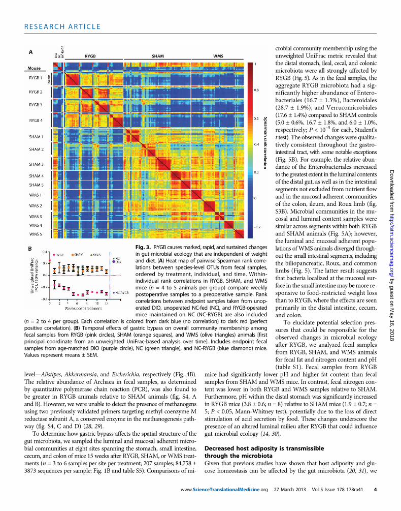
RYGB markedly altered the composition of the distal gut mi-crobiota as early as 1 week after surgery, a change that progressedover time and stabilized after 5 weeks (Fig. 3 and fig. S2). The shamprocedure also affected the fecal microbial communities but to asubstantially lesser extent than RYGB; furthermore, the differences inmicrobial ecology between the SHAM and WMS groups were mini-mal (Fig. 3 and fig. S2). These observations suggest that rearrange-ment of the gastrointestinal tract by RYGB had a substantiallygreater effect on the fecal microbiota than either food restriction–mediated weight loss or the limited intestinal disruption caused bythe sham procedure.
Additionally, we analyzed fecal samples at a single time point fromunoperated DIO, unoperated lean NC-fed mice, and RYGB-operatedmice maintained on NC (NC-RYGB). As expected, the abundance ofspecies-level operational taxonomic units (OTUs) in samples from DIOand lean animals was significantly different (Fig. 3A; Spearman corre-lation range was 0.58 to 0.84 and 0.16 to 0.24 for within-group versusbetween-group comparisons, respectively; P < 10−7, Student’s t test).However, the effect of RYGB on the gut microbiota was similar re-gardless of whether mice were fed NC or HFD (Fig. 3), suggesting thatthe impact of surgery can overwhelm even the well-recognized effectof dietary composition on the gut microbiota (14).
www.Scien
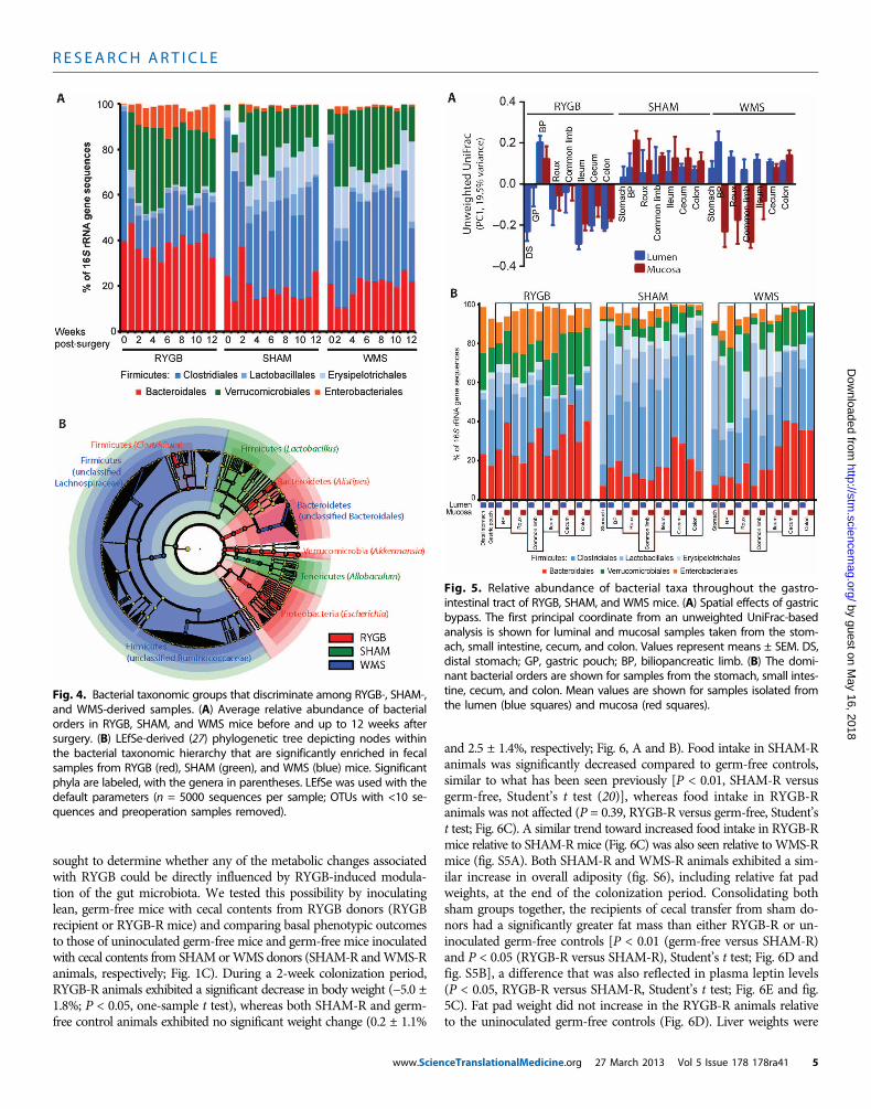
Weight loss after dietary restriction or RYGB was accompaniedby similar changes in the abundance of taxonomic groups within theFirmicutes phylum as a proportion of the total sequences observed inthe phylum. Compared to preoperative levels, both the RYGB andWMS microbiota were dominated by the order Clostridiales (78.9 ±2.7% and 72.9 ± 2.7% of Firmicutes 16S rRNA gene sequences, respec-tively), whereas the SHAM microbiota had a significantly greaterabundance of the orders Lactobacillales and Erysipelotrichales[42.5 ± 4.0% (SHAM), 27.1 ± 3.8% (WMS), and 21.1 ± 2.7% (RYGB);P < 0.01, SHAM versus WMS and SHAM versus RYGB, Student’s ttest; Fig. 4A and table S4]. In contrast, the relative abundance ofBacteroidales remained stable when comparing pre- and postoperativeabundances across all treatment groups over time (Fig. 4A).
RYGB induced several specific changes in the gut microbial ecol-ogy including an increased abundance in the Enterobacteriales withinthe first 2 weeks after surgery; there was no change in the Entero-bacteriales in either the SHAM or WMS groups (Fig. 4A and fig. S3A).There was also a significantly greater increase in Verrucomicrobialeswithin the first 2 weeks after RYGB (10,000-fold on average; P < 0.05,Student’s t test) compared to preoperative levels that was more markedthan that observed after either SHAM or WMS treatment (3-fold, P <0.05 for both; Fig. 4A). Using the linear discriminant analysis (LDA)effect size (LEfSe) (27) method, we identified more specific bacterialtaxa and species-level phylotypes whose relative abundance varied sig-nificantly among fecal samples taken from the RYGB, SHAM, andWMS treatment groups. This analysis revealed 50 discriminative fea-tures (LDA score >2; Fig. 4B and table S4). RYGB microbiotas wereenriched for three distinct taxonomic groups, evident from phylumlevel—Bacteroidetes, Verrucomicrobia, and Proteobacteria—to genus
Fig. 2. Phenotypic data from the RYGB mouse model. Character-istics of DIO C57BL/6J mice undergoing either RYGB (n = 11 to 17),sham operation (SHAM; n = 4 to 6), or sham operation with weight
matching to the RYGB group by food restriction (WMS; n = 5 to 6). (A) Bodyweight curves after surgery. (B) Body composition analysis. (C) Adiposityindex calculated from epididymal and retroperitoneal fat pad weights. (D)Liver triglyceride content. (E) Liver histology (×20). (F to H) One-weekcumulative (F) food intake, (G) fecal energy output, and (H) net energy intakein RYGB (n = 14), SHAM (n = 11), and WMS (n = 6) animals. Measurements for(B) to (E) were taken at the end of study, 15 weeks after surgery. Food intakeand energy output studies were performed 4 to 6 weeks after surgery. *P <0.05, **P < 0.01, ***P < 0.001, one-way analysis of variance (ANOVA) and posthoc Tukey test. Values represent means ± SEM.
ceTranslationalMedicine.org 27 March 2013 Vol 5 Issue 178 178ra41 3
R E S EARCH ART I C L E
by guest on May 16, 2018
http://stm.sciencem
ag.org/D
ownloaded from
level—Alistipes, Akkermansia, and Escherichia, respectively (Fig. 4B).The relative abundance of Archaea in fecal samples, as determinedby quantitative polymerase chain reaction (PCR), was also found tobe greater in RYGB animals relative to SHAM animals (fig. S4, Aand B). However, we were unable to detect the presence of methanogensusing two previously validated primers targeting methyl coenzyme Mreductase subunit A, a conserved enzyme in the methanogenesis path-way (fig. S4, C and D) (28, 29).
To determine how gastric bypass affects the spatial structure of thegut microbiota, we sampled the luminal and mucosal adherent micro-bial communities at eight sites spanning the stomach, small intestine,cecum, and colon of mice 15 weeks after RYGB, SHAM, or WMS treat-ments (n = 3 to 6 samples per site per treatment; 207 samples; 84,758 ±3873 sequences per sample; Fig. 1B and table S5). Comparisons of mi-
www.ScienceTranslationalMedicine.org
crobial community membership using theunweighted UniFrac metric revealed thatthe distal stomach, ileal, cecal, and colonicmicrobiota were all strongly affected byRYGB (Fig. 5). As in the fecal samples, theaggregate RYGB microbiota had a sig-nificantly higher abundance of Entero-bacteriales (16.7 ± 1.3%), Bacteroidales(28.7 ± 1.9%), and Verrucomicrobiales(17.6 ± 1.4%) compared to SHAM controls(5.0 ± 0.6%, 16.7 ± 1.8%, and 6.0 ± 1.0%,respectively; P < 10−5 for each, Student’st test). The observed changes were qualita-tively consistent throughout the gastro-intestinal tract, with some notable exceptions(Fig. 5B). For example, the relative abun-dance of the Enterobacteriales increasedto the greatest extent in the luminal contentsof the distal gut, as well as in the intestinalsegments not excluded from nutrient flowand in the mucosal adherent communitiesof the colon, ileum, and Roux limb (fig.S3B). Microbial communities in the mu-cosal and luminal content samples weresimilar across segments within both RYGBand SHAM animals (Fig. 5A); however,the luminal and mucosal adherent popu-lations ofWMS animals diverged through-out the small intestinal segments, includingthe biliopancreatic, Roux, and commonlimbs (Fig. 5). The latter result suggeststhat bacteria localized at the mucosal sur-face in the small intestinemay bemore re-sponsive to food-restricted weight lossthan to RYGB, where the effects are seenprimarily in the distal intestine, cecum,and colon.
To elucidate potential selection pres-sures that could be responsible for theobserved changes in microbial ecologyafter RYGB, we analyzed fecal samplesfrom RYGB, SHAM, and WMS animalsfor fecal fat and nitrogen content and pH(table S1). Fecal samples from RYGB
mice had significantly lower pH and higher fat content than fecalsamples from SHAM and WMS mice. In contrast, fecal nitrogen con-tent was lower in both RYGB and WMS samples relative to SHAM.Furthermore, pH within the distal stomach was significantly increasedin RYGB mice (3.8 ± 0.6; n = 8) relative to SHAMmice (1.9 ± 0.7; n =5; P < 0.05, Mann-Whitney test), potentially due to the loss of directstimulation of acid secretion by food. These changes underscore thepresence of an altered luminal milieu after RYGB that could influencegut microbial ecology (14, 30).
Decreased host adiposity is transmissiblethrough the microbiotaGiven that previous studies have shown that host adiposity and glu-cose homeostasis can be affected by the gut microbiota (20, 31), we
Fig. 3. RYGB causes marked, rapid, and sustained changesin gut microbial ecology that are independent of weightand diet. (A) Heat map of pairwise Spearman rank corre-lations between species-level OTUs from fecal samples,ordered by treatment, individual, and time. Within-individual rank correlations in RYGB, SHAM, and WMSmice (n = 4 to 5 animals per group) compare weeklypostoperative samples to a preoperative sample. Rankcorrelations between endpoint samples taken from unop-erated DIO, unoperated NC-fed (NC), and RYGB-operatedmice maintained on NC (NC-RYGB) are also included
(n = 2 to 4 per group). Each correlation is colored from dark blue (no correlation) to dark red (perfectpositive correlation). (B) Temporal effects of gastric bypass on overall community membership amongfecal samples from RYGB (pink circles), SHAM (orange squares), and WMS (olive triangles) animals [firstprincipal coordinate from an unweighted UniFrac-based analysis over time]. Includes endpoint fecalsamples from age-matched DIO (purple circle), NC (green triangle), and NC-RYGB (blue diamond) mice.Values represent means ± SEM.
27 March 2013 Vol 5 Issue 178 178ra41 4
R E S EARCH ART I C L E
by guest on May 16, 2018
http://stm.sciencem
ag.org/D
ownloaded from
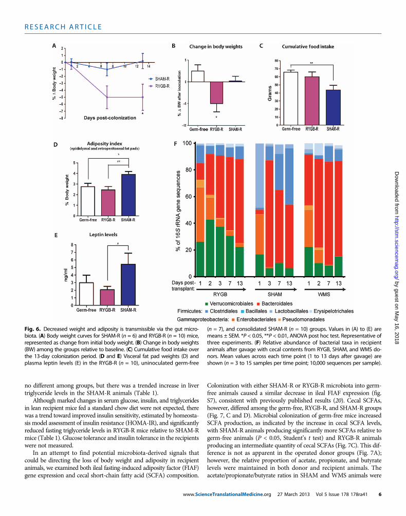
sought to determine whether any of the metabolic changes associatedwith RYGB could be directly influenced by RYGB-induced modula-tion of the gut microbiota. We tested this possibility by inoculatinglean, germ-free mice with cecal contents from RYGB donors (RYGBrecipient or RYGB-R mice) and comparing basal phenotypic outcomesto those of uninoculated germ-free mice and germ-free mice inoculatedwith cecal contents from SHAMorWMS donors (SHAM-R andWMS-Ranimals, respectively; Fig. 1C). During a 2-week colonization period,RYGB-R animals exhibited a significant decrease in body weight (−5.0 ±1.8%; P < 0.05, one-sample t test), whereas both SHAM-R and germ-free control animals exhibited no significant weight change (0.2 ± 1.1%
www.Scien
and 2.5 ± 1.4%, respectively; Fig. 6, A and B). Food intake in SHAM-Ranimals was significantly decreased compared to germ-free controls,similar to what has been seen previously [P < 0.01, SHAM-R versusgerm-free, Student’s t test (20)], whereas food intake in RYGB-Ranimals was not affected (P = 0.39, RYGB-R versus germ-free, Student’st test; Fig. 6C). A similar trend toward increased food intake in RYGB-Rmice relative to SHAM-Rmice (Fig. 6C) was also seen relative to WMS-Rmice (fig. S5A). Both SHAM-R and WMS-R animals exhibited a sim-ilar increase in overall adiposity (fig. S6), including relative fat padweights, at the end of the colonization period. Consolidating bothsham groups together, the recipients of cecal transfer from sham do-nors had a significantly greater fat mass than either RYGB-R or un-inoculated germ-free controls [P < 0.01 (germ-free versus SHAM-R)and P < 0.05 (RYGB-R versus SHAM-R), Student’s t test; Fig. 6D andfig. S5B], a difference that was also reflected in plasma leptin levels(P < 0.05, RYGB-R versus SHAM-R, Student’s t test; Fig. 6E and fig.5C). Fat pad weight did not increase in the RYGB-R animals relativeto the uninoculated germ-free controls (Fig. 6D). Liver weights were
Fig. 4. Bacterial taxonomic groups that discriminate among RYGB-, SHAM-,and WMS-derived samples. (A) Average relative abundance of bacterial
orders in RYGB, SHAM, and WMS mice before and up to 12 weeks aftersurgery. (B) LEfSe-derived (27) phylogenetic tree depicting nodes withinthe bacterial taxonomic hierarchy that are significantly enriched in fecalsamples from RYGB (red), SHAM (green), and WMS (blue) mice. Significantphyla are labeled, with the genera in parentheses. LEfSe was used with thedefault parameters (n = 5000 sequences per sample; OTUs with <10 se-quences and preoperation samples removed).Fig. 5. Relative abundance of bacterial taxa throughout the gastro-intestinal tract of RYGB, SHAM, and WMS mice. (A) Spatial effects of gastric
bypass. The first principal coordinate from an unweighted UniFrac-basedanalysis is shown for luminal and mucosal samples taken from the stom-ach, small intestine, cecum, and colon. Values represent means ± SEM. DS,distal stomach; GP, gastric pouch; BP, biliopancreatic limb. (B) The domi-nant bacterial orders are shown for samples from the stomach, small intes-tine, cecum, and colon. Mean values are shown for samples isolated fromthe lumen (blue squares) and mucosa (red squares).ceTranslationalMedicine.org 27 March 2013 Vol 5 Issue 178 178ra41 5
R E S EARCH ART I C L E
by guest on May 16, 2018
http://stm.sciencem
ag.org/D
ownloaded from
no different among groups, but there was a trended increase in livertriglyceride levels in the SHAM-R animals (Table 1).
Although marked changes in serum glucose, insulin, and triglyceridesin lean recipient mice fed a standard chow diet were not expected, therewas a trend toward improved insulin sensitivity, estimated by homeosta-sis model assessment of insulin resistance (HOMA-IR), and significantlyreduced fasting triglyceride levels in RYGB-R mice relative to SHAM-Rmice (Table 1). Glucose tolerance and insulin tolerance in the recipientswere not measured.
In an attempt to find potential microbiota-derived signals thatcould be directing the loss of body weight and adiposity in recipientanimals, we examined both ileal fasting-induced adiposity factor (FIAF)gene expression and cecal short-chain fatty acid (SCFA) composition.
www.Scien
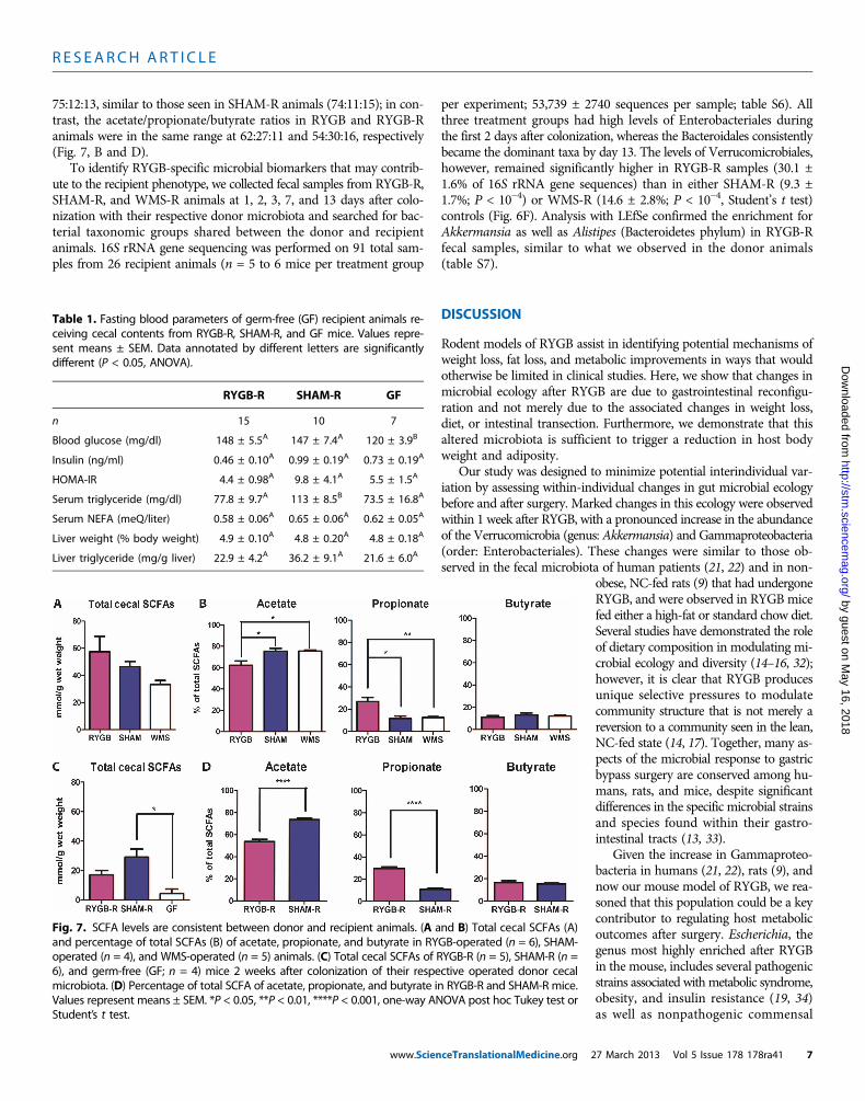
Colonization with either SHAM-R or RYGB-R microbiota into germ-free animals caused a similar decrease in ileal FIAF expression (fig.S7), consistent with previously published results (20). Cecal SCFAs,however, differed among the germ-free, RYGB-R, and SHAM-R groups(Fig. 7, C and D). Microbial colonization of germ-free mice increasedSCFA production, as indicated by the increase in cecal SCFA levels,with SHAM-R animals producing significantly more SCFAs relative togerm-free animals (P < 0.05, Student’s t test) and RYGB-R animalsproducing an intermediate quantity of cecal SCFAs (Fig. 7C). This dif-ference is not as apparent in the operated donor groups (Fig. 7A);however, the relative proportion of acetate, propionate, and butyratelevels were maintained in both donor and recipient animals. Theacetate/propionate/butyrate ratios in SHAM and WMS animals were
Fig. 6. Decreased weight and adiposity is transmissible via the gut micro-biota. (A) Body weight curves for SHAM-R (n = 6) and RYGB-R (n = 10) mice,
(n = 7), and consolidated SHAM-R (n = 10) groups. Values in (A) to (E) aremeans ± SEM. *P < 0.05, **P < 0.01, ANOVA post hoc test. Representative of
represented as change from initial body weight. (B) Change in body weights(BW) among the groups relative to baseline. (C) Cumulative food intake overthe 13-day colonization period. (D and E) Visceral fat pad weights (D) andplasma leptin levels (E) in the RYGB-R (n = 10), uninoculated germ-free
three experiments. (F) Relative abundance of bacterial taxa in recipientanimals after gavage with cecal contents from RYGB, SHAM, and WMS do-nors. Mean values across each time point (1 to 13 days after gavage) areshown (n = 3 to 15 samples per time point; 10,000 sequences per sample).
ceTranslationalMedicine.org 27 March 2013 Vol 5 Issue 178 178ra41 6
R E S EARCH ART I C L E
75:12:13, similar to those seen in SHAM-R animals (74:11:15); in con-trast, the acetate/propionate/butyrate ratios in RYGB and RYGB-Ranimals were in the same range at 62:27:11 and 54:30:16, respectively(Fig. 7, B and D).
To identify RYGB-specific microbial biomarkers that may contrib-ute to the recipient phenotype, we collected fecal samples from RYGB-R,SHAM-R, and WMS-R animals at 1, 2, 3, 7, and 13 days after colo-nization with their respective donor microbiota and searched for bac-terial taxonomic groups shared between the donor and recipientanimals. 16S rRNA gene sequencing was performed on 91 total sam-ples from 26 recipient animals (n = 5 to 6 mice per treatment group
www.Scien
per experiment; 53,739 ± 2740 sequences per sample; table S6). Allthree treatment groups had high levels of Enterobacteriales duringthe first 2 days after colonization, whereas the Bacteroidales consistentlybecame the dominant taxa by day 13. The levels of Verrucomicrobiales,however, remained significantly higher in RYGB-R samples (30.1 ±1.6% of 16S rRNA gene sequences) than in either SHAM-R (9.3 ±1.7%; P < 10−4) or WMS-R (14.6 ± 2.8%; P < 10−4, Student’s t test)controls (Fig. 6F). Analysis with LEfSe confirmed the enrichment forAkkermansia as well as Alistipes (Bacteroidetes phylum) in RYGB-Rfecal samples, similar to what we observed in the donor animals(table S7).
http://stm.sciencem
ag.oD
ownloaded from
DISCUSSION
Rodent models of RYGB assist in identifying potential mechanisms ofweight loss, fat loss, and metabolic improvements in ways that wouldotherwise be limited in clinical studies. Here, we show that changes inmicrobial ecology after RYGB are due to gastrointestinal reconfigu-ration and not merely due to the associated changes in weight loss,diet, or intestinal transection. Furthermore, we demonstrate that thisaltered microbiota is sufficient to trigger a reduction in host bodyweight and adiposity.
Our study was designed to minimize potential interindividual var-iation by assessing within-individual changes in gut microbial ecologybefore and after surgery. Marked changes in this ecology were observedwithin 1 week after RYGB, with a pronounced increase in the abundanceof the Verrucomicrobia (genus: Akkermansia) and Gammaproteobacteria(order: Enterobacteriales). These changes were similar to those ob-served in the fecal microbiota of human patients (21, 22) and in non-
ceTranslationalMedicine.org 2
by guest on May 16, 2018
rg/
obese, NC-fed rats (9) that had undergoneRYGB, and were observed in RYGB micefed either a high-fat or standard chow diet.Several studies have demonstrated the roleof dietary composition in modulating mi-crobial ecology and diversity (14–16, 32);however, it is clear that RYGB producesunique selective pressures to modulatecommunity structure that is not merely areversion to a community seen in the lean,NC-fed state (14, 17). Together, many as-pects of the microbial response to gastricbypass surgery are conserved among hu-mans, rats, and mice, despite significantdifferences in the specific microbial strainsand species found within their gastro-intestinal tracts (13, 33).Given the increase in Gammaproteo-bacteria in humans (21, 22), rats (9), andnow our mouse model of RYGB, we rea-soned that this population could be a keycontributor to regulating host metabolicoutcomes after surgery. Escherichia, thegenus most highly enriched after RYGBin the mouse, includes several pathogenicstrains associated withmetabolic syndrome,obesity, and insulin resistance (19, 34)as well as nonpathogenic commensal
Table 1. Fasting blood parameters of germ-free (GF) recipient animals re-ceiving cecal contents from RYGB-R, SHAM-R, and GF mice. Values repre-sent means ± SEM. Data annotated by different letters are significantlydifferent (P < 0.05, ANOVA).
RYGB-R
SHAM-R GFn
15 10 7Blood glucose (mg/dl)
148 ± 5.5A 147 ± 7.4A 120 ± 3.9BInsulin (ng/ml)
0.46 ± 0.10A 0.99 ± 0.19A 0.73 ± 0.19AHOMA-IR
4.4 ± 0.98A 9.8 ± 4.1A 5.5 ± 1.5ASerum triglyceride (mg/dl)
77.8 ± 9.7A 113 ± 8.5B 73.5 ± 16.8ASerum NEFA (meQ/liter)
0.58 ± 0.06A 0.65 ± 0.06A 0.62 ± 0.05ALiver weight (% body weight)
4.9 ± 0.10A 4.8 ± 0.20A 4.8 ± 0.18ALiver triglyceride (mg/g liver)
22.9 ± 4.2A 36.2 ± 9.1A 21.6 ± 6.0AFig. 7. SCFA levels are consistent between donor and recipient animals. (A and B) Total cecal SCFAs (A)and percentage of total SCFAs (B) of acetate, propionate, and butyrate in RYGB-operated (n = 6), SHAM-
operated (n = 4), and WMS-operated (n = 5) animals. (C) Total cecal SCFAs of RYGB-R (n = 5), SHAM-R (n =6), and germ-free (GF; n = 4) mice 2 weeks after colonization of their respective operated donor cecalmicrobiota. (D) Percentage of total SCFA of acetate, propionate, and butyrate in RYGB-R and SHAM-R mice.Values represent means ± SEM. *P < 0.05, **P < 0.01, ****P < 0.001, one-way ANOVA post hoc Tukey test orStudent’s t test.7 March 2013 Vol 5 Issue 178 178ra41 7
R E S EARCH ART I C L E
by guest on May 16, 2018
http://stm.sciencem
ag.org/D
ownloaded from
species that have been used as probiotic agents to prevent gastro-intestinal inflammatory conditions (35). It is possible that the specificEscherichia population enriched after RYGB may have some beneficialrole in driving host metabolic improvements after surgery.
The Verrucomicrobium Akkermansia was also substantially in-creased in RYGB mice and was maintained after transfer to germ-freerecipients. Given this observation, it is possible that Akkermansia mayhave a substantial role in regulating host adiposity and weight loss.Akkermansia can use mucus as a sole source of carbon and nitrogenin times of health and particularly in times of caloric restriction (36).This observation likely explains why Akkermansia is selectively in-creased in the mucus layer of WMS mice. The physiological relevanceof the overall increase in Akkermansia throughout the RYGB-alteredgastrointestinal tract, and how this may differ from the site-specificincreases seen in WMS animals, remains to be determined. Additionalstudies are needed to elucidate whether increased foraging on hostmucins or altered inflammation and insulin sensitivity in responseto Akkermansia (37, 38) contribute to the metabolic outcomes afterRYGB. In addition, we cannot discount the potential interactionsamong Enterobacteriales, Akkermansia, and other enriched bacterialgroups (such as Alistipes) in the RYGB model and how they maywork independently or interdependently to influence host metabol-ic improvements.
Microbial community structure is likely altered after RYGB throughrerouting of nutrients and biliopancreatic secretions. Although we didsee changes in fecal fat and pH profiles in the RYGB animal, thesechanges would be expected to promote the growth of Firmicutes(17, 30) rather than the observed decrease in Firmicutes after surgery.Therefore, the precise factors responsible for modulating microbialcommunities have yet to be defined.
Phenotypic characterization of germ-free recipient mice receiving amicrobiota transplant from RYGB-operated donors highlights the po-tential functional and metabolic contributions of the RYGB micro-biota. We found that RYGB-R animals had reduced body weight anddecreased fat deposition compared to SHAM-R and uninoculatedgerm-free controls. WMS-R animals shared similar body weight andadiposity phenotypes as the SHAM-R animals (fig. S5), likely due tothe similar cecal microbial profiles between the WMS and SHAM do-nors (Fig. 3C and fig. S3), suggesting that the effects of the RYGBmicrobiota on germ-free recipients are unique to this particular mi-crobial profile. This was a surprising observation because inoculationof the entire complex microbial communities (14, 19, 20) or specificmicroorganisms (39) from either lean or obese mice has previouslybeen shown to cause an increase in epididymal fat pad weight andoverall adiposity in the face of decreased food intake (10, 20). Al-though the degree of weight loss by the RYGB microbiota into leanrecipient mice is not as profound as the effect of RYGB in obese mice(5% versus 29% weight loss, respectively), these findings are consistentwith the hypothesis that alterations in the gut microbiota after RYGBat least partially modulate body weight and adiposity.
A decrease in adiposity and body weight without a change in foodintake suggests that the RYGB-associated microbiota may either re-duce the ability to harvest energy from the diet or produce signalsregulating energy expenditure and/or lipid metabolism. Previousstudies had identified ileal FIAF as a microbiota-influenced mecha-nism promoting lipid uptake into adipose tissue (20, 31). Colonizationof germ-free recipients with either a RYGB or SHAM microbiota re-sulted in equally reduced ileal FIAF expression despite the differences
www.Scien
in fat pad weight, suggesting that the effect of the RYGB microbiotaon metabolic outcomes is independent of FIAF.
SCFAs are by-products of microbial fermentation that affect hostphysiology in several ways, including serving as a primary energysource for colonocytes, substrates for lipid storage in adipose tissue,immune cell modulators, and molecules regulating lipid and glucosemetabolism and appetitive drives via G protein (heterotrimeric guaninenucleotide–binding protein)–coupled receptor (GPR41 and GPR43)activation and signaling (40, 41). Increased adiposity has been demon-strated in previously germ-free recipients of gut microbiota contents,likely due to increased availability of SCFAs (10, 17). Indeed, an in-creased production of SCFAs in SHAM-R animals relative to RYGB-Rand germ-free animals may explain the differences in adiposity amongthe groups. However, these observations do not explain the physiologicaldifferences among the RYGB, SHAM, and WMS groups, which do notexhibit significant differences in total cecal SCFA content.
It is possible that the different SCFAs affect host metabolic phys-iology in different ways. Both RYGB and RYGB-R animals had rela-tively greater propionate and lower acetate production than the WMS,SHAM, and SHAM-R groups, likely a consequence of the differentmicrobial profiles seen in the RYGB and RYGB-R animals (tablesS4 and S7) (42). The decreased adiposity in RYGB-R animals relativeto SHAM-R animals could be due to the reduced level of acetate avail-able for lipogenesis by adipose tissue and peripheral tissues (43). Fur-thermore, propionate is known to inhibit acetate conversion into lipidin the liver (44) and adipose tissue (45), and may contribute to thelowered adiposity and serum triglyceride levels and a trend toward de-creased hepatic triglyceride content. Propionate is also thought to in-hibit food intake through GPR41- and GPR43-induced secretion ofsatiety-regulating gastrointestinal hormones (46), although this is lesslikely given the lack of change in food intake in the RYGB-R animals.
Given the well-established effect of RYGB to increase energy ex-penditure (7, 8), we cannot discount the possibility that a reductionin body weight without a change in food intake is due to altered mi-crobial signaling that up-regulates host energy expenditure. Male micelacking GPR41 have reduced energy expenditure and increased adi-posity (47), suggesting that SCFAs regulate energy expenditure viaGPR41. Among the known SCFAs, propionate has the highest affinityfor GPR41, and its administration has been shown to increase sympa-thetic activity, resulting in elevated energy expenditure (48). Therefore,the increase in energy expenditure after RYGB could be mediated, inpart, through enhanced propionate activation of the GPR41 pathway.Another potential mechanism is differential microbial modification ofbile acids, which are also signaling molecules that can influence energyexpenditure (49). Additional studies are necessary to further clarify thespecific contributions of these various microbially influenced metabo-lites on changes in host energy balance and metabolism after RYGB.
It is clear from studies in our laboratory and others that RYGB im-proves glucose metabolism in animals and in people (50). However, thedegree to which the RYGB-altered microbiota mediates those improve-ments when administered to germ-free mice remains uncertain. In theseexperiments, recipient mice were lean and maintained on NC. Althoughthere were no changes in fasting glucose levels between RYGB-R andSHAM-R mice, the RYGB-R group exhibited a trend toward lower fastinginsulin levels than either SHAM-R or germ-free mice. It is possible that,in this model system, the effect of the microbial community on host glu-cose metabolism is diet-dependent. Therefore, future microbiota trans-plantation studies in germ-free recipients fed a HFD are warranted.
ceTranslationalMedicine.org 27 March 2013 Vol 5 Issue 178 178ra41 8
R E S EARCH ART I C L E
In summary, these studies have given us preliminary insights to thepotential contributions of the RYGB-associated gut microbiota, andparticularly SCFAs, in regulating host physiology, energy balance,and metabolism (fig. S8). Future studies incorporating surgery, meta-genomics, gnotobiotics, and genetic deletions for key metabolic sign-aling pathways promise to expand our understanding of the gutmicrobiota in shaping host energy balance and the metabolic responseto surgical intervention.
by guest on May 16, 2018
http://stm.sciencem
ag.org/D
ownloaded from
MATERIALS AND METHODS
AnimalsMale C57BL/6J DIO mice were purchased at 22 to 26 weeks of age(Jackson Laboratories) and maintained on a 60% HFD (ResearchDiets, D12492) until they reached a preoperative weight of 40 to 50 g.All surgically operated animals were individually housed in cageswith wire floor throughout the study period. Male, age-matched (7 to10 weeks old), germ-free Swiss Webster mice were obtained fromTaconic. All animal studies were performed under protocols approvedby the Massachusetts General Hospital and Harvard Medical SchoolInstitutional Animal Care and Use Committees.
Surgery, postoperative care, and dietThe surgical approach for RYGB mice is a modified version of thatpreviously described (8, 23), the only difference being that the glandularand nonglandular portions of the stomach were double-sutured andtransected to form the distal stomach and gastric pouch, respectively(Fig. 1B). With this approach, we achieved less than 20% mortalityover the course of the experiment. Sham animals were treated in amanner similar to the RYGB animals, with a single transection justdistal to the ligament of Treitz followed by reanastomosis to restorethe preoperative intestinal anatomy.
Operated animals did not receive postoperative antibiotics or anti-inflammatory drugs, but were given buprenorphine (0.05 mg/kg, in-tramuscularly) for pain. Animals were maintained on a liquid diet(Vital HN, Abbott Laboratories) for 2 weeks until weaned back ontosolid HFD. Body weights were monitored weekly. Three weeks aftersurgery, a group of weight-stabilized SHAM animals was randomlychosen and maintained on a restricted diet of about 75% of the caloriesconsumed daily by RYGB-treated mice so as to match the weight of theRYGB animals (WMS). With constant adjustment of the daily foodintake of these animals, weight matching with the RYGB group was sta-bilized by 6 weeks after surgery (3 weeks after start of food restriction).
Food intake and fecal analysisA subgroup of animals was housed singly on wire floors, and food inthe hopper and food spilled were weighed every 3 to 4 days. All fecalpellets were collected, weighed, and submitted to the University of Ar-kansas Central Analytical Laboratory (http://www.uark.edu/ua/cal/)for measurements of fecal calorie, crude fat, and protein (nitrogen)and pH. The net intake for each component (energy, fat, or protein)was calculated by subtracting the energy expelled in the stool from thetotal amount consumed during a 1-week period.
Fecal sampling, processing, and analysisFecal samples were collected weekly and stored at −80°C until process-ing. DNA was extracted with the PowerSoil bacterial DNA extraction
www.Scien
kit (MO-BIO) and PCR-amplified with universal bacterial primers tar-geting variable region 4 of the 16S rRNA gene as described previously(51). Amplicons were sequenced with the Illumina HiSeq platform(52). Multivariable statistical analysis was used to compare microbialcomposition among the treatment groups. 16S rRNA gene sequences wereanalyzed with the QIIME [Quantitative Insights Into Microbial Ecol-ogy (53)] software package along with custom Perl scripts to analyze a(within-sample) and b (between-sample) diversity. The LEfSe packagewas used to identify taxonomic groups significantly associated witheach treatment (27). Spearman rank correlations were calculated basedon the number of sequences assigned to abundant species-level phylo-types in each fecal microbiota (phylotypes with≥100 sequences acrossa combined data set were included; 10,000 randomly subsampled se-quences were included per sample).
Tissue harvest and intestinal axis samplingMice were sacrificed between 12 and 15 weeks after surgery. Beforesacrifice, animals were fasted for 2 hours and blood glucose was mea-sured from the tail tip with a handheld glucometer (AlphaTRAK, AbbottLaboratories). The animals were then anesthetized with sodiumpentobarbital (100 mg/kg, intraperitoneally) and euthanized by cardiacexsanguination. Plasma was isolated and stored at −80°C. The fol-lowing intestinal sections were collected for bacterial DNA analysis:gastric pouch, distal stomach remnant, biliopancreatic limb, Roux limb,common limb, ileum, cecum, and colon. Sections representative of theintestinal segments comprising each limb were also collected in SHAManimals (Fig. 1B). Each segment was flushed with extraction buffer(200 mMNaCl, 200 mM tris, 20 mMEDTA, pH 8.0) to retrieve luminalbacterial contents. Each section was then cut longitudinally, and themucosa was scraped off with slides for assessment of mucosal adherentbacterial populations. All contents and mucosal adherent samples wereflash-frozen in liquid nitrogen and stored at −80°C until DNA extrac-tion. In a separate group of animals, pH was measured in different seg-ments of the gastrointestinal tract with a micro-pH electrode (ThermoScientific). Epididymal and retroperitoneal fat pads were collected andweighed as a biomarker for visceral adiposity. Whole-body lean and fatmasses were determined by time-domain nuclear magnetic resonance(Bruker TD Minispec).
Germ-free mouse experimentsCecal contents from a donor animal representing each group (RYGB,WMS, and SHAM)were saved in reduced anaerobic phosphate-bufferedsaline and homogenized within an anaerobic chamber. The resultantslurry was administered by oral gavage to germ-free mice (five to six ani-mals per group). Animals were housed one to two per cage on wirefloors and fed autoclaved rodent breeder chow.Uninoculated germ-freemice were used as controls. Body weight and cumulative food intakewere measured and recorded weekly. Fecal pellets were collected atdays −1, 1, 2, 3, 7, and 13 days after gavage. On day 13, food was with-held from animals overnight. The next morning, fasting blood glucosewas measured in blood collected from the tail tip. After euthanasia, aterminal blood collection was taken, tissue was harvested, the liver andvisceral (retroperitoneal and epididymal) fat pads were weighed, andcecal contents were obtained for SCFA analysis.
Biochemical assaysPlasma insulin was measured with the mouse ultrasensitive insulinenzyme-linked immunosorbent assay (ELISA) (Alpco). Fasting triglyceride
ceTranslationalMedicine.org 27 March 2013 Vol 5 Issue 178 178ra41 9
R E S EARCH ART I C L E
Dow
nlo
and nonesterified fatty acids (NEFAs) were assayed in serum with kitsfor these analytes (Wako Chemicals). Liver triglycerides were mea-sured with free glycerol reagent (Sigma) after ethanolic KOH extrac-tion. Plasma leptin was measured by ELISA (Crystal Chem). HOMA-IR,an estimate of insulin resistance, was calculated as follows: [fasting glu-cose (mg/dl) × fasting insulin (mU/ml)]/405. Cecal SCFA content wasdetermined by gas chromatography.
Gene expressionTotal RNA was extracted with TRIzol reagent (Invitrogen), and com-plementary DNA was produced with the SuperScript III First-StrandSynthesis System (Invitrogen). TaqMan quantitative PCR was performedwith primers for FIAF (Mm00480431_m1, Applied Biosystems). Geneexpression was normalized to the housekeeping gene b-actin.
Statistical analysisFor in vivo physiology experiments, all data are expressed as means ±SEM. Statistical analyses were performed with GraphPad Prism (v. 5).The threshold of statistical significance was set at P < 0.05.
by guest on May 16, 2018
http://stm.sciencem
ag.org/aded from
SUPPLEMENTARY MATERIALSwww.sciencetranslationalmedicine.org/cgi/content/full/5/178/178ra41/DC1Materials and MethodsFig. S1. Effect of RYGB on glucose metabolism.Fig. S2. RYGB alters the gut microbiota independently of weight loss.Fig. S3. Relative abundance of Enterobacteriales over time and space.Fig. S4. Determination and quantification of Archaea and methanogens in fecal DNA samplesin RYGB, SHAM, and WMS animals.Fig. S5. Phenotypic changes between germ-free mice colonized with microbiota from eitherRYGB or WMS donors.Fig. S6. Comparisons of body weight and adiposity markers in SHAM-R and WMS-R mice.Fig. S7. Ileal FIAF gene expression in germ-free recipient mice.Fig. S8. Potential mechanisms by which altered microbiota after RYGB affect host metabolicfunction.Table S1. One-week food intake and fecal calorimetry analysis of a cohort of RYGB, SHAM, andWMS animals.Table S2. Two-hour fasted blood concentrations of glucose, insulin, calculated HOMA-IR, tri-glycerides, and NEFAs in mice maintained on HFD.Table S3. 16S rRNA gene sequencing metadata from fecal time series.Table S4. Taxonomic groups with differential relative abundance between treatments.Table S5. 16S rRNA gene sequencing metadata from intestinal axis sampling.Table S6. 16S rRNA gene sequencing metadata from fecal transplants.Table S7. Taxonomic groups with relative abundance between transplant recipients.
REFERENCES AND NOTES
1. I. J. Hatoum, D. M. Greenawalt, C. Cotsapas, M. L. Reitman, M. J. Daly, L. M. Kaplan,Heritability of the weight loss response to gastric bypass surgery. J. Clin. Endocrinol. Metab.96, E1630–E1633 (2011).
2. S. M. Ahn, A. Pomp, F. Rubino, Metabolic surgery for type 2 diabetes. Ann. N. Y. Acad. Sci.1212, E37–E45 (2010).
3. F. Rubino, M. Gagner, P. Gentileschi, S. Kini, S. Fukuyama, J. Feng, E. Diamond, The earlyeffect of the Roux-en-Y gastric bypass on hormones involved in body weight regulationand glucose metabolism. Ann. Surg. 240, 236–242 (2004).
4. J. P. Thaler, D. E. Cummings, Minireview: Hormonal and metabolic mechanisms of diabetesremission after gastrointestinal surgery. Endocrinology 150, 2518–2525 (2009).
5. C. W. le Roux, S. J. Aylwin, R. L. Batterham, C. M. Borg, F. Coyle, V. Prasad, S. Shurey, M. A. Ghatei,A. G. Patel, S. R. Bloom, Gut hormone profiles following bariatric surgery favor an anorecticstate, facilitate weight loss, and improve metabolic parameters. Ann. Surg. 243, 108–114 (2006).
6. N. Stylopoulos, A. G. Hoppin, L. M. Kaplan, Roux-en-Y gastric bypass enhances energy ex-penditure and extends lifespan in diet-induced obese rats. Obesity 17, 1839–1847 (2009).
www.Scienc
7. M. Bueter, C. Löwenstein, T. Olbers, M. Wang, N. L. Cluny, S. R. Bloom, K. A. Sharkey, T. A. Lutz,C. W. le Roux, Gastric bypass increases energy expenditure in rats. Gastroenterology 138,1845–1853 (2010).
8. E. Nestoridi, S. Kvas, J. Kucharczyk, N. Stylopoulos, Resting energy expenditure and energeticcost of feeding are augmented after Roux-en-Y gastric bypass in obese mice. Endocrinology153, 2234–2244 (2012).
9. J. V. Li, H. Ashrafian, M. Bueter, J. Kinross, C. Sands, C. W. le Roux, S. R. Bloom, A. Darzi,T. Athanasiou, J. R. Marchesi, J. K. Nicholson, E. Holmes, Metabolic surgery profoundly influ-ences gut microbial-host metabolic cross-talk. Gut 60, 1214–1223 (2011).
10. P. J. Turnbaugh, R. E. Ley, M. A. Mahowald, V. Magrini, E. R. Mardis, J. I. Gordon, An obesity-associated gut microbiome with increased capacity for energy harvest. Nature 444, 1027–1031(2006).
11. P. J. Turnbaugh, M. Hamady, T. Yatsunenko, B. L. Cantarel, A. Duncan, R. E. Ley, M. L. Sogin,W. J. Jones, B. A. Roe, J. P. Affourtit, M. Egholm, B. Henrissat, A. C. Heath, R. Knight, J. I. Gordon,A core gut microbiome in obese and lean twins. Nature 457, 480–484 (2009).
12. S. Greenblum, P. J. Turnbaugh, E. Borenstein, Metagenomic systems biology of the humangut microbiome reveals topological shifts associated with obesity and inflammatory boweldisease. Proc. Natl. Acad. Sci. U.S.A. 109, 594–599 (2012).
13. R. E. Ley, F. Bäckhed, P. Turnbaugh, C. A. Lozupone, R. D. Knight, J. I. Gordon, Obesity altersgut microbial ecology. Proc. Natl. Acad. Sci. U.S.A. 102, 11070–11075 (2005).
14. P. J. Turnbaugh, V. K. Ridaura, J. J. Faith, F. E. Rey, R. Knight, J. I. Gordon, The effect of dieton the human gut microbiome: A metagenomic analysis in humanized gnotobiotic mice.Sci. Transl. Med. 1, 6ra14 (2009).
15. M. A. Hildebrandt, C. Hoffmann, S. A. Sherrill-Mix, S. A. Keilbaugh, M. Hamady, Y. Y. Chen,R. Knight, R. S. Ahima, F. Bushman, G. D. Wu, High-fat diet determines the composition ofthe murine gut microbiome independently of obesity. Gastroenterology 137, 1716–1724.e2 (2009).
16. Y. Ravussin, O. Koren, A. Spor, C. LeDuc, R. Gutman, J. Stombaugh, R. Knight, R. E. Ley,R. L. Leibel, Responses of gut microbiota to diet composition and weight loss in leanand obese mice. Obesity 20, 738–747 (2012).
17. P. J. Turnbaugh, F. Bäckhed, L. Fulton, J. I. Gordon, Diet-induced obesity is linked to markedbut reversible alterations in the mouse distal gut microbiome. Cell Host Microbe 3, 213–223(2008).
18. F. Bäckhed, R. E. Ley, J. L. Sonnenburg, D. A. Peterson, J. I. Gordon, Host-bacterial mutualism inthe human intestine. Science 307, 1915–1920 (2005).
19. M. Vijay-Kumar, J. D. Aitken, F. A. Carvalho, T. C. Cullender, S. Mwangi, S. Srinivasan, S. V. Sitaraman,R. Knight, R. E. Ley, A. T. Gewirtz, Metabolic syndrome and altered gut microbiota in mice lackingToll-like receptor 5. Science 328, 228–231 (2010).
20. F. Bäckhed, H. Ding, T. Wang, L. V. Hooper, G. Y. Koh, A. Nagy, C. F. Semenkovich, J. I. Gordon,The gut microbiota as an environmental factor that regulates fat storage. Proc. Natl. Acad. Sci.U.S.A. 101, 15718–15723 (2004).
21. J. P. Furet, L. C. Kong, J. Tap, C. Poitou, A. Basdevant, J. L. Bouillot, D. Mariat, G. Corthier, J. Doré,C. Henegar, S. Rizkalla, K. Clément, Differential adaptation of human gut microbiota to bariatricsurgery-induced weight loss: Links with metabolic and low-grade inflammation markers.Diabetes 59, 3049–3057 (2010).
22. H. Zhang, J. K. DiBaise, A. Zuccolo, D. Kudrna, M. Braidotti, Y. Yu, P. Parameswaran, M. D. Crowell,R. Wing, B. E. Rittmann, R. Krajmalnik-Brown, Human gut microbiota in obesity and after gastricbypass. Proc. Natl. Acad. Sci. U.S.A. 106, 2365–2370 (2009).
23. J. Kucharczyk, E. Nestoridi, S. Kvas, R. Andrews, N. Stylopoulos, Probing the mechanisms ofthe metabolic effects of weight loss surgery in humans using a novel mouse model system.J. Surg. Res. 179, e91–e98 (2013).
24. I. J. Hatoum, N. Stylopoulos, A. M. Vanhoose, K. L. Boyd, D. P. Yin, K. L. Ellacott, L. L. Ma,K. Blaszczyk, J. M. Keogh, R. D. Cone, I. S. Farooqi, L. M. Kaplan, Melanocortin-4 receptorsignaling is required for weight loss after gastric bypass surgery. J. Clin. Endocrinol.Metab. 97, E1023–E1031 (2012).
25. S. Collins, T. L. Martin, R. S. Surwit, J. Robidoux, Genetic vulnerability to diet-induced obesity inthe C57BL/6J mouse: Physiological and molecular characteristics. Physiol. Behav. 81, 243–248(2004).
26. B. T. Bikman, D. Zheng, W. J. Pories, W. Chapman, J. R. Pender, R. C. Bowden, M. A. Reed,R. N. Cortright, E. B. Tapscott, J. A. Houmard, C. J. Tanner, J. Lee, G. L. Dohm, Mechanismfor improved insulin sensitivity after gastric bypass surgery. J. Clin. Endocrinol. Metab.93, 4656–4663 (2008).
27. N. Segata, J. Izard, L. Waldron, D. Gevers, L. Miropolsky, W. S. Garrett, C. Huttenhower,Metagenomic biomarker discovery and explanation. Genome Biol. 12, R60 (2011).
28. E. E. Hansen, C. A. Lozupone, F. E. Rey, M. Wu, J. L. Guruge, A. Narra, J. Goodfellow, J. R. Zaneveld,D. T. McDonald, J. A. Goodrich, A. C. Heath, R. Knight, J. I. Gordon, Pan-genome of the dominanthuman gut-associated archaeon, Methanobrevibacter smithii, studied in twins. Proc. Natl. Acad.Sci. U.S.A. 108 (Suppl. 1), 4599–4606 (2011).
29. B. A. Hales, C. Edwards, D. A. Ritchie, G. Hall, R. W. Pickup, J. R. Saunders, Isolation andidentification of methanogen-specific DNA from blanket bog peat by PCR amplificationand sequence analysis. Appl. Environ. Microbiol. 62, 668–675 (1996).
eTranslationalMedicine.org 27 March 2013 Vol 5 Issue 178 178ra41 10

R E S EARCH ART I C L E
by guest on May 16, 2018
http://stm.sciencem
ag.org/D
ownloaded from
30. S. H. Duncan, P. Louis, J. M. Thomson, H. J. Flint, The role of pH in determining the speciescomposition of the human colonic microbiota. Environ. Microbiol. 11, 2112–2122 (2009).
31. F. Bäckhed, J. K. Manchester, C. F. Semenkovich, J. I. Gordon, Mechanisms underlying theresistance to diet-induced obesity in germ-free mice. Proc. Natl. Acad. Sci. U.S.A. 104, 979–984(2007).
32. C. De Filippo, D. Cavalieri, M. Di Paola, M. Ramazzotti, J. B. Poullet, S. Massart, S. Collini,G. Pieraccini, P. Lionetti, Impact of diet in shaping gut microbiota revealed by a com-parative study in children from Europe and rural Africa. Proc. Natl. Acad. Sci. U.S.A. 107,14691–14696 (2010).
33. R. E. Ley, M. Hamady, C. Lozupone, P. J. Turnbaugh, R. R. Ramey, J. S. Bircher, M. L. Schlegel,T. A. Tucker, M. D. Schrenzel, R. Knight, J. I. Gordon, Evolution of mammals and their gutmicrobes. Science 320, 1647–1651 (2008).
34. P. D. Cani, M. Osto, L. Geurts, A. Everard, Involvement of gut microbiota in the development oflow-grade inflammation and type 2 diabetes associated with obesity. Gut Microbes 3, 279–288(2012).
35. I. Trebichavsky, I. Splichal, V. Rada, A. Splichalova, Modulation of natural immunity in thegut by Escherichia coli strain Nissle 1917. Nutr. Rev. 68, 459–464 (2010).
36. C. Belzer, W. M. de Vos, Microbes inside—From diversity to function: The case of Akkermansia.ISME J. 6, 1449–1458 (2012).
37. M. Derrien, P. Van Baarlen, G. Hooiveld, E. Norin, M. Müller, W. M. de Vos, Modulation ofmucosal immune response, tolerance, and proliferation in mice colonized by the mucin-degrader Akkermansia muciniphila. Front. Microbiol. 2, 166 (2011).
38. C. H. Hansen, L. Krych, D. S. Nielsen, F. K. Vogensen, L. H. Hansen, S. J. Sørensen, K. Buschard,A. K. Hansen, Early life treatment with vancomycin propagates Akkermansia muciniphila andreduces diabetes incidence in the NOD mouse. Diabetologia 55, 2285–2294 (2012).
39. B. S. Samuel, A. Shaito, T. Motoike, F. E. Rey, F. Backhed, J. K. Manchester, R. E. Hammer,S. C. Williams, J. Crowley, M. Yanagisawa, J. I. Gordon, Effects of the gut microbiota onhost adiposity are modulated by the short-chain fatty-acid binding G protein-coupledreceptor, Gpr41. Proc. Natl. Acad. Sci. U.S.A. 105, 16767–16772 (2008).
40. E. N. Bergman, Energy contributions of volatile fatty acids from the gastrointestinal tract invarious species. Physiol. Rev. 70, 567–590 (1990).
41. B. T. Layden, A. R. Angueira, M. Brodsky, V. Durai, W. L. Lowe Jr., Short chain fatty acids andtheir receptors: New metabolic targets. Transl. Res. 161, 131–140 (2013).
42. G. T. Macfarlane, S. Macfarlane, Bacteria, colonic fermentation, and gastrointestinal health.J. AOAC Int. 95, 50–60 (2012).
43. Y. H. Hong, Y. Nishimura, D. Hishikawa, H. Tsuzuki, H. Miyahara, C. Gotoh, K. C. Choi, D. D. Feng,C. Chen, H. G. Lee, K. Katoh, S. G. Roh, S. Sasaki, Acetate and propionate short chain fatty acidsstimulate adipogenesis via GPCR43. Endocrinology 146, 5092–5099 (2005).
44. T. M. Wolever, P. Spadafora, H. Eshuis, Interaction between colonic acetate and propionatein humans. Am. J. Clin. Nutr. 53, 681–687 (1991).
45. L. Reshef, J. Niv, B. Shapiro, Effect of propionate on lipogenesis in adipose tissue. J. LipidRes. 8, 682–687 (1967).
46. S. H. Al-Lahham, M. P. Peppelenbosch, H. Roelofsen, R. J. Vonk, K. Venema, Biologicaleffects of propionic acid in humans; metabolism, potential applications and underlyingmechanisms. Biochim. Biophys. Acta 1801, 1175–1183 (2010).
47. M. Bellahcene, J. F. O’Dowd, E. T. Wargent, M. S. Zaibi, D. C. Hislop, R. A. Ngala, D. M. Smith,M. A. Cawthorne, C. J. Stocker, J. R. Arch, Male mice that lack the G-protein-coupled re-ceptor GPR41 have low energy expenditure and increased body fat content. Br. J. Nutr. 1–10(2012).
48. I. Kimura, D. Inoue, T. Maeda, T. Hara, A. Ichimura, S. Miyauchi, M. Kobayashi, A. Hirasawa,G. Tsujimoto, Short-chain fatty acids and ketones directly regulate sympathetic nervoussystem via G protein-coupled receptor 41 (GPR41). Proc. Natl. Acad. Sci. U.S.A. 108, 8030–8035(2011).
www.Scienc
49. M. Watanabe, S. M. Houten, C. Mataki, M. A. Christoffolete, B. W. Kim, H. Sato, N. Messaddeq,J. W. Harney, O. Ezaki, T. Kodama, K. Schoonjans, A. C. Bianco, J. Auwerx, Bile acids induceenergy expenditure by promoting intracellular thyroid hormone activation. Nature 439,484–489 (2006).
50. F. Rubino, P. R. Schauer, L. M. Kaplan, D. E. Cummings, Metabolic surgery to treat type 2diabetes: Clinical outcomes and mechanisms of action. Annu. Rev. Med. 61, 393–411(2010).
51. J. G. Caporaso, C. L. Lauber, W. A. Walters, D. Berg-Lyons, C. A. Lozupone, P. J. Turnbaugh,N. Fierer, R. Knight, Global patterns of 16S rRNA diversity at a depth of millions of sequencesper sample. Proc. Natl. Acad. Sci. U.S.A. 108 (Suppl. 1), 4516–4522 (2011).
52. J. G. Caporaso, C. L. Lauber, W. A. Walters, D. Berg-Lyons, J. Huntley, N. Fierer, S. M. Owens,J. Betley, L. Fraser, M. Bauer, N. Gormley, J. A. Gilbert, G. Smith, R. Knight, Ultra-high-throughputmicrobial community analysis on the Illumina HiSeq and MiSeq platforms. ISME J. 6, 1621–1624(2012).
53. J. G. Caporaso, J. Kuczynski, J. Stombaugh, K. Bittinger, F. D. Bushman, E. K. Costello, N. Fierer,A. G. Peña, J. K. Goodrich, J. I. Gordon, G. A. Huttley, S. T. Kelley, D. Knights, J. E. Koenig,R. E. Ley, C. A. Lozupone, D. McDonald, B. D. Muegge, M. Pirrung, J. Reeder, J. R. Sevinsky,P. J. Turnbaugh, W. A. Walters, J. Widmann, T. Yatsunenko, J. Zaneveld, R. Knight, QIIME allowsanalysis of high-throughput community sequencing data. Nat. Methods 7, 335–336 (2010).
54. P. D. Scanlan, F. Shanahan, J. R. Marchesi, Human methanogen diversity and incidence inhealthy and diseased colonic groups using mcrA gene analysis. BMC Microbiol. 8, 79 (2008).
55. B. Deplancke, K. R. Hristova, H. A. Oakley, V. J. McCracken, R. Aminov, R. I. Mackie, H. R. Gaskins,Molecular ecological analysis of the succession and diversity of sulfate-reducing bacteria in themouse gastrointestinal tract. Appl. Environ. Microbiol. 66, 2166–2174 (2000).
56. S. Devkota, Y. Wang, M. W. Musch, V. Leone, H. Fehlner-Peach, A. Nadimpalli, D. A. Antonopoulos,B. Jabri, E. B. Chang, Dietary-fat-induced taurocholic acid promotes pathobiont expansion andcolitis in Il10−/− mice. Nature 487, 104–108 (2012).
Acknowledgments: We thank C. Maurice for helpful advice; D. Gootenberg, S. Lajoie, andA. Pfalzer for technical assistance; N. Stylopoulos for contributions to surgical model develop-ment; C. Reardon and C. Daly of the Bauer Core Facility for sequencing; L. Kirby (University ofArkansas Central Analytical Laboratory) for fecal analysis; and V. Yeliseyev, M. Delaney, and L. Bryof the Harvard Digestive Diseases Center Gnotobiotic Core Facility for technical assistance, ad-vice with the microbiota transfer studies, and SCFA analysis of cecal samples. Funding: NIHDK088661 (L.M.K.), NIH P50 GM068763 (P.J.T.), F32 DK095561 (A.P.L.), P30DK034854 (HarvardDigestive Diseases Center), and Ethicon Surgical Care (L.M.K.). Author contributions: A.P.L., P.J.T.,and L.M.K. designed the experiments and wrote the paper; M.P. produced surgical mouse models;J.-M.L. performed 16S sequencing of samples; P.J.T. analyzed 16S sequencing data; A.P.L. collectedand processed the samples, performed metabolic phenotyping experiments, and analyzed relateddata; S.M. provided technical assistance and critical reading and editing of the manuscript.Competing interests: L.M.K. receives funding as a consultant for Ethicon Surgical Care. A.P.L.,L.M.K., and P.J.T. are named inventors on a patent related to this work (serial 13/780,284). M.P.,J.-M.L., and S.M. declare no competing financial interests. Data and materials availability:16S rRNA gene sequencing reads are deposited in MG-RAST (accession no. 1331).
Submitted 11 January 2013Accepted 5 March 2013Published 27 March 201310.1126/scitranslmed.3005687
Citation: A. P. Liou, M. Paziuk, J.-M. Luevano Jr., S. Machineni, P. J. Turnbaugh, L. M. Kaplan,Conserved shifts in the gut microbiota due to gastric bypass reduce host weight and adiposity.Sci. Transl. Med. 5, 178ra41 (2013).
eTranslationalMedicine.org 27 March 2013 Vol 5 Issue 178 178ra41 11
AdiposityConserved Shifts in the Gut Microbiota Due to Gastric Bypass Reduce Host Weight and
Alice P. Liou, Melissa Paziuk, Jesus-Mario Luevano, Jr., Sriram Machineni, Peter J. Turnbaugh and Lee M. Kaplan
DOI: 10.1126/scitranslmed.3005687, 178ra41178ra41.5Sci Transl Med
physiology.obesity and related metabolic diseases that harness the ability of the gut microbiota to influence host metaboliceffects of bariatric surgery on energy balance and obesity. They suggest new approaches to the treatment of animals. These observations demonstrate that specific alterations in the gut microbiota contribute to the beneficialchanges that were conveyed to the previously germ-free mice that received the microbiota from these operated decreased body fat. Gastric bypass was also associated with changes in the production of short-chain fatty acids,of the surgically altered microbial community to nonoperated, germ-free mice resulted in weight loss and changes in this mouse model were similar to those previously observed in human gastric bypass patients. Transfercommunities that were independent of both diet and the weight loss associated with this procedure. The observed the gastrointestinal tract. Gastric bypass induced substantial, rapid, and sustained changes to the gut microbialof gastric bypass surgery to characterize changes in the gut microbiota, both temporally and along the length of
use a mouse modelet al.bypass, but the functional importance of these changes is unknown. In a new study, Liou population structure of the trillions of microorganisms that reside in the human gut is markedly altered after gastric its application to a large population of obese patients, prompting a search for less invasive treatments. Thepowerful effect on weight loss and remission of diabetes, the cost and associated risk of this procedure prevents
One of the most durably effective treatments for severe obesity is gastric bypass surgery. Despite itsGetting By(pass) with Help from Our Little Friends
ARTICLE TOOLS http://stm.sciencemag.org/content/5/178/178ra41
MATERIALSSUPPLEMENTARY http://stm.sciencemag.org/content/suppl/2013/03/25/5.178.178ra41.DC1
CONTENTRELATED
file:/contenthttp://science.sciencemag.org/content/sci/343/6178/1439.fullhttp://science.sciencemag.org/content/sci/342/6163/1243.fullhttp://science.sciencemag.org/content/sci/345/6200/1048.fullhttp://science.sciencemag.org/content/sci/343/6178/1249288.fullhttp://science.sciencemag.org/content/sci/342/6165/1440.2.fullhttp://science.sciencemag.org/content/sci/341/6150/1069.fullhttp://science.sciencemag.org/content/sci/341/6144/351.fullhttp://science.sciencemag.org/content/sci/341/6144/406.fullhttp://science.sciencemag.org/content/sci/341/6150/1241214.fullhttp://stm.sciencemag.org/content/scitransmed/5/199/199ra111.fullhttp://science.sciencemag.org/content/sci/341/6145/569.fullhttp://science.sciencemag.org/content/sci/341/6141/1237439.full
Terms of ServiceUse of this article is subject to the
is a registered trademark of AAAS.Science Translational Medicinetitle licensee American Association for the Advancement of Science. No claim to original U.S. Government Works. TheScience, 1200 New York Avenue NW, Washington, DC 20005. 2017 © The Authors, some rights reserved; exclusive
(ISSN 1946-6242) is published by the American Association for the Advancement ofScience Translational Medicine
by guest on May 16, 2018
http://stm.sciencem
ag.org/D
ownloaded from
REFERENCES
http://stm.sciencemag.org/content/5/178/178ra41#BIBLThis article cites 55 articles, 20 of which you can access for free
PERMISSIONS http://www.sciencemag.org/help/reprints-and-permissions
Terms of ServiceUse of this article is subject to the
is a registered trademark of AAAS.Science Translational Medicinetitle licensee American Association for the Advancement of Science. No claim to original U.S. Government Works. TheScience, 1200 New York Avenue NW, Washington, DC 20005. 2017 © The Authors, some rights reserved; exclusive
(ISSN 1946-6242) is published by the American Association for the Advancement ofScience Translational Medicine
by guest on May 16, 2018
http://stm.sciencem
ag.org/D
ownloaded from


![Mucosal microbiota and metabolome along the intestinal ... · microbiota through fermentation of complex carbohydrates [13]. Without a functional microbiota, the colon epithelia undergo](https://img.pdfslide.us/doc/110x75/5f430db1384e9c41ca5af5ab/mucosal-microbiota-and-metabolome-along-the-intestinal-microbiota-through-fermentation.jpg)