Embed Size (px)
Citation preview
REVIEW
Arabinda Kumar Choudhary1 & Sabah Servaes2 & Thomas L. Slovis3 & Vincent J. Palusci4 & Gary L. Hedlund5&
Sandeep K. Narang6& Joëlle Anne Moreno7
& Mark S. Dias8 & Cindy W. Christian9& Marvin D. Nelson Jr10 &
V. Michelle Silvera11 & Susan Palasis12 & Maria Raissaki13 & Andrea Rossi14 & Amaka C. Offiah15
Received: 16 November 2017 /Revised: 22 March 2018 /Accepted: 25 April 2018 /Published online: 23 May 2018# Springer-Verlag GmbH Germany, part of Springer Nature 2018
AbstractAbusive head trauma (AHT) is the leading cause of fatal head injuries in children younger than 2 years. A multidisciplinary teambases this diagnosis on history, physical examination, imaging and laboratory findings. Because the etiology of the injury ismultifactorial (shaking, shaking and impact, impact, etc.) the current best and inclusive term is AHT. There is no controversyconcerning the medical validity of the existence of AHT, with multiple components including subdural hematoma, intracranialand spinal changes, complex retinal hemorrhages, and rib and other fractures that are inconsistent with the provided mechanismof trauma. The workup must exclude medical diseases that can mimic AHT. However, the courtroom has become a forum forspeculative theories that cannot be reconciled with generally accepted medical literature. There is no reliable medical evidencethat the following processes are causative in the constellation of injuries of AHT: cerebral sinovenous thrombosis, hypoxic–
Thomas L. Slovis passed away before publication of this work wascompleted.
* Arabinda Kumar [email protected]
1 Department of Radiology,Nemours AI duPont Hospital for Children,1600 Rockland Road, Wilmington, DE 19803, USA
2 Department of Radiology, The Children’s Hospital of Philadelphia,University of Pennsylvania,Philadelphia, PA, USA
3 Department of Radiology, Children’s Hospital of Michigan,Wayne State University,Detroit, MI, USA
4 New York University School of Medicine,New York, NY, USA
5 Department of Medical Imaging, Primary Children’s Hospital,Intermountain Healthcare, Department of Radiology,University of Utah School of Medicine,Salt Lake City, UT, USA
6 Division of Child Abuse Pediatrics,Ann & Robert H. Lurie Children’s Hospital of Chicago,Chicago, IL, USA
7 Florida International University College of Law,Miami, FL, USA
8 Departments of Neurosurgery and Pediatrics,Penn State Health Children’s Hospital,Hershey, PA, USA
9 Department of Pediatrics, Child Abuse and Neglect Prevention,The Children’s Hospital of Philadelphia,The Perelman School of Medicine at the University of Pennsylvania,Philadelphia, PA, USA
10 Department of Radiology,Children’s Hospital of Los Angeles,Los Angeles, CA, USA
11 Department of Radiology,Boston Children’s Hospital,Boston, MA, USA
12 Pediatric Neuroradiology, Children’s Healthcare of Atlanta,Scottish Rite Campus, Department of Radiology,Emory University School of Medicine,Atlanta, GA, USA
13 Department of Radiology,University Hospital of Heraklion, University of Crete,Crete, Greece
14 Neuroradiology Unit,Istituto Giannina Gaslini,Genoa, Italy
15 Paediatric Musculoskeletal Imaging, Academic Unit of Child Health,Sheffield Children’s NHS Foundation Trust, Western Bank,University of Sheffield,Sheffield, UK
Pediatric Radiology (2018) 48:1048–1065https://doi.org/10.1007/s00247-018-4149-1
Consensus statement on abusive head trauma in infantsand young children
ischemic injury, lumbar puncture or dysphagic choking/vomiting. There is no substantiation, at a time remote from birth, that anasymptomatic birth-related subdural hemorrhage can result in rebleeding and sudden collapse. Further, a diagnosis of AHT is amedical conclusion, not a legal determination of the intent of the perpetrator or a diagnosis of murder.We hope that this consensusdocument reduces confusion by recommending to judges and jurors the tools necessary to distinguish genuine evidence-basedopinions of the relevant medical community from legal arguments or etiological speculations that are unwarranted by the clinicalfindings, medical evidence and evidence-based literature.
Keywords Abusive head trauma . Child abuse . Children . Computed tomography . Consensus statement . Infants . Magneticresonance imaging .Mimics . Unsubstantiated theories
Executive summary
This consensus statement, supported by the Society forPediatric Radiology (SPR), European Society of PaediatricRadiology (ESPR), American Society of PediatricNeuroradiology (ASPNR), American Academy of Pediatrics(AAP), European Society of Neuroradiology (ESNR),American Professional Society on the Abuse of Children(APSAC), Swedish Paediatric Society, Norwegian PediatricAssociation and Japanese Pediatric Society addresses signifi-cant misconceptions about the diagnosis of abusive head trau-ma (AHT) in infants and children. It builds on 15 major na-tional and international professional medical societies’ andorganizations’ consensus statements confirming the validityof the AHT diagnosis. The statement also exposes the fallacyof simplifying the diagnostic process to a “triad of findings”— a legal argument and not a medically valid term.
AHT is the leading cause of fatal head injuries in childrenyounger than 2 years and is responsible for 53% of serious orfatal traumatic brain injury cases. The etiology of injury ismultifactorial (shaking, shaking and impact, impact, etc.) sothat the current best and most inclusive term is AHT, as ad-vanced by the American Academy of Pediatrics.
No single injury is diagnostic of AHT. Rather the multi-plicity of findings including evidence of intracranial and spi-nal involvement, complex retinal hemorrhages, rib and otherfractures inconsistent with the provided mechanism of trauma,as well as the severity and age of the findings provide clues tothe diagnosis. Subdural hematoma is the most frequently iden-tified intracranial lesion but brain parenchymal injury is themost significant cause of morbidity and mortality in this set-ting. There is a high incidence of ligamentous cervical spineinjury among victims of inflicted injury. However, it is impor-tant to emphasize that absence of ligamentous injury does notexclude AHT. In suspected cases of AHT, alternative diagno-ses must be considered and when appropriate explored. Thequestion to be answered is, “Is there a medical cause to explainall the findings or did this child suffer from inflicted injury?”
Despite courtroom arguments by defense lawyers and theirretained physician witnesses, there is no reliable medical evi-dence that the following processes are precise mimics or
causative in the constellation of injuries characteristic ofAHT: cerebral sinovenous thrombosis, hypoxic–ischemic in-jury, lumbar puncture or dysphagic choking/vomiting. Thereis also no substantiation, at a time remote from birth, of theproposal that birth-related subdural hemorrhages can result insudden collapse, coma or death caused by acute rebleedinginto a previously asymptomatic chronic collection. In addi-tion, subdural hematoma is uncommon in the setting of benignenlargement of the subarachnoid space (BESS), and whensubdural hematoma is present, AHT should be considered.
The diagnosis of AHT is a medical diagnosis made by amultidisciplinary team of pediatricians and pediatric subspe-cialty physicians, social workers and other professionals basedon consideration of all the facts and evidence. AHT is a sci-entifically non-controversial medical diagnosis broadly recog-nized and managed throughout the world. When diagnosed, itsignifies that accidental and disease processes cannot plausi-bly explain the etiology of the infant/child’s injuries. Adiagnosis of AHT is a medical conclusion, not a legaldetermination of the intent of the perpetrator or, in thefalse hyperbole of the courtroom and sensationalisticmedia, “a diagnosis of murder.”
The question in civil and criminal court cases involvingallegations of unwitnessed abuse is the quality of the medicalevidence and the integrity and expertise of the medicalwitness’s testimony. Over the last decade, the courtroom hasbecome a forum for medical opinions on the etiology ofinfant/child head injuries that runs the gamut from the well-founded evidence-based conclusions of multidisciplinarymedical teams to speculative theories that cannot be recon-ciled with the medical evidence that is generally accepted inthe relevant medical community. When pivotal medical testi-mony is contradictory, the message to the courts, the newsmedia and the general public about infant injuries and safecaregiving is often confusing and inaccurate.
Professional medical societies use consensus statements tocommunicate general physician acceptance on a particulartopic. These statements are vetted by the membership anddesigned to help physicians, news media and the public dis-tinguish accurate medical information from non-evidence-based or “courtroom-only” causation theories. The formal
Pediatr Radiol (2018) 48:1048–1065 1049
dissemination of this information via a consensus statement isintended to help courts improve the scientific accuracy of theirdecisions involving vital public health issues. Consensusstatements reduce confusion by recommending to judgesand jurors the tools necessary to distinguish genuineevidence-based opinions of the relevant medical communityfrom legal arguments or etiological speculations that are un-warranted by the clinical findings, medical evidence andevidence-based literature.
Introduction
This consensus statement addresses significant misconcep-tions and misrepresentations about the diagnosis of abusivehead trauma (AHT) in infants and young children. Major na-tional and international professional medical societies and or-ganizations have consistently confirmed the validity of theAHT diagnosis, its classic features and its severity [1–4].
Recently, denialism of child abuse has become a significantmedical, legal and public health problem. In courtrooms in theUnited States defense attorneys and the medical witnesseswho testify for them have been disseminating inaccurate anddangerous messages that are often repeated by the news me-dia. Instead of arguing that there is reasonable doubt that phy-sicians made a mistake in this case, they are arguing that childabuse is routinely overdiagnosed. The deliberate dissemina-tion of this misinformation will deter caregivers from seekingmedical services for infants and children — even in caseswhere there has been no abuse or neglect. The accompanyingdefense message — that shaking an infant cannot cause seri-ous injury — will create the additional risk of encouragingdangerous or even life-threatening caregiver behavior. Themajority of the expert witnesses practice evidence-based med-icine; they base their testimony on clinical expertise and peer-reviewed evidence in the medical literature. However in somelegal AHT cases, defense arguments (frequently supported byopinion testimony provided by a small group of medical wit-nesses) have offered a scientific-sounding critique of the AHTdiagnosis by offering a laundry list of alternative causationhypotheses [5]. Efforts to create doubt about AHT includethe deliberate mischaracterization and replacement of thecomplex and multifaceted diagnostic process by a near-mechanical determination based on the “triad”— the findingsof subdural hemorrhage, retinal hemorrhage and encephalop-athy [1]. This critique has been sensationalized in the massmedia in an attempt to create the appearance of a “medicalcontroversy”where there is none [6, 7]. The strawman “triad”argument ignores the fact that the AHT diagnosis typically ismade only after careful consideration of all historical, clinicaland laboratory findings as well as radiologic investigations bythe collaboration of a multidisciplinary team.
This consensus statement reviews and synthesizes relevantscientific data. This statement is supported by the SPR ChildAbuse Imaging Committee and endorsed by the boards ofdirectors of the Society for Pediatric Radiology (SPR),European Society of Paediatric Radiology (ESPR),American Society of Pediatric Neuroradiology (ASPNR),American Academy of Pediatrics (AAP), European Societyof Neuroradiology (ESNR), American Professional Societyon the Abuse of Children (APSAC), Swedish PaediatricSociety, Norwegian Pediatric Association and JapanesePediatric Society. This statement is derived from an empiricalassessment of the quality and accuracy of the medical litera-ture and addresses the threshold question of when such liter-ature is generally medically accepted in the pediatric healthcare community. This review of the medical literature alsoconsiders the court admissibility and the reliability of expertmedical opinions based on such literature. The contributingboard-certified physician authors each have one or more pe-diatric subspecialty board certifications from the AmericanBoard of Radiology or the American Board of Pediatrics orAmerican Board of Neurosurgery (all member organizationsof the American Board of Medical Specialties) or RoyalCollege of Radiologists (UK) or equivalent boards in Greeceand Italy. Additionally, all authors have 10–40 years of indi-vidual clinical experience diagnosing and treating children.The non-physician author is a law professor with nearly twodecades of experience researching and writing on the appro-priate use of child abuse evidence in court.
We address the following questions:
1. What are the causes of head injury in infants and youngchildren? Why has AHT terminology evolved (shakenbaby syndrome, battered child, abusive head trauma,etc.)?
2. What are the presenting features of AHT?3. How is the diagnosis of AHT made?4. What unsubstantiated alternative diagnoses are being
proffered in the court?5. What is the role of the multidisciplinary child protection
team in the determination of AHT?6. What are the issues that perpetuate misconceptions in the
courtroom?7. What can be done to provide the courts accurate informa-
tion about the state of medical knowledge in AHT?
Etiology of head trauma in infants and young childrenand nomenclature of abusive head trauma (AHT)
When data are evaluated from head trauma in children youn-ger than 2 years old, AHT is recognized as the leading cause offatal head injuries and is responsible for 53% of the serious orfatal traumatic brain injury cases [8]. The peak incidence of
1050 Pediatr Radiol (2018) 48:1048–1065
fatal AHT is at 1–2 months of age [9]. Terms used to describethis form of head injury have evolved as scientific data haveadvanced [10] (Table 1 with references [11–16]). This abusiveform of head trauma occurs most frequently with other formsof abuse and less often in isolation [17].
In 1946, Caffey [11] described six children with chronicsubdural hematoma and fractures of the long bones. Two ofthe six children had retinal hemorrhages. Multiple authorssubsequently confirmed this association [18–21]. In 1962,Kempe et al. [12] coined the term “battered-child syndrome”to include “discrepancy between clinical findings and histor-ical data.… subdural hematomas with or without fractures ofthe skull … even in the absence of fractures of the longbones.” Caffey [13] in 1972 suggested the term “parent-infanttraumatic stress syndrome.”
In 1972 and again in 1974, Caffey [14, 15] postulated thatthe practice of “whiplash shaking and jerking of abused in-fants are common causes of the skeletal as well as the cere-brovascular lesion.” He referred to the earlier work ofOmmaya and Yarnell [22] and that of Guthkelch [23] to showthe effects of rotational acceleration/deceleration of whiplashas the etiology of subdural hematomas. This mechanism ex-plains why there are frequently no external marks of injuryand also provides a reason for the retinal hemorrhages foundin abused children [24–26]. In these papers, Caffey mentionedthat whiplash/shaking may cause “protracted, repeated breathholding spells which may be similarly damaging to the brain”and was prescient to theories and data published decades laterregarding hypoxic–ischemic injury associated with AHT [14,15, 27–29]. Of note, whiplash/shaking has been repeatedlyreaffirmed by confessions of perpetrators in which violentshaking was the most commonly reported mechanism of in-jury (68–100%) [30–32].
In 1987, Duhaime et al. [16] postulated that based on clin-ical, pathological data and biomechanical models, rotationalacceleration/deceleration whiplash injuries do not provideenough force to account for the severe injuries of these chil-dren and that in severe cases blunt trauma must be involved.From this article, the term shaken baby/shaken impactemerged. There still remains discussion over whether shakingalone or shaking with blunt trauma is necessary for the injuriesof these abused children, but confessional evidence is quitestriking that shaking alone can cause AHT [30–32]. Dias [33]
made the case that shaking alone can be a causative mecha-nism and significantly questioned the validity of the biome-chanical model of Duhaime et al. [16]. In 2016, Narang et al.[3] documented that both AHT and shaken baby syndrome(SBS) are generally accepted diagnoses in the medical com-munity. Currently, the medical literature and overwhelmingclinical experience and judgment demonstrate that AHT canbe caused by shaking alone, shaking with impact, or bluntimpact alone.
In 2009, the Committee on Child Abuse and Neglect of theAmerican Academy of Pediatrics issued a statementrecommending the medical use of the term abusive head trau-ma (AHT) [10]. This policy statement did not negate themechanism of shaking as a significant mechanism of injurybut instead merely clarified that the term “shaking” alone wasnot inclusive of the full range of injury mechanisms. AHT isthe most comprehensive term for the intracranial and spinallesions in abused infants and children. In various forms, AHThas been in the modern medical literature for more than60 years [34], “with over 1,000 peer-reviewed clinical medicalarticles written by over 1,000 medical authors from more than25 different countries” [2]. Inflicted brain injuries are multi-factorial in origin. It is the role of physicians to determinewhether the injuries and the history for the injuries are suspi-cious for AHTand whether the child should be evaluated by amultidisciplinary child protection team with the goal ofprotecting the child.We note that the repeated defense counselargument that the 2009 AAP statement constitutes arejection of the medical evidence for shaking as amechanism of infant injury is false and misleading legalrhetoric without any factual support in the statement orin any other statement from the AAP.
The presenting features of AHT
The clinical presenting features of AHT include severe headinjury; death; less severe trauma with an unexplained mecha-nism; unsuspected finding on imaging or assessment formacrocephaly, developmental delay, seizures or other neuro-logic concerns; or discovery during the workup as a sibling ofan abused child. The clinical findings might include neurolog-ic signs and symptoms such as irritability/lethargy, alteredmental status, seizures, respiratory compromise and apnea,
Table 1 Nomenclature for inflicted, non-accidental trauma in infants and children
1946 Caffey [11] Multiple fractures in long bones of infants suffering from chronic subdural hematoma
1962 Kempe [12] Battered child syndrome
1972 Caffey [13] Parent-infant traumatic stress syndrome
1972,1974 Caffey [14, 15] Whiplash shaking baby syndrome
1987 Duhaime [16] Shaken-impact syndrome
2009 Christian [10] Abusive head trauma
Pediatr Radiol (2018) 48:1048–1065 1051
fractures, varying degrees of pattern marks or bruises in un-usual locations, vomiting and poor feeding [35].
Children with fatal head injuries have altered mental statusimmediately after the injury [36]. However on rare occasionsyoung victims of fatal head trauma present with Glasgowcoma scale (GCS) of >12 for a short time before death, al-thoughGCS is a very rough guide of normalcy in the youngestage group [36, 37]. There is no evidence that children withfatal head trauma have prolonged asymptomatic lucid inter-vals prior to neurologic collapse. Some victims of AHT whosustain non-fatal injuries have nonspecific symptoms for sev-eral hours or more before developing either seizures or coma,while others remain relatively asymptomatic. Sixty-five per-cent of AHT cases present with neurologic abnormality whilethe remainder present with nonspecific symptoms [38]. Thislack of specificity and other factors can lead to inaccuratediagnosis unless the evaluating physician understands thebroad clinical spectrum of AHT [39].
Kemp et al. [40] described the predictive power of differentneuroradiologic features to aid in the distinction of AHT fromother causes. The clinical certainty for AHT is higher for chil-dren with more severe presentations or with multiple findings[17, 41]. Several characteristic findings have most frequentlybeen identified in AHT including subdural hematoma (SDH),brain parenchymal injuries, retinal hemorrhages and rib frac-tures [2, 10, 41, 42]. In the review by Maguire et al. [41], anycombination of three or more of the significant diagnosticfeatures yielded a positive predictive value of 85%. Kellyet al. [43], in their review of referrals to a child protection teamover a 20-year period, reported that in children younger than2 years the characteristics of particular interest for AHT in-cluded no history of trauma (90%), no external evidence ofimpact to the head (90%), complex skull fractures with intra-cranial injury (79%), subdural hemorrhage (89%) and hypox-ic–ischemic injury (97%).
How the diagnosis of AHT is made
The diagnosis of AHT is made like any other medicaldiagnosis, by considering all the information acquiredvia clinical history, physical examination, and laboratoryand imaging data.
History
Inconsistency of the presenting history with the clinical find-ings is a concern for child maltreatment including AHT.Therefore, detailed history including a follow-up history oncethe acute illness has been addressed is vital to diagnostic ac-curacy [44, 45]. The two most common histories provided incases of confirmed AHT are a low-height fall (of less than 4–6 ft) and no specific history of trauma [46]. Severe head injury
or moderate to large non-focal SDH are rarely consistent witha history of a short fall of less than 4 ft [47].
There are significant limitations with published biome-chanical studies evaluating falls including a lack of completebiofidelic integrity [48–51]. The data for injury thresholds inthese studies were derived from adult primates undergoingsingle, non-impact accelerations [48–51]. The differences inintrinsic material properties of the infant skull, brain, cerebro-spinal fluid (CSF) and blood vessels versus an adult human orprimate were not considered, nor were the effects of repeatedinjury [33]. We need to develop a better understanding ofthese critical differences to develop better biomechanical stud-ies approximating real-life situations in order to provide moreaccurate and reliable information.
Review of extensive literature demonstrates that severe in-tracranial injury from short falls is rare, and the predictionsfrom any biomechanical study/model should not deviate toomuch from established extensive real-life data to be consid-ered valid [25, 47, 52–86]. For example, Chadwick et al. [52]in their study of short falls demonstrated a mortality of 0.48per million per year in children younger than 5 years. A re-view of 26 studies of accidental falls from various heights [25,72–85] involving 1,902 children found 23 fatal injuries, ofwhich only 0.26% (5/1,902) were from falls less than threestories [47]. In a review of 24 in-hospital newborn falls fromless than 1 m height, 2 babies had non-depressed linear pari-etal fractures and 2 babies without skull fracture hadinfratentorial SDH, which was thought to be birth-trauma-related SDH and unrelated to the fall. All the babies had anormal or benign physical examination post fall and had nor-mal findings on examination at discharge [86].
Review of the extensive literature informs us that mortalityfrom short falls is extremely rare, and the majority of these arebenign occurrences with no significant neurologic dysfunc-tion. Linear skull fracture, associated epidural hemorrhage,focal contusion and rarely small focal SDH or subarachnoidhemorrhage might be seen on imaging, but significant intra-cranial hemorrhage, parenchymal contusion or diffuse hypox-ic–ischemic injury is uncommon in contrast to findings seenin AHT. When significant neurologic dysfunction ormortality does occur with short falls, it is related to alarge extra-axial hematoma or vascular dissection andsecondary stroke [33, 52].
Physical examination and importance of ocular findings
Clinicians should perform a meticulous examination for ex-ternal bruises and tenderness. Bruises to the head and facehave been associated with AHT, and patterns of injury consis-tent with grabbing, choking and blunt trauma should besought [69, 87]. The absence of external trauma to thehead and neck is common, however, and sometimes
1052 Pediatr Radiol (2018) 48:1048–1065
soft-tissue injuries including scalp hematomas are onlyevident at autopsy [88].
Ocular findings in AHT include orbital and lid ecchymosis,subconjunctival hemorrhage, anisocoria and disconjugate eyemovements and retinal hemorrhages. Retinal hemorrhages arean important finding in AHT and when abuse is suspected, aprompt complete examination including full indirectopthalmoscopic examination through a dilated pupil shouldbe obtained [87]. The incidence of retinal hemorrhage inAHT is approximately 85% [89, 90]. “Hemorrhages that aretoo numerous to count, multilayered and extending to the oraserrata are specific” [91]. A number of conditions have beenassociated with retinal hemorrhages, but this quoted descrip-tion is highly suspicious for AHT [87] (Table 2; also see ref-erence [92]). The retina is multilayered and traumatic retinos-chisis occurs from vitreo-retinal traction sustained from re-peated rapid acceleration/deceleration forces [93]. Deep splitsof the retina and even focal retinal detachment can occur.Retinal folds are hypopigmented ridges, usually around themacula. In the absence of severe documented head trau-ma, retinal folds and retinoschisis are more specific forAHT [93]. These types of retinal lesions do not occurfrom birth trauma or papilledema (papilledema occurs in10% of AHT) [87].
A prompt evaluation for retinal hemorrhages is importantbecause they can fade rapidly. Generally, intraretinal hemor-rhages clear rapidly, whereas preretinal hemorrhages mightpersist for many weeks [94]. The presence of too-numerous-to-count intraretinal hemorrhages might indicate that traumaoccurred within a few days prior to examination, whereas thepresence of preretinal with no or few intraretinal hemorrhagessuggests days to weeks since trauma [94]. To identify thesepatterns accurately, the health care team should complete eyeexaminations as soon as possible after admission, preferablywithin 24–48 h [94].
Laboratory studies and imaging
Although the history and physical examination are paramount,appropriate use of laboratory studies and imaging is vital foraccurate diagnosis and treatment. Recent papers discuss theevaluation of bleeding and bone diseases when there is a sus-picion of abuse [95, 96]. Skeletal survey following currentguidelines should be performed for all children with potentialAHT, particularly those younger than 2 years [4]. In olderchildren, long-bone fractures can be more reliably suspectedin the presence of extremity tenderness, swelling or refusal tobear weight.
For an acutely ill child with neurologic impairment, anoptimal imaging strategy involves initial unenhanced CTwith3-D reformatted images of the calvarium [97], followed by afull multi-sequence MRI of the brain and the cervical, thoracicand lumbar spine as soon as feasible. Children who are intact
neurologically can be imaged with MR first [98–101].Suspicion of AHT warrants comprehensive imaging, and thedecision rule developed from a network of emergency depart-ments regarding the use of imaging in low-risk blunt headtrauma does not apply when there are concerns for AHT[102–104]. Intracranial bleeding is common in AHTand oftenpresents as subdural hematoma. Magnetic resonance imagingof the brain and spine with a variety of sequences is useful incharacterizing extra-axial bleeds and defining cerebral contu-sion, laceration and other parenchymal brain injuries.
A number of comparative studies in young children haveelucidated the statistical differences in the types and severityof intracranial injuries from accidental versus abusive headtrauma [25, 32, 43, 46, 72, 76, 77, 79, 83, 105–108]. Thesestudies collectively demonstrate that: (1) skull fractures areequally as common following accidental trauma and AHT,but the complex skull fractures are more common followingAHT; (2) epidural hematomas are more common followingaccidental trauma; (3) subdural hematomas are far more com-mon following AHT; and (4) subarachnoid, intra-parenchymal and intraventricular hemorrhage are equallycommon in both AHT and accidental trauma [25, 32, 43, 46,72, 76, 77, 79, 83, 105–107].
Subdural hematoma is the most commonly observed intra-cranial lesion (in up to 90%) in young infants with AHTand ismost commonly parafalcine in location [109, 110]. Theinflicted injury (acceleration/deceleration +/- impact) can leadto tearing of convexity bridging veins at the junction of thebridging vein and superior sagittal sinus. Additionally, ruptureof the arachnoid membrane allows cerebrospinal fluid to enterthe subdural space, mixing with subdural blood(hematohygroma) [111, 112]. SDH might have a mixed atten-uation at presentation (Table 3). Mixed-attenuation subduralhematomas are found with greater prevalence in AHT than inaccidental head trauma [109]. In a review by Bradford et al.[110], of 105 confirmed AHT cases, intracranial SDH wasidentified in 92% of cases. On the initial diagnostic CT study,the SDH was of homogeneous hyperattenuation in 28% ofcases, mixed attenuation in 58% of cases and homogeneoushypoattenuation in 14% of cases. In the cases with homoge-neous hyperattenuation SDH on the initial CT, the first hypo-attenuated component was seen between 0.3 days and 16 daysafter injury and the disappearance of the last hyperattenuatedcomponent was identified between 2 days and 40 days afterinjury. For these reasons, precise estimation of age of themixed-attenuation SDH on the initial CT should be avoided.
While SDH is the most frequent intracranial lesion in AHT,parenchymal brain injury is the most significant cause of mor-bidity and mortality [113]. The injury might be direct mechan-ical injury such as contusion, direct axonal injury, laceration orparenchymal hematoma or indirect in nature, resulting fromhypoxia and ischemia [113]. MRI is more sensitive than CT indelineation of parenchymal injures. Timing parenchymal and
Pediatr Radiol (2018) 48:1048–1065 1053
extra-axial injury can be challenging, and because injuriesevolve over time, repeat MRI is frequently indicated.
Venous injury is strongly associated with AHT. It is com-mon at the junction of the bridging vein and superior sagittalsinus complex and is considered the source of SDH [109,114]. Choudhary et al. [114] found that nearly 70% of childrenwith AHT had some sort of venous abnormality. Findingsconsisted of cortical vein injury (44%) and mass effect oncortical draining veins or dural sinuses (69%). Specifically,disruption of bridging veins at their insertion into the superiorsagittal sinus is a common source of SDH in AHT. Rupture ofsmaller intradural vessels resulting in subdural hemorrhage,likely caused by trauma, has also been proposed as an etiology[115, 116]. Trauma of both types, accidental and AHT, causesvenous injury including intracranial venous thrombosis.
Young infants are at an increased risk of upper cervicalspinal injury. Such injury is more likely to be soft-tissueor ligamentous in nature [117]. Imaging of bony cervicalspine is infrequently positive (0.3–2.7%) in children in-vestigated for suspected child abuse [118]. Non-bony spi-nal abnormalities have, however, been identified in up to2/3 of victims of AHT, in both clinical and autopsy series[117, 119, 120]. Choudhary et al. [119] has shown onMRI that 78% of these infants have spinal findings, most-ly ligamentous, and up to 75% have spinal subdural he-matoma that tracks from the posterior fossa [117, 119,121]. It is apparent that cervical, thoracic and lumbarMRI should be included in the diagnostic workup whenthere is evidence of intracranial injury. Prior to knowledgeof the ligamentous injury, those who denied the existence
Table 2 Processes associatedwith retinal bleeding (modifiedfrom Levin et al. [87])
Injury or condition Discussion
Accidental trauma Few in number except in very severe trauma, usually limited to posteriorpole, predominantly intraretinal and pre-retinal, extremely rare (moststudies <3% incidence) after short falls except if there has been anepidural hemorrhage or occipital impact
Birth Between 19.2% and 37.3% incidence in vaginal birth, 6% incidence afterC-section
Motor vehicle crash or severecrush injury
Easily determined by history
Cardiopulmonary resuscitation Extremely rare, few in number, posterior pole
Extracorporeal membraneoxygenation (ECMO)
5 of 37 (13%) ECMO patients had retinal hemorrhage
Prematurity Retinal hemorrhage occurs at the peripheral circumferential demarcationbetween the vascularized and avascular retina
Intracranial hypertension orpapilledema
Small number of retinal hemorrhages on or around the optic disc
Coagulopathy/anemia Uncommon, few in number, posterior pole severe anemia and usuallythrombocytopenia required, often with cotton wool spotsa
Meningitis More often if coagulopathy or sepsis is present. Only severe retinalhemorrhage if purulent meningitis, otherwise few in number, posteriorpole
Rupturesaneurysm/arteriovenousmalformation
May have severe extensive retinal hemorrhage; vascular malformationeasily recognized on neuroimaging
Hypoxia Few in posterior pole
Menkes disease Causes blue sclera
Galactosemia Vitreous hemorrhages reported
Glutaric aciduria Rarely occurs and is confined to posterior pole
a Rare in critically ill children with fatal accidental trauma, severe coagulopathy sepsis and myeloid leukemia [92]
Table 3 Various appearances ofsubdural collection as seen on CT[109]
Appearance of subdural collection on CT Possible time frame
Iso-attenuation Hyperacute, acute
Hyperattenuation Acute, early subacute
Mixed hyper- and hypoattenuation Hyperacute, acute, subacute and chronic
Hypoattenuation Chronic
1054 Pediatr Radiol (2018) 48:1048–1065
of the shaken baby mechanism used “lack of spinal inju-ry” to boost their unfounded theory [122–124]. However,it is important to emphasize that absence of ligamentousinjury does not exclude AHT.
Unsubstantiated alternative theories profferedin the court [109]
The determination of whether certain theories are puta-tive explanations for AHT must at least recognize thelong and storied medical history of the many etiologiesalready investigated as reasonable explanations. Withthose historical investigations as a foundation, traumahas come to be uniformly recognized as the primaryetiology of pediatric and adult SDHs [46]. Dependingon the health history, clinical presentation and pertinentlaboratory testing, there are diseases that are consideredin the differential diagnosis of subdural hematoma andappropriate medical evaluation is required for allchildren.
Because medicine and science are dynamic, it is important tocontinually evaluate new hypotheses and, consequently, re-evaluate previously confirmed scientific understanding, thusavoiding a rush to judgment. In this section, we discuss selectedcurrent theories proffered as causative bases for AHT that report-edly “mimic” the injuries seen. However, the lack of scientificevidence for these assertions underscores the general consensusopinion of pediatricians and pediatric subspecialists against thesetheories as reasonable explanations for AHT [1, 125]. Most ofthese unsubstantiated alternative theories just focus on one aspectof the range of injuries seen in AHTwhile conveniently ignoringother injuries that cannot be explained away. For instance, thosepostulating cerebral sinovenous thrombosis (CSVT) theory as analternative diagnosis of AHT focus on retinal hemorrhage andintracranial SDHwhile they ignore concomitant skeletal injuries,neck injury and visceral injury.
The theories have included association of common proce-dures such as lumbar puncture and common symptoms suchas cough with uncommon clinical presentations such as CSVTor hypoxic–ischemic injuries (HII) in the newborn. The theoryof lumbar puncture leading to intracranial hemorrhage pre-cisely mimicking AHT speculates that loss of CSF pressureleads to intracranial hypotension and resultant SDH, but theonly evidence provided has been couple of case reports inolder children and adult literature [126–128]. Meanwhile lum-bar puncture is a routine procedure performed safely acrossoutpatient and inpatient settings without intracranial sequela.Complications from lumbar puncture are rare, and in fact arecent study in adults has documented that an underlying issuesuch as coagulopathy is typically present when complicationsarise [129].
Similarly, sustained cough, choking and dysphagic chok-ing have been speculated to cause SDH and retinal
hemorrhage mimicking AHT. The theory speculates that anycause of sustained raised intrathoracic pressure such as chok-ing, paroxysmal coughing, gagging or vomiting can causeincreased intracranial and retinal venous pressure by impedingthoracic venous return, leading to traumatic venous rupturewith retinal hemorrhage and SDH [130, 131]. However acomputer model developed to prove this hypothesis was lack-ing because it did not have a clearly defined threshold forfailure of bridging vein in infants and because it was devel-oped from data obtained mostly from adult and animal studies[109, 131]. An isolated case report of SDH in an infant withpertussis has also been cited to support this theory, but thisparticular case also had a confounding history of a fall a weekbefore presentation, which might have been responsible forthe SDH [109, 132]. Additionally, this theory has been negat-ed by prospective studies in 83 infants suffering from pertussisdemonstrating no evidence of retinal hemorrhages seen inAHT [133, 134]. Dysphagic choking-type of acute life-threatening event (ALTE) mimicking AHT was described ina Barnes et al. [135] case report and review [136]. The casereport has been criticized for failing to disclose the source ofinformation, for the author’s role as defense expert witness,for omission and misrepresentations of certain facts and legaloutcome, for lacking proper evidence base and for use ofinaccurate information to support speculative explanations[137, 138]. ALTE, which has been replaced with the newterminology “brief resolved unexplained events,” has beenshown to have a low prevalence of retinal hemorrhage orSDH and cannot be considered to be the cause of SDH orretinal hemorrhage [139–141]. Similarly, retinal hemorrhagewas not identified in a prospective study of vomiting infantswith hypertrophic pyloric stenosis [142]. These prospectivestudies underline the fact that while the cough/dyphagic chok-ing/vomiting theory is supported by no recent solid evidencebase, there are strong prospective studies providing evidencethat refutes these theories. In a retrospective study, childrenwho presented with ALTE and subdural hemorrhages werefound to be nearly 5 times more likely to have at least onesuspicious extracranial injury, supporting the diagnosis ofAHT and thereby negating the role of ALTE as a causativemechanism for findings concerning AHT [141].
Hypoxic–ischemic injury is another diagnosis proposed asan etiology of intracranial SDH and retinal hemorrhage, positedby some to precisely mimic AHT [143, 144]. This is basedupon Geddes et al.’s [143] unified hypoxia theory, which de-rived its findings from the commonality between intracranialpostmortem findings of pediatric patients who suffered fromhypoxia and people with AHT. However, this theory has beenrefuted by a number of studies in which SDHwas not identifiedon pathology or imaging or either in the clinical context ofhypoxic injury [145–148]. Besides, traumatic AHTcan be pres-ent without hypoxia, and AHTwith hypoxic injury can coexistwith other clinical findings such as visceral or skeletal injuries
Pediatr Radiol (2018) 48:1048–1065 1055
and paraspinal soft-tissue injuries supporting the diagno-sis of AHT [117]. Although hypoxia is frequently seenin traumatic injury of the brain, it is likely a comorbidassociation similar to other traumatic injuries of thebrain and spine.
Cerebral sinovenous thrombosis has been proposed as acause of intracranial injury in children. This unsupported the-ory proposes that raised intracranial venous pressure resultingfrom cerebral sinovenous thrombosis leads to bursting ofbridging veins resulting in brain parenchymal injury, SDHand retinal hemorrhage similar to the pattern of injuries seenin AHT [114, 149–151]. CSVT is an uncommon disorder inchildhood but fortunately has been well reported in the litera-ture and thereby provides us with a robust evidence base toconclusively refute this theory [109, 152–157]. AlthoughCSVT has been associated with parenchymal hemorrhagicinfarct, resulting in significant morbidity and mortality, thereis no evidence in the literature where primary CSVT throm-bosis has been identified as the cause of acute SDH or a pre-sentation with abrupt collapse with prolonged coma in a pre-viously healthy child [114]. CSVT has been identified in sit-uations where it is secondary in nature, consistent with themechanism of pathology such as iron deficiency anemia oran inherited predisposition toward coagulation and trauma[109, 114]. We should not confuse thrombosis with subcorti-cal hemorrhage; similarly, absence of veins on MR venogramdoesn’t equate to thrombosis, and demonstration ofintraluminal thrombosis is equally important [114].
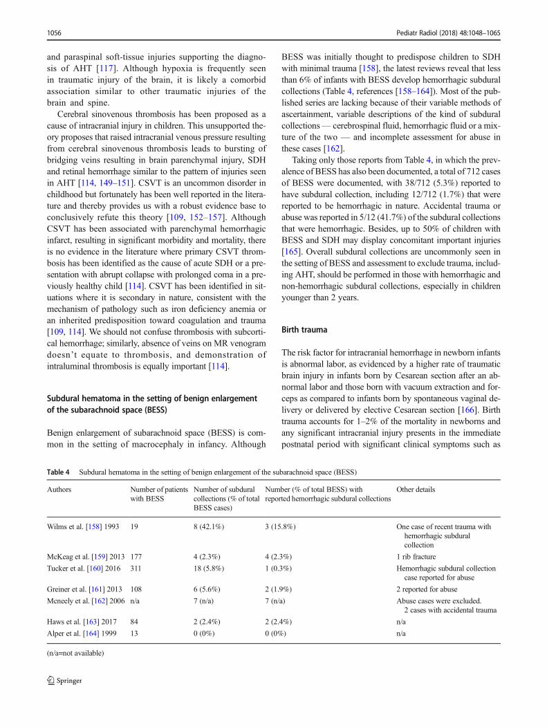
Subdural hematoma in the setting of benign enlargementof the subarachnoid space (BESS)
Benign enlargement of subarachnoid space (BESS) is com-mon in the setting of macrocephaly in infancy. Although
BESS was initially thought to predispose children to SDHwith minimal trauma [158], the latest reviews reveal that lessthan 6% of infants with BESS develop hemorrhagic subduralcollections (Table 4, references [158–164]). Most of the pub-lished series are lacking because of their variable methods ofascertainment, variable descriptions of the kind of subduralcollections— cerebrospinal fluid, hemorrhagic fluid or a mix-ture of the two — and incomplete assessment for abuse inthese cases [162].
Taking only those reports from Table 4, in which the prev-alence of BESS has also been documented, a total of 712 casesof BESS were documented, with 38/712 (5.3%) reported tohave subdural collection, including 12/712 (1.7%) that werereported to be hemorrhagic in nature. Accidental trauma orabuse was reported in 5/12 (41.7%) of the subdural collectionsthat were hemorrhagic. Besides, up to 50% of children withBESS and SDH may display concomitant important injuries[165]. Overall subdural collections are uncommonly seen inthe setting of BESS and assessment to exclude trauma, includ-ing AHT, should be performed in those with hemorrhagic andnon-hemorrhagic subdural collections, especially in childrenyounger than 2 years.
Birth trauma
The risk factor for intracranial hemorrhage in newborn infantsis abnormal labor, as evidenced by a higher rate of traumaticbrain injury in infants born by Cesarean section after an ab-normal labor and those born with vacuum extraction and for-ceps as compared to infants born by spontaneous vaginal de-livery or delivered by elective Cesarean section [166]. Birthtrauma accounts for 1–2% of the mortality in newborns andany significant intracranial injury presents in the immediatepostnatal period with significant clinical symptoms such as
Table 4 Subdural hematoma in the setting of benign enlargement of the subarachnoid space (BESS)
Authors Number of patientswith BESS
Number of subduralcollections (% of totalBESS cases)
Number (% of total BESS) withreported hemorrhagic subdural collections
Other details
Wilms et al. [158] 1993 19 8 (42.1%) 3 (15.8%) One case of recent trauma withhemorrhagic subduralcollection
McKeag et al. [159] 2013 177 4 (2.3%) 4 (2.3%) 1 rib fracture
Tucker et al. [160] 2016 311 18 (5.8%) 1 (0.3%) Hemorrhagic subdural collectioncase reported for abuse
Greiner et al. [161] 2013 108 6 (5.6%) 2 (1.9%) 2 reported for abuse
Mcneely et al. [162] 2006 n/a 7 (n/a) 7 (n/a) Abuse cases were excluded.2 cases with accidental trauma
Haws et al. [163] 2017 84 2 (2.4%) 2 (2.4%) n/a
Alper et al. [164] 1999 13 0 (0%) 0 (0%) n/a
(n/a=not available)
1056 Pediatr Radiol (2018) 48:1048–1065
irritability, poor feeding, emesis, apnea or disorderedbreathing, bradycardia, and seizures or disordered men-tation [167–184].
Small birth-related subdural hematomas, most commonlyalong the tentorium, parietal occipital convexity,retrocerebellar posterior fossa or interhemispheric fissure, areobserved in 8–46% of asymptomatic newborn infants[185–187]. This has led to the unsubstantiated theory thatrebleeding, months later, in persistent birth-related asymptom-atic SDH can present acutely with clinical features mimickingAHT [188]. Rooks et al. [186] in 2008 reported MRI findingswithin 72 h of birth and serial developmental evaluations of101 asymptomatic neonates, 79 born by vaginal delivery and22 by Cesarean delivery. SDH was present in 46 (46%) of theinfants, most of whom resolved on follow-upMRI by 1monthand all resolved by 3 months. There were no significant dif-ferences in clinical outcomes in this cohort, as compared to thenormal population, on serial developmental examinations[186]. Other authors have reported similar findings [187, 189].
To summarize, asymptomatic birth-related subdural hema-tomas are relatively frequent and resolve in the overwhelmingmajority of infants within the first 4–6 postnatal weeks, and donot appear to rebleed. If there is significant birth-related trau-ma, neonates are symptomatic in the immediate postnatal pe-riod. In particular, there is no merit to the unsubstantiatedproposal that acute collapse, coma or death, occurring monthsafter delivery, is caused by a parturitional SDHwith secondaryrebleeding.
Multidisciplinary assessment and long-term outcome
The medical diagnosis of AHT is made by pediatricians andpediatric subspecialists based on medical evaluation. In manychildren’s hospitals, an interdisciplinary team of specialiststhat includes physicians, nurses, hospital social workers andothers works together to evaluate cases. Hospital-based mul-tidisciplinary teams have existed in many communities to pro-vide comprehensive assessments and services for families formore than 60 years. The overriding goal of the work of theseteams is to diagnose and to treat child abuse and neglect,assess for alternative diagnoses when appropriate, and assistin the efforts of the many agencies involved. The Children’sHospital Association (formerly the National Association ofChildren’s Hospitals and Related Institutions) has releasedguidelines for team composition and function to aid in provid-ing services [101, 190]. In addition, in some jurisdictions,multidisciplinary teams of hospital and community profes-sionals review injuries, medical history, family and social riskto reach a more comprehensive assessment. These hospital–community partnerships are composed of physicians, nurses,social workers, clergy, psychologists, child protection ser-vices, law enforcement and other professionals with relevantexperience. These multidisciplinary teams can review all of
the data related to the case from different perspectives to gain amore complete understanding of the issues [8, 45, 191–194].Whenever members of these teams present testimony in alegal setting, there has usually been much in-depth consider-ation of the diagnosis, and the probability of the correct diag-nosis is high.
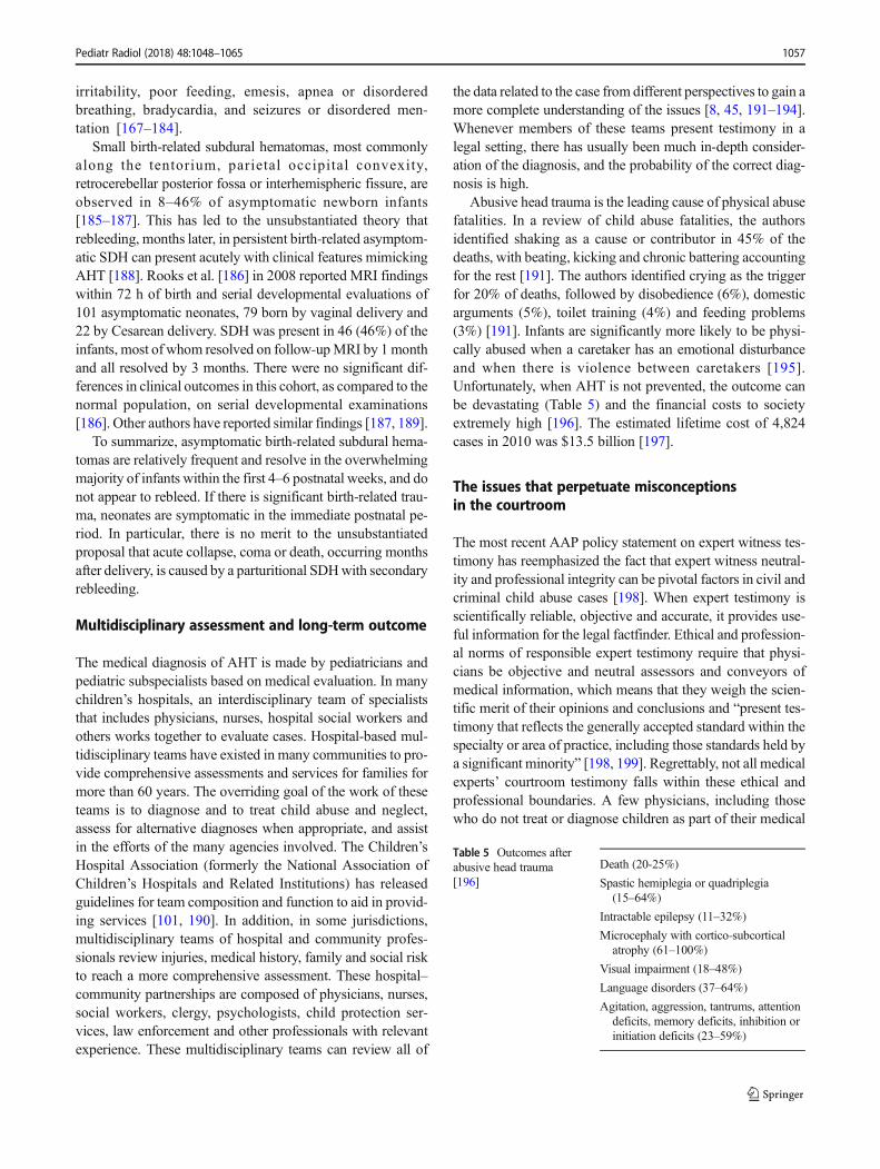
Abusive head trauma is the leading cause of physical abusefatalities. In a review of child abuse fatalities, the authorsidentified shaking as a cause or contributor in 45% of thedeaths, with beating, kicking and chronic battering accountingfor the rest [191]. The authors identified crying as the triggerfor 20% of deaths, followed by disobedience (6%), domesticarguments (5%), toilet training (4%) and feeding problems(3%) [191]. Infants are significantly more likely to be physi-cally abused when a caretaker has an emotional disturbanceand when there is violence between caretakers [195].Unfortunately, when AHT is not prevented, the outcome canbe devastating (Table 5) and the financial costs to societyextremely high [196]. The estimated lifetime cost of 4,824cases in 2010 was $13.5 billion [197].
The issues that perpetuate misconceptionsin the courtroom
The most recent AAP policy statement on expert witness tes-timony has reemphasized the fact that expert witness neutral-ity and professional integrity can be pivotal factors in civil andcriminal child abuse cases [198]. When expert testimony isscientifically reliable, objective and accurate, it provides use-ful information for the legal factfinder. Ethical and profession-al norms of responsible expert testimony require that physi-cians be objective and neutral assessors and conveyors ofmedical information, which means that they weigh the scien-tific merit of their opinions and conclusions and “present tes-timony that reflects the generally accepted standard within thespecialty or area of practice, including those standards held bya significant minority” [198, 199]. Regrettably, not all medicalexperts’ courtroom testimony falls within these ethical andprofessional boundaries. A few physicians, including thosewho do not treat or diagnose children as part of their medical
Table 5 Outcomes afterabusive head trauma[196]
Death (20-25%)
Spastic hemiplegia or quadriplegia(15–64%)
Intractable epilepsy (11–32%)
Microcephaly with cortico-subcorticalatrophy (61–100%)
Visual impairment (18–48%)
Language disorders (37–64%)
Agitation, aggression, tantrums, attentiondeficits, memory deficits, inhibition orinitiation deficits (23–59%)
Pediatr Radiol (2018) 48:1048–1065 1057
practice, frequently proffer various speculative causation the-ories (described in prior sections) camouflaged as alternativeor mimic diagnoses in child maltreatment cases. These medi-cal witnesses run afoul of professional norms and standardsand, when their arguments are repeated by the news media,create a grave public health risk by promulgating dangerousmisinformation regarding safe infant and child care.
What can be done to provide the court accurateinformation about the state of medical knowledgein AHT
The admissibility of expert evidence
In current day jurisprudence, admissibility of medical or sci-entific expert testimony requires some judicial assessment ofthe “reliability” of that testimony. In some jurisdictions, thestandard for assessing admissible expert testimony is the Fryestandard (or whether a particular concept or methodology is“generally accepted” in the medical/scientific community); inothers, it is a Daubert standard (where judges consider addi-tional criteria other than just “general acceptance,” such astestability, peer review and publication and error rate). But inany legal jurisdiction, the medical precept that is considered“generally accepted” holds significant weight with courts.Unfortunately, courts are generally ill-equipped to measurethe general consensus of physician thought on a particularconcept, which makes them susceptible to more speculativetheories unsupported by the medical evidence and medicalliterature. Thus, consensus statements present a unique oppor-tunity to provide courts with a way to know general medicalthought about a particular medical topic.
Professional society consensus statements
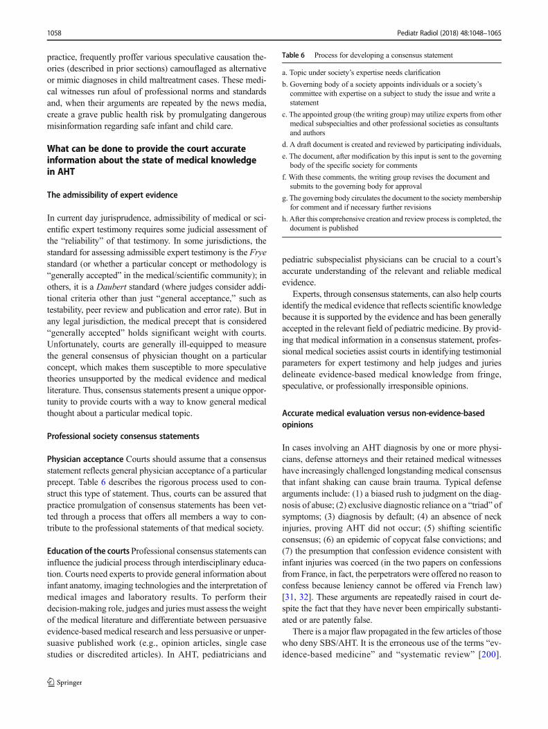
Physician acceptance Courts should assume that a consensusstatement reflects general physician acceptance of a particularprecept. Table 6 describes the rigorous process used to con-struct this type of statement. Thus, courts can be assured thatpractice promulgation of consensus statements has been vet-ted through a process that offers all members a way to con-tribute to the professional statements of that medical society.
Education of the courts Professional consensus statements caninfluence the judicial process through interdisciplinary educa-tion. Courts need experts to provide general information aboutinfant anatomy, imaging technologies and the interpretation ofmedical images and laboratory results. To perform theirdecision-making role, judges and juries must assess the weightof the medical literature and differentiate between persuasiveevidence-based medical research and less persuasive or unper-suasive published work (e.g., opinion articles, single casestudies or discredited articles). In AHT, pediatricians and
pediatric subspecialist physicians can be crucial to a court’saccurate understanding of the relevant and reliable medicalevidence.
Experts, through consensus statements, can also help courtsidentify the medical evidence that reflects scientific knowledgebecause it is supported by the evidence and has been generallyaccepted in the relevant field of pediatric medicine. By provid-ing that medical information in a consensus statement, profes-sional medical societies assist courts in identifying testimonialparameters for expert testimony and help judges and juriesdelineate evidence-based medical knowledge from fringe,speculative, or professionally irresponsible opinions.
Accurate medical evaluation versus non-evidence-basedopinions
In cases involving an AHT diagnosis by one or more physi-cians, defense attorneys and their retained medical witnesseshave increasingly challenged longstanding medical consensusthat infant shaking can cause brain trauma. Typical defensearguments include: (1) a biased rush to judgment on the diag-nosis of abuse; (2) exclusive diagnostic reliance on a “triad” ofsymptoms; (3) diagnosis by default; (4) an absence of neckinjuries, proving AHT did not occur; (5) shifting scientificconsensus; (6) an epidemic of copycat false convictions; and(7) the presumption that confession evidence consistent withinfant injuries was coerced (in the two papers on confessionsfrom France, in fact, the perpetrators were offered no reason toconfess because leniency cannot be offered via French law)[31, 32]. These arguments are repeatedly raised in court de-spite the fact that they have never been empirically substanti-ated or are patently false.
There is a major flaw propagated in the few articles of thosewho deny SBS/AHT. It is the erroneous use of the terms “ev-idence-based medicine” and “systematic review” [200].
Table 6 Process for developing a consensus statement
a. Topic under society’s expertise needs clarification
b. Governing body of a society appoints individuals or a society’scommittee with expertise on a subject to study the issue and write astatement
c. The appointed group (the writing group) may utilize experts from othermedical subspecialties and other professional societies as consultantsand authors
d. A draft document is created and reviewed by participating individuals,
e. The document, after modification by this input is sent to the governingbody of the specific society for comments
f. With these comments, the writing group revises the document andsubmits to the governing body for approval
g. The governing body circulates the document to the societymembershipfor comment and if necessary further revisions
h. After this comprehensive creation and review process is completed, thedocument is published
1058 Pediatr Radiol (2018) 48:1048–1065
Because the suggestion that denialist views are supported bythe evidence is likely to confuse judges and juries, we addresstwo purported literature reviews: Donohoe in 2003 [201] andLynoe et al. in 2017 [202]. Both articles are flawed by “(1)improper search and systemic review questions, (2) impropercriteria for assessing bias and (3) inequitable application ofquality of study assessment standards” [137, 203].
It is unprecedented that Donohoe’s “systematic review”chose to exclude the voluminous literature before 1999 de-spite the fact that AHTwas well described by multiple authorsworldwide and the incidence of the disease was quite similarworldwide [204]. In the final analysis, Donohoe used only 23articles to reach his erroneous conclusions [201]. As Greeley[204] showed, evidence supporting the AHT medical diagno-sis “clearly fits the Bradford Hill criteria for causation” [205].Similarly, despite the vast medical literature, Lynoe et al. [202]chose to use only 30 publications. Narang et al. [203] revealedthe severe prejudicial bias of the authors of the Lynoeet al. [202] study. Additional publications have also re-futed the Lynoe report [206–210]. This alternative agen-da has no role in true science and can result in infantharm through shaking and neglect, through avoidance ofemergency medical intervention.
In contrast, a 2016 study published in The Journal ofPediatrics found a high degree of medical consensus thatshaking a young child can cause subdural hematoma, severeretinal hemorrhage, coma or death [3]. The study, which sur-veyed 628 physicians at 10 leading U.S. children’s hospitals,found that 88% of physicians believe that SBS is a validevidence-based diagnosis and 93% believe that the somewhatmore comprehensive diagnosis of AHT is a valid evidence-based diagnosis [3].
AHT is a medical diagnosis, not a legal finding of murder
It is increasingly popular for defense lawyers to argue thatAHT is a medical diagnosis of murder. This evocative court-room hyperbole deliberately distorts the judicial process bymischaracterizing the physician expert’s role. The medicalexpert in a child abuse case plays just one role — to help thejudge or jury answer the medical question of whether an in-fant’s injuries were most likely caused by abuse or they couldbe plausibly explained by a recognized disease or by one ormore of the myriad hypothetical alternative causal explana-tions typically proffered by the defense. It is absurd to arguethat a medical diagnosis proves murder. Medical expert testi-mony on the etiology of the injury cannot answer the twofoundational legal questions of actus reus (Latin for guiltyact) ormens rea (Latin for guilty mind). That is because, evenafter the factfinder decides that the medical evidence supportsa finding that an infant’s injuries were inflicted, non-medicalevidence is required to determine who committed the act andto determine the level of intent (e.g., knowing, reckless or
negligent). “The debate surrounding AHT is neither scientificnor medical but legal” [204]. The denialists have tried to cre-ate a medical controversy where there is none.
The “diagnosis of murder” argument is obviously wrongbecause it falsely implies that medical opinion testimony, byits nature, resolves all legal issues. To cite an analogous ex-ample that disproves the argument’s premise, the toxicologistwho testifies that the victim was poisoned does not diagnosemurder because the court must still decide the actus reus (howwas the poison ingested?) and the mens rea (was the victim’spoisoning accidental, negligent, reckless or intentional?).
Defense attorneys and few medical witnesses who promul-gate scientifically unsubstantiated theories about abuse“mimics” in an effort to manufacture a scientific-soundingcontroversy run afoul of professional norms and standards,can distort the view of the relevant medical community, andcreate a grave public health risk by promulgating dangerousmisinformation regarding safe infant and child care (i.e. infantshaking is safe). As professional medical societies continue toissue evidence-based consensus statements to help courts, thenews media and the public to address these issues, we antic-ipate that they will also play a greater role in curbing andsanctioning members whose testimony impedes the goals ofscientific, adjudicative and public health accuracy.
Conclusions
1. Abusive head trauma (AHT) is the current most appro-priate and inclusive diagnostic term for infants andyoung children who suffer from inflicted intracranialand associated spinal injury. This does not negate themechanisms of shaking or shaking with impact as a sig-nificant mechanism of injury but merely indicates thatthe term “shaken baby” is not all-inclusive.
2. Lack of history, changing history or the incompatibilityof history (i.e. short falls) with the severity of injury raiseconcerns for possible AHT.
3. Relatively few infants with AHT have isolated intracra-nial injury without retinal hemorrhages, fractures or oth-er manifestations of child abuse. These children need acomprehensive evaluation to rule out other diseases.However, isolated intracranial injuries occur in a smallpercentage of children with AHT.
4. No single injury is diagnostic of AHT. A compilation ofinjuries most often including SDH, complex retinal hem-orrhage and/or retinoschisis, rib, metaphyseal or otherfractures and soft-tissue injury leads to the diagnosis.
5. Each infant suspected of suffering AHT must be furtherevaluated for other diseases that might present with sim-ilar findings. The question to be answered is, “Is there amedical cause to explain the findings or did this childsuffer from inflicted injury?”
Pediatr Radiol (2018) 48:1048–1065 1059
6. There is no reliable medical evidence that the followingprocesses cause the constellation of injuries associatedwith AHT: cerebral sinovenous thrombosis, isolatedhypoxic–ischemic injury, lumbar puncture anddysphagic choking/vomiting. There is no reliable evi-dence to support speculation that long-term conse-quences of birth-related subdural hematoma can resultin later collapse, coma or death from acute rebleedinginto a previously asymptomatic chronic subdural hema-toma. In addition, subdural hematoma is uncommon inthe setting of benign enlargement of the subarachnoidspace, and when present, AHT should be considered inthe differential diagnosis.
7. After medical diagnosis, in many hospitals a multidisci-plinary team provides comprehensive assessmentand services to the family, based on considerationof all the facts.
8. There is no controversy about the methodology used todiagnose AHT as a medical disease.
9. AHT is a medical diagnosis unrelated to the legal deter-mination by a judge or jury of a charge of murder. Theterm “triad” is a legal convention that falsely mischarac-terizes a complex AHT diagnosis process.
10. A professional medical society’s consensus statementeducates judicial factfinders, the news media and thepublic about “general acceptance,” what is accuratemedical information and what is non-evidence,speculative or professionally irresponsible etiologi-cal hypotheses.
11. The professional societies' consensus statement on AHTshould help the court recognize unsubstantiated medicalexpert testimony.
Acknowledgments We are grateful to the fellows at Children’s Hospitalof Philadelphia who reviewed the references for this manuscript (AndrewJ. Degnan, Rachelle Durand, Edward Fenlon, Ami Gokli, Aditi Hendi,James Hogan, Fang Lu, Ian Mills, Christy Pomeranz, Jordan Rapp andMichele Retrouvey).
Compliance with ethical standards
Conflicts of interest Dr. Narang has been paid as an expert consultant/witness in cases of abusive head trauma. Drs. Choudhary, Servaes,Christian, Hedlund, Dias, Nelson, Palasis, Rossi and Offiah provide med-ical–legal expert work in child abuse cases.
References
1. Narang S (2011) A Daubert analysis of abusive head trauma/shaken baby syndrome. Hous J Health L Pol'y 11:505–633
2. American Academy of Pediatrics (2015) Understanding abusivehead trauma in infants and children: answers from America’s pe-diatricians. https://www.aap.org/en-us/Documents/cocan_
understanding_aht_in_infants_children.pdf. Accessed 17 April2018
3. Narang SK, Estrada C, Greenberg S et al (2016) Acceptance ofshaken baby syndrome and abusive head trauma as medical diag-noses. J Pediatr 177:273–278
4. Meyer JS, Gunderman R, Coley BD et al (2011) ACR appropri-ateness criteria® on suspected physical abuse— child. J Am CollRadiol 8:87–94
5. Strouse PJ (2016) Child abuse: we have problems. Pediatr Radiol46:587–590
6. Tuerkheimer D (2009) The next innocence project: shaken babysyndrome and the criminal courts. Wash U L Rev 87:1–58
7. Bazelon E (2011) Shaken-baby syndrome faces new questions incourt. NewYork TimesMagazine. http://www.nytimes.com/2011/02/06/magazine/06baby-t.html. Accessed 17 Dec 2015
8. Keenan HT, Runyan DK, Marshall SWet al (2003) A population-based study of inflicted traumatic brain injury in young children.JAMA 290:621–626
9. Parks SE, Kegler SR, Annest JL et al (2012) Characteristics offatal abusive head trauma among children in the USA: 2003-2007:an application of the CDC operational case definition to nationalvital statistics data. Inj Prev 18:193–199
10. Christian CW, Block R, Committee on Child Abuse and Neglect,American Academy of Pediatrics (2009) Abusive head trauma ininfants and children. Pediatrics 123:1409–1411
11. Caffey J (1946) Multiple fractures in the long bones of infantssuffering from chronic subdural hematoma. Am J RoentgenolRadium Ther 56:163–173
12. Kempe CH, Silverman FN, Steele BF et al (1962) The battered-child syndrome. JAMA 181:17–24
13. Caffey J (1972) The parent-infant traumatic stress syndrome;(Caffey-Kempe syndrome), (battered babe syndrome). Am JRoentgenol Radium Therapy Nucl Med 114:218–229
14. Caffey J (1972) On the theory and practice of shaking infants. Itspotential residual effects of permanent brain damage and mentalretardation. Am J Dis Child 124:161–169
15. Caffey J (1974) The whiplash shaken infant syndrome: manualshaking by the extremities with whiplash-induced intracranial andintraocular bleedings, linked with residual permanent brain dam-age and mental retardation. Pediatrics 54:396–403
16. Duhaime AC, Gennarelli TA, Thibault LE et al (1987) The shakenbaby syndrome. A clinical, pathological and biomechanical study.J Neurosurg 66:409–415
17. Kemp AM (2011) Abusive head trauma: recognition and the es-sential investigation. Arch Dis Child Educ Pract Ed 96:202–208
18. Smith MJ (1950) Subdural hematoma with multiple fractures.AJR Am J Roentgenol 63:342–344
19. Lis EF, Frauenberger GS (1950)Multiple fractures associatedwithsubdural hematoma in infancy. Pediatrics 6:890–892
20. Silverman FN (1953) The roentgen manifestations of unrecog-nized skeletal trauma in infants. Am J Roentgenol RadiumTherapy Nucl Med 69:413–427
21. Woolley PV Jr, Evans WA Jr (1955) Significance of skeletal le-sions in infants resembling those of traumatic origin. J Am MedAssoc 158:539–543
22. Ommaya AK, Yarnell P (1969) Subdural haematoma after whip-lash injury. Lancet 2:237–239
23. Guthkelch AN (1971) Infantile subdural haematoma and its rela-tionship to whiplash injuries. Br Med J 2:430–431
24. Kiffney GT Jr (1964) The eye of the "battered child". ArchOphthalmol 72:231–233
25. Duhaime AC, Alario AJ, LewanderWJ et al (1992) Head injury invery young children: mechanisms, injury types and ophthalmolog-ic findings in 100 hospitalized patients younger than 2 years ofage. Pediatrics 90:179–185
1060 Pediatr Radiol (2018) 48:1048–1065

26. Levin AV, Christian CW, Committee on Child Abuse and Neglect,Section on Ophthalmology (2010) The eye examination in theevaluation of child abuse. Pediatrics 126:376–380
27. Johnson DL, Boal D, Baule R (1995) Role of apnea innonaccidental head injury. Pediatr Neurosurg 23:305–310
28. Ichord RN, Naim M, Pollock AN et al (2007) Hypoxic-ischemicinjury complicates inflicted and accidental traumatic brain injuryin young children: the role of diffusion-weighted imaging. JNeurotrauma 24:106–118
29. Bayir H, Kochanek PM, Kagan VE (2006) Oxidative stress inimmature brain after traumatic brain injury. Dev Neurosci 28:420–431
30. Starling SP, Patel S, Burke BL et al (2004) Analysis of perpetratoradmissions to inflicted traumatic brain injury in children. ArchPediatr Adolesc Med 158:454–458
31. Adamsbaum C, Grabar S, Mejean N et al (2010) Abusive headtrauma: judicial admissions highlight violent and repetitive shak-ing. Pediatrics 126:546–555
32. Vinchon M, de Foort-Dhellemmes S, Desurmont M et al (2010)Confessed abuse versus witnessed accidents in infants: compari-son of clinical, radiological and ophthalmological data in corrob-orated cases. Childs Nerv Syst 26:637–645
33. Dias MS (2011) The case for shaking. In: Jenny C (ed) Childabuse and neglect: diagnosis, treatment and evidence. Saunders/Elsevier, St. Louis, pp 364–372
34. Silverman FN (1972) Unrecognized trauma in infants, the batteredchild syndrome and the syndrome of Ambroise Tardieu. RiglerLecture Radiology 104:337–353
35. Jenny C (2003) Modes of presentation of inflicted childhood trau-ma. In: Reece RM, Nicholson CE (eds) Inflicted childhoodneurotrauma: proceedings of a conference sponsored byDepartment of Health and Human Services, Oct. 10–11, 2002,Bethesda, Maryland American Academy of Pediatrics, S.l., pp48–63
36. Arbogast KB, Margulies SS, Christian CW (2005) Initial neuro-logic presentation in young children sustaining inflicted and unin-tentional fatal head injuries. Pediatrics 116:180–184
37. Borgialli DA, Mahajan P, Hoyle JD et al (2016) Performance ofthe pediatric Glasgow coma scale score in the evaluation of chil-dren with blunt head trauma. Acad Emerg Med 23:878–884
38. Hettler J, Greenes DS (2003) Can the initial history predict wheth-er a child with a head injury has been abused? Pediatrics 111:602–607
39. Jenny C, Hymel KP, Ritzen A et al (1999) Analysis of missedcases of abusive head trauma. JAMA 281:621–626
40. Kemp AM, Jaspan T, Griffiths J et al (2011) Neuroimaging: whatneuroradiological features distinguish abusive from non-abusivehead trauma? A systematic review. Arch Dis Child 96:1103–1112
41. Maguire SA, Kemp AM, Lumb RC et al (2011) Estimating theprobability of abusive head trauma: a pooled analysis. Pediatrics128:e550–e564
42. Palifka LA, Frasier LD, Metzger RR, Hedlund GL (2016)Parenchymal brain laceration as a predictor of abusive head trau-ma. AJNR Am J Neuroradiol 37:163–168
43. Kelly P, John S, Vincent AL et al (2015) Abusive head trauma andaccidental head injury: a 20-year comparative study of referrals toa hospital child protection team. Arch Dis Child 100:1123–1130
44. Hennes H, Kini N, Palusci VJ (2001) The epidemiology, clinicalcharacteristics and public health implications of shaken baby syn-drome. J Aggress Maltreat Trauma 5:19–40
45. Palusci VJ (2011) Risk factors and services for child maltreatmentamong infants and young children. Child Youth Serv Rev 33:1374–1382
46. Feldman KW, Bethel R, Shugerman RP et al (2001) The cause ofinfant and toddler subdural hemorrhage: a prospective study.Pediatrics 108:636–646
47. Dias MS (2002) Inflicted head injury: future directions and pre-vention. Neurosurg Clin 13:247–257
48. Abel JM, Gennarelli TA, Segawa H (1978) Incidence and severityof cerebral concussion in the rhesus monkey following sagittalplane angular acceleration. In Proceeding of 22nd Stapp CarCrash Conference, Society for Automotive Engineers, pp 35–53
49. Margulies SS, Thibault LE, Gennarelli TA (1990) Physical modelsimulations of brain injury in the primate. J Biomech 23:823–836
50. Nahum AM, Smith RW (1976) An experimental model for closedhead impact injury. In: Proceedings of 20th Stapp Car CrashConference. Society of Automotive Engineers, pp 783–814
51. Prange MT, Coats B, Duhaime AC et al (2003) Anthropomorphicsimulations of falls, shakes and inflicted impacts in infants. JNeurosurg 99:143–150
52. Chadwick DL, Bertocci G, Castillo E et al (2008) Annual risk ofdeath resulting from short falls among young children: less than 1in 1 million. Pediatrics 121:1213–1224
53. Aoki N,MasuzawaH (1984) Infantile acute subdural hematoma. JNeurosurg 61:273–280
54. Benoit R, Watts DD, Dwyer K et al (2000) Windows 99: a sourceof suburban pediatric trauma. J Trauma 49:477–481
55. Claydon SM (1996) Fatal extradural hemorrhage following a fallfrom a baby bouncer. Pediatr Emerg Care 12:432–434
56. Denton S, Mileusnic D (2003) Delayed sudden death in an infantfollowing an accidental fall: a case report with review of the liter-ature. Am J Forensic Med Pathol 24:371–376
57. Docherty E, Hassan A, Burke D (2010) Things that go bump ...bump ... bump: an analysis of injuries from falling down stairs inchildren based at Sheffield Children’s Hospital. Emerg Med J 27:207–208
58. Gruskin KD, Schutzman SA (1999) Head trauma in childrenyounger than 2 years: are there predictors for complications?Arch Pediatr Adolesc Med 153:15–20
59. Hall JR, Reyes HM, Horvat M et al (1989) The mortality of child-hood falls. J Trauma 29:1273–1275
60. KimKA,WangMY, Griffith PM et al (2000) Analysis of pediatrichead injury from falls. Neurosurg Focus 8:1–5
61. Levene S, Bonfield G (1991) Accidents on hospital wards. ArchDis Child 66:1047–1049
62. Joffe M, Ludwig S (1988) Stairway injuries in children. Pediatrics82:457–461
63. Murray JA, Chen D, Velmahos GC et al (2000) Pediatric falls: isheight a predictor of injury and outcome? Am Surg 66:863–865
64. Park SH, Cho BM, Oh SM (2004) Head injuries from falls inpreschool children. Yonsei Med J 45:229
65. Partington MD, Swanson JA, Meyer FB (1991) Head injury andthe use of baby walkers: a continuing problem. Ann Emerg Med20:652–654
66. Reiber GD (1993) Fatal falls in childhood. How far must childrenfall to sustain fatal head injury? Report of cases and review of theliterature. Am J Forensic Med Pathol 14:201–207
67. Ruddick C, Platt MW, Lazaro C (2010) Head trauma outcomes ofverifiable falls in newborn babies. Arch Dis Child Fetal NeonatalEd 95:F144–F145
68. Sturm V, Knecht PB, Landau K, Menke MN (2009) Rare retinalhaemorrhages in translational accidental head trauma in children.Eye 23:1535–1541
69. Trenchs V, Curcoy AI, Morales M et al (2008) Retinalhaemorrhages in head trauma resulting from falls: differentialdiagnosis with non-accidental trauma in patients younger than 2years of age. Childs Nerv Syst 24:815–820
70. Williams RA (1991) Injuries in infants and small childrenresulting from witnessed and corroborated free falls. J Trauma31:1350–1352
Pediatr Radiol (2018) 48:1048–1065 1061
71. Zielinski AE, Rochette LM, Smith GA (2012) Stair-related inju-ries to young children treated in US emergency departments,1999-2008. Pediatrics 129:721–727
72. Billmire ME, Myers PA (1985) Serious head injury in infants:accident or abuse? Pediatrics 75:340–342
73. Chadwick DL, Chin S, Salerno C et al (1991) Deaths from falls inchildren: how far is fatal? J Trauma 31:1353–1355
74. Chiaviello CT, Christoph RA, Bond GR (1994) Stairway-relatedinjuries in children. Pediatrics 94:679–681
75. Chiaviello CT, Christoph RA, Bond GR (1994) Infant walker-related injuries: a prospective study of severity and incidence.Pediatrics 93:974–976
76. Ewing-Cobbs L, Kramer L, Prasad M et al (1998) Neuroimaging,physical and developmental findings after inflicted andnoninflicted traumatic brain injury in young children. Pediatrics102:300–307
77. Goldstein B, Kelly MM, Bruton D et al (1993) Inflicted versusaccidental head injury in critically injured children. Crit Care Med21:1328–1332
78. Helfer RE, Slovis TL, Black M (1977) Injuries resulting whensmall children fall out of bed. Pediatrics 60:533–535
79. Kelly P, Hayes I (2004) Infantile subdural haematoma inAuckland, New Zealand: 1988-1998. N Z Med J 117:U1047
80. Kravitz H, Driessen G, Gomberg R et al (1969) Accidental fallsfrom elevated surfaces in infants from birth to one year of age.Pediatrics 44:869–876
81. Lyons TJ, Oates RK (1993) Falling out of bed: a relatively benignoccurrence. Pediatrics 92:125–127
82. Nimityongskul P, Anderson LD (1987) The likelihood of injurieswhen children fall out of bed. J Pediatr Orthop 7:184–186
83. Reece RM, Sege R (2000) Childhood head injuries: accidental orinflicted? Arch Pediatr Adolesc Med 154:11–15
84. Selbst SM, Baker MD, ShamesM (1990) Bunk bed injuries. Am JDis Child 144:721–723
85. Smith MD, Burrington JD, Woolf AD (1975) Injuries in childrensustained in free falls: an analysis of 66 cases. J Trauma 15:987–991
86. KahnDJ, Fisher PD, Hertzler DA (2017)Variation in managementof in-hospital newborn falls: a single-center experience. JNeurosurg Pediatr 20:176–182
87. Levin AV, Luyet FM, Knox BL (2016) Ophthalmologic concernsin abusive head trauma. J Fam Violence 31:797–804
88. Gill JR, Goldfeder LB, Armbrustmacher Vet al (2009) Fatal headinjury in children younger than 2 years in New York City and anoverview of the shaken baby syndrome. Arch Pathol Lab Med133:619–627
89. Kivlin JD, Simons KB, Lazoritz S, Ruttum MS (2000) Shakenbaby syndrome. Ophthalmology 107:1246–1254
90. Morad Y, Kim YM, Armstrong DC et al (2002) Correlation be-tween retinal abnormalities and intracranial abnormalities in theshaken baby syndrome. Am J Ophthalmol 134:354–359
91. Morad Y, Wygnansky-Jaffe T, Levin AV (2010) Retinal haemor-rhage in abusive head trauma. Clin Exp Ophthalmol 38:514–520
92. Agrawal S, Peters MJ, Adams GGW et al (2012) Prevalence ofretinal hemorrhages in critically ill children. Pediatrics 129:e1388–e1396
93. Levin AV (2010) Retinal hemorrhage in abusive head trauma.Pediatrics 126:961–970
94. Binenbaum G, Chen W, Huang J et al (2016) The natural historyof retinal hemorrhage in pediatric head trauma. J AAPOS 20:131–135
95. Servaes S, Brown SD, Choudhary AK et al (2016) The etiologyand significance of fractures in infants and young children: a crit-ical multidisciplinary review. Pediatr Radiol 46:591–600
96. Anderst JD, Carpenter SL, Abshire TC et al (2013) Evaluation forbleeding disorders in suspected child abuse. Pediatrics 131:e1314–e1322
97. Choudhary AK, Jha B, Boal DK, Dias M (2010) Occipital suturesand its variations: the value of 3D-CT and how to differentiate itfrom fractures using 3D-CT? Surg Radiol Anat 32:807–816
98. Kemp AM, Rajaram S, Mann M et al (2009) What neuroimagingshould be performed in children in whom inflicted brain injury(iBI) is suspected? A systematic review. Clin Radiol 64:473–483
99. Barber I, Kleinman PK (2014) Imaging of skeletal injuries asso-ciated with abusive head trauma. Pediatr Radiol 44:S613–S620
100. Girard N, Brunel H, Dory-Lautrec P et al (2016) Neuroimagingdifferential diagnoses to abusive head trauma. Pediatr Radiol 46:603–614
101. Slovis TL, Strouse PJ, Strauss KJ (2015) Radiation exposure inimaging of suspected child abuse: benefits versus risks. J Pediatr167:963–968
102. Kuppermann N, Holmes JF, Dayan PS et al (2009) Identificationof children at very low risk of clinically-important brain injuriesafter head trauma: a prospective cohort study. Lancet 374:1160–1170
103. Magana JN, Kuppermann N (2017) The PECARN TBI rules donot apply to abusive head trauma. Acad Emerg Med 24:382–384
104. National Association of Children’s Hospitals and RelatedInstitutions (2011) Defining the children’s hospital role in childmaltreatment, 2nd edn. https://www.childrenshospitals.org/~/media/Files/CHA/Main/Issues_and_Advocacy/Key_Issues/Child_Health/Child_Abuse/child_abuse_guidelines_100111.pdf.Accessed 05 June 2017
105. Bechtel K, Stoessel K, Leventhal JM et al (2004) Characteristicsthat distinguish accidental from abusive injury in hospitalizedyoung children with head trauma. Pediatrics 114:165–168
106. Ewing-Cobbs L, Prasad M, Kramer L et al (2000) Acuteneuroradiologic findings in young children with inflicted ornoninflicted traumatic brain injury. Childs Nerv Syst 16:25–33
107. Myhre MC, Grogaard JB, Dyb GA et al (2007) Traumatic headinjury in infants and toddlers. Acta Paediatr 96:1159–1163
108. Tung GA, Kumar M, Richardson RC et al (2006) Comparison ofaccidental and nonaccidental traumatic head injury in children onnoncontrast computed tomography. Pediatrics 118:626–633
109. Hedlund G (2015) Abusive head trauma: extraaxial hemorrhageand nonhemic collections. In: Kleinman PK (ed) Diagnostic im-aging of child abuse. Cambridge University Press, Cambridge, pp394–452
110. Bradford R, Choudhary AK, Dias MS (2013) Serial neuroimagingin infants with abusive head trauma: timing abusive injuries. JNeurosurg Pediatr 12:110–119
111. Zouros A, Bhargava R, Hoskinson M et al (2004) Further charac-terization of traumatic subdural collections of infancy. J NeurosurgPediatr 100:512–518
112. Vezina G (2009) Assessment of the nature and age of subduralcollections in nonaccidental head injury with CTandMRI. PediatrRadiol 39:586–590
113. Grant P (2015) Abusive head trauma: parenchymal injury. In:Kleinman PK (ed) Diagnostic imaging of child abuse.Cambridge University Press, Cambridge, pp 453–486
114. Choudhary AK, Bradford R, Dias MS et al (2015) Venous injuryin abusive head trauma. Pediatr Radiol 45:1803–1813
115. Nelson MD Jr (2009) Unraveling the puzzle. Pediatr Radiol 39:199
116. Mack J, Squier W, Eastman J (2009) Anatomy and developmentof the meninges: implications for subdural collections and CSFcirculation. Pediatr Radiol 39:200–210
117. Choudhary AK, Ishak R, Zacharia TT et al (2014) Imaging ofspinal injury in abusive head trauma: a retrospective study.Pediatr Radiol 44:1130–1140
1062 Pediatr Radiol (2018) 48:1048–1065
118. Kemp A, Cowley L, Maguire S (2014) Spinal injuries in abusivehead trauma: patterns and recommendations. Pediatr Radiol 44:S604–S612
119. Choudhary AK, Bradford RK, Dias MS et al (2012) Spinal sub-dural hemorrhage in abusive head trauma: a retrospective study.Radiology 262:216–223
120. Brennan LK, Rubin D, Christian CWet al (2009) Neck injuries inyoung pediatric homicide victims. J Neurosurg Pediatr 3:232–239
121. Koumellis P, McConachie NS, Jaspan T (2009) Spinal subduralhaematomas in children with non-accidental head injury. Arch DisChild 94:216–219
122. Bandak FA (2005) Shaken baby syndrome: biomechanics analysisof injury mechanisms. Forensic Sci Int 151:71–79
123. Woodall TT (2009) In the court of criminal appeals of Tennesseeat Nashville. www.tba.org/tba_files/TCCA/2010/mazer_110210.pdf. Accessed on 30 July 2017
124. Gabaeff SC (2011) Challenging the pathophysiologic connectionbetween subdural hematoma, retinal hemorrhage and shaken babysyndrome. West J Emerg Med 12:144–158
125. Leventhal JM, Edwards GA (2017) Flawed theories to explainchild physical abuse: what are the medical-legal consequences?JAMA 318:1317–1318
126. Martin-Millan M, Hernandez JL, Matorras P et al (2005) Multiplesubdural haematomas following lumbar puncture. Eur Neurol 53:159–160
127. Lee AC, Lau Y, Li CH et al (2007) Intraspinal and intracranialhemorrhage after lumbar puncture. Pediatr Blood Cancer 48:233–237
128. OpenshawH, Ressler JA, Snyder DS (2008) Lumbar puncture andsubdural hygroma and hematomas in hematopoietic cell transplantpatients. Bone Marrow Transplant 41:791–795
129. Brown MW, Yilmaz TS, Kasper EM (2016) Iatrogenic spinal he-matoma as a complication of lumbar puncture: what is the risk andbest management plan? Surg Neurol Int 7:S581–S589
130. Talbert DG (2005) Paroxysmal cough injury, vascular rupture and'shaken baby syndrome'. Med Hypotheses 64:8–13
131. Geddes JF, Talbert DG (2006) Paroxysmal coughing, subdural andretinal bleeding: a computer modelling approach. NeuropatholAppl Neurobiol 32:625–634
132. Watts CC, Acosta C (1969) Pertussis and bilateral subdural hema-tomas. Am J Dis Child 118:518–519
133. Curcoy AI, Trenchs V, Morales M et al (2012) Is pertussis ininfants a potential cause of retinal haemorrhages? Arch DisChild 97:239–240
134. Raoof N, Pereira S, Dai S et al (2017) Retinal haemorrhage ininfants with pertussis. Arch Dis Child 102:1158–1160
135. Barnes PD, Galaznik J, Gardner H, ShumanM (2010) Infant acutelife-threatening event –dysphagic choking versus nonaccidentalinjury. Semin Pediatr Neurol 17:7–11
136. Barnes PD (2011) Imaging of nonaccidental injury and themimics: issues and controversies in the era of evidence-basedmedicine. Radiol Clin N Am 49:205–229
137. Greeley CS (2010) Infant fatality. Semin Pediatr Neurol 17:275–278
138. Edwards GA (2015) Mimics of child abuse: can choking explainabusive head trauma? J Forensic Legal Med 35:33–37
139. Curcoy AI, Trenchs V, Morales M et al (2010) Retinal hemor-rhages and apparent life-threatening events. Pediatr Emerg Care26:118–120
140. Bonkowsky JL, Guenther E, Filloux FM et al (2008) Death, childabuse and adverse neurological outcome of infants after an appar-ent life-threatening event. Pediatrics 122:125–131
141. Hansen JB, Frazier T, Moffatt M et al (2017) Evaluation of thehypothesis that choking/ALTE may mimic abusive head trauma.Acad Pediatr 17:362–367
142. Herr S, Pierce MC, Berger RP et al (2004) Does valsalva retinop-athy occur in infants? An initial investigation in infants withvomiting caused by pyloric stenosis. Pediatrics 113:1658–1661
143. Geddes JF, Tasker RC, Hackshaw AK et al (2003) Dural haemor-rhage in non-traumatic infant deaths: does it explain the bleedingin 'shaken baby syndrome'? Neuropathol Appl Neurobiol 29:14–22
144. Cohen MC, Scheimberg I (2009) Evidence of occurrence ofintradural and subdural hemorrhage in the perinatal and neonatalperiod in the context of hypoxic ischemic encephalopathy: anobservational study from two referral institutions in the UnitedKingdom. Pediatr Dev Pathol 12:169–176
145. Punt J, Bonshek RE, Jaspan Tet al (2004) The 'unified hypothesis'of Geddes et al. is not supported by the data. Pediatr Rehabil 7:173–184
146. Rao P, Carty H, Pierce A (1999) The acute reversal sign: compar-ison of medical and non-accidental injury patients. Clin Radiol 54:495–501
147. Byard RW, Blumbergs P, Rutty G et al (2007) Lack of evidence fora causal relationship between hypoxic-ischemic encephalopathyand subdural hemorrhage in fetal life, infancy and early childhood.Pediatr Dev Pathol 10:348–350
148. Hurley M, Dineen R, Padfield CJH et al (2010) Is there a causalrelationship between the hypoxia-ischaemia associated with car-diorespiratory arrest and subdural haematomas? An observationalstudy. Br J Radiol 83:736–743
149. Bailey OT, Hass GM (1937) Dural sinus thrombosis in early life:recovery from acute thrombosis of the superior longitudinal sinusand its relation to certain acquired cerebral lesions in childhood.Brain 60:293–314
150. Matsuda M, Matsuda I, Sato M et al (1982) Superior sagittal sinusthrombosis followed by subdural hematoma. Surg Neurol 18:206–211
151. Bucy PC, Lesemann FJ (1942) Idiopathic recurrent thrombophle-bitis: with cerebral venous thromboses and an acute subdural he-matoma. JAMA 119:402–405
152. Hedlund GL (2013) Cerebral sinovenous thrombosis in pediatricpractice. Pediatr Radiol 43:173–188
153. Sebire G, Tabarki B, Saunders DE et al (2005) Cerebral venoussinus thrombosis in children: risk factors, presentation, diagnosisand outcome. Brain 128:477–489
154. Bracken J, Barnacle A, Ditchfield M (2013) Potential pitfalls inimaging of paediatric cerebral sinovenous thrombosis. PediatrRadiol 43:219–231
155. Heller C, Heinecke A, Junker R et al (2003) Cerebral venousthrombosis in children: a multifactorial origin. Circulation 108:1362–1367
156. deVeber G, AndrewM,Adams C et al (2001) Cerebral sinovenousthrombosis in children. N Engl J Med 345:417–423
157. McLean LA, Frasier LD, Hedlund GL (2012) Does intracranialvenous thrombosis cause subdural hemorrhage in the pediatricpopulation? AJNR Am J Neuroradiol 33:1281–1284
158. Wilms G, Vanderschueren G, Demaerel PH et al (1993) CT andMR in infants with pericerebral collections and macrocephaly:benign enlargement of the subarachnoid spaces versus subduralcollections. AJNR Am J Neuroradiol 14:855–860
159. McKeag H, Christian CW, Rubin D et al (2013) Subdural hemor-rhage in pediatric patients with enlargement of the subarachnoidspaces. J Neurosurg Pediatr 11:438–444
160. Tucker J, Choudhary AK, Piatt J (2016) Macrocephaly in infancy:benign enlargement of the subarachnoid spaces and subdural col-lections. J Neurosurg Pediatr 18:16–20
161. Greiner MV, Richards TJ, Care MM et al (2013) Prevalence ofsubdural collections in children with macrocrania. AJNR Am JNeuroradiol 34:2373–2378
Pediatr Radiol (2018) 48:1048–1065 1063
162. McNeely PD, Atkinson JD, Saigal G et al (2006) Subdural hema-tomas in infants with benign enlargement of the subarachnoidspaces are not pathognomonic for child abuse. AJNR Am JNeuroradiol 27:1725–1728
163. Haws ME, Linscott L, Thomas C et al (2017) A retrospectiveanalysis of the utility of head computed tomography and/or mag-netic resonance imaging in the management of benignmacrocrania. J Pediatr 182:283–289
164. Alper G, Ekinci G, Yilmaz Y et al (1999) Magnetic resonanceimaging characteristics of benign macrocephaly in children. JChild Neurol 14:678–682
165. Hansen JB, Frazier T,Moffatt M et al (2018) Evaluations for abusein young children with subdural hemorrhages: findings based onsymptom severity and benign enlargement of the subarachnoidspaces. J Neurosurg Pediatr 21:31–37
166. Towner D, CastroMA, Eby-Wilkens E et al (1999) Effect of modeof delivery in nulliparouswomen on neonatal intracranial injury. NEngl J Med 341:1709–1714
167. Abroms IF, McLennan JE, Mandell F (1977) Acute neonatal sub-dural hematoma following breech delivery. Am J Dis Child 131:192–194
168. Bergman I, Bauer RE, Barmada MA et al (1985) Intracerebralhemorrhage in the full-term neonatal infant. Pediatrics 75:488–496
169. Gresham EL (1975) Birth trauma. Pediatr Clin N Am 22:317–328170. Hanigan WC, Morgan AM, Stahlberg LK et al (1990) Tentorial
hemorrhage associated with vacuum extraction. Pediatrics 85:534–539
171. Hernansanz J, Munoz F, Rodriguez D et al (1984) Subdural he-matomas of the posterior fossa in normal-weight newborns.Report of two cases. J Neurosurg 61:972–974
172. Hickey K, McKenna P (1996) Skull fracture caused by vacuumextraction. Obstet Gynecol 88:671–673
173. Hill A, Martin DJ, Daneman A et al (1983) Focal ischemic cere-bral injury in the newborn: diagnosis by ultrasound and correlationwith computed tomographic scan. Pediatrics 71:790–793
174. Hovind KH (1986) Traumatic birth injuries. In: Raimondi AJ,Choux M, Di Rocco C (eds) Head injuries in the newborn andinfant. Springer, New York, pp 87–109
175. Leblanc R, O'Gorman AM (1980) Neonatal intracranial hemor-rhage. A clinical and serial computerized tomographic study. JNeurosurg 53:642–651
176. Mannino FL, Trauner DA (1983) Stroke in neonates. J Pediatr102:605–610
177. Menezes AH, Smith DE, Bell WE (1983) Posterior fossa hemor-rhage in the term neonate. Neurosurgery 13:452–456
178. Natelson SE, Sayers MP (1973) The fate of children sustainingsevere head trauma during birth. Pediatrics 51:169–174
179. Perrin RG, Rutka JT, Drake JM et al (1997) Management andoutcomes of posterior fossa subdural hematomas in neonates.Neurosurgery 40:1190–1199
180. Pollina J, Dias MS, Li Vet al (2001) Cranial birth injuries in termnewborn infants. Pediatr Neurosurg 35:113–119
181. Ravenel SD (1979) Posterior fossa hemorrhage in the term new-born: report of two cases. Pediatrics 64:39–42
182. Roessmann U, Miller RT (1980) Thrombosis of the middle cere-bral artery associated with birth trauma. Neurology 30:889–892
183. Romodanov AP, Brodsky Yu S (1987) Subdural hematomas in thenewborn. Surgical treatment and results. Surg Neurol 28:253–258
184. Takagi T, Fukuoka H, Wakabayashi S et al (1982) Posterior fossasubdural hemorrhage in the newborn as a result of birth trauma.Childs Brain 9:102–113
185. Looney CB, Smith JK, Merck LH et al (2007) Intracranial hem-orrhage in asymptomatic neonates: prevalence onMR images andrelationship to obstetric and neonatal risk factors. Radiology 242:535–541
186. Rooks VJ, Eaton JP, Ruess L et al (2008) Prevalence and evolutionof intracranial hemorrhage in asymptomatic term infants. AJNRAm J Neuroradiol 29:1082–1089
187. Whitby EH, Griffiths PD, Rutter S et al (2004) Frequency andnatural history of subdural haemorrhages in babies and relationto obstetric factors. Lancet 363:846–851
188. Uscinski RH, McBride DK (2008) The shaken baby syndrome: anodyssey. II Origins and further hypotheses. Neurol Med Chir 48:151–155
189. Holden KR, Titus MO, Van Tassel P (1999) Cranial magneticresonance imaging examination of normal term neonates: a pilotstudy. J Child Neurol 14:708–710
190. Kolbo JR, Strong E (1997) Multidisciplinary team approaches tothe investigation and resolution of child abuse and neglect: a na-tional survey. Child Maltreat 2:61–72
191. Palusci VJ, Covington TM (2014) Child maltreatment deaths inthe U.S. National Child Death Review Case Reporting System.Child Abuse Negl 38:25–36
192. Schnitzer PG, Ewigman BG (2005) Child deaths resulting frominflicted injuries: household risk factors and perpetrator character-istics. Pediatrics 116:e687–e693
193. Spies EL, Klevens J (2016) Fatal abusive head trauma amongchildren aged <5 years — United States, 1999–2014. MMWRMorb Mortal Wkly Rep 65:505–509
194. U. S. Department of Health and Human Services; Administrationfor Children and Families; Administration on Children, Youth andFamilies; Children’s Bureau (2018) Child maltreatment 2016.https://www.acf.hhs.gov/sites/default/files/cb/cm2016.pdf.Accessed 29 March 2018
195. Palusci VJ, Smith EG, Paneth N (2005) Predicting and respondingto physical abuse in young children using NCANDS. Child YouthServ Rev 27:667–682
196. Chevignard MP, Lind K (2014) Long-term outcome of abusivehead trauma. Pediatr Radiol 44:S548–S558
197. Miller TR, Steinbeigle R, Lawrence BA et al (2017) Lifetime costof abusive head trauma at ages 0–4, USA. Prev Sci. https://doi.org/10.1007/s11121-017-0815-z
198. Paul SR, Narang SK, Committee on Medical Liability and RiskManagement (2017) Expert witness participation in civil and crim-inal proceedings. Pediatrics 139(3)
199. Chadwick DL, Krous HF (1997) Irresponsible testimony by med-ical experts in cases involving the physical abuse and neglect ofchildren. Child Maltreat 2:313–321
200. Whiting P, Rutjes AW, Reitsma JB et al (2003) The developmentof QUADAS: a tool for the quality assessment of studies of diag-nostic accuracy included in systematic reviews. BMC Med ResMethodol 3:25
201. Donohoe M (2003) Evidence-based medicine and shaken babysyndrome: part I: literature review, 1966-1998. Am J ForensicMed Pathol 24:239–242
202. Lynoe N, Elinder G, Hallberg B et al (2017) Insufficient evidencefor 'shaken baby syndrome'— a systematic review. Acta Paediatr106:1021–1027
203. Narang SK, Greeley CS (2017) Lynoe et al.- #theRestoftheStory.Acta Paediatr 106:1047–1049
204. Greeley CS (2015) Abusive head trauma: a review of the evidencebase. AJR Am J Roentgenol 204:967–973
205. Hill AB (1965) The environment and disease: association or cau-sation? Proc R Soc Med 58:295–300
206. Bilo RAC, Banaschak S, Herrmann B et al (2017) Using the tablein the Swedish review on shaken baby syndrome will not helpcourts deliver justice. Acta Paediatr 106:1043–1045
207. Hellgren K, Hellstrom A, Hard AL et al (2017) The new Swedishreport on shaken baby syndrome is misleading. Acta Paediatr 106:1040
1064 Pediatr Radiol (2018) 48:1048–1065
208. Lucas S, Bärtås A, BonamyA-KE et al (2017) The way forward inaddressing abusive head trauma in infants— current perspectivesfrom Sweden. Acta Paediatr 106:1033–1035
209. Levin AV (2017) The SBU report: a different view. Acta Paediatr106:1037–1039
210. Saunders D, RaissakiM, Servaes S et al (2017) Throwing the babyout with the bath water — response to the Swedish Agency forHealth Technology Assessment and Assessment of SocialServices (SBU) report on traumatic shaking. Pediatr Radiol 47:1386–1389
Pediatr Radiol (2018) 48:1048–1065 1065