Embed Size (px)
Citation preview
CONSCIOUSNESS RESEARCH Bottom-up influences are responsible for certain iilusions in which ;he brain is tricked into perceiving something distinctly Visua 1 System Provides Cl ues different from the image received bv the reti-
To How the Brain Perceives Most people would agree that conscious- ness is one of the great achievementsand great mysterie-f the human brain. But 10 years or so ago, any neurophysiologist who claimed to be looking for the neural basis of consciousness would have had a tough time being taken seriously. While everyone assumed that consciousness arises out of the activitv of neurons in our brains. the concept seeked too ill-defined and tangled up with subjective self-awareness to be explored in a quantitative way. As California Institute of Technology (Cal- tech) neuroscientist Christof Koch puts it, a self-respecting neuroscientist had to "wait until afLer hours and drink several beers" before even discussing consciousness.
But that is chang- ing, partly because of the efforts of Koch and his frequent co-author, Nobel laureate Francis Crick, to persuade neur-
the images, even though their visual systems were receiving information from the retina and extracting features such as color, move- ment, and form, In conscious animals, re- searchers expected the picture would be more complicated; psychological experiments had shown &it an individual's mental state could alter visual awareness, enhancing certain fea- tures of a scene, for example, while down- playing others.
In the past decade, researchers studying the brains of fully conscious monkeys have learned that those changes are occurring at the level of neural activity in the visual cortex. "What we actuailv perceive is not the image
oscientists that consciousness can be broken down into pieces that can be tackled by the techniques of modem neurobiology. One simple form of consciousness, they say, is awarenessof one's surroundings, of sensa- tions, and of one's relationship to those stimuli. Researchers are well equipped to ex- plore +e underpinnings of awareness, espe- cially in the brain's visual system, where years of work in monkeys and other animals have traced the pathways by which informa- tion from the retina enters the brain's cere- bral cortex and is analyzed there.
Now, neuroscientists studying visual awareness are beeinnine to understand how
nas. In one such illusion, called an"bistable percept," a single image produces two distinct
on h e retina, but a 'neural
perceptions that alternate involuntarily in the viewer's mind, although the image itself never changes. Familiar examples include the Necker cube, which creates the impression of a three-dimensional cube, alternately pro- truding from the page or recessed into it, and the well-known white-on-black picture that looks like either two faces or a vase.
Perceptual trickery. Behavioral experi- ments have shown that monkeys experience alternating perceptions when presented with bistable percepts just as humans do, and that has allowed researchers to use the illu- sions to search for neurons in a monkey's brain whose activity shifts with the monkey's perception, suggesting that they represent- and perhaps contribute to-that perception.
l l " ;
In a'receit experiment, ~ i cha rd Andersen at Caltech, along with postdoc David Bradley and graduate student Grace Chang, found such neurons in the MT
f region of the monkey's visual system, which responds to movement.
The researchers showed monkeys a 1 field of moving dots on a screen.that @ appears to represent a cylinder that is
rotating either clockwise or counter-
" " neurons along the visual pathways come to represent what we "seem-not just what reg- isters on our retinas-and how the brain itself influences those perceptions. The emerging picture, although still not clear in detail, suggbsts that "there is not one place where you have [perception], right at the top of the visual system," says Crick, but instead that the brain begins to select and alter the pure signals coming from the retina at early stages in the processing Stream. The resulting picture in our mind's eye may be built up from contributions from all those processing levels.
Most of our understanding of visual pro- cessing pathways came from work done on anesthetized animals, who were not aware of
$ clockwise. "The stimulus is physically
a the same each time, and yet the monkey I sees it either one way or another, as we
do," says Andersen. As with the Necker cube or the faces and the vase, the per- [ ceived image alternates between the
9 two possibilities every few seconds, and the monkeys were trained to indicate with an eye movement which way the cylinder appeared to be turning.
Diverging paths. tdentifying faces activates a hu- By recording from the animals' MT man subject's "whar visual processing pathway neurons, the team found that about half (red); judging their location within the boxes activates of the cells that fired vigorously in re- the "Where" Path (green). Both tasks activate early sponse to one percept, for example, areas (yellow) in the visual-processing stream. when the monkev saw the dots as a
image' formed in the cortex," explains John Maunsell, who studies visual attention at Baylor College of Medicine in Houston. And that neural image, he adds, "is not a com- pletely accurate representkrion of what is go- ing on in the world; it has been adjusted."
That adjustment can occur either by so- called "top-down" processes involving vol- untary decisions-such as the choice to focus our attention on searchine for a red book on the shelf, or a friend's face-in a crowd-r by "bottom-up" influences over which we may have no control, such as the brain's involun- tary mechanisms for resolving competition between conflicting interpretations of infor- mation it receives.
clockwise-turning 'cylinder, would fire much less when the cylinder appeared to turn counterclockwise. That means, Ander- sen says, that about half of the neurons in MT refleet not the image .on the animal's retha, which was the &me fbt each trial, but what the animal perceives.
What distinguishes the neurons that shift with the DerceDt from those that don't re- . . mains a mystery. One guess, says Crick, is that the shifting neurons are wired to other parts of the brain that direct the visual per- ceptions. What does appear clear, from ex- periments from the laboratory of Nikos Logothetis at Baylor, is that the percentage of neurons that reflect the animal's percep- tion increases as visual information works its
http://www.sciencemag.org SCIENCE VOL. 275 14 MARCH 1997
way up the visual processing pathway. Logothetis's team subjected monkeys to
binocular rivalry, a situation in which the two eves are simultaneouslv shown com- pletely different images, such as a tree and a face. Although each image is continually present on one of the viewer's retinas, the monkey, like a human viewer, is conscious only of one image at a time. The images alternate spontaneously, and monkeys can be trained to indicate which image they see.
In work over the past 8 years, Logothetis's team has sampled the activity of neurons in visual-processing areas ranging from the pri- mary visual cortex, where retinal signals first enter the brain, to an area called IT, which is at the very end of one fork of the visual processing stream. In the primary visual cor- tex, only 18% of the tested neurons changed their response according to which image the animal was perceiving, suggesting that most of the neurons in that very early processing stage merely report what is happening on the retina. In areas midway in the processing stream, nearly half of the neurons' responses correlated with the animal's ~erce~tion-a . . result comparable to what Andersen sees in MT, which is also a midway area. At the end of the line in IT, virtually all did. "There you have a perfect reflection of your perceived stimulus," says Logothetis.
Influences everywhere. The observation suggests that the "neural mechanisms under- lying visual awareness are actually distrib- uted over the entire visual pathway," Logo- thetis adds. "One could have thought that a sensory pathway just does its job, and aware- ness is the business of some other center, but this does not seem to be the case at all." However, the question remains: Are the neu- ral responses that the researchers see through- out the visual cortex actually shaping the animal's perception, or are they somehow shaped by it?
Andersen notes that an experiment per- formed by William Newsome and his col- leagues at Stanford University in 1991 sug- gests that neural activity in a midlevel area like MT can indeed influence perception. In that experiment, monkeys were given a task of telling whether, in a field of randomly moving dots, a subset of dots was moving up or down. The researchers deliberately made the task very difficult, so the monkeys were uncertain about the dots' movement, and got the answer wrong almost as often as they got it right. But Newsome's team found that stimulating neurons in MT that registered movement in a particular direction, "up," for example, biased the monkeys' decision in that direction, suggesting that these MT neurons actually shape perception. Stimu- lating MT neurons while the animal is view- ing the bistable percept might shed light on whether MT is creating or just reflecting the
monkey's perception in that case as well, the experience of paying such close atten- Andersen suggests. tion to a gripping passage in a book or a
Researchers working on human perception friend's captivating conversation that we can't use electrodes to determine whether are oblivious to other activity going on individual neurons in the human visual sys- around us. Indeed, one of the main things tem display the same traces of perception. attention does is filter out unnecessary in- But brain-imaging techniques such as posi- formation and sharpen our perceptions of
the things we are at;ending ti . FO; the past decade, a number of labs have been working to understand how visual attention modi- fies neural responses to get this job done.
In 1985, Robert Desimone, of the Na- tional Institute of Mental Health (NIMH), and his then-student Jeffrey Moran were the first to show the influence of attention on - - - -
neurons in the early processing stages of the visual cortex. They trained monkeys to keep their eyes fixed on a spot in the center of a screen. while the researchers recorded the electrical activity of individual neurons in the visual processing area called V4. This area contains neurons selective for color and form. and is one of the earlv s t o ~ s for visual , .
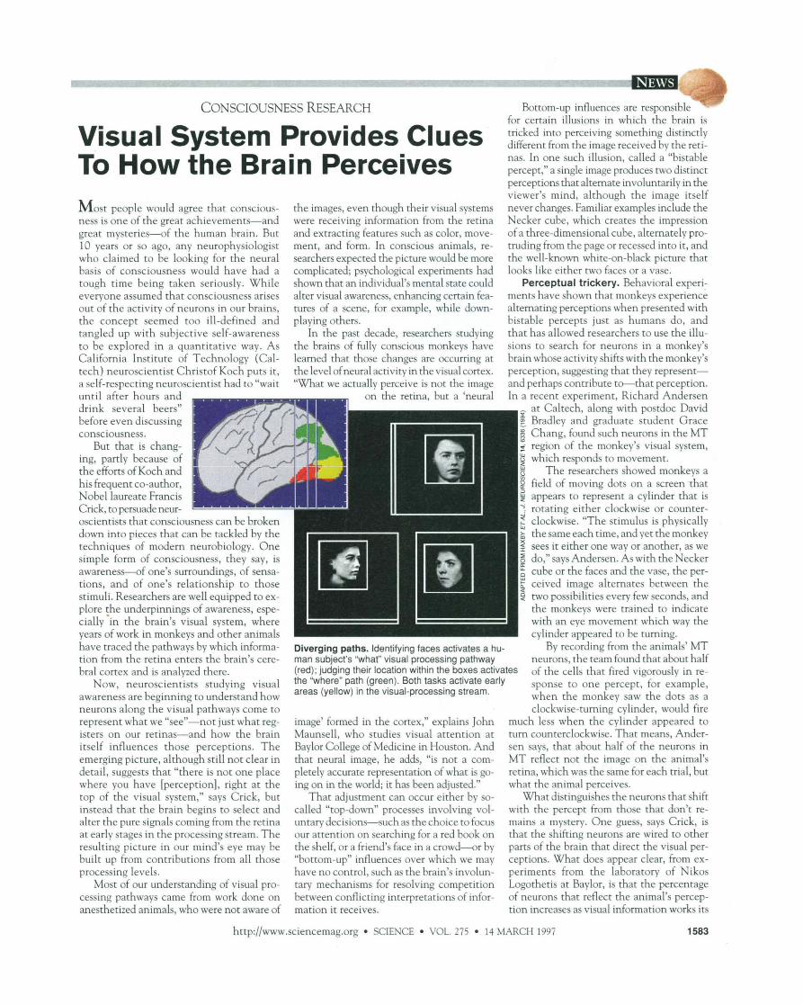
The mind's eye. Monkey experiments suggest information as it makes its way along the so- that the neural activity in the visual cortex called "what" pathway, which analyzes im- changes when the image perceived shifts from ages for their identity. the faces to the vase.
Neurons in V4, like all visual cortical neu- rons. have rece~tive fields-
tron emission tomography or functional magnetic resonance - imaging offer a coarser look at activity in entire visual areas. And in at least one experi- ment, researchers have found activity in MT that correlates with the subjects' perceptions when they experience visual il-
0 areas of the visual scene that 5 thev monitor for features thev F
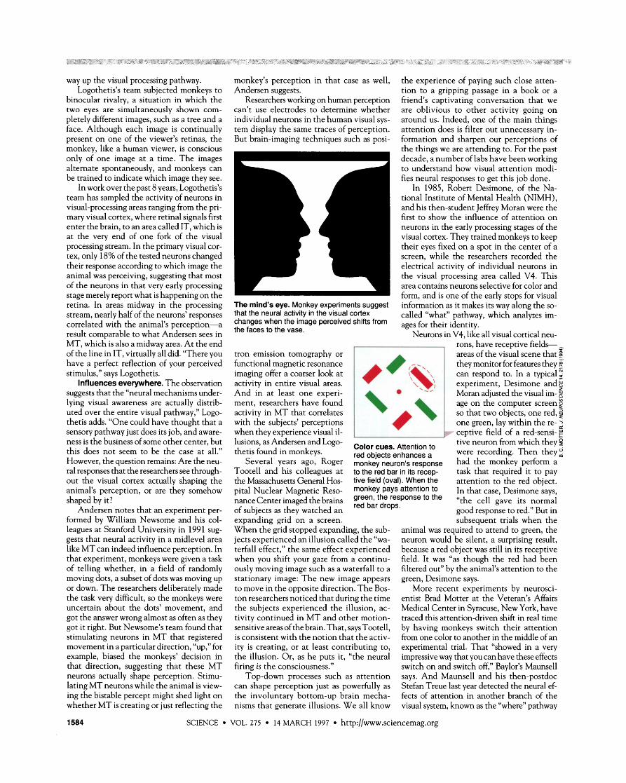
I - can respond to. In a typical 2 experiment, Desimone and 8 Moran adjusted the visual im- 0 age on the computer screen 0 so that two objects, one red, $ - one green, lay within the re-
pr ceptive field of a red-sensi- g lusions, as Andersen and Logo-
Color cues. Attention to tive neuron from which they $
thetis found in monkeys. red objects enhances a were recording. Then they: Several' years ago, Roger monkey neuron's response had the monkey perform a
Tootell and his colleagues at to the red bar in its recep- task that required it to pay the Massachusetts General Hos- tive field (oval). When the attention to the red object. pita1 Nuclear Magnetic Reso- monkey Pays attention to In that case, Desimone says, nance Center imaged the brains ~ ~ ~ ~ ' ~ ~ d ~ ~ P o n s e the "the cell gave its normal of subjects as they watched an good response to red." But in expanding grid on a screen. subsequent trials when the When the grid stopped expanding, the sub- animal was required to attend to green, the jects experienced an illusion called the "wa- neuron would be silent, a surprising result, terfall effect," the same effect experienced because a red object was still in its receptive when vou shift vour gaze from a continu- field. It was "as though the red had been , - ously moving image such as a waterfall to a stationary image: The new image appears to move in the opposite direction. The Bos- ton researchers noticed that during the time the subjects experienced the illusion, ac- tivity continued in MT and other motion- sensitive areas of the brain. That, says Tootell, is consistent with the notion that the activ- ity is creating, or at least contributing to, the illusion. Or, as he puts it, "the neural firing is the consciousness."
- filtered out" by the animal's attention to the green, Desimone says.
More recent experiments by neurosci- entist Brad Motter at the Veteran's Affairs Medical Center in Syracuse, New York, have traced this attention-driven shift in real time by having monkeys switch their attention from one color to another in the middle of an experimental trial. That "showed in a very impressive way that you can have these effects switch on and switch off." Bavlor's Maunsell
~ L ~ - d o w n processes such as attention says. And Maunsell and his' then-postdoc can shape perception just as powerfully as Stefan Treue last year detected the neural ef- the involuntary bottom-up brain mecha- fects of attention in another branch of the nisms that generate illusions. We all know visual system, known as the "where" pathway
SCIENCE VOL. 275 14 MARCH 1997 http://www.sciencemag.org
because it focuses on an ob- I Ig Where the attent1 ject's location in space.
Indeed, following Desi- mone and Moran's milestone experiment came a flood of studies confirmine that an cz
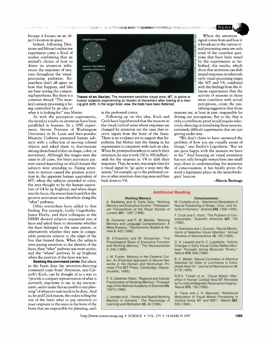
animal's choice of how to direct its attention influ- I ences the responses of neu- rons throughout the visual- I - processing pathways. Re- i early visual processing stages searchers don't all agree on like MT and V4, combined how that happens, and labs with the findings from the il- are busy testing the compet- lusion experiments that the ing But there is a Traces of an illusion. The movement-sensitive visual area, MT, is active in activity of neurons in Common thread: "The man- human subjects experiencing an illusion of movement after staring at a mov- areas correlates with actual key's sensory processing is be- ing grid (left). In the larger brain view, the folds have been flattened. perceptions, create the tan-
signal comes from and how it is broadcast to the various vi- sual processing areas are only some of the countless ques- tions that have been raised by the experiments so far. Indeed. the results. which show that attention can shift neural res~onses in relativelv
ing controlled by an idea of what it is lookine for." savs Motter. as the re frontal cortex.
talizing suggestion that those neurons are. at least in Dart. res~onsible for - , , L , L
As with the perception experiments, oliow win^ up on this idea, Koch and driving our'perceptions. But so far, that is the monkev results on attention have been Crick have hv~othesized that the neurons in onlv a correlation: c roof would reauire selec- paralleled in humans. In a 1990 experi- ment, Steven Petersen of Washington University in St. Louis and then-postdoc Maurizio Corbetta presented human sub- jects with a collection of moving colored objects and asked them to discriminate among them based either on shape, color, or movement. Although the images were the same in all cases, the brain activation pat- tern varied depending on which feature the subjects were attending to. Paying atten- tion to motion caused the greatest activa- tion in the apparent human equivalent of MT; when the subjects attended to color, the area thought to be the human equiva- lent of V4 lit up brightest; and when shape was the focus, the researchers found that the greatest activation was elsewhere along the "what" pathway.
Other researchers have added to that finding. For example, Leslie Ungerleider, James Haxby, and their colleagues at the NIMH showed subjects sequential sets of faces and asked them to determine whether the faces belonged to the same person, or alternatively whether they were in compa- rable positions relative to the edges of the box that framed them. When the subiects were paying attention to the identity of the faces, their "what" pathway was more active, and the "where" pathway lit up brightest when the position of the faces was key.
Seeking the command center. But where in the brain does the attention-directing command come from? Attention, says Cal- tech's Koch, can be thought of as a way to "provide a compact representation of what is currently important to me in my environ- ment, and to make this accessible to my plan- ning" of whatever task needs to be done. And so, he and Crick reason, the orders telling the rest of the brain what to pay attention to must originate in the areas in the front of the brain that are responsible for planning, such
, . the visual cortical areas whose responses are changed by attention are the ones that re- ceive inputs from the front of the brain. There is no evidence yet to support that hy- pothesis, but Motter says the timing in his experiments is consistent with such an idea. When he prompted monkeys to switch their attention, he says it took 150 to 300 millisec- onds for the neurons in V4 to shift their responses. That, he notes, was ample time for a visual signal to "go quite a ways into the system," for example, up to the prefrontal cor- tex or other attention-directing areas and then back down to V4.
, . tively silencing or stimulating those neurons, extremely difficult experiments that are just getting under way.
"We don't claim to have answered the problem of how you are visually aware of things," says Baylor's Logothetis. "But we are quite happy with the answers we have so far." And focusing on visual awareness has not only brought researchers one small step closer to understanding the mysteries of consciousness; it has finally given the word a legitimate place in the neurobiolo- gists' lexicon.
-Marcia Barinaga
I[ A. Baddele" and ~YCIella &la. Workina M. Corbetta eta/., "Attentbnal Modulation of 11 I ~emory a d Executive Control: ~hi~osophF Neural Processing of Shape. W r , and Ve- cal Transadions of the Row1 Societv of locitv in Humans.' tkieme248.1556 11 9901. I
7 ; London 551,1397 (1 996).
M. Daneman and P. M. Meriide, 'Working Memory and Language Comprehension: A Meta-Analysis," Ps- Bulktin & Re- view 3,422 (1 996).
M. D'Esposito and M. Grossman, 'The Physiological Basis of Executive Function and Working Memory," The Neuroscientist 2,345 (1 996).
' J. M. Fuster, Memory in the Cerebral Cor- 1 tex: An Empirical Approach to Neural Net- I works in the Human and Nonhumen Pri- 1 mate (The MIT Press. Carnbrldge, Massa- : chusetts, 1995).
P. S. GokhnakRakic, 'Regional and Cellular Fractionation of Working Memory,' Praceed- ings oflhe ElafionalAcademy ofSciencss93, 1 3473 (1 996).
J. Jonides et al., "Verbal and Spatial Working Mernorv in Humans." The Psvcholm of
" . F. Crick and C. Koch, "The Problem of Con. sdousness," Scientific American 267, 152 (1 992).
R. Desirnone and J. Duncan, 'Neural Mecha- nisms of Selective Visual Attention,' Annual Reviews of Eleuroscim 18,193 (1 995). I D. A. ~eopoid and N. K. ~ogothetis, %ctivity I Changes in Early Visual Cortex Reflect Mon. keys' Percepts during Binocular Rivalry,' Nature 379,549 (I 996).
B. C. Mow, "Neural Correlates of Attentive I Selection for Cdor or Luminance in Extra- d
striate Area V4: Jarma1 ofNeums&m 14, 'I 21 78 (1994). 1 1
I
R.B.H. Tootell et a/., Visual Motion After- effect in Human Cortical Area MT Revealed by Functional Magnetic Resonance Imaging; Nature 375, I 39 (1 995).
S. Treue and J. H. Maunsell, 'Attentionill Modulation of Visual Motion Proceasing in
http://www.sciencemag.org SCIENCE VOL. 275 14 MARCH 1997